the potential role of follicle-stimulating hormone in the
TRANSCRIPT
Urologic Oncology: Seminars and Original Investigations ] (2017) ∎∎∎–∎∎∎
http://dx.doi.org/10.1016/j1078-1439/r 2017 The A(http://creativecommons.o
* Corresponding authorE-mail address: edc@e
Review article
The potential role of follicle-stimulating hormone in the cardiovascular,metabolic, skeletal, and cognitive effects associated with androgen
deprivation therapy
E. David Crawford, M.D.a,*, Andrew V. Schally, Ph.D., M.Dhc. (Multi), D.Sc., hcb,c,d,Jehonathan H. Pinthus, M.D., Ph.D.e, Norman L. Block, M.D.b,c,d, Ferenc G. Rick, M.D., Ph.D.b,f,Marc B. Garnick, M.D.g, Robert H. Eckel, M.D.h, Thomas E. Keane, M.B.B.Ch., F.R.C.S.I., F.A.C.S.i,
Neal D. Shore, M.D., F.A.C.S., C.P.I.j, David N. Dahdal, M.S., Ph.D.k,Thomas J.R. Beveridge, M.Sc., Ph.D.k, Dennis C. Marshall, R.N., M.S., Ph.D.k
a Department of Urologic Oncology, School of Medicine, University of Colorado, Denver, Denver, COb Endocrine, Polypeptide and Cancer Institute, Miami Veterans Affairs Medical Center, Miami, FL
c Department of Pathology, University of Miami School of Medicine, Miami, FLd Department of Medicine, University of Miami School of Medicine, Miami, FL
e Department of Surgery, Juravinski Cancer Centre, McMaster University, Hamilton, Ontario, Canadaf Department of Urology, Herbert Wertheim College of Medicine, Florida International University, Miami, FL
g Department of Medicine, Harvard Medical School, Boston, MAh Division of Endocrinology, Metabolism and Diabetes, University of Colorado, Denver, CO
i Department of Urology, Medical University of South Carolina, Charleston, SCj Carolina Urologic Research Center, Myrtle Beach, SC
k Ferring Pharmaceuticals Inc., Parsippany, NJ
Received 1 August 2016; received in revised form 20 January 2017; accepted 24 January 2017
Abstract
Purpose: To explore how follicle-stimulating hormone (FSH) may contribute to cardiovascular, metabolic, skeletal, and cognitive eventsin men treated for prostate cancer, with various forms of androgen deprivation therapy (ADT).Materials and methods: A colloquium of prostate cancer experts was convened in May 2015, to discuss the role of FSH in the
development of unwanted effects associated with ADT. Subsequently, a literature review (Medline, PubMed, and relevant congress abstractdatabases) was performed to further explore and evaluate the collected evidence.Results: It has become evident that, in the setting of ADT, FSH can promote the development of atherosclerotic plaque formation,
metabolic syndrome, and insulin resistance. Data also suggest that FSH is an important mediator of bone remodeling, particularly boneresorption, and thereby increases the risk for bone fracture. Additional evidence implicates a role for FSH in bone metastasis as well. Theinfluence of FSH on ADT-induced cognitive deficits awaits further elucidation; however, the possibility that FSH may be involved thereincannot be ruled out.Conclusions: The widespread molecular and physiological consequences of FSH system activation in normal and pathological conditions
are becoming better understood. Progress in this area has been achieved by the development of additional investigative and clinical measures tobetter evaluate specific adverse effects. More research is needed on FSH function in the development of cancer as well as its association withcardiovascular, metabolic, musculoskeletal, and cognitive effects in ADT. r 2017 The Authors. Published by Elsevier Inc. This is an openaccess article under the CC BY-NC-ND license (http://creativecommons.org/licenses/by-nc-nd/4.0/).
Keywords: Follicle-stimulating hormone (FSH); Prostate cancer; Cardiovascular; Bone; Metabolic syndrome; Cognition
.urolonc.2017.01.025uthors. Published by Elsevier Inc. This is an open access article under the CC BY-NC-ND licenserg/licenses/by-nc-nd/4.0/).
.davidcrawford.com (E.D. Crawford).

E.D. Crawford et al. / Urologic Oncology: Seminars and Original Investigations ] (2017) ∎∎∎–∎∎∎2
1. Introduction
Recent advances, which have elucidated the role offollicle-stimulating hormone (FSH) in various malignancies,have also expanded our understanding of the effect of FSH-related effects produced by gonadotropin-releasing hor-mone/luteinizing hormone (LH)–releasing hormone(GnRH/LHRH) receptor agonists and antagonists used inthe treatment of prostate cancer. Normally, GnRH/LHRH isreleased in a pulsatile manner from the hypothalamus, bindsto the GnRH/LHRH receptor in the anterior pituitary, andinduces the release of LH and FSH. The prostate, as well asother tissues, can also synthesize and release FSH, andexpress FSH receptors (FSHR) [1–3]. There is evidence thatbenign prostatic hyperplasia (BPH), as well as advancedand metastatic prostate cancer tissue, has either greater FSHor FSHR expression or both than healthy tissue. Usingimmunohistochemical techniques in primates and rodents,Garde et al. [4] demonstrated that FSHR antibody stainingwas greater in BPH and malignant prostate cancer tissue,compared with healthy tissue. In a similar experiment, Ben-Josef et al. [5] reported FSHR expression increasedas a function of disease severity (normal prostate tissue o B-PH o primary carcinoma), and prostate cancer cell lines thatdo not express the androgen receptor (AR), but not normalprostatic tissue, also stained positive for FSHR. Consistentwith these findings, clinical studies demonstrated positivecorrelations among serum FSH concentration, tumor malig-nancy status, and tumor size [6]. Moreover, following tumordevelopment, serum FSH was a significant predictor ofextraprostatic extension [7] and the time to the developmentof castrate-resistant prostate cancer [8].
Functional FSHR expression has been identified in theandrogen insensitive prostate cancer cell lines, PC3, andDU145 [5]. Both prostatic- and pituitary-derived FSH actdirectly on prostatic FSHR, which may then modulatehormones and growth factors involved in the developmentof BPH [5]. Interestingly, FSHR expression, together withvascular endothelial growth factor (VEGF) expression, hasbeen identified on endothelial cells of a wide array oftumors (e.g., breast, urinary bladder, colon, pancreas, andtestes) [9], and likely contributes to metastatic processesincluding intravasation and angiogenesis [10,11]. FSH is apotent inducer of reactive oxygen species [12], which arealso involved in the expression and regulation of VEGF andangiogenesis [13]. VEGF has a critical role in enhancingneovascularization of growing tumor cells and was found to
TableRepresentative articles summarizing the unwanted effects associated with androge
Effect of ADT Potential role of FSH
Cardiovascular morbidity and mortality Dyslipidemia, plaque formationMetabolic syndrome Adipocyte rearrangement, metaBone loss, fracture, and metastasis Increased osteoclast expressionCognitive impairment Associated with decreased testo
be overexpressed in BPH and highly overexpressed inprostate cancer (for reviews on VEGF and prostate cancersee Refs. [14–17]). In summary, FSH acts as a mitogen [18]and a positive trophic factor in tumor angiogenesis [19–21].This combined effect is important to consider becausedetailed studies have linked stimulation of FSHR withdownstream activation of VEGF [22], and the transmigra-tion of malignant prostate cancer cells into circulation [23].
Although androgen deprivation therapy (ADT) improvesoutcomes in men diagnosed with advanced prostate cancer,and those treated with radiation for high-risk localizeddisease, it is also associated with adverse treatment-relatedmetabolic effects, increased cardiovascular morbidity andmortality [24,25], and decreased bone mineral density[26,27]. Accumulating experimental and clinical data indicatethat FSH may contribute to development of these unwantedeffects through its role in inflammation, atherosclerosis,insulin resistance, formation of reactive oxygen species, andadipocyte rearrangement [12,28–30]. The purpose of thisreview is to investigate the potential associations betweenFSH and the cardiovascular, metabolic, skeletal, and cognitiveeffects associated with ADT for prostate cancer (Table).
2. Methods
A colloquium of world experts in FSH, GnRH/LHRH,endocrinology, cardiovascular function, and prostate cancerwas convened in May 2015 to discuss current knowledge ofFSH, the relevant evidence for its role in the progression ofprostate cancer, and the unwanted effects associated withADT. We also conducted a comprehensive literature searchof Medline, PubMed, and relevant congress abstract data-bases using combinations of the keywords such as prostatecancer, follicle-stimulating hormone, metabolic syndrome,cognition, cardiovascular disease (CVD), vascular endothe-lial growth factor, neoangiogenesis, bone metabolism,metastases, androgen deprivation therapy, and gonadotropinreleasing hormone/luteinizing hormone-releasing hormoneagonists/antagonists. Basic science and clinical studies thatreported an association between the FSH system, andadverse consequences of prostate cancer, and its treatmentwith ADT were selected for further review. Data fromrelevant and FSH-focused studies were presented, reviewed,and discussed in-detail by the authors. In addition, anupdated review of the literature was conducted periodicallyduring the writing of this article.
n deprivation therapy and their relationship to follicle-stimulating hormone.
References
, and disruption [25,49–51,57]bolic derangement, and insulin resistance [28,29,48,52,54]through RANK- and TNF-α-mediated pathways [26,70,71,78,81]sterone and increased FSH and LH levels [84,85,87,89,91]

E.D. Crawford et al. / Urologic Oncology: Seminars and Original Investigations ] (2017) ∎∎∎–∎∎∎ 3
3. Basis of chemical ADT in the treatment of prostatecancer
The GnRH/LHRH receptor is the target of agonists andantagonists in the treatment of androgen-dependent prostatecancer. During the past 43 years, more than 3,000 agonisticanalogs of GnRH/LHRH have been synthesized, and muchhas been learned about the structure-activity relationship ofthese molecules, thus enabling the synthesis of ligands withgreater potency, selectivity, and resistance to enzymaticdegradation [31–33]. Several of these analogs have pro-gressed into clinical application and have been successfullyused for ADT in the treatment of prostate cancer. At thesame time, hundreds of GnRH/LHRH receptor antagonistswere also synthesized and evaluated in preclinical andclinical studies [31,34,35]. Following early setbacks withthe use of molecules that produced anaphylactic reactions,several improved GnRH/LHRH receptor antagonists weresynthesized that demonstrated clinical efficacy, withoutthose side effects, in patients with prostate cancer andBPH [31,34,36–39]. Preclinical studies demonstrated thatGnRH/LHRH receptor antagonists inhibited tumor growth,lowered levels of testosterone, and decreased prostate-specific antigen to a greater extent than did GnRH/LHRHreceptor agonists [31,32,40,41].
Preliminary clinical trials with GnRH/LHRH receptorantagonists confirmed their positive therapeutic profile [42],and subsequent side-by-side comparisons with GnRH/LHRH receptor agonists showed a modest advantage forantagonist therapy. GnRH/LHRH receptor antagonists low-ered testosterone to castrate levels more rapidly, without“flare” reactions, and dramatically decreased both LH andFSH [38,43–45]. GnRH/LHRH antagonists also suppressedserum levels of FSH and LH more rapidly than did agonists.However, although there were no appreciable differencesbetween treatments in LH concentrations measured over1-year treatment period, serum FSH concentration increasedin the agonist arm relative to the antagonist by day 28; thisdifference continued through day 364 [45]. Importantly, therelative difference in magnitude and duration of FSHsuppression with GnRH/LHRH antagonists is not ligandspecific (i.e., exclusive to a specific molecule), but rather isa class effect associated with the mechanism of action of allclinically evaluated antagonists [46]. These GnRH/LHRHanalogs have served as both therapeutics and as tools forinvestigating the underlying mechanisms responsible fortreatment differences.
4. Association between FSH and cardiometabolicmorbidity
For several years accumulating clinical data have indi-cated that ADT has been associated with potentially seriousunwanted effects, including an increased risk for cardio-vascular morbidity and mortality [24,25,47,48]. Most
reports are based on observational data, but potentiallyowing to differences in treatment interventions and diseaseheterogeneity, post hoc analyses performed on randomizedtrials have not unequivocally supported such an associationbetween ADT and cardiac death [49,50]. Particularly,patients with pre-existing CVD seemed to be at risk forthe development of cardiovascular events with ADT.Accordingly, data from O’Farrell et al. [51] found thatmen undergoing ADT, who have pre-existing risk factorsfor CVD are at greater risk for adverse cardiovascularoutcomes compared with those without pre-existing riskfactors. It has been suggested that the risk for CVD andcardiac events may be stratified based on the patient’scardiac history, and that symptoms can develop soon afterinitiating ADT [51].
In patients without established cardiovascular risk fac-tors, ADT can promote the development of proatherogenicmetabolic derangements, such as glucose intolerance, dys-lipidemia, and increased adiposity [52], in an exposure-dependent manner [53,54]. This increased cardiovascularrisk associated with chemical ADT was once thought to bea consequence of hypogonadism [54,55]. More recently,however, reports highlighting the potential differences incardiovascular risk between GnRH/LHRH agonists andantagonists [56] have led some investigators to focus onligand-specific mechanisms such as T lymphocyte (Fig. 1A)or cardiac GnRH/LHRH receptor activation, as well as therole of the FSH system in mediating cardiovascular effects(reviewed by Zareba et al. [57]).
There are a number of processes involved with ADT-associated cardiometabolic morbidity that FSH may influ-ence, including metabolic alterations favoring adiposity. Forexample, Liu et al. [29] studied age-dependent changes incirculating FSH levels and their correlation with body massindex. They reported the presence of FSHR in humanadipose tissue and on adipocytes, along with a positivecorrelation between FSH levels and an increase in bodymass index. Further support for the role of FSH incardiometabolic morbidity can be found in preclinical andin vitro experiments. Murine 3T3-L1 preadipocytes treatedwith recombinant FSH in vitro manifested an up-regulationin the expression of lipogenic genes, including fatty acidsynthase and lipoprotein lipase, the major mediators oflipoprotein-dependent fatty acid uptake in adipose tissue[58]. Additionally, studies in mice showed that dysfunc-tional fat in vivo accumulates in a FSHR concentration–dependent manner [29]. Adipose tissue itself may have arole in cardiovascular dysfunction. Adipokines, such astumor necrosis factor alpha (TNF-α) and interleukin-6(IL-6), are proinflammatory molecules secreted by adipo-cytes that are associated with the development of insulinresistance and atherosclerotic disease [28].
Data from in vivo preclinical research support thepotential ligand-dependent effect of ADT on cardiometa-bolic morbidity. Low-density lipoprotein receptor knockout(LDLR�/�) mice treated with different modes of ADT

Fig. 1. Potential interactions among chemical ADT, immune system, FSH, and atherosclerotic plaques. (A) GnRH/LHRH receptor agonists bind GnRH/LHRH receptors on circulating T cells to stimulate proliferation and differentiation to the Th1 phenotype; in contrast, GnRH/LHRH receptor antagonists blockGnRH/LHRH receptors. Consequently, GnRH/LHRH receptor agonists induce a proinflammatory environment within the plaque by stimulating Th1 cells torelease RANKL, IFN-γ, and TNF-α; macrophages and IFN-γ in the atherosclerotic plaque release collagenases and contribute to plaque instability. (B) FSH,binding to its receptor, can increase expression of RANK on peripheral blood monocytes. Monocytes can infiltrate from the blood vessel lumen into theplaque. RANK can be activated by RANKL released by Th1 cells, stimulating the differentiation to osteoclasts, which can resorb calcified regions within theplaque, further contributing to plaque instability. (C) Ultimately, these mechanisms can weaken the thin fibrous cap of the plaque, increasing the risk ofrupture, release of internal thrombogenic lipids, and subsequent thrombotic complications. FSH ¼ follicle-stimulating hormone (receptor); GnRH/LHRH ¼gonadotropin-releasing hormone/luteinizing hormone–releasing hormone; INFγ ¼ interferon gamma; RANK (L) ¼ receptor activator of NFκB (ligand);TNF-α ¼ tumor necrosis factor alpha. (Color version of figure is available online.)
E.D. Crawford et al. / Urologic Oncology: Seminars and Original Investigations ] (2017) ∎∎∎–∎∎∎4
(orchiectomy, GnRH/LHRH antagonist, or agonist) for4 months, showed no appreciable differences in testosteronesuppression [59]; however, mice treated with antagonist hadsignificantly lower levels of FSH compared with animalstreated with agonist or surgical castration. Mice treated withGnRH/LHRH antagonist accumulated the lowest percent-age of adipose tissue compared with mice treated withagonist or orchiectomy; those treated with GnRH/LHRHagonist or orchiectomy had more than double the number ofatherosclerotic plaques compared with those of the antag-onist arm. The fasting serum lipid profiles of these miceshowed treatment with the GnRH/LHRH receptor antago-nist resulted in the highest levels of high-density lipoproteinand the lowest low-density lipoprotein concentrationscompared with the other treatment groups [59]. Lastly, allmice developed fatty streaks in the ascending aorta, but thenecrotic areas within the atherosclerotic plaques weresignificantly smaller in mice treated with GnRH/LHRHantagonist, compared with those treated with agonist ororchiectomy. This study demonstrates that the cardiometa-bolic differences among modes of ADT can be recapitulatedin murine models, and the results suggest that importantchanges in cardiometabolic markers in the context of ADTmay not be explained by the effects of low testosteronealone.
Knutsson et al. [60] reported similar effects on lipids inapolipoprotein knockout (ApoE�/�) mice, where theGnRH/LHRH receptor agonist significantly increased thesize of areas of necrosis and the degree of inflammation inatherosclerotic plaques compared with controls. Consistent
with these findings, Moulton et al. [61] reported that anangiogenesis inhibitor reduced plaque growth and intimalneovascularization in ApoE�/� mice, suggesting neovascu-larization itself is associated with plaque development. Thisis noteworthy because FSHRs are located on neovascularendothelium [9] and FSH promotes angiogenesis through aVEGF-dependent mechanism [56,62]. Lastly, vascular cal-cification, which is also associated with atherosclerosis andsubsequent plaque disruption, may be partially driven byFSH signaling. Data have also shown that calcified athero-sclerotic plaques are at least 4- to 5-times more stiff thancellular plaques [63], and physical stress directed at theinterface between calcified and vulnerable, adjacent, non-calcified regions, are more likely to result in plaqueinstability and subsequent rupture [64]. Osteoclasts, througha signaling cascade involving FSH (detailed in the follow-ing section), can reabsorb calcified regions within plaquesfurther increasing the likelihood of rupture (Fig.1B and C).
In summary, the proinflammatory effects of FSH, as wellas FSH-mediated alterations in adipocyte composition andadipokine release, provide a potential mechanism for ADT-induced cardiometabolic changes observed in prostatecancer treatment. Experimental research in mice, supportedby emerging clinical data, demonstrates GnRH/LHRHagonists and antagonists not only differ in their mechanismof action at the receptor level but also in their effectsdownstream (e.g., FSH). These differences may be impor-tant when treating patients with pre-existing risk factors forCVD [56]. Additional clinical research is needed to confirmthese hypothesized differences between pharmacologic

E.D. Crawford et al. / Urologic Oncology: Seminars and Original Investigations ] (2017) ∎∎∎–∎∎∎ 5
modes of ADT and the specific effect of FSH on cardio-metabolic morbidity.
5. Association among FSH, bone metabolism, and bonemetastasis
The association between ADT and bone loss has beenrecognized for many years [26,65]. Increased bone loss andfracture incidence are partially the result of lower levels oftestosterone available for conversion to estrogen, which isinversely related to osteoclast activity [65,66]. Although theposited influence of FSH on cardiometabolic morbidity isnovel, and perhaps surprising, an association between FSHand bone metabolism was previously identified in womenwith postmenopausal osteoporosis. Clinical studies in post-menopausal women revealed an inverse relationshipbetween serum FSH and bone mineral density as well asmarkers of bone resorption [30,67]. Likewise, in a cross-sectional, case-control study of 156 men, a significantnegative correlation was identified between FSH and bonemineral densities in the lumbar spine and femoral neck [68].
Under normal physiologic conditions, bone metabolisminvolves a complex sequence of bone turnover (via osteo-clasts) and bone formation (via osteoblasts). Osteoclastsresorb the mineral components of bone via an acidicextracellular mechanism, resulting in the release of anumber of factors from the bone matrix, including trans-forming growth factor beta (TGFβ) [69]. Osteoblasts arestimulated by these osteoclastic soluble factors, and thendeposit osteoids (organic, young bone matrix that has notundergone calcification) at the resorption site [70]. Researchhas identified a number of cytokines and signaling mole-cules, such as receptor activator of nuclear factor kappaB(RANK), RANK ligand (RANKL), osteoprotegerin, TNF-α, and IL-6, that are critical in this process; however,several of these proteins are either directly or indirectlyaffected by FSH (Fig. 2A) [30,71–73].
Preclinical and in vitro studies also support a role forFSH in bone metabolism. FSHR activation directly stim-ulates the expression of RANK on the surface of monocyteosteoclast precursors, which can be transformed intoosteoclasts following activation by RANKL [72]. Theexpression of RANKL may be increased through FSHsignaling or GnRH/LHRH receptor agonist stimulation of Tcells (Fig. 2B) [71,74]. FSH has also been shown to inducerelease of TNF-α from mononuclear cells, bone marrowgranulocytes, and macrophages, which can independentlystimulate osteoclast precursors to differentiate into osteo-clasts (Fig. 2C) [71]. FSH has also been shown to stimulaterelease of IL-1β from monocytes [30], which increasessurvival time of mature osteoclasts, thereby allowing themto participate in additional rounds of resorption [75]. Thesedata provide hypothesized mechanisms by which FSH maymediate bone loss and increase fracture risk in patients withprostate cancer.
Increased FSH-mediated osteoclast activity also has thepotential to enhance the growth and progression of bonemetastases [72]. Once metastatic prostate cancer cells arrivein bone, they are stimulated by growth factors, such asTGFβ, present in the noncellular fraction of bone marrow[76]. RANKL, also present in the bone microenvironment,stimulates metastatic prostate cancer cells to release IL-6[73], which signals continued release of IL-6 througha feed-forward mechanism, as well as matrixmetalloproteinase-9 (MMP9) [77], a protease known tofacilitate tumor cell migration and invasion. Factorsreleased from the bone matrix in response to osteoclasticdegradation (e.g., calcium, endothelial growth factor, basicfibroblast growth factor , and TGFβ) then stimulate osteo-blastic differentiation and activity (Fig. 2D). Clinically,prostate cancer skeletal metastases are usually characterizedradiographically as osteoblastic and result in new bone thatis of poor quality, immature, and of a woven configuration[78]. The combination of dysregulated osteoblastic andosteoclastic function coupled with metastatic invasion ofbone, is consistent with the hypothesized alterations in bonemetabolism mediated through FSH signaling [78].
In patients with metastatic prostate cancer, elevatedlevels of serum bone–specific alkaline phosphatase, amarker of osteoblastic activity, are significantly associatedwith shorter overall survival [79]. Clinical and experimentalevidence indicates bone resorption, which is also paradoxi-cally increased in osteoblastic metastases. Concentrations ofthe bone resorption marker, N-telopeptide, likewise isknown to be elevated in patients with prostate cancer andis also a strong predictor of morbidity and mortality [80].Distinct ADT modalities may have different effects on theclinical course of bone metastases. In a retrospectiveanalysis of a prospective, randomized, pivotal trial compar-ing GnRH/LHRH receptor agonists and antagonist in 610patients with advanced prostate cancer, among patientswith metastases or baseline prostate-specific antigenlevels 4 50 ng/ml or both, serum alkaline phosphataselevels were significantly reduced in patients treated withthe GnRH/LHRH receptor antagonist vs. agonist [81,82].At the completion of the study (day 364), there remained asignificant difference in levels of serum alkaline phospha-tase between the 2 kinds of ADT. Although these findingsneed to be confirmed with larger-scale, well-controlled,clinical trials, these data suggest that FSH suppression in thecontext of ADT should be further evaluated in conjunctionwith a strategy for optimizing bone health.
6. Association between ADT and cognitive impairment
Clinical research on the effect of ADT continues toexpand as the average male life expectancy increases. Asystematic review of the literature investigating effects ofADT on cognition indicates that upwards of 69% of patientsexhibit a measurable decline in at least 1 cognitive domain
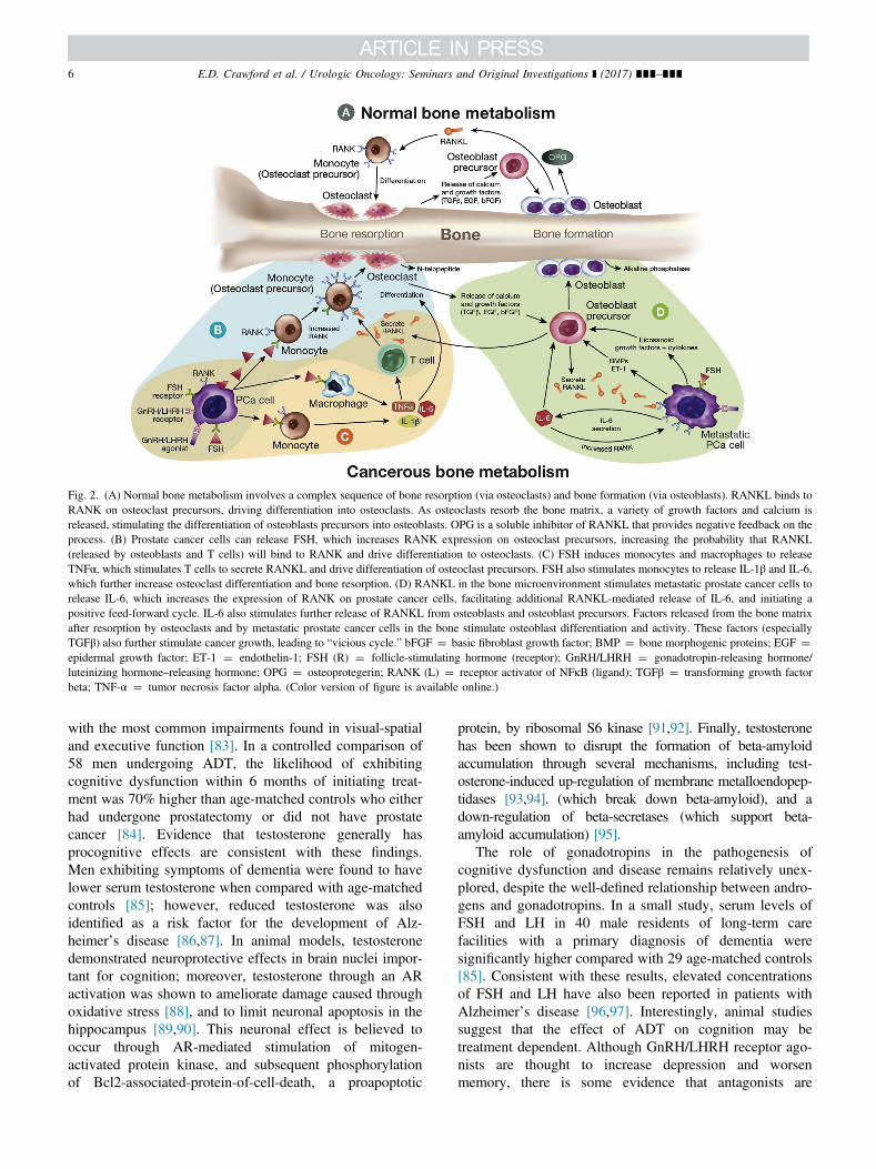
Fig. 2. (A) Normal bone metabolism involves a complex sequence of bone resorption (via osteoclasts) and bone formation (via osteoblasts). RANKL binds toRANK on osteoclast precursors, driving differentiation into osteoclasts. As osteoclasts resorb the bone matrix, a variety of growth factors and calcium isreleased, stimulating the differentiation of osteoblasts precursors into osteoblasts. OPG is a soluble inhibitor of RANKL that provides negative feedback on theprocess. (B) Prostate cancer cells can release FSH, which increases RANK expression on osteoclast precursors, increasing the probability that RANKL(released by osteoblasts and T cells) will bind to RANK and drive differentiation to osteoclasts. (C) FSH induces monocytes and macrophages to releaseTNFα, which stimulates T cells to secrete RANKL and drive differentiation of osteoclast precursors. FSH also stimulates monocytes to release IL-1β and IL-6,which further increase osteoclast differentiation and bone resorption. (D) RANKL in the bone microenvironment stimulates metastatic prostate cancer cells torelease IL-6, which increases the expression of RANK on prostate cancer cells, facilitating additional RANKL-mediated release of IL-6, and initiating apositive feed-forward cycle. IL-6 also stimulates further release of RANKL from osteoblasts and osteoblast precursors. Factors released from the bone matrixafter resorption by osteoclasts and by metastatic prostate cancer cells in the bone stimulate osteoblast differentiation and activity. These factors (especiallyTGFβ) also further stimulate cancer growth, leading to “vicious cycle.” bFGF ¼ basic fibroblast growth factor; BMP ¼ bone morphogenic proteins; EGF ¼epidermal growth factor; ET-1 ¼ endothelin-1; FSH (R) ¼ follicle-stimulating hormone (receptor); GnRH/LHRH ¼ gonadotropin-releasing hormone/luteinizing hormone–releasing hormone; OPG ¼ osteoprotegerin; RANK (L) ¼ receptor activator of NFκB (ligand); TGFβ ¼ transforming growth factorbeta; TNF-α ¼ tumor necrosis factor alpha. (Color version of figure is available online.)
E.D. Crawford et al. / Urologic Oncology: Seminars and Original Investigations ] (2017) ∎∎∎–∎∎∎6
with the most common impairments found in visual-spatialand executive function [83]. In a controlled comparison of58 men undergoing ADT, the likelihood of exhibitingcognitive dysfunction within 6 months of initiating treat-ment was 70% higher than age-matched controls who eitherhad undergone prostatectomy or did not have prostatecancer [84]. Evidence that testosterone generally hasprocognitive effects are consistent with these findings.Men exhibiting symptoms of dementia were found to havelower serum testosterone when compared with age-matchedcontrols [85]; however, reduced testosterone was alsoidentified as a risk factor for the development of Alz-heimer’s disease [86,87]. In animal models, testosteronedemonstrated neuroprotective effects in brain nuclei impor-tant for cognition; moreover, testosterone through an ARactivation was shown to ameliorate damage caused throughoxidative stress [88], and to limit neuronal apoptosis in thehippocampus [89,90]. This neuronal effect is believed tooccur through AR-mediated stimulation of mitogen-activated protein kinase, and subsequent phosphorylationof Bcl2-associated-protein-of-cell-death, a proapoptotic
protein, by ribosomal S6 kinase [91,92]. Finally, testosteronehas been shown to disrupt the formation of beta-amyloidaccumulation through several mechanisms, including test-osterone-induced up-regulation of membrane metalloendopep-tidases [93,94]. (which break down beta-amyloid), and adown-regulation of beta-secretases (which support beta-amyloid accumulation) [95].
The role of gonadotropins in the pathogenesis ofcognitive dysfunction and disease remains relatively unex-plored, despite the well-defined relationship between andro-gens and gonadotropins. In a small study, serum levels ofFSH and LH in 40 male residents of long-term carefacilities with a primary diagnosis of dementia weresignificantly higher compared with 29 age-matched controls[85]. Consistent with these results, elevated concentrationsof FSH and LH have also been reported in patients withAlzheimer’s disease [96,97]. Interestingly, animal studiessuggest that the effect of ADT on cognition may betreatment dependent. Although GnRH/LHRH receptor ago-nists are thought to increase depression and worsenmemory, there is some evidence that antagonists are

E.D. Crawford et al. / Urologic Oncology: Seminars and Original Investigations ] (2017) ∎∎∎–∎∎∎ 7
protective of brain function [98]. In a murine model, theGnRH/LHRH receptor antagonist, cetrorelix, reversed beta-amyloid-induced memory impairment and was also foundto exhibit anxiolytic and antidepressive effects [99].
The effect of ADT on cognitive performance, especiallyin the elderly, is intriguing, although the mechanismsaccounting for these differences are currently unknown.Nonetheless, as we holistically evaluate individualizedtreatment options in the context of ADT, it is reasonableto hypothesize that FSH might play a role. Further studiescomparing cognitive functions in patients with prostatecancer treated with GnRH/LHRH receptor agonists andantagonists are warranted to learn whether cognitive impair-ment varies as a function of ADT modality.
7. Summary and conclusions
The availability of different pharmacologic strategies toattain ADT has progressively helped to identify variousmechanisms, beyond testosterone suppression, to helpoptimize treatment and potentially reduce unwanted sideeffects. Men who undergo ADT for prostate cancer appearto be at higher risk for developing cardiovascular morbidity,metabolic syndrome, musculoskeletal events, and cognitiveimpairment. Converging lines of research have revealed apotential role for FSH in several of these processes.Evidence that GnRH/LHRH receptor antagonist effectscan be differentiated from those of agonists in suppressingthe FSH system in ADT might provide a selective mech-anism of therapeutic action to consider in optimizingtreatment for patients with prostate cancer.
Acknowledgments
The authors would like to thank Phillip A. Saccone, Ph.D.for his valuable insight and editing of the article.
References
[1] Hurkadli KS, Shah MG, Pardanani DS, Sheth AR. De novobiosynthesis of FSH like peptide by the human prostate. Life Sci1990;47:391.
[2] Hurkadli KS, Sheth AR, Garde SV, Doctor VM, Sheth NA.Immunocytochemical localisation of follicle stimulating hormone(FSH) in normal, benign and malignant human prostates. Br JCancer 1990;61:225.
[3] Mandrekar PS, Sheth AR, Doctor VM, Zaveri JP, Sheth NA.Immunocytochemical localization of follicle stimulating hormonein normal human stomach. Anat Rec 1990;227:334.
[4] Garde SV, Sheth AR, Shah MG, Kulkarni SA. Prostate—anextrapituitary source of follicle-stimulating hormone (FSH): occur-rence, localization, and de novo biosynthesis and its hormonalmodulation in primates and rodents. Prostate 1991;18:271.
[5] Ben-Josef E, Yang SY, Ji TH, Bidart JM, Garde SV, Chopra DP.Hormone-refractory prostate cancer cells express functional follicle-stimulating hormone receptor (FSHR). J Urol 1999;161:970.
[6] Heracek J, Urban M, Sachova J, Kuncova J, Eis V, Mandys V, et al.The endocrine profiles in men with localized and locally advancedprostate cancer treated with radical prostatectomy. Neuro EndocrinolLett 2007;28:45.
[7] Ide H, Terado Y, Sakamaki K, Inoue M, Nakajima A, Lu Y. Serumlevel of follicle-stimulating hormone is associated with extraprostaticextension of prostate cancer. Prostate Int 2013;1:109.
[8] Hoare D, Skinner TA, Black A, Robert Siemens D. Serum follicle-stimulating hormone levels predict time to development ofcastration-resistant prostate cancer. Can Urol Assoc J 2015;9:122.
[9] Radu A, Pichon C, Camparo P, Antoine M, Allory Y, Coulevard A.Expression of follicle-stimulating hormone receptor in tumor bloodvessels. N Engl J Med 2010;363:1621.
[10] Siraj A, Desestret V, Antoine M, Fromont G, Huerre M, Sanson M.Expression of follicle-stimulating hormone receptor by the vascularendothelium in tumor metastases. BMC Cancer 2013;13:246.
[11] Crawford ED, Rove KO, Schally AV, Rick FG, Block NL,Beveridge TJR. The role of the FSH system in the developmentand progression of prostate cancer. Am J Hematol Oncol 2014;10:5.
[12] Zhang Z, Wang Q, Ma J, Yi X, Zhu Y, Xi X, et al. Reactive oxygenspecies regulate FSH-induced expression of vascular endothelialgrowth factor via Nrf2 and HIF1alpha signaling in human epithelialovarian cancer. Oncol Rep 2013;29:1429.
[13] Ushio-Fukai M, Nakamura Y. Reactive oxygen species and angio-genesis: NADPH oxidase as target for cancer therapy. Cancer Lett2008;266:37.
[14] Roberts E, Cossigny DA, Quan GM. The role of vascular endothelialgrowth factor in metastatic prostate cancer to the skeleton. ProstateCancer 2013;2013:418340.
[15] Soulitzis N, Karyotis I, Delakas D, Spandidos DA. Expressionanalysis of peptide growth factors VEGF, FGF2, TGFB1, EGF andIGF1 in prostate cancer and benign prostatic hyperplasia. Int J Oncol2006;29:305.
[16] Delongchamps NB, Peyromaure M, Dinh-Xuan AT. Role ofvascular endothelial growth factor in prostate cancer. Urology2006;68:244.
[17] de Brot S, Ntekim A, Cardenas R, James V, Allegrucci C, HeeryDM, et al. Regulation of vascular endothelial growth factor inprostate cancer. Endocr Relat Cancer 2015;22:R107.
[18] Emons G, Muller V, Ortmann O, Schulz KD. Effects of LHRH-analogues on mitogenic signal transduction in cancer cells. J SteroidBiochem Mol Biol 1998;65:199.
[19] Gartrell BA, Tsao CK, Galsky MD. The follicle-stimulatinghormone receptor: a novel target in genitourinary malignancies.Urol Oncol 2013;31:1403.
[20] Kuo SW, Ke FC, Chang GD, Lee MT, Hwang JJ. Potential role offollicle-stimulating hormone (FSH) and transforming growth factor(TGFbeta1) in the regulation of ovarian angiogenesis. J Cell Physiol2011;226:1608.
[21] Huang Y, Hua K, Zhou X, Jin H, Chen X, Lu X, et al. Activationof the PI3K/AKT pathway mediates FSH-stimulated VEGF ex-pression in ovarian serous cystadenocarcinoma. Cell Res 2008;18:780.
[22] Alam H, Weck J, Maizels E, Park Y, Lee EJ, Ashcroft M, et al. Roleof the phosphatidylinositol-3-kinase and extracellular regulated kinasepathways in the induction of hypoxia-inducible factor (HIF)-1 activityand the HIF-1 target vascular endothelial growth factor in ovariangranulosa cells in response to follicle-stimulating hormone. Endocri-nology 2009;150:915.
[23] Liu W, Xu J, Wang M, Wang Q, Bi Y, Han M. Tumor-derivedvascular endothelial growth factor (VEGF)—a facilitates tumormetastasis through the VEGF-VEGFR1 signaling pathway. Int JOncol 2011;39:1213.
[24] Tsai HK, D’Amico AV, Sadetsky N, Chen MH, Carroll PR.Androgen deprivation therapy for localized prostate cancer and therisk of cardiovascular mortality. J Natl Cancer Inst 2007;99:1516.

E.D. Crawford et al. / Urologic Oncology: Seminars and Original Investigations ] (2017) ∎∎∎–∎∎∎8
[25] Bosco C, Bosnyak Z, Malmberg A, Adolfsson J, Keating NL,Van Hemelrijck M. Quantifying observational evidence for risk of fataland nonfatal cardiovascular disease following androgen deprivationtherapy for prostate cancer: a meta-analysis. Eur Urol 2015;68:386.
[26] Goldray D, Weisman Y, Jaccard N, Merdler C, Chen J, Matzkin H.Decreased bone density in elderly men treated with thegonadotropin-releasing hormone agonist decapeptyl (D-Trp6-GnRH). J Clin Endocrinol Metab 1993;76:288.
[27] Mittan D, Lee S, Miller E, Perez RC, Basler JW, Bruder JM. Boneloss following hypogonadism in men with prostate cancer treatedwith GnRH analogs. J Clin Endocrinol Metab 2002;87:3656.
[28] Choi SH, Hong ES, Lim S. Clinical implications of adipocytokinesand newly emerging metabolic factors with relation to insulinresistance and cardiovascular health. Front Endocrinol (Lausanne)2013;4:97.
[29] Liu XM, Chan HC, Ding GL, Cai J, Song Y, Wang TT, et al. FSHregulates fat accumulation and redistribution in aging through theGa/Ca(2þ)/CREB pathway. Aging Cell 2015;14:409.
[30] Cannon JG, Cortez-Cooper M, Meaders E, Stallings J, Haddow S,Kraj B. Follicle-stimulating hormone, interleukin-1, and bonedensity in adult women. Am J Physiol Regul Integr Comp Physiol2010;298:R790.
[31] Schally AV. Luteinizing hormone-releasing hormone analogs: theirimpact on the control of tumorigenesis. Peptides 1999;20:1247.
[32] Schally AV, Comaru-Schally AM, Plonowski A, Nargy A, HalmosG, Rekasi Z. Peptide analogs in the therapy of prostate cancer.Prostate 2000;45:158.
[33] Karten MJ, Rivier JE. Gonadotropin-releasing hormone analogdesign. Structure-function studies toward the development ofagonists and antagonists: rationale and perspective. Endocr Rev1986;7:44.
[34] Schally AV, Comaru-Schally AM. Hypothalamic and other peptidehormones. In: Cancer medicine, 5th ed. Edited by J.F. Holland, E.I.Frei, R.C.J. Bast et al. Hamilton, Ontario: B.C. Decker; 2000;pp.715–26.
[35] Engel JB, Schally AV. Drug insight: clinical use of agonists andantagonists of luteinizing-hormone-releasing hormone. Nat ClinPract Endocrinol Metab 2007;3:157.
[36] Rick FG, Block NL, Schally AV. An update on the use of degarelixin the treatment of advanced hormone-dependent prostate cancer.Onco Targets Ther 2013;6:391.
[37] Schally AV, Halmos G, Rekasi Z, Arencibia JM. The actions of LH-RH agonists, antagonists, and cytotoxic analogs on the LH-RHreceptors on the pituitary and tumors. In: Infertility and reproduc-tive medicine clinics of North America: GnRH analogs Edited byP. Devroey. Philadelphia: Saunders, vol. 12, pp. 17-44, 2001.
[38] Gonzalez-Barcena D, Vadillo Buenfil M, Garcia Procel E,Guerra-Arguero L, Cardenas Cornejo I, Comar-Schally AM, et al.Inhibition of luteinizing hormone, follicle-stimulating hormone andsex-steroid levels in men and women with a potent antagonist analogof luteinizing hormone-releasing hormone, Cetrorelix (SB-75). Eur JEndocrinol 1994;131:286.
[39] Gonzalez-Barcena D, Vadillo-Buenfil M, Cortez-Morales A,Fuentes-Garcia M, Cardenas-Cornejo I, Comaru-Schally AM, et al.Luteinizing hormone-releasing hormone antagonist cetrorelix asprimary single therapy in patients with advanced prostatic cancerand paraplegia due to metastatic invasion of spinal cord. Urology1995;45:275.
[40] Schally AV, Comaru-Schally AM, Nagy A, Kovacs M, SzepeshazK, Plonowski A, et al. Hypothalamic hormones and cancer. FrontNeuroendocrinol 2001;22:248.
[41] Redding TW, Schally AV, Radulovic S, Milovanovic S, SzepeshaziK, Issacs JT. Sustained release formulations of luteinizing hormone-releasing hormone antagonist SB-75 inhibit proliferation andenhance apoptotic cell death of human prostate carcinoma (PC-82)in male nude mice. Cancer Res 1992;52:2538.
[42] Gonzalez-Barcena D, Vadillo-Buenfil M, Gomez-Orta F, FuentesGarcia M, Cardenas-Cornejo I, Graef-Sanchez A, et al. Responses tothe antagonistic analog of LH-RH (SB-75, Cetrorelix) in patients withbenign prostatic hyperplasia and prostatic cancer. Prostate 1994;24:84.
[43] Garnick MB, Campion M. Abarelix Depot, a GnRH antagonist,v LHRH superagonists in prostate cancer: differential effects onfollicle-stimulating hormone. Abarelix Depot study group. Mol Urol2000;4:275.
[44] Garnick MB, Mottet N. New treatment paradigm for prostate cancer:abarelix initiation therapy for immediate testosterone suppressionfollowed by a luteinizing hormone-releasing hormone agonist. BJUInt 2012;110:499.
[45] Klotz L, Boccon-Gibod L, Shore ND, Andreou C, Persson BE,Cantor P, et al. The efficacy and safety of degarelix: a 12-month,comparative, randomized, open-label, parallel-group phase III studyin patients with prostate cancer. BJU Int 2008;102:1531.
[46] Trachtenberg J, Gittleman M, Steidle C, Barzell W, Friedel W, Pessis D,et al. A phase 3, multicenter, open label, randomized study of abarelixversus leuprolide plus daily antiandrogen in men with prostate cancer.J Urol 2002;167:1670.
[47] Zhao J, Zhu S, Sun L, Meng F, Zhao L, Zhao Y, et al. Androgendeprivation therapy for prostate cancer is associated with cardiovas-cular morbidity and mortality: a meta-analysis of population-basedobservational studies. PLoS One 2014;9:e107516.
[48] Keating NL, O’Malley AJ, Freedland SJ, Smith MR. Diabetes andcardiovascular disease during androgen deprivation therapy: observationalstudy of veterans with prostate cancer. J Natl Cancer Inst 2010;102:39.
[49] Nguyen PL, Je Y, Schutz FA, Hoffman KE, Hu JC, Parekh A, et al.Association of androgen deprivation therapy with cardiovasculardeath in patients with prostate cancer: a meta-analysis of randomizedtrials. J Am Med Assoc 2011;306:2359.
[50] Voog JC, Paulus R, Shipley WU, Smith MR, McGowan DG,Jones CU, et al. Cardiovascular mortality following short-termandrogen deprivation in clinically localized prostate cancer: ananalysis of RTOG 94-08. Eur Urol 2016;69:204–10.
[51] O’Farrell S, Garmo H, Holmberg L, Adolfsson J, Stattin P,Van Hemelrijck M. Risk and timing of cardiovascular disease afterandrogen-deprivation therapy in men with prostate cancer. J ClinOncol 2015;33:1243.
[52] Smith MR, Finkelstein JS, McGovern FJ, Zietman AL, Fallon MA,Schoenfeld DA, et al. Changes in body composition duringandrogen deprivation therapy for prostate cancer. J Clin EndocrinolMetab 2002;87:599.
[53] Basaria S, Muller DC, Carducci MA, Egan J, Dobs AS. Hyper-glycemia and insulin resistance in men with prostate carcinoma whoreceive androgen-deprivation therapy. Cancer 2006;106:581.
[54] Braga-Basaria M, Dobs AS, Muller DC, Carducci MA, John M,Egan J, et al. Metabolic syndrome in men with prostate cancerundergoing long-term androgen-deprivation therapy. J Clin Oncol2006;24:3979.
[55] Conteduca V, Di Lorenzo G, Tartarone A, Aieta M. The cardiovascularrisk of gonadotropin releasing hormone agonists in men with prostatecancer: an unresolved controversy. Crit Rev Oncol Hematol 2013;86:42.
[56] Albertsen PC, Klotz L, Tombal B, Brady J, Olesen TK, Nilsson J.Cardiovascular morbidity associated with gonadotropin releasinghormone agonists and an antagonist. Eur Urol 2014;65:565.
[57] Zareba P, Duivenvoorden W, Leong DP, Pinthus JH. Androgendeprivation therapy and cardiovascular disease: what is the linkingmechanism? Ther Adv Urol 2015;1.
[58] Wang H, Eckel RH. Lipoprotein lipase: from gene to obesity. Am JPhysiol Endocrinol Metab 2009;297:E271.
[59] Hopmans SN, Duivenvoorden WC, Werstuck GH, Klotz L, Pinthus JH.GnRH antagonist associates with less adiposity and reduced character-istics of metabolic syndrome and atherosclerosis compared withorchiectomy and GnRH agonist in a preclinical mouse model. UrolOncol 2014;32:1126.
E.D. Crawford et al. / Urologic Oncology: Seminars and Original Investigations ] (2017) ∎∎∎–∎∎∎ 9
[60] Knutsson A, Hsiung S, Celik S, Wigren M, Nilsson J, Hultgårdh-NilssonA. Treatment with an LHRH agonist, but not the LHRH antagonistdegarelix, induces atherosclerotic plaque instability in ApoE�/� mice. EurJ Urol Suppl 2015;14:e558.
[61] Moulton KS, Heller E, Konerding MA, Flynn E, Palinski W,Folkman J. Angiogenesis inhibitors endostatin or TNP-470 reduceintimal neovascularization and plaque growth in apolipoproteinE-deficient mice. Circulation 1999;99:1726.
[62] Stilley JA, Guan R, Duffy DM, Segaloff DL. Signaling through FSHreceptors on human umbilical vein endothelial cells promotesangiogenesis. J Clin Endocrinol Metab 2014;99:E813.
[63] Lee RT, Grodzinsky AJ, Frank EH, Kamm RD, Schoen FJ.Structure-dependent dynamic mechanical behavior of fibrous capsfrom human atherosclerotic plaques. Circulation 1991;83:1764.
[64] Richardson PD, Davies MJ, Born GV. Influence of plaque config-uration and stress distribution on fissuring of coronary atheroscler-otic plaques. Lancet 1989;2:941.
[65] Wei JT, Gross M, Jaffe CA, Gravlin K, Lahaie M, Faerber GJ, et al.Androgen deprivation therapy for prostate cancer results in signifi-cant loss of bone density. Urology 1999;54:607.
[66] Smith MR, Fallon MA, Goode MJ. Cross-sectional study of boneturnover during bicalutamide monotherapy for prostate cancer.Urology 2003;61:127.
[67] Garcia-Martin A, Reyes-Garcia R, Garcia-Castro JM, Rozas-MorenoP, Escobar-Jiménez F, Muñoz-Torres M. Role of serum FSHmeasurement on bone resorption in postmenopausal women. Endo-crine 2012;41:302.
[68] Karim N, MacDonald D, Dolan AL, Fogelman I, Wierzbicki AS,Hampson G. The relationship between gonadotrophins, gonadalhormones and bone mass in men. Clin Endocrinol (Oxf) 2008;68:94.
[69] Kawai M, Mödder UI, Khosla S, Rosen CJ. Emerging therapeuticopportunities for skeletal restoration. Nat Rev Drug Discov2011;10:141.
[70] Kwan Tat S, Padrines M, Theoleyre S, Heymann D, Fortun Y. IL-6,RANKL, TNF-alpha/IL-1: interrelations in bone resorption patho-physiology. Cytokine Growth Factor Rev 2004;15:49.
[71] Iqbal J, Sun L, Kumar TR, Blair HC, Zaidi M. Follicle-stimulatinghormone stimulates TNF production from immune cells to enhanceosteoblast and osteoclast formation. Proc Natl Acad Sci U S A 2006;103:14925.
[72] Cannon JG, Kraj B, Sloan G. Follicle-stimulating hormone promotesRANK expression on human monocytes. Cytokine 2011;53:141.
[73] Zheng Y, Basel D, Chow SO, Fong-Yee C, Kim S, Buttgereit F,et al. Targeting IL-6 and RANKL signaling inhibits prostate cancergrowth in bone. Clin Exp Metastasis 2014;31:921.
[74] Tivesten Å, Pinthus JH, Clarke N, Nilsson J. Cardiovascular riskwith androgen deprivation therapy for prostate cancer: Potentialmechanisms. Urol Oncol 2015;33:464.
[75] Rodan GA. Bone homeostasis. Proc Natl Acad Sci U S A 1998;95:13361.
[76] Ibrahim T, Flamini E, Mercatali L, Amadori D. Pathogenesis ofosteoblastic bone metastases from prostate cancer. Cancer2010;116:1406.
[77] Armstrong AP, Miller RE, Jones JC, Zhang J, Keller ET, DougallWC. RANKL acts directly on RANK-expressing prostate tumorcells and mediates migration and expression of tumor metastasisgenes. Prostate 2008;68:92.
[78] Clarke NW, McClure J, George NJ. Morphometric evidence forbone resorption and replacement in prostate cancer. Br J Urol1991;68:74.
[79] Cook RJ, Coleman R, Brown J, Lipton A, Major P, Hei YJ, et al.Markers of bone metabolism and survival in men with hormone-refractory metastatic prostate cancer. Clin Cancer Res 2006;12:3361.
[80] Coleman RE, Major P, Lipton A, Brown JE, Lee KA, Smith M,et al. Predictive value of bone resorption and formation markers incancer patients with bone metastases receiving the bisphosphonatezoledronic acid. J Clin Oncol 2005;23:4925.
[81] Schroder FH, Tombal B, Miller K, Boccon-Gibod L, Shore ND,Crawford ED, et al. Changes in alkaline phosphatase levels inpatients with prostate cancer receiving degarelix or leuprolide:results from a 12-month, comparative, phase III study. BJU Int2010;106:182.
[82] Schroder F, Crawford ED, Axcrona K, Payne H, Keane TE.Androgen deprivation therapy: past, present and future. BJU Int2012;109(Suppl. 6):1.
[83] Nelson CJ, Lee JS, Gamboa MC, Roth AJ. Cognitive effects ofhormone therapy in men with prostate cancer: a review. Cancer2008;113:1097–106.
[84] Gonzalez BD, Jim HS, Booth-Jones M, Small BJ, Sutton SK, LinHY, et al. Course and predictors of cognitive function in patientswith prostate cancer receiving androgen-deprivation therapy:a controlled comparison. J Clin Oncol 2015;33:2021.
[85] Bowen RL, Isley JP, Atkinson RL. An association of elevated serumgonadotropin concentrations and Alzheimer disease? J Neuroendoc-rinol 2000;12:351.
[86] Moffat SD. Effects of testosterone on cognitive and brain aging inelderly men. Ann N Y Acad Sci 2005;1055:80.
[87] Moffat SD, Zonderman AB, Metter EJ, Kawas C, Blackman MR,Harman SM, et al. Free testosterone and risk for Alzheimer diseasein older men. Neurology 2004;62:188.
[88] Ahlbom E, Prins GS, Ceccatelli S. Testosterone protects cerebellargranule cells from oxidative stress-induced cell death through areceptor mediated mechanism. Brain Res 2001;892:255.
[89] Nguyen TV, Jayaraman A, Quaglino A, Pike CJ. Androgensselectively protect against apoptosis in hippocampal neurones.J Neuroendocrinol 2010;22:1013.
[90] Ramsden M, Shin TM, Pike CJ. Androgens modulate neuronalvulnerability to kainate lesion. Neuroscience 2003;122:573.
[91] Pike CJ, Carroll JC, Rosario ER, Barron AM. Protective actions ofsex steroid hormones in Alzheimer’s disease. Front Neuroendocrinol2009;30:239.
[92] Nguyen TV, Yao M, Pike CJ. Androgens activate mitogen-activatedprotein kinase signaling: role in neuroprotection. J Neurochem2005;94:1639.
[93] Yao M, Nguyen TV, Rosario ER, Ramsden M, Pike CJ. Androgensregulate neprilysin expression: role in reducing beta-amyloid levels.J Neurochem 2008;105:2477.
[94] Kanemitsu H, Tomiyama T, Mori H. Human neprilysin is capable ofdegrading amyloid beta peptide not only in the monomeric form butalso the pathological oligomeric form. Neurosci Lett 2003;350:113.
[95] McAllister C, Long J, Bowers A, Walker A, Cao P, Honda S, et al.Genetic targeting aromatase in male amyloid precursor proteintransgenic mice down-regulates beta-secretase (BACE1) and pre-vents Alzheimer-like pathology and cognitive impairment. J Neuro-sci 2010;30:7326.
[96] Hogervorst E, Bandelow S, Combrinck M, Smith AD. Low freetestosterone is an independent risk factor for Alzheimer’s disease.Exp Gerontol 2004;39:1633.
[97] Short RA, Bowen RL, O’Brien PC, Graff-Radford NR. Elevatedgonadotropin levels in patients with Alzheimer disease. Mayo ClinProc 2001;76:906.
[98] Telegdy G, Adamik A, Tanaka M, Schally AV. Effects of the LHRHantagonist Cetrorelix on affective and cognitive functions in rats.Regul Pept 2010;159:142.
[99] Telegdy G, Tanaka M, Schally AV. Effects of the LHRH antagonistCetrorelix on the brain function in mice. Neuropeptides 2009;43:229.