the phenotypic spectrum of polymerase gamma (polg) disease
TRANSCRIPT

Omar Hikmat
The phenotypic spectrum ofpolymerase gamma (POLG)disease from birth to lateadulthood
2020
Thesis for the degree of Philosophiae Doctor (PhD)University of Bergen, Norway

at the University of Bergen
Avhandling for graden philosophiae doctor (ph.d )
ved Universitetet i Bergen
.
2017
Dato for disputas: 1111
Omar Hikmat
The phenotypic spectrum of polymerasegamma (POLG) disease from birth to late
adulthood
Thesis for the degree of Philosophiae Doctor (PhD)
Date of defense: 25.09.2020

The material in this publication is covered by the provisions of the Copyright Act.
Print: Skipnes Kommunikasjon / University of Bergen
© Copyright Omar Hikmat
Name: Omar Hikmat
Title: The phenotypic spectrum of polymerase gamma (POLG) disease from birth to late adulthood
Year: 2020

2
CONTENT
1. PREFACE ……………………………………………………………………...4
1.1 ACKNOWLEDGEMENTS …………………………………………………….4
1.2 SCIENTIFIC ENVIRONMENT ………………………………………………..7
1.3 SUMMARY OF THE THESIS ………………….…………………………….. 9
1.4 LIST OF PUBLICATIONS …..………………………………………………. 11
1.5 LIST OF ABBREVIATIONS ………………………………………………....12
2. INTRODUCTION ...……………………………………….............................14
2.1 HISTORICAL OVERVIEW.........……………………………………………. 14
2.2 MITOCHONDRIAL STRUCTURE AND FUNCTIONS…………………......15
2.3 THE GENETICS OF MITOCHONDRIAL DISEASE ………………………..19
2.3.1 Mitochondrial DNA …………………………………………………….. 19
2.3.2 Nuclear DNA …………………………………………………………… 24
2.4 MITOCHONDRIAL POLYMERASE GAMMA (polγ)………………………26
2.5 POLG DISEASE…………………………………………………………….....28
2.5.1 General overview………………………………………………………...28
2.5.2 Major clinical phenotypes………………………………………………..30
2.5.3 Diagnosis……………………………………………………………….. 35
2.5.4 Management……………………………………….................................. 39
3. AIMS OF THE THESIS……………………………………………….......…43
3.1 GENERAL AIMS…………………………………………….......................... 43
3.2 SPECIFIC AIMS ……………………………………………………………. . 43
4. METHODS………………………………………………………………. …..44
4.1 STUDY DESIGN AND POPULATIO……...…………………………………44
4.2 INCLUSION CRITERIA …………………………………………………….48
4.3 DATA COLLECTION …………………………………………………….......48
4.4 STUDY DATA………………………………………………...………........... 48
4.5 STATISTICAL ANAYLSIS …………………………………………............. 49

3
4.6 ETHICAL STATEMENT ……………………………………………………..50
5. RESULTS …………………………………………………………………….. 51
5.1. PAPER I ………………………………………………………………..............51
5.2. PAPER II …………………………………………………………………........ 53
5.3 PAPER III ………………………………………………………………….......55
5.4 PAPER IV ……………………………………………………………...............55
6. DISCUSSION …………………………………………………………………57
6.1 NATURAL HISTORY AND PHENOTYPIC SPECTRUM ……………….....58
6.1.1 Paper I…………………………………………………………………….58
6.1.2 Paper II…………………………………………………………………... 60
6.2 NOVEL DIAGNOSTIC AND PROGNOSTIC BIOMARKERS…………....... 62
6.2.1 Paper III………………………………………………………………….. 63
6.2.2 Paper IV…………………………………………………………………..64
6.3 LIMITATIONS OF THE PRESENT STUDIES…………………………….....66
7. CONCLUSION ……………………………………………………………….67
8. FURTHER PROSPECTS ………………………………………………….....68
9. REFERENCES ………………………………………………………………..69
10. APPENDIX …………………………………………………………………....80
11. ORIGINAL PUBLICATIONS (PAPERS I, II, III AND IV)……………......81

4
1. PREFACE
1.1 ACKNOWLEDGEMENTS
This thesis would not have been possible without the invaluable contributions of my
supervisors, the national and international collaborators, and my colleagues. I wish to
express my sincere gratitude to:
My supervisor Professor Laurence A. Bindoff for introducing me to the field of clinical
research and to this fascinating world of neuro-genetics. You are a never-ending source
of ideas, your stimulating discussion and constructive criticism and outstanding
guidance throughout my PhD journey have helped me to stand gradually on my own
and produce in this complex and difficult field of medicine. Your excellent supervision
has helped me to acquire knowledge not only in the field of clinical research, but also in
clinical neurology. Laurence, it is really a privilege to be one of your students. I am also
grateful to know you as a person; you are very caring and supportive and not least have
a very good sense of humour. I have really enjoyed our meetings, travels and the time
we have spent together; thank you and I am looking forward to continuing our success.
My co-supervisor Professor Charalampos Tzoulis for your invaluable support
particularly in the early stage of my PhD project, and for your help in establishing the
National Norwegian POLG Registry.
Professor Shamima Rahman for your unending enthusiasm and support. Your scientific
input and your problem-solving and positive attitude have been invaluable, particularly
in the difficult phases of my PhD project. Thank you for your unlimited generosity
during all of my visits to London and for including me in the scientific community of
Great Ormond Street Hospital and considering me as a member of your research group.
I owe you my deepest gratitude.
All the members of the Mitochondrial Clinical Research Network (MCRN) for their
collaboration, discussion and scientific input. Special thanks to: Professor Niklas Darin,
Dr. Karin Naess, Dr. Martin Engvall, Dr.Elsebet Ostergaard, Dr. I.F.M de Coo, Dr. Pirjo
Isohanni, Professor Johanna Uusimaa, Dr. Leticia Pias-Peleteiro for their active

5
contribution to the POLG database and being actively involved in my PhD-related
studies and publications.
The national collaborators of the POLG study; Professor Claus Klingenberg, Dr.
Magnhild Rasmussen, Professor Chantal ME Tallaksen and Professor Eylert Brodtkorb
for their ongoing contribution in recruiting patients to the National Norwegian POLG
Registry and being actively involved in the studies and publications related to this thesis.
The Genetic Department, Haukeland University Hospital for recruiting patients to the
National Norwegian POLG Registry.
Professor Geir Egil Eide for his help with some of the statistical analysis.
My colleagues Dr. Sura Aziz, Dr. Erling Tjora and Novin Balafkan for helping me in
editing some of the figures and the tables related to my papers and this thesis, and in
running some of the statistical analysis.
The former director of the paediatric department, Dr. Britt Skadberg for her kind support
and for giving me the opportunity to do my research beside the clinical work.
The director of the paediatric department, Professor Ansgar Berg, and the head of the
clinic, Dr. Karin Birgitta Tylleskär, for their positive attitude, being supportive, flexible,
and for facilitating my research and creating such a good atmosphere for both research
and clinical work.
Western Norway Health Authority (Helse-Vest) for the financial support.
My former tutor during my paediatric training, Dr. Hallvard Reigstad, for his never-
ending support. Your knowledge covers every aspect of the paediatric medicine.
My tutor in paediatric neurology, Professor Kristian Sommerfelt, for introducing me to
the amazing field of paediatric neurology. I really admire your clinical expertise and
your compassion for the patients and their families.
My colleagues and friends in the paediatric neurology section, Dr. Ånen Aarli, Dr.
Nanna Margrethe Mjellem, Dr. Katrine Leversen, Dr. Silja Griffithis, Dr.Lene Aase, Dr.

6
Gudrun Henriksen and Dr. Kathinka Aslaksen, for your flexibility, understanding and
support.
Sincere thanks to my family, especially my parents, my great source of inspiration. You
always encouraged me to work hard and never give up, I am forever grateful. It was my
father’s wish to see me receiving my PhD, and so I dedicate my thesis to his memory.
My beloved daughters, Lara and Leen, “the salt and the pepper of my life”; I feel so
lucky to have such wonderful daughters like you. Thank you for filling our lives with
joy and happiness and thank you for your kindness and understanding.
My beautiful wife Sura, you are the best thing that happened to me, your beautiful face
is the remedy of all my stress and your love gives me the power to carry on. Thank you
for being alone with Lara and Leen for considerably long periods during my PhD study,
your ability to carry the extra weight on your shoulders while I am doing my PhD has
been essential. Thank you for your understanding, patience, positive attitude,
encouragement, and for being with me every step in my life, thank you for your love.

7
1.2 SCIENTIFIC ENVIRONMENT
The present PhD project was carried out at the Centre for Mitochondrial Medicine &
Neurogenetics, Department of Clinical Medicine (K1), University of Bergen,
Department of Neurology and the Department of Paediatrics and Adolescent Medicine,
Haukeland University Hospital.
The project was conducted in collaboration with the members of the Mitochondrial
Clinical Research Network (MCRN) and in close collaboration with the Mitochondrial
Research Group, Genetics and Genomic Medicine Program at UCL Great Ormond
Street Institute of Child Health, London, United Kingdom.
1.2.1 National collaborating centres include:
Department of Paediatric and Adolescent Medicine, University Hospital of North
Norway and Paediatric Research Group, Department of Clinical Medicine, UiT-
The Arctic University of Norway, Tromsø, Norway.
Women and Children's Division, Department of Clinical Neuroscience for
Children and Department of Neurology at Oslo University Hospital, and Institute
of Clinical Medicine, Faculty of Medicine, University of Oslo, Oslo, Norway.
Department of Neurology and Clinical Neurophysiology, St. Olav's University
Hospital and Department of Neuroscience, Norwegian University of Science and
Technology, Trondheim, Norway.
1.2.2. International collaborating centres include:
Mitochondrial Research Group, UCL Great Ormond Street Institute of Child
Health, and Metabolic Unit, Great Ormond Street Hospital for Children NHS
Foundation Trust, London, UK.
Centre for Inherited Metabolic Diseases, Department of Medical Biochemistry
and Biophysics, and Department of Molecular Medicine and Surgery, Karolinska
Institutet, Stockholm, Sweden.
Department of Clinical Genetics, Copenhagen University Hospital
Rigshospitalet, Copenhagen, Denmark.

8
Department of Neurology, Medical Spectrum Twente, Enschede, and
Department of Genetics and Cell Biology, University of Maastricht, Maastricht,
The Netherlands.
Department of Neurology, Sant Joan de Déu Children´s Hospital, Barcelona,
Spain.
Department of Paediatric Neurology, Children's Hospital, Helsinki University
Hospital and Stem Cells and Metabolism Research Program, Faculty of
Medicine, University of Helsinki, Helsinki, Finland.
PEDEGO Research Unit and the Department of Paediatric Neurology, Clinic for
Children and Adolescents, Medical Research Centre, Oulu University Hospital,
Oulu, Finland.
Department of Paediatrics, The Queen Silvia Children's Hospital, University of
Gothenburg, Gothenburg, Sweden.

9
1.3 SUMMARY OF THE THESIS
Variants in POLG, the gene encoding the catalytic subunit of DNA-polymerase gamma
(polγ), the enzyme that replicates and repairs the mitochondrial genome, are among the
most common causes of inherited mitochondrial disease. The clinical phenotypes of
POLG disease are overlapping and extremely heterogeneous, making early clinical
recognition challenging. The aim of my PhD project was to study the clinical spectrum
and natural course of POLG disease in a large cohort of patients in order to provide a
reliable clinical classification that was useful in both paediatric and adult populations,
and to identify robust diagnostic and prognostic biomarkers which could facilitate early
diagnosis and/or predict the prognosis.
Multinational, retrospective studies of individuals recruited from 13 centres in seven
European countries (Norway, Sweden, Denmark, Finland, Netherlands, Spain and the
United Kingdom) were performed. Clinical, laboratory, neurophysiological, neuro-
imaging, and genetic data were systematically collected using a standardized electronic,
web-based clinical record form.
The results of this project provide clear evidence that the clinical features of POLG
disease are a continuum, i.e. the same spectrum of symptoms/features is found in all age
groups. This allowed us to classify POLG disease more simply than the earlier attempts
which only generated a plethora of syndromes with overlapping features.
The project provides also an extensive phenotypic characterisation of patients with early
onset disease and demonstrated the breadth of clinical manifestations and natural history
of the disease in this age group. Highlighting the existence of POLG disease without
seizures will improve diagnosis of those with early onset disease.
The study cohort included individuals with disease onset from birth to late adulthood;
this enabled us to study the clinical spectrum of the disease through all the ages. We
could identify clear phenotypic and prognostic differences by grouping the patients
simply using age of onset to: early onset, juvenile and adult, and late onset disease. We
believe that our simplified classification will facilitate early clinical recognition, guide
the investigation and predict the prognosis of the disease.

10
Further, the results of this project showed that POLG disease is associated with blood
brain barrier dysfunction and that the presence of raised cerebrospinal fluid
protein/albumin can be used as a biomarker both for early diagnosis and to predict those
who will be at risk to develop epilepsy. Moreover, the project revealed, for the first time,
that anaemia is a feature of POLG disease, and the presence of anaemia is associated
with significantly worse survival and can be used as a predictor for poor prognosis.

11
1.4 LIST OF PUBLICATIONS
Paper I
Hikmat O, Tzoulis C, Chong WK, Chentouf L, Klingenberg C, Fratter C, Carr LJ,
Prabhakar P, Kumaraguru N, Gissen P, Cross JH, Jacques TS, Taanman JW, Bindoff
LA, Rahman S. The clinical spectrum and natural history of early-onset diseases due to
DNA polymerase gamma mutations. Genet Med. 2017 Nov;19(11):1217-1225. doi:
10.1038/gim.2017.35. Epub 2017 Apr 27.
Paper II
Hikmat O, Naess K, Engvall M, Klingenberg C, Rasmussen M, Tallaksen CME,
Brodtkorb E, Ostergaard E, de Coo I.F.M, Pias-Peleteiro L, Isohanni P, Uusimaa J,
Darin N, Rahman S, Bindoff LA. Simplifying the clinical classification of polymerase
gamma (POLG) disease based on age of onset; studies using a cohort of 155 cases. J
Inherit Metab Dis. 2020; 43:726-736. doi: 10.1002/jimd.12211. Epub 2020 May 12.
Paper III
Hikmat O, Naess K, Engvall M, Klingenberg C, Rasmussen M, Tallaksen CME,
Brodtkorb E, Fiskerstrand T, Isohanni P, Uusimaa J, Darin N, Rahman S, Bindoff LA.
Elevated cerebrospinal fluid protein in POLG-related epilepsy: Diagnostic and
prognostic implications. Epilepsia. 2018 Jun 19. doi: 10.1111/epi.14459.
Paper IV
Hikmat O, Tzoulis C, Klingenberg C, Rasmussen M, Tallaksen CME, Brodtkorb E,
Fiskerstrand T, McFarland R, Rahman S, Bindoff LA. The presence of anaemia
negatively influences survival in patients with POLG disease. J Inherit Metab Dis. 2017
Nov; 40 (6):861-866. doi: 10.1007/s10545-017-0084-9. Epub 2017 Sep 1.
“The above papers are reprinted with permission from Springer Nature and John Wiley and Sons. All
rights reserved”

12
1.5 LIST OF ABBREVIATIONS
AD: Autosomal dominant.
ADP: Adenosine diphosphate.
adPEO: autosomal dominant chronic progressive external ophthalmoplegia.
AED: Antiepileptic drug.
ALAT: Alanine aminotransferase.
AR: Autosomal recessive.
ASAT: Aspartate aminotransferase.
ATP: Adenosine triphosphate.
BBB: Blood brain barrier.
CFL: Cortical focal lesion.
CNS: Central nervous system.
CSF: Cerebrospinal fluid.
DNA: Deoxyribonucleic acid.
dNTP: Deoxyribonucleotide triphosphate.
eCRF: electronic-Case Report Form.
EEG: Electroencephalogram.
EPC: Epilepsia partialis continua.
FAD: Flavin adenine dinucleotide.
FADH2: Reduced flavin adenine dinucleotide.
FDG-PET: 18F fluoro-deoxy-glucose positron emission tomography.
FGF21: Fibroblast growth factor 21.
GDF15: Growth differentiation factor 15.

13
MCHS: Myocerebrohepatopathy spectrum.
MELAS: Mitochondrial encephalomyopathy, lactic acidosis and stroke-like episodes.
MEMSA: Myoclonic epilepsy, myopathy, sensory ataxia.
MNGIE: Mitochondrial neurogastro-intestinal encephalomyopathy.
MRI: Magnetic resonance imaging.
MRS: Magnetic resonance spectroscopy.
mtDNA: mitochondrial DNA.
mtSSB: mitochondrial single-strand binding protein.
NAD+: Nicotinamide adenine dinucleotide.
NADH: Reduced nicotinamide adenine dinucleotide
nDNA: Nuclear DNA.
NF-L: Neurofilament light chain.
OXPHOS: Oxidative phosphorylation.
PEO: Progressive external ophthalmoplegia.
POLG: The catalytic subunit of Pol γ.
Pol γ: Polymerase gamma.
RBC: Red blood cell.
RC: Respiratory chain.
SE: Status epilepticus.
tRNA: Transfer RNA.
14
2. INTRODUCTION
2.1 HISTORICAL OVERVIEW
Mitochondria were first described as intracellular structures with vital cellular functions
by Altmann in 1890. The name mitochondria was first introduced in 1898 and originates
from The Greek “mitos” (threads) and “chondros” (granules)(1). The early recognition
of the nature of cell respiration started in the 1910s and the reconstitution of the
respiratory chain was first described early in the 1960s (1-3). Mitochondria were for the
first time linked to human disease in 1962 by the Swedish endocrinologist Rolf Luft
who described a young woman with hyper-metabolic syndrome and biochemical and
histological findings suggesting mitochondrial dysfunction (4). In 1963 it was shown
that mitochondria have their own genome, mitochondrial DNA (mtDNA) (5, 6).
Initially, it was thought that variants in mtDNA were the most common cause of human
mitochondrial disease (7, 8). However, it was subsequently shown that the respiratory
chain is under dual genetic control, mtDNA and nuclear DNA (nDNA), and a new class
of mitochondrial disease emerged: disorders of nuclear-mitochondrial intergenomic
cross talk (9), which is becoming the most common form of mitochondrial disease (10).
Almost four decades have passed since the discovery that mtDNA is inherited
exclusively from the maternal side (11) and mtDNA point mutations (12) or deletions
(13) can cause human disease. Since then, a growing number of mtDNA defects and
variants in nuclear genes encoding proteins essential for mitochondrial structure and
function have been identified and associated with human disease both in paediatric and
adult populations. Advances in the laboratory diagnostic methods, including in recent
years whole-exome sequencing, have enhanced this process. Approximately 1500 genes
are currently known to encode the mitochondria-related proteins (14) and approximately
300 of those are known to cause a disease (http://www.mitomap.org). It is now well
established that mitochondrial disorders due to variants in either mtDNA or nDNA
genes are the most common inborn error of metabolism with an estimated prevalence of
>1:5000 (15).

15
Mitochondria have received increasing attention in recent decades as evidenced by the
expanding number of mitochondrial related publications compared to other organelles
such as the nucleus, Golgi apparatus and endoplasmic reticulum (16) . This reflects the
increasing relevance of mitochondrial disease in modern medicine.
Pathogenic variants in any of the mtDNA genes encoding for the 13 subunits of the
oxidative phosphorylation (OXPHOS) complexes, the 22 mitochondrial tRNAs, two
rRNAs, or in any of the nuclear genes encoding the rest of the approximately 1500
proteins essential for mitochondrial structure and function, may result in mitochondrial
dysfunction and disease. Clinically, affected individuals can present with a spectrum of
heterogeneous phenotypes and disease onset at any time during their life span, and often
with multi-organ involvement. However organs with high energy demand, such as the
brain, heart and the skeletal muscles, are the most vulnerable (17).
Despite the advances in diagnostic methods and better understanding of mitochondrial
biology, early clinical recognition of patients with mitochondrial disorders is still
challenging, demonstrating the translational gap between the advances in science and
clinical practice.
2.2 MITOCHONDRIAL STRUCTURE AND FUNCTIONS
Mitochondria are complex organelles present in the cytoplasm of almost all human cells,
apart from mature erythrocytes. Each mitochondrion is enclosed by two highly
specialized, phospholipid membranes known as the outer and the inner membranes.
These create two separate compartments; the inter-membrane space and the matrix. The
outer mitochondrial membrane contains many porin molecules forming large aqueous
channels which are freely permeable to all molecules of 5000 Daltons or less. These
molecules can enter into the inter-membrane space, but most of them cannot proceed
further into the matrix as the inner membrane is far less permeable and highly
specialized, allowing only very small molecules to cross into the matrix. It is
impermeable to most charged and hydrophilic substances such as ADP, ATP and
pyruvate. The inner membrane is highly convoluted and forms specialised folds known
as cristae, which provide an increased surface area available for chemical reactions and

16
higher capacity for ATP production. The inner membrane also contains transport
proteins to allow molecules to cross into the matrix, however, short chain fatty acids
appear able to permeate the inner mitochondrial membrane without specialized transport
mechanisms (18). The matrix contains mtDNA, mitochondrial ribosomes, tRNA and
proteins involved in many biochemical pathways, including the Krebs cycle (also
known as the citric acid cycle, or tricarboxylic acid), and beta-oxidation of fatty acids
(19) (Figure 1).
Figure 1. Mitochondrial structure.
Mitochondria are cytoplasmic organelles with an inner and outer membrane, between
which is the intermembrane space. The mitochondrial matrix lies within the inner
membrane which is highly convoluted to form cristae. (Created with Biorender.com).
Mitochondria are highly dynamic organelles and continuously change their size, shape
and position, often forming networks through the opposing processes of fusion and
fission (20). This machinery has an important quality control function as fusion
contributes to mitochondrial maintenance, and fission allows the elimination of
dysfunctional mitochondria. Dynamin-related GTPases on the outer mitochondrial
membrane (mitofusins, MFN1 and MFN2) and inner mitochondrial membrane (OPA1)

17
are involved in the control of the fusion process, while the cytosolic soluble dynamin-
related protein 1 (DRP1) is involved in the fission process. (21, 22).
The main function of the mitochondria is energy production in the form of ATP via the
process of oxidative phosphorylation (OXPHOS) carried out by the respiratory chain,
an enzyme pathway consisting of five multi-subunit protein-complexes located within
the inner mitochondrial membrane. This pathway consists of the electron transport chain
(complexes I -IV) and ATPase (complex V). Thirteen of the respiratory chain subunits
are encoded by mtDNA while the remaining are encoded by nDNA (23).
Mitochondria use both pyruvate and fatty acids as fuel. Glucose is metabolised to
pyruvate by the process of glycolysis and then it is either converted to lactate or enters
the mitochondrial matrix where it is oxidised by the pyruvate dehydrogenase (PDH)
complex to form acetyl-CoA. This acetyl–CoA is then metabolized via the Krebs cycle,
a process that generates NADH from NAD+ and FADH2 from FAD.
The metabolism of fatty acids, which includes fatty acid oxidation or beta (β)-
oxidation, starts in the cytoplasm, where fatty acids are first converted into fatty acyl-
CoA molecules. The fatty acyl-CoA combines with carnitine to form a fatty acyl
carnitine molecule, which is an important step in the transport of the fatty acid across
the mitochondrial membrane. Once inside the mitochondrial matrix, the fatty acyl
carnitine molecule is converted back into fatty acyl-CoA and then into acetyl-CoA by
repeated cycles of β-oxidation. The newly formed acetyl-CoA enters the Krebs cycle. In
contrast to long chain fatty acids (>C8) that are activated to acyl-CoA in the cytosol and
transferred to the mitochondrial matrix by the carnitine shuttle, short and medium chain
fatty acids, at least those of carbon atom number up to C8, permeate the inner
mitochondrial membrane in the non-esterified form and are activated to their CoA-
derivatives in the mitochondrial matrix (18, 19).
Complex I (NADH ubiquinone oxidoreductase) reoxidises NADH and the electrons
released by this process shuttle to coenzyme Q10 (CoQ10, ubiquinone). Similarly,
complex II (succinate ubiquinone oxidoreductase) oxides FADH2 and provides electrons
to CoQ10. CoQ10 moves along the inner membrane, carrying the electrons from complex

18
I and II to complex III (ubiquinol cytochrome c oxidoreductase) which subsequently
transfers the electrons from reduced CoQ10 (ubiquinol) to cytochrome c. These electrons
will finally be donated to molecular oxygen (O2) via complex IV (cytochrome c oxidase)
with the formation of water (H2O). During this process hydrogen cations (protons) are
pumped from the mitochondrial matrix to the intermembranous space creating an
electrochemical gradient across the mitochondrial inner membrane. The movement of
these protons back into the matrix through complex V (ATP-synthase) provides the
energy that generates ATP from ADP (24, 25) (figure 2).
Figure 2: Mitochondrial metabolism and ATP production through the process of
OXPHOS (Created with Biorender.com).
19
Besides energy production, mitochondria play an important role in several processes
essential for maintaining cellular homeostasis including; iron-sulphur biogenesis,
porphyrin and pyrimidine biosynthesis, calcium buffering and phospholipid metabolism
(26, 27). Moreover, mitochondria are considered to be the major producers of reactive
oxygen species (ROS), which are directly involved in programmed cell death
(apoptosis) (28-30).
2.3 THE GENETICS OF MITOCHONDRIAL DISEASE
The mitochondrial respiratory chain is under dual genetic control. Components of four
of the five complexes contain subunits encoded by mtDNA and nDNA. This means that
the cross-talk between these two genomes is essential for the function of the respiratory
chain (9). Primary mitochondrial disease can be caused by pathogenic variants in either
mtDNA or nDNA, and manifests any mode of inheritance including maternal, autosomal
recessive, autosomal dominant and X-linked (31).
2.3.1 Mitochondrial DNA
A. What is mitochondrial DNA (mtDNA)?
Mitochondria are the only organelles, other than the nucleus, that contain genetic
information in the form of the mtDNA. Human mtDNA is a 16.5 kb, double stranded,
circular molecule encoding 37 genes (Figure 3). Thirteen of these genes encode
OXPHOS subunits, the remaining 24 encode RNAs, two ribosomal RNAs (rRNA) and
22 transfer RNAs (tRNA), that are needed for synthesis of mtDNA encoded proteins
(32). Purine and pyrimidine content are unequally distributed between the two mtDNA-
strands resulting in a purine rich (heavy strand) and purine poor (light strand). MtDNA
is a compact genome and contains little non-coding sequence apart from the
displacement loop (D-Loop), which contains the promoters for transcription of both
heavy and light strands (33, 34).

20
Figure 3: Human mtDNA.
Human mtDNA, double stranded, circular and consists of 16 569base pair. (Adapted
and modified from https://en.wikipedia.org/wiki/Mitochondrial_DNA, created with
biorender.com).
B. Replication of mtDNA
Replication of mtDNA is independent of the cell cycle (35, 36). The exact mechanism
is still unclear and this has led to different theories: the asynchronous strand
displacement and the strand coupled models (33, 34, 37). The POLG gene encodes the
catalytic subunit of the mitochondrial DNA polymerase gamma (pol γ) which, together
with two other proteins (a helicase called Twinkle and the mitochondrial single stranded
binding protein (mtSSB), is required for mtDNA replication (38, 39). POLG also
contains an exonuclease function that proofreads newly synthesised DNA and which is
important for mtDNA repair.

21
C. Inheritance of mtDNA
Mitochondrial DNA is thought to be inherited exclusively from the mother (11). One
case of paternal transmission of mtDNA mutation has been reported in a patient with
myopathy (40) and paternal mtDNA sequences have been identified in next generation
sequencing studies of healthy individuals (41).
D. MtDNA is a multicopy genome
Each cell contains multiple copies of mtDNA varying from approximately 100 in a
sperm to more than 100,000 in a mature oocyte (42). Normally, all mtDNA copies
within a cell have the same sequence, a situation called homoplasmy. Pathogenic
variants of mtDNA can affect some or all mtDNA copies. The situation in which there
are two populations of mtDNA, one mutated and one wild-type is called heteroplasmy
(43). Whether a phenotype manifests or not depends on there being a critical proportion
of mutant mtDNA present in the cell; this is called the threshold, and this varies
depending on the mutation and is usually between levels of 60-90 % mutated mtDNA
(44).
During the formation of the female germline, which occurs during early fetal
development, the number of copies of mtDNA in the female primordial germ cell falls.
The exact number is unclear but estimated to be in the order of 100-200 copies.
Expansion of the copy number occurs during oocyte maturation such that the mature
oocyte contains 100,000 copies. This contraction of the number of mtDNA copies with
subsequent expansion is the basis of the bottleneck and the reason why mtDNA mutation
heteroplasmy level can change dramatically from one generation to the next (45) (Figure
4).
Following fertilisation, varying amounts of wild-type and mutant mtDNA are randomly
segregated to each of the daughter cells and since there is initially no replication, there
is another decrease in mtDNA copy number per cell. Lastly, not all the cells in the
blastocyst are destined to go into the fetus. Thus, if there is any cell to cell variation in
the level of heteroplasmy, this can lead to different levels in the tissues that develop
from the different germ layers. (45).

22
Figure 4: The mitochondrial genetic bottleneck.
The mitochondrial genetic bottleneck. Reduction followed by rapid replication of
mtDNA copy number occurs during the process of oocyte maturation. This restriction
and then amplification leads to variable level of mutant mtDNA being passed from one
generation to the next. A: mature oocyte with high level of mutation (affected), B:
mature oocyte with a medium level of mutation (mildly affected), C: mature oocyte with
low level of mutation (probably not affected) (created with Biorender.com).
E. MtDNA and disease
The majority (80%) of primary mitochondrial disorders in adults and about 25% of
mitochondrial disorders in the paediatric population are caused by pathogenic variants
in mtDNA (46, 47) and more than 300 pathogenic mtDNA variants have been described
(http://www.mitomap.org). Mutations in mtDNA can affect specific OXPHOS proteins
or tRNA/rRNA leading to disruption of the synthesis of the mitochondrial proteins (48).
Mutations can be divided to large-scale rearrangement (duplications/deletions) or point

23
mutations (49). The genotype-phenotype correlation is generally poor and this reflects
several factors not least the different level of heteroplasmy.
Large-scale mtDNA rearrangement syndromes: While the size of the deletions can vary
from small to several kilobases, duplications are usually large. Rearrangements usually
affect several genes including protein coding and tRNA genes. Syndromes associated
with the rearrangement of mtDNA include: Pearson syndrome (PS), which is an early
onset and often life-threatening condition associated with transfusion-dependent
sideroblastic anaemia and exocrine pancreatic dysfunction; Kearns-Sayre syndrome
(KSS), a childhood or juvenile onset multi-systemic syndrome characterized by PEO,
ptosis, mitochondrial myopathy with ragged red fibres, ataxia and life-threatening
abnormalities of cardiac rhythm. Children surviving the pancytopenic phase of PS show
evolution of clinical features into early onset KSS; Chronic Progressive External
Ophthalmoplegia (CPEO) which may be an isolated paralysis of eye muscles and ptosis
or associated with other extra-ocular manifestations such as myopathy, hearing loss,
cataract (49-53).
Point mutations of mtDNA. These can be maternally inherited or sporadic (54) and
have been found in all mtDNA-encoding genes. Common syndromes include:
Mitochondrial Encephalopathy, Lactic Acidosis, Stroke-like episodes (MELAS) often
caused by m.3243A>G in MT-TL1; Myoclonus Epilepsy with Ragged Red Fibre
(MERRF) caused mainly by m.8344A>G in MT-TK; and Leber Heredity Optic
Neuropathy (LHON) due to m.3460G>A in MT-ND1, m.11778G>A in MT-ND4 or
m.14484T>C in MT-ND6. Mutations in the MT-ATP6 encoding subunit 6 of the ATP
synthase (complex V) typically give rise to maternally inherited Leigh syndrome or a
milder phenotype characterized by neurogenic muscle weakness, ataxia, retinitis
pigmentosa (NARP), depending on the level of heteroplasmy. Several other clinical
manifestations are seen with mutations in cytochrome b of complex III and the mtDNA
encoded protein subunits of cytochrome oxidase (49, 53).

24
2.3.2 Nuclear DNA
Pathogenic variants in the nuclear genome account for the majority of mitochondrial
disorders. Approximately 1500 nuclear genes are necessary for proper mitochondrial
function and maintenance (14). Only a small proportion of these encode structural
subunits of the OXPHOS complexes (Table 1). The majority of nuclear genes encode
factors involved in mtDNA maintenance, transcription and translation, or proteins
involved in biosynthesis of lipids and cofactors, proteins involved in mitochondrial
protein import and dynamics, assembly factors of OXPHOS complexes, enzymes
involved in detoxification pathways or factors involved in mitochondrial dynamics,
apoptosis, ion transport and protein import (23, 49, 55) .
Table 1: Encoding of OXPHOS complexes
Complex I II III IV V Total
mtDNA encoding subunits 7 0 1 3 2 13
nDNA encoding subunits 37 4 10 11 17 79
Total 44 4 11 14 19 92
Encoding of OXPHOS complexes. Mitochondrial OXPHOS complexes comprise more
than 90 proteins, 13 of those encoded by mtDNA while the remaining subunits are
encoded by nDNA.
The most frequently affected nuclear gene causing mitochondrial disease is POLG, the
main focus of this thesis and discussed in detail in the following sections. Other nuclear
genes that are known to be associated with disorders of mtDNA stability, and which
may mimic POLG disease clinically, are summarized in table 2.

25
Table 2: Summary of the genes, other than POLG, associated with disorders of mtDNA
maintenance and stability.
Age of onset Gene Pathway Clinical features
Neonatal, infancy,
early childhood
TWNK
mtDNA replication
Ataxia, encephalopathy, neuropathy,
hepatopathy(56)
TFAM mtDNA replication Hepatopathy(57)
TK2 dNTP metabolism Myopathy(58)
DGUOK dNTP metabolism Encephalopathy, hepatopathy(59)
SUCLA2 dNTP metabolism Encephalopathy, myopathy, ↑MMA(60)
SUCLG1 dNTP metabolism Encephalopathy, myopathy, ↑MMA(61)
ABAT dNTP metabolism Encephalopathy, myopathy, ↑MMA(62)
RRM2B dNTP metabolism Encephalopathy, myopathy(63)
AGK dNTP metabolism
Cardiac and skeletal myopathy,
cataract(64)
MPV17
dNTP metabolism
Encephalopathy, hepatopathy,
myopathy, neuropathy(65)
OPA1
Mitochondrial dynamics
Optic atrophy, neuropathy,
spinocerebellar degeneration(66)
GFER
Mitochondrial dynamics
Myopathy, developmental delay,
cataract, hearing loss(67)
FBXL4 Mitochondrial dynamics Encephalopathy, myopathy(68)
MGME1 mtDNA repair Progressive ataxia(69)
Adolescence, early
adulthood
RNASEH1
mtDNA replication
Encephalopathy, myopathy(70)
MGME1 mtDNA repair Myopathy, PEO(71)
DNA2 mtDNA repair Myopathy(72)
TK2 dNTP metabolism Ophthalmoplegia, myopathy(58)
DGUOK dNTP metabolism Myopathy(59)
TYMP
dNTP metabolism
Gastrointestinal dysmotility,
encephalopathy, myopathy(73)
RRM2B
dNTP metabolism
Gastrointestinal dysmotility,
encephalopathy, myopathy(74)
SLC25A4 dNTP metabolism Myopathy(75)
MPV17 dNTP metabolism Neuropathy, myopathy(76)
OPA1 Mitochondrial dynamics Optic atrophy(77)
AFG3L2
Mitochondrial dynamics
Spinocerebellar ataxia, progressive
spasticity, dystonia(78)
SPG7 Mitochondrial dynamics Progressive spastic paraplegia(79)
MFN2 Mitochondrial dynamics Optic atrophy and neuropathy(80)
Adulthood TWNK mtDNA replication AD ophthalmoplegia, myopathy(56)
TOP3A mtDNA replication AR ophthalmoplegia(81)
RRM2B dNTP metabolism AD/AR ophthalmoplegia(74)
SLC25A4 dNTP metabolism AD ophthalmoplegia(82)
AD: autosomal dominant, AR: autosomal recessive, MMA: methylmalonic acid, PEO:
progressive external ophthalmoplegia. The table is adapted and updated from REF(83).
26
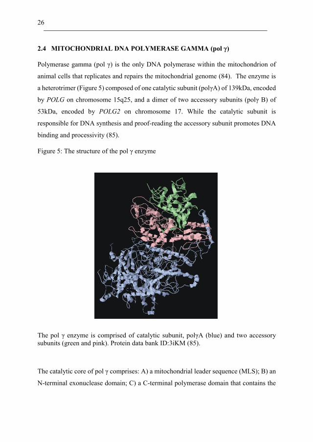
2.4 MITOCHONDRIAL DNA POLYMERASE GAMMA (pol γ)
Polymerase gamma (pol γ) is the only DNA polymerase within the mitochondrion of
animal cells that replicates and repairs the mitochondrial genome (84). The enzyme is
a heterotrimer (Figure 5) composed of one catalytic subunit (polγA) of 139kDa, encoded
by POLG on chromosome 15q25, and a dimer of two accessory subunits (polγ B) of
53kDa, encoded by POLG2 on chromosome 17. While the catalytic subunit is
responsible for DNA synthesis and proof-reading the accessory subunit promotes DNA
binding and processivity (85).
Figure 5: The structure of the pol γ enzyme
The pol γ enzyme is comprised of catalytic subunit, polγA (blue) and two accessory
subunits (green and pink). Protein data bank ID:3iKM (85).
The catalytic core of pol γ comprises: A) a mitochondrial leader sequence (MLS); B) an
N-terminal exonuclease domain; C) a C-terminal polymerase domain that contains the

27
polymerase active site; and D) the spacer (linker region) which separates the
exonuclease and polymerase domains (Figure 6).
Figure 6: The linearized structure of the catalytic pol γ subunit
Linear schematic diagram. The catalytic subunit comprises MLS, exonuclease, palm,
thumb, fingers and linker domains.
The polymerase domain consists of three sub-domains: A) the palm sub-domain
(residues 816-910 and 1096-1239), a positively charged domain stabilizing the
negatively charged DNA backbone and containing the polymerase catalytic site and two
Mg2+ ions, which are vital for formation of the phosphodiester bond between the 3’OH
end and the phosphate group of the incoming nucleotide (dNTP); B) The fingers sub-
domain (residues 911-1095), which is involved in binding the incoming dNTP substrate;
C) the thumb sub-domain (residues 441-475 and 785-815), which forms the major
surface of the DNA binding channel. The linker domain (residues 476-785) comprises
two sub-domains: the accessory interacting sub-domain, which forms a major
hydrophobic contact with the proximal accessory subunits and the intrinsic processivity
domain which forms a region for the upstream DNA binding. The exonuclease domain
repairs replication errors by 3’-5’ excision and is important for fidelity (39, 85, 86).

28
2.5 POLG DISEASE
2.5.1 General overview
The first pathogenic variant in the POLG gene was identified in families with autosomal
dominant Progressive External Ophthalmoplegia (adPEO) (OMIN 157640) in 2001
(87). Since then, an increasing number of overlapping phenotypes with wide variation
in the age of disease onset have been linked to pathogenic variants in the POLG gene.
The true prevalence of POLG disease is unknown, however, it has been estimated to be
about 10-25 % of all adult patients with mitochondrial disorders (10, 83). The most
common reported variants causing human diseases are c.2243G>C (p.Trp748Ser),
c.1399G>A (p.Ala467Thr) and c.2542G>A (p.Gly848Ser) (88-91). The carrier
frequency of p.Trp748Ser is estimated to be 1:125 in Finland (92), while for
p.Ala467Thr it is estimated at 0.6% in Belgium and 1% in Norway (93, 94). Thus, the
combined frequency of p.Trp748Ser and p.Ala467Thr maybe is as high as 1:50 in the
Norwegian population (88). Variants in POLG have also been described in other non-
European ethic groups (95).
The majority of POLG-related phenotypes are inherited as autosomal recessive traits.
Pathogenic variants within the POLG gene can be homozygous or compound
heterozygous. These variants can either decrease the processivity of polymerase gamma,
its affinity for native DNA or the speed at which it incorporates nucleotides. Autosomal
recessive phenotypes usually present early in life, however, onset late in adulthood has
also been reported (96).
Almost all the variants which are associated with adPEO are in the polymerase domain
(97). Variants in this domain usually interfere with the translocation and binding affinity
to incoming nucleotide, which may result in increased mtDNA replication errors and
decreased catalytic function. AdPEO patients usually present in adulthood (90). One
patient with the p.Tyr955His mutation and early onset disease with bilateral
sensorineural hearing loss, cataract, myopathy, and liver failure has however been
previously reported (98).
29
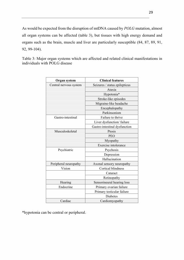
As would be expected from the disruption of mtDNA caused by POLG mutation, almost
all organ systems can be affected (table 3), but tissues with high energy demand and
organs such as the brain, muscle and liver are particularly susceptible (84, 87, 89, 91,
92, 99-104).
Table 3: Major organ systems which are affected and related clinical manifestations in
individuals with POLG disease
Organ system Clinical features
Central nervous system Seizures / status epilepticus
Ataxia
Hypotonia*
Stroke-like episodes
Migraine-like headache
Encephalopathy
Parkinsonism
Gastro-intestinal Failure to thrive
Liver dysfunction/ failure
Gastro-intestinal dysfunction
Musculoskeletal Ptosis
PEO
Myopathy
Exercise intolerance
Psychiatric Psychosis
Depression
Hallucination
Peripheral neuropathy Axonal sensory neuropathy
Vision Cortical blindness
Cataract
Retinopathy
Hearing Sensorineural hearing loss
Endocrine Primary ovarian failure
Primary testicular failure
Diabetes
Cardiac Cardiomyopathy
*hypotonia can be central or peripheral.

30
2.5.2 Major clinical phenotypes
A. MyoCerebroHepatopathy Spectrum (MCHS):
MCHS is a severe and fatal phenotype that usually presents very early in the neonatal
period with a triad of myopathy/hypotonia, encephalopathy/developmental delay and
liver failure. Other findings include failure to thrive, renal tubular acidosis, cataract, and
hearing loss. Diagnostic criteria for MCHS include: absence of hepatic
histopathological features of classical Alpers-Hunttenlocher syndrome and at least two
of the following: neuropathy, seizures, elevated blood or cerebrospinal fluid (CSF)
lactate, dicarboxylic aciduria, renal tubular dysfunction with aminoaciduria, glycosuria
or bicarbonaturia, hearing loss, abnormal MRI with either cerebral volume loss, delayed
myelination or white matter disease, and either isolated deficiency of complex IV or a
combined defect of two or more OXPHOS complexes in skeletal muscle or liver biopsy
(84, 90, 100, 102, 105).
B. Alpers-Huttenlocher syndrome (AHS):
Alpers-Huttenlocher syndrome (AHS) (OMIM # 203700) is the most frequently
reported phenotype in infancy and early childhood (106), although disease onset can
occur at any time during childhood or early adulthood (107, 108). AHS was first
recognised by Bernard Alpers in 1931, long before its genetic basis was identified, and
diagnosis was based on typical neuropathological findings. Subsequently, when the
association with liver involvement was described by Petter Huttenlocher, it was called
Alpers-Huttenlocher syndrome (109, 110). It was not until the 1980s that the link to
mitochondrial dysfunction was made (111) and the link to polymerase gamma was made
first in 1999 (99). Pathogenic POLG variants causing AHS were first reported in 2004
(112).
AHS is characterized clinically by a triad of progressive encephalopathy with
psychomotor regression, refractory epilepsy and liver disease (103). A prodromal phase
with mild developmental delay, hypotonia and failure to thrive may occur and an
infectious illness may precede the disease onset (113). Focal seizures, commonly
evolving into bilateral convulsive seizures, are the most common seizure types with

31
epileptiform discharges predominantly seen over the occipital regions, at least initially
(89, 114). The clinical features of occipital lobe involvement such as visual
hallucination, vomiting and headache are less clearly manifested in young children than
older children and adults. The majority of the patients develop myoclonic seizures and
episodes of epilepsia partialis continua (EPC) and/or generalized status epilepticus (SE).
Patients with AHS may also present with refractory SE from which they might never
recover (84, 89, 115).
Patients with AHS may develop episodes of acute exacerbation that previously have
been called stroke-like episodes (SLEs). These episodes are characterized by acute or
subacute neurological dysfunction and are often associated with EPC. The aetiology of
these episodes is neuronal dysfunction leading to damage, not vascular occlusion (116).
In the older age group, prodromal symptoms such as migraine-like headaches, visual
disturbance, and mental changes may occur. Clinically, such episodes are less often
reported in children compared with adults, but radiological evidence of cortical lesions
is common in both (117).
Hepatic involvement is a major feature of early onset POLG disease and may progress
rapidly to end stage liver failure. Affected children with normal, mild, and transient
abnormalities of liver function are, however, well recognized (91, 102). Liver failure
can occur spontaneously or be triggered by sodium valproate (88, 118). Recovery after
transient liver failure and after the discontinuation of sodium valproate has been reported
(114). Nevertheless, sodium valproate clearly accelerates the development of liver
failure in patients with pathogenic variants in the POLG gene and its use is absolutely
contraindicated.
C. Myoclonic epilepsy myopathy sensory ataxia (MEMSA):
MEMSA (also referred to as mitochondrial spinocerebellar ataxia with epilepsy -
MSCAE) includes a spectrum of manifestations: mainly epilepsy, ataxia and myopathy.
Seizures, including SE, can be the first manifestation. Ataxia, which is often present at
onset, is usually due to a combined central and evolving sensory polyneuropathy.

32
Myopathy and ophthalmoplegia develop later, if the patient survives the consequences
of the epilepsy (89, 94, 100).
Seizure semiology is similar to AHS with focal seizures, commonly evolving into
bilateral convulsive seizures, being the most common seizure type. However, myoclonic
seizures, epilepsia partialis continua and generalized SE are frequently reported (119).
Occipital lobe features, including visual hallucinations, scotomata, hemianopia and
amaurosis, are common (88, 94). Episodes of encephalopathy, previously called SLEs,
are more frequently reported than with AHS (117). Vomiting and migraine-like
headache with aura occur and may precede the acute episodes. Hepatic dysfunction
including liver failure is also a feature of MEMSA and may occur spontaneously or can
be triggered by usage of sodium valproate as in AHS (88, 120).
D: Ataxia Neuropathy Spectrum (ANS):
Ataxia Neuropathy Spectrum is characterized by ataxia, neuropathy and encephalopathy
that usually presents in adulthood and which can be associated with prolonged survival.
The neuropathy may be sensory, motor or mixed and is usually severe enough to
contribute to ataxia. The encephalopathy is slowly progressive (100). Individuals with
ANS may also develop cognitive decline and psychiatric symptoms including
depression. Ophthalmoplegia is more often a late feature and predominant myopathy is
rare (91, 92, 101). Liver dysfunction may also occur, ranging from mildly elevated liver
enzymes to liver failure. Seizures have also been reported, however, these are not a
major feature of ANS (88, 90, 100). Other terms for this phenotype include
mitochondrial recessive ataxia syndrome (MIRAS) and sensory ataxia neuropathy
dysarthria and ophthalmoplegia (SANDO).
E: Progressive external ophthalmoplegia (PEO):
Pathogenic variants in the POLG gene can cause both autosomal dominant and
autosomal recessive PEO. Affected individuals suffer from progressive weakness of the
extraocular muscles resulting in unilateral or bilateral symmetrical ptosis and loss of eye
movements both in the vertical and horizontal directions (87). Other system involvement

33
such as ataxia, peripheral neuropathy and generalized myopathy occur more frequently
in the recessive form (arPEO) than in the dominant form (adPEO), and usually develop
later during the disease course (87, 90, 100, 121-123). Other features which can be seen
in individuals with adPEO may include sensorineural hearing loss (91), parkinsonism
(101, 124), premature menopause (101, 121), male infertility (125), cataract (101) and
depression (101).
In addition to the above described major clinical syndromes, variants in POLG have
been associated with a spectrum of overlapping clinical phenotypes. The terminology
used to describe these phenotypes has evolved haphazardly, becoming more
complicated and difficult to use in an everyday clinical setting. A summary of the
reported POLG related phenotypes with major clinical features and age of onset is
summarized in table 4.
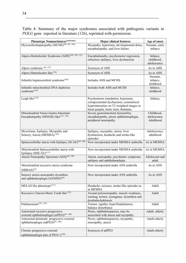
34
Table 4: Summary of the major syndromes associated with pathogenic variants in
POLG gene reported in literature (126), reprinted with permission.
Phenotype Nomenclatures(reference) Major clinical features Age of onset
Myocerebrohepatopathy (MCHS)(90, 105, 106)
Myopathy, hypotonia, developmental delay,
encephalopathy, and liver failure.
Neonate, early
infancy
Alpers-Huttenlocher Syndrome (AHS)(103, 105, 112)
Encephalopathy, psychomotor regression,
refractory epilepsy, liver dysfunction
Infancy,
childhood,
adolescence
Alpers syndrome (84, 127) Synonym of AHS As in AHS
Alpers-Huttenlocher like(128) Synonym of AHS As in AHS
Infantile hepatocerebral syndrome(105)
Includes AHS and MCHS
Neonate,
infancy,
childhood
Infantile mitochondrial DNA depletion
syndrome(129)
Includes both AHS and MCHS
Infancy,
childhood
Leigh like(129)
Psychomotor retardation, hypotonia,
extrapyramidal dysfunction, symmetrical
Infancy
hyperintensities on T2 weighted images in
basal ganglia, brain stem, thalamus Mitochondrial Neuro-Gastro-Intestinal
Encephalopathy (MNGIE) like(127, 130)
Severe gastrointestinal dysmotility,
encephalopathy, ptosis ophthalmoplegia,
peripheral neuropathy
Childhood,
adolescence
Adulthood
Myoclonus, Epilepsy, Myopathy and
Sensory Ataxia (MEMSA) (84)
Epilepsy, myopathy, ataxia, liver
dysfunction, headache and stroke-like
episodes
Adolescence,
adulthood
Spinocerebellar ataxia with Epilepsy (SCAE)(84, 100)
Now incorporated under MEMSA umbrella
As in MEMSA
Mitochondrial Spinocerebellar ataxia with
Epilepsy (MSCAE)(115)
Now incorporated under MEMSA umbrella
As in MEMSA
Ataxia Neuropathy Spectrum (ANS)(84, 100)
Ataxia, neuropathy, psychiatric symptoms,
epilepsy and ophthalmoplegia
Adolescent and
adult
Mitochondrial recessive ataxia syndrome Now incorporated under ANS umbrella As in ANS
(MIRAS)(92)
Sensory ataxia neuropathy dysarthria Now incorporated under ANS umbrella As in ANS
and ophthalmoplegia (SANDO)(84)
MELAS like phenotype(131)
Headache, seizures, stroke-like episodes as
in MEMSA
Adult
Recessive Charcot-Marie Tooth like(132)
Axonal polyneuropathy, muscle weakness,
wasting, tremor, nystagmus, dysarthria and
dysdiadochokinesis
Adult
Parkinsonism(101, 124)
Tremor, rigidity, hypo/bradykinesia,
balance disturbance
Adult
Autosomal recessive progressive
external ophthalmoplegia (arPEO)(87, 100)
Ptosis, ophthalmoparesis, may be
associated with ataxia and myopathy
Adult, elderly
Autosomal dominant progressive external
ophthalmoplegia (adPEO)(87, 100)
Ptosis, ophthalmoparesis, myopathy,
neuropathy, ataxia
Adult-elderly
Chronic progressive external Synonym of adPEO Adult-elderly
ophthalmoplegia plus (CPEO+)(100)
35
2.5.3 Diagnosis
A. Clinical awareness:
As with most mitochondrial disorders, the diagnosis of POLG disease is challenging
owing to the extreme clinical heterogeneity of presentation, particularly in the paediatric
population. There is no single clinical feature that is diagnostic for POLG disease,
however, certain groups of symptoms and signs may provide a clue: for instance, disease
onset soon after birth with hypotonia, failure to thrive and liver failure, but no seizures,
would suggest MCHS. Presentation with acute-onset status epilepticus preceded by
headache and visual disturbances and MRI changes suggestive of ischaemia in an
adolescent or adults may raise the suspicion of MEMSA. EEG, biochemical and neuro-
imaging findings, as described below, may also provide clues to the diagnosis. However
none of those are diagnostic for POLG disease.
A detailed family and medical history and a thorough physical examination of central
and peripheral nervous systems, as well as evaluation of the possible involvement of
other organ systems and assessment of vision, hearing, growth and psycho-motor
development are still essential initial steps to achieve the diagnosis.
The diagnosis of POLG disease should not only be considered in individuals presenting
with one of the classic POLG phenotypes, since a large proportion of patients with
POLG disease do not present with a discrete clinical syndrome (83). POLG disease
should also be considered in patients with therapy resistant epilepsy, unexplained
encephalopathy or ataxia. The difficulties associated with making the diagnosis of
POLG disease are not helped by the unwieldy classification.

36
B. Biochemical analysis:
There are no specific blood or urine biomarkers for POLG disease. Peripheral blood
lactate is a marker for mitochondrial disease generally, however, normal blood lactate
does not exclude the diagnosis of POLG disease as elevation can be mild, transient or
even absent. Further, inappropriate collection may result in a falsely pathologic high
value. Pyruvate/lactate ratio can be used to indicate OXPHOS impairment (127, 133).
Both lactate and pyruvate can be measured in the CSF, however, lack of specificity
limits their diagnostic value since elevated CSF lactate can occur in other conditions
such as seizures of other cause, and CNS inflammation and infection (134).
Fibroblast growth factor 21 (FGF21) and growth differentiation factor 15 (GDF15) are
both potential biomarkers for mitochondrial disorders (135, 136). FGF21 has been
reported to be elevated mainly in individuals with manifestations in skeletal muscle and
appears rarely elevated in POLG disease. Values ranging from 25pg/ml to > 4000 pg/ml
have however been reported in patients with AHS and in a single patient with ANS with
terminal SE (135). FGF21 and GDF15 can be used as additional biomarkers in the initial
diagnostic process and might have prognostic implications as FGF21 and GDF15 values
appear to correlate with disease severity (135, 136). Nevertheless, negative results
should not exclude the diagnosis. Owing to the observation of normal values in many
patients with POLG disease, FGF21 and GDF15 are not considered useful markers to
establish the diagnosis. Other findings such as low CSF folate, the presence of CSF
oligoclonal bands, and raised CSF NF-L have also been reported, however, none is
specific for POLG disease (137-139).
General blood and urine investigations including full blood count, glucose, creatine
kinase (CK), liver transaminases, liver and renal function tests and urine analysis should
be performed to evaluate the systemic involvement of the disease. Metabolic screening
with measurement of plasma amino acid and acylcarnitine profiles and urinary organic
acids are helpful to exclude other metabolic/mitochondrial disorders that may mimic
POLG disease.

37
C. Histopathology and respiratory chain enzymology:
Classical mitochondrial muscle pathology findings such as ragged-red and cytochrome
oxidase (COX) negative fibres can be seen in patients with POLG disease, however,
these can be absent in patients with early onset disease (140). Adolescent patients may
have less than 1% of COX-negative fibres, emphasizing that the major manifestations
are in the CNS. Further, infants with normal muscle biopsy may have severe
pathological liver changes (141). The characteristic hepatic histopathological changes
of AHS which are required for the diagnosis are namely the presence of at least two of
the following: microvesicular steatosis, bile ductular proliferation, hepatocyte dropout,
bridging fibrosis or cirrhosis, collapse of liver cell plates, parenchymal lobular
architecture, regenerative nodules and oncocytic changes in scattered hepatocytes not
affected by steatosis (106).
In addition to morphological examination, mitochondrial RC analysis in muscle may
provide a diagnostic clue. RC enzyme analysis may show isolated enzyme deficiency or
combined deficiencies of multiple enzymes, especially in patients with primary muscle
involvement, but the results may also be normal. Pathogenic variants in the POLG gene
demonstrate tissue specific predilections and thus RC enzyme deficiencies may only be
identified in clinically affected tissues such as liver or brain (83, 84).
D. Neurophysiological findings:
EEG findings may give a clue to the diagnosis of POLG disease in individuals with
seizures. Ictal and inter-ictal occipital epileptic activity are highly suggestive of POLG
disease. Focal epileptic discharges over the temporal and frontal regions can however
also be observed and multifocal or generalized epileptic activity may occur during
seizure evolution and SE (89, 119). Other EEG changes such as rhythmic high
amplitude delta (RHADs) and focal slowing are frequently observed (114, 119).
Nerve conduction studies will confirm the presence of peripheral neuropathy
particularly in juvenile and adult onset disease (88). POLG disease is mostly associated
with axonal changes and a predominantly sensory and some motor component
neuropathy. Demyelinating motor neuropathy has also been reported (142, 143).
38
E. Neuro-imaging:
Brain MRI is the modality of choice and the recommended sequences are T2 fluid-
attenuated inversion recovery (FLAIR-T2) and diffusion weighted imaging (DWI). The
most prevalent abnormalities are T2 / T2-FLAIR hyperintensities in the cerebral cortex,
also known as cortical focal lesions (CFLs), which occur in patients with epilepsy and
mainly affect the occipital regions although involvement of other regions such as
parietal, temporal and frontal lobes may also occur (119). These changes may evolve
over days or weeks and subsequent partial or complete regression may occur (88, 117,
119). Neuroimaging studies may, however, be normal early in the disease course and
should not exclude the possibility of POLG disease.
Other neuroimaging abnormalities include lesions in the thalamus, olivary nucleus, and
cerebellar white matter which may remain stable throughout the disease course.
Generalized brain atrophy develops later during the disease course and is progressive,
reflecting the clinical progression of the disease (117, 119, 120).
MRS of CFLs often shows a prominent lactate peak due to impaired aerobic respiration
and decreased N-acetyl aspartate concentration which reflects neuronal loss (117). Ictal
Cerebral FDG-PET imaging in the acute phase shows increased glucose uptake over
these lesions (120).
F. Molecular genetic:
When the diagnosis of POLG disease is suspected clinically, direct sequencing of the
POLG gene is the most appropriate first-line investigation. The inclusion of POLG in
next generation sequencing (NGS) gene panels for epilepsy, ataxia and mitochondrial
disorders should facilitate early diagnosis. In some regions, screening for the common
founder mutations; p.Ala467Thr, p.Trp784Ser, and p.Gly848Ser may still be
appropriate (87, 144). The finding of pathological variants in POLG, biallelic in
recessive disease and heterozygous in dominant disease, establishes the diagnosis.
New variants in the POLG gene continue to be identified. Determining the pathogenicity
of such variants as ‘pathogenic’, ‘likely pathogenic’ or of ‘uncertain significance’ can
be challenging. Useful resources for judging pathogenicity are: the Human DNA

39
Polymerase Gamma Mutation Database (https://tools.niehs.nih.gov/polg/), and the
POLG Pathogenicity Prediction Server (http://polg.bmb.msu.edu/) (145). Further, the
identification of new variants either requires extensive laboratory research to prove its
pathogenicity or verification by finding more families. Ideally, all pathological variants
should be reported in ClinVar (https://www.ncbi.nlm.nih.gov/clinvar/), however many
laboratories are either slow or forget to submit variants. These challenges highlight the
difficulties in establishing the definite genetic diagnosis in some individuals with
POLG-related phenotypes and new variants in POLG gene.
2.5.4 Management
Currently, there are no cures for POLG disease. Clinical management is mainly
symptomatic and based on conventional approaches to treat the clinical manifestations
and associated complications. As in other rare diseases with high mortality, randomized
controlled clinical trials are still lacking and may be challenging to perform as designing
a trial is likely to be extremely difficult in the view of clinical heterogeneity,
unpredictable clinical course, and spontaneous resolution of features such as SLE or
cerebral ischaemia. Understanding the natural history of the disease may help to
establish a clinical baseline which can be used in comparison in single-arm clinical trials
for potential therapies. Unfortunately, however, earlier descriptions of the natural
history of POLG disease were often based on case reports or cohorts with limited
numbers of patients, making large cohorts such as ours even more valuable.
A. Management of epilepsy:
The presence of epilepsy is associated with increased mortality and morbidity in patients
with POLG disease (88, 146). Early recognition and immediate, aggressive seizure
treatment are crucial to improve patient survival. The majority develop therapy resistant
seizures (88, 119) and this is particularly true for infants and children. Treatment with a
single AED is usually not effective and high dose, multiple AED treatment is often
required (119). There are currently no consensus recommendations for epilepsy

40
management in POLG disease and several AEDs have been used in various dosages and
combinations.
No specific single or combined AEDs have been shown to be particularly effective in
treating the seizures in POLG disease. AEDs known to be effective in treatment of focal
seizures such as oxcarbazepine, carbamazepine, lacosamide and perampanel are
appropriate, as focal and focal evolving to bilateral convulsive seizures are among the
most common types. Lamotrigine, topiramate and levetiracetam have also been used,
alone and in combination with a benzodiazepine such as clobazam or clonazepam.
Lamotrigine can worsen mycolonic seizures and should be used with caution (89, 115,
146).
Sodium valproate is absolutely contraindicated due to the risk of acute and progressive
hepatic necrosis (88). Transient liver failure with recovery after discontinuation of
sodium valproate has been reported (114). If the clinical presentation raises the suspicion
of POLG disease, sequencing of the POLG gene should be considered before prescribing
sodium valproate, particularly in those with status epilepticus of unknown aetiology.
(118).
Management of SE is challenging; benzodiazepines, phenytoin and levetiracetam can
be used as first line treatment, however, failure to control the seizures is common. In a
case of refractory SE, anaesthetic agents as propofol or a barbiturate (pentothal) should
be instituted promptly. Propofol should be used with caution due to the risk of propofol
infusion syndrome particularly in the paediatric population. Other agents as ketamine
(147), magnesium infusion (148) and corticosteroids (114) have been reported to be
effective in terminating SE in single cases, however, data available regarding the
effectiveness are currently insufficient. Epilepsia partialis continua is generally resistant
to pharmacotherapy.
Other non-pharmacological alternatives including ketogenic diet and vagus nerve
stimulation have been used (149), but currently, there are insufficient data confirming
the benefit of either of these entities in patients with POLG disease. Transcranial direct
current stimulation gave promising results in one previously published case (150),

41
however a recent publication showed it was not effective (151). Palliative functional
hemispherectomy can be an option when the short-term benefits outweigh the risk of
surgery (152).
There is no clear evidence showing any significant clinical effect of nutritional
supplements such as co-enzyme Q10, folic acid, carnitine, L-arginine, EPI-734 or other
vitamins, although these are widely used. Further studies need to be performed to
investigate the effectiveness of these agents. (153, 154).
B: Gastrointestinal and nutritional:
Feeding difficulties and failure to thrive, mainly in the very young, and vomiting/gastric
dysmotility regardless of the age of onset are common gastro-intestinal features of
POLG disease (84, 127, 155). Evaluation by a gastroenterologist and dietician and the
use of enteral nutrition via gastric tube/gastrostomy should be considered early during
the disease course, particularly in young individuals.
C. Liver dysfunction:
Liver dysfunction is a common feature of POLG disease regardless of the age of onset
and can range from acute and progressive liver failure to mild/transient elevation of liver
enzymes. Liver dysfunction can also occur spontaneously or as a consequence of sodium
valproate exposure. Spontaneous resolution of liver failure after exposure to sodium
valproate has been reported (156). Close monitoring of the liver function by measuring
liver enzymes (AST, ALT, GGT) and other liver function parameters such as ammonia,
albumin, bilirubin, prothrombin time, is highly recommended and should be performed
routinely at least every 3 to 4 months, especially in infants, children and adolescents.
Liver transplantation remains an option and it has been performed in more 40 patients
with POLG disease (83, 157-159). However, there is some controversy around the use
of liver transplantation, particularly in early onset disease, due to the aggressive nature
of the disease and early death due to neurological decline occurring within one year of
the liver transplantation. Survival after liver transplantation in adult onset disease is
42
better than with early onset disease (88, 90, 160). Thorough evaluation of the ethical
aspects and an individualized risk-benefit analysis in all cases, regardless of age, are
needed before proceeding to transplantation (160-162).
D. Movement disorders:
Parkinsonism is a feature of late-onset disease (101) and usually occurs together with
PEO and peripheral neuropathy. However, early onset parkinsonism was reported in two
sisters who also had neuropathy, but not PEO (124). Treatment with L-Dopa (163)
appeared to be effective in some cases. Benzodiazepines may reduce the severity of
other non-epileptic movement disorders including myoclonus and tremor. In individuals
with dystonia, local injections with botulinum toxin and oral/intrathecal baclofen can be
useful.
E. Ophthalmological manifestations:
Individuals with POLG disease may develop cortical blindness, nystagmus, ptosis and
ophthalmoplegia and should be referred for ophthalmological evaluation. Surgery for
ptosis may be considered and may provide some symptomatic relief, although post-
operative relapse is frequent.

43
3. AIMS OF THE THESIS
3.1 GENERAL AIMS
The overarching aim of the work in this thesis was to bridge the translational gap
between research and clinical practice in order to facilitate faster and more accurate
diagnosis, and impact the management of patients with POLG disease.
3.2 SPECIFIC AIMS
- To study the clinical spectrum and the natural course of POLG disease in a large
cohort of patients to provide a detailed description of the disease’s phenotypic-
spectrum and a reliable clinical classification which can be used both in paediatric
and adult populations.
- To identify robust diagnostic and prognostic biomarkers which may help to
facilitate the diagnosis and to predict the outcome of the disease.

44
4. METHODS
4.1 STUDY DESIGN AND POPULATION
In these multinational, retrospective studies 159 patients were recruited from 13 centres
in seven European countries: Norway (Haukeland University Hospital, Oslo University
Hospital, St. Olav’s Hospital and University Hospital of Northern Norway); United
Kingdom (Great Ormond Street Hospital, London and Welcome Trust Centre for
Mitochondrial Research, Institute of Neuroscience, Newcastle University); Sweden
(Centre for Inherited Metabolic Diseases, Karolinska University Hospital, Stockholm
and The Queen Silvia Children's Hospital, University of Gothenburg), Denmark
(Department of Clinical Genetics, Copenhagen University Hospital); Finland
(Children's Hospital, Helsinki University Hospital and Clinic for Children and
Adolescents, Oulu University Hospital); Netherlands (Department of Genetics and Cell
Biology, Maastricht University, Maastricht) and Spain (Sant Joan de Déu Children´s
Hospital, Barcelona) (figure 7).
Figure 7: The European countries participating in this study highlighted in green.

45
The Norwegian patients were recruited from the National Norwegian POLG Registry,
(www.polgregister.no). Collaboration was established with all Norwegian university
hospitals and a local investigator responsible for data collection was allocated in each
centre. By June 2018, 76 patients had been enrolled in the registry. Distribution of
patients according to the health-regions (Central, Northern, Southern and Eastern and
Western Norway) is provided in figure 8.
Figure 8: Distribution of patients with POLG disease in Norway according to the four
health regions; Central: red, Northern: blue, Southern and Eastern: yellow, Western:
green.
The majority of patients included in this study were Northern European (n=150), but
there were three from Iraq, two from Cyprus and one each from Croatia, Pakistan, Spain
and the United Arab Emirates.

46
In view of the rarity of MCHS, and to provide a better understanding of this particular
phenotype, a systematic literature review (using search terms “POLG”, “mitochondria”,
“Alpers”, “infantile hepatocerebral syndromes”, “mtDNA depletion”,
“myocerebrohepatopathy syndrome” performed in PubMed, June 2016) was used to
identify previously published cases. Cases fulfilling the criteria for MCHS (106) and
confirmed biallelic pathogenic POLG variants were included. These additional MCHS
cases were just included in paper I.
A large database of patients with POLG disease was assembled and analysed in step-
wise fashion. Details regarding number of patients, recruiting-centres and period of data
entry for each part of the study are provided in table 5. In paper I, only data from
individuals with disease onset before 12 years of age was included, while in papers II,
III and IV available data from all the individuals, regardless of the age of onset were
included. Patients (n=4) recruited from the Centre for Mitochondrial Research,
Newcastle, were only included in paper IV.

47
Table 5: Recruiting-centres, number of patients and period of data entry.
Study part
Recruiting centres
Number of
patients
Data-entry period
Paper I May 2015- July 2016
Norway: 8
Haukeland University Hospital, Bergen
University Hospital of Northern Norway, Tromsø
United Kingdom: 19
Great Ormond Street Hospital, London
Total 27 Paper II May 2015- December 2017
Norway: 76
Haukeland University Hospital, Bergen
University Hospital of Northern Norway, Tromsø
St. Olav’s Hospital, Trondheim
Oslo University Hospital, Oslo
United Kingdom: 19
Great Ormond Street Hospital, London
Sweden: 44
Karolinska University Hospital, Stockholm
The Queen Silvia Children's Hospital, Gothenburg
Denmark: 5
Copenhagen University Hospital, Copenhagen
Finland: 8
Helsinki University Hospital, Helsinki
Oulu University Hospital, Oulu
Netherlands: 2
Maastricht University, Maastricht
Spain: 1
Sant Joan de Déu Children´s Hospital, Barcelona
Total 155 Paper III May 2015- June 2017
Norway: 50
Haukeland University Hospital, Bergen
University Hospital of Northern Norway, Tromsø
St. Olav’s Hospital, Trondheim
Oslo University Hospital, Oslo
United Kingdom: 9
Great Ormond Street Hospital, London
Sweden: 19
Karolinska University Hospital Stockholm
The Queen Silvia Children's Hospital, Gothenburg
Finland: 5
Helsinki University Hospital, Helsinki
Oulu University Hospital, Oulu
Total 83 Paper IV May2015- December2016
Norway: 45
Haukeland University Hospital, Bergen
University Hospital of Northern Norway, Tromsø
St. Olav’s Hospital, Trondheim
Oslo University Hospital, Oslo
United Kingdom: 16
Great Ormond Street Hospital, London
Centre for Mitochondrial Research, Newcastle Total 61

48
4.2 INCLUSION CRITERIA
Individuals diagnosed and followed at the participating centres were considered eligible
if they had recessive disease and confirmed biallelic pathogenic POLG variants or
dominant disease and a heterozygous, confirmed pathogenic variant. Individuals with
novel variants considered pathological and clinical features consistent with POLG
disease were also included.
4.3 DATA COLLECTION
Data were recorded on an electronic-case report form (eCRF) completed by the
responsible investigator(s) at each centre and reviewed by the study-monitor (O.H).
The eCRF was developed and tested in a pilot study of 10 patients conducted at
Haukeland University Hospital, Bergen, Norway. The electronic version of the eCRF
(WebCRF) was developed by the Unit for Applied Clinical Research Faculty of
Medicine, Norwegian University of Science and Technology, Trondheim, Norway.
(https://webcrf3.medisin.ntnu.no/).
4.4 STUDY DATA
Detailed clinical, biochemical, muscle biopsy, neurophysiological, neuroimaging and
genetics data were obtained (Appendix 1). The date of disease onset was defined by the
date of symptom(s) necessitating the patient’s first medical evaluation. The age of onset
of each individual symptom was also identified. End of follow up was defined as the
date of the patients’ last visit to the treating centre or death. Preterm birth was defined
as birth before a gestational age of 37 completed weeks. Microcephaly was defined as
head circumference more than two standard deviations below the mean for age and sex.
Anaemia was defined as an abnormally low haemoglobin value according to age and
sex adjusted values (164). Renal tubulopathy was defined by the presence of at least
three of the following: tubular acidosis with generalised aminoaciduria, tubular
proteinuria, glycosuria, increase in urinary N-acetyl-beta-D-glucosaminidase
(NAG)/creatinine ratio, or Fanconi syndrome. Liver involvement was defined by the
presence of two or more of the following in at least two different time points: elevated

49
aspartate aminotransferase (ASAT), gamma-glutamyltransferase (GGT), bilirubin or
ammonia, low serum albumin, or pathological histological findings on liver biopsy.
CSF protein and/or albumin values at disease onset and during the disease course were
obtained. Since the reference normal range for CSF protein may be age dependent, we
looked at the values according to age (165-167). Since albumin is produced exclusively
in the liver, all albumin detected in CSF originates, by definition, from blood. The level
of CSF albumin provides, therefore, a parameter with which to evaluate the permeability
of the blood-CSF barrier. The ratio of CSF to serum albumin (Q-alb) corrects for the
individual’s albumin level and provides a reflection of the diffusion gradient of albumin
(165, 168-170). This ratio was calculated as CSF albumin (milligram (mg)/L)/serum
albumin (g/L). The reference range for Q-alb is also age dependent. The cut-offs used
in this study were reported in supplementary file 2, paper III (171).
Hearing impairment was defined as abnormal auditory evoked response test. Therapy
resistant epilepsy was defined using the International League Against Epilepsy (ILAE)
definition (172).
POLG variant(s) for each case were identified either by targeted mutation analysis for
specific common mutations (c.1399G>A, p.Ala467Thr and c.2243G>C, p.Trp748Ser,
and Gly848Ser) or by sequence analysis of all coding regions of POLG gene.
4.5 STATISTICAL ANALYSIS
Data were analysed using SPSS (Statistical Package of Social Sciences). A two sided P
value less than 0.05 was considered to be statistically significant. Categories were
compared using Fisher’s Exact tests when appropriate.
In paper II, Correspondence Analysis (CA) was performed to examine the relationship
between two variables graphically in a multidimensional space. These variables were:
A) groups of patients who were classified according to the age of disease onset (early
onset, juvenile and adult, and late onset disease), and B) the age of onset of each
individual symptom. This enabled examination of the clustering of symptoms around

50
each age group. Further, mosaic plots were performed to study the differences between
these groups.
For survival analysis, the end-point was time to death which was defined as the time in
months from the date of onset to the date of death. Univariate survival analysis was
performed using log-rank test (Kaplan Meier) to compare differences in survival time
between categories. Adobe illustrator CS6 and BioRender (biorender.com) were used to
create and edit the figures.
4.6 ETHICAL STATEMENT
The ethical approval for the study was obtained from the Regional Committee for
Medical and Health Research Ethics, Western Norway (REK 2014/1783-4). Each
participating country obtained approval from their local ethical committee. The study
was registered as an audit at Great Ormond Street Hospital, London, UK (Registration
Number 1675). Anonymised data regarding Newcastle patients was provided courtesy
of the MRC Mitochondrial Disease Patient Cohort (Ethics ref:13/NE/0326). All
procedures followed were in accordance with the ethical standards of the responsible
committee both in Norway and in the other collaborating countries and with the Helsinki
Declaration of 1975, as revised in 2000.

51
5. RESULTS
5.1 Paper I: The clinical spectrum and natural history of early-onset diseases due to
DNA polymerase gamma mutations
The primary aim was to study the natural history of paediatric onset POLG disease in a
sufficiently large cohort of patients to provide a better and more detailed description of
the clinical spectrum in this age group. Clinical, biochemical, neuro-imaging and genetic
data of 27 individuals with recessive disease and biallelic variants in POLG in whom
initial symptomatology manifested prior to the age of 12 years were obtained.
The median age of disease onset was 11 months (range 2 weeks to 7 years). Extensive
phenotypic characterisation was performed. Six patients fulfilled diagnostic criteria for
MCHS, 19 for AHS, and one had a MNGIE-like phenotype. One patient was
unclassified owing to insufficient data. The majority presented with global
developmental delay (n=24/24, 100%), hypotonia (n=22/23, 96%), liver dysfunction
(n=23/25, 92%) and failure to thrive (n=24/27, 89 %). Seizures were also common
(n=19/26, 73%) and were present in 89% (n=17/19) at disease onset. Focal and focal
evolving to bilateral convulsive seizures with epileptiform activities predominantly seen
over the occipital regions were the most commonly reported seizure types. Notably,
none of the patients with the MCHS phenotype had seizures.
Lactate was elevated in blood in 60% of those in whom this was measured (n=12/20)
and in CSF in 40% of those in whom CSF studies were performed (n=2/5). Abnormal
respiratory chain enzyme activities were observed in 9 of 14 cases in whom muscle
biopsy was performed. Interestingly, most AHS patients had normal respiratory chain
enzyme activities or only an isolated deficiency of a single enzyme complex, while those
with MCHS had multiple respiratory chain complex deficiencies.
The most common MRI findings were cortical focal lesions manifesting as T2/FLAIR
hyperintensities involving cortical and subcortical areas, predominantly affecting the
occipital lobes.

52
All cases had two biallelic pathogenic POLG variants identified either by targeted
mutation analysis for specific common variants as p.Ala467Thr and p.Trp748Ser or by
sequence analysis of all coding regions of POLG. Two patients (no. 23 and no. 26,
supplementary file 1, paper I) with classical AHS phenotype and novel POLG gene
variants (p.Lys498Thr, p.Gly621Asp) were included. All POLG gene variants reported
in this part of the study and associated phenotypes are illustrated in Figure 2 paper I.
Survival analysis showed that median age at death was 15.8 months (range 1.0 to 184.6
months), whereas median time from disease onset to death was 4.9 months (range 0.5 to
181.2 months). The main causes of death were liver failure, followed by sepsis and status
epilepticus. Median survival time of patients with disease onset ≤12 months was 3.6
months ( range 0.5-181) compared to 10 months (range 1.4-82) for those with disease
onset >12 months. Survival analysis by phenotype showed that the median survival time
for patients with MCHS was 5 months (range 0.6-22) and 4 months (range 0.5- 181) for
AHS.
This study showed the multi-systemic nature and natural history of early onset POLG
disease and highlighted the existence of a disease group without seizures - i.e. those with
the MCHS phenotype. The common founder variants p.Ala467Thr and p.Trp748Ser
were present in only one of our cases with MCHS and a review of the literature showed
only one additional case with this variant, confirming the need to sequence the whole
POLG gene rather than perform targeted variant analysis. Further, we extended the
phenotypic spectrum of childhood POLG disease to include a phenotype with prominent
gastro-intestinal symptoms mimicking mitochondrial neurogastro-intestinal
encephalomyopathy (MNGIE), in addition to the AHS and MCHS phenotypes. Based
on the results of this study, we observed that early onset POLG disease could be
classified simply into those with and those without epilepsy.
53
5.2 Paper II: Simplifying the clinical classification of polymerase gamma (POLG)
disease based on age of onset: studies using a cohort of 155 cases
The aim of this study was to provide a simple clinical classification that would facilitate
early clinical recognition of patients with POLG disease. To do this, we systematically
reviewed detailed longitudinal data from 155 patients both at disease onset and later
during the disease course. The cohort included patients with disease onset from birth to
late adulthood, providing us with a unique opportunity to study the clinical spectrum of
the disease during the whole life span.
The age of onset of each individual symptom was identified. Median age at disease onset
for the whole study cohort was 10 years (range: birth – 71 years). Fifty-four percent
(n=83/155) had onset prior to the age of 12 years, 34% (n=53/155) had onset between
12 and 40 years of age, and 12% (n=19/155) had onset after the age of 40 years. Disease
debut was apparently spontaneous in 113/155 (73%), followed an infectious illness in
32/155 (21%) and not clearly reported in 10/155 (6%) of the patients.
Neurological (90%, n=139/155), ophthalmological (74%, n=112/151) and
gastrointestinal (63%, n=92/146) symptoms were the predominant clinical features.
Epilepsy was reported in 69% (n=107/155), with focal and focal evolving to bilateral
tonic-clonic seizures being the most common seizure types (92%, n=94/102). Ataxia
(63%, n=87/138), peripheral neuropathy (53%, n= 65/123), and hypotonia (50%,
n=68/135) were frequently reported. Nystagmus (38%, n=55/146), PEO (38%,
n=56/146) and ptosis (34%, n=51/149) were the most commonly reported
ophthalmological features. Liver involvement was identified in 64% (n=96/151) of the
patients. More than half of the study cohort had feeding difficulties (52%, n=75/145),
regardless of the age of onset. A detailed description of the clinical features, laboratory,
muscle-biopsy, neuro-physiology and neuro-imaging findings is provided in table 2 and
supplementary tables 1 and 2, paper II.
Our data confirmed that POLG disease comprises a continuum of clinical features rather
than a set of separate clinical identities (see figure 1 paper II). Nevertheless, by grouping
the patients simply using age of onset, we could identify clear phenotypic and prognostic

54
differences (table 3 paper II). Liver involvement (87%), seizures (84%) and feeding
difficulties (84%) were the major features in those with onset prior to the age of 12 years,
while ataxia (90%), peripheral neuropathy (84%) and seizures (71%) occurred most in
those with onset between the age 12 and 40 years, and ptosis (95%), progressive external
ophthalmoplegia (89) and ataxia (58%) in those with onset after the age of 40 years.
Early onset was associated with a worse prognosis. Cluster analysis showed there was
obvious clustering of the symptoms around the three different age groups and further
mosaic plots confirmed statistically significant differences in the phenotypes seen in the
above mentioned groups (supplementary figure 2, paper II).
A total of 41 different POLG variants were identified in the 155 individuals described
in this study. Ninety patients had compound heterozygous variants, 59 had homozygous
variants and six patients had a heterozygous variant associated with autosomal dominant
disease, mainly adPEO. The majority of patients with early onset disease (70%) had
compound heterozygous pathogenic POLG variants (regardless of the variant types),
while 62% of those with juvenile/adult onset had homozygous pathogenic variants.
POLG gene variants reported in this part of the study are summarized in supplementary
figure 3, paper II.
The main cause of death for the whole cohort was liver failure, followed by
infection/sepsis, multi-organ failure and SE, and was unknown in 13% of the
individuals. The presence of epilepsy was associated with significantly worse survival,
and the median survival time from seizure onset to death was 37 months (range < 1 -
487). Survival after the onset of seizures in those with early onset disease was
significantly worse than those who developed seizures as part of juvenile/adult onset
disease. Analysis also showed that patients with pathogenic compound heterozygous
POLG variants had significantly worse survival than those with pathogenic homozygous
variants. Further, individuals with liver involvement showed a significantly worse
survival than those without liver impairment (supplementary figure 4, paper II).

55
5.3 Paper III: Elevated cerebrospinal fluid protein in POLG-related epilepsy:
Diagnostic and prognostic implications
Since epilepsy is common in individuals with POLG disease and the disruption of blood
brain barrier (BBB) has been described in many neurological disorders that include
epilepsy (Table 1, paper III), the aim of this study was to assess whether BBB
dysfunction occurs in POLG disease and what clinical implications it has for patients.
Our study cohort contained 83 patients with details of CSF protein/albumin. We used
the presence of a raised CSF/serum ratio of albumin (Q-alb) to evaluate the integrity of
BBB.
This study demonstrated that elevated CSF protein was a common feature of POLG
disease; raised CSF protein was observed in 70% of patients in whom data were
available (n=58 /83) and was, moreover, associated with the most severe phenotypes.
The finding of a significantly elevated Q-albumin ratio indicated a clear dysfunction of
the BBB. We found that the majority of those with epilepsy (n=50/66, 76%) had raised
CSF protein and interestingly, this preceded seizure debut in 75% (n=15/20). This study
showed that elevated CSF protein can be used as a biomarker both to facilitate early
diagnosis and to identify those at high risk of developing epilepsy.
5.4 Paper IV: The presence of anaemia negatively influences survival in patients
with POLG disease
Mitochondria are known to play an important role in iron metabolism and thus
haematopoietic cell homeostasis. Little attention has been given to the haematological
manifestations in patients with mitochondrial disease particularly those with POLG
disease. Recent studies performed in the POLG mutator mouse model demonstrated that
variants in POLG can also drive haematopoietic abnormalities including anaemia (173-
177). This study aimed to determine the frequency of and outcomes associated with
anaemia in a large cohort of patients with POLG disease.

56
Approximately two third of the patients (n= 41/61, 67%) enrolled in this study
developed anaemia at some stage and, importantly, almost one in four (n=14/61, 23 %)
had anaemia already at presentation. This was particularly true for patients with early
onset POLG disease (AHS and MCHS phenotypes) who had a high frequency of
anaemia (n= 18/ 25, 72%) with 35% (n=8/23) having anaemia at presentation. Details
regarding the number of individuals with anaemia stratified according to age group is
provided in table 2, paper IV.
Survival analysis (Figure 1, paper IV) revealed that the presence of anaemia was
associated with a significantly worse survival. This part of the study demonstrated for
the first time that anaemia is indeed a feature of POLG disease in humans and is
associated with significantly worse survival and can be used as a predictor for poor
prognosis.

57
6. DISCUSSION
The aim of this study was to provide a thorough description of the natural history and
the phenotypic spectrum of POLG disease, and to elaborate a robust, but simple clinical
classification to guide clinicians and help predict the prognosis.
The cornerstone of clinical diagnosis is a relevant clinical, phenotypic classification that
is useful in everyday clinical practice. This, however, is challenging in mitochondrial
disorders due to poor phenotype:genotype correlation, namely that defects in a single
gene, such as POLG, may give rise to multiple clinical phenotypes. This means that even
affected individuals in the same family, sharing the same pathogenic variant may present
with different phenotypes, and that unrelated individuals with different pathogenic
variants may share the same phenotypes (83, 84, 100). Understanding the natural history
of a disease is essential to address these challenges since this will provide information
about the evolution of clinical features and the related morbidity and mortality.
Classification of POLG disease has developed haphazardly and resulted in a plethora of
eponymous and syndromic categories (Table 3). This has made clinical recognition
challenging and can, potentially, have resulted in delayed or even missed diagnosis.
Since data describing the natural history of POLG disease in a sufficiently large cohort
of patients were lacking, we established the largest known cohort of individuals with
POLG disease by recruiting individuals from the National Norwegian POLG Registry
and from our international collaborating centres. Clinical, laboratory, neuro-
physiological and neuroimaging findings were reviewed both at the disease onset and
later during the disease course. This enabled us to describe in detail the clinical spectrum
and the natural history of the disease in a sufficiently large number of patients. Further,
data from our thorough national history studies allowed us to classify POLG disease
more simply than the earlier attempts. We believe that such a classification is easier to
implement in clinical practice and will facilitate early clinical recognition. We were also
able to provide clinical, biochemical and genetic prognostic indicators of the disease.
58
6.1 NATURAL HISTORY AND THE PHENOTYPIC SPECTRUM OF POLG
DISEASE
6.1.1 Paper I: The clinical spectrum and natural history of early-onset diseases due
to DNA polymerase gamma mutations
In this study, we focused on early onset disease. We looked at paediatric patients with
disease onset prior to the age of 12 years and generated a comprehensive clinical and
laboratory description of the features present at onset and later during the disease course.
In addition, we reviewed the neurophysiological, histopathological and neuro-imaging
findings and performed a phenotype-genotype correlation study.
We found that the majority of patients with early onset POLG disease had relatively
diffuse clinical manifestations such as hypotonia, failure to thrive, developmental delay
and seizures. These findings highlight the challenges associated with making the
diagnosis of POLG disease in the paediatric population. These non-specific clinical
manifestations overlap with the manifestations of many other disorders including
mitochondrial and non-mitochondrial disorders. For example, pathogenic variants in
nuclear genes such as PARS2, NARS2, FARS2, SURF1, PDHA1 and ADCK3 (83, 178-
180), or those affecting mtDNA stability (summarized in table 2), may all mimic POLG
phenotypes clinically. Defects in mtDNA such as the m.3243A>G mutation causing
MELAS (181), may also mimic the early and juvenile forms of POLG disease. The
differential diagnosis of non-mitochondrial disorders resembling POLG disease is also
wide and includes febrile infection related epilepsy syndrome (FIRES) (182),
autoimmune encephalitis/Rasmussen encephalitis with EPC (183), and other inborn
errors of metabolism with recurrent liver failure such as that caused by pathogenic
mutations in NBAS gene (184) or neonatal hypoglycaemia and non-ketotic
hyperglycinaemia (185).
Survival analysis showed that early onset disease is associated with high mortality
(n=22/26, 85%) and the median survival time from disease onset to death was 5 months,
which highlights the rapid disease progression.

59
The majority of individuals in this paediatric cohort were classified as AHS or MCHS.
We also found one patient who fulfilled the criteria for MNGIE and were, therefore,
able to expand the POLG phenotypic spectrum in this age category. One patient
remained unclassified owing to insufficient data. Our findings were also in broad
agreement with the earlier understanding that MCHS and AHS can be considered as two
separate entities; we found clear differences in the age of onset and the survival between
these two patient groups. Nevertheless, since few of the patients had liver biopsy
performed (liver biopsy was only performed in 3 cases), it was not possible to
differentiate these two phenotypes as previously suggested (106). This probably reflects
everyday clinical practice where liver biopsy is rarely performed in early onset POLG
disease due to the associated risks and is contraindicated in the case of severe liver
dysfunction. We did find one important feature that differentiated these two phenotypes:
none of the individuals with MCHS phenotype had epilepsy (Table 3, paper I). This was
true not only in our cohort, but also when we collected all previously published cases of
MCHS (Table 2, paper I). Based on these findings, we concluded that the previously
described clinical and histopathological criteria used to differentiate between AHS and
MCHS phenotypes were not applicable and we suggested a simplified classification of
early onset disease into those with or without epilepsy.
Patients with MCHS who underwent biochemical measurements also showed the most
severe respiratory chain defects (supplementary file 2, paper I). We hypothesised that
these patients were the more severely affected and, possibly because of this, died before
the onset of seizures. Discordance in the outcome observed in one sibling pair (cases 15
and 16) provides further evidence for this hypothesis. Case 15 with an AHS phenotype
died at 9 months with intractable seizures whereas his sister (case 16) with a MCHS
phenotype died at 5 months and never developed seizures.
Our data also led us to believe that the MCHS phenotype (i.e. early onset disease without
epilepsy) may be under diagnosed due to the presence of non-specific clinical features
and absence of epilepsy, the most common factor that triggers the clinical suspicion of
POLG disease in the paediatric population. Thus, we feel that highlighting the
60
possibility of POLG disease arising in the absence of epilepsy, will improve the clinical
ascertainment of patients in this age group.
When we looked at the genotypes in the group of MCHS patients, we found that the
common variants (c.2243G>C, p.Trp748Ser and c.1399G>A, p.Ala467Thr) occurred
only once (patient 16, paper I) in our MCHS group. Moreover, we found only one other
previously published MCHS case with these variants (186). These findings suggest that
screening only for common POLG mutations in children younger than 12 years with
unexplained encephalopathy, or multi-system neurological disorders, as previously
suggested (91, 144), will be unsuccessful.
6.1.2 Paper II: Simplifying the clinical classification of polymerase gamma (POLG)
disease based on age of onset: studies using a cohort of 155 cases
In the second study, we wished to extend our work to include all age groups and at the
same time include sufficient numbers of patients to enable us to make robust statements
concerning the natural history and the clinical spectrum of POLG disease. To do this we
used all the available data from the National Norwegian POLG Registry, data from our
ongoing collaboration in the Mitochondrial Clinical Research Network (MCRN)
(www.mcrnet.org ) and data from patients in our first study that were recruited from
Great Ormond Street Hospital, London, UK. We were able to recruit 155 patients and
thus establish the largest known cohort of patients with POLG disease. This large
multicentre/international cohort gave us a unique opportunity to study the natural history
of POLG disease through all ages, to increase the power of statistical analysis, and
eliminate any potential geographical bias.
Since our cohort contained longitudinal data, including clinical, laboratory and neuro-
imaging data at disease onset and during the disease course, we could show that POLG
disease comprised a continuum of clinical features (Figure 1, paper II). These data
showed clearly that features such as ataxia, seizures, liver dysfunction and peripheral
neuropathy could arise at any point from infancy to older age. Indeed, it was clear that
many of the features that were associated with age-defined phenotypes/syndromes could

61
arise at different times and overlap with one another. This suggested to us that POLG
disease was a continuum rather than a set of separate clinical identities (Table 1).
While we felt that the finding of a continuum of features mitigated against multiple
different clinical entities, we observed that when we looked at the median age of onset
of the major features, there was a tendency for these to cluster according to age (Figure
1 paper II). We, therefore, re-analysed the data using three age groups (Table 3, paper
II). Based on these findings, we showed that the clinical spectrum of POLG disease
could be described by stratifying the patients simply according to the age of disease
onset. 1.) An early onset disease (onset prior to the age of 12 years) - liver involvement,
feeding difficulties, seizures, hypotonia, and muscle weakness were the most common/
easily recognizable clinical features and this group had the worst prognosis. 2.) A
juvenile/adult onset (onset from 12 - 40 years of age) - characterized by peripheral
neuropathy, ataxia, seizures and SLE. Patients in this group carried a better prognosis
than the early onset group. 3.) A late onset disease (onset after the age of 40 years) -
characterized by ptosis and PEO. Other features such as peripheral neuropathy ataxia
and muscle weakness may also occur. This group had the best prognosis.
To confirm our findings, we performed cluster analysis which showed a clear clustering
of the symptoms around the age groups mentioned above (supplementary figure 1, paper
II). Further, mosaic plots confirmed statistically significant differences in the
phenotypes observed in these groups (supplementary figure 2, paper II).
Based on our age groups, clear clinical patterns could be identified and these dictated
which investigations were appropriate and useful to reach the diagnosis. We used these
data to generate a diagnostic algorithm (Figure 3, paper II) to show how recognition of
the key clinical features could be used to direct the diagnostic investigations in the
different age groups.
We believe that our simple clinical classification of POLG disease into these three
groups - early onset, juvenile to adult, and late onset disease - will facilitate early clinical
recognition and provide data that will help predict the outcome of patients with POLG
disease.

62
Age of disease onset was clearly correlated to the survival time (Table 3, paper II), the
earlier the onset, the worse the prognosis. Moreover, survival analysis showed that the
presence of epilepsy was significantly associated with worse prognosis (Figure 2, paper
II). Further, individuals who developed liver involvement showed significantly worse
survival than those without liver involvement (supplementary figure 4, paper II).
Regarding the impact of the genotype on survival, we found that individuals with
compound heterozygous POLG variants had a worse prognosis compared to those with
homozygous variants, regardless of variant type (Figure 2, paper II). We believe that
data concerning the features mentioned above, age of onset, seizure, liver involvement
and/or the identification of specific genotype (i.e. compound heterozygous), can inform
everyday clinical practice and help us guide patients/families regarding the prognosis of
POLG disease.
6.2 NOVEL DIAGNOSTIC AND PROGNOSTIC BIOMARKERS
Biomarkers (biological markers) are defined in general as: ‘A characteristic that is
objectively measured and evaluated as an indicator of normal biologic processes,
pathogenic processes, or pharmacologic responses to a therapeutic intervention’ (187).
In mitochondrial disorders, biomarkers have been used to facilitate early diagnosis,
selecting patients who should undergo more invasive diagnostic procedures and
providing information about disease outcome (188).
Our studies in POLG disease showed that laboratory biomarkers commonly used in the
initial diagnostic work up such as blood and CSF lactate, the presence of ragged-
red/COX-negative fibres and abnormal respiratory chain activities in the skeletal muscle
had low diagnostic sensitivity, being present in less than 50% of the individuals. This is
particularly true in the paediatric population.
In the third and fourth studies of this thesis, we examined two novel biomarkers and
evaluated their impact on the management of individuals with POLG disease.

63
6.2.1 Paper III: Elevated cerebrospinal fluid protein in POLG-related epilepsy:
Diagnostic and prognostic implications
This study investigated whether POLG disease is associated with disruption of BBB
and, if so, what the diagnostic and/or prognostic implications were for the affected
individuals.
Clinical, laboratory and genetic data of 83 individuals were available for this study. The
laboratory data included CSF protein and/or albumin values. The study revealed that
more than two thirds of those studied (70%) had raised CSF protein and/or albumin.
When we used the ratio of CSF/serum albumin (Q-alb) to evaluate the integrity of the
BBB, we found markedly raised levels (median value of 21.5 x 10⁻³) in the 18
individuals in whom these were measured. Indeed, the Q-alb ratios were higher in
individuals with POLG disease than in other disorders in which BBB dysfunction also
occurs (Table 1, paper III) suggesting a profound disruption of BBB integrity. Further,
the availability of longitudinal clinical and laboratory data allowed us to follow disease
progression and enabled us to show that there was a progressive deterioration of BBB
integrity as both the proportion of those with abnormal raised CSF protein and the actual
CSF protein value increased with time.
Raised CSF protein was also associated with worse prognosis (regardless of the age of
disease onset). Median survival time for those with raised CSF protein was markedly
decreased (13 months) compared to those with normal CSF protein (32 months).
Further, our analysis showed that raised CSF protein mainly occurred in patients with
epilepsy (76%) and interestingly, its presence preceded the onset of the seizures (n=
15/20). This finding has important clinical implications and suggests that raised CSF
protein can be used as a biomarker to identify those at risk of developing epilepsy.
We also found that the proportion of individuals with elevated protein/albumin was
higher than the proportion of patients having abnormalities of other commonly used
diagnostic biomarkers (Table 3, paper III). This suggests that elevated CSF protein has
a higher diagnostic sensitivity than tests such as blood lactate and may be useful as a
diagnostic biomarker.

64
The role of mitochondria in the integrity of the BBB has been investigated; in vitro
inhibition of complex I with rotenone, impairing ATP production using carbonyl
cyanide 4- (trifluoromethoxy) phenylhydrazone (FCCP) or inhibiting complex V with
oligomycin can result in disruption of BBB integrity and increase its permeability
(189). Further, inhibition of mitochondrial respiratory chain enzyme activity in mice by
epidural administration of rotenone impaired BBB integrity and was associated with
marked increase in BBB permeability (189). Disruption of the BBB permits circulating
large molecular proteins such as albumin to enter into the brain microenvironment.
Uptake of these molecules by neurons or astrocytes (190) is followed by down-
regulation of extracellular potassium buffering capacity (191), which in turn facilitates
N-methyl-D-aspartate receptor mediated neuronal hyperexcitability (192, 193), and the
release of proinflammatory cytokines (190, 194, 195). All of these mechanisms may
lead either to seizures or lower the threshold for seizure initiation (192, 196, 197). Based
on our findings, and work by others (189, 190, 192, 197), we hypothesized that
mitochondrial dysfunction induced by pathogenic variants in POLG gene leads to
disruption of the BBB tight junctions and leakage of proteins such as albumin. Uptake
of albumin by astrocytes and neurons initiates a cascade of events that contribute to
seizure development (Figure 1, paper III) and subsequent worsening of the BBB
dysfunction.
6.2.2 Paper IV: The presence of anaemia negatively influences survival in patients
with POLG disease
This study was inspired by previous work showing that polγ-deficient mice manifested
haematopoietic abnormalities including anaemia (173-177). The aim of the study was
to investigate whether anaemia was a feature of POLG disease in humans.
Haematological abnormalities associated with POLG disease have not previously been
reported. We assumed, therefore, it was likely that the serious nature of the neurological
and hepatic manifestations may have obscured the significance of the haematological
abnormalities. In the absence of human data, we analysed the frequency of anaemia in
the 61 individuals in whom data were available when this study was performed.

65
We showed that anaemia is indeed a feature of POLG disease in humans with
approximately two third of the individuals studied developing anaemia during the course
of their disease, and almost one in four having anaemia at disease onset. The frequency
of anaemia was even higher (72%) in individuals with early onset disease, and 35% of
these had anaemia at the disease onset. This may reflect the severity of the disease in
this age group. Further, survival analysis showed that the presence of anaemia was
significantly associated with worse survival (Figure 1, paper IV). This finding has an
important clinical implication as the presence of anaemia can be used as a negative
prognostic biomarker.
When thinking about how the anaemia arose in POLG disease, we hypothesised that it
was due to deceased RBC production based on the following observations: A) None of
the individuals enrolled in this study had any clinical or laboratory evidence of
haemolysis or blood loss at the time that anaemia was recorded. B) Those who
developed terminal multi-organ failure already had anaemia registered at an earlier time
point. Individuals who developed anaemia due to bleeding as a part of multi-organ
failure were not included. C) The presence of anaemia at disease onset argues strongly
against an “anaemia of chronic disease” or it being due to the influence of therapy e.g.
AEDs. D) Only 35 % (5/14) of those with anaemia at disease onset had evidence of liver
impairment (three with only mild elevation of ASAT or ALAT, and two with early onset
disease without epilepsy with clear evidence of liver failure), suggesting liver
dysfunction was most probably not the cause of the anaemia. Lastly, our hypothesis is
supported by the animal studies showing that POLG mutation is associated with
anaemia, haematopoietic progenitor dysfunction and erythroid dysplasia (173-177).
We were unable to classify the type of anaemia in the majority of individuals enrolled
in this study owing to insufficient data, however available data suggested iron deficiency
was the major cause.
The question of whether we are missing a potential therapeutic opportunity by not
treating anaemia remains unresolved. A study using matched quantitative genomic and
proteomic analysis in mouse muscle cells showed iron deprivation resulted in a rapid,

66
dose-dependent decrease of mitochondrial proteins and oxidative capacity which was
fully reversed when iron was reintroduced (198). A subsequent study performed by the
same group showed that the transcriptional changes were accompanied by alterations
to histone acetylation and methylation levels that were largely reversible by
reintroduction of iron (199). Prospective studies are needed to investigate whether
treating anaemia and optimizing iron level will improve the survival and decrease the
morbidity in patients with POLG disease.
6.3 LIMITATIONS OF THE PRESENT STUDIES:
As shown by our studies, one strength of retrospective studies is the ability to
accumulate data from a large number of patients. Another strength is that patients are
unselected. A major weakness of such studies is that there is often a great deal of missed
data, which may have the effect of reducing the size and power of the study. This
disadvantage would be compounded if the missing data were not distributed randomly.
Our study population was diverse enough to include all the known POLG clinical
phenotypes. This provided us with a unique opportunity to produce robust and
statistically significant conclusions. Nevertheless, missing data did influence some of
our sub-analyses such as in paper III where CSF/serum ratio of albumin could only be
measured in 18 of the 83 individuals enrolled in the study. Another example of this was
seen in paper IV where we were unable to identify the cause of anaemia in the majority
of the individuals, again due to missing data.
The clinical forms used to acquire data were completed by many investigators based in
different centres. This too might have generated discrepancies in the data collected. We
were, however, able to minimize this potential disadvantage by having regular study-
meetings in which we first went through the study form in detail, and then discussed the
quality of the collected data and the results of the studies as the project progressed.
Further, all the forms were quality checked by O.H.
There is also the question of ethnicity. Individuals included in this study were mainly of
Northern European descent. While this may limit the impact of this study to other ethnic
groups, we feel that the numbers included in our studies provide a counterbalance. Our
67
cohort is the largest known group of patients with POLG disease and included all the
known phenotypes previously associated with POLG disease and more than 40 different
POLG variants. Moreover, there is no evidence in the literature of marked ethnic
differences in phenotypic expression of POLG disease. We can therefore suppose that
our findings are relevant to patients with POLG disease, regardless of the ethnic
background.
7. CONCLUSION
The studies performed as part of the PhD thesis allow us to make some important
observations. We found that the clinical data were best explained by POLG disease
being a continuum rather than a collection of separate phenotypes. Nevertheless, we did
see that age played an important role: we found that by classifying patients simply
according to age of onset it was possible to elucidate three groups; early onset, juvenile
/adult onset and late onset disease with clear phenotypic and prognostic differences. We
hope that our classification and thorough description of the natural history will help
facilitate early clinical recognition of the disease, help physicians to direct the laboratory
investigations and be useful in predicting the outcome of patients with POLG disease
(Figure 3, paper II).
Highlighting the existence of early onset POLG disease without seizures will hopefully
improve diagnosis of this subgroup of patients with rapidly fatal disease. Since affected
individuals rarely have the so-called ‘common’ POLG variants, we state the need for
full screening of the POLG gene. This applies equally to patients in the neonatal period
and during childhood with unexplained encephalopathy and multi-system disorders with
or without epilepsy.
Our studies also enabled us to identify commonly available prognostic biomarkers such
as CSF protein and haemoglobin concentration. Further, we showed that age of disease
onset had important negative impact on the severity of the disease and that patients with

68
epilepsy and/or liver impairment, and those who were compound heterozygous for
POLG variants had a significantly worse prognosis.
In light of the difficulties associated with performing randomized controlled clinical
trials in rare disorders such as POLG disease, we believe our natural history data, based
on a large number of individuals, will help physicians to establish a clinical baseline for
comparison in single-arm clinical trials for potential novel therapies in the future.
8. FUTURE PROSPECTS
The available data from this large cohort of individuals with POLG disease is still not
completely analysed. The impact of gender is an important question we need to address:
based on clinical observations, females with POLG disease appear more severely
affected during puberty and pregnancy. Further, we need to analyse our data regarding
the efficacy of AED(s) to reduce seizure frequency and severity so that we may provide
an evidence based guideline for treatment of epilepsy in patients with POLG disease.
Previous studies have shown conflicting results regarding the effect of nutritional
supplements such as co-enzyme Q10, carnitine and vitamin cocktails, which are widely
used in the treatment of mitochondrial disorders. Using our cohort, we hope to address
the question of whether these supplements have any positive effect to stop the disease
progression, reduce seizure frequency or improve the survival in patients with POLG
disease.
Another important area that has not been sufficiently studied is the quality of life in
patients with POLG disease. We are currently conducting a study using standardized
questionnaires to evaluate the quality of life and mental health status of individuals with
POLG disease.
Our ongoing collaboration with the members of the MCRN (Mitochondrial Clinical
Research Network) and with the Mitochondrial Research Group, Genetics and Genomic
Medicine Program at UCL Great Ormond Street Institute of Child Health, will enable
us to continue our clinical studies within the field of mitochondrial medicine.

69
9. REFERENCES
1. Ernster L, Schatz G. Mitochondria: a historical review. The Journal of cell biology. 1981;91(3 Pt 2):227s-55s. 2. Keilin D, King TE. Reconstitution of the succinic oxidase system from soluble succinic dehydrogenase and a particulate cytochrome system preparation. Nature. 1958;181(4622):1520-2. 3. Hatefi Y, Haavik AG, Fowler LR, Griffiths DE. Studies on the electron transfer system. XLII. Reconstitution of the electron transfer system. The Journal of biological chemistry. 1962;237:2661-9. 4. Luft R, Ikkos D, Palmieri G, Ernster L, Afzelius B. A case of severe hypermetabolism of nonthyroid origin with a defect in the maintenance of mitochondrial respiratory control: a correlated clinical, biochemical, and morphological study. The Journal of clinical investigation. 1962;41:1776-804. 5. Nass S, Nass MM. INTRAMITOCHONDRIAL FIBERS WITH DNA CHARACTERISTICS. II. ENZYMATIC AND OTHER HYDROLYTIC TREATMENTS. The Journal of cell biology. 1963;19:613-29. 6. Nass MM, Nass S. INTRAMITOCHONDRIAL FIBERS WITH DNA CHARACTERISTICS. I. FIXATION AND ELECTRON STAINING REACTIONS. The Journal of cell biology. 1963;19:593-611. 7. Thorburn DR, Dahl HH. Mitochondrial disorders: genetics, counseling, prenatal diagnosis and reproductive options. American journal of medical genetics. 2001;106(1):102-14. 8. Schaefer AM, Taylor RW, Turnbull DM, Chinnery PF. The epidemiology of mitochondrial disorders--past, present and future. Biochimica et biophysica acta. 2004;1659(2-3):115-20. 9. Spinazzola A, Zeviani M. Disorders from perturbations of nuclear-mitochondrial intergenomic cross-talk. Journal of internal medicine. 2009;265(2):174-92. 10. Chinnery PF, Zeviani M. 155th ENMC workshop: polymerase gamma and disorders of mitochondrial DNA synthesis, 21-23 September 2007, Naarden, The Netherlands. Neuromuscul Disord. 2008;18(3):259-67. 11. Giles RE, Blanc H, Cann HM, Wallace DC. Maternal inheritance of human mitochondrial DNA. Proceedings of the National Academy of Sciences of the United States of America. 1980;77(11):6715-9. 12. Wallace DC, Zheng XX, Lott MT, Shoffner JM, Hodge JA, Kelley RI, et al. Familial mitochondrial encephalomyopathy (MERRF): genetic, pathophysiological, and biochemical characterization of a mitochondrial DNA disease. Cell. 1988;55(4):601-10. 13. Holt IJ, Harding AE, Morgan-Hughes JA. Deletions of muscle mitochondrial DNA in patients with mitochondrial myopathies. Nature. 1988;331(6158):717-9. 14. Calvo SE, Clauser KR, Mootha VK. MitoCarta2.0: an updated inventory of mammalian mitochondrial proteins. Nucleic acids research. 2016;44(D1):D1251-7. 15. Skladal D, Halliday J, Thorburn DR. Minimum birth prevalence of mitochondrial respiratory chain disorders in children. Brain : a journal of neurology. 2003;126(Pt 8):1905-12. 16. Picard M, Wallace DC, Burelle Y. The rise of mitochondria in medicine. Mitochondrion. 2016;30:105-16. 17. Wallace DC, Fan W, Procaccio V. Mitochondrial energetics and therapeutics. Annual review of pathology. 2010;5:297-348. 18. Schonfeld P, Wojtczak L. Short- and medium-chain fatty acids in energy metabolism: the cellular perspective. Journal of lipid research. 2016;57(6):943-54. 19. Alberts B. JA, Lewis J., Raff M., Roberts K., Walter P. Energy Conversion: Mitochondria and Chloroplasts. Molecular Biology of the cell. 5th ed. USA: Garland Science, Tayler & Francis Group,LLC, an informa business; 2008. p. 816-7. 20. Mishra P, Chan DC. Mitochondrial dynamics and inheritance during cell division, development and disease. Nature reviews Molecular cell biology. 2014;15(10):634-46. 21. Youle RJ, Narendra DP. Mechanisms of mitophagy. Nature reviews Molecular cell biology. 2011;12(1):9-14.

70
22. Chen H, Chan DC. Physiological functions of mitochondrial fusion. Annals of the New York Academy of Sciences. 2010;1201:21-5. 23. Smeitink J, van den Heuvel L, DiMauro S. The genetics and pathology of oxidative phosphorylation. Nature reviews Genetics. 2001;2(5):342-52. 24. Saraste M. Oxidative phosphorylation at the fin de siecle. Science (New York, NY). 1999;283(5407):1488-93. 25. Chaban Y, Boekema EJ, Dudkina NV. Structures of mitochondrial oxidative phosphorylation supercomplexes and mechanisms for their stabilisation. Biochimica et biophysica acta. 2014;1837(4):418-26. 26. Stehling O, Lill R. The role of mitochondria in cellular iron-sulfur protein biogenesis: mechanisms, connected processes, and diseases. Cold Spring Harbor perspectives in biology. 2013;5(8):a011312. 27. Lu YW, Claypool SM. Disorders of phospholipid metabolism: an emerging class of mitochondrial disease due to defects in nuclear genes. Frontiers in genetics. 2015;6:3. 28. Schieber M, Chandel NS. ROS function in redox signaling and oxidative stress. Current biology : CB. 2014;24(10):R453-62. 29. Green DR, Kroemer G. The pathophysiology of mitochondrial cell death. Science (New York, NY). 2004;305(5684):626-9. 30. Fleury C, Mignotte B, Vayssiere JL. Mitochondrial reactive oxygen species in cell death signaling. Biochimie. 2002;84(2-3):131-41. 31. Zeviani M, Di Donato S. Mitochondrial disorders. Brain : a journal of neurology. 2004;127(Pt 10):2153-72. 32. Anderson S, Bankier AT, Barrell BG, de Bruijn MH, Coulson AR, Drouin J, et al. Sequence and organization of the human mitochondrial genome. Nature. 1981;290(5806):457-65. 33. Brown TA, Cecconi C, Tkachuk AN, Bustamante C, Clayton DA. Replication of mitochondrial DNA occurs by strand displacement with alternative light-strand origins, not via a strand-coupled mechanism. Genes & development. 2005;19(20):2466-76. 34. Yasukawa T, Kang D. An overview of mammalian mitochondrial DNA replication mechanisms. Journal of biochemistry. 2018;164(3):183-93. 35. Bogenhagen D, Clayton DA. Mouse L cell mitochondrial DNA molecules are selected randomly for replication throughout the cell cycle. Cell. 1977;11(4):719-27. 36. Robberson DL, Kasamatsu H, Vinograd J. Replication of mitochondrial DNA. Circular replicative intermediates in mouse L cells. Proceedings of the National Academy of Sciences of the United States of America. 1972;69(3):737-41. 37. Wanrooij S, Fuste JM, Farge G, Shi Y, Gustafsson CM, Falkenberg M. Human mitochondrial RNA polymerase primes lagging-strand DNA synthesis in vitro. Proceedings of the National Academy of Sciences of the United States of America. 2008;105(32):11122-7. 38. Ropp PA, Copeland WC. Cloning and characterization of the human mitochondrial DNA polymerase, DNA polymerase gamma. Genomics. 1996;36(3):449-58. 39. Yakubovskaya E, Chen Z, Carrodeguas JA, Kisker C, Bogenhagen DF. Functional human mitochondrial DNA polymerase gamma forms a heterotrimer. The Journal of biological chemistry. 2006;281(1):374-82. 40. Schwartz M, Vissing J. Paternal inheritance of mitochondrial DNA. The New England journal of medicine. 2002;347(8):576-80. 41. Luo S, Valencia CA, Zhang J, Lee NC, Slone J, Gui B, et al. Biparental Inheritance of Mitochondrial DNA in Humans. Proceedings of the National Academy of Sciences of the United States of America. 2018;115(51):13039-44. 42. Shoubridge EA, Wai T. Mitochondrial DNA and the mammalian oocyte. Current topics in developmental biology. 2007;77:87-111. 43. Taylor RW, Turnbull DM. Mitochondrial DNA mutations in human disease. Nature reviews Genetics. 2005;6(5):389-402.

71
44. Lightowlers RN, Chinnery PF, Turnbull DM, Howell N. Mammalian mitochondrial genetics: heredity, heteroplasmy and disease. Trends in genetics : TIG. 1997;13(11):450-5. 45. Shoubridge EA. Mitochondrial DNA segregation in the developing embryo. Human reproduction (Oxford, England). 2000;15 Suppl 2:229-34. 46. Gorman GS, Chinnery PF, DiMauro S, Hirano M, Koga Y, McFarland R, et al. Mitochondrial diseases. Nature reviews Disease primers. 2016;2:16080. 47. Thorburn DR. Mitochondrial disorders: prevalence, myths and advances. Journal of inherited metabolic disease. 2004;27(3):349-62. 48. Yarham JW, Elson JL, Blakely EL, McFarland R, Taylor RW. Mitochondrial tRNA mutations and disease. Wiley interdisciplinary reviews RNA. 2010;1(2):304-24. 49. Viscomi C, Zeviani, M. Mitochondria in health and disease. In: Chinnery PF, Keogh, M.J., editor. Clinical mitochondrial medicine Cambridge, UK: Cambridge University press; 2018. p. 6-7. 50. Pearson HA, Lobel JS, Kocoshis SA, Naiman JL, Windmiller J, Lammi AT, et al. A new syndrome of refractory sideroblastic anemia with vacuolization of marrow precursors and exocrine pancreatic dysfunction. J Pediatr. 1979;95(6):976-84. 51. Lestienne P, Ponsot G. Kearns-Sayre syndrome with muscle mitochondrial DNA deletion. Lancet (London, England). 1988;1(8590):885. 52. Moraes CT, DiMauro S, Zeviani M, Lombes A, Shanske S, Miranda AF, et al. Mitochondrial DNA deletions in progressive external ophthalmoplegia and Kearns-Sayre syndrome. The New England journal of medicine. 1989;320(20):1293-9. 53. Kremer LS, McCormick, E.M., Prokisch, H. , Falk, M.J. . Mitochondrial Disease Genetics In: Mancuso M, Klopstock, T., editor. Diagnosis and Management of Mitochondrial Disorders Springer Nature Switzerland AG 2019; 2019. p. 44. 54. Campos Y, Martin MA, Lorenzo G, Aparicio M, Cabello A, Arenas J. Sporadic MERRF/MELAS overlap syndrome associated with the 3243 tRNA(Leu(UUR)) mutation of mitochondrial DNA. Muscle & nerve. 1996;19(2):187-90. 55. Chinnery PF, Hudson G. Mitochondrial genetics. British medical bulletin. 2013;106:135-59. 56. Spelbrink JN, Li FY, Tiranti V, Nikali K, Yuan QP, Tariq M, et al. Human mitochondrial DNA deletions associated with mutations in the gene encoding Twinkle, a phage T7 gene 4-like protein localized in mitochondria. Nature genetics. 2001;28(3):223-31. 57. Stiles AR, Simon MT, Stover A, Eftekharian S, Khanlou N, Wang HL, et al. Mutations in TFAM, encoding mitochondrial transcription factor A, cause neonatal liver failure associated with mtDNA depletion. Molecular genetics and metabolism. 2016;119(1-2):91-9. 58. Saada A, Shaag A, Mandel H, Nevo Y, Eriksson S, Elpeleg O. Mutant mitochondrial thymidine kinase in mitochondrial DNA depletion myopathy. Nature genetics. 2001;29(3):342-4. 59. Mandel H, Szargel R, Labay V, Elpeleg O, Saada A, Shalata A, et al. The deoxyguanosine kinase gene is mutated in individuals with depleted hepatocerebral mitochondrial DNA. Nature genetics. 2001;29(3):337-41. 60. Elpeleg O, Miller C, Hershkovitz E, Bitner-Glindzicz M, Bondi-Rubinstein G, Rahman S, et al. Deficiency of the ADP-forming succinyl-CoA synthase activity is associated with encephalomyopathy and mitochondrial DNA depletion. American journal of human genetics. 2005;76(6):1081-6. 61. Ostergaard E, Christensen E, Kristensen E, Mogensen B, Duno M, Shoubridge EA, et al. Deficiency of the alpha subunit of succinate-coenzyme A ligase causes fatal infantile lactic acidosis with mitochondrial DNA depletion. American journal of human genetics. 2007;81(2):383-7. 62. Besse A, Wu P, Bruni F, Donti T, Graham BH, Craigen WJ, et al. The GABA transaminase, ABAT, is essential for mitochondrial nucleoside metabolism. Cell metabolism. 2015;21(3):417-27. 63. Bourdon A, Minai L, Serre V, Jais JP, Sarzi E, Aubert S, et al. Mutation of RRM2B, encoding p53-controlled ribonucleotide reductase (p53R2), causes severe mitochondrial DNA depletion. Nature genetics. 2007;39(6):776-80.

72
64. Haghighi A, Haack TB, Atiq M, Mottaghi H, Haghighi-Kakhki H, Bashir RA, et al. Sengers syndrome: six novel AGK mutations in seven new families and review of the phenotypic and mutational spectrum of 29 patients. Orphanet journal of rare diseases. 2014;9:119. 65. Spinazzola A, Viscomi C, Fernandez-Vizarra E, Carrara F, D'Adamo P, Calvo S, et al. MPV17 encodes an inner mitochondrial membrane protein and is mutated in infantile hepatic mitochondrial DNA depletion. Nature genetics. 2006;38(5):570-5. 66. Hudson G, Amati-Bonneau P, Blakely EL, Stewart JD, He L, Schaefer AM, et al. Mutation of OPA1 causes dominant optic atrophy with external ophthalmoplegia, ataxia, deafness and multiple mitochondrial DNA deletions: a novel disorder of mtDNA maintenance. Brain : a journal of neurology. 2008;131(Pt 2):329-37. 67. Di Fonzo A, Ronchi D, Lodi T, Fassone E, Tigano M, Lamperti C, et al. The mitochondrial disulfide relay system protein GFER is mutated in autosomal-recessive myopathy with cataract and combined respiratory-chain deficiency. American journal of human genetics. 2009;84(5):594-604. 68. Almannai M, Dai H, El-Hattab AW, Wong LJC. FBXL4-Related Encephalomyopathic Mitochondrial DNA Depletion Syndrome. In: Adam MP, Ardinger HH, Pagon RA, Wallace SE, Bean LJH, Stephens K, et al., editors. GeneReviews((R)). Seattle (WA): University of Washington, Seattle
University of Washington, Seattle. GeneReviews is a registered trademark of the University of Washington, Seattle. All rights reserved.; 1993. 69. Hebbar M, Girisha KM, Srivastava A, Bielas S, Shukla A. Homozygous c.359del variant in MGME1 is associated with early onset cerebellar ataxia. European journal of medical genetics. 2017;60(10):533-5. 70. Reyes A, Melchionda L, Nasca A, Carrara F, Lamantea E, Zanolini A, et al. RNASEH1 Mutations Impair mtDNA Replication and Cause Adult-Onset Mitochondrial Encephalomyopathy. American journal of human genetics. 2015;97(1):186-93. 71. Kornblum C, Nicholls TJ, Haack TB, Scholer S, Peeva V, Danhauser K, et al. Loss-of-function mutations in MGME1 impair mtDNA replication and cause multisystemic mitochondrial disease. Nature genetics. 2013;45(2):214-9. 72. Ronchi D, Di Fonzo A, Lin W, Bordoni A, Liu C, Fassone E, et al. Mutations in DNA2 link progressive myopathy to mitochondrial DNA instability. American journal of human genetics. 2013;92(2):293-300. 73. Hirano M, Nishino I, Nishigaki Y, Marti R. Thymidine phosphorylase gene mutations cause mitochondrial neurogastrointestinal encephalomyopathy (MNGIE). Internal medicine (Tokyo, Japan). 2006;45(19):1103. 74. Finsterer J, Zarrouk-Mahjoub S. Phenotypic and Genotypic Heterogeneity of RRM2B Variants. Neuropediatrics. 2018;49(4):231-7. 75. King MS, Thompson K, Hopton S, He L, Kunji ERS, Taylor RW, et al. Expanding the phenotype of de novo SLC25A4-linked mitochondrial disease to include mild myopathy. Neurology Genetics. 2018;4(4):e256. 76. Baumann M, Schreiber H, Schlotter-Weigel B, Loscher WN, Stucka R, Karall D, et al. MPV17 mutations in juvenile- and adult-onset axonal sensorimotor polyneuropathy. Clinical genetics. 2019;95(1):182-6. 77. Pretegiani E, Rosini F, Rufa A, Gallus GN, Cardaioli E, Da Pozzo P, et al. Genotype-phenotype and OCT correlations in Autosomal Dominant Optic Atrophy related to OPA1 gene mutations: Report of 13 Italian families. Journal of the neurological sciences. 2017;382:29-35. 78. Di Bella D, Lazzaro F, Brusco A, Plumari M, Battaglia G, Pastore A, et al. Mutations in the mitochondrial protease gene AFG3L2 cause dominant hereditary ataxia SCA28. Nature genetics. 2010;42(4):313-21. 79. Elleuch N, Depienne C, Benomar A, Hernandez AM, Ferrer X, Fontaine B, et al. Mutation analysis of the paraplegin gene (SPG7) in patients with hereditary spastic paraplegia. Neurology. 2006;66(5):654-9.

73
80. Rouzier C, Bannwarth S, Chaussenot A, Chevrollier A, Verschueren A, Bonello-Palot N, et al. The MFN2 gene is responsible for mitochondrial DNA instability and optic atrophy 'plus' phenotype. Brain : a journal of neurology. 2012;135(Pt 1):23-34. 81. Nicholls TJ, Nadalutti CA, Motori E, Sommerville EW, Gorman GS, Basu S, et al. Topoisomerase 3alpha Is Required for Decatenation and Segregation of Human mtDNA. Molecular cell. 2018;69(1):9-23.e6. 82. Park KP, Kim HS, Kim ES, Park YE, Lee CH, Kim DS. SLC25A4 and C10ORF2 Mutations in Autosomal Dominant Progressive External Ophthalmoplegia. Journal of clinical neurology (Seoul, Korea). 2011;7(1):25-30. 83. Rahman S, Copeland WC. POLG-related disorders and their neurological manifestations. Nature reviews Neurology. 2019;15(1):40-52. 84. Saneto RP, Naviaux RK. Polymerase gamma disease through the ages. Developmental disabilities research reviews. 2010;16(2):163-74. 85. Lee YS, Kennedy WD, Yin YW. Structural insight into processive human mitochondrial DNA synthesis and disease-related polymerase mutations. Cell. 2009;139(2):312-24. 86. Yakubovskaya E, Lukin M, Chen Z, Berriman J, Wall JS, Kobayashi R, et al. The EM structure of human DNA polymerase gamma reveals a localized contact between the catalytic and accessory subunits. The EMBO journal. 2007;26(19):4283-91. 87. Van Goethem G, Dermaut B, Lofgren A, Martin JJ, Van Broeckhoven C. Mutation of POLG is associated with progressive external ophthalmoplegia characterized by mtDNA deletions. Nat Genet. 2001;28(3):211-2. 88. Tzoulis C, Engelsen BA, Telstad W, Aasly J, Zeviani M, Winterthun S, et al. The spectrum of clinical disease caused by the A467T and W748S POLG mutations: a study of 26 cases. Brain. 2006;129(Pt 7):1685-92. 89. Engelsen BA, Tzoulis C, Karlsen B, Lillebo A, Laegreid LM, Aasly J, et al. POLG1 mutations cause a syndromic epilepsy with occipital lobe predilection. Brain : a journal of neurology. 2008;131(Pt 3):818-28. 90. Wong LJ, Naviaux RK, Brunetti-Pierri N, Zhang Q, Schmitt ES, Truong C, et al. Molecular and clinical genetics of mitochondrial diseases due to POLG mutations. Human mutation. 2008;29(9):E150-72. 91. Horvath R, Hudson G, Ferrari G, Futterer N, Ahola S, Lamantea E, et al. Phenotypic spectrum associated with mutations of the mitochondrial polymerase gamma gene. Brain. 2006;129(Pt 7):1674-84. 92. Hakonen AH, Heiskanen S, Juvonen V, Lappalainen I, Luoma PT, Rantamaki M, et al. Mitochondrial DNA polymerase W748S mutation: a common cause of autosomal recessive ataxia with ancient European origin. Am J Hum Genet. 2005;77(3):430-41. 93. Van Goethem G, Martin JJ, Dermaut B, Lofgren A, Wibail A, Ververken D, et al. Recessive POLG mutations presenting with sensory and ataxic neuropathy in compound heterozygote patients with progressive external ophthalmoplegia. Neuromuscular disorders : NMD. 2003;13(2):133-42. 94. Winterthun S, Ferrari G, He L, Taylor RW, Zeviani M, Turnbull DM, et al. Autosomal recessive mitochondrial ataxic syndrome due to mitochondrial polymerase gamma mutations. Neurology. 2005;64(7):1204-8. 95. Gonzalez-Vioque E, Blazquez A, Fernandez-Moreira D, Bornstein B, Bautista J, Arpa J, et al. Association of novel POLG mutations and multiple mitochondrial DNA deletions with variable clinical phenotypes in a Spanish population. Archives of neurology. 2006;63(1):107-11. 96. Weiss MD, Saneto RP. Sensory ataxic neuropathy with dysarthria and ophthalmoparesis (SANDO) in late life due to compound heterozygous POLG mutations. Muscle & nerve. 2010;41(6):882-5. 97. Graziewicz MA, Longley MJ, Bienstock RJ, Zeviani M, Copeland WC. Structure-function defects of human mitochondrial DNA polymerase in autosomal dominant progressive external ophthalmoplegia. Nature structural & molecular biology. 2004;11(8):770-6.

74
98. Siibak T, Clemente P, Bratic A, Bruhn H, Kauppila TES, Macao B, et al. A multi-systemic mitochondrial disorder due to a dominant p.Y955H disease variant in DNA polymerase gamma. Human molecular genetics. 2017;26(13):2515-25. 99. Naviaux RK, Nyhan WL, Barshop BA, Poulton J, Markusic D, Karpinski NC, et al. Mitochondrial DNA polymerase gamma deficiency and mtDNA depletion in a child with Alpers' syndrome. Annals of neurology. 1999;45(1):54-8. 100. Cohen BH, Chinnery PF, Copeland WC. POLG-Related Disorders. In: Pagon RA, Adam MP, Ardinger HH, Wallace SE, Amemiya A, Bean LJH, et al., editors. GeneReviews(R). Seattle (WA): University of Washington, Seattle
University of Washington, Seattle. All rights reserved.; 1993. 101. Luoma P, Melberg A, Rinne JO, Kaukonen JA, Nupponen NN, Chalmers RM, et al. Parkinsonism, premature menopause, and mitochondrial DNA polymerase gamma mutations: clinical and molecular genetic study. Lancet. 2004;364(9437):875-82. 102. de Vries MC, Rodenburg RJ, Morava E, van Kaauwen EP, ter Laak H, Mullaart RA, et al. Multiple oxidative phosphorylation deficiencies in severe childhood multi-system disorders due to polymerase gamma (POLG1) mutations. European journal of pediatrics. 2007;166(3):229-34. 103. Harding BN. Progressive neuronal degeneration of childhood with liver disease (Alpers-Huttenlocher syndrome): a personal review. Journal of child neurology. 1990;5(4):273-87. 104. Filosto M, Mancuso M, Nishigaki Y, Pancrudo J, Harati Y, Gooch C, et al. Clinical and genetic heterogeneity in progressive external ophthalmoplegia due to mutations in polymerase gamma. Archives of neurology. 2003;60(9):1279-84. 105. Ferrari G, Lamantea E, Donati A, Filosto M, Briem E, Carrara F, et al. Infantile hepatocerebral syndromes associated with mutations in the mitochondrial DNA polymerase-gammaA. Brain : a journal of neurology. 2005;128(Pt 4):723-31. 106. Nguyen KV, Sharief FS, Chan SS, Copeland WC, Naviaux RK. Molecular diagnosis of Alpers syndrome. Journal of hepatology. 2006;45(1):108-16. 107. Uusimaa J, Hinttala R, Rantala H, Paivarinta M, Herva R, Roytta M, et al. Homozygous W748S mutation in the POLG1 gene in patients with juvenile-onset Alpers syndrome and status epilepticus. Epilepsia. 2008;49(6):1038-45. 108. Wiltshire E, Davidzon G, DiMauro S, Akman HO, Sadleir L, Haas L, et al. Juvenile Alpers disease. Archives of neurology. 2008;65(1):121-4. 109. Alpers BJ. DIffuse progressive degeneration of the gray matter of the cerebrum. Archives of Neurology & Psychiatry. 1931;25(3):469-505. 110. Huttenlocher PR, Gilles FH. Infantile neuroaxonal dystrophy. Clinical, pathologic, and histochemical findings in a family with 3 affected siblings. Neurology. 1967;17(12):1174-84. 111. Gabreels FJ, Prick MJ, Trijbels JM, Renier WO, Jaspar HH, Janssen AJ, et al. Defects in citric acid cycle and the electron transport chain in progressive poliodystrophy. Acta neurologica Scandinavica. 1984;70(3):145-54. 112. Naviaux RK, Nguyen KV. POLG mutations associated with Alpers' syndrome and mitochondrial DNA depletion. Ann Neurol. 2004;55(5):706-12. 113. Saneto RP, Cohen BH, Copeland WC, Naviaux RK. Alpers-Huttenlocher syndrome. Pediatric neurology. 2013;48(3):167-78. 114. Wolf NI, Rahman S, Schmitt B, Taanman JW, Duncan AJ, Harting I, et al. Status epilepticus in children with Alpers' disease caused by POLG1 mutations: EEG and MRI features. Epilepsia. 2009;50(6):1596-607. 115. Bindoff LA, Engelsen BA. Mitochondrial diseases and epilepsy. Epilepsia. 2012;53 Suppl 4:92-7. 116. Ng YS, Bindoff LA, Gorman GS, Horvath R, Klopstock T, Mancuso M, et al. Consensus-based statements for the management of mitochondrial stroke-like episodes. Wellcome open research. 2019;4:201.
75
117. Tzoulis C, Neckelmann G, Mork SJ, Engelsen BE, Viscomi C, Moen G, et al. Localized cerebral energy failure in DNA polymerase gamma-associated encephalopathy syndromes. Brain. 2010;133(Pt 5):1428-37. 118. Saneto RP, Lee IC, Koenig MK, Bao X, Weng SW, Naviaux RK, et al. POLG DNA testing as an emerging standard of care before instituting valproic acid therapy for pediatric seizure disorders. Seizure. 2010;19(3):140-6. 119. Anagnostou ME, Ng YS, Taylor RW, McFarland R. Epilepsy due to mutations in the mitochondrial polymerase gamma (POLG) gene: A clinical and molecular genetic review. Epilepsia. 2016. 120. Janssen W, Quaegebeur A, Van Goethem G, Ann L, Smets K, Vandenberghe R, et al. The spectrum of epilepsy caused by POLG mutations. Acta neurologica Belgica. 2016;116(1):17-25. 121. Pagnamenta AT, Taanman JW, Wilson CJ, Anderson NE, Marotta R, Duncan AJ, et al. Dominant inheritance of premature ovarian failure associated with mutant mitochondrial DNA polymerase gamma. Human reproduction (Oxford, England). 2006;21(10):2467-73. 122. Milone M, Brunetti-Pierri N, Tang LY, Kumar N, Mezei MM, Josephs K, et al. Sensory ataxic neuropathy with ophthalmoparesis caused by POLG mutations. Neuromuscular disorders : NMD. 2008;18(8):626-32. 123. Pitceathly RD, Tomlinson SE, Hargreaves I, Bhardwaj N, Holton JL, Morrow JM, et al. Distal myopathy with cachexia: an unrecognised phenotype caused by dominantly-inherited mitochondrial polymerase gamma mutations. Journal of neurology, neurosurgery, and psychiatry. 2013;84(1):107-10. 124. Davidzon G, Greene P, Mancuso M, Klos KJ, Ahlskog JE, Hirano M, et al. Early-onset familial parkinsonism due to POLG mutations. Annals of neurology. 2006;59(5):859-62. 125. Nowak R, Zub R, Skoneczna I, Sikora K, Ligaj M. CAG repeat polymorphism in the DNA polymerase gamma gene in a Polish population: an association with testicular cancer risk. Annals of oncology : official journal of the European Society for Medical Oncology. 2005;16(7):1211-2. 126. Hikmat O, Naess K, Engvall M, Klingenberg C, Rasmussen M, Tallaksen CM, et al. Simplifying the clinical classification of polymerase gamma (POLG) disease based on age of onset; studies using a cohort of 155 cases.n/a(n/a). 127. Hikmat O, Tzoulis C, Chong WK, Chentouf L, Klingenberg C, Fratter C, et al. The clinical spectrum and natural history of early-onset diseases due to DNA polymerase gamma mutations. Genetics in medicine : official journal of the American College of Medical Genetics. 2017. 128. Uusimaa J, Finnila S, Vainionpaa L, Karppa M, Herva R, Rantala H, et al. A mutation in mitochondrial DNA-encoded cytochrome c oxidase II gene in a child with Alpers-Huttenlocher-like disease. Pediatrics. 2003;111(3):e262-8. 129. Taanman JW, Rahman S, Pagnamenta AT, Morris AA, Bitner-Glindzicz M, Wolf NI, et al. Analysis of mutant DNA polymerase gamma in patients with mitochondrial DNA depletion. Human mutation. 2009;30(2):248-54. 130. Tang S, Dimberg EL, Milone M, Wong LJ. Mitochondrial neurogastrointestinal encephalomyopathy (MNGIE)-like phenotype: an expanded clinical spectrum of POLG1 mutations. Journal of neurology. 2012;259(5):862-8. 131. Deschauer M, Tennant S, Rokicka A, He L, Kraya T, Turnbull DM, et al. MELAS associated with mutations in the POLG1 gene. Neurology. 2007;68(20):1741-2. 132. Harrower T, Stewart JD, Hudson G, Houlden H, Warner G, O'Donovan DG, et al. POLG1 mutations manifesting as autosomal recessive axonal Charcot-Marie-Tooth disease. Archives of neurology. 2008;65(1):133-6. 133. Debray FG, Mitchell GA, Allard P, Robinson BH, Hanley JA, Lambert M. Diagnostic accuracy of blood lactate-to-pyruvate molar ratio in the differential diagnosis of congenital lactic acidosis. Clinical chemistry. 2007;53(5):916-21. 134. Chow SL, Rooney ZJ, Cleary MA, Clayton PT, Leonard JV. The significance of elevated CSF lactate. Archives of disease in childhood. 2005;90(11):1188-9.

76
135. Suomalainen A, Elo JM, Pietilainen KH, Hakonen AH, Sevastianova K, Korpela M, et al. FGF-21 as a biomarker for muscle-manifesting mitochondrial respiratory chain deficiencies: a diagnostic study. Lancet neurology. 2011;10(9):806-18. 136. Yatsuga S, Fujita Y, Ishii A, Fukumoto Y, Arahata H, Kakuma T, et al. Growth differentiation factor 15 as a useful biomarker for mitochondrial disorders. Annals of neurology. 2015;78(5):814-23. 137. Hasselmann O, Blau N, Ramaekers VT, Quadros EV, Sequeira JM, Weissert M. Cerebral folate deficiency and CNS inflammatory markers in Alpers disease. Mol Genet Metab. 2010;99(1):58-61. 138. Echaniz-Laguna A, Chassagne M, de Seze J, Mohr M, Clerc-Renaud P, Tranchant C, et al. POLG1 variations presenting as multiple sclerosis. Archives of neurology. 2010;67(9):1140-3. 139. Sofou K, Shahim P, Tulinius M, Blennow K, Zetterberg H, Mattsson N, et al. Cerebrospinal fluid neurofilament light is associated with survival in mitochondrial disease patients. Mitochondrion. 2019;46:228-35. 140. Isohanni P, Hakonen AH, Euro L, Paetau I, Linnankivi T, Liukkonen E, et al. POLG1 manifestations in childhood. Neurology. 2011;76(9):811-5. 141. McKiernan P, Ball S, Santra S, Foster K, Fratter C, Poulton J, et al. Incidence of Primary Mitochondrial Disease in Children Younger Than 2 Years Presenting With Acute Liver Failure. J Pediatr Gastroenterol Nutr. 2016;63(6):592-7. 142. Menezes MP, Rahman S, Bhattacharya K, Clark D, Christodoulou J, Ellaway C, et al. Neurophysiological profile of peripheral neuropathy associated with childhood mitochondrial disease. Mitochondrion. 2016;30:162-7. 143. Horga A, Pitceathly RD, Blake JC, Woodward CE, Zapater P, Fratter C, et al. Peripheral neuropathy predicts nuclear gene defect in patients with mitochondrial ophthalmoplegia. Brain : a journal of neurology. 2014;137(Pt 12):3200-12. 144. Hakonen AH, Davidzon G, Salemi R, Bindoff LA, Van Goethem G, Dimauro S, et al. Abundance of the POLG disease mutations in Europe, Australia, New Zealand, and the United States explained by single ancient European founders. Eur J Hum Genet. 2007;15(7):779-83. 145. Nurminen A, Farnum GA, Kaguni LS. Pathogenicity in POLG syndromes: DNA polymerase gamma pathogenicity prediction server and database. BBA clinical. 2017;7:147-56. 146. Hikmat O, Eichele T, Tzoulis C, Bindoff LA. Understanding the Epilepsy in POLG Related Disease. International journal of molecular sciences. 2017;18(9). 147. Pruss H, Holtkamp M. Ketamine successfully terminates malignant status epilepticus. Epilepsy research. 2008;82(2-3):219-22. 148. Visser NA, Braun KPJ, Leijten FSS, van Nieuwenhuizen O, Wokke JHJ, van den Bergh WM. Magnesium treatment for patients with refractory status epilepticus due to POLG1-mutations. Journal of neurology. 2011;258(2):218-22. 149. Martikainen MH, Paivarinta M, Jaaskelainen S, Majamaa K. Successful treatment of POLG-related mitochondrial epilepsy with antiepileptic drugs and low glycaemic index diet. Epileptic disorders : international epilepsy journal with videotape. 2012;14(4):438-41. 150. Ng YS, van Ruiten H, Lai HM, Scott R, Ramesh V, Horridge K, et al. The adjunctive application of transcranial direct current stimulation in the management of de novo refractory epilepsia partialis continua in adolescent-onset POLG-related mitochondrial disease. Epilepsia open. 2018;3(1):103-8. 151. Marquardt L, Eichele T, Bindoff LA, Olberg HK, Veiby G, Eichele H, et al. No effect of electrical transcranial direct current stimulation adjunct treatment for epilepsia partialis continua in POLG disease. Epilepsy & behavior reports. 2019;12:100339. 152. Lupashko S, Malik S, Donahue D, Hernandez A, Perry MS. Palliative functional hemispherectomy for treatment of refractory status epilepticus associated with Alpers' disease. Child's nervous system : ChNS : official journal of the International Society for Pediatric Neurosurgery. 2011;27(8):1321-3. 153. Pfeffer G, Horvath R, Klopstock T, Mootha VK, Suomalainen A, Koene S, et al. New treatments for mitochondrial disease-no time to drop our standards. Nature reviews Neurology. 2013;9(8):474-81.

77
154. Pfeffer G, Majamaa K, Turnbull DM, Thorburn D, Chinnery PF. Treatment for mitochondrial disorders. The Cochrane database of systematic reviews. 2012(4):Cd004426. 155. Van Goethem G, Schwartz M, Lofgren A, Dermaut B, Van Broeckhoven C, Vissing J. Novel POLG mutations in progressive external ophthalmoplegia mimicking mitochondrial neurogastrointestinal encephalomyopathy. European journal of human genetics : EJHG. 2003;11(7):547-9. 156. McFarland R, Hudson G, Taylor RW, Green SH, Hodges S, McKiernan PJ, et al. Reversible valproate hepatotoxicity due to mutations in mitochondrial DNA polymerase gamma (POLG1). Archives of disease in childhood. 2008;93(2):151-3. 157. Thomson MA, Lynch S, Strong R, Shepherd RW, Marsh W. Orthotopic liver transplantation with poor neurologic outcome in valproate-associated liver failure: a need for critical risk-benefit appraisal in the use of valproate. Transplantation proceedings. 2000;32(1):200-3. 158. Mindikoglu AL, King D, Magder LS, Ozolek JA, Mazariegos GV, Shneider BL. Valproic acid-associated acute liver failure in children: case report and analysis of liver transplantation outcomes in the United States. The Journal of pediatrics. 2011;158(5):802-7. 159. Kelly DA. Liver transplantation: to do or not to do? Pediatric transplantation. 2000;4(3):170-2. 160. Hynynen J, Komulainen T, Tukiainen E, Nordin A, Arola J, Kalviainen R, et al. Acute liver failure after valproate exposure in patients with POLG1 mutations and the prognosis after liver transplantation. Liver transplantation : official publication of the American Association for the Study of Liver Diseases and the International Liver Transplantation Society. 2014;20(11):1402-12. 161. Parikh S, Karaa A, Goldstein A, Ng YS, Gorman G, Feigenbaum A, et al. Solid organ transplantation in primary mitochondrial disease: Proceed with caution. Molecular genetics and metabolism. 2016;118(3):178-84. 162. Park S, Kang HC, Lee JS, Park YN, Kim S, Koh H. Alpers-Huttenlocher Syndrome First Presented with Hepatic Failure: Can Liver Transplantation Be Considered as Treatment Option? Pediatric gastroenterology, hepatology & nutrition. 2017;20(4):259-62. 163. Martikainen MH, Ng YS, Gorman GS, Alston CL, Blakely EL, Schaefer AM, et al. Clinical, Genetic, and Radiological Features of Extrapyramidal Movement Disorders in Mitochondrial Disease. JAMA neurology. 2016;73(6):668-74. 164. Nordin G, Martensson A, Swolin B, Sandberg S, Christensen NJ, Thorsteinsson V, et al. A multicentre study of reference intervals for haemoglobin, basic blood cell counts and erythrocyte indices in the adult population of the Nordic countries. Scandinavian journal of clinical and laboratory investigation. 2004;64(4):385-98. 165. Brettschneider J, Claus A, Kassubek J, Tumani H. Isolated blood-cerebrospinal fluid barrier dysfunction: prevalence and associated diseases. Journal of neurology. 2005;252(9):1067-73. 166. Hegen H, Auer M, Zeileis A, Deisenhammer F. Upper reference limits for cerebrospinal fluid total protein and albumin quotient based on a large cohort of control patients: implications for increased clinical specificity. Clin Chem Lab Med. 2016;54(2):285-92. 167. Deisenhammer F, Sellebjerg F, Teunissen CE, Hayrettin T. Cerebrospinal fluid in clinical neurology. Cerebrospinal Fluid in Clinical Neurology
Part 1. Switzerland Springer International Publishing Switzerland 2015. p. 107-9. 168. Link H, Tibbling G. Principles of albumin and IgG analyses in neurological disorders. III. Evaluation of IgG synthesis within the central nervous system in multiple sclerosis. Scandinavian journal of clinical and laboratory investigation. 1977;37(5):397-401. 169. Blennow K, Fredman P, Wallin A, Gottfries CG, Karlsson I, Langstrom G, et al. Protein analysis in cerebrospinal fluid. II. Reference values derived from healthy individuals 18-88 years of age. European neurology. 1993;33(2):129-33. 170. Reiber H. Flow rate of cerebrospinal fluid (CSF)--a concept common to normal blood-CSF barrier function and to dysfunction in neurological diseases. Journal of the neurological sciences. 1994;122(2):189-203.

78
171. Hikmat O, Naess K, Engvall M, Klingenberg C, Rasmussen M, Tallaksen CME, et al. Elevated cerebrospinal fluid protein in POLG-related epilepsy: Diagnostic and prognostic implications. Epilepsia. 2018. 172. Kwan P, Arzimanoglou A, Berg AT, Brodie MJ, Allen Hauser W, Mathern G, et al. Definition of drug resistant epilepsy: consensus proposal by the ad hoc Task Force of the ILAE Commission on Therapeutic Strategies. Epilepsia. 2010;51(6):1069-77. 173. Trifunovic A, Wredenberg A, Falkenberg M, Spelbrink JN, Rovio AT, Bruder CE, et al. Premature ageing in mice expressing defective mitochondrial DNA polymerase. Nature. 2004;429(6990):417-23. 174. Chen ML, Logan TD, Hochberg ML, Shelat SG, Yu X, Wilding GE, et al. Erythroid dysplasia, megaloblastic anemia, and impaired lymphopoiesis arising from mitochondrial dysfunction. Blood. 2009;114(19):4045-53. 175. Ahlqvist KJ, Hamalainen RH, Yatsuga S, Uutela M, Terzioglu M, Gotz A, et al. Somatic progenitor cell vulnerability to mitochondrial DNA mutagenesis underlies progeroid phenotypes in Polg mutator mice. Cell metabolism. 2012;15(1):100-9. 176. Ahlqvist KJ, Leoncini S, Pecorelli A, Wortmann SB, Ahola S, Forsstrom S, et al. MtDNA mutagenesis impairs elimination of mitochondria during erythroid maturation leading to enhanced erythrocyte destruction. Nature communications. 2015;6:6494. 177. Li-Harms X, Milasta S, Lynch J, Wright C, Joshi A, Iyengar R, et al. Mito-protective autophagy is impaired in erythroid cells of aged mtDNA-mutator mice. Blood. 2015;125(1):162-74. 178. Hikmat O, Tzoulis C, Knappskog PM, Johansson S, Boman H, Sztromwasser P, et al. ADCK3 mutations with epilepsy, stroke-like episodes and ataxia: a POLG mimic? Eur J Neurol. 2016;23(7):1188-94. 179. Sofou K, Kollberg G, Holmstrom M, Davila M, Darin N, Gustafsson CM, et al. Whole exome sequencing reveals mutations in NARS2 and PARS2, encoding the mitochondrial asparaginyl-tRNA synthetase and prolyl-tRNA synthetase, in patients with Alpers syndrome. Molecular genetics & genomic medicine. 2015;3(1):59-68. 180. Wedatilake Y, Brown RM, McFarland R, Yaplito-Lee J, Morris AA, Champion M, et al. SURF1 deficiency: a multi-centre natural history study. Orphanet journal of rare diseases. 2013;8:96. 181. Iizuka T, Sakai F. Pathogenesis of stroke-like episodes in MELAS: analysis of neurovascular cellular mechanisms. Current neurovascular research. 2005;2(1):29-45. 182. Gaspard N, Hirsch LJ, Sculier C, Loddenkemper T, van Baalen A, Lancrenon J, et al. New-onset refractory status epilepticus (NORSE) and febrile infection-related epilepsy syndrome (FIRES): State of the art and perspectives. Epilepsia. 2018;59(4):745-52. 183. Mameniškienė R, Wolf P. Epilepsia partialis continua: A review. Seizure - European Journal of Epilepsy.44:74-80. 184. Staufner C, Peters B, Wagner M, Alameer S, Baric I, Broue P, et al. Defining clinical subgroups and genotype-phenotype correlations in NBAS-associated disease across 110 patients. Genetics in medicine : official journal of the American College of Medical Genetics. 2019. 185. Bathla G, Policeni B, Agarwal A. Neuroimaging in patients with abnormal blood glucose levels. AJNR American journal of neuroradiology. 2014;35(5):833-40. 186. Tang S, Wang J, Lee NC, Milone M, Halberg MC, Schmitt ES, et al. Mitochondrial DNA polymerase gamma mutations: an ever expanding molecular and clinical spectrum. J Med Genet. 2011;48(10):669-81. 187. Biomarkers and surrogate endpoints: preferred definitions and conceptual framework. Clinical pharmacology and therapeutics. 2001;69(3):89-95. 188. Steele HE, Horvath R, Lyon JJ, Chinnery PF. Monitoring clinical progression with mitochondrial disease biomarkers. Brain : a journal of neurology. 2017;140(10):2530-40. 189. Doll DN, Hu H, Sun J, Lewis SE, Simpkins JW, Ren X. Mitochondrial crisis in cerebrovascular endothelial cells opens the blood-brain barrier. Stroke. 2015;46(6):1681-9.

79
190. Ralay Ranaivo H, Wainwright MS. Albumin activates astrocytes and microglia through mitogen-activated protein kinase pathways. Brain research. 2010;1313:222-31. 191. Kofuji P, Newman EA. Potassium buffering in the central nervous system. Neuroscience. 2004;129(4):1045-56. 192. van Vliet EA, Aronica E, Gorter JA. Blood-brain barrier dysfunction, seizures and epilepsy. Seminars in cell & developmental biology. 2015;38:26-34. 193. Coulter DA, Eid T. Astrocytic regulation of glutamate homeostasis in epilepsy. Glia. 2012;60(8):1215-26. 194. Cacheaux LP, Ivens S, David Y, Lakhter AJ, Bar-Klein G, Shapira M, et al. Transcriptome profiling reveals TGF-beta signaling involvement in epileptogenesis. The Journal of neuroscience : the official journal of the Society for Neuroscience. 2009;29(28):8927-35. 195. Ralay Ranaivo H, Patel F, Wainwright MS. Albumin activates the canonical TGF receptor-smad signaling pathway but this is not required for activation of astrocytes. Experimental neurology. 2010;226(2):310-9. 196. Han H, Mann A, Ekstein D, Eyal S. Breaking Bad: the Structure and Function of the Blood-Brain Barrier in Epilepsy. The AAPS journal. 2017;19(4):973-88. 197. Obermeier B, Daneman R, Ransohoff RM. Development, maintenance and disruption of the blood-brain barrier. Nature medicine. 2013;19(12):1584-96. 198. Rensvold JW, Ong SE, Jeevananthan A, Carr SA, Mootha VK, Pagliarini DJ. Complementary RNA and protein profiling identifies iron as a key regulator of mitochondrial biogenesis. Cell reports. 2013;3(1):237-45. 199. Rensvold JW, Krautkramer KA, Dowell JA, Denu JM, Pagliarini DJ. Iron Deprivation Induces Transcriptional Regulation of Mitochondrial Biogenesis. The Journal of biological chemistry. 2016.

80
10. APPENDIX
POLG study – Clinical record form (CRF)

The mitochondrial & neurogenetics research group, Bergen, Norway. CRF-POLG study 1
POLG related disorders CASE REPORT FORM (CRF)
Site ID: _ _ Patient ID: _ _ _ _ Date of registration: _ _ /_ _ / _ _ _ _
Date of re-entry: _ _ /_ _ /_ _ _ _
Mito-Server ID: _ _ _ Bio-bank ID: _ _ _
Registration performed by: _ _ _ _ Registration controlled by: _ _ _ _
Inclusion Criteria
Genetically verified pathologic POLG mutations □ Yes □ No
Demographics
Date of birth:_ _ / _ _ /_ _ _ _ (dd/ mm/yyyy)
Gender: □ male □ female □ unknown
Ethnic group:
□ Caucasian □ Black □Asian □ other, specify ______ □unknown
Resident country: ________________, □ unknown
Resident county ( for Norwegian patients only) : ________________, □ unknown
Hospital name ( for Norwegian patients only) : ________________, □ unknown
Family History
Parental consanguinity: □ yes □ no □ Unknown
If yes specify: __________________________
Presence of a known mitochondrial disease phenotype in relative:
□ yes □ no □ unknown
If yes specify:____________________________
Phenotype: specify,________________________, □ unknown
Gender: □ male □ female □ unknown
Origin of relatedness: □ paternal □ maternal □ both □ unknown
Degree of relatedness: □ 1st □ 2nd □ other ______ □ unknown

The mitochondrial & neurogenetics research group, Bergen, Norway. CRF-POLG study 2
Disease onset and last follow up Dates
Onset date : _ _ /_ _ / _ _ _ _ ( dd/mm/yyyy)
Last follow up: _ _ / _ _ / _ _ _ _ ( dd/ mm/yyyy)
Growth
At disease onset :
Weight: ____ (kg) □ unknown
Height: ____ (cm) □ unknown
Head cf. : ____ (cm) □ unknown
At last follow up:
Weight: ____ (kg) □ unknown
Height: ____ (cm) □ unknown
Head cf. : ____ (cm) □ unknown
Diagnostic Testing For Suspected Mitochondrial Disease
Muscle biopsy: □ performed, date _ _/_ _ /_ _ _ _ (dd/ mm/yyyy) □ not done
Liver biopsy: □ performed, date _ _/_ _ /_ _ _ _ ( dd/mm/yyyy) □ not done
Fibroblast: □ performed, date_ _/ _ _ /_ _ _ _ ( dd/mm/yyyy) □ not done
Genetic investigation:□ performed, date _ _/_ _ /_ _ _ _ ( dd/mm/yyyy) □ not done
Other test: □ performed, date _ _ /_ _ /_ _ _ _ ( dd/mm/yyyy) □ not done
Genetic Findings
POLG mutations :
Specify: ________________
The mitochondrial & neurogenetics research group, Bergen, Norway. CRF-POLG study 3
Perinatal History
Mother age at birth: _ _ (years ) □ unknown
Gestational age (weeks) : □ < 35 □ 35-37 □ 38-41 □ > 41 □ unknown
Intra-uterine growth retardation: □ yes □ no □ unknown
Placenta pathology: □ yes □ no □ unknown
Birth weight: ____ kg □ AGA □ SGA □ LGA □ unknown
Head cf. at birth: ___cm □ normal □ microcephaly □ macrocephaly □ unknown
APGAR 1-5-10 : ____-____-____ □ unknown
Pathological signs at birth: □ yes □ no □ unknown
If yes:
Need for resuscitation: □ yes □ no □ unknown
Hypotonia: □ yes □ no □ unknown
Hypertonia: □ yes □ no □ unknown
Seizures: □ yes □ no □ unknown
Respiratory complications: □ yes □ no □ unknown
Cardiac complications: □ yes □ no □ unknown
Dysmorphic features: □ yes □ no □ unknown
Hyperlactatemia: □ yes □ no □ unknown
pH value at birth : ______ □ unknown
Base access value at birth : ____ □ unknown
Hyperglycaemia: □ yes □ no □ unknown
Hypoglycaemia: □ yes □ no □ unknown
Feeding difficulties : □ yes □ no □ unknown
Vomiting : □ yes □ no □ unknown
Anaemia: □ yes □ no □ unknown
Other : □ yes □ no □ unknown
If yes, specify: _______

The mitochondrial & neurogenetics research group, Bergen, Norway. CRF-POLG study 4
Disease Debut
Spontaneous: □ yes □ no □ unknown
Post infectious: □ yes □ no □ unknown
Post epileptic: □ yes □ no □ unknown
Post puberty: □ yes □ no □ not applicable □ unknown
Post Menarche: □ yes □ no □ not applicable □ unknown
Other: □ yes □ no □ unknown
If yes, specify: ___________
Registered Mitochondrial disease diagnosis
ICD code : __ _ _ _ □ unknown
Date of definitive diagnosis: _ _ /_ _/_ _ □ unknown
Alpers-Hunttenlocher syndrome: □ yes □ no □ unknown
Myocerebrohepatopathy spectrum: □ yes □ no □ unknown
Myoclonic epilepsy myopathy sensory ataxia: □ yes □ no □ unknown
Ataxia Neuropathy spectrum: □ yes □ no □ unknown
Progressive external ophthalmoplegia( PEO): □ yes □ no □ unknown
Autosomal recessive PEO: □ yes □ no □ unknown
Autosomal dominant PEO: □ yes □ no □ unknown
Unclassified: □ yes □ no □ unknown
Other: □ yes □ no □ unknown
If yes specify: ______________
Unclassified : □ yes □ no □ unknown
The mitochondrial & neurogenetics research group, Bergen, Norway. CRF-POLG study 5
Clinical Features
Neurological disorders:
If yes specify:
Seizures: □ yes □ no □ unknown
If yes, specify:
Generalized: □ yes □ no □ unknown
If yes, Debut: □ at onset □ later, Date_ _ / _ _/_ _ □ unknown
Present at last follow up: □ yes □ no □ unknown
Infantile spasms: □ yes □ no □ unknown
If yes, Debut: □ at onset □ later, Date_ _/ _ _/_ _□ unknown
Present at last follow up: □ yes □ no □ unknown
Myoclonus epilepsy: □ yes □ no □ unknown
If yes, Debut: □ at onset □ later, Date_ _/ _ _/_ _ □ unknown
Present at last follow up: □ yes □ no □ unknown
Status epilepticus: □ yes □ no □ unknown
If yes, Debut: □ at onset □ later, Date _ _/ _ _/_ _□ unknown
Present at last follow up: □ yes □ no □ unknown
Focal: □ yes □ no □ unknown
If yes, Specify: _________
Debut: □ at onset □ later, Date _ _/ _ _/_ _□ unknown
Present at last follow up: □ yes □ no □ unknown
Epilepsia partialis continue: □ yes □ no □ unknown
If yes, Debut: □ at onset □ later, Date _ _ /_ _/_ _□ unknown
Present at last follow up: □ yes □ no □ unknown
Other: □ yes □ no □ unknown
If yes, Specify: ____________
Debut: □ at onset □ later, Date _ _ /_ _/_ _ □ unknown
Present at last follow up: □ yes □ no □ unknown

The mitochondrial & neurogenetics research group, Bergen, Norway. CRF-POLG study 6
Therapy resistant: □ yes □ no □ unknown
If yes, specify; ____________
Debut: □ at onset □ later, Date _ _/_ _/_ _ □ unknown
Present at last follow up: □ yes □ no □ unknown
Cerebrovascular: □ yes □ no □ unknown
If yes, specify:
Stroke: □ yes □ no □ unknown
If yes, Specify: _________
Debut: □ at onset □ later, Date_ _/ _ _/_ _ □ unknown
Present at last follow up: □ yes □ no □ unknown
Stroke like episodes: □ yes □ no □ unknown
If yes, Specify: _________
Debut: □ at onset □ later, Date _ _/_ _/_ _ □ unknown
Present at last follow up: □ yes □ no □ unknown
Migraine: □ yes □ no □ unknown
If yes, Specify: _________
Debut: □ at onset □ later, Date _ _/_ _/_ _ □ unknown
Present at last follow up: □ yes □ no □ unknown
Motor: □ yes □ no □ unknown
If yes, specify:
Limb weakness: □ yes □ no □ unknown
If yes, specify character: _________
Debut: □ at onset □ later, Date _ _/_ _/_ _ □ unknown
Present at last follow up: □ yes □ no □ unknown
Muscle atrophy: □ yes □ no □ unknown
If yes, specify character: _________
Debut: □ at onset □ later, Date _ _/_ _/_ _ □ unknown
Present at last follow up: □ yes □ no □ unknown
Hypotonia: □ yes □ no □ unknown
If yes, specify character: _________
Debut: □ at onset □ later, Date _ _/_ _/_ _ □ unknown
Present at last follow up: □ yes □ no □ unknown

The mitochondrial & neurogenetics research group, Bergen, Norway. CRF-POLG study 7
Hypertonia: □ yes □ no □ unknown
If yes, specify character: _________
Debut: □ at onset □ later, Date _ _/_ _/_ _ □ unknown
Present at last follow up: □ yes □ no □ unknown
Dystonia: □ yes □ no □ unknown
If yes, Specify: _________
Debut: □ at onset □ later, Date_ _/ _ _/_ _ □ unknown
Present at last follow up: □ yes □ no □ unknown
Ataxia: □ yes □ no □ unknown
If yes, Specify: _________
Debut: □ at onset □ later, Date _ _/_ _/_ _ □ unknown
Present at last follow up: □ yes □ no □ unknown
Spasticity : □ yes □ no □ unknown
If yes, Specify: _________
Debut: □ at onset □ later, Date _ _/_ _/_ _ □ unknown
Present at last follow up: □ yes □ no □ unknown
Weakness □ yes □ no □ unknown
If yes, specify: _________
Debut: □ at onset □ later, Date _ _/_ _/_ _ □ unknown
Present at last follow up: □ yes □ no □ unknown
Bradykinesia: □ yes □ no □ unknown
If yes, specify: ________
Debut: □ at onset □ later, Date _ _/_ _/_ _ □ unknown
Present at last follow up: □ yes □ no □ unknown
Chorea/athetosis: □ yes □ no □ unknown
If yes, specify: ______
Debut: □ at onset □ later, Date _ _/_ _/_ _ □ unknown
Present at last follow up: □ yes □ no □ unknown
Other motor disorders: □ yes □ no □ unknown
If yes, specify: _________
Debut: □ at onset □ later, Date _ _/_ _/_ _ □ unknown
Present at last follow up: □ yes □ no □ unknown

The mitochondrial & neurogenetics research group, Bergen, Norway. CRF-POLG study 8
Other: □ yes □ no □ unknown
If yes, specify:
Hearing impairment: □ yes □ no □ unknown
If yes, specify: _________
Debut: □ at onset □ later, Date_ _/ _ _/_ _ □ unknown
Present at last follow up: □ yes □ no □ unknown
Sensorineural hearing loss: □ yes □ no □ unknown
If yes, specify: _________
Debut: □ at onset □ later, Date _ _/_ _/_ _ □ unknown
Present at last follow up: □ yes □ no □ unknown
Peripheral neuropathy: □ yes □ no □ unknown
If yes, specify: _________
Debut: □ at onset □ later, Date _ _/_ _/_ _ □ unknown
Present at last follow up: □ yes □ no □ unknown
Fatigue: □ yes □ no □ unknown
If yes, specify: _________
Debut: □ at onset □ later, Date _ _/_ _/_ _ □ unknown
Present at last follow up: □ yes □ no □ unknown
Exertional myalgia: □ yes □ no □ unknown
If yes, specify: _________
Debut: □ at onset □ later, Date _ _/_ _/_ _ □ unknown
Present at last follow up: □ yes □ no □ unknown
Other not otherwise specified: □ yes □ □ unknown
If yes, specify: _________
Debut: □ at onset □ later, Date _ _/_ _/_ _ □ unknown
Present at last follow up: □ yes □ no □ unknown
Psychomotor developmental disorders: □ yes □ no □ unknown
If yes, specify:
Global developmental delay: □ yes □ no □ unknown
If no, specify:
□Gross motor delay □ fine motor delay □ speech □ social □ Unknown
The mitochondrial & neurogenetics research group, Bergen, Norway. CRF-POLG study 9
Neuropsychiatric and cognitive disorders: □ yes □ no □ unknown
If yes, specify:
Depression: □ yes □ no □ unknown
If yes, debut: □ at onset □ later, Date _ _/_ _/_ _ □ unknown
Present at last follow up: □ yes □ no □ unknown
Psychosis: □ yes □ no □ unknown
If yes, debut: □ at onset □ later, Date _ _/_ _/_ _ □ unknown
Present at last follow up: □ yes □ no □ unknown
Dementia: □ yes □ no □ unknown
If yes, debut: □ at onset □ later, Date _ _/_ _/_ _ □ unknown
Present at last follow up : □ yes □ no □ unknown
Visual hallucination: □ yes □ no □ unknown
If yes, debut: □ at onset □ later, Date _ _/_ _/_ _ □ unknown
Present at last follow up : □ yes □ no □ unknown
Mental retardation: □ yes □ no □ unknown
If yes, debut: □ at onset □ later, Date _ _/_ _/_ _ □ unknown
WPPSI III test□ performed □ not performed □ unknown
WISC IV test □ performed □ not performed □ unknown
WAIS test □ performed □ not performed □ unknown
WMS test □ performed □ not performed □ unknown
Ophthalmological disorders: □ yes □ no □ unknown
If yes, specify:
Ptosis: □ yes □ no □ unknown
If yes, symmetry: □ bilateral □unilateral □ unknown
Debut: □ at onset □ later, Date _ _/_ _/_ _ □ unknown
Present at last follow up: □ yes □ no □ unknown

The mitochondrial & neurogenetics research group, Bergen, Norway. CRF-POLG study 10
Progressive external ophthalmoplegia: □ yes □ no □ unknown
If yes, symmetry: □bilateral □unilateral □unknown
Debut: □ at onset □ later, Date _ _/_ _/_ _ □ unknown
Present at last follow up: □ yes □ no □ unknown
Cataract: □ yes □ no □ unknown
If yes, symmetry: □bilateral □unilateral □unknown
Debut: □ at onset □ later, Date _ _/_ _/_ _ □ unknown
Present at last follow up: □ yes □ no □ unknown
Strabismus: □ yes □ no □ unknown
If yes, debut: □ at onset □ later, Date _ _/_ _/_ _ □ unknown
Present at last follow up : □ yes □ no □ unknown
Nystagmus: □ yes □ no □ unknown
If yes, debut: □ at onset □ later, Date_ _/ _ _/_ _ □ unknown
Present at last follow up: □ yes □ no □ unknown
Pigmentary retinopathy: □ yes □ no □ unknown
If yes, specify:____________
Debut: □ at onset □ later, Date_ _/ _ _/_ _ □ unknown
Present at last follow up: □ yes □ no □ unknown
Visual impairment: □ yes □ no □ unknown
If yes, debut: □ at onset □ later, Date _ _/_ _/_ _ □ unknown
Present at last follow up: □ yes □ no □ unknown
Cortical blindness : □ yes □ no □ unknown
If yes, debut: □ at onset □ later, Date _ _/_ _/_ _ □ unknown
Present at last follow up: □ yes □ no □ unknown
Other ophthalmological disorder: □ yes □ no □ unknown
If yes, specify:____________
Debut: □ at onset □ later, Date _ _/_ _/_ _ □ unknown
State at last follow up: □ yes □ no □ unknown

The mitochondrial & neurogenetics research group, Bergen, Norway. CRF-POLG study 11
Cardiac disorders: □ yes □ no □ unknown
If yes, specify:
Cardiomyopathy: □ yes □ no □ unknown
If yes, specify;
□ Dilated cardiomyopathy
Debut: □ at onset □ later, Date _ _/_ _/_ _ □ unknown
Present at last follow up: □ yes □ no □ unknown
□ Hypertrophic cardiomyopathy
Debut: □ at onset □ later, Date _ _/_ _/_ _ □ unknown
Present at last follow up: □ yes □ no □ unknown
□ Hypertension
Debut: □ at onset □ later, Date _ _/_ _/_ _ □ unknown
Present at last follow up: □ yes □ no □ unknown
□ Other____
Debut: □ at onset □ later, Date _ _/_ _/_ _ □ unknown
Present at last follow up: □ yes □ no □ unknown
□Unknown
Debut: □ at onset □ later, Date _ _/_ _/_ _ □ unknown
Present at last follow up: □ yes □ no □ unknown
Other cardiac disorder: specify _________________
If yes, Specify: ____________
Debut: □ at onset □ later, Date _ _/ _ _/_ _ □ unknown
Present at last follow up: □ yes □ no □ unknown
Endocrine disorders: □ yes □ no □ unknown
If yes, specify:
Diabetes mellitus type 1: □ yes □ no □ unknown
If yes, specify: _________
Debut: □ at onset □ later, Date _ _/_ _/_ _ □ unknown
Present at last follow up: □ yes □ no □ unknown

The mitochondrial & neurogenetics research group, Bergen, Norway. CRF-POLG study 12
Diabetes mellitus type 2: □ yes □ no □ unknown
If yes, specify: _________
Debut: □ at onset □ later, Date _ _/_ _/_ _ □ unknown
Present at last follow up: □ yes □ no □ unknown
Exocrine pancreatic insufficiency: □ yes □ no □ unknown
If yes, specify: _________
Debut: □ at onset □ later, Date _ _/_ _/_ _ □ unknown
Present at last follow up: □ yes □ no □ unknown
Hypogonadism: □ yes □ no □ unknown
If yes, specify: _________
Debut: □ at onset □ later, Date _ _/_ _ / _ _ □ unknown
Present at last follow up: □ yes □ no □ unknown
Delayed puberty: □ yes □ no □ unknown
If yes, specify: _________
Debut: □ at onset □ later, Date _ _/_ _ /_ _ □ unknown
Present at last follow up: □ yes □ no □ unknown
Irregular menses: □ yes □ no □ not applicable □ unknown
If yes, specify: _________
Debut: □ at onset □ later, Date _ _/_ _ /_ _ □ unknown
Present at last follow up: □ yes □ no □ unknown
Repeated miscarriage: □ yes □ no □ not applicable □ unknown
If yes, specify: _________
Debut: □ at onset □ later, Date _ _/_ _ /_ _ □ unknown
Present at last follow up: □ yes □ no □ unknown
Adrenal insufficiency: □ yes □ no □ unknown
If yes, specify: _________
Debut: □ at onset □ later, Date _ _/_ _ /_ _ □ unknown
Present at last follow up: □ yes □ no □ unknown
Growth hormone deficiency: □ yes □ no □ unknown
If yes, specify: _________
Debut: □ at onset □ later, Date _ _/_ _ /_ _ □ unknown
Present at last follow up: □ yes □ no □ unknown

The mitochondrial & neurogenetics research group, Bergen, Norway. CRF-POLG study 13
Other endocrine disorder : □ yes □ no □ unknown
If yes, specify: _________
Debut: □ at onset □ later, Date _ _/_ _ /_ _ □ unknown
Present at last follow up: □ yes □ no □ unknown
Gastrointestinal and nutritional disorders: □ yes □ no □ unknown
If yes, specify:
Failure to thrive: □ yes □ no □ unknown
If yes, specify: _________
Debut: □ at onset □ later, Date _ _/_ _ /_ _ □ unknown
Present at last follow up: □ yes □ no □ unknown
Feeding difficulties: □ yes □ no □ unknown
If yes, specify: _________
Debut: □ at onset □ later, Date _ _/_ _ / _ _ □ unknown
Present at last follow up: □ yes □ no □ unknown
Gastrointestinal dysmotility: □ yes □ no □ unknown
If yes, specify: _________
Debut: □ at onset □ later, Date _ _/_ _ /_ _ □ unknown
Present at last follow up: □ yes □ no □ unknown
Chronic diarrhoea : □ yes □ no □ unknown
If yes, specify: _________
Debut: □ at onset □ later, Date _ _/_ _ /_ _ □ unknown
Present at last follow up: □ yes □ no □ unknown
Vomiting: □ yes □ no □ unknown
If yes, specify: _________
Debut: □ at onset □ later, Date _ _/_ _ /_ _ □ unknown
Present at last follow up: □ yes □ no □ unknown
Other gastrointestinal disorder : □ yes □ no □ unknown
If yes, specify: _________
Debut: □ at onset □ later, Date _ _/_ _ /_ _ □ unknown
Present at last follow up: □ yes □ no □ unknown

The mitochondrial & neurogenetics research group, Bergen, Norway. CRF-POLG study 14
Hematologic disorders: □ yes □ no □ unknown
If yes, specify:
Anaemia: □ yes □ no □ unknown
If yes, specify: □Microcytic □ Macrocytic □ normocytic □ unknown
Debut: □ at onset □ later, Date _ _/_ _ /_ _ □ unknown
Present at last follow up: □ yes □ no □ unknown
Leukopenia: □ yes □ no □ unknown
If yes, specify: _________
Debut: □ at onset □ later, Date _ _/_ _ /_ _ □ unknown
Present at last follow up: □ yes □ no □ unknown
Thrombocytopenia: □ yes □ no □ unknown
If yes, specify: _________
Debut: □ at onset □ later, Date _ _/_ _ /_ _ □ unknown
Present at last follow up: □ yes □ no □ unknown
Other haematological disorders: □ yes □ no □ unknown
If yes, specify: _________
Debut: □ at onset □ later, Date _ _/_ _ /_ _ □ unknown
Present at last follow up: □ yes □ no □ unknown
Other disorders: □ yes □ no □ unknown
If yes, specify:
Renal disorder: □ yes □ no □ unknown
If yes, specify: _________
Debut: □ at onset □ later, Date _ _/_ _ /_ _ □ unknown
Present at last follow up: □ yes □ no □ unknown
Hepatic disorder : □ yes □ no □ unknown
If yes, specify: _________
Debut: □ at onset □ later, Date _ _/_ _ /_ _ □ unknown
Present at last follow up: □ yes □ no □ unknown
Respiratory disorder : □ yes □ no □ unknown
If yes, specify: _________
Debut: □ at onset □ later, Date _ _/_ _ /_ _ □ unknown
Present at last follow up: □ yes □ no □ unknown

The mitochondrial & neurogenetics research group, Bergen, Norway. CRF-POLG study 15
Other disorder not otherwise specified: □ yes □ no □ unknown
If yes, specify: _________
Debut: □ at onset □ later, Date _ _/_ _ /_ _ □ unknown
Present at last follow up: □ yes □ no □ unknown
Smoking : □ yes □ no □ unknown
If yes, specify: □ at onset □ later, Date _ _/_ _/_ _ □ unknown
Smoking at last follow up: □ yes □ no □ unknown
Disease Course
Has the patient experienced acute exacerbation(s)/relapse(s):
□ yes □ no □ unknown
Main cause : □ infection □ side effect of medication
□ other, specify_____________ □unknown
Treatment required :
□ outpatient care □hospitalization □ICU □ unknown
Type(s) of exacerbation symptoms: □Motor □seizures
□ cognitive □other, specify_______. □unknown
Survival Status
Last known status : □alive □deceased □ unknown/ lost to follow up
If deceased, specify
Date of death _ _ / _ _ / _ _ _ _ ( dd/ mm/yyyy)
Cause of death : □Status epilepticus □Liver failure □Multi-organ failure
□Sepsis □other____________________ □ unknown
Post-mortem investigation perform □ yes □ no □ unknown

The mitochondrial & neurogenetics research group, Bergen, Norway. CRF-POLG study 16
Laboratory Findings
Respiratory chain enzyme activity : □ assayed □ not assayed/unknown
If assayed, Date: _ _/ _ _/ _ _ □normal □abnormal
If abnormal, specify: _________________________________
In muscle Ref. Range In liver Ref. Range In Fibroblast Ref. Range
Complex I Complex
II+III
Complex IV Complex V
Blue native gel perform : □ yes □ no □ unknown
If yes, specify: _________________________________________
At onset :
Absolute values in blood/ serum:
pH: ____ , ref. __-__ □normal □not taken/ unknown
Base excess: : ____ , ref. __-__ □normal □abnormal
□not taken/ unknown
Lactate: ____ mmol/L, ref. __-__ □normal □abnormal
□not taken/ unknown
Pyruvate: ____ mmol/L, ref. __-__ □normal □abnormal
□not taken/ unknown
Glucose : ____ mmol/L, ref. __-__ □normal □abnormal
□not taken/ unknown
Creatinine kinase: ____ IU/L, ref. __-__ □normal □abnormal
□not taken/ unknown
Albumin: ____ g/L, ref. __-__ □normal □abnormal
□not taken/ unknown
ASAT: ____ IU/L , ref. __-__ □normal □abnormal
□not taken/ unknown
ALAT: ____IU/L , ref. __-__ □normal □abnormal
□not taken/ unknown

The mitochondrial & neurogenetics research group, Bergen, Norway. CRF-POLG study 17
GGT : ____ IU/L, ref. __-__ □normal □abnormal
□not taken/ unknown
Hb: ____ g/dL, ref. __-__ □normal □abnormal
□not taken/ unknown
MCV ____ fl, ref. __-__ □normal □abnormal
□not taken/ unknown
Vit B12 ____ pmol/L, ref. __-__ □normal
□abnormal □not taken/ unknown
Folate ____ nmol/L, ref. __-__ □normal □abnormal
□not taken/ unknown
Ferritin: ____ microgram/L, ref. __-__ □normal
□abnormal □not taken/ unknown
Iron: ____ mircromol/L, ref. __-__ □normal
□abnormal □not taken/ unknown
Absolute values in CSF:
Lactate: ____ mmol/L, ref. __-__ □normal □abnormal
□not taken/ unknown
Pyruvate: ____ mmol/L, ref. __-__ □normal □abnormal
□not taken/ unknown
Albumin: ____ mg/L, ref. __-__ □normal □abnormal
□not taken/ unknown
Protein: ____ g/L, ref. __-__ □normal □abnormal
□not taken/ unknown
Most abnormal values noted over disease course:
Absolute values in blood/ serum :
pH: ____ , ref. __-__ , date: _ _/ _ _/ _ _ □normal
□abnormal □not taken/ unknown
Base excess: : ____ , ref. __-__ , date: _ _/ _ _/ _ _ □normal
□abnormal □not taken/ unknown
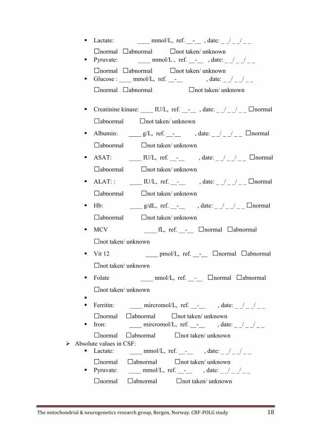
The mitochondrial & neurogenetics research group, Bergen, Norway. CRF-POLG study 18
Lactate: ____ mmol/L, ref. __-__ , date: _ _/ _ _/ _ _
□normal □abnormal □not taken/ unknown
Pyruvate: ____ mmol/L , ref. __-__ , date: _ _/ _ _/ _ _
□normal □abnormal □not taken/ unknown
Glucose : ____ mmol/L, ref. __-__ , date: _ _/ _ _/ _ _
□normal □abnormal □not taken/ unknown
Creatinine kinase: ____ IU/L, ref. __-__ , date: _ _/ _ _/ _ _ □normal
□abnormal □not taken/ unknown
Albumin: ____ g/L, ref. __-__ , date: _ _/ _ _/ _ _ □normal
□abnormal □not taken/ unknown
ASAT: ____ IU/L, ref. __-__ , date: _ _/ _ _/ _ _ □normal
□abnormal □not taken/ unknown
ALAT: : ____ IU/L, ref. __-__ , date: _ _/ _ _/ _ _ □normal
□abnormal □not taken/ unknown
Hb: ____ g/dL, ref. __-__ , date: _ _/ _ _/ _ _ □normal
□abnormal □not taken/ unknown
MCV ____ fL, ref. __-__ □normal □abnormal
□not taken/ unknown
Vit 12 ____ pmol/L, ref. __-__ □normal □abnormal
□not taken/ unknown
Folate ____ nmol/L, ref. __-__ □normal □abnormal
□not taken/ unknown
Ferritin: ____ mircromol/L, ref. __-__ , date: _ _/ _ _/ _ _
□normal □abnormal □not taken/ unknown
Iron: ____ mircromol/L, ref. __-__ , date: _ _/ _ _/ _ _
□normal □abnormal □not taken/ unknown
Absolute values in CSF:
Lactate: ____ mmol/L, ref. __-__ , date: _ _/ _ _/ _ _
□normal □abnormal □not taken/ unknown
Pyruvate: ____ mmol/L, ref. __-__ , date: _ _/ _ _/ _ _
□normal □abnormal □not taken/ unknown

The mitochondrial & neurogenetics research group, Bergen, Norway. CRF-POLG study 19
Albumin: ____ mg/L, ref. __-__ , date: _ _/ _ _/ _ _ □normal
□abnormal □not taken/ unknown
Protein: ____ g/L, ref. __-__ , date: _ _/ _ _/ _ _ □normal
□abnormal □not taken/ unknown
Pathological total or free carnitine: □ yes □ no □ unknown
If yes, date of the test : _ _/ _ _/_ _, absolute value:_____ ref: __-___
Pathological acyl carnitine: □ yes □ no □ unknown
If yes, date of the test : _ _/_ _ /_ _ (, absolute value:_____ ref: __-___
Muscle pathology abnormality: □ yes □ no □ unknown / not perform
If yes
Date: _ _/ _ _/ _ _ ( dd/ mm/yy)
Regged red
fibers
SDH positive
fibers
COX negative
fibers
Excess lipid
□ yes □ no
□ unknown
□ yes □ no
□ unknown
□ yes □ no
□ unknown
□ yes □ no
□ unknown
Other Specify:_______________________________________________
Electron microscopy performed : □ yes □ no □ unknown / not perform
If yes , specify __________________________________________
Liver pathology abnormality: □ yes □ no □ unknown / not perform
If yes
Date: _ _/ _ _/ _ _ ( dd/ mm/yy)
Specify:_______________________________________________
Pathological EEG: □ yes □ no □ unknown / not perform
If yes:
Date_ _/ _ _/_ _ ( dd/mm/yy)
Finding _____________________________________
Epi.activity: □ yes □ no □ unknown
If yes : □frontal□ parietal □temporal □occipital □ multifocal □unknown
Repeated examination: □ no □progression□ no clear change □unknown
Pathological EMG: □ yes □ no □ unknown / not perform
If yes:
Date _ _ /_ _/_ _ ( dd/mm/yy)

The mitochondrial & neurogenetics research group, Bergen, Norway. CRF-POLG study 20
Finding: □ Myopathic □ neuropathic □ other ______ □ unknown
Pathological nerve conduction : □ yes □ no □ unknown / not perform
If yes:
Date _ _/_ _/_ _ ( dd/ mm/yy)
Finding: □ axonal □demyelinating □combination □other____ □ unknown
• Pathological VER : □ yes □ no □ unknown / not perform
If yes: Date _ _/_ _/_ _ ( dd/ mm/yy)
• Pathological ERG : □ yes □ no □ unknown / not perform
If yes: Date _ _/_ _/_ _ ( dd/ mm/yy)
MtDNA Findings
Genetic analysis method :
□ Long PCR □ Real Time PCR □Other, specify _________
Tissue mtDNA deletion (YES /
NO / % if available)
mtDNA depletion (YES /
NO / % if available)
Skeletal muscle □yes□ no, %: □yes □no; %:
Liver □yes□no, %: □yes□no, %:
Brain □yes□no, %: □yes□no, %:
Other □yes□no, %: □yes□no, %:
Other □yes□no, %: □yes□no, %:
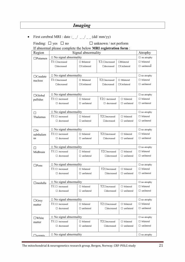
The mitochondrial & neurogenetics research group, Bergen, Norway. CRF-POLG study 21
Imaging
First cerebral MRI : date :_ _/ _ _/ _ _ (dd/ mm/yy)
Finding: □ yes □ no □ unknown / not perform
If abnormal please complete the below MRI registration form :
Region Signal abnormality Atrophy
□Putamen □ No signal abnormality □ no atrophy
□ bilateral
□ unilateral T1:□increased
□decreased
□ Bilateral
□Unilateral
T2:□increased
□decreased
□Bilateral
□Unilateral
□Caudate
nucleus □ No signal abnormality □ no atrophy
□ bilateral
□ unilateral T1:□increased
□decreased
□ Bilateral
□Unilateral
T2□increased
□decreased
□ Bilateral
□Unilateral
□Global
pallidus □ No signal abnormality □ no atrophy
□ bilateral
□ unilateral T1:□ increased
□ decreased
□ bilateral
□ unilateral
T2□ increased
□ decreased
□ bilateral
□ unilateral
□ Thalamus
□ No signal abnormality □ no atrophy
□ bilateral
□ unilateral T1:□ increased
□ decreased
□ bilateral
□ unilateral
T2□increased
□decreased
□ bilateral
□ unilateral
□N
subthalam
us
□ No signal abnormality □ no atrophy
□ bilateral
□ unilateral T1:□ increased
□ decreased
□ bilateral
□ unilateral
T2□increased
□decreased
□ bilateral
□ unilateral
□ Midbrain
□ No signal abnormality □ no atrophy
□ bilateral
□ unilateral T1:□ increased
□ decreased
□ bilateral
□ unilateral
T2□increased
□decreased
□ bilateral
□ unilateral
□Pons □ No signal abnormality □ no atrophy
□ bilateral
□ unilateral T1:□ increased
□ decreased
□ bilateral
□ unilateral
T2□increased
□decreased
□ bilateral
□ unilateral
□medulla □ No signal abnormality □ no atrophy
□ bilateral
□ unilateral T1:□ increased
□ decreased
□ bilateral
□ unilateral
T2□increased
□decreased
□ bilateral
□ unilateral
□Grey
matter □ No signal abnormality □ no atrophy
□ bilateral
□ unilateral T1:□ increased
□ decreased
□ bilateral
□ unilateral
T2:□increased
□decreased
□ bilateral
□ unilateral
□White
matter □ No signal abnormality □ no atrophy
□ bilateral
□ unilateral T1:□ increased
□ decreased
□ bilateral
□ unilateral
T2□increased
□decreased
□ bilateral
□ unilateral
□vermis □ No signal abnormality □ no atrophy

The mitochondrial & neurogenetics research group, Bergen, Norway. CRF-POLG study 22
T1:□ increased
□ decreased
□ bilateral
□ unilateral
T2□increased
□decreased
□ bilateral
□ unilateral
□ bilateral
□ unilateral
□ peduncles
□ No signal abnormality □ no atrophy
□ bilateral
□ unilateral T1:□ increased
□ decreased
□ bilateral
□ unilateral
T2□increased
□decreased
□ bilateral
□ unilateral
□Dentae
nucleus □ No signal abnormality □ no atrophy
□ bilateral
□ unilateral T1:□ increased
□ decreased
□ bilateral
□ unilateral
T2□increased
□decreased
□ bilateral
□ unilateral
□cerebral
cortex □ No signal abnormality □ no atrophy
□ bilateral
□ unilateral T1:□ increased
□ decreased
□ bilateral
□ unilateral
T2□increased
□decreased
□ bilateral
□ unilateral
□supraten
torial
white
matter
□ No signal abnormality □ no atrophy
□ bilateral
□ unilateral T1:□ increased
□ decreased
□ bilateral
□ unilateral
T2□increased
□decreased
□ bilateral
□ unilateral
□corpus
callosum □ No signal abnormality □ no atrophy
□ bilateral
□ unilateral T1:□ increased
□ decreased
□ bilateral
□ unilateral
T2□increased
□decreased
□ bilateral
□ unilateral
□ other
2nd cerebral MRI: □ performed □ unknown / not perform
If performed, date :_ _/ _ _/ _ _ (dd/ mm/yy)
□ progression,__________ □ regression,__________ □ no changed
Other notes :
If this is the first abnormal MRI please complete the MRI registration form
Last cerebral MR: □ performed □ unknown / not perform
If performed, date :_ _/ _ _/ _ _ ( dd/mm/yy)
□ progression,__________ □ regression,__________ □ no changed
If this is the first abnormal MRI please complete the MRI registration form
Other notes:
The mitochondrial & neurogenetics research group, Bergen, Norway. CRF-POLG study 23
MRI of the thigh: □ performed □ unknown / not perform
If performed, date : _ _/ _ _/ _ _ ( dd/mm/yy)
Specify:______________________
MRS: □ yes □ no □ unknown / not perform
If normal , date :_ _/ _ _/ _ _ ( dd/ mm/yy)
If abnormal , date :_ _/ _ _ / _ _ ( dd/ mm/yy)
□ elevated lactate region ____________
□ elevated choline region ____________
□ elevated succinate region _____________
□ decreased NAA region _____________
□ other, specify _______ region _____________
CT finding 1st CT : □ normal □ abnormal □ unknown / not perform
If normal, date : _ _/_ _/ _ _ ( dd/ mm/yy)
If abnormal, date : _ _/ _ _ / _ _ ( dd/ mm/yy)
□ calcifications region ____________
□ high attenuation region _____________
□ low attenuation region _____________
□ other, specify _______ region _____________

The mitochondrial & neurogenetics research group, Bergen, Norway. CRF-POLG study 24
Treatment
Antiepileptic treatment: □ yes □ no □ unknown
□ single antiepileptic drug, specify ___________ date started : _ _/ _ _/_ _
□ combination of antiepileptic drugs , specify ____________________________
□ antiepileptic drugs used with no effect : _____, _______,_______,______,_____
□ valproate , date started: _ _/ _ _/_ _.
□ on-going antiepileptic drugs: effect : □ no apparent □positive □ unknown
□ discontinued, reason(s):______________________________
Ketogenic diet : □ yes □ no □ unknown
If yes : date started : _ _/ _ _ / _ _ ( dd/ mm/yy)
□ on-going: effect : □ no apparent □positive □ unknown
□ discontinued, reason(s):______________________________
Coenzyme Q10: □ yes □ no □ unknown
If yes : date started : _ _/ _ _ / _ _ ( dd/mm/yy)
□ on-going: effect : □ no apparent □positive □ unknown
□ discontinued, reason(s):______________________________
Thiamine: □ yes □ no □ unknown
If yes : date started : _ _/ _ _ / _ _ ( dd/mm/yy)
□ on-going: effect : □ no apparent □positive □ unknown
□ discontinued, reason(s):______________________________
Nicotinamide: □ yes □ no □ unknown
If yes : date started : _ _ / _ _ / _ _ ( dd/ mm/yy)
□ on-going: effect : □ no apparent □positive □ unknown
□ discontinued, reason(s):______________________________
Riboflavin: □ yes □ no □ unknown
If yes: date started : _ _/ _ _ / _ _ ( dd/ mm/yy)
□ on-going: effect: □ no apparent □positive □ unknown
□ discontinued, reason(s):______________________________
Carnitine: □ yes □ no □ unknown
If yes: date started : _ _/ _ _ / _ _ ( dd/ mm/yy)
□ on-going: effect: □ no apparent □positive □ unknown
The mitochondrial & neurogenetics research group, Bergen, Norway. CRF-POLG study 25
□ discontinued, reason(s):______________________________
Folic acid: □ yes □ no □ unknown
If yes: date started : _ _/ _ _ / _ _ ( dd/ mm/yy)
□ on-going: effect: □ no apparent □positive □ unknown
□ discontinued, reason(s):______________________________
Antidiabetic treatment: □ yes □ no □ unknown
If yes: date started: _ _/ _ _ / _ _ ( dd/ mm/yy)
□on-going: effect: □ no apparent □positive □ unknown
□discontinued, reason(s):______________________________
Oral contraceptive: □ yes □ no □ unknown
If yes: date started: _ _/ _ _ / _ _ ( dd/ mm/yy)
Other: □ yes □ no □ unknown
If yes: date started: _ _ / _ _ ( mm/yy)
□on-going: effect: □ no apparent □positive □ unknown
□discontinued, reason(s):______________________________
Other: □ yes □ no □ unknown
If yes: date started: _ _/ _ _ / _ _ ( dd/ mm/yy)
□ on-going: effect: □ no apparent □ positive □ unknown
□ discontinued, reason(s):______________________________
Other: □ yes □ no □ unknown
If yes: date started: _ _/ _ _ / _ _ ( dd/ mm/yy)
□ on-going: effect: □ no apparent □ positive □ unknown
□ discontinued, reason(s):______________________________
Other details :

The mitochondrial & neurogenetics research group, Bergen, Norway. CRF-POLG study 26
Investigator signature
I have carefully checked all the data recorded in the present case report form and I confirm
that they are true, complete and accurate to the best of my knowledge.
Investigator name: ________________________________
Registration completed: yes □ no □
Signature date: ________________________________

81
11. Original publications (PAPERS I, II, III and IV)

PAPER II
Simplifying the clinical classification of polymerase gamma (POLG)
disease based on age of onset; studies using a cohort of 155 cases.
Hikmat O, Naess K, Engvall M, Klingenberg C, Rasmussen M, Tallaksen
CME, Brodtkorb E, Ostergaard E, de Coo I.F.M, Pias-Peleteiro L, Isohanni P,
Uusimaa J, Darin N, Rahman S, Bindoff LA.
J Inherit Metab Dis. DOI: 10.1002/jimd.12211.
Erratum: Page 7; 3.6 Genetic findings: the correct cDNA position is c.1399G>A (p.Ala467Thr).


OR I G I N A L AR T I C L E
Simplifying the clinical classification of polymerase gamma(POLG) disease based on age of onset; studies using a cohortof 155 cases
Omar Hikmat1,2 | Karin Naess3,4 | Martin Engvall3,5 | Claus Klingenberg6,7 |
Magnhild Rasmussen8,9 | Chantal ME Tallaksen10,11 | Eylert Brodtkorb12,13 |
Elsebet Ostergaard14 | I. F. M de Coo15,16 | Leticia Pias-Peleteiro17 |
Pirjo Isohanni18,19 | Johanna Uusimaa20,21,22 | Niklas Darin23 |
Shamima Rahman24,25 | Laurence A. Bindoff2,26
1Department of Pediatrics, Haukeland University Hospital, Bergen, Norway2Department of Clinical Medicine (K1), University of Bergen, Bergen, Norway3Centre for Inherited Metabolic Diseases, Karolinska University Hospital, Stockholm, Sweden4Department of Medical Biochemistry and Biophysics, Karolinska Institutet, Stockholm, Sweden5Department of Molecular Medicine and Surgery, Karolinska Institutet, Stockholm, Sweden6Department of Paediatric and Adolescent Medicine, University Hospital of North Norway, Tromso, Norway7Paediatric Research Group, Department of Clinical Medicine, UiT - The Arctic University of Norway, Tromso, Norway8Women and Children's Division, Department of Clinical Neurosciences for Children, Oslo University Hospital, Oslo, Norway9Unit for Congenital and Hereditary Neuromuscular Disorders, Department of Neurology, Oslo University Hospital, Oslo, Norway10Department of Neurology, Oslo University Hospital, Oslo, Norway11Faculty of Medicine, Institute of Clinical Medicine, University of Oslo, Oslo, Norway12Department of Neuroscience, Norwegian University of Science and Technology, Trondheim, Norway13Department of Neurology and Clinical Neurophysiology, St. Olav's University Hospital, Trondheim, Norway14Department of Clinical Genetics, Copenhagen University Hospital Rigshospitalet, Copenhagen, Denmark15Department of Neurology, Medical Spectrum Twente, Enschede, The Netherlands16Department of Genetics and Cell Biology, University of Maastricht, Maastricht, The Netherlands17Department of Neurology, Sant Joan de Déu Children´s Hospital, Barcelona, Spain18Department of Pediatric Neurology, Children's Hospital and Pediatric Research Center, University of Helsinki and Helsinki University Hospital,Helsinki, Finland19Stem Cells and Metabolism Research Program, Faculty of Medicine, University of Helsinki, Helsinki, Finland20PEDEGO Research Unit, University of Oulu, Oulu, Finland21Biocenter Oulu, University of Oulu, Oulu, Finland22Department of Pediatric Neurology, Clinic for Children and Adolescents, Medical Research Center, Oulu University Hospital, Oulu, Finland23Department of Pediatrics, The Queen Silvia Children's Hospital, University of Gothenburg, Gothenburg, Sweden24Mitochondrial Research Group, UCL Great Ormond Street Institute of Child Health, London, UK25Metabolic Unit, Great Ormond Street Hospital for Children NHS Foundation Trust, London, UK26Department of Neurology, Haukeland University Hospital, Bergen, Norway
Received: 14 August 2019 Revised: 2 January 2020 Accepted: 3 January 2020
DOI: 10.1002/jimd.12211
This is an open access article under the terms of the Creative Commons Attribution License, which permits use, distribution and reproduction in any medium, provided
the original work is properly cited.
© 2020 The Authors. Journal of Inherited Metabolic Disease published by John Wiley & Sons Ltd on behalf of SSIEM
J Inherit Metab Dis. 2020;1–11. wileyonlinelibrary.com/journal/jimd 1

CorrespondenceLaurence A. Bindoff, Department ofNeurology, University of Bergen,Haukeland University Hospital, 5021Bergen, Norway.Email: [email protected]
Funding informationHelse Vest Regionalt Helseføretak, Grant/Award Number: 91144; NeMOfoundation, Grant/Award Number:17_P19; Lily Foundation; NIHR GreatOrmond Street Hospital BiomedicalResearch Centre; Great Ormond StreetHospital Children's Charity
Communicating Editor: Saskia BrigitteWortmann
Summary
Background: Variants in POLG are one of the most common causes of
inherited mitochondrial disease. Phenotypic classification of POLG disease
has evolved haphazardly making it complicated and difficult to implement in
everyday clinical practise. The aim of our study was to simplify the classifica-
tion and facilitate better clinical recognition.
Methods: A multinational, retrospective study using data from 155 patients
with POLG variants recruited from seven European countries.
Results: We describe the spectrum of clinical features associated with POLG
variants in the largest known cohort of patients. While clinical features
clearly form a continuum, stratifying patients simply according to age of
onset—onset prior to age 12 years; onset between 12 and 40 years and onset
after the age of 40 years, permitted us to identify clear phenotypic and prog-
nostic differences. Prior to 12 years of age, liver involvement (87%), seizures
(84%), and feeding difficulties (84%) were the major features. For those with
onset between 12 and 40 years, ataxia (90%), peripheral neuropathy (84%),
and seizures (71%) predominated, while for those with onset over 40 years,
ptosis (95%), progressive external ophthalmoplegia (89%), and ataxia (58%)
were the major clinical features. The earlier the onset the worse the progno-
sis. Patients with epilepsy and those with compound heterozygous variants
carried significantly worse prognosis.
Conclusion: Based on our data, we propose a simplified POLG disease classi-
fication, which can be used to guide diagnostic investigations and predict dis-
ease course.
KEYWORD S
Alpers syndrome, epilepsy, mitochondrial disease, POLG, stroke-like episodes
1 | INTRODUCTION
Mitochondria are intracellular organelles found in almostall human cells. Their key function is the production ofadenosine triphosphate through the process of oxidativephosphorylation performed by the mitochondrial respira-tory chain (MRC). The MRC comprises more than 90 pro-teins organised into five complexes (I-V). MitochondrialDNA (mtDNA) codes for 13 proteins while the remainingproteins are encoded by nuclear DNA.1 The enzyme thatreplicates and repairs mtDNA, polymerase γ,2 is aheterotrimer comprising a catalytic subunit (POLG) andtwo accessory subunits (POLG2). Mutations in POLG(OMIM * 174763), the nuclear gene encoding the cata-lytic subunit, interfere with mtDNA maintenance.2,3
Variants in POLG are the single most common cause ofinherited mitochondrial disease.4 The first POLG variantassociated with disease was described in a family with auto-somal dominant progressive external ophthalmoplegia
(PEO5), but since then, more than 190 disease-causingvariants have been identified (http://tools.niehs.nih.giv/polg). POLG variants are associated with a wide spectrumof overlapping phenotypes ranging from devastating fatalneonatal disease to a mild late onset disease with myopa-thy and PEO. A summary of the major POLG-relatedphenotypes reported in the literature4,6-22 is provided inTable 1.
The clinical features of POLG disease are extremelyheterogeneous making early clinical recognition chal-lenging. The increasing numbers of terms that have beenused to describe the clinical phenotypes (Table 1) haveadded to this confusion.
The clinical reports published so far were basedon small numbers of patients and did not describethe clinical spectrum through the whole life span.Longitudinal studies describing the natural history ofthe disease in a large cohort of patients are stilllacking.
2 HIKMAT ET AL.

In this study, we aimed to describe the natural historyof POLG disease in the largest cohort of patients withconfirmed POLG variants, focusing on the clinical fea-tures and the biomarkers which may predict the long-term prognosis. We aimed to provide a simpler clinicalclassification to facilitate early clinical recognition ofpatients with POLG disease.
2 | METHODS
2.1 | Study design, population, anddata collection
We performed a multinational, retrospective study ofpatients from 12 centres in seven European countries:
TABLE 1 Summary of the major syndromes associated with POLG mutations reported in the literature
Phenotype nomenclatures(reference) Major clinical features Age of onset
1 Myocerebrohepatopathy (MCHS)9,18,22 Myopathy, hypotonia, developmental delay,encephalopathy, liver failure.
Neonate, early infancy
2 Alpers-Huttenlocher Syndrome(AHS)9,11,17
Encephalopathy, psychomotor regression, refractoryepilepsy liver dysfunction
Infancy, childhood,adolescence
3 Alpers syndrome4,13,14 Synonym of AHS As in AHS
4 Alpers-Huttenlocher like21 Synonym of AHS As in AHS
5 Infantile hepatocerebral syndrome9 Includes AHS and MCHS Neonate, infancy,childhood
6 Infantile mitochondrial DNA depletionsyndrome19
Includes both AHS and MCHS Infancy, childhood
7 Leigh like19 Psychomotor retardation, hypotonia, extrapyramidaldysfunction, symmetrical hyperintensities on T2weighted images in basal ganglia, brain stem,thalamus
Infancy
8 Mitochondrial Neuro-Gastro-IntestinalEncephalopathy (MNGIE) like13,14,20
Severe gastrointestinal dysmotility, encephalopathy,ptosis ophthalmoplegia, peripheral neuropathy
Childhood, adolescenceadulthood
9 Myoclonus, epilepsy, myopathy, andsensory ataxia (MEMSA)4
Epilepsy, myopathy, ataxia, liver dysfunction,headache and stroke-like episodes
Adolescence, adulthood
10 Spinocerebellar ataxia with epilepsy(SCAE)4,7
Now incorporated under MEMSA umbrella As in MEMSA
11 Mitochondrial spinocerebellar ataxiawith epilepsy (MSCAE)6
Now incorporated under MEMSA umbrella As in MEMSA
12 Ataxia neuropathy spectrum (ANS)4,7 Ataxia, neuropathy, psychiatric symptoms, cognitiveimpairment, epilepsy, and ophthalmoplegia
Adolescent and adult
13 Mitochondrial recessive ataxiasyndrome (MIRAS)10
Now incorporated under ANS umbrella As in ANS
14 Sensory ataxia neuropathy dysarthriaand ophthalmoplegia (SANDO)4
Now incorporated under ANS umbrella As in ANS
15 Mitochondrial encephalomyopathy,lactic acidosis, and stroke-likeepisodes (MELAS) like phenotype23
Headache, seizures, stroke-like episodes as inMEMSA
Adult
16Recessive Charcot-Marie tooth like12 Axonal polyneuropathy, muscle weakness, wasting,
tremor nystagmus, dysarthria, dysmetria, anddysdiadochokinesis.
Adult
17 Parkinsonism8,16 Tremor, rigidity, hypo/bradykinesia, balancedisturbance
Adult
18 Autosomal recessive progressiveexternal ophthalmoplegia (arPEO)5,7
Ptosis, ophthalmoparesis, may be associated withataxia and myopathy
Adult, elderly
19 Autosomal dominant progressiveexternal ophthalmoplegia (adPEO)5,7
Ptosis, ophthalmoparesis, myopathy, neuropathy,ataxia
Adult-elderly
20 Chronic progressive externalophthalmoplegia plus(CPEO+)7
Synonym of adPEO Adult-elderly
HIKMAT ET AL. 3

Norway (Haukeland University Hospital, Oslo UniversityHospital, St. Olav's Hospital and University Hospital ofNorthern Norway); United Kingdom (Great OrmondStreet Hospital, London); Sweden (Centre for InheritedMetabolic Diseases, Karolinska University Hospital,Stockholm and The Queen Silvia Children's Hospital,University of Gothenburg); Denmark (Department ofClinical Genetics, Copenhagen University Hospital); Fin-land (Children's Hospital, Helsinki University Hospitaland Clinic for Children and Adolescents, Oulu UniversityHospital); Netherlands (Department of Genetics and CellBiology, Maastricht University, Maastricht); and Spain(Sant Joan de Déu Children's Hospital, Barcelona).Patients diagnosed and followed at the participating cen-tres were considered eligible if they had recessive diseaseand confirmed biallelic pathogenic/likely pathogenicPOLG variants or dominant disease and heterozygousconfirmed pathogenic variants. Data entry was completedin December 2017.
Detailed clinical, biochemical, muscle biopsy, neuro-physiological, neuroimaging, and genetic data wereobtained by using an electronic-case report form com-pleted by the responsible investigator(s) at each centreand reviewed by the study-principal investigator (O.H.).
The date of disease onset was defined by the date ofthe first symptom(s) requiring medical evaluation. End offollow up was defined as the date of the patient's last visitto the follow-up centre or death. Available longitudinaldata, both at disease onset and later during the diseasecourse, were collected. Liver involvement was defined bythe presence of two or more of the following parametersin at least two different time points; elevated aspartateaminotransferase, gamma-glutamyltransferase, bilirubinor ammonia, low serum albumin, or pathological histo-logical findings of liver biopsy. The presence of anaemiaand abnormal cerebrospinal fluid (CSF) protein and/oralbumin was identified as described in previous publica-tions.13,14,24 We use the recent International LeagueAgainst Epilepsy (ILAE) classification25 for seizureclassification.
2.2 | Data and statistical analysis
Detailed descriptive data analysis was performed on theentire study cohort using SPSS (Statistical Package ofSocial Sciences), Version 23.0. A two sided P value lessthan .05 was considered to be statistically significant.Mosaic plots was performed by using R (The R founda-tion for statistical computing), version 3.6.1.
In order to simplify the clinical classification patientswere grouped according to the age of disease onset intothree groups: (a) those with disease onset prior to the age
of 12 years (before puberty), (b) those with disease onsetbetween 12 and 40 years, and (c) those with disease onsetafter the age of 40 years. The age of onset of each individ-ual symptom was recorded and classified according tothese three defined age-groups. Correspondence analysiswas performed to examine the relationship between twovariables (groups of patients who were classifiedaccording to the age of onset as described and the age ofonset of each individual symptom) graphically in a multi-dimensional space; this allowed examination of the clus-tering of symptoms around each age group. Further,mosaic plots was performed to study the differencesbetween the above mentioned groups. The study cohortwas also classified according to the presence or absenceof epilepsy, regardless the age of onset.
For survival analysis, the end-point was time to deathwhich was defined as the time in months from the dateof disease onset to the date of death. Univariate survivalanalysis was performed using log-rank test (Kaplan-Meier) to compare differences in survival time betweencategories.
3 | RESULTS
3.1 | Demography
One hundred and fifty-five patients, (males n = 76 [49%],females n = 79 [51%]) with confirmed pathogenic POLGvariants were identified. Seventy-six were diagnosed inNorway, 44 in Sweden, 19 in the United Kingdom,8 in Finland, 5 in Denmark, 2 in The Netherlands, and1 in Spain. The majority of patients were NorthernEuropean (n = 146), while three patients were from Iraq,two from Cyprus and one from Croatia, Pakistan, Spain,and the United Arab Emirates.
3.2 | Major clinical features
Median age at disease onset for the whole study cohortwas 10 years (range: birth—71 years). Disease onset priorto the age of 12 years occurred in 54% (n = 83/155),between 12 and 40 years of age in 34% (n = 53/155), andafter the age of 40 years in 12% (n = 19/155) had. Diseasedebut was apparently spontaneous in 113/155 (73%),followed an infectious illness in 32/155 (21%) and notclearly reported in 10/155 (6%) of the patients.
Neurological (90%, n = 139/155), ophthalmological(74%, n = 112/151), and gastrointestinal symptoms (63%,n = 92/146) were the most predominant clinical features.Epilepsy was reported in 69% (n = 107/155), with focaland focal evolving to bilateral tonic-clonic seizures being
4 HIKMAT ET AL.

TABLE 2 Major clinical features of patients reported in this study
Major clinical featuresNumber ofpatients
Number ofpatients at onset
Number ofpatients later
1. Neurological
Seizure 107/154 (69%) 73/106 (69%) 33/106 (31%)
Focal 94/102 (92%) 60/91 (66%) 31/91 (34%)
Focal evolving to bilateral tonic-clonic 85/100 (85%) 43/85 (51%) 42/85 (49%)
Myoclonic 73/98 (74%) 26/71 (37%) 45/71 (63%)
Epilepsia partialis continua 52/91 (57%) 15/52 (29%) 37/52 (71%)
Convulsive status epilepticus 79/101 (78%) 30/78 (38%) 48/78 (62%)
Othersa 11/80 (14%) 5/10 (50%) 5/10 (50%)
Ataxia 87/138 (63%) 53/85 (62%) 32/85 (38%)
Hypotonia 68/135 (50%) 50/66 (76%) 16/66 (24%)
Limb weakness 89/125 (71%) 33/83 (40%) 50/83 (60%)
Migraine-like headache 52/143 (36%) 38/52 (73%) 14/52 (27%)
Peripheral neuropathy 65/123 (53%) 23/63 (36%) 40/63 (64%)
Sensorineural hearing loss 16/146 (11%) 9/16 (56%) 7/16 (44%)
2. Ophthalmological
Ptosis 51/149 (34%) 28/49 (57%) 21/49 (43%)
Progressive external ophthalmoplegia 56/146 (38%) 24/56 (43%) 32/56 (57%)
Nystagmus 55/146 (38%) 29/53 (55%) 24/53 (45%)
Cataract 11/148 (7%) 2/9 (22%) 7/9 (78%)
Cortical blindness 32/111 (29%) 17/29 (59%) 12/29 (41%)
Pigmentary retinopathy 3/140 (2%) 1/2 (50%) 1/2 (50%)
3. Gastrointestinal
Feeding difficulties 75/145 (52%) 36/73 (49%) 37/73 (51%)
Vomiting 52/137 (38%) 31/49 (63%) 18/49 (37%)
Chronic diarrhoea 8/136 (6%) 2/8 (25%) 6/8 (75%)
Liver involvement 95/151 (64%) 35/93 (38%) 58/93 (62%)
Othersb 7/128 (5%) 2/7 (29%) 5/7 (71%)
4. Endocrinological
Diabetes mellitus type 1 1/151 (1%) 0/1 (0%) 1/1 (100%)
Diabetes mellitus type 2 2/151 (1%) 2/2 (100%) 0/2 (0%)
Adrenal insufficiency 2/151 (1%) 1/2 (50%) 1/2 (50%)
Growth hormone deficiency 2/147 (1%) 1/2 (50%) 1/2 (50%)
Othersc 3/140 (2%) 0/3 (0%) 3/3 (100%)
5. Others
Anaemia 77/136 (57%) 20/77 (26%) 57/77 (74%)
Renal disordersd 13/149 (8%) 3/12 (25%) 9/12 (75%)
Respiratory disorderse 18/149 (12%) 5/18 (28%) 13/18 (72%)
aOne infantile spasms, eight absence, one atonic seizure, and one non convulsive status epilepticus.bOne Coeliac disease, one constipation, one paralytic ileus, one acute colon necrosis, one gastrointestinal bleeding, one colitis, and one milk proteinintolerance.cOne hypothyroidism, one hypoparathyroidism, and one pseudo hypoparathyroidism.dSeven renal tubular acidosis, five renal failure, and one renal stone.eTwo asthma, one chest deformity, two recurrent chest infections, two sleep apnoea, elven hypoventilation/respiratory insufficiency.
HIKMAT ET AL. 5

the most common seizure types (92%, n = 94/102). Ataxia(63%, n = 87/138), peripheral neuropathy (53%,n = 65/123), and hypotonia (50%, n = 68/135) were fre-quently reported. Nystagmus (38%, n = 55/146), PEO(38%, n = 56/146), and ptosis (34%, n = 51/149) were themost commonly reported ophthalmological features.Liver involvement was identified in 64% (n = 96/151) ofthe patients. More than half of the study cohort had feed-ing difficulties (52%, n = 75/145), regardless of the age of
onset. A detailed description of the clinical features isprovided in Table 2.
3.3 | Age-related clinical features
We found clear evidence that the clinical features ofPOLG disease form a continuum (Figure 1) rather thandistinct phenotypes (Table 1). Nevertheless, by groupingthe patients into three groups according to the age ofonset (early, juvenile/adult, and late onset groups), wecould identify clear phenotypic and prognostic differ-ences (Table 3). To confirm this finding, correspondenceanalysis was performed and demonstrated clear cluster-ing of the symptoms around the three different agegroups as illustrated in Figure S1. Further, mosaic plotsshowed there was a statistically significance differencesin the phenotypes between the above mentioned groups(Figure S2).
3.4 | Laboratory, muscle biopsy, andneurophysiological findings
The percentage of those with raised lactate in serum andCSF at disease onset was 35% (n = 29/84) and 40%(n = 19/47), respectively. Abnormal elevated CSF proteinat disease onset was reported in 68% (n = 44/68) ofpatients. In muscle biopsies, the presence of ragged-redfibres, COX-negative fibres, and abnormal respiratorychain activities was reported in fewer than the half ofthose who had been investigated (Table S1).
Electroencephalogram recordings showed thatapproximately half (54%, n = 58/107) of patients withepilepsy had epileptiform activities over the occipitallobes. Abnormal nerve conduction was observed in 70%
0 24 48 72
Anaemia
PEO
Ptosis
Muscle weakness
Peripheral neuropathy
Ataxia
Liver dysfunction
Stroke like episodes
Seizure
Age at onset in years
FIGURE 1 Age of onset of each
individual symptom in patients with
POLG disease. Range I, Median. PEO,
progressive external ophthalmoplegia
TABLE 3 The onset of symptoms according to three age
groups
Symptoms<12 years 12–40 years >40 years83/155 (54%) 53/155 (34%) 19/155 (12%)
Seizures 69/82 (84%) 37/52 (71%) 1/19 (5%)
Ataxia 30/67 (45%) 46/51 (90%) 11/19 (58%)
Hypotonia 57/72 (79%) 9/44 (20%) 2/18 (11%)
Stroke-likeepisodes
26/73 (36%) 26/48 (54%) None (0%)
Peripheralneuropathy
17/57 (30%) 38/45 (84%) 10/18 (65%)
Migraine-likeheadache
14/52 (27%) 36/52 (69%) 2/52 (4%)
Feedingdifficulties
58/69 (84%) 13/47 (28%) 4/18 (22%)
Liverinvolvement
71/82 (87%) 23/49 (47%) 2/19 (11%)
Anaemia 49/71 (69%) 25/45 (56%) 3/16 (19%)
Ptosis 12/78 (15%) 21/50 (42%) 18/19 (95%)
PEO 7/83 (8%) 32/48 (67%) 17/19 (89%)
Survival timein months
19 (0.5–600) 151 (4–487) 191 (17–336)
Median (Range)
Abbreviation: PEO, progressive external ophthalmoplegia.
6 HIKMAT ET AL.
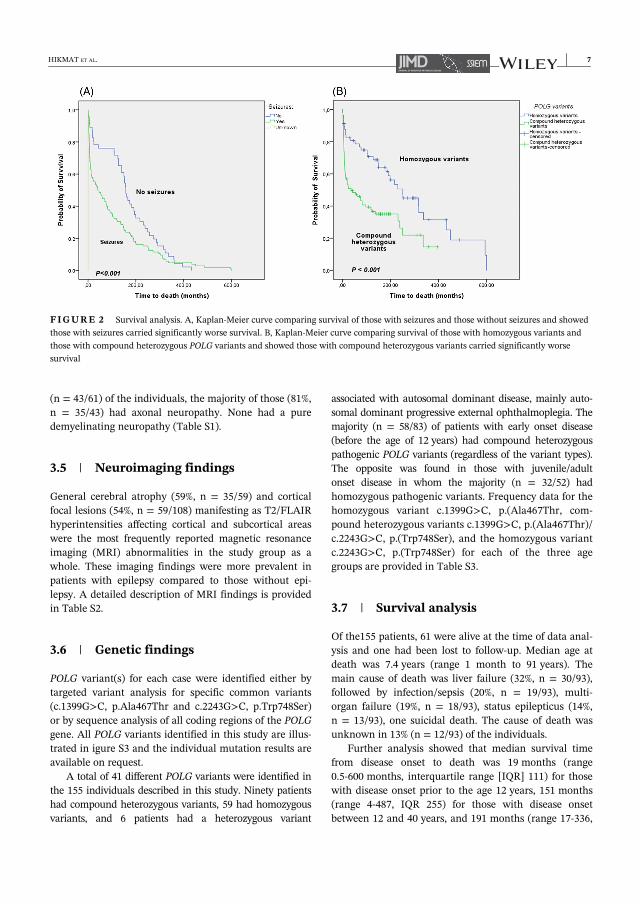
(n = 43/61) of the individuals, the majority of those (81%,n = 35/43) had axonal neuropathy. None had a puredemyelinating neuropathy (Table S1).
3.5 | Neuroimaging findings
General cerebral atrophy (59%, n = 35/59) and corticalfocal lesions (54%, n = 59/108) manifesting as T2/FLAIRhyperintensities affecting cortical and subcortical areaswere the most frequently reported magnetic resonanceimaging (MRI) abnormalities in the study group as awhole. These imaging findings were more prevalent inpatients with epilepsy compared to those without epi-lepsy. A detailed description of MRI findings is providedin Table S2.
3.6 | Genetic findings
POLG variant(s) for each case were identified either bytargeted variant analysis for specific common variants(c.1399G>C, p.Ala467Thr and c.2243G>C, p.Trp748Ser)or by sequence analysis of all coding regions of the POLGgene. All POLG variants identified in this study are illus-trated in igure S3 and the individual mutation results areavailable on request.
A total of 41 different POLG variants were identified inthe 155 individuals described in this study. Ninety patientshad compound heterozygous variants, 59 had homozygousvariants, and 6 patients had a heterozygous variant
associated with autosomal dominant disease, mainly auto-somal dominant progressive external ophthalmoplegia. Themajority (n = 58/83) of patients with early onset disease(before the age of 12 years) had compound heterozygouspathogenic POLG variants (regardless of the variant types).The opposite was found in those with juvenile/adultonset disease in whom the majority (n = 32/52) hadhomozygous pathogenic variants. Frequency data for thehomozygous variant c.1399G>C, p.(Ala467Thr, com-pound heterozygous variants c.1399G>C, p.(Ala467Thr)/c.2243G>C, p.(Trp748Ser), and the homozygous variantc.2243G>C, p.(Trp748Ser) for each of the three agegroups are provided in Table S3.
3.7 | Survival analysis
Of the155 patients, 61 were alive at the time of data anal-ysis and one had been lost to follow-up. Median age atdeath was 7.4 years (range 1 month to 91 years). Themain cause of death was liver failure (32%, n = 30/93),followed by infection/sepsis (20%, n = 19/93), multi-organ failure (19%, n = 18/93), status epilepticus (14%,n = 13/93), one suicidal death. The cause of death wasunknown in 13% (n = 12/93) of the individuals.
Further analysis showed that median survival timefrom disease onset to death was 19 months (range0.5-600 months, interquartile range [IQR] 111) for thosewith disease onset prior to the age 12 years, 151 months(range 4-487, IQR 255) for those with disease onsetbetween 12 and 40 years, and 191 months (range 17-336,
FIGURE 2 Survival analysis. A, Kaplan-Meier curve comparing survival of those with seizures and those without seizures and showed
those with seizures carried significantly worse survival. B, Kaplan-Meier curve comparing survival of those with homozygous variants and
those with compound heterozygous POLG variants and showed those with compound heterozygous variants carried significantly worse
survival
HIKMAT ET AL. 7
IQR 101) for those with disease onset after the age of40 years.
The presence of epilepsy was associated with signifi-cantly worse survival (P < .001), and the median survivaltime from seizure onset to death was 37 months (range<1-487). Survival analysis also showed that patients withpathogenic compound heterozygous POLG variants hadsignificantly (P < .001) worse survival compared to thosewith pathogenic homozygous variants, regardless of spe-cific variant types (Figure 2). Further analysis showedthat survival after the onset of seizures in those with earlyonset disease was significantly worse than those whodeveloped seizures as part of juvenile/adult onset disease.Further, patients who developed liver involvementshowed a significantly worse survival than those withoutliver impairment (Figure S4).
4 | DISCUSSION
We present the detailed description of 155 patients withconfirmed pathogenic POLG variants focusing on theclinical features, but including laboratory, genetic, andneuroimaging findings. As far as we can ascertain, this isthe largest cohort of patients with POLG disease so fardescribed. In addition to the descriptive element, we havealso analysed factors, which may predict the prognosis.
We defined the age of onset of each individual symp-tom and our data confirms that POLG disease comprisesa continuum of clinical features rather than a set of sepa-rate clinical identities (Figure 1). Apart from PEO/ptosis,all other symptoms could start from infancy to adult-hood. While hypotonia and feeding difficulties in infantsare likely due to different pathological processes thanthese features appearing in adults, seizures, peripheralneuropathy, ataxia, muscle weakness, and hepatic distur-bance have a similar basis and all could present at anyage. PEO/ptosis starts later and appears mainly inpatients with dominantly inherited disease or in thosewith juvenile/early adult onset disease who do notdevelop epilepsy or, less often, survive despite it. Stroke-like episodes appear to start slightly later than most otherfeatures. This may reflect the nature of the process26,27
namely that these represent prolonged seizure activity orstatus epilepticus.
If we look at the median ages of onset, instead oflooking at the age range, we do see a tendency for thefeatures to cluster according to age. We, therefore,reanalysed the data using different age groups. Based onthese findings, we found that the clinical spectrum ofPOLG disease was best described by grouping patientsinto three categories of early, juvenile/adult, and lateonset. Early onset disease was classified as beginning
prior to the age of 12 years. In these patients, liverinvolvement, feeding difficulties, seizures, hypotonia, andmuscle weakness were the most dominant/importantclinical features and this group had the worst prognosis.The juvenile/adult onset form (12-40 years of age) wascharacterised by peripheral neuropathy, ataxia, seizures,stroke-like episodes and, in patients with longer survival,PEO. This group carried a better prognosis than the earlyonset group. Late onset disease (after the age of 40 years)was characterised by ptosis and PEO, with additional fea-tures such as peripheral neuropathy ataxia and muscleweakness occurring frequently. This group had the bestprognosis. Thus, while the clinical features associatedwith POLG variants can present at any age, age of diseaseonset provides both clues to the diagnosis and informa-tion about the outcome (Table 3).
The most frequently reported neurological features,included seizures, ataxia, and peripheral neuropathy.Focal evolving to bilateral tonic-clonic seizures were themost common seizure types, with epileptiform activitiespredominantly seen in occipital regions. These findingsare consistent with previous reports (8,10,13-15,28,29), how-ever, our results also showed that seizures were the mostpredominant clinical feature in patients with early onsetdisease (<12 years), common in those with juvenile/adultonset (12-40 years), but infrequent in those with lateonset disease (>40 years). Ataxia, peripheral neuropathy,and migraine-like headache were most predominant inindividuals with juvenile/adult onset disease, althoughreported in both early and late onset disease. Ptosis andPEO were common in late onset disease as reportedpreviously,22 however our data showed that the onset ofptosis and PEO occurred in all age groups. The onset ofgastrointestinal features such as feeding difficulties andliver involvement occurred at any age, but was predomi-nantly seen in patients with early onset disease.
Demographic data showed that more than half of theindividuals included in this study had onset during child-hood (prior to the age of 12), and the incidence of the dis-ease decreased with age. Contrary to previouspublications,9,15 which demonstrate some male predomi-nance, we observed no gender difference.
Survival analysis demonstrated a clear correlationbetween the age of disease onset and the survival time;earlier onset was associated with worse prognosis(Table 3). Further analysis showed that the presence ofepilepsy was significantly associated with worse progno-sis regardless of the age of disease onset, and individualsharbouring compound heterozygous POLG variants hadworse prognosis compared to those with homozygousvariants.
Our study showed that laboratory investigations whichare commonly used in the initial diagnostic work-up of
8 HIKMAT ET AL.

mitochondrial disorders, for example, raised blood andCSF lactate, the presence of ragged-red/COX-negativefibres and abnormal respiratory chain activities in the skel-etal muscle have low diagnostic sensitivity, being presentin fewer than 50% of the individuals. As we showed in aprevious publication,24 elevated CSF protein was the mostsensitive (68%, n = 44/65) laboratory diagnostic biomarkerat disease onset.
The majority of the patients included in this study wereof Northern European descent; thus, a possible limitationof this study is that it might not be possible to extrapolateour findings to other ethnic groups. However, we providedetailed description of all the known phenotypes associ-ated with POLG disease related to more than 40 differentPOLG variants. Moreover, there is little evidence in the lit-erature of marked ethnic differences in phenotypic
expression of POLG disease. Based on the diverse geno-typic background of our population, we consider that thefindings of our study are relevant to patients with POLGdisease, regardless of the ethnic background.
A simple and robust clinical classification is the cor-nerstone of early diagnosis. Such a classification, togetherwith diagnostic investigations, should facilitate easy rec-ognition of the disease and be useful for both experts andphysicians with limited experience of the field. Currentnomenclature describing the phenotypic spectrum ofPOLG disease (Table 1) is complicated and includes over-lapping clinical syndromes. This makes implementationin everyday clinical practice difficult. A clear and accu-rate classification that describes the full spectrum of dis-ease taking account of age-related features is essentialnot only for optimal management, but also for research
FIGURE 3 Diagnosing POLG disease; clinical suspicion and relevant investigations according to the age of onset. While we have
shown that POLG clinical features form a continuum, but it is also clear that age plays a role in which features predominate. Based on our
age groups, we can see clear clinical patterns and these will dictate which investigations are appropriate and useful. For example, in the
older age category, PEO and ataxia dominate the clinical spectrum and in these cases one can choose either to screen the known genes or to
take a muscle biopsy which give both structural clues (COX negative fibres) and the possibility to examine mtDNA (for multiple deletions).
We also see that the typical occipital epilepsy occurs in the younger two categories and it is in these that MRI imaging also provides
important clues. Peripheral neuropathy occurs in all age groups. In earlier studies, we showed that elevated CSF protein can be helpful, for
example in a child with epilepsy and focal MRI changes it can be an important indicator of poor prognosis. a: Direct POLG gene sequence
analysis is recommended to confirm the diagnosis in a case of strong clinical suspicion. b: Absence of these findings does not exclude the
diagnosis of POLG disease. c: targeted variant analysis for the most common variants (p. Ala467Thr and p.Trp748Ser) can be performed first
in juvenile and late onset disease, whole POLG gene sequence analysis is recommended for all early onset disease and those with strong
clinical suspicion of POLG disease regardless of the age of onset. CSF, cerebrospinal fluid; RRF, ragged-red fibres; PEO, progressive external
ophthalmoplegia; P. neuropathy, peripheral neuropathy; SLE, stroke- like episodes
HIKMAT ET AL. 9

and, when treatments become available, for use in clini-cal trials.
We provide a robust and simplified clinical classifica-tion based on data from the largest cohort of patientswith POLG disease published to date. This classificationhighlights three distinct age groups and within thesegroups the major clinical features. Earlier classificationsof POLG disease have focused primarily on phenotypicelements; for example, the presence of ataxia with orwithout myoclonus or epilepsy has variously beenreferred to as SANDO, ANS, or MIRAS/MSCAE. Earlyonset diseases have been separated into Alpers or MCHSor Leigh-like syndromes. The presence of mtDNA deple-tion has also been used to define POLG related diseasealthough the presence of this is known to be tissuedependent and depletion in brain and liver is found inboth young and older patients. We feel that these pheno-typic labels create an unnecessarily complicated classifi-cation. Age alone appears robust enough to delineate theimportant features of POLG disease such that we wouldrecommend simplifying classification to early onset, juve-nile onset and late onset POLG disease. The algorithm(Figure 3) shows how recognition of these key clinicalfeatures could be used to direct clinical investigation inthe different age groups.
ACKNOWLEDGMENTSThis work was supported by grants from the Western Nor-way Regional Health Authority (Helse-Vest, grantno. 911944). P.I. is supported by grant from the special gov-ernmental subsidy for health sciences research of the Hel-sinki University Hospital. S.R. is supported by researchgrant funding from Great Ormond Street Hospital Chil-dren's Charity, the NIHR Great Ormond Street HospitalBiomedical Research Centre, and the Lily Foundation.I.d.C. was supported by the NeMO foundation (no.17_P19).We would also thank professor Geir Egil Eide, Centre forClinical Research, Haukeland University Hospital, Bergen,Norway for his help with some of the statistical analysis.
CONFLICT OF INTERESTAll declare that they have no conflict of interest.
AUTHOR CONTRIBUTIONSO.H. and L.B. designed the study, were responsible fordata collection, analysed the data, and drafted the initialmanuscript, and approved the final manuscript as sub-mitted. K.N., M. E., C. K., M.R., C.M.E.T., E.B., T.F.,E.O., I.F.M.D., L.P., P.I., J.U., N.D., and S.R., wereresponsible for data acquisition and analysis, revising themanuscript critically, and approving the final manuscriptas submitted. All authors are responsible for accuracyand integrity of the work.
COMPLIANCE WITH ETHICALSTANDARDSAll procedures followed were in accordance with the eth-ical standards of the responsible committee on humanexperimentation (institutional and national) and with theHelsinki Declaration of 1975, as revised in 2000. Ethicalapproval for the study was obtained from the RegionalCommittee for Medical and Health Research Ethics,Western Norway (REK 2014/1783-4). Each participatingcountry has obtained approval from their local ethicalcommittee. The study was registered as an audit at GreatOrmond Street Hospital, London, UK (Registration Num-ber 1675). This article does not contain any studies withanimal subjects performed by any of the authors.
ORCIDLaurence A. Bindoff https://orcid.org/0000-0003-0988-276X
REFERENCES1. Smeitink J, van den Heuvel L, DiMauro S. The genetics and pathol-
ogy of oxidative phosphorylation. Nat Rev Genet. 2001;2:342-352.2. Longley MJ, Graziewicz MA, Bienstock RJ, Copeland WC.
Consequences of mutations in human DNA polymerasegamma. Gene. 2005;354:125-131.
3. Rahman S, Copeland WC. POLG-related disorders and theirneurological manifestations. Nat Rev Neurol. 2019;15:40-52.
4. Saneto RP, Naviaux RK. Polymerase gamma disease throughthe ages. Dev Disabil Res Rev. 2010;16:163-174.
5. Van Goethem G, Dermaut B, Lofgren A, Martin JJ, VanBroeckhoven C. Mutation of POLG is associated with progres-sive external ophthalmoplegia characterized by mtDNA dele-tions. Nat Genet. 2001;28:211-212.
6. Bindoff LA, Engelsen BA. Mitochondrial diseases and epilepsy.Epilepsia. 2012;53(suppl 4):92-97.
7. Cohen BH, Chinnery PF, Copeland WC. POLG-related disor-ders. In: Pagon RA, Adam MP, Ardinger HH, et al., eds.GeneReviews(R). Seattle, WA: University of Washington. 1993;1993-2020.
8. Davidzon G, Greene P, Mancuso M, et al. Early-onset familial par-kinsonism due to POLG mutations. Ann Neurol. 2006;59:859-862.
9. Ferrari G, Lamantea E, Donati A, et al. Infantile hepa-tocerebral syndromes associated with mutations in the mito-chondrial DNA polymerase-gammaA. Brain. 2005;128:723-731.
10. Hakonen AH, Heiskanen S, Juvonen V, et al. MitochondrialDNA polymerase W748S mutation: a common cause of autoso-mal recessive ataxia with ancient European origin. Am J HumGenet. 2005;77:430-441.
11. Harding BN. Progressive neuronal degeneration of childhoodwith liver disease (Alpers-Huttenlocher syndrome): a personalreview. J Child Neurol. 1990;5:273-287.
12. Harrower T, Stewart JD, Hudson G, et al. POLG1 mutationsmanifesting as autosomal recessive axonal Charcot-Marie-toothdisease. Arch Neurol. 2008;65:133-136.
13. Hikmat O, Tzoulis C, Klingenberg C, et al. The presence ofanaemia negatively influences survival in patients with POLGdisease. J Inherit Metab Dis. 2017;40:861-866.
10 HIKMAT ET AL.

14. Hikmat O, Tzoulis C, Chong WK, et al. The clinical spectrumand natural history of early-onset diseases due to DNA poly-merase gamma mutations. Genet Med. 2017;19:1217-1225.
15. Horvath R, Hudson G, Ferrari G, et al. Phenotypic spectrumassociated with mutations of the mitochondrial polymerasegamma gene. Brain. 2006;129:1674-1684.
16. Luoma P, Melberg A, Rinne JO, et al. Parkinsonism, prematuremenopause, and mitochondrial DNA polymerase gammamutations: clinical and molecular genetic study. Lancet(London, England). 2004;364:875-882.
17. Naviaux RK, Nguyen KV. POLG mutations associated withAlpers' syndrome and mitochondrial DNA depletion. Ann Neu-rol. 2004;55:706-712.
18. Nguyen KV, Sharief FS, Chan SS, Copeland WC, Naviaux RK.Molecular diagnosis of Alpers syndrome. J Hepatol. 2006;45:108-116.
19. Taanman JW, Rahman S, Pagnamenta AT, et al. Analysis ofmutant DNA polymerase gamma in patients with mitochon-drial DNA depletion. Hum Mutat. 2009;30:248-254.
20. Tang S, Dimberg EL, Milone M, Wong LJ. Mitochondrial neu-rogastrointestinal encephalomyopathy (MNGIE)-like pheno-type: an expanded clinical spectrum of POLG1 mutations.J Neurol. 2012;259:862-868.
21. Uusimaa J, Finnila S, Vainionpaa L, et al. A mutation in mito-chondrial DNA-encoded cytochrome c oxidase II gene in achild with Alpers-Huttenlocher-like disease. Pediatrics. 2003;111:e262-e268.
22. Wong LJ, Naviaux RK, Brunetti-Pierri N, et al. Molecular andclinical genetics of mitochondrial diseases due to POLG muta-tions. Hum Mutat. 2008;29:E150-E172.
23. Deschauer M, Tennant S, Rokicka A, et al. MELAS associatedwith mutations in the POLG1 gene. Neurology. 2007;68:1741-1742.
24. Hikmat O, Naess K, Engvall M, et al. Elevated cerebrospinalfluid protein in POLG-related epilepsy: diagnostic and prognos-tic implications. Epilepsia. 2018;59:1595-1602.
25. Scheffer IE, Berkovic S, Capovilla G, et al. ILAE classificationof the epilepsies: position paper of the ILAE Commission forClassification and Terminology. Epilepsia. 2017;58:512-521.
26. Tzoulis C, Bindoff LA. Melas associated with mutations in thepolg1 gene. Neurology. 2008;70:1054-1055.
27. Tzoulis C, Tran GT, Coxhead J, et al. Molecular pathogenesisof polymerase gamma-related neurodegeneration. Ann Neurol.2014;76:66-81.
28. Tzoulis C, Engelsen BA, Telstad W, et al. The spectrum of clini-cal disease caused by the A467T and W748S POLG mutations:a study of 26 cases. Brain. 2006;129:1685-1692.
29. Winterthun S, Ferrari G, He L, et al. Autosomal recessive mito-chondrial ataxic syndrome due to mitochondrial polymerasegamma mutations. Neurology. 2005;64:1204-1208.
SUPPORTING INFORMATIONAdditional supporting information may be found onlinein the Supporting Information section at the end of thisarticle.
How to cite this article: Hikmat O, Naess K,Engvall M, et al. Simplifying the clinicalclassification of polymerase gamma (POLG)disease based on age of onset; studies using acohort of 155 cases. J Inherit Metab Dis. 2020;1–11.https://doi.org/10.1002/jimd.12211
HIKMAT ET AL. 11

1
Supplementary table 1: Summary of the diagnostic investigations
Diagnostic investigations Number of patients with pathological findings
At onset Later
1-Blood
Raised lactate 29/84(35%) 63/98(64%)
Raised creatine kinase 5/88(6%) 21/73(29%)
Low albumin 18/83(22%) 66/95(69%)
Raised aspartate aminotransferase 33/107(31%) 83/124(67%)
Raised alanine aminotransferase 29/120(24%) 89/137(65%)
2-Cerebrospinal fluid
Raised lactate 19/47(40%) 26/39(67%)
Raised albumin 14/23(61%) 12/18(67%)
Raised protein 44/65(68%) 22/30(73%)
Number of patients with pathological findings
during the disease course
3- Muscle biopsy
Pathological finding in general 61/103(59%)
Ragged-Red fibres 22/85(26%)
COX-negative fibres 37/87(45%)
Excessive lipid accumulation 22/82(27%)
Abnormal respiratory chain activities 25/54(46%)
4-EEG findings -epileptiform activities
Frontal lobe 18/107(17%)
Parietal lobe 25/107(23%)
Temporal lobe 33/107(31%)
Occipital lobe 58/107(54%)
Multifocal 15/107(14%)
5- EMG
Pathological findings in general 25/55(45%)
Myopathic 6/25(24%)
Neuropathic 18/25(72%)
Non-specified 1/25(4%)
6-Nerve conduction study
Pathological findings in general 43/61(70%)
Axonal 35/43(81%)
Demyelinating 0/43(0%)
Combined 5/43(12%)
Non-specified 3/43(7%)

2
Supplementary table 2: Neuroimaging findings
Cerebral MRI findings General Epilepsy+ Epilepsy -
Cortical focal lesions 59/108(54%) 54/80(68%) 2/29(7%) Generalised cerebral atrophy 35/59(59%) 14/27(52%) 3/8(38%)
Putamen lesions 4/106(4%) 3/77(4%) 1/29(3%) Caudate nucleus lesions 6/104(6%) 6/75(8%) 0/29(0%)
Thalamus lesions 44/109(4%) 39/80(49%) 5/29(17%)
Pons lesions 10/105(10%) 4/76(5%) 6/29(21%)
White matter lesions 8/105(8%) 6/79(8%) 2/29(7%)
Vermis lesions 4/103(4%) 3/74(4%) 1/29(3%) Dentate nucleus lesions 5/103(5%) 3/74(4%) 2/29(7%)
MRI: Magnetic Resonance Imaging, Epilepsy+: Subjects with epilepsy. Epilepsy -: subjects without
epilepsy
Supplementary table 3: The frequency of Homozygous variant c.1399G>C,
p.(Ala467Thr, Hetrozygous variants c.1399G>C, p.(Ala467Thr)/ c.2243G>C,
p.(Trp748Ser) and Homozygous variant c.2243G>C, p.(Trp748Ser) according to
3 age groups.
POLG variants Early onset ( < 12 years )
Juvenile/adult onset (12-40 years )
Late onset (> 40 years )
Homozygous variant c.1399G>C, p.(Ala467Thr)
7 % (no.6 /83) 13%( no. 7/52) 5% (no.1/19)
Hetrozygous variants c.1399G>C, p.(Ala467Thr)/ c.2243G>C, p.(Trp748Ser)
6% (no.5/83) 23%( no.12/52) 5% (no. 1/19)
Homozygous variant c.2243G>C, p.(Trp748Ser)
13%(no.11/83) 51(no.27/52) 5%( no.1/19)

3
Supplementary Figure 1: Correspondence analysis showed clustering of
variable symptoms around the three different age groups (onset prior to the
age of 12 years, onset between the age of 12-40 years and onset after the age
of 40 years).
PEO: Progressive external ophthalmoplegia

4
Supplementary Figure 2 (A-K): Moisac plots shows there are a statistically
significant differences between the 3 age groups ( those with onset before 12
years, onset 12-40 years and onset after 40 years of age ).
A) Seizures ( P-value= 5,7496e-10)
B) Ataxia (p-Value= 1,73e-08)

5
C) Hypotonia (P-Value= 1.2032e-08)
D) Stroke-like episodes ( P-Value = 0.0016769 )

6
E) Peripheral neuropathy ( P-value = 1.7285e-06)
F) Migraine-like headache (P-value=8.3503e-11)

7
G) Feeding difficulties (P-value = 1.2111e-05
H) Liver involvement (P-value= 8.1461e-10)

8
I) Anaemia (p-Value=1.2111e-05)
J) Ptosis ( P-Value= 3.062e-10)

9
K) Progressive external ophthalmoplagia ( P-value = 3.7896e-12)

10
Supplementary figure 3: Genetic findings (variants) of patients with POLG
disease reported in this study

11
Supplementary figure 4 (A-B): Survival analysis.
3- A: Kaplan-Meier curve comparing survival after the onset of seizure in those with early
onset disease and those with juvenile/ adult onset ,and showed those with early onset had
significantly worse survival
3-B: Kaplan-Meier curve comparing survival of those with hepatopathy and those without,
and showed those with hepatopathy had significantly worse survival


Graphic design: Comm
unication Division, UiB / Print: Skipnes Komm
unikasjon AS
uib.no
ISBN: 9788230845752 (print)9788230850718 (PDF)