the p21-activated kinases cla4 and ste20 regulate …...the p21-activated kinases cla4 and ste20...
TRANSCRIPT

THE P21-ACTIVATED KINASES CLA4 AND STE20 REGULATE VACUOLE
INHERITANCE IN S. cerevisiae.
By
Clinton Ron Bartholomew
Dissertation
Submitted to the Faculty of the
Graduate School of Vanderbilt University
in partial fulfillment of the requirements
for the degree of
DOCTOR OF PHILOSOPHY
in
Cell and Developmental Biology
May, 2008
Nashville, Tennessee
Approved:
Professor Christopher F.J. Hardy
Professor Kathleen L. Gould
Professor James R. Goldenring
Professor Todd R. Graham
ii
ACKNOWLEDGEMENTS
First I like to thank my mentor, Chris Hardy. When I graduated from
Brigham Young University he was gracious enough to take me under his wing
and give me my first experience studying budding yeast as a technician in his lab
at Washington University School of Medicine in St. Louis. After entering
Washington University School of Medicine in St. Louis as a graduate student, I
chose to return to his lab for my graduate work and transferred with him to
Vanderbilt University School of Medicine. Chris is not only has an ingenuous and
brilliant mind but I consider him a personal friend.
I would additionally like to thank Susan Wente and the members of her
lab. I have thoroughly enjoyed sharing lab space with the Wente Lab members
for the last several years and they have gone out of the way to make me feel a
part of their lab family. I would also like to express my appreciation for their
willingness to listen to countless discussions on vacuole inheritance and for their
camaraderie and encouragement which each member of the lab has offered me
over the last few years.
I would additionally like to express my thanks to my committee members
Kathy Gould, Jim Goldenring, and Todd Graham and past committee member
David Greenstein. Their encouragement and useful comments during committee
meetings have been very helpful. I would also like to thank my committee
members for their advice and help in choosing a postdoctoral fellowship.

iii
Finally I would like to thank my wife for her love, support, and willingness
to follow me to Nashville. She has always been supportive of me through this
entire tough process and I will love her forever.

iv
TABLE OF CONTENTS
Page
ACKNOWLEDGEMENT ...................................................................................ii
LIST OF TABLES .............................................................................................vii
LIST OF FIGURES...........................................................................................viii
LIST OF ABBREVIATIONS ..............................................................................x
Chapter
I. INTRODUCTION ...................................................................................1
Organelle inheritance .......................................................................1 Budding and actin cable formation ...................................................2 Vacuole inheritance in budding yeast...............................................3 Transportation of the segregation structure......................................7 Formation of the segregation structure.............................................10 Resolution of the segregation structure............................................14 Coordination of vacuole inheritance with the cell cycle ....................16 Mitogen-activated protein kinases ....................................................17 Cellular functions of Ste20 and Cla4 ................................................20 Signal transduction......................................................................20 Polarized growth .........................................................................23 Septin formation..........................................................................24 Cell cycle progression.................................................................25 Vacuole fusion ............................................................................27 Conclusions......................................................................................27 References.......................................................................................29
II. BOI1 AND BOI2 REGULATE VACUOLE INHERITANCE .....................38
Abstract ............................................................................................38 Introduction.......................................................................................39 Materials and Methods .....................................................................41 Results .............................................................................................44 Boi1 and Boi2 are inherited by the daughter cell.........................44 boi1 boi2 cells display a daughter-specific budding delay...........45 boi1 boi2 cells have a vacuole inheritance defect .......................47 boi1 boi2 daughter-specific budding delay and vacuole..............49 inheritance defects is suppressed by CLA4 or STE20 deletion

v
The PH domain of Boi1 is required for vacuole inheritance ........52 Discussion ........................................................................................54 References.......................................................................................58 III. THE P21-ACTIVATED KINASES CLA4 AND STE20 REGULATE........63 THE DESTRUCTION OF THE VACUOLE SPECIFIC MYOSIN RECEPTOR VAC17
Abstract ............................................................................................63 Introduction.......................................................................................64 Materials and Methods .....................................................................66 Results .............................................................................................71 Cells overexpressing CLA4 have a daughter-specific .................71 budding delay and a vacuole inheritance defect Cells overexpressing STE20 have a vacuole inheritance defect.75 vac17!PEST expression suppresses the vacuole .....................77
inheritance defect of CLA4 and STE20 overexpressing and boi1 boi2 cells Vac17 is a phosphoprotein degraded in late-M...........................79 PAK function is required for Vac17-ProA degradation in late-M..81 CLA4 overexpression promotes Vac17 degradation...................81 CLA4 or STE20 overexpression inhibits peroxisome ..................84 but not late Golgi inheritance Discussion ........................................................................................87 References.......................................................................................92 IV. THE P21-ACTIVATED KINASES CLA4 AND STE20 ARE....................95 REQUIRED FOR RESOLUTION OF THE VACUOLE SEGREGATION STRUCTURE Abstract ............................................................................................95 Introduction.......................................................................................96 Materials and Methods .....................................................................97 Results .............................................................................................100 Cell cycle progression is not required for segregation.................100 Structure resolution Cla4 localizes to the segregation structure .................................100 PAKs are required for segregation structure resolution...............104 Cells overexpressing CLA4t fail to form a daughter vacuole.......106 and have a partial in vivo fusion defect Discussion ........................................................................................108 References.......................................................................................113

vi
V. THE VACUOLE SEGREGATION STRUCTURE IS A SENSOR ...........117 FOR REGULATING ENTRY INTO MITOSIS Abstract.......................................................................................117 Introduction .................................................................................118 Materials and Methods................................................................120 Results ........................................................................................123 cla4-75 ste20 cells fail to form a daughter vacuole and ........123 arrest at G2/M. The metaphase arrest of cla4-75 ste20 is linked to the .........125 vacuole segregation structure. Deletion of SWE1 uncouples daughter vacuole formation.....125 and mitotic progression. Hsl1 is mislocalized in cla4-75 ste20 and ..............................127 cla4-75 ste20 vac8 cells. Cdc5 through an interaction with Atg11 is a target of the ......128 vacuole segregation checkpoint. Deletion of ATG11 suppresses the metaphase arrest of .......129 cla4-75 ste20 cells in a Cdc5 dependent manner. Discussion...................................................................................132 References..................................................................................135 VI. FUTURE DIRECTIONS .........................................................................137 How do Cla4 and Ste20 promote resolution of the ...........................137 segregation structure? How do Boi1 and Boi2 regulate Cla4 and Ste20? ............................139 How do Cla4 and Ste20 regulate Vac17?.........................................141 Is PAK regulation of vacuole inheritance connected to Vac17 .........143 degradation? Do PtdIns(3,5)P2 levels increase during segregation structure........144 formation? How do PAKs regulate peroxisome inheritance? .............................145 Vacuole inheritance checkpoint........................................................145 References.......................................................................................147
vii
LIST OF TABLES
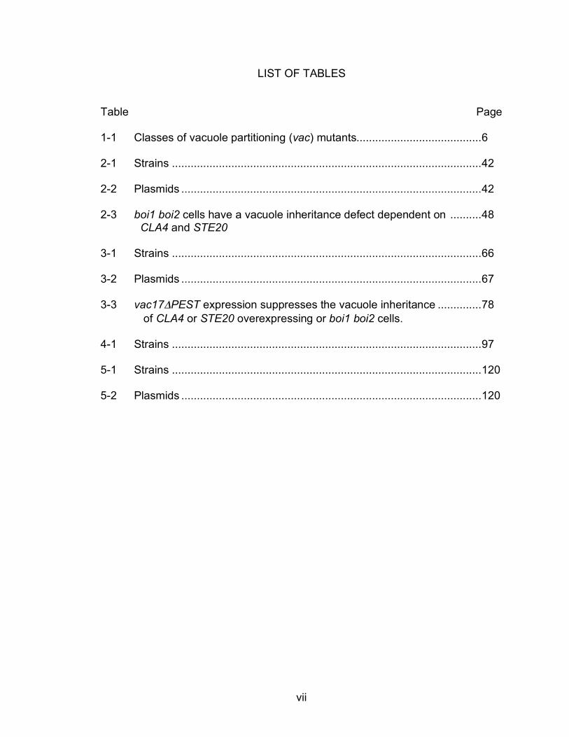
Table Page 1-1 Classes of vacuole partitioning (vac) mutants........................................6
2-1 Strains ...................................................................................................42
2-2 Plasmids ................................................................................................42
2-3 boi1 boi2 cells have a vacuole inheritance defect dependent on ..........48 CLA4 and STE20
3-1 Strains ...................................................................................................66
3-2 Plasmids ................................................................................................67
3-3 vac17!PEST expression suppresses the vacuole inheritance ..............78
of CLA4 or STE20 overexpressing or boi1 boi2 cells.
4-1 Strains ...................................................................................................97
5-1 Strains ...................................................................................................120
5-2 Plasmids ................................................................................................120

viii
LIST OF FIGURES
Figure Page 1-1 Proteins involved in transport of the vacuole into the bud......................8
1-2 Ste20 and Cla4 are involved in signal transduction. ..............................21
2-1 Boi1-GFP has a dynamic localization during the cell cycle and is .........44 inherited by daughter cells.
2-2 boi1 boi2 and vac8 cells have a daughter-specific delay .......................46
2-3 boi1 boi2 cells have a vacuole inheritance defect..................................49
2-4 boi1 boi2 daughter-specific budding delay and vacuole inheritance......51 defects are suppressed by CLA4 or STE20 deletion.
2-5 The PH domain of Boi1 is required for vacuole inheritance in ...............53 the absence of BOI2.
3-1 Proposed model for the bud-specific degradation of Vac17 ..................64
3-2 Cells overexpressing CLA4 have a daughter-specific budding delay ....72
3-3 Cells overexpressing CLA4 have a vacuole inheritance defect .............74
3-4 Cells overexpressing STE20 have a vacuole inheritance defect ...........76
3-5 Vac17 is a phosphoprotein and is degraded in late-M phase ................80
3-6 PAK function is required for Vac17-ProA degradation in late-M ............83 and CLA4 overexpression promotes Vac17-ProA degradation.
3-7 PAK overexpression inhibits peroxisome but not late Golgi...................85 inheritance.
3-8 Model for PAK priming of Vac17 for degradation...................................89
4-1 Cell cycle progression is not required for segregation structure ............100 resolution.
4-2 Cla4 localizes with the vacuole..............................................................101
4-3 Cla4 remains on the daughter vacuole until late-M phase .....................102

ix
4-4 Cla4-GFP requires vacuole inheritance for vacuole associated.............103 punctate structure localization.
4-5 Cells lacking PAK function form but do not resolve segregation............105 structures.
4-6 Cells overexpressing CLA4t fail to form a daughter vacuole and ..........107 have a partial in vivo vacuole fusion defect.
5-1 cla4-75 ste20 cells fail to form a daughter vacuole and arrest at G2/M .123
5-2 cla4-75 ste20 vac8 and cla4-75 ste20 vac17 cells do not arrest............124
5-3 Deletion of SWE1 partially suppresses the cla4-75 ste20 G2/M............126 arrest but not the failure to resolve segregation structures.
5-4 Hsl1 is mislocalized in cla4-75 ste20 and cla4-75 ste20 vac8 cells .......128
5-5 Cdc5 is a target of the vacuole segregation checkpoint.........................129
5-6 Model of the vacuole segregation checkpoint ........................................132 partial in vivo vacuole fusion defect.

x
LIST OF ABBREVIATIONS
!F alpha factor
! null
µm micrometer
µM micromolar
Async asynchronous
ATP adenosine triphosphate
C carboxyl
CDK Cyclin-dependent Kinase
DIC differential contrast interference
cER cortical endoplasmic reticulum
DAPI 4’,6-diamidino-2-phenylindole
EDTA ethylenediaminetetraacetic acid
FACS fluorescence-activated cell sorting
FM 4-64 N-(3-triethylammoniumpropyl)-4-(p-diethylaminophenyl- hexatrienyl) pyridinium dibromide
FRAP fluorescence recovery after photobleaching
G1 gap 1 phase
G2 gap 2 phase
GAL galactose
GCK germinal center kinase
GDP guanosine-5’-diphosphate
GEF guanine nucleotide exchange factor

xi
GFP green fluorescent protein
GLU gluose
GPCR G-protein coupled receptor
GTP guanosine-5’-triphosphate
HA influenza hemaggluttinin epitope
Hr. hour
HU hydroxyurea
IgG immunoglobulin G
M molar
MAPK mitogen-activated protein kinase
MAPKK mitogen-activated protein kinase kinase
MAPKKK mitogen-activated protein kinase kinase kinase
MAPKKKK mitogen-activated protein kinase kinase kinase kinase
MEN mitotic exit network
Min. minutes
mM millimolar
N amino
NZ nocodazole
PAGE polyacrylamide gel electrophoresis
PAK p21-activated kinase
PBD p21 binding domain
PCR polymerase chain reaction
PH pleckstrin homology

xii
PMSF phenylmethanesulphonylfluoride
PtdIns phosphatidylinositol
UT untagged
vac vacuole partitioning mutant
WT wild type

1
CHAPTER I
INTRODUCTION
Organelle Inheritance
Eukaryotic cells contain membrane bound compartments called organelles
that perform various essential cellular functions. Organelle function is critical for
proper cellular function; and each time eukaryotic cells divides they ensure that
each of the progeny cells inherits a full complement of organelles.
There are two main non mutually exclusive strategies used by cells to
ensure high fidelity transmission of organelles to their progeny. Stochastic
inheritance requires that an organelle is present in multiple copies and evenly
distributed throughout the cell. Division of the cell approximately in half by
cytokinesis then ensures that both progeny contain a nearly equal amount of a
multicopy organelle. The second strategy used by eukaryotic cells is an ordered
process requiring the active transport of organelles to ensure equal partitioning.
This second strategy is required for the accurate dispersal of low copy number
organelles 1.
My studies have focused on how the ordered process of organelle
inheritance is regulated. To study the regulation of organelle inheritance I have
used the genetically tractable organism S. cerevisiae. One advantage to studying
organelle inheritance in budding yeast is that they divide asymmetrically, as
opposed to symmetrically down the center. Therefore, organelles must be

2
actively transported into the bud to ensure that the daughter cell receives an
equal share of organelles.
Budding and actin cable formation
Budding occurs through a stereotypical pattern of growth and division.
Just prior to entry into S-phase polarized growth forms the nascent bud which
emerges from the mother cell. This bud continues to grow throughout G2 and
mitosis until cytokinesis. Cytokinesis yields a mother and a smaller daughter cell.
Under nutrient rich conditions the mother cell quickly initiates another round of
budding. The daughter undergoes a brief period of growth until it reaches a
critical size and begins budding.
Budding yeast require the polarization of the actin cytoskeleton to
establish polarity. There are three different actin structures found in budding
yeast: patches, cables, and rings 2. Actin cables are composed of bundled actin
filaments and are the primary tracks along which secretory vesicles are
transported to direct polarized growth. During late G1, the Rho-like GTPase
Cdc42 is activated at a predetermined cortical site. Active GTP bound Cdc42
activates the formins Bni1 and Bnr1. Actin cables are then formed by the actin-
nucleating activity of formins and the actin binding protein profilin 3-6. Secretory
vesicles are transported along actin cables by the processive myosin V motor,
Myo2.
Actin cables, which are almost exclusively localized along the cortex of the
cell, are dynamic structures and undergo spatial rearrangements during the cell

3
cycle. During late G1, actin cables are polarized to the nascent bud site. While
the bud is growing actin cables are polarized within the bud. Finally, as cells exit
mitosis the actin cytoskeleton polarizes towards the mother bud neck in
preparation for cytokinesis 7.
Vacuole inheritance in budding yeast
My research has focused on the regulation of vacuole inheritance in
budding yeast. The yeast vacuole is a membrane bound organelle that is often
considered analogous to the mammalian lysosome because of its acidification
and the hydrolase activities that are localized to the vacuole. The vacuole is also
the terminal degradative compartment of the endocytic pathway. In addition to its
degradative capacity, the vacuole serves as a storage site for amino acids and
polyphosphate 8. Additionally, the vacuole plays a role in regulating cytoplasmic
pH and in modulating water and ion concentration within the cell 8-11.
Vacuoles can be observed in living cells by at least three different
methods. First, differential interference contrast (DIC) microscopy is used to
examine vacuole morphology and partitioning; however, unambiguous
identification of the vacuole can often be difficult with this method 12. Second,
ade2 mutant cells accumulate amino-imidazole ribotide polymers within the
vacuole that are fluorescent and allow vacuole visualization by fluorescence
microscopy 13 14. Third, vacuoles can be visualized by pulse-chase of cells with
the fluorescent dye N-(3-triethylammoniumpropyl)-4-(p-diethylaminophenyl-
hexatrienyl) pyridinium dibromide (FM 4-64). FM 4-64 is imported into cells by

4
endocytosis and accumulates specifically on the vacuole membrane. FM 4-64
staining has several advantages in comparison to other methods. Specifically,
FM 4-64 staining of the vacuole is quick, strain background independent, and the
membrane localization of FM 4-64, as opposed to luminal accumulation of
amino-imidazole ribotide polymers, allows for unencumbered visualization of
organelle structure especially when differentiating clusters of small vacuoles 15.
On average wild type yeast cells contain 1-3 large vacuole lobes 16. These
vacuole lobes are dynamic and undergo continuous fission and fusion 17. Early
experiments designed to visualize the vacuole showed that a portion of the
vacuole forms a tubular and vesicular structure, termed the “segregation
structure”, that is partitioned into the bud to found the daughter vacuole 13, 18. This
high fidelity partitioning of vacuole material from the mother into the bud
prompted the search for mutants defective for vacuole partitioning.
Two large-scale methods have been developed to search for vacuole
partitioning (vac) mutants. The yeast vacuole contains many proteinases and
these proteinases are transported to the vacuole as inactive precursors that must
be cleaved for activation. One of these proteinases, carboxypeptidase Y, is
activated by a proteinase cascade consisting of proteinase A cleaving and
activating proteinase B, which in turn cleaves and activates carboxypeptidase Y.
A simple plate assay exists to measure carboxypeptidase Y activity. To screen
for vacuole inheritance mutants, cells are mutagenized and proteinase A is
produced for a short period of time under the control of the GAL promoter in a
pep4 (proteinase A gene) mutant. This leads to proteinase B and subsequent

5
carboxypeptidase Y cleavage and activation. Proteinase A transcription is then
squelched by the addition of glucose. Daughter cells that inherit vacuolar material
from their mothers maintain carboxypeptidase Y activity for up to 20 generations
due to transmission of activated proteinase B from mother into daughter cells.
However, vac mutants have to make a vacuole de novo and all of their
proteinase B is therefore inactive. Screening mutants for loss of
carboxypeptidase Y after proteinase A was shutoff by glucose has revealed
multiple vac mutants 19-21.
FM 4-64 has also been used to screen for vac mutants. After FM 4-64
pulsing cells, FM 4-64 accumulates on the vacuole membrane. Each cell division
approximately half the mother’s FM 4-64 stained vacuole membrane is
partitioned to the daughter. Mother cells that fail to partition vacuole material to
the daughter will maintain high levels of FM 4-64 fluorescent membrane after
several divisions and daughters will have very little or no stained vacuole
membrane. To screen for vac mutants, mutagenized cells are FM 4-64 stained
and sorted by FACS to isolate highly fluorescent and very low fluorescent cells.
Isolates then are subjected to secondary screens to confirm that mutants are
legitimate vac mutants 22, 23.

6
Table 1-1. Classes of vacuole partitioning (vac) mutants WT Gene Corresponding gene product
Class I mutants act1 Actin
24
pfy1 Profilin 24
Normal vacuole morphology, often
multilobed. myo2 Myo2, class V myosin 24
vac8 Vac8, vacuole membrane protein binds Vac17
25
vac9 Unknown 25
vac10 Unknown
25
vac16 Unknown 26
vac17 Vac17, vacuole specific Myo2 receptor
20, 27
vac19 Unknown 26
Class II mutants
vac5 Pho80 28
vac6 Unknown
19
Enlarged vacuole, often the vacuole is positioned towards bud. vac11 Unknown
25
vac12 Unknown 25
vps3 Vps3, subunit of CORVET tethering complex
18
pep12/vps6 Syntaxin, t-SNARE vps8 Vps8, subunit of CORVET tethering complex vps9 Vps9, GEF for Vps21 pep7/vps19/vac1 Pep7, Vps21 effector
vps21/ypt51 Vps21, Rab vps45 Vps45, Sec1-like
Class III mutants fab1 Fab1, PtdIns(3)P 5-Kinase
29, 30 Highly enlarged vacuole
membrane fission defects. vac7 Vac7, regulator of Fab1 31
vac14 Vac14, regulator of Fab1
10
fig4 Fig4, PtdIns 5’ phosphatase 10
atg18 PtdIns(3,5)P2 binding protein
32
vps15 Vps15, activator of Vps34 33
vps34 Vps34, PtdIns 3-Kinase 34
Based on vacuole morphology all of the presently known vac mutants can
be divided into one of three different classes (Table 1-1) 22. Class I vacuole
partitioning mutants have normally sized vacuoles but often contain multiple
vacuole lobes. To date, each of the class I vac mutants, for which the
corresponding gene has been identified, identifies a part of the physical
machinery necessary for transport of the segregation structure into the bud.
Class II mutants display a moderately enlarged vacuole, normal vacuole

7
morphology, and a portion of the vacuole is often positioned towards the bud.
Class II vac mutants include a number of vacuole protein sorting (vps) mutants
which fail to transport carboxypeptidase Y to the vacuole. The mechanism by
which class II mutants abrogate vacuole inheritance is not known 14. Class III
mutants have a greatly enlarged vacuole which is defective for vacuole fission.
All of the known class III mutants identify proteins required for the production of,
or that bind to PtdIns(3,5)P2.
Transport of the segregation structure
Vesicle transport requires both a track along which a vesicle must travel
and a motor protein to physically transport the vesicle. Eukaryotic cells have
three main cytoskeletal tracks: actin filaments, microtubules, and intermediate
filaments. In mammalian cells, organelle transport commonly utilizes
microtubules 35. However, microtubules are not necessary for vacuole inheritance
as cell treated with the microtubule depolymerizing drug nocodazole (NZ)
partition vacuoles normally 36. Instead, budding yeast transport the vacuole along
polarized actin cables 24. Multiple actin mutants are defective for vacuole
inheritance. In particular act1-!DSE, which eliminates three NH2-terminal amino
acids, shows no observable cytoskeletal defects except a vacuole inheritance
defect. Importantly, the three NH2-terminal amino acids form a portion of the
myosin binding site and other actin mutants in the actin-myosin binding site also
show vacuole inheritance defects 24, 37. Immunofluorescence also shows the
segregation structure is juxtaposed along actin cables 24.
8
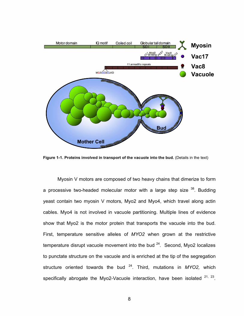
Figure 1-1. Proteins involved in transport of the vacuole into the bud. (Details in the text)
Myosin V motors are composed of two heavy chains that dimerize to form
a processive two-headed molecular motor with a large step size 38. Budding
yeast contain two myosin V motors, Myo2 and Myo4, which travel along actin
cables. Myo4 is not involved in vacuole partitioning. Multiple lines of evidence
show that Myo2 is the motor protein that transports the vacuole into the bud.
First, temperature sensitive alleles of MYO2 when grown at the restrictive
temperature disrupt vacuole movement into the bud 24. Second, Myo2 localizes
to punctate structure on the vacuole and is enriched at the tip of the segregation
structure oriented towards the bud 24. Third, mutations in MYO2, which
specifically abrogate the Myo2-Vacuole interaction, have been isolated 21, 23.

9
Further analysis of the Myo2 globular tail domain, that is known to bind cargo,
has revealed it to be composed of two subdomains, SDI and SDII. SDI and SDII
dimerize in vitro and in vivo and this dimerization is required for cargo binding.
SDI and SDII bind different cargo. SDI binds to the vacuole-specific myosin
receptor Vac17 (described below). Mutants in MYO2 that disrupt vacuole
inheritance all map to SDI. SDII binds to secretory vesicles 39-41.
Attachment of Myo2 to the vacuole is accomplished through the vacuole
transport complex consisting of Myo2-Vac17-Vac8. Vac8 is both myristoylated
and palmitoylated on residues at the NH2-terminus, and these modifications are
necessary for Vac8 localization to the vacuole 25, 42-45. Vac8 plays many roles in
the cell, including roles in caffeine resistance, homotypic vacuole fusion, nucleus-
vacuole junction (NVJ) formation, cytoplasm to vacuole transport (CVT), and
vacuole inheritance 11, 25, 42, 43, 46-49. Vac8 contains eleven armadillo repeat
domains. Armadillo repeats are known to mediate protein-protein interaction and
Vac8 is known to bind at least nine different receptors 49. Vac8 is enriched on
three different subdomains of the vacuole associating with the NVJ, sites of
homotypic vacuole fusion, and domains that act in vacuole transport 44.
The Vac8 receptor protein Vac17 connects Vac8 to Myo2 and is the final
member of the vacuole transport complex. Vac17 binds directly to both Myo2 and
Vac8 through separable protein domains 20, 27. Vac17 production and degradation
play key roles in the spatial and temporal regulation of vacuole inheritance.
VAC17 mRNA levels oscillate with the cell cycle and increase as cells begin
budding 50, 51. Concordantly Vac17 levels increase during early budding. Vac17

10
contains a region abundant in proline (P), glutamic acid (E), serine (S), and
threonine (E) known as a PEST sequence which acts as a signal for rapid
turnover. Intriguingly, turnover of Vac17 is spatially dependent and requires
transport of Vac17 into the bud. The vacuole inheritance mutants vac8 and
myo2-2 fail to degrade Vac17 and Vac17 accumulates to high levels 20.
Examination of the timing of Vac17 degradation shows that it is normally
degraded prior to cytokinesis. The spatial and temporal regulation of Vac17 is
necessary to ensure that the vacuole is not transported back to the mother-bud
neck when the actin cytoskeleton concentrates at the mother-bud neck in
preparation for cytokinesis. When Vac17 missing the PEST sequence
(vac17!PEST) is expressed in cells, Vac17!PEST localizes to the vacuole but is
not degraded. Furthermore, both the mother and daughter vacuole are
transported to the mother-bud neck 20.
Vac17 is degraded in a bud-specific manner, but how this is achieved
remains unknown. One model posits that factors specifically localized to or
activated in the bud promote Vac17 degradation 14. Thus Vac17 does not come
into contact with these factors until Vac17 enters the bud. The identity of these
factors remains unknown.
Formation of the segregation structure
Isolation of the class III vac mutants shed light on the machinery
necessary for forming the segregation structure. Like wild type yeast, class III vac
mutants transport a portion of the vacuole into the bud. Unlike wild type cells the

11
class III mutants usually contain an enlarged single lobed vacuole. During
inheritance the vacuole is transported into the bud, but the vacuole is commonly
seen as one enlarged contiguous vacuole stretched from the mother into the bud
that is constricted at the neck, forming what looks like an “open-figure-eight”
(Table 1-1) 29, 30. The enlarged vacuole and the persistence of the vacuole in the
neck of the most severe class III vac mutant, fab1, adversely affects spindle
positioning leading to aploid and binucleate cells 29. Proteins identified by the
class III vac mutants have been proposed to form the tubules and vesicles
making up the segregation structure, allowing vacuolar material to pass easily
through the confined space of the mother bud neck 14, 26.
Cloning of the class III vac mutants helped elucidate the mechanism of
segregation structure formation (Table 1-1). Each of the class III vac mutants are
in genes that code for proteins that produce, regulate the production of, or bind to
PtdIns(3,5)P2. Vps15 binds to and activates the sole PtdIns 3’-Kinase in yeast,
Vps34, and PtdIns(3)P is required as a precursor for PtdIns(3,5)P2 production 33,
34, 52, 53. Fab1 is the sole PtdIns(3) 5-kinase in yeast 54 and it is positively
regulated by Vac7 and Vac14 10, 31.
Importantly PtdIns(3,5)P2 production has physiological affects on vacuole
morphology. PtdIns(3,5)P2 levels are 18-28 fold lower than PtdIns(3)P,
PtdIns(4)P, and PtdIns(4,5)P2 under normal physiological conditions but rapidly
rise 20-fold after hyperosmotic stress 10. Concomitant with the rise in
PtdIns(3,5)P2 levels the vacuole fissions to form multiple small lobes. The
fissioning of the vacuole is linked to the production of PtdIns(3,5)P2; and fab1

12
and vac14 mutants do not fission under hyperosmotic conditions 10, 31. The rapid
fissioning of the vacuole may mitigate problems caused by the rapid efflux of
water out of the cell under hyperosmotic conditions by releasing water from the
vacuole. A sphere with a given diameter can produce four spheres with a total of
half the volume 10. Under hyposmotic conditions the vacuole undergoes fusion
and vac8 mutants are defective for hyposmotic induced homotypic vacuole fusion
11.
How does PtdIns(3,5)P2 promote the fissioning of the vacuole? Fission of
the vacuole requires, at least in part, the recruitment of PtdIns(3,5)P2 effector
proteins. One effector, Atg18, binds directly to PtdIns(3,5)P2 and atg18 mutants
display an enlarged vacuole phenotype like all known class III vac mutants 32, 55.
Atg18 was originally isolated in a screen for mutants defective in autophagy.
However, PtdIns(3,5)P2 does not appear necessary for autophagy as fab1
mutants are not defective for autophagy 32. Cells lacking ATG18 additionally have
a 5-10 fold higher levels of PtdIns(3,5)P2 under normal physiological conditions
while retaining an enlarged vacuole 32. Atg18 and other fission machinery (Fab1
and Vac14) localize to punctate structures on the vacuole. In a small number of
cells FM 4-64 stained vesicles bud from the Atg18-GFP punctate structures and
Atg18-GFP containing vacuoles travel into the bud and fuse with what is
presumably the daughter vacuole. This is especially intriguing as Atg18 binds
Vac17 by 2-hybrid and coimmunoprecipitation 55. Thus it has been proposed that
Atg18 serves a nonessential role in facilitating the formation of the segregation
structure by promoting vesicle fission 55.

13
Inhibition of vacuole fusion also plays a role in maintaining vacuole
fragmentation after exposure to hyperosmotic conditions and in forming the
segregation structure. The yeast casein kinase, Yck3, negatively regulates
homotypic vacuole fusion by inhibiting the tethering of vacuoles. Inhibition of
homotypic vacuole fusion is essential to maintain vacuole fragmentation after
PtdIns(3,5)P2 levels have returned to normal after hyperosmotic shock 56.
Inhibition of vacuole fusion is also important for the efficient partitioning of the
vacuole into the bud. Mutant yck3 cells have a moderate vacuole inheritance
defect, reduced daughter vacuole size in cells inheriting vacuole material from
the mother, and tubular and vesicular segregation structures were not observed
in the mutant 56. These data combined lead to the current model for segregation
structure formation which proposes that vacuole fission is required to form the
vesicles and tubules that compose the segregation structure 57.
Despite the added clarity that recent experiments have shed on how the
segregation structure is formed, many unanswered questions exist about the
process and several key experiments to test the model remain undone. One of
the major roadblocks to analyzing vacuole partitioning revolves around the
transience of segregation structures. Based on the rate of vacuole movement
and the distance of travel, segregation structures can be around for as little as 30
seconds during the cell cycle 14. During this 30 seconds a complex set of
reactions has been proposed to occur, including the production of PtdIns(3,5)P2
14, the recruitment PtdIns(3,5)P2 binding proteins, segregation structure formation
32, 55, 58, 59, transport of the segregation structure into the bud, and resolution of

14
the segregation structure (discussed below). The transient nature of the
segregation structure and the impossibility of reliably synchronizing a population
with intact segregation structures have made testing if PtdIns(3,5)P2 levels
transiently rise during segregation structure formation difficult. Visualization and
quantification of Atg18, or other potential PtdIns(3,5)P2 effectors, on segregation
structures is also difficult or impossible due to the transience of the segregation
structure. Identification of mutants defective for resolving the segregation,
therefore extending the amount of time the segregation structure persists, could
potentially be useful to further study these problems.
Resolution of the segregation structure
Once formed, the segregation structure is transported into the bud. Upon
entry into the bud the segregation structure is resolved to form a discrete mother
and daughter vacuole. Over the years various strides have been made in forming
a model for how the segregation structure is resolved.
Together the experimental data suggests that the segregation structure is
resolved by the fusion of tubular and vesicular structures in the bud. The data
that support this model are reviewed below. (1) Formation of the vacuole
segregation structure requires PtdIns(3,5)P2 production on the vacuole which
promotes fission of the vacuole, suggesting that the opposing process fusion is
required for resolution 10, 30, 31, 60-62. (2) Examination of labeled vacuoles by
microscopy showed transport of vesicular structures from the mother into the
daughter and fusion of these vesicles to form a larger daughter vacuole 63. (3)

15
Studies on vacuoles in semi-permeabilized cells show the formation of tubular
and vesicular structures followed by vacuole fusion 63-66. (4) Resolution of
segregation structure in zygotes involves vacuole fusion and merging of vacuole
contents 67. (5) Formation of the segregation structure is promoted by Yck3 which
maintains fragmentation through inhibition of homotypic vacuole fusion 56.
Together these data suggest a model in which the vesicles and tubules
composing the segregation structure are formed by fission machinery and
resolved by fusion of the tubules and vesicles in the bud. A non-exclusive
alternate model suggests that both formation and resolution of the segregation
structure is accomplished by fission of the tubules to form the daughter vacuole.
However, this possibility has not been rigorously tested and is solely supported
by the observation that class III vac mutant often have vacuoles which span the
mother bud neck 57. Missing from these models of segregation resolution, is a
mechanism for not only promoting, but insuring, that the segregation structure is
only resolved after arriving in the bud. As was proposed for Vac17 degradation
14, there may be proteins localized specifically to the bud which promote vacuole
fusion (or fission) to resolve the segregation structure as it enters the bud.
However, as with the daughter-specific degradation of Vac17, potential daughter
localized proteins that promote resolution of the segregation structure after it has
entered the bud have not been identified.

16
Coordination of vacuole inheritance with the cell cycle
Cells must coordinate organelle inheritance with other cell cycle driven
events including mitosis and cytokinesis. This coordination ensures that each
time an eukaryotic cell divides, each of the progeny receives an accurate copy of
each of the chromosomes and a full complement of organelles. Checkpoints can
delay cell cycle progression in response to a failure to complete earlier events
through transduction of a negative signal to the cell cycle machinery 68, 69. Recent
evidence has shown that checkpoints exist to ensure the faithful propagation of
organelles to each progeny cell.
In mammalian cells the Golgi apparatus is composed of 4-8 cisternae
anchored at pericentriolar region of the cell. During prophase this Golgi ribbon
fragments into smaller vesicles and tubules that are subsequently dispersed
during metaphase 35. Failure to fragment the Golgi activates the Golgi mitotic
checkpoint and delays cells at G2/M 70.
Organelle partitioning checkpoints are not exclusive to mammalian cells.
In budding yeast the cortical endoplasmic reticulum (cER) is transported along
actin cables by the myosin V family motor Myo4 into the bud where it becomes
anchored at the tip of the bud and expands to fill the bud 35. Cells lacking proteins
required for cER inheritance also delay in G2/M. This cER inheritance
checkpoint requires the morphogenesis checkpoint proteins which acts to delay
the cell cycle by negatively regulating the cyclin B-CDK complex essential for
mitotic progression 71.

17
Checkpoints may also be required to ensure that segregation structure
resolution occurs prior to mitosis. Budding yeast have a relatively narrow neck
through which both the segregation structure and the nucleus must pass. Early
studies noted that segregation structures are oriented towards the emerging bud
early in the cell cycle and inheritance was completed prior to nuclear division 12,
13, 36. This opens the possibility that budding yeast monitor and delay cell cycle
progression until segregation structures are resolved, thus clearing the neck for
karyokinesis. How segregation structure resolution is coordinated with the cell
cycle is unknown 57.
Currently there are two known checkpoints that delay mitotic progression
in response to failure to complete organelle inheritance: the Golgi mitotic
checkpoint and the cER inheritance checkpoint. Mitogen-activated protein
kinases regulate (MAPKs) regulate the inheritance of both of these organelles. In
mammalian cells the Golgi mitotic checkpoint delays cells in G2/M 70. While the
mechanism for this checkpoint has not been fully elucidated, inhibition of MAP
kinases involved in Golgi fragmentation results in checkpoint activation 72, 73. cER
inheritance is also regulated by a MAP kinase pathway 74.
Mitogen-activated protein kinases
The Ste20 group kinases are regulators of mitogen-activated protein
kinase (MAPK) cascades and are divided into two main groups the p21-activated
kinases (PAKs) and the germinal center kinases (GCKs) 75. My studies have

18
focused on the role of PAKs in regulating vacuole inheritance. Yeast contain two
main p21-activated kinases, the founding member of the family, Ste20, and Cla4.
Ste20 and Cla4 contain multiple domains and share similar domain
structures. Both Ste20 and Cla4 have a kinase domain towards the C-terminus of
the protein. There are multiple known targets of the PAKs in budding yeast. Both
Ste20 and Cla4 are known to phosphorylate the myosin-I isoforms Myo3 and
Myo5 76, 77. Ste20 phosphorylates the MAPKKK Ste11 78. Cla4 has several
known targets including the Cdc42 guanine nucleotide exchange factor (GEF)
Cdc24 79, 80, septins 81, Swe1 82, 83, and Lte1 84-86. Despite the increasing data
about Ste20 and Cla4 substrates, it must be noted that a complete catalog of
substrates for either kinase remains undetermined and the data on the
redundancy of Ste20 and Cla4 substrates remains elusive.
Ste20 and Cla4 activity is carefully controlled by both positive and
negative regulation. Regions in the N-terminal halves of the PAKs interact with
the kinase domain in the C-terminal half of the PAKs leading to autoinhibition.
This model is supported by genetic and structural data on Ste20, Cla4, and other
PAKs 87-89. Autoinhibition is relieved when PAKs bind to activated GTP bound
Cdc42 through the p21 binding domain (PBD) located within the N-terminal half
of the protein 89-91. In budding yeast the adaptor protein Bem1 facilitates the
interactions between Cdc42 and the PAKs. Bem1 is a multidomain protein and it
binds both Cdc42 and Cdc24 92-95. Bem1 contains two SH3 domains and the
second SH3 domain interacts with proline rich domains in Ste20 and Cla4 79, 80,
96. The adaptor protein Bem1 thus brings together Cdc42 with its GEF leading to

19
Cdc42 activation. Activated Cdc42’s interaction with Ste20 and Cla4 relieves
PAK autoinhibition and activates the PAKs. Cla4 activity is also cell cycle
regulated and peaks near mitosis 88.
Both Cla4 and Ste20 localize to sites of polarized growth 87, 97-99. To
ensure that Ste20 and Cla4 localize to regions where potential targets reside,
both proteins contain domains necessary and sufficient to bind
phosphatidylinositides. Cla4 contains a pleckstrin homology (PH) domain. PH
domains are known to vary in their specificity for different phosphatidylinositides
100. In vitro analysis by protein-lipid overlay analysis demonstrates that Cla4’s PH
domain binds to PtdIns(3)P, PtdIns(3,5)P2, PtdIns(4)P, and PtdIns(5)P. Surface
plasmon resonance showed strong binding to both PtdIns(4,5)P2 and
PtdIns(3,5)P2. In vivo analysis finds that Cla4-GFP localization to the plasma
membrane requires PtdIns(4)P but not PtdIns(4,5)P2 100, 101. Functional analysis
of Cla4 mutants unable to bind phosphatidylinositides shows that
phosphatidylinositide binding acts in conjunction with Cdc42 localization and
activation to promote Cla4 activity 101. Ste20 also binds to phosphatidylinositides
using a basic-rich (BR) domain, but the phosphatidylinositide specificity of the
Ste20’s BR domain has not been tested. Like the PH domain in Cla4, the BR
domain in Ste20 acts in conjunction with the PBD to both localize and activate
Ste20 102.
Ste20 was originally isolated in a screen for suppressors of a dominant
negative mutant of the "-subunit of a heterotrimeric G-protein coupled receptor,

20
Ste4, involved in mating 103. Ste4 binds to Ste20 through a noncatalytic region in
the extreme C-terminus of Ste20 104.
Cellular functions of Ste20 and Cla4
Ste20 and Cla4 are not only similar in their domain structure but they also
share many overlapping functions. However, they also perform nonoverlapping
functions. In budding yeast PAKs are known to play roles in signal transduction
involved in mating, filamentous growth, and in responding to osmotic stress.
Additionally, they promote polarized growth, septin formation, and cell cycle
progression.
Signal transduction
Signal transduction networks are necessary for cells to respond to
external signals and to coordinate intracellular changes with the extracellular
environment. In budding yeast the best studied of these signal transduction
networks is the pathway required for mating. Haploid yeast excrete peptide
pheromones which bind to cell-surface receptors on yeast of the opposite mating
type. Pheromone binding causes G1 cell cycle arrest and polarized growth
towards a partner cell of the opposite mating type. Once the two cells come into
contact they fuse to form a diploid cell. A G-protein coupled receptor (GPCR)
activated MAPK cascade is required to coordinate the process of mating. This
kinase cascade requires the MAPKKKK Ste20, a MAPKKK Ste11, a MAPKK
Ste7, and two MAPKs Fus3 and Kss1 which are all connected by the scaffolding

21
protein Ste5 105. Ste20 directly interacts with Ste4, the "-subunit of the GPCR 104.
After binding Ste4, Ste20 directly phosphorylates Ste11, relieving its
autoinhibition and thus promoting Ste7 phosphorylation 78. The scaffolding
protein Ste5 also binds to Bem1 allowing for the efficient activation of Ste20 by
GTP-Cdc42 106. Cla4 is not known to bind to Ste4. However, genetic and
biochemical data suggest that Cla4 does participate in the mating type pathway "
though probably by negatively regulating it 107.
Figure 1-2. Ste20 and Cla4 are involved in signal transduction. (Details in the text)
Although less understood than the mating type cascade, Ste20 also plays key
roles in regulating filamentous growth (also called invasive growth in haploid cells
and pseudohyphal growth development in diploid) under nutrient deprivation
conditions. The filamentous growth MAPK cascade causes cells to become
elongated and fail to separate after division causing cells to penetrate into the
agar. The filamentous growth MAPK cascade consists of Ste20 (MAPKKKK) #

22
Ste11 (MAPKKK) # Ste7 (MAPKK) # Kss1 (MAPK) 108. Unlike in the mating
type cascade, Fus3 is not activated, the scaffolding protein Ste5 is not used, and
Cdc42 is activated by Ras2 105. Thus Ste20 plays key roles in regulating both
mating and filamentous growth.
In response to hyperosmotic stress, vacuoles fragment immediately to
accommodate osmotic changes. Prolonged hyperosmotic change requires long-
term cellular adaptations to the new environment. In response to hyperosmotic
shock cells arrest and begin producing the compatible solute glycerol 109.
Budding yeast contain two putative osmosensors, Sln1 and Sho1. Sln1 and Sho1
activate different branches of the pathway but both culminate in the activation of
the same MAPK cascade consisting of Pbs2 (MAPKK) # Hog1 (MAPK). The
Sho1 branch of the pathway requires Ste11 (MAPKKK) activation of Pbs2. Both
Ste20 and Cla4 play roles in activating Ste11 in response to hyperosmotic stress
as assayed in a reporter strain 110. Further evidence supporting a role for Cla4 in
hyperosmotic stress response is found in the early findings that cells lacking
CLA4 are normally elongated and this elongation is suppressed by growth under
hyperosmotic conditions but not in the absence of either PBS2 or HOG1 91.
Intriguingly hyperosmotic stress can cause cells to arrest in G1 or delay in G2.
This G2 delay requires the Hog1 dependent phosphorylation of the Nim1-related
protein kinase in yeast Hsl1 (discussed further below) 111.

23
Polarized Growth
Polarized growth requires the activation of Cdc42 by Cdc24 and
temperature sensitive cdc42 and cdc24 mutants arrest as large unbudded cells
at the restrictive temperature 112, 113. Cdc24 is normally localized to the nucleus
during G1 and is exported into the cytoplasm at bud emergence 114, 115. Cdc24 is
initially localized to the site of bud emergence by proteins that mark the
presumptive bud site 116. Stabilization of Cdc24 at the site of polarized growth is
accomplished by interactions with the adaptor protein Bem1 93, 117. Cla4 and
Ste20 interact with Bem1, and Cla4 (and possibly Ste20) phosphorylates Cdc24.
The effect of Cdc24 phosphorylation, however, is an unresolved issue with
separate groups reporting contradictory results. Gulli et al. find that
phosphorylation of Cdc24 by Cla4 causes Cdc24 to dissociate from Bem1 79. In
contrast Bose et al. find that phosphorylation of Cdc24 by Cla4 causes a
stabilization of the Cdc24-Bem1 interaction 80. Therefore, Cla4 regulates
polarized growth by regulating Cdc24 localization, though the consequences of
this regulation are disputed.
Ste20 and Cla4 also indirectly regulate actin branching. Neither Ste20 nor
Cla4 are necessary for the polarization of actin cables required for bud
emergence 91, 118, 119. However, Ste20 and Cla4 are involved in promoting actin
polymerization through the Arp2/3 complex. Cdc42 activates Arp2/3 dependent
branching through a defined pathway. Cdc42 activates the Wiskott-Aldrich
syndrome protein (Las17) bound to the WASP-interacting protein (Vrp1) 120-124
and Las17/Vrp1 activates the Arp2/3 complex to promote actin polymerization 3,

24
125. Ste20 and Cla4 phosphorylate the type I myosins, Myo3 and Myo5 76, 77, 126.
In turn, phosphorylated Myo3 and Myo5 activate the Las17/Vrp1 complex 126, 127.
Through modulating Arp2/3 mediated branching it has been proposed that the
PAKs regulate the actin cytoskeleton in S. cerevisiae and other organisms 128.
Septin Formation
Septins are evolutionarily conserved GTP-binding proteins that form
hetero-oligomeric filaments. In budding yeast these septin filaments form at the
mother bud neck where they act as a scaffold on which other proteins assemble
129. In G1 cells, septins are localized to the nascent bud site but are highly mobile
and fluid as judged by fluorescence recovery after photobleaching (FRAP) but
they quickly reorganize to form a protein collar concomitant with bud emergence
130, 131. During cytokinesis the septin collar splits into two rings.
A role for PAKs in septin organization was recognized in early studies. In
cells lacking both Ste20 and Cla4 activity the mother bud neck appears
particularly wide and the septin Cdc3 is mislocalized 91. PAKs appear to promote
reorganization of the septins leading to their immobilization and in both cdc42
and cla4 mutant cells septins remain more dynamic throughout budding 130-132.
Cla4 directly phosphorylates the septins Cdc3, Cdc10, and Cdc11, and removal
of the Cla4 phosphorylation site on Cdc10 causes septin organization defects 81.
Ste20 also plays a role in regulating septin formation. Evidence supporting this
conclusion include the findings that Cdc10 has residual phosphorylation in a cla4
mutant, Ste20 phosphorylates Cdc10 in vitro, and septins mislocalize to a greater

25
extent in cells lacking both CLA4 and STE20 than in cells lacking either PAK
individually 81, 91.
Cell Cycle Progression
Each time budding yeast reproduce, bud formation must be coordinated
with organelle partitioning and mitotic progression. In the event of a failure to
properly form a bud, checkpoints ensure that mitotic progression is delayed until
a bud is formed. The checkpoint linking bud formation with cell cycle progression
is called the morphogenesis checkpoint and it delays cells in G2 133. The
morphogenesis checkpoint is thought to monitor proper bud formation by
monitoring proper formation of the septin collar. In response to a failure to
properly form the septin scaffold, cells delay or arrest at G2. To delay cell cycle
progression the morphogenesis checkpoint modulates the localization and
stability of the Wee1 kinase in budding yeast, Swe1. Swe1 is a dual specificity
kinase that prevents cell cycle progression by phosphorylating the mitosis-
promoting cyclin B/CDK complex on a conserved tyrosine residue 134. When
septin rings are properly formed at the mother-bud neck the Nim1-family protein
kinase Hsl1 recruits the protein methyltransferase Hsl7 to the mother bud neck.
Hsl7 interacts directly with Swe1 and recruits Swe1 to the mother bud neck
where it is negatively regulated 135-140.
Multiple lines of evidence show that Cla4 and Ste20 regulate Swe1.
Failure to negatively regulate Swe1 leads to cells with elongated buds. The
Cdc42 mutant cdc42V44A has elongated buds. This elongated bud phenotype is

26
suppressed by deletion of SWE1 or by overexpression of CLA4 or STE20 141.
Cells lacking both Cla4 and Ste20 activity arrest in G2 in a SWE1 dependent
manner 91, 118, 119. Cells lacking CLA4 are elongated and this elongation is
suppressed by SWE1 deletion 135. Finally, Cla4 is targeted to the mother bud
neck and directly phosphorylates Swe1 82, 83. In this way PAKs act to promote cell
cycle progression through G2. The yeast Polo-like kinase, Cdc5, also interacts
with Swe1 and phosphorylates Swe1 to promote cell cycle progression 82, 83, 142.
In addition to promoting progression into mitosis PAKs act to promote
mitotic exit. In budding yeast mitotic exit is driven by the protein phosphatase
Cdc14 that acts to reverse CDK phosphorylation events. From the beginning of
the cell cycle until metaphase, Cdc14 is kept inactive within the nucleolus. In
order to promote mitotic exit, sustained Cdc14 release from the nucleolus
requires a network of proteins called the MEN (mitotic exit network). The MEN
consists of the GTPase Tem1; its putative GEF Lte1; a two-component GTPase
activating protein (GAP) Bub2/Bfa1; the protein kinases Cdc5, Cdc15, Dbf2; a
Dbf2 associated factor Mob1, and a scaffold protein Nud1 143. The GTPase Tem1
is localized to the spindle pole body entering the bud. Once transported into the
bud Tem1 comes into contact with and probably activated by Lte1 localized at
bud cortex. Cla4 phosphorylates Lte1 regulating its initial localization and binding
to the bud cortex 84-86. Ste20 is not necessary for Lte1 phosphorylation or
localization but is synthetically lethal with lte1. The ste20 lte1 synthetic lethality
combined with other genetic and molecular biological data show that Cla4 and
Ste20 act in parallel pathways to promote mitotic exit 84, 119.

27
Vacuole Fusion
The yeast vacuole is a dynamic structure and constantly undergoes fusion
and fission. Fusion of separate lobes of the vacuole requires a multistep process.
First vacuoles are primed for fusion by the ATP dependent activation of fusion
factors. Second, vacuoles are reversibly tethered together. Next unpaired
SNAREs assemble into a trans-complexes leading to docking. Finally, lipid
bilayer mixing and fusion of vacuoles occurs 144. Both Cdc42 and actin
polymerization are required for the docking stage of vacuole fusion 122, 124, 145, 146.
In vitro and in vivo studies show that inhibition of actin branching by inhibiting
either Las17 or the Arp2/3 complex inhibits docking 122. Importantly, Cla4 and
Ste20 both indirectly activate Las17 by phosphorylating Myo3 and Myo5 76, 77, 126,
127. Therefore, PAKs may play a role in vacuole fusion. In further support of a role
for PAKs in vacuole fusion, Cla4 localizes to purified vacuole and cla4 mutants
have fragmented vacuoles 16, 122. Additionally, the Cla4 and Ste20 interacting
protein Bem1 promotes vacuole fusion 147, 148.
Conclusions
Cla4 and Ste20 play many roles in regulating various biological processes
in S. cerevisiae. In the chapters that follow I will present data showing that Cla4
and Ste20 act to regulate vacuole inheritance. In chapter two I identify a novel
class I vac mutant. Boi1 and Boi2 are functionally redundant proteins that interact
with Bem1. Mutant boi1 boi2 cells display a class I vac defect which is

28
suppressible by deletion of CLA4 or STE20. Unlike all other known class I vac
mutants which are part of the physical machinery transporting the segregation
structure into the bud, Boi1 and Boi2 play no known structural role in inheritance
and may instead be regulators of vacuole inheritance.
In chapter three I address an open question in the vacuole inheritance
field of how Vac17 is degraded in a spatially dependent fashion. It has been
previously hypothesized that bud-specific degradation of Vac17 is promoted by
bud localized factors. However, the identity of these factors was not known. In
chapter three I show that Cla4 and Ste20, which localize specifically to the bud,
are required for Vac17 degradation. I also show that CLA4 and STE20
overexpression causes a vacuole inheritance defect that is suppressed by non-
degradable Vac17.
In chapter four I address the question of how the segregation structure is
resolved. I show that Cla4 localizes to the segregation structure just prior to
segregation structure resolution placing it in the right time and place to resolve
the segregation structure. I additionally show that PAKs are required for daughter
vacuole formation.
Checkpoints delay cell cycle progression in response to a failure to
complete earlier events through transduction of a negative signal to the cell cycle
machinery. Vacuole inheritance is finished before mitosis occurs. In chapter five I
present data supporting a novel vacuole inheritance checkpoint. Together this
data advances our understanding of how vacuole inheritance is regulated in a
spatially dependent manner and how it is coordinated with cell cycle progression.

29
References
1. Warren, G. & Wickner, W. Organelle inheritance. Cell 84, 395-400 (1996). 2. Moseley, J.B. & Goode, B.L. The yeast actin cytoskeleton: from cellular
function to biochemical mechanism. Microbiol Mol Biol Rev 70, 605-45 (2006).
3. Evangelista, M., Pruyne, D., Amberg, D.C., Boone, C. & Bretscher, A. Formins direct Arp2/3-independent actin filament assembly to polarize cell growth in yeast. Nat Cell Biol 4, 260-9 (2002).
4. Sagot, I., Klee, S.K. & Pellman, D. Yeast formins regulate cell polarity by controlling the assembly of actin cables. Nat Cell Biol 4, 42-50 (2002).
5. Pruyne, D. et al. Role of formins in actin assembly: nucleation and barbed-end association. Science 297, 612-5 (2002).
6. Sagot, I., Rodal, A.A., Moseley, J., Goode, B.L. & Pellman, D. An actin nucleation mechanism mediated by Bni1 and profilin. Nat Cell Biol 4, 626-31 (2002).
7. Amberg, D.C. Three-dimensional imaging of the yeast actin cytoskeleton through the budding cell cycle. Mol Biol Cell 9, 3259-62 (1998).
8. Klionsky, D.J., Herman, P.K. & Emr, S.D. The fungal vacuole: composition, function, and biogenesis. Microbiol Rev 54, 266-92 (1990).
9. Dove, S.K. et al. Osmotic stress activates phosphatidylinositol-3,5-bisphosphate synthesis. Nature 390, 187-92 (1997).
10. Bonangelino, C.J. et al. Osmotic stress-induced increase of phosphatidylinositol 3,5-bisphosphate requires Vac14p, an activator of the lipid kinase Fab1p. J Cell Biol 156, 1015-28 (2002).
11. Wang, Y.X., Kauffman, E.J., Duex, J.E. & Weisman, L.S. Fusion of docked membranes requires the armadillo repeat protein Vac8p. J Biol Chem 276, 35133-40 (2001).
12. Jones, H.D., Schliwa, M. & Drubin, D.G. Video microscopy of organelle inheritance and motility in budding yeast. Cell Motil Cytoskeleton 25, 129-42 (1993).
13. Weisman, L.S., Bacallao, R. & Wickner, W. Multiple methods of visualizing the yeast vacuole permit evaluation of its morphology and inheritance during the cell cycle. J Cell Biol 105, 1539-47 (1987).
14. Weisman, L.S. Yeast vacuole inheritance and dynamics. Annu Rev Genet 37, 435-60 (2003).
15. Vida, T.A. & Emr, S.D. A new vital stain for visualizing vacuolar membrane dynamics and endocytosis in yeast. J Cell Biol 128, 779-92 (1995).
16. Seeley, E.S., Kato, M., Margolis, N., Wickner, W. & Eitzen, G. Genomic analysis of homotypic vacuole fusion. Mol Biol Cell 13, 782-94 (2002).
17. Wickner, W. Yeast vacuoles and membrane fusion pathways. Embo J 21, 1241-7 (2002).
18. Raymond, C.K., O'Hara, P.J., Eichinger, G., Rothman, J.H. & Stevens, T.H. Molecular analysis of the yeast VPS3 gene and the role of its product

30
in vacuolar protein sorting and vacuolar segregation during the cell cycle. J Cell Biol 111, 877-92 (1990).
19. Gomes de Mesquita, D.S., van den Hazel, H.B., Bouwman, J. & Woldringh, C.L. Characterization of new vacuolar segregation mutants, isolated by screening for loss of proteinase B self-activation. Eur J Cell Biol 71, 237-47 (1996).
20. Tang, F. et al. Regulated degradation of a class V myosin receptor directs movement of the yeast vacuole. Nature (2003).
21. Catlett, N.L., Duex, J.E., Tang, F. & Weisman, L.S. Two distinct regions in a yeast myosin-V tail domain are required for the movement of different cargoes. J Cell Biol 150, 513-26 (2000).
22. Wang, Y.X. et al. Multiple classes of yeast mutants are defective in vacuole partitioning yet target vacuole proteins correctly. Mol Biol Cell 7, 1375-89 (1996).
23. Catlett, N.L. & Weisman, L.S. The terminal tail region of a yeast myosin-V mediates its attachment to vacuole membranes and sites of polarized growth. Proc Natl Acad Sci U S A 95, 14799-804 (1998).
24. Hill, K.L., Catlett, N.L. & Weisman, L.S. Actin and myosin function in directed vacuole movement during cell division in Saccharomyces cerevisiae. J Cell Biol 135, 1535-49 (1996).
25. Wang, Y.X., Catlett, N.L. & Weisman, L.S. Vac8p, a vacuolar protein with armadillo repeats, functions in both vacuole inheritance and protein targeting from the cytoplasm to vacuole. J Cell Biol 140, 1063-74 (1998).
26. Catlett, N.L. & Weisman, L.S. Divide and multiply: organelle partitioning in yeast. Curr Opin Cell Biol 12, 509-16 (2000).
27. Ishikawa, K. et al. Identification of an organelle-specific myosin V receptor. J Cell Biol 160, 887-97 (2003).
28. Nicolson, T.A., Weisman, L.S., Payne, G.S. & Wickner, W.T. A truncated form of the Pho80 cyclin redirects the Pho85 kinase to disrupt vacuole inheritance in S. cerevisiae. J Cell Biol 130, 835-45 (1995).
29. Yamamoto, A. et al. Novel PI(4)P 5-kinase homologue, Fab1p, essential for normal vacuole function and morphology in yeast. Mol Biol Cell 6, 525-39 (1995).
30. Bonangelino, C.J., Catlett, N.L. & Weisman, L.S. Vac7p, a novel vacuolar protein, is required for normal vacuole inheritance and morphology. Mol Cell Biol 17, 6847-58 (1997).
31. Gary, J.D., Wurmser, A.E., Bonangelino, C.J., Weisman, L.S. & Emr, S.D. Fab1p is essential for PtdIns(3)P 5-kinase activity and the maintenance of vacuolar size and membrane homeostasis. J Cell Biol 143, 65-79 (1998).
32. Dove, S.K. et al. Svp1p defines a family of phosphatidylinositol 3,5-bisphosphate effectors. Embo J 23, 1922-33 (2004).
33. Herman, P.K., Stack, J.H., DeModena, J.A. & Emr, S.D. A novel protein kinase homolog essential for protein sorting to the yeast lysosome-like vacuole. Cell 64, 425-37 (1991).

31
34. Herman, P.K. & Emr, S.D. Characterization of VPS34, a gene required for vacuolar protein sorting and vacuole segregation in Saccharomyces cerevisiae. Mol Cell Biol 10, 6742-54 (1990).
35. Lowe, M. & Barr, F.A. Inheritance and biogenesis of organelles in the secretory pathway. Nat Rev Mol Cell Biol 8, 429-39 (2007).
36. Gomes de Mesquita, D.S., ten Hoopen, R. & Woldringh, C.L. Vacuolar segregation to the bud of Saccharomyces cerevisiae: an analysis of morphology and timing in the cell cycle. J Gen Microbiol 137 ( Pt 10), 2447-54 (1991).
37. Sutoh, K. Identification of myosin-binding sites on the actin sequence. Biochemistry 21, 3654-61 (1982).
38. Desnos, C. et al. Myosin va mediates docking of secretory granules at the plasma membrane. J Neurosci 27, 10636-45 (2007).
39. Pashkova, N., Catlett, N.L., Novak, J.L. & Weisman, L.S. A point mutation in the cargo-binding domain of myosin V affects its interaction with multiple cargoes. Eukaryot Cell 4, 787-98 (2005).
40. Pashkova, N. et al. Myosin V attachment to cargo requires the tight association of two functional subdomains. J Cell Biol 168, 359-64 (2005).
41. Pashkova, N., Jin, Y., Ramaswamy, S. & Weisman, L.S. Structural basis for myosin V discrimination between distinct cargoes. Embo J (2006).
42. Pan, X. & Goldfarb, D.S. YEB3/VAC8 encodes a myristylated armadillo protein of the Saccharomyces cerevisiae vacuolar membrane that functions in vacuole fusion and inheritance. J Cell Sci 111 ( Pt 15), 2137-47 (1998).
43. Fleckenstein, D., Rohde, M., Klionsky, D.J. & Rudiger, M. Yel013p (Vac8p), an armadillo repeat protein related to plakoglobin and importin alpha is associated with the yeast vacuole membrane. J Cell Sci 111 ( Pt 20), 3109-18 (1998).
44. Peng, Y., Tang, F. & Weisman, L.S. Palmitoylation plays a role in targeting Vac8p to specific membrane subdomains. Traffic 7, 1378-87 (2006).
45. Subramanian, K. et al. Palmitoylation determines the function of Vac8 at the yeast vacuole. J Cell Sci 119, 2477-85 (2006).
46. Pan, X. et al. Nucleus-vacuole junctions in Saccharomyces cerevisiae are formed through the direct interaction of Vac8p with Nvj1p. Mol Biol Cell 11, 2445-57 (2000).
47. Scott, S.V. et al. Apg13p and Vac8p are part of a complex of phosphoproteins that are required for cytoplasm to vacuole targeting. J Biol Chem 275, 25840-9 (2000).
48. Veit, M., Laage, R., Dietrich, L., Wang, L. & Ungermann, C. Vac8p release from the SNARE complex and its palmitoylation are coupled and essential for vacuole fusion. Embo J 20, 3145-55 (2001).
49. Tang, F., Peng, Y., Nau, J.J., Kauffman, E.J. & Weisman, L.S. Vac8p, an armadillo repeat protein, coordinates vacuole inheritance with multiple vacuolar processes. Traffic 7, 1368-77 (2006).

32
50. Spellman, P.T. et al. Comprehensive identification of cell cycle-regulated genes of the yeast Saccharomyces cerevisiae by microarray hybridization. Mol Biol Cell 9, 3273-97 (1998).
51. Zhu, G. et al. Two yeast forkhead genes regulate the cell cycle and pseudohyphal growth. Nature 406, 90-4 (2000).
52. Schu, P.V. et al. Phosphatidylinositol 3-kinase encoded by yeast VPS34 gene essential for protein sorting. Science 260, 88-91 (1993).
53. Stack, J.H., Herman, P.K., Schu, P.V. & Emr, S.D. A membrane-associated complex containing the Vps15 protein kinase and the Vps34 PI 3-kinase is essential for protein sorting to the yeast lysosome-like vacuole. Embo J 12, 2195-204 (1993).
54. Cooke, F.T. et al. The stress-activated phosphatidylinositol 3-phosphate 5-kinase Fab1p is essential for vacuole function in S. cerevisiae. Curr Biol 8, 1219-22 (1998).
55. Efe, J.A., Botelho, R.J. & Emr, S.D. Atg18 regulates organelle morphology and fab1 kinase activity independent of its membrane recruitment by phosphatidylinositol 3,5-bisphosphate. Mol Biol Cell 18, 4232-44 (2007).
56. LaGrassa, T.J. & Ungermann, C. The vacuolar kinase Yck3 maintains organelle fragmentation by regulating the HOPS tethering complex. J Cell Biol 168, 401-14 (2005).
57. Weisman, L.S. Organelles on the move: insights from yeast vacuole inheritance. Nat Rev Mol Cell Biol 7, 243-52 (2006).
58. Ito, T. et al. A comprehensive two-hybrid analysis to explore the yeast protein interactome. Proc Natl Acad Sci U S A 98, 4569-74 (2001).
59. Georgakopoulos, T. et al. Functional analysis of the Saccharomyces cerevisiae YFR021w/YGR223c/YPL100w ORF family suggests relations to mitochondrial/peroxisomal functions and amino acid signalling pathways. Yeast 18, 1155-71 (2001).
60. Efe, J.A., Botelho, R.J. & Emr, S.D. The Fab1 phosphatidylinositol kinase pathway in the regulation of vacuole morphology. Curr Opin Cell Biol 17, 402-8 (2005).
61. Rudge, S.A., Anderson, D.M. & Emr, S.D. Vacuole size control: regulation of PtdIns(3,5)P2 levels by the vacuole-associated Vac14-Fig4 complex, a PtdIns(3,5)P2-specific phosphatase. Mol Biol Cell 15, 24-36 (2004).
62. Gary, J.D. et al. Regulation of Fab1 phosphatidylinositol 3-phosphate 5-kinase pathway by Vac7 protein and Fig4, a polyphosphoinositide phosphatase family member. Mol Biol Cell 13, 1238-51 (2002).
63. Conradt, B., Shaw, J., Vida, T., Emr, S. & Wickner, W. In vitro reactions of vacuole inheritance in Saccharomyces cerevisiae. J Cell Biol 119, 1469-79 (1992).
64. Haas, A., Conradt, B. & Wickner, W. G-protein ligands inhibit in vitro reactions of vacuole inheritance. J Cell Biol 126, 87-97 (1994).
65. Haas, A. & Wickner, W. Organelle inheritance in a test tube: the yeast vacuole. Seminars in Cell & Developmental Biology 7, 517-24 (1996).
66. Xu, Z. & Wickner, W. Thioredoxin is required for vacuole inheritance in Saccharomyces cerevisiae. J Cell Biol 132, 787-94 (1996).

33
67. Weisman, L.S. & Wickner, W. Intervacuole exchange in the yeast zygote: a new pathway in organelle communication. Science 241, 589-91 (1988).
68. Hartwell, L.H. & Weinert, T.A. Checkpoints: Controls That Ensure the Order of Cell Cycle Events. Science 246, 629-634 (1989).
69. Elledge, S.J. Cell cycle checkpoints: preventing an identity crisis. Science 274, 1664-72 (1996).
70. Sutterlin, C., Hsu, P., Mallabiabarrena, A. & Malhotra, V. Fragmentation and dispersal of the pericentriolar Golgi complex is required for entry into mitosis in mammalian cells. Cell 109, 359-69 (2002).
71. Loewen, C.J., Young, B.P., Tavassoli, S. & Levine, T.P. Inheritance of cortical ER in yeast is required for normal septin organization. J Cell Biol 179, 467-83 (2007).
72. Feinstein, T.N. & Linstedt, A.D. Mitogen-activated protein kinase kinase 1-dependent Golgi unlinking occurs in G2 phase and promotes the G2/M cell cycle transition. Mol Biol Cell 18, 594-604 (2007).
73. Shaul, Y.D. & Seger, R. ERK1c regulates Golgi fragmentation during mitosis. J Cell Biol 172, 885-97 (2006).
74. Du, Y., Walker, L., Novick, P. & Ferro-Novick, S. Ptc1p regulates cortical ER inheritance via Slt2p. Embo J 25, 4413-22 (2006).
75. Dan, I., Watanabe, N.M. & Kusumi, A. The Ste20 group kinases as regulators of MAP kinase cascades. Trends Cell Biol 11, 220-30 (2001).
76. Wu, C., Lytvyn, V., Thomas, D.Y. & Leberer, E. The phosphorylation site for Ste20p-like protein kinases is essential for the function of myosin-I in yeast. J Biol Chem 272, 30623-6 (1997).
77. Wu, C. et al. Activation of myosin-I by members of the Ste20p protein kinase family. J Biol Chem 271, 31787-90 (1996).
78. Drogen, F. et al. Phosphorylation of the MEKK Ste11p by the PAK-like kinase Ste20p is required for MAP kinase signaling in vivo. Curr Biol 10, 630-9 (2000).
79. Gulli, M.P. et al. Phosphorylation of the Cdc42 exchange factor Cdc24 by the PAK-like kinase Cla4 may regulate polarized growth in yeast. Mol Cell 6, 1155-67 (2000).
80. Bose, I. et al. Assembly of scaffold-mediated complexes containing Cdc42p, the exchange factor Cdc24p, and the effector Cla4p required for cell cycle-regulated phosphorylation of Cdc24p. J Biol Chem 276, 7176-86 (2001).
81. Versele, M. & Thorner, J. Septin collar formation in budding yeast requires GTP binding and direct phosphorylation by the PAK, Cla4. J Cell Biol 164, 701-15 (2004).
82. Sakchaisri, K. et al. Coupling morphogenesis to mitotic entry. Proc Natl Acad Sci U S A 101, 4124-9 (2004).
83. Asano, S. et al. Concerted mechanism of Swe1/Wee1 regulation by multiple kinases in budding yeast. Embo J 24, 2194-204 (2005).
84. Hofken, T. & Schiebel, E. A role for cell polarity proteins in mitotic exit. Embo J 21, 4851-62 (2002).

34
85. Seshan, A., Bardin, A.J. & Amon, A. Control of lte1 localization by cell polarity determinants and cdc14. Curr Biol 12, 2098-110 (2002).
86. Seshan, A. & Amon, A. Ras and the Rho effector Cla4 collaborate to target and anchor Lte1 at the bud cortex. Cell Cycle 4, 940-6 (2005).
87. Lamson, R.E., Winters, M.J. & Pryciak, P.M. Cdc42 regulation of kinase activity and signaling by the yeast p21-activated kinase Ste20. Mol Cell Biol 22, 2939-51 (2002).
88. Benton, B.K., Tinkelenberg, A., Gonzalez, I. & Cross, F.R. Cla4p, a Saccharomyces cerevisiae Cdc42p-activated kinase involved in cytokinesis, is activated at mitosis. Mol Cell Biol 17, 5067-76 (1997).
89. Bokoch, G.M. Biology of the p21-Activated Kinases. Annu Rev Biochem (2003).
90. Simon, M.N. et al. Role for the Rho-family GTPase Cdc42 in yeast mating-pheromone signal pathway. Nature 376, 702-5 (1995).
91. Cvrckova, F., De Virgilio, C., Manser, E., Pringle, J.R. & Nasmyth, K. Ste20-like protein kinases are required for normal localization of cell growth and for cytokinesis in budding yeast. Genes Dev 9, 1817-30 (1995).
92. Leeuw, T. et al. Pheromone response in yeast: association of Bem1p with proteins of the MAP kinase cascade and actin. Science 270, 1210-3 (1995).
93. Peterson, J. et al. Interactions between the bud emergence proteins Bem1p and Bem2p and Rho-type GTPases in yeast. J Cell Biol 127, 1395-406 (1994).
94. Ito, T., Matsui, Y., Ago, T., Ota, K. & Sumimoto, H. Novel modular domain PB1 recognizes PC motif to mediate functional protein-protein interactions. Embo J 20, 3938-46 (2001).
95. Yamaguchi, Y., Ota, K. & Ito, T. A novel Cdc42-interacting domain of the yeast polarity establishment protein Bem1. Implications for modulation of mating pheromone signaling. J Biol Chem 282, 29-38 (2007).
96. Winters, M.J. & Pryciak, P.M. Interaction with the SH3 domain protein Bem1 regulates signaling by the Saccharomyces cerevisiae p21-activated kinase Ste20. Mol Cell Biol 25, 2177-90 (2005).
97. Holly, S.P. & Blumer, K.J. PAK-family kinases regulate cell and actin polarization throughout the cell cycle of Saccharomyces cerevisiae. J Cell Biol 147, 845-56 (1999).
98. Huh, W.K. et al. Global analysis of protein localization in budding yeast. Nature 425, 686-91 (2003).
99. Peter, M., Neiman, A.M., Park, H.O., van Lohuizen, M. & Herskowitz, I. Functional analysis of the interaction between the small GTP binding protein Cdc42 and the Ste20 protein kinase in yeast. Embo J 15, 7046-59 (1996).
100. Yu, J.W. et al. Genome-wide analysis of membrane targeting by S. cerevisiae pleckstrin homology domains. Mol Cell 13, 677-88 (2004).
101. Wild, A.C., Yu, J.W., Lemmon, M.A. & Blumer, K.J. The p21-activated protein kinase-related kinase Cla4 is a coincidence detector of signaling

35
by Cdc42 and phosphatidylinositol 4-phosphate. J Biol Chem 279, 17101-10 (2004).
102. Takahashi, S. & Pryciak, P.M. Identification of novel membrane-binding domains in multiple yeast Cdc42 effectors. Mol Biol Cell 18, 4945-56 (2007).
103. Leberer, E., Dignard, D., Harcus, D., Thomas, D.Y. & Whiteway, M. The protein kinase homologue Ste20p is required to link the yeast pheromone response G-protein beta gamma subunits to downstream signalling components. Embo J 11, 4815-24 (1992).
104. Leeuw, T. et al. Interaction of a G-protein beta-subunit with a conserved sequence in Ste20/PAK family protein kinases. Nature 391, 191-5 (1998).
105. Elion, E.A. Pheromone response, mating and cell biology. Curr Opin Microbiol 3, 573-81 (2000).
106. Lyons, D.M., Mahanty, S.K., Choi, K.Y., Manandhar, M. & Elion, E.A. The SH3-domain protein Bem1 coordinates mitogen-activated protein kinase cascade activation with cell cycle control in Saccharomyces cerevisiae. Mol Cell Biol 16, 4095-106 (1996).
107. Heinrich, M., Kohler, T. & Mosch, H.U. Role of Cdc42-Cla4 interaction in the pheromone response of Saccharomyces cerevisiae. Eukaryot Cell 6, 317-27 (2007).
108. Rua, D., Tobe, B.T. & Kron, S.J. Cell cycle control of yeast filamentous growth. Curr Opin Microbiol 4, 720-7 (2001).
109. Hohmann, S., Krantz, M. & Nordlander, B. Yeast osmoregulation. Methods Enzymol 428, 29-45 (2007).
110. Tatebayashi, K. et al. Adaptor functions of Cdc42, Ste50, and Sho1 in the yeast osmoregulatory HOG MAPK pathway. Embo J 25, 3033-44 (2006).
111. Clotet, J. et al. Phosphorylation of Hsl1 by Hog1 leads to a G2 arrest essential for cell survival at high osmolarity. Embo J 25, 2338-46 (2006).
112. Adams, A.E., Johnson, D.I., Longnecker, R.M., Sloat, B.F. & Pringle, J.R. CDC42 and CDC43, two additional genes involved in budding and the establishment of cell polarity in the yeast Saccharomyces cerevisiae. J Cell Biol 111, 131-42 (1990).
113. Sloat, B.F. & Pringle, J.R. A mutant of yeast defective in cellular morphogenesis. Science 200, 1171-3 (1978).
114. Nern, A. & Arkowitz, R.A. Nucleocytoplasmic shuttling of the Cdc42p exchange factor Cdc24p. J Cell Biol 148, 1115-22 (2000).
115. Shimada, Y., Gulli, M.P. & Peter, M. Nuclear sequestration of the exchange factor Cdc24 by Far1 regulates cell polarity during yeast mating. Nat Cell Biol 2, 117-24 (2000).
116. Gulli, M.P. & Peter, M. Temporal and spatial regulation of Rho-type guanine-nucleotide exchange factors: the yeast perspective. Genes Dev 15, 365-79 (2001).
117. Butty, A.C. et al. A positive feedback loop stabilizes the guanine-nucleotide exchange factor Cdc24 at sites of polarization. Embo J 21, 1565-76 (2002).

36
118. Weiss, E.L., Bishop, A.C., Shokat, K.M. & Drubin, D.G. Chemical genetic analysis of the budding-yeast p21-activated kinase Cla4p. Nat Cell Biol 2, 677-85 (2000).
119. Chiroli, E. et al. Budding yeast PAK kinases regulate mitotic exit by two different mechanisms. J Cell Biol 160, 857-74 (2003).
120. Tedrick, K., Trischuk, T., Lehner, R. & Eitzen, G. Enhanced membrane fusion in sterol-enriched vacuoles bypasses the Vrp1p requirement. Mol Biol Cell 15, 4609-21 (2004).
121. Isgandarova, S. et al. Stimulation of actin polymerization by vacuoles via Cdc42p-dependent signaling. J Biol Chem 282, 30466-75 (2007).
122. Eitzen, G., Wang, L., Thorngren, N. & Wickner, W. Remodeling of organelle-bound actin is required for yeast vacuole fusion. J Cell Biol 158, 669-79 (2002).
123. Eitzen, G., Will, E., Gallwitz, D., Haas, A. & Wickner, W. Sequential action of two GTPases to promote vacuole docking and fusion. Embo J 19, 6713-20 (2000).
124. Muller, O., Johnson, D.I. & Mayer, A. Cdc42p functions at the docking stage of yeast vacuole membrane fusion. Embo J 20, 5657-65 (2001).
125. Winter, D., Lechler, T. & Li, R. Activation of the yeast Arp2/3 complex by Bee1p, a WASP-family protein. Curr Biol 9, 501-4 (1999).
126. Lechler, T., Shevchenko, A. & Li, R. Direct involvement of yeast type I myosins in Cdc42-dependent actin polymerization. J Cell Biol 148, 363-73 (2000).
127. Evangelista, M. et al. A role for myosin-I in actin assembly through interactions with Vrp1p, Bee1p, and the Arp2/3 complex. J Cell Biol 148, 353-62 (2000).
128. Hofmann, C., Shepelev, M. & Chernoff, J. The genetics of Pak. J Cell Sci 117, 4343-54 (2004).
129. Versele, M. & Thorner, J. Some assembly required: yeast septins provide the instruction manual. Trends Cell Biol 15, 414-24 (2005).
130. Caviston, J.P., Longtine, M., Pringle, J.R. & Bi, E. The role of Cdc42p GTPase-activating proteins in assembly of the septin ring in yeast. Mol Biol Cell 14, 4051-66 (2003).
131. Dobbelaere, J., Gentry, M.S., Hallberg, R.L. & Barral, Y. Phosphorylation-dependent regulation of septin dynamics during the cell cycle. Dev Cell 4, 345-57 (2003).
132. Kadota, J., Yamamoto, T., Yoshiuchi, S., Bi, E. & Tanaka, K. Septin ring assembly requires concerted action of polarisome components, a PAK kinase Cla4p, and the actin cytoskeleton in Saccharomyces cerevisiae. Mol Biol Cell 15, 5329-45 (2004).
133. Keaton, M.A. & Lew, D.J. Eavesdropping on the cytoskeleton: progress and controversy in the yeast morphogenesis checkpoint. Curr Opin Microbiol 9, 540-6 (2006).
134. Booher, R.N., Deshaies, R.J. & Kirschner, M.W. Properties of Saccharomyces cerevisiae wee1 and its differential regulation of p34CDC28 in response to G1 and G2 cyclins. Embo J 12, 3417-26 (1993).

37
135. Longtine, M.S. et al. Septin-dependent assembly of a cell cycle-regulatory module in Saccharomyces cerevisiae. Mol Cell Biol 20, 4049-61 (2000).
136. Shulewitz, M.J., Inouye, C.J. & Thorner, J. Hsl7 localizes to a septin ring and serves as an adapter in a regulatory pathway that relieves tyrosine phosphorylation of Cdc28 protein kinase in Saccharomyces cerevisiae. Mol Cell Biol 19, 7123-37 (1999).
137. McMillan, J.N. et al. The morphogenesis checkpoint in Saccharomyces cerevisiae: cell cycle control of Swe1p degradation by Hsl1p and Hsl7p. Mol Cell Biol 19, 6929-39 (1999).
138. Barral, Y., Parra, M., Bidlingmaier, S. & Snyder, M. Nim1-related kinases coordinate cell cycle progression with the organization of the peripheral cytoskeleton in yeast. Genes Dev 13, 176-87 (1999).
139. Cid, V.J., Shulewitz, M.J., McDonald, K.L. & Thorner, J. Dynamic localization of the Swe1 regulator Hsl7 during the Saccharomyces cerevisiae cell cycle. Mol Biol Cell 12, 1645-69 (2001).
140. Lew, D.J. The morphogenesis checkpoint: how yeast cells watch their figures. Curr Opin Cell Biol 15, 648-53 (2003).
141. Richman, T.J., Sawyer, M.M. & Johnson, D.I. The Cdc42p GTPase is involved in a G2/M morphogenetic checkpoint regulating the apical-isotropic switch and nuclear division in yeast. J Biol Chem 274, 16861-70 (1999).
142. Bartholomew, C.R., Woo, S.H., Chung, Y.S., Jones, C. & Hardy, C.F. Cdc5 interacts with the Wee1 kinase in budding yeast. Mol Cell Biol 21, 4949-59 (2001).
143. Stegmeier, F. & Amon, A. Closing mitosis: the functions of the Cdc14 phosphatase and its regulation. Annu Rev Genet 38, 203-32 (2004).
144. Ostrowicz, C.W., Meiringer, C.T. & Ungermann, C. Yeast vacuole fusion: a model system for eukaryotic endomembrane dynamics. Autophagy 4, 5-19 (2008).
145. Eitzen, G., Thorngren, N. & Wickner, W. Rho1p and Cdc42p act after Ypt7p to regulate vacuole docking. Embo J 20, 5650-6 (2001).
146. Eitzen, G. Actin remodeling to facilitate membrane fusion. Biochim Biophys Acta 1641, 175-81 (2003).
147. Han, B.K. et al. Bem1p, a scaffold signaling protein, mediates cyclin-dependent control of vacuolar homeostasis in Saccharomyces cerevisiae. Genes Dev 19, 2606-18 (2005).
148. Xu, H. & Wickner, W. Bem1p is a positive regulator of the homotypic fusion of yeast vacuoles. J Biol Chem 281, 27158-66 (2006).

38
CHAPTER II
BOI1 AND BOI2 REGULATE VACUOLE INHERITANCE.
Abstract
Each time eukaryotic cells divide, they ensure that each of the progeny
cells receives a full complement of organelles. The budding yeast vacuole
(lysosome) fissions to form a tubular-vesicular structure called the “segregation
structure” which is transported along actin cables by myosin into the bud. Upon
entering the bud the segregation structure is resolved, probably by fusion of the
tubules and vesicles, to form a discrete daughter vacuole. The machinery
ensuring the resolution of the segregation structure in a spatially dependent
manner, specifically in the bud, has not been identified. Boi1 and Boi2 are a pair
of functionally redundant proteins that localize to the bud cortex. boi1 boi2
mutant cells were found to be defective for vacuole inheritance. The boi1 boi2
vacuole inheritance defect was dependent on the presence of the p21-activated
kinases, Cla4 and Ste20. These results suggested a model in which Boi1 and
Boi2 negatively regulate Cla4 and Ste20 insuring they are only active in the bud
where they potentially act as daughter-specific factors to provide spatial
regulation of vacuole inheritance.

39
Introduction
Under normal physiological conditions the yeast vacuole is a low-copy
number organelle containing on average 1-3 lobes and requires ordered
transport to ensure its inheritance by the daughter cell 1. At the time of bud
emergence, proteins necessary for PtdIns(3,5)P2 production are required to form
the tubular and vesicular structure called the segregation structure 2, 3. Transport
of the segregation structure into the bud requires attachment of the vacuole to
the myosin V motor Myo2 4-6. Attachment between the vacuole and myosin
requires the vacuole-specific Myo2 receptor Vac17. Vac17 binds both Myo2 and
Vac8 directly and is degraded after transport into the bud thus providing
directionality to the transport process 7, 8. Vac8 is myristoylated and palmitoylated
and is inserted into the vacuole membrane 9, 10. Once attached to myosin,
vacuoles, like most of the organelles in budding yeast, are transported into the
bud along polarized actin cables 11-16. Upon entering the bud, the segregation
structure is resolved, probably by fusion of the tubules and vesicles, to found the
daughter vacuole 17-21.
While the machinery necessary for the formation and transport of the
segregation structure has been identified, the machinery regulating segregation
structure resolution in a spatially dependent manner has not been identified. The
fusion of tubules and vesicles leading to the formation of a separate daughter
vacuole does not take place until after entering the bud. What restricts fusion to
the bud? How Vac17 is degraded in a bud-specific manner is unknown. It has

40
been hypothesized that the bud specific degradation of Vac17 is promoted by
factors specifically localized to the bud 22. However, these factors have not been
identified. Similarly, I hypothesized that factors promoting vacuole fusion were
specifically localized to the bud, thus creating spatial regulation of segregation
structure resolution. In support of this hypothesis I identified Boi1 and Boi2 as
factors localized to the bud. Double mutant boi1 boi2 cells have a vacuole
inheritance defect. The boi1 boi2 vacuole inheritance defect was dependent on
the p21-activated kinases Cla4 and Ste20.
Vacuole inheritance requires transport along polarized actin cables. Actin
cable formation requires the Rho-like GTPase Cdc42 activated by the guanine
nucleotide exchange factor (GEF) Cdc24 23, 24. Cdc24 is brought into contact with
Cdc42 by its direct interaction with the scaffolding protein Bem1 25, 26. Bem1 also
binds the p21-activated kinases Cla4 and Ste20 through Bem1’s second SH3
domain 27-30. Cla4 and Ste20 are activated by Cdc42 and act as Cdc42 effectors
30-34. However, neither Cla4 nor Ste20 are required for polarization of the actin
cytoskeleton, but they do promote septin organization 31, 35-37.
Boi1 and Boi2 are a pair of functionally redundant proteins which also bind
Bem1 on the second SH3 domain 38-40 and which show multiple direct and
indirect protein-protein interactions with Cla4 and Ste20 38, 40-42. Cells deleted for
both BOI1 and BOI2 grow slowly, have poor viability, and populations of boi1
boi2 cells contain a high percentage of unbudded cells 38, 40. A spontaneously
faster growing boi1 boi2 strain was isolated to further investigate the roles of Boi1
and Boi2 in living cells, as has been done previously 40, 43.

41
Materials and Methods
Plasmid and Strain Construction
Yeast strains and sources are listed in Table 2-1. Yeast strains were
constructed by genetic crosses followed by tetrad dissection or by transformation
using the lithium acetate method 44. GFP and the adjacent HIS3 marker were
amplified by PCR and integrated to generate BOI1-GFP and BOI2-GFP as
described 45, 46. Plasmid sources and construction methods are listed in Table 2-2
and DNA manipulations were performed as described 47.
Microscopy
Digital images were taken with a 100X objective on an Olympus
microscope. NIH image 1.62 (written by Wayne Rasband) was used for image
acquisition. Cells were visualized by fluorescence and differential interference
contrast microscopy (DIC). Within each experiment, all images were collected
and scaled identically. Images were processed with Photoshop 9.0 software
(Adobe). Video microscopy was performed with mid-log phase cells placed on a
drop of 2% agarose in YPD flattened by an uncoated glass slide as described 48.
Cells were visualized every three to five minutes. Small, medium, and large
budded cells were grouped into size categories as described 49. Scale bar = 2.5
mm throughout all figures.

42
Table 2-1. Strains Strain Genotype Source
BY4741 MATa his3D1 leu2D0 met15D0 ura3D0 (S288c) 50
YCH463 MAT" BOI2-GFP-HIS3 his3 leu2 ura3 (S288c) This Study YCH928 MAT" BOI1-GFP-HIS3 his3 leu2 ura3 (S288c) This Study YCH1043 MATa boi1::kanMX4 his3D1 leu2D0 met15D0 ura3D0 (S288c)
51
YCH1220 MATa boi1::kanMX4 boi2::URA3 (S288c) This Study YCH1342 MATa boi1::LEU2 boi2::URA3 cla4::LEU2 (S288c) This Study YCH1478 MATa vac8::kanMX4 his3D1 leu2D0 met15D0 ura3D0 (S288c)
51
YCH1511 MATa ste20::kanMX4 his3D1 leu2D0 met15D0 ura3D0 (S288c) 51
YCH1642 MATa cla4::kanMX4 his3D1 leu2D0 met15D0 ura3D0 (S288c) 51
YCH1872 MATa boi1::LEU2 boi2::URA3 ste20::KanMX4 (S288c) This Study YCH1959 MATa boi1::kanMX4 his3D1 leu2D0 met15D0 ura3D0 (S288c)
51
YCH3576 MATa pRS414-pBOI1-BOI1 boi1::kanMX4 boi2::URA3 (S288c) This Study YCH3578 MATa pRS414-pBOI1-boi1-W53K boi1::kanMX4 boi2::URA3 (S288c) This Study YCH3580 MATa pRS414-pBOI1-boi1-P7A7 boi1::kanMX4 boi2::URA3 (S288c) This Study YCH3582 MATa pRS414-pBOI1-boi1-K795A,R797A boi1::kanMX4 boi2::URA3
(S288c) This Study
YCH3584 MATa pRS414-pBOI1-boi1-D5-733 boi1::kanMX4 boi2::URA3 (S288c)
This Study
Table 2-2. Plasmids Plasmids Description Source
pCH1542 pRS414-pBOI1-BOI1. Made by insertion of a 4.5 kB BamHI-SalI fragment of pPB939
52 into the BamHI-SalI site of pRS414
53.
This Study
pCH1543 pRS414-pBOI1-boi1-W53K. Made by insertion of a 4.5 kB BamHI-SalI fragment of pPB952
52 into the BamHI-SalI site of pRS414
53
This Study
pCH1544 pRS414-pBOI1-boi1-P7A7. Made by insertion of a 4.5 kB BamHI-SalI fragment of pPB954
52 into the BamHI-SalI site of pRS414
53
This Study
pCH1545 pRS414-pBOI1-boi1-K795A,R797A.Made by insertion of a 4.5 kB BamHI-SalI fragment of pPB967
52 into the BamHI-SalI site of
pRS414 53
This Study
pCH1546 pRS414-pBOI1-boi1D5-733. Made by insertion of a 4.5 kB BamHI-SalI fragment of pPB1004
52 into the BamHI-SalI site of pRS414
53
This Study
In vivo labeling of vacuoles
To visualize vacuoles, yeast cells were concentrated and incubated for 1
hour with N-(3-triethylammoniumpropyl)-4(6(4(diethylamino)phenyl)hexatrienyl)
pyridium dibromide (FM 4-64; Molecular Probes) at a final concentration of 16.5
#M 54. Cells were then washed with appropriate media and grown for $3 hours
before being viewed by fluorescence microscopy.

43
Other methods
YP media containing 1% yeast extract and 2% Bacto Peptone was used
with addition of glucose (YPD) as a carbon source at 2% final concentration.
Alternately, strains with plasmids were grown on synthetic media (SC) lacking
tryptophan to maintain plasmids.

44
Results
Boi1 and Boi2 are inherited by the daughter cell
To determine Boi1 and Boi2 localization, BOI1-GFP cells and BOI2-GFP
cells were visualized by fluorescence microscopy. Boi1-GFP and Boi2-GFP had
identical localization patterns (data not shown) and only Boi2-GFP localization is
presented (Figure 2-1). Boi2-GFP localized to the (I) mother-bud neck prior to
separation. (II-III) Boi2-GFP then localizes to the nascent bud site of mother cells
and to the cortex of buds but remained on the daughter after cell separation. (IV-
VI) Boi2-GFP localized to the nascent bud site and the cortex of the bud in
daughter cells and a small amount of Boi2-GFP remained at what is presumably
the birth scar where it gradually disappeared as the new bud grew. These
findings have been corroborated by other researchers 55. However, the daughter-
specific inheritance of Boi1 and Boi2 has not been previously reported.
Figure 2-1. Boi2-GFP has a dynamic localization during the cell cycle and is inherited by daughter cells. Boi2-GFP (YCH463) localization was visualized by fluorescence microscopy and represenative images are shown. Arrows indicate Boi2-GFP localization to nascent bud sites and arrowheads indicate Boi2-GFP inheritance. Pedigree analysis: M (mother) d (daughter).

45
boi1 boi2 mutant cells display a daughter-specific budding delay
The daughter-specific inheritance of Boi1 and Boi2 led me to reexamine
previous phenotypes of boi1 boi2 mutant cells. Asynchronous cultures of boi1
boi2 cells, in addition to other phenotypes, display a high proportion of unbudded
cells 38, 40. After confirming this result (data not shown) I hypothesized that in the
absence of BOI1 and BOI2, daughter cells were delayed for budding and this
might account for the high proportion of unbudded cells.
To test this hypothesis wild type and boi1 boi2 cells were observed by
time-lapse video microscopy. Cell division led to the production of a larger
mother and a smaller daughter cell. After cell separation, wild type mother cells,
which have already reached a critical size, almost immediately rebudded.
However, smaller daughter cells were delayed for bud emergence an average of
42 minutes while they grew to a critical size (Figure 2-2A,C). The boi1 boi2
mutant mother cells budded with similar kinetics to wild type mother cells. In
contrast the boi1 boi2 daughter cells were greatly delayed for bud emergence
and were delayed on average 88 minutes longer than wild type daughter cells
(Figure 2-2A,C). This daughter-specific budding delay suggested that there was
some difference between boi1 boi2 mother and daughter cells that led to the
daughter-specific budding delay.

46
Figure 2-2. boi1 boi2 and vac8 cells have a daughter-specific budding delay. (A-B) Wild type (BY4741), boi1 boi2 (YCH1220), and vac8 (YCH1478) cells were grown to early log phase and examined by video microscopy and pedigree analysis was performed. (C) The budding time, defined as the interval in minutes between cell separation and subsequent bud emergence for mother (light bar) and daughter (dark bar) cells, was determined. Black dots represent independent bud emergence events. Pedigree analysis: M (mother), d1-3 (primary - tertiary daughters) and gd1 (primary granddaughter).

47
boi1 boi2 cells have a vacuole inheritance defect
It has been previously shown that cells which fail to inherit a vacuole are
delayed for bud emergence until daughter cells form a vacuole de novo and thus
exhibit a daughter-specific budding delay 56. I therefore hypothesized that
vacuole inheritance mutants, when examined by time-lapse video microscopy,
should have similar delays in daughter, but not mother, budding times after cell
separation.
To test this hypothesis I examined a Class I vacuole inheritance mutant
vac8. Vac8 contains 11 armadillo repeats serving in protein-protein interactions
and is both myristoylated and multiply palmitoylated allowing its insertion into the
vacuole membrane. At the vacuole membrane it interacts with the vacuole
specific myosin receptor Vac17 enabling transport of vacuolar material into the
bud 7-9. Examination of vac8 mutants revealed that, similar to boi1 boi2 mutants,
vac8 mutants have a daughter-specific budding delay and on average are
delayed 65 minutes longer than wild type daughter cells (Figure 2-2B,C).
Because vac8 cells have a daughter-specific budding delay and daughter
cells bud with similar kinetics to boi1 boi2, I hypothesized that, like vac8 mutants,
boi1 boi2 cells had a vacuole inheritance defect. Vacuole inheritance can be
visualized by pulse chase experiment using the fluorescent dye FM 4-64 which
accumulates specifically on the vacuole membrane. FM 4-64 stained vacuoles
can be seen to form segregation structures which transport vacuolar material into
the bud, and cells which fail to inherit a vacuole have daughter cells devoid of FM
4-64 stained vacuole membranes 54. Pulse chase experiments were performed to

48
determine if boi1 boi2 cells had a vacuole inheritance defect. Wild type and boi1
or boi2 single mutants formed segregation structures and 100% of large budded
cells contained FM 4-64 stained vacuoles (Table 2-3). In contrast, segregation
structures were only very infrequently observed in boi1 boi2 cells and only 13%
of large budded cells contained FM 4-64 stained vacuole material. Additionally,
boi1 boi2 vacuoles were frequently multilobed (Figure 2-3A).
Table 2-3. boi1 boi2 cells have a vacuole inheritance defect dependent on CLA4 and STE20.
Genotype Vacuole Inheritance of Large Budded Cells
Wild type 100% boi1 100% boi2 100% ste20 100% cla4 100% boi1 boi2 13% boi1 boi2 ste20 100% boi1 boi2 cla4 100%
Other yeast organelles have been reported to have cortical anchors within
the bud that ensure organelle retention. Organelles in cells lacking these cortical
anchors are not retained within the bud and are subsequently transported back
into the mother 57, 58. Because Boi1 and Boi2 are cortically localized they could
potentially be cortical anchors within the bud, and the vacuole inheritance defect I
saw was solely due to a failure to retain the daughter vacuole. To assess this
possibility time-lapse video microscopy was performed on FM 4-64 stained boi1
boi2 cells and cells were examined every 5 minutes. Over the time course of the
experiment vacuole inheritance structures did not form and FM 4-64 stained
vacuole materials were never observed in buds (Figure 2-3C). Thus it is unlikely
that Boi1 and Boi2 are acting, at least solely, as cortical anchors.

49
Figure 2-3. boi1 boi2 cells have a vacuole inheritance defect. (A) Wild type (BY4741) and boi1 boi2 (YCH1220) strains were FM 4-64 labeled with the vacuole specific dye FM 4-64 and then observed by fluorescence microscopy. (B) Pedigree analysis was performed by video microscopy of FM 4-64 stained boi1 boi2 (YCH1220) cells and DIC and fluorescent images are shown. Pedigree analysis: M (mother), d1-3 (primary - tertiary daughters). Min (minutes). Cell Outline Color Code, Red: bud contains segregation structure; Blue: bud has inherited vacuole material from mother cell; Yellow: medium or large bud lacking inherited vacuole material from mother cell.
boi1 boi2 daughter-specific budding and vacuole inheritance defects are suppressed by CLA4 or STE20 deletion Because Boi1 and Boi2 are not known to have a catalytic function, I
hypothesized that Boi1 and Boi2 effects on vacuole transport might be indirect
through an interacting protein. Boi1 and Boi2 have multiple direct and indirect
protein-protein interactions with the p21-activated kinases Ste20 and Cla4 in

50
budding yeast 38, 40-42. I hypothesized that Boi1 and Boi2 might negatively
regulate Cla4 and or Ste20, and that in the absence of BOI1 and BOI2, that
Ste20 and Cla4 were overactive, or were active somewhere in the cell that they
were not usually active, and therefore the PAKs were responsible for the vacuole
inheritance defect.
To test this model I determined if the deletion of STE20 or CLA4
suppressed the boi1 boi2 daughter-specific budding delay and/or vacuole
inheritance defects. Time-lapse video microscopy was performed on boi1 boi2
ste20 and boi1 boi2 cla4 cells (Figure 2-4A) and the elapsed time between cell
separation and mother and daughter budding events was determined (Figure 2-
4B). The deletion of either STE20 or CLA4 fully suppressed the boi1 boi2
daughter-specific budding delay.
In addition I determined if the deletion of STE20 or CLA4 suppressed the
boi1 boi2 vacuole inheritance defect. Mutant boi1 boi2 ste20 and boi1 boi2 cla4
cells were FM 4-64 stained and examined by fluorescence microscopy.
Segregation structures were common in these strains (Figure 2-4C) and 100% of
large budded cells contained a daughter vacuole (Table 2-3). As expected ste20
and cla4 cells did not have a vacuole inheritance defects (Table 2-3). Thus I
conclude that CLA4 and STE20 are required for the boi1 boi2 daughter-specific
budding delay and vacuole inheritance defect.
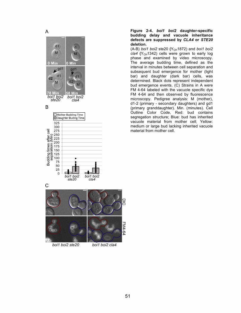
51
Figure 2-4. boi1 boi2 daughter-specific budding delay and vacuole inheritance defects are suppressed by CLA4 or STE20 deletion. (A-B) boi1 boi2 ste20 (YCH1872) and boi1 boi2 cla4 (YCH1342) cells were grown to early log phase and examined by video microscopy. The average budding time, defined as the interval in minutes between cell separation and subsequent bud emergence for mother (light bar) and daughter (dark bar) cells, was determined. Black dots represent independent bud emergence events. (C) Strains in A were FM 4-64 labeled with the vacuole specific dye FM 4-64 and then observed by fluorescence microscopy. Pedigree analysis: M (mother), d1-2 (primary - secondary daughters) and gd1 (primary granddaughter). Min. (minutes). Cell Outline Color Code, Red: bud contains segregation structure; Blue: bud has inherited vacuole material from mother cell; Yellow: medium or large bud lacking inherited vacuole material from mother cell.

52
The PH domain of Boi1 is required for vacuole inheritance
Finally, in hopes of further elucidating how Boi1 and Boi2 regulated
vacuole inheritance, I determined what portion of Boi1 was necessary for vacuole
inheritance. Boi1 and Boi2 contain an SH3 domain, a proline-rich domain, and a
PH domain. To determine the region necessary for vacuole inheritance boi1 boi2
cells were transformed with plasmids containing full length BOI1 (pBOI1), BOI1
with a specific mutation in the coding sequence for the SH3 domain which coding
for a conserved tryptophan codon (pboi1-W53K), BOI1 with mutations in seven of
the nine prolines in the proline-rich domain changed to alanines (pboi1-P7A7),
BOI1 containing only the coding sequence for the PH domain and the C-terminus
of the protein (pboi1D5-733), and full length BOI1 with mutations in the PH
domain coding sequence which disrupt the PH domain of other PH domain
containing proteins 40, 55.
Vacuole inheritance was examined by FM 4-64 straining. Cells
transformed with plasmids contained wild type BOI1, BOI1 with SH3 domain or
proline-rich domains mutations, and the PH domain alone all inherited vacuoles
(Figure 2-5). In contrast, cells transformed with a BOI1 containing plasmids with
mutations affecting the PH domain alone were not able to restore vacuole
inheritance. Taken together this data suggests that the SH3 and proline-rich
domains of Boi1 are not required for vacuole inheritance, but an active PH
domain is required for vacuole inheritance in cells lacking BOI2.

53
Figure 2-5. The PH domain of Boi1 is required for vacuole inheritance in the absence of BOI2. Asynchronous cultures of FM 4-64 stained boi1 boi2 pBOI1 (YCH3576), boi1 boi2 pboi1-W53K (YCH3578), boi1 boi2 pboi1-P7A7 (YCH3580), boi1 boi2 pboi1-D5-733 (YCH3584), and boi1 boi2 pboi1-K795A,R797A (YCH3582) were examined by fluorescence microscopy, and representative pictures are shown. Cell Outline Color Code, Red: bud contains segregation structure; Blue: bud has inherited vacuole material from mother cell; Yellow: medium or large bud lacking inherited vacuole material from mother cell.

54
Discussion
I here report that boi1 boi2 mutants display a vacuole partitioning defect.
Previously vacuole partitioning mutants have been classified into three classes
based on vacuole morphology and transport. Class I mutants contain multilobed
vacuoles and fail to form a segregation structure or transport any vacuole
material into the bud. Class II mutants have moderately enlarged vacuoles and
often have a node on the vacuole oriented towards the bud. Class III mutants fail
to form lobes, contain a single highly enlarged vacuole and fail to form a
segregation structure; but they do partition a portion of the vacuole into the
daughter. Based on its vacuole morphology and failure to inherit I propose that
boi1 boi2 is a Class I vac mutant.
All of Class I vac mutants for which the corresponding gene has been
identified identify components of the physical machinery necessary for transport
of the vacuole into the bud, such as actin, profillin, and the vacuole transport
complex (Myo2, Vac8, and Vac17). Currently there are no known Class I mutants
identified that regulate the transport process. I propose Boi1 and Boi2 as the first
Class I vacuole partitioning mutants that play a role in regulating vacuole
inheritance. In support of this proposal I found that deletion of CLA4 or STE20
suppressed both the daughter-specific budding delay and the vacuole partitioning
defect of boi1 boi2 cells. Therefore, it is unlikely that, like other Class I vac
mutant, Boi1 and Boi2 are part of the physical machinery necessary for vacuole
inheritance.

55
How do Boi1 and Boi2 regulate vacuole inheritance? Boi1 and Boi2 show
multiple direct and indirect interactions with Cla4 and Ste20 as assayed by either
two-hybrid or coimmunoprecipitation 38, 40-42. Cla4 and Ste20 are autoinhibitory
and only become active by binding activated GTP-Cdc42 31, 33, 59-61. Cla4 and
Ste20 activation is facilitated by binding of the proline-rich domain in Cla4 and
Ste20 to the second SH3 domain of the adaptor protein Bem1 28-30. Bem1 also
binds to Cdc42 and its GEF Cdc24, thus bringing together GTP-Cdc42 with its
effectors Cla4 and Ste20 62-65. Importantly Boi1 and Boi2 also bind to the second
SH3 domain of Bem1 38-40 and therefore could potentially negatively regulate
Cla4 and Ste20 by competitive inhibition of PAKs for access to Bem1. In this
model the vacuole inheritance defect in boi1 boi2 cells would be caused by
overactive PAKs in the bud. I do not favor such a model since boi1 boi2 cells
expressing plasmid born BOI1 lacking the proline-rich domain necessary for
Bem1 binding do not exhibit a vacuole inheritance defect (Figure 2-5).
While boi1 boi2 pboi1-P7A7 cells did not have a vacuole inheritance
defect, I found that boi1 boi2 cells with boi1 covered by a plasmid expressing
BOI1 with a mutation in the PH domain or BOI1 containing only the PH domain
and the C-terminus of the protein did not suppress and suppressed respectively
the boi1 boi2 vacuole inheritance defect (Figure 2-5). How does the PH domain
alone act to regulate vacuole inheritance? PH domains bind
phosphatidylinositides and are the 11th most common domain in the human
genome. Studies examining the PH domain of all 33 proteins in yeast showed
that most PH domains, including the PH domains of Boi1 and Boi2, have little

56
specificity towards different phosphatidylinositide species and usually bound with
very low affinity 66. However, a growing number of examples indicate that PH
domains also bind to activated small GTPases and specifically to Rho type
GTPases, of which Cdc42 is a member 67-72. In support of this possibility, Bender
et al. show that the PH domain of Boi1 interacts with GTP but not GDP bound
Cdc42. Additionally, the overexpression of the PH domain and C-terminus of
Boi1 inhibits bud emergence and this defect is suppressed by co-overexpression
of CDC42 40. Thus Boi1 and Boi2 may negatively regulate PAKs by directly
interacting with Cdc42 and either negatively regulating Cdc42 or more likely
restricting access to Cdc42.
Formation of the tubular and vesicular segregation structure requires
PtdIns(3,5)P2. Cells defective for PtdIns(3,5)P2 production have one contiguous
vacuole stretched from the mother into the bud that is constricted at the neck 2, 73.
PtdIns(3,5)P2 is produced in response to hyperosmotic shock and leads to
vacuole fission, thus leading researchers to hypothesize that the segregation
structure is formed using the fission machinery 3, 5, 22. In addition to fission,
efficient segregation structure formation also requires the inhibition of fusion 21.
How then might deletion of Boi1 and Boi2 prevent segregation structure
formation? Cdc42 and Bem1 localize to the vacuole and promote homotypic
vacuole fusion in vitro and in vivo 74-77. Cla4 is found on isolated vacuoles and
cla4 mutants have a vacuole fusion defect in vivo 1, 78. Therefore, I suggest that
Boi1 and Boi2 negatively regulate Cla4 and Ste20 by restricting access of PAKs
to Cdc42. In boi1 boi2 cells residual Cla4 and Ste20 in the mother cell may bind

57
Cdc24 on the vacuole promoting vacuole fusion thus inhibiting vacuole
segregation structure formation. Deletion of either CLA4 or STE20 suppresses
the boi1 boi2 vacuole inheritance defect. This suggests that Cla4 and Ste20 are
required for vacuole fusion and that Cla4 and Ste20 likely have an additive or
synergistic affect on vacuole fusion.

58
References
1. Seeley, E.S., Kato, M., Margolis, N., Wickner, W. & Eitzen, G. Genomic
analysis of homotypic vacuole fusion. Mol Biol Cell 13, 782-94 (2002). 2. Bonangelino, C.J., Catlett, N.L. & Weisman, L.S. Vac7p, a novel vacuolar
protein, is required for normal vacuole inheritance and morphology. Mol Cell Biol 17, 6847-58 (1997).
3. Bonangelino, C.J. et al. Osmotic stress-induced increase of phosphatidylinositol 3,5-bisphosphate requires Vac14p, an activator of the lipid kinase Fab1p. J Cell Biol 156, 1015-28 (2002).
4. Catlett, N.L. & Weisman, L.S. The terminal tail region of a yeast myosin-V mediates its attachment to vacuole membranes and sites of polarized growth. Proc Natl Acad Sci U S A 95, 14799-804 (1998).
5. Catlett, N.L. & Weisman, L.S. Divide and multiply: organelle partitioning in yeast. Curr Opin Cell Biol 12, 509-16 (2000).
6. Hill, K.L., Catlett, N.L. & Weisman, L.S. Actin and myosin function in directed vacuole movement during cell division in Saccharomyces cerevisiae. J Cell Biol 135, 1535-49 (1996).
7. Tang, F. et al. Regulated degradation of a class V myosin receptor directs movement of the yeast vacuole. Nature (2003).
8. Ishikawa, K. et al. Identification of an organelle-specific myosin V receptor. J Cell Biol 160, 887-97 (2003).
9. Wang, Y.X., Catlett, N.L. & Weisman, L.S. Vac8p, a vacuolar protein with armadillo repeats, functions in both vacuole inheritance and protein targeting from the cytoplasm to vacuole. J Cell Biol 140, 1063-74 (1998).
10. Pan, X. & Goldfarb, D.S. YEB3/VAC8 encodes a myristylated armadillo protein of the Saccharomyces cerevisiae vacuolar membrane that functions in vacuole fusion and inheritance. J Cell Sci 111 ( Pt 15), 2137-47 (1998).
11. Lowe, M. & Barr, F.A. Inheritance and biogenesis of organelles in the secretory pathway. Nat Rev Mol Cell Biol 8, 429-39 (2007).
12. Fagarasanu, M., Fagarasanu, A. & Rachubinski, R.A. Sharing the wealth: peroxisome inheritance in budding yeast. Biochim Biophys Acta 1763, 1669-77 (2006).
13. Barr, F.A. Inheritance of the endoplasmic reticulum and Golgi apparatus. Curr Opin Cell Biol 14, 496-9 (2002).
14. Weisman, L.S. Organelles on the move: insights from yeast vacuole inheritance. Nat Rev Mol Cell Biol 7, 243-52 (2006).
15. Boldogh, I.R., Fehrenbacher, K.L., Yang, H.C. & Pon, L.A. Mitochondrial movement and inheritance in budding yeast. Gene 354, 28-36 (2005).
16. Bretscher, A. Polarized growth and organelle segregation in yeast: the tracks, motors, and receptors. J Cell Biol 160, 811-6 (2003).
17. Conradt, B., Shaw, J., Vida, T., Emr, S. & Wickner, W. In vitro reactions of vacuole inheritance in Saccharomyces cerevisiae. J Cell Biol 119, 1469-79 (1992).
59
18. Haas, A., Conradt, B. & Wickner, W. G-protein ligands inhibit in vitro reactions of vacuole inheritance. J Cell Biol 126, 87-97 (1994).
19. Haas, A. & Wickner, W. Organelle inheritance in a test tube: the yeast
vacuole. Seminars in Cell & Developmental Biology 7, 517-24 (1996). 20. Xu, Z. & Wickner, W. Thioredoxin is required for vacuole inheritance in
Saccharomyces cerevisiae. J Cell Biol 132, 787-94 (1996). 21. LaGrassa, T.J. & Ungermann, C. The vacuolar kinase Yck3 maintains
organelle fragmentation by regulating the HOPS tethering complex. J Cell Biol 168, 401-14 (2005).
22. Weisman, L.S. Yeast vacuole inheritance and dynamics. Annu Rev Genet 37, 435-60 (2003).
23. Moseley, J.B. & Goode, B.L. The yeast actin cytoskeleton: from cellular function to biochemical mechanism. Microbiol Mol Biol Rev 70, 605-45 (2006).
24. Park, H.O. & Bi, E. Central roles of small GTPases in the development of cell polarity in yeast and beyond. Microbiol Mol Biol Rev 71, 48-96 (2007).
25. Zheng, Y., Bender, A. & Cerione, R.A. Interactions among proteins involved in bud-site selection and bud-site assembly in Saccharomyces cerevisiae. J Biol Chem 270, 626-30 (1995).
26. Toenjes, K.A., Sawyer, M.M. & Johnson, D.I. The guanine-nucleotide-exchange factor Cdc24p is targeted to the nucleus and polarized growth sites. Curr Biol 9, 1183-6 (1999).
27. Leeuw, T. et al. Interaction of a G-protein beta-subunit with a conserved sequence in Ste20/PAK family protein kinases. Nature 391, 191-5 (1998).
28. Winters, M.J. & Pryciak, P.M. Interaction with the SH3 domain protein Bem1 regulates signaling by the Saccharomyces cerevisiae p21-activated kinase Ste20. Mol Cell Biol 25, 2177-90 (2005).
29. Gulli, M.P. et al. Phosphorylation of the Cdc42 exchange factor Cdc24 by the PAK-like kinase Cla4 may regulate polarized growth in yeast. Mol Cell 6, 1155-67 (2000).
30. Bose, I. et al. Assembly of scaffold-mediated complexes containing Cdc42p, the exchange factor Cdc24p, and the effector Cla4p required for cell cycle-regulated phosphorylation of Cdc24p. J Biol Chem 276, 7176-86 (2001).
31. Cvrckova, F., De Virgilio, C., Manser, E., Pringle, J.R. & Nasmyth, K. Ste20-like protein kinases are required for normal localization of cell growth and for cytokinesis in budding yeast. Genes Dev 9, 1817-30 (1995).
32. Butty, A.C. et al. A positive feedback loop stabilizes the guanine-nucleotide exchange factor Cdc24 at sites of polarization. Embo J 21, 1565-76 (2002).
33. Simon, M.N. et al. Role for the Rho-family GTPase Cdc42 in yeast mating-pheromone signal pathway. Nature 376, 702-5 (1995).
34. Zhao, Z.S., Leung, T., Manser, E. & Lim, L. Pheromone signalling in Saccharomyces cerevisiae requires the small GTP-binding protein Cdc42p and its activator CDC24. Mol Cell Biol 15, 5246-57 (1995).

60
35. Chiroli, E. et al. Budding yeast PAK kinases regulate mitotic exit by two different mechanisms. J Cell Biol 160, 857-74 (2003).
36. Weiss, E.L., Bishop, A.C., Shokat, K.M. & Drubin, D.G. Chemical genetic analysis of the budding-yeast p21-activated kinase Cla4p. Nat Cell Biol 2, 677-85 (2000).
37. Schmidt, M., Varma, A., Drgon, T., Bowers, B. & Cabib, E. Septins, under Cla4p regulation, and the chitin ring are required for neck integrity in budding yeast. Mol Biol Cell 14, 2128-41 (2003).
38. Matsui, Y., Matsui, R., Akada, R. & Toh-e, A. Yeast src homology region 3 domain-binding proteins involved in bud formation. J Cell Biol 133, 865-78 (1996).
39. Hertveldt, K., Robben, J. & Volckaert, G. Whole genome phage display selects for proline-rich Boi polypeptides against Bem1p. Biotechnol Lett 28, 1233-9 (2006).
40. Bender, L. et al. Associations among PH and SH3 domain-containing proteins and Rho-type GTPases in Yeast. J Cell Biol 133, 879-94 (1996).
41. Ptacek, J. et al. Global analysis of protein phosphorylation in yeast. Nature 438, 679-84 (2005).
42. Drees, B.L. et al. A protein interaction map for cell polarity development. J Cell Biol 154, 549-71 (2001).
43. Norden, C. et al. The NoCut pathway links completion of cytokinesis to spindle midzone function to prevent chromosome breakage. Cell 125, 85-98 (2006).
44. Burke, D., Dawson, D. & Stearns, T. Methods in yeast genetics: a Cold Spring Harbor Larboratory course manual (Cold Spring Harbor Laboratory Press, Cold Spring Harbor, 2000).
45. Cormack, B.P., Valdivia, R.H. & Falkow, S. FACS-optimized mutants of the green fluorescent protein (GFP). Gene 173, 33-8 (1996).
46. Aitchison, J.D., Rout, M.P., Marelli, M., Blobel, G. & Wozniak, R.W. Two novel related yeast nucleoporins Nup170p and Nup157p: complementation with the vertebrate homologue Nup155p and functional interactions with the yeast nuclear pore-membrane protein Pom152p. J Cell Biol 131, 1133-48 (1995).
47. Sambrook, J. & Russell, D.W. Molecular Cloning: a laboratory manual (Cold Spring Harbor Laboratory Press, Cold Spring Harbor, 2001).
48. Waddle, J.A., Karpova, T.S., Waterston, R.H. & Cooper, J.A. Movement of cortical actin patches in yeast. J Cell Biol 132, 861-70 (1996).
49. Saito, T.L. et al. SCMD: Saccharomyces cerevisiae Morphological Database. Nucleic Acids Res 32, D319-22 (2004).
50. Brachmann, C.B. et al. Designer deletion strains derived from Saccharomyces cerevisiae S288C: a useful set of strains and plasmids for PCR-mediated gene disruption and other applications. Yeast 14, 115-32 (1998).
51. Winzeler, E.A. et al. Functional characterization of the S. cerevisiae genome by gene deletion and parallel analysis. Science 285, 901-6 (1999).

61
52. Bender, A. & Pringle, J.R. Use of a screen for synthetic lethal and multicopy suppressee mutants to identify two new genes involved in morphogenesis in Saccharomyces cerevisiae. Mol Cell Biol 11, 1295-305 (1991).
53. Sikorski, R.S. & Hieter, P. A system of shuttle vectors and yeast host strains designed for efficient manipulation of DNA in Saccharomyces cerevisiae. Genetics 122, 19-27 (1989).
54. Vida, T.A. & Emr, S.D. A new vital stain for visualizing vacuolar membrane dynamics and endocytosis in yeast. J Cell Biol 128, 779-92 (1995).
55. Hallett, M.A., Lo, H.S. & Bender, A. Probing the importance and potential roles of the binding of the PH-domain protein Boi1 to acidic phospholipids. BMC Cell Biol 3, 16 (2002).
56. Gomes De Mesquita, D.S. et al. Vacuole segregation in the Saccharomyces cerevisiae vac2-1 mutant: structural and biochemical quantification of the segregation defect and formation of new vacuoles. Yeast 13, 999-1008 (1997).
57. Boldogh, I.R., Ramcharan, S.L., Yang, H.C. & Pon, L.A. A type V myosin (Myo2p) and a Rab-like G-protein (Ypt11p) are required for retention of newly inherited mitochondria in yeast cells during cell division. Mol Biol Cell 15, 3994-4002 (2004).
58. Fagarasanu, M., Fagarasanu, A., Tam, Y.Y., Aitchison, J.D. & Rachubinski, R.A. Inp1p is a peroxisomal membrane protein required for peroxisome inheritance in Saccharomyces cerevisiae. J Cell Biol 169, 765-75 (2005).
59. Lamson, R.E., Winters, M.J. & Pryciak, P.M. Cdc42 regulation of kinase activity and signaling by the yeast p21-activated kinase Ste20. Mol Cell Biol 22, 2939-51 (2002).
60. Benton, B.K., Tinkelenberg, A., Gonzalez, I. & Cross, F.R. Cla4p, a Saccharomyces cerevisiae Cdc42p-activated kinase involved in cytokinesis, is activated at mitosis. Mol Cell Biol 17, 5067-76 (1997).
61. Bokoch, G.M. Biology of the p21-Activated Kinases. Annu Rev Biochem (2003).
62. Leeuw, T. et al. Pheromone response in yeast: association of Bem1p with proteins of the MAP kinase cascade and actin. Science 270, 1210-3 (1995).
63. Peterson, J. et al. Interactions between the bud emergence proteins Bem1p and Bem2p and Rho-type GTPases in yeast. J Cell Biol 127, 1395-406 (1994).
64. Ito, T., Matsui, Y., Ago, T., Ota, K. & Sumimoto, H. Novel modular domain PB1 recognizes PC motif to mediate functional protein-protein interactions. Embo J 20, 3938-46 (2001).
65. Yamaguchi, Y., Ota, K. & Ito, T. A novel Cdc42-interacting domain of the yeast polarity establishment protein Bem1. Implications for modulation of mating pheromone signaling. J Biol Chem 282, 29-38 (2007).
66. Yu, J.W. et al. Genome-wide analysis of membrane targeting by S. cerevisiae pleckstrin homology domains. Mol Cell 13, 677-88 (2004).

62
67. Lemmon, M.A. Pleckstrin homology domains: not just for phosphoinositides. Biochem Soc Trans 32, 707-11 (2004).
68. Lemmon, M.A. Pleckstrin homology (PH) domains and phosphoinositides. Biochem Soc Symp, 81-93 (2007).
69. Rossman, K.L. et al. A crystallographic view of interactions between Dbs and Cdc42: PH domain-assisted guanine nucleotide exchange. Embo J 21, 1315-26 (2002).
70. Rossman, K.L. et al. Functional analysis of cdc42 residues required for Guanine nucleotide exchange. J Biol Chem 277, 50893-8 (2002).
71. Kim, O., Yang, J. & Qiu, Y. Selective activation of small GTPase RhoA by tyrosine kinase Etk through its pleckstrin homology domain. J Biol Chem 277, 30066-71 (2002).
72. Jaffe, A.B., Aspenstrom, P. & Hall, A. Human CNK1 acts as a scaffold protein, linking Rho and Ras signal transduction pathways. Mol Cell Biol 24, 1736-46 (2004).
73. Yamamoto, A. et al. Novel PI(4)P 5-kinase homologue, Fab1p, essential for normal vacuole function and morphology in yeast. Mol Biol Cell 6, 525-39 (1995).
74. Xu, H. & Wickner, W. Bem1p is a positive regulator of the homotypic fusion of yeast vacuoles. J Biol Chem 281, 27158-66 (2006).
75. Han, B.K. et al. Bem1p, a scaffold signaling protein, mediates cyclin-dependent control of vacuolar homeostasis in Saccharomyces cerevisiae. Genes Dev 19, 2606-18 (2005).
76. Eitzen, G., Thorngren, N. & Wickner, W. Rho1p and Cdc42p act after Ypt7p to regulate vacuole docking. Embo J 20, 5650-6 (2001).
77. Muller, O., Johnson, D.I. & Mayer, A. Cdc42p functions at the docking stage of yeast vacuole membrane fusion. Embo J 20, 5657-65 (2001).
78. Eitzen, G., Wang, L., Thorngren, N. & Wickner, W. Remodeling of organelle-bound actin is required for yeast vacuole fusion. J Cell Biol 158, 669-79 (2002).

63
CHAPTER III
THE P21-ACTIVATED KINASES CLA4 AND STE20 REGULATE THE DESTRUCTION OF THE VACUOLE SPECIFIC MYOSIN RECEPTOR VAC17
Abstract
The budding yeast S. cerevisiae vacuole (lysosome) is transported along
actin cables by the myosin V motor, Myo2, bound to the vacuole-specific myosin
receptor Vac17 1, 2. Directionality in vacuole transport is ensured by the bud-
specific degradation of Vac17. It has been proposed that bud-specific
degradation of Vac17 is promoted by proteins localized to and/or activated solely
in the bud 3. However, these factors have not been identified. The p21-activated
kinases (PAKs) Cla4 and Ste20 are localized to and are activated in the bud, and
overexpression of either inhibited vacuole inheritance. Inhibition of vacuole
inheritance by CLA4 or STE20 overexpression was suppressed by the
expression of non-degradable VAC17. Additionally, PAK activity was required for
Vac17 degradation in late-M and CLA4 overexpression promoted Vac17
degradation. I conclude that Cla4 and Ste20 are the bud-specific proteins that
promote the bud-specific degradation of Vac17.
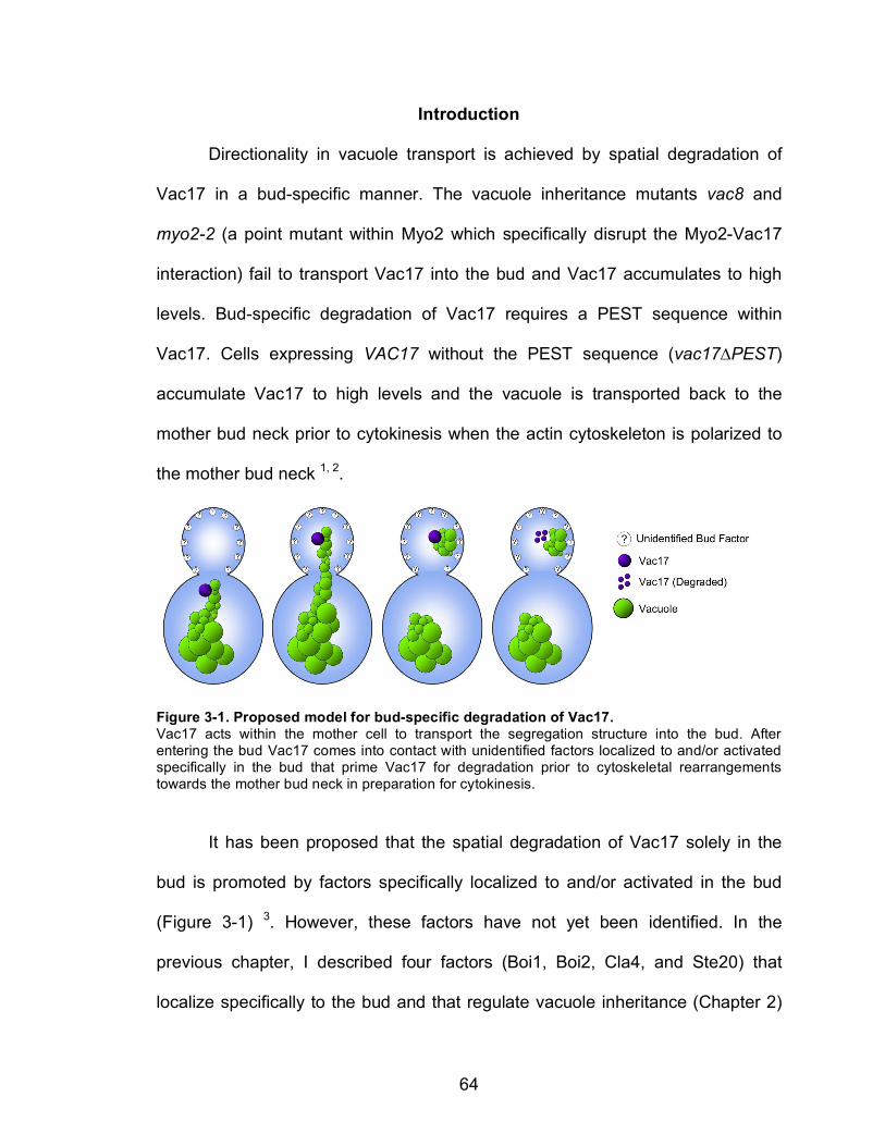
64
Introduction
Directionality in vacuole transport is achieved by spatial degradation of
Vac17 in a bud-specific manner. The vacuole inheritance mutants vac8 and
myo2-2 (a point mutant within Myo2 which specifically disrupt the Myo2-Vac17
interaction) fail to transport Vac17 into the bud and Vac17 accumulates to high
levels. Bud-specific degradation of Vac17 requires a PEST sequence within
Vac17. Cells expressing VAC17 without the PEST sequence (vac17#PEST)
accumulate Vac17 to high levels and the vacuole is transported back to the
mother bud neck prior to cytokinesis when the actin cytoskeleton is polarized to
the mother bud neck 1, 2.
Figure 3-1. Proposed model for bud-specific degradation of Vac17. Vac17 acts within the mother cell to transport the segregation structure into the bud. After entering the bud Vac17 comes into contact with unidentified factors localized to and/or activated specifically in the bud that prime Vac17 for degradation prior to cytoskeletal rearrangements towards the mother bud neck in preparation for cytokinesis.
It has been proposed that the spatial degradation of Vac17 solely in the
bud is promoted by factors specifically localized to and/or activated in the bud
(Figure 3-1) 3. However, these factors have not yet been identified. In the
previous chapter, I described four factors (Boi1, Boi2, Cla4, and Ste20) that
localize specifically to the bud and that regulate vacuole inheritance (Chapter 2)

65
4-8. Here, I provide evidence that the p21-activated kinases Cla4 and Ste20 are
bud-localized factors promoting Vac17 degradation.

66
Materials and Methods
Table 3-1. Strains
Strain Genotype Source
W303-1A MATa ade2-1 his3-11,15 leu2-3,112 trp1-1 ura3-1 (W303) 9
YCH4774 MATa VAC17-ProA-HIS3-URA3 (W303) This Study YCH4826 MATa boi1::LEU2 boi2::URA3 vac17::kanMX4 pRS313-pVAC17-
VAC17 (W303) This Study
YCH4827 MATa boi1::LEU2 boi2::URA3 vac17::kanMX4 pRS313-pVAC17-vac17DPEST (W303)
This Study
YCH4837 MATa pRS314-GAL-GST-STE20 (W303) This Study YCH4843 MATa pRS314-GAL-GST-STE20 VAC17-ProA-HIS3-URA3 (W303) This Study YCH4852 MATa cdc14-1 VAC17-ProA-HIS3-URA3 (W303) This Study YCH4862 MATa cdc15-2 VAC17-ProA-HIS3-URA3 (W303) This Study YCH4869 MATa dbf2-2 VAC17-ProA-HIS3-URA3 (W303) This Study YCH4882 MATa his3::GAL-CLA4-HIS3 VAC17-ProA-HIS3-URA3 (W303) This Study YCH4893 MATa ura3::4X URA3::GAL1-CLA4t swe1::LEU2 VAC17-ProA-
HIS3-URA3 (W303) This Study
YCH4894 MATa ura3::4X URA3::GAL1-CLA4t VAC17-ProA-HIS3-URA3 (W303)
This Study
YCH4912 MATa his3::GAL-CLA4-HIS3 ura3::GFP-PTS1-URA3 (W303) This Study YCH4914 MATa ura3::GFP-PTS1-URA3 (W303) This Study YCH4974 MATa his3::GAL-CLA4-HIS3 vac17::kanMX4 pRS414-pVAC17-
VAC17 (W303) This Study
YCH4975 MATa his3::GAL-CLA4-HIS3 vac17::kanxMX4 pRS414-pVAC17-
vac17!PEST (W303)
This Study
YCH4978 MATa SEC7-GFP-HIS3 pRS316 (S288c) This Study YCH4980 MATa SEC7-GFP-HIS3 pRS31-GAL-GST-STE20 (S288c) This Study YCH4992 MATa pRS316-GST-STE20 vac17::kanMX4 pRS414-pVAC17-
VAC17 (W303) This Study
YCH4993 MATa pRS316-GST-STE20 vac17::kanMX4 pRS414-pVAC17-
vac17!PEST (W303)
This Study
YCH5162 MATa PDS1-HA-LEU2::leu2 VAC17-ProA-HIS3 (W303) This Study YMG694 MATa bar1-1 his3::GAL-CLA4-HIS3 (W303)
10
Plasmid and strain construction
Yeast strains and sources are listed in Table 3-1. Yeast strains were
constructed by genetic crosses followed by tetrad dissection or by transformation
using the lithium acetate method 11. ProA and the adjacent HIS3 and URA3
marker were amplified by PCR and integrated to generate VAC17-ProA as
described 12. Plasmid sources and construction methods are listed in Table 3-2
and DNA manipulations were performed as described 13.

67
Table 3-2. Plasmids Plasmid Description Source
pCH1915 pRS414-pVAC17-VAC17. Made by insertion of 1.94 kB BamHI-SalI fragment of pVAC17
1 into the BamHI-SalI
site of pRS414 14
.
This Study
pCH1916 pRS414-pVAC17-vac17#PEST Made by insertion of 1.75 kB BamHI-SalI fragment of pVAC17($97-259)
1 into
the BamHI-SalI site of pRS414 14
.
This Study
pCH1924 pRS313-pVAC17-VAC17 Made by insertion of 1.94 kB BamHI-SalI fragment of pVAC17
1 into the BamHI-SalI
site of pRS313 14
.
This Study
pCH1925 pRS313-pVAC17-vac17#PEST. Made by insertion of 1.75 kB BamHI-SalI fragment of pVAC17($97-259)
1 into
the BamHI-SalI site of pRS414 14
.
This Study
pCH1926 pRS314-GAL-GST-STE20. Made by insertion of a 4.53 kB SacII-KpnI fragment of pRD20-STE20-ATG
8 into the
SacII-KpnI site of pRS314 14
.
This Study
pRD20-STE20-ATG pRS316-GST-STE20. 8
pRS316 pRS316 14
Microscopy
Digital images were taken with a 100X objective on an Olympus
microscope. NIH image 1.62 (written by Wayne Rasband) was used for image
acquisition. Cells were visualized by fluorescence and differential interference
contrast microscopy (DIC). Within each experiment, all images were collected
and scaled identically. Images were processed with Photoshop 9.0 software
(Adobe). Video microscopy was performed with mid-log phase cells placed on a
drop of 2% agarose in YPD was flattened by an uncoated glass slide as
described 15. Cells were visualized every three to five minutes. Scale bar = 2.5
#m throughout all figures.
In vivo labeling of vacuoles and nuclei staining
To visualize vacuoles, yeast cells were concentrated and incubated for 1
hour with N-(3-triethylammoniumpropyl)-4(6(4(diethylamino)phenyl)hexatrienyl)

68
pyridium dibromide (FM 4-64; Molecular Probes) at a final concentration of 16.5
#M 16. Cells were then washed with appropriate media and grown for $3 hours
before being viewed by fluorescence microscopy. Nuclei were visualized by
fixation in 70% ethanol followed by staining with DAPI at 0.1 mg/ml.
Sizing cells and quantification of organelle inheritance
Small, medium, and large budded cells were grouped into size categories
as described 17. Peroxisomes and late Golgi inheritance were determined by
fluorescence microscopy and bud sizes were determined by measuring the cross
sectional area of mothers and buds using NIH-Image 1.62. Each cell was
grouped into a category according to bud cross-sectional area expressed as a
percentage of mother cell cross-sectional area. Bud size categories for late Golgi
inheritance were: category I 4.0 -7.4%, II, 7.5–10.9%; category III, 11.0–14.4%;
category IV, 14.5–17.9%; and category V, 18.0–21.4% as previously described
18. Bud size categories for peroxisome inheritance were: category I, 0–24%;
category II, 24–39%; category III, 39–50%; category IV, 50–61%, and category V
61-100% as previously described 19. Six different focal planes were examined for
each bud to quantify organelle inheritance.
SDS-PAGE and western blot analysis
Yeast protein levels and gel mobility were assayed by TCA precipitation
followed by SDS-PAGE (7.5%) and western blot analysis using Rabbit-a-Mouse

69
IgG 1:1000 (Promega), Mouse-a-HA 16B12 1:1000 (Covance), and Goat-a-Actin
antibodies 1:2000.
Vac17-ProA immunoprecipitation and phosphatase treatment
Pelleted cells where resuspended in lysis buffer (20mM Tris-HCl (pH 6.5),
5mM MgCl2, 2% Triton X-100, 150 mM NaCl, 1x protease inhibitor (Roche), 1mM
PMSF, 5mM EDTA, 50 mM NaF, 10 mM Na4P2O7, 0.5mM NaVO4, and 1X
phosphatase inhibitor (Calbiochem)). Cells were lysed using a French Press,
lysates were incubated with 300#l of IgG sepharose beads (Amersham
Biosciences), and washed with wash buffer (20mM Tris-HCl (pH6.8), 5mM
MgCl2, 2% Triton X-100, 150 mM NaCl). Immunoprecipitates were then split and
treated with l-protein phosphatase (NEB) and phosphatase inhibitor
(Calbiochem) as indicated.
Cell cycle arrests
Cell cycle arrests were performed by the addition a-factor (ZymoResearch) or
nocodazole (Sigma) at the final concentration of 3 #M and 15 #g/ml respectively
as described or by shifting temperature sensitive mutants from the permissive
temperature (23°C) to the nonpermissive temperature (37°C) for three hours.
Conditions for growth and release of synchronous cultures from arrest by a-factor
were as described 20.

70
Other methods
YP media containing 1% yeast extract and 2% Bacto Peptone was used
with addition of glucose (YPD), raffinose (YPR), and/or galactose (YPG) as
carbon sources at 2% final concentration. Alternately, strains with plasmids were
grown on synthetic media (SC) lacking tryptophan, uracil, or histidine to maintain
plasmids.

71
Results
Cells overexpressing CLA4 have a daughter-specific budding delay and a vacuole inheritance defect
I have previously shown that boi1 boi2 and vac8 mutant cells had a
daughter-specific budding delay (Figure 2-2) and boi1 boi2 cells had a vacuole
inheritance defect suppressed by CLA4 (Figure 2-4). In light of those results, I
hypothesized that Cla4 regulates vacuole inheritance. To test this hypothesis I
determined if cells overexpressing CLA4 had a daughter-specific budding delay
and/or a vacuole inheritance defect like boi1 boi2 and vac8 cells.
CLA4 overexpression from a MET promoter causes defects in growth and
actin polymerization 10. However, overexpression of CLA4 from the GAL
promoter integrated at the HIS3 locus was not lethal and cells grew well on
YP+2%Galactose plates (data not shown). I used this strain to determine if CLA4
overexpressing cells had a daughter-specific budding delay like boi1 boi2 and
vac8 (Figure 2-2).
Wild type and GAL-CLA4 cells were observed by time lapse video
microscopy with cells grown on agar pads containing YP+2% glucose or YP+2%
galactose to induce CLA4 overexpression. Cell division led to the production of a
larger mother and a smaller daughter cell. After cell separation, wild type mother
cells, grown on media containing either glucose or galactose quickly rebudded
(Figure 3-2A). Smaller daughter cells grown on either glucose or galactose
budded with similar kinetics but were delayed for bud emergence an average of
48 and 65 minutes respectively while they grew to a critical size (Figure 3-2A).

72
GAL-CLA4 mother cells grown on either glucose or galactose budded with similar
kinetics to wild type mother cells. In contrast the GAL-CLA4 daughter cells grown
on either glucose or galactose did not bud with similar kinetics and daughter cells
overexpressing CLA4 were delayed on average 135 minutes longer than GAL-
CLA4 daughter cells grown on glucose (Figure 3-2).
Figure 3-2. Cells overexpressing CLA4 have a daughter-specific budding delay. (A-B) Wild type (W303-1A) and GAL-CLA4 (YMG694) cells were grown to early log phase in either YP+2% Glucose or YP+2% Galactose and where examined by video microscopy. The budding time, defined as the interval in minutes between cell separation and subsequent bud emergence for mother (light bar) and daughter (dark bar) cells, was determined. Black dots represent independent bud emergence events. Pedigree analysis was performed. Pedigree analysis: M (mother), d1-3 (primary - tertiary daughters) and gd1 (primary granddaughter).

73
Video microscopy of FM 4-64 stained wild type and GAL-CLA4 cells was
performed to determine if CLA4 overexpression inhibited vacuole inheritance.
Wild type (data not shown) and GAL-CLA4 cells were grown on YP+2%
galactose media. Unlike wild type cells in which segregation structures were
frequently observed and 100% of cells inherited a vacuole (data not shown),
segregation structures were formed in cells overexpressing CLA4 (Figure 3-3A).
To further explore the ability of CLA4 to inhibit vacuole inheritance, FM 4-
64 stained wild type and GAL-CLA4 cells were grown in YP+2% raffinose to mid-
log phase at which time the cultures were split and 2% galactose was added to
half the sample, and samples were examined every two hours. The presence of
a segregation structure in small and medium budded cells and the presence of a
daughter vacuole in large budded cells was scored for each sample. Segregation
structures were present in ~30% of all wild type small and medium budded cells
and 100% of all large budded cells contained a daughter vacuole in the presence
or absence of galactose throughout the time course of the experiment (data not
shown). GAL-CLA4 cells grown in non-inducing conditions had similar vacuole
inheritance percentages as wild type cells. In contrast, GAL-CLA4 cells grown
under inducing conditions exhibited a decrease in vacuole inheritance as early as
2 hours, rapidly decreasing vacuole inheritance between 2 and 4 hours, and by 6
hours post induction 0% of small and medium budded cells had segregation
structures, and only 2% of large budded cells contained a daughter vacuole
(Figure 3-3B,C).

74

75
Figure 3-3. Cells overexpressing CLA4 have a vacuole inheritance defect. (A) Video microscopy and pedigree analysis of FM 4-64 stained GAL-CLA4 (YMG694) cells was performed on cells grown in YP+2%Galactose. (B-C) FM 4-64 strained GAL-CLA4 (YMG694) cells were grown in YP+2% raffinose to mid-logarithmic stage. At time 0 2% galactose was added to half the clture and the presence of segregation structures in small and medium budded cells and of daughter vacuoles in large budded cells was quantified by fluorescence microscopy. Representative pictures at the 6 hour time point are shown. Cell Outline Color Code, Red: bud contains segregation structure; Blue: bud has inherited vacuole material from mother cell; Yellow: medium or large bud lacking inherited vacuole material from mother cell. Pedigree analysis: M (mother), d1-3 (primary - tertiary daughters).
Cells overexpressing STE20 have a vacuole inheritance defect
Cla4 and Ste20 play many unique and overlapping roles in the cell 21, 22.
Since deletion of either CLA4 or STE20 suppressed the boi1 boi2 daughter-
specific budding and vacuole inheritance defects (Figure 2-4). I therefore
hypothesized that STE20 overexpressing cells might also have a daughter-
specific budding delay and vacuole inheritance defect.
When examined, GAL-STE20 cells grown under either inducing or
noninducing conditions did not display a daughter-specific budding delay (data
not shown). However, because not all vacuole inheritance mutants have a
daughter-specific budding delay 3, I proceeded to test for a vacuole inheritance
defect. A culture of FM 4-64 stained GAL-STE20 cells was grown to mid-log
phase at which time the culture was split and 2% galactose was added to one
100% of all large budded cells contained a daughter vacuole throughout the time
course of the experiment (Figure 3-4). GAL-STE20 cells grown under inducing
conditions had decreasing amounts of vacuole inheritance as early as 2 hours,
rapidly decreasing vacuole inheritance between 2 and 4 hours, and by 6 hours
post induction only 4% of small and medium budded cells had a segregation

76
structure and only 18% of large-budded cells had a discrete daughter vacuole
(Figure 3-4).
Figure 3-4. Cells overexpressing STE20 have a vacuole inheritance defect. (A-B) FM 4-64 strained GAL-STE20 (YCH4837) cells were grown in SC-TRP+2% raffinose to mid-logarithmic stage. At time 0 2% galactose was added to half the culture and the presence of segregation structures in small and medium budded cells and of daughter vacuoles in large budded cells was quantified by fluorescence microscopy. Representative pictures at the 6 hour time point are shown. Cell Outline Color Code, Red: bud contains segregation structure; Blue: bud has inherited vacuole material from mother cell; Yellow: medium or large bud lacking inherited vacuole material from the mother cell.

77
vac17!PEST expression suppresses the vacuole inheritance defect of
CLA4 or STE20 overexpressing and boi1 boi2 cells One possible explanation for the vacuole inheritance defect exhibited in
cells overexpressing CLA4 or STE20 was that the PAKs are the bud-specific
factors that promote Vac17 degradation. I hypothesized that CLA4 or STE20
overexpression caused a vacuole inheritance defect by promoting the premature
degradation of Vac17. If this model was correct then I would have expected that
expression of the non-degradable VAC17 (vac17!PEST) should suppress the
vacuole inheritance defect caused by CLA4 or STE20 overexpression.
FM 4-64 stained GAL-CLA4 vac17 and GAL-STE20 vac17 cells covered
by a plasmid containing VAC17 or vac17!PEST were examined for vacuole
inheritance. As expected 100% of all GAL-CLA4 and GAL-STE20 large budded
cells contained a daughter vacuole in the presence of VAC17 or vac17!PEST
under noninducing conditions. Under inducing conditions GAL-CLA4 and GAL-
STE20 cells containing VAC17 exhibited vacuole inheritance defects after 6
hours of overexpression. However expression of vac17!PEST fully suppressed
the vacuole inheritance defect in GAL-CLA4 and GAL-STE20 cells under
inducing conditions (Table 3-3).
I have previously shown that boi1 boi2 exhibited a vacuole inheritance
defect suppressible by CLA4 or STE20 deletion (Table 2-3). To further test my
hypothesis I determined if the boi1 boi2 vacuole inheritance defect was
suppressible by vac17!PEST expression. FM 4-64 stained boi1 boi2 vac17
covered by a VAC17 containing plasmid displayed vacuole inheritance defects as

78
expected (Table 3-3). Expression of vac17!PEST in the boi1 boi2 vac17 cells
fully suppressed the boi1 boi2 vacuole inheritance defect (Table 3-3).
Table 3-3. vac17#PEST expression suppresses the vacuole inheritance defect of CLA4 or STE20 overexpressing or boi1 boi2 cells.
Genotype Vacuole Inheritance of Large Budded Cells
GAL-CLA4 vac17 pVAC17 (-GAL) 100% GAL-CLA4 vac17 pvac17#PEST (-GAL) 100% GAL-CLA4 vac17 pVAC17 (+GAL) 2% GAL-CLA4 vac17 pvac17#PEST (+GAL) 100% GAL-STE20 vac17 pVAC17 (-GAL) 100% GAL-STE20 vac17 pvac17#PEST (-GAL) 100% GAL-STE20 vac17 pVAC17 (+GAL) 19% GAL-STE20 vac17 pvac17#PEST (+GAL) 100% boi1 boi2 vac17 pVAC17 15%
boi1 boi2 vac17 pvac17!PEST 100%
The ability of vac17!PEST expression to suppress the vacuole inheritance
defect of CLA4 or STE20 overexpressing and boi1 boi2 cells provided me with
support for a model that PAKs are the bud specific factors that promote Vac17
degradation. I then set out to test two predictions of this model. (1) If PAKs are
required for Vac17 degradation then cells lacking PAK activity should not
degrade Vac17. (2) Cells overexpressing CLA4 or STE20 should degrade Vac17.
However, to perform these experiments further information was required
concerning the timing of Vac17 degradation.

79
Vac17 is a phosphoprotein degraded in late-M phase
Vac17 levels oscillate during the cell cycle. Vac17 levels increase as cells
bud and decreases prior to cytokinesis 1. I examined Vac17 levels throughout the
cell cycle to further characterize when Vac17 was degraded.
I tagged Vac17 with a ProA tag. Vacuole inheritance was assayed in
VAC17-ProA cells and the kinetics of vacuole inheritance were identical to wild
type cells suggesting that Vac17-ProA was functional (data not shown). Vac17-
ProA levels were then examined in asynchronous cells by SDS-PAGE followed
by western-blot analysis. I found that Vac17 ran as multiple bands suggesting the
possibility that Vac17-ProA was a phosphoprotein (Figure 3-5A). Vac17-ProA
was immunoprecipitated and treated with %-phosphatase or %-phosphatase plus
phosphatase inhibitor. Vac17-ProA treated with %-phosphatase collapsed down
to a single band and phosphatase inhibitors blocked this collapse (Figure 3-5B).
The timing of Vac17 degradation was further examined during the cell
cycle. VAC17-ProA PDS1-HA cells were arrested in G1 with a-factor and
released. Samples were taken every 10 minutes, and Vac17 and Pds1 protein
levels were examined throughout a single cell cycle. Pds1 is degraded as cells
enter anaphase 23 and Vac17 degradation occurred after this point (Figure 3-5C).
Additionally, phosphorylated Vac17 appeared common in G2/M cells arrested
with nocodazole (Figure 3-5D). Together these data suggested that Vac17
destruction did not occur until after entry into anaphase.

80
Figure 3-5. Vac17 is a phosphoprotein and is degraded in late-M phase. (A) Vac17-ProA and actin proteins were examined by western blot analysis in untagged (W303-1A) and VAC17-ProA (YCH4774) cells. (B) Vac17-ProA was immunoprecipitated from protein extracts of 4X GAL-CLA4t swe1! VAC17-ProA (YCH4893) or untagged (UT) 4X GAL-CLA4t swe1! (Piatti2711) strains grown on YP+2%galactose media for 6 hours. Immunoprecipitated
material was split and treated with buffer alone, %-phosphatase, or %-phosphatase plus
phosphatase inhibitors. (B) Vac17-ProA and actin proteins were examined by western blot analysis in untagged (W303-1A) and VAC17-ProA (YCH4774) cells. (C) VAC17-ProA PDS1-HA (YCH5162) cells were arrested in G1 with a-factor, released, samples were taken every 10 minutes, and Vac17-ProA and Pds1-HA were examined by western blot analysis. (D) Vac17-ProA levels were assayed by western blot in cdc15-2 (YCH4862), dbf2-2 (YCH4869), and cdc14-1 (YCH4852) cells grown at 23°C or arrested at telophase in 37°C media. Additionally, Vac17-ProA levels were examined in asynchronous and nocodazole arrested cells (YCH4774).
Because Vac17 degradation occurred after anaphase onset I examined if
mitotic exit was required for Vac17 degradation. CDC15, DBF2, and CDC14 are
required for mitotic exit and temperature sensitive mutants in these genes arrest
prior to mitotic exit 24, 25. Vac17-ProA levels were examined in cdc15-2, dbf2-2,
and cdc14-1 cells grown at the permissive and non-permissive temperature to
arrest cells at mitotic exit. Phosphorylated Vac17-ProA was common in cells
grown at the non-permissive temperature. However, cells arrested at mitotic exit
only contained a small amount of what appeared to be unphosphorylated Vac17

81
(Figure 3-5D). This suggested that Vac17 was destroyed after anaphase entry
and before mitotic exit during Late-M phase.
PAK function is required for Vac17-ProA degradation in late-M
If PAKs are required for Vac17 degradation, then in the absence of Cla4
and Ste20 activity Vac17-ProA should not be degraded. A CLA4 allele with a
truncated C-terminus (CLA4t) when overexpressed causes Cla4 and Ste20 to
delocalize from the bud cortex and cells display similar defect as seen in cells
lacking PAKs 26. 4X GAL-CLA4t VAC17-ProA cells were arrested in G1 and
released, and Vac17 levels and mitotic progression were examined. As in wild
type cells (Figure 3-5C), Vac17-ProA is phosphorylated and cells arrest at G2/M
in a SWE1 dependent manner (Figure 3-6A). Since Vac17 is stable at G2/M, the
same experiment was performed in the absence of SWE1, a situation in which
cells progress to Late-M when Vac17 protein levels were low (Figure 3-5D). I
observed that even though cells progressed to late-M, Vac17-ProA levels
remained high with large amounts of phosphorylated Vac17 present (Figure 3-
6B). Thus I concluded that active PAKs were required for Vac17 degradation in
late-M.
CLA4 overexpression promotes Vac17-ProA degradation
After examining the requirement for PAKs in Vac17 degradation I
determined if PAK overexpression caused Vac17 degradation. Asynchronous
cultures of wild type and GAL-CLA4 cells were grown to mid-log phase, 2%

82
galactose was added at the zero time point, and samples were taken every hour
for examination of Vac17-ProA levels. Over the 6 hour time course of the
experiment, Vac17 remained constant in wild type cells (Figure 3-6D). However,
Vac17-ProA levels decreased in GAL-CLA4 cells over the 6 hours time course. A
reduction in Vac17-ProA levels was observed as early as 2 hours with the
phosphorylated Vac17-ProA disappearing quickest and Vac17-ProA levels
reached very low levels at the six hour time point (Figure 3-6C). Importantly the
decrease in Vac17-ProA levels occurred with similar kinetics to vacuole
inheritance defects (Figure 3-3C). Therefore, CLA4 overexpressing cells have a
severe vacuole inheritance defect like the Class I vacuole inheritance mutants
vac8 and myo2-2 which fail to inherit vacuoles. However, unlike vac8 and myo2-
2 cells, CLA4 overexpressing cells do not accumulate Vac17 to high levels like
other Class I vacuole inheritance mutants 1, but instead Vac17 levels drop after
CLA4 overexpression is initiated. Thus I concluded that CLA4 promotes Vac17
degradation.
Additionally the ability of STE20 to promote Vac17-ProA degradation was
examined. Asynchronous cultures of GAL-STE20 were grown under inducing
conditions, and while vacuole inheritance defects were seen as in Figure 3-4B
(data not shown), Vac17-ProA levels remained constant throughout the six hour
time course of the experiment (Figure 3-6E).

83
Figure 3-6. PAK function is required for Vac17-ProA degradation in late-M and CLA4 overexpression promotes Vac17-ProA degradation. (A-B) 4X GAL-CLA4t VAC17-ProA (YCH4894) and 4X GAL-CLA4t swe1! VAC17-ProA (YCH4893)
were a-factor arrested, released, samples taken at the indicated times, and subjected to western blot analysis. Additionally the percentage of telophase cells was determined by DAPI staining and scoring the number of cells with mother and bud nuclei staining. (C-D) Mid-logarithmic phase GAL-CLA4 VAC17-ProA (YCH4882) and VAC17-ProA (YCH4774) were grown in YP+2% raffinose. At the 0 time point 2% galactose was added to induce CLA4 overexpression, samples were taken every hour, and western blot analysis was performed. (E) Mid-logarithmic phase GAL-STE20 VAC17-ProA (YCH4843) was grown in SC-TRP+2% raffinose. At the 0 time point 2% galactose was added to induce STE20 overexpression, samples were taken every hour, and western blot analysis was performed.

84
CLA4 or STE20 overexpression inhibits peroxisome but not late Golgi inheritance In addition to the yeast vacuole, Myo2 transports the late Golgi and
peroxisomes along actin cables into the bud 18, 27. I therefore investigated if
CLA4 or STE20 overexpression perturbed either late Golgi or peroxisome
inheritance as they had vacuole inheritance.
Inheritance of the late Golgi was investigated by examination of SEC7-
GFP cells. The SEC7 gene codes for a guanine nucleotide exchange factor
(GEF) for ADP ribosylation factors and is found on Golgi associated vesicles and
serves a marker for the late Golgi 18. Inheritance of the late Golgi is not an
essential process, and in the absence of transport into the bud late Golgi is made
de novo. Therefore, I used standard criteria for assaying late Golgi inheritance
relying on the examination of small buds of various sizes 18. Overexpression of
either CLA4 or STE20 had no effects on the inheritance of the late Golgi in
comparison to wild type cells (Figure 3-7A,C).
Inheritance of peroxisomes was examined using GFP fused in frame with
the peroxisome localization signal, PTS1 19. Cultures of wild type, GAL-CLA4,
and GAL-STE20 cells were grown under inducing conditions and peroxisome
inheritance was examined by fluorescence microscopy. Peroxisome transport
into buds of different sizes was quantified using established criteria 19, 27. While
wild type cells inherit peroxisomes, GAL-CLA4 cells showed a severe
peroxisome inheritance defect with only 25% of large budded cells having
peroxisomes (Figure 3-7B,D). By comparison 30% of type IV cells lacking the
peroxisome-specific Myo2 receptor Inp2 inherit a peroxisome 27.

85
86
Figure 3-7. PAK overexpression inhibits peroxisome but not late Golgi inheritance. (A) SEC7-GFP (YCH4978), GAL-CLA4 SEC7-GFP (YCH4979), GAL-STE20 SEC7-GFP (YCH4980), (B) GFP-PTS1 (YCH4914), GAL-CLA4 GFP- PTS1 (YCH4912), and GAL-STE20 GFP-PTS1 (YCH 4913) were grown in SC or SC-URA +2% raffinose/ 2% galactose for 6 hours for wild type and GAL-STE20 and 12 hours for GAL-CLA4 cells. Sec7-GFP and GFP-PTS1 were visualized by fluorescence microscopy. (C-D) Quantization of Sec7-GFP and GFP-PTS1 inheritance was done as described in Materials and Methods.
GAL-STE20 overexpressing cells also have a peroxisome inheritance
defect with only 58% of large budded cells having peroxisomes in the bud (Figure
3-7B,D). Thus STE20 overexpression caused a substantial, but lesser, inhibition
of peroxisome inheritance in comparison to CLA4 overexpression.

87
Discussion
Organelles are often localized to specific areas of the cell, placing them in
the correct locale to perform their function and in correct spatial relation to other
organelles. In order to be localized correctly, many organelles are actively
transported along the actin or microtubule cytoskeletons by motor proteins. While
many aspects of organelle transport are understood, there are still many
unanswered questions about the delivery process. (1) How does the organelle
ensure that once it has reached its destination that it is not subsequently
transported to an incorrect location when it comes in contact with other motor
proteins or when the cytoskeleton rearranges? (2) How does the transport
complex and organelle know that they have arrived at the correct destination?
Studies in budding yeast focusing on vacuole transport during cell division
have provided insights into these questions. Transport of vacuoles occurs during
a short span of time from the time of budding to mitosis. Transportation is
restricted to this brief period of time in part by carefully controlling the production
and destruction of the vacuole specific myosin receptor Vac17. Transcription of
VAC17 oscillates with the cell cycle increasing very early in the cell cycle and
subsequently decreasing 28, 29. Vac17 protein level parallel its RNA level and
increase as cells begin budding (Figure 3-5C) 1. Once Vac17 accumulates, the
vacuole transport complex consisting of Myo2, Vac17, and Vac8 assembles, the
segregation structure forms, and it is transported into the bud.
Once it has arrived in the bud the segregation structure is resolved
possibly by fusion of tubules and vesicles making up the segregation structure to

88
found the daughter vacuole. The cell ensures that the vacuole, which has already
arrived at its correct destination, is not incorrectly repositioned after mitotic exit,
when the actin cytoskeleton rearranges at the mother bud neck, by degrading
Vac17 during late-M phase (Figure 3-5) 1. The degradation of Vac17 requires
transport of Vac17 into the bud as vac8 and myo2-2 accumulate high levels of
Vac17.
However, how does the vacuole know it has arrived at its destination in
the first place? This question may be answered by answering another question
about Vac17. How is bud-specific degradation of Vac17 achieved? It has been
proposed that factors specifically localized to or activated in the bud promote the
bud-specific degradation of Vac17 thus acting as destination markers 3.
I have shown here that the p21-activated kinases CLA4 and STE20
regulate vacuole inheritance (Figure 3-3,4). Both CLA4 and Ste20 are localized
to the bud thus spatially separating them from the forming segregation structure
4-6, 8. Additionally, Cla4 and Ste20 are normally kept inactive by autoinhibition
relieved by binding the activated Rho-GTPase Cdc42 30-35. I propose a model in
which Cla4 and Ste20 localization and activation in the bud marks the bud as the
destination for the transported vacuole. Upon entry of the segregation structure
into the bud, the vacuole transport complex comes into contact with activated
Cla4 and Ste20, or proteins activated by Cla4 and Ste20 phosphorylation, that
prime Vac17 for degradation (Figure 3-8). In this study I have provided multiple
experimental lines of evidence supporting this model.

89
Figure 3-8. Model for PAK priming of Vac17 for degradation. Vac17 acts within the mother cell to transport the segregation structure into the bud. After entering the bud Vac17 comes into contact with Cla4 and Ste20 localized to and activated specifically in the bud that prime Vac17 for degradation prior to cytoskeletal rearrangements towards the mother bud neck in preparation for cytokinesis.
Overexpression of either CLA4 or STE20 led to vacuole inheritance
defects (Figure 3-3,4). Overexpression of the PAKs may have led to Cla4 and
Ste20 localizing in the mother. In support of this I found that GFP-CLA4 when
overexpressed was easily seen in both the mother and daughter cytoplasm (data
not shown). Additionally Cla4 and Ste20 could potentially be activated at the
mother vacuole where Cdc42 is constitutively localized 36-38. In further support of
this possibility, the scaffolding protein Bem1 that brings together Cdc42 with its
guanine nucleotide exchange factor (GEF) Cdc24 39, 40 is localized to the vacuole
where it plays a role with Cdc42 to promote homotypic vacuole fusion 41, 42. Cla4
and Ste20 bind directly to the second SH3 domain of Bem1 10, 43-45 therefore
bringing it into the proximity of the Cdc42 allowing potential PAK activation. Thus
overexpression of PAKs disrupts the spatial separation necessary for the bud-
specific degradation of Vac17.
Overexpression of CLA4 led to the enhanced degradation of Vac17
(Figure 3-6C) and the timing of this degradation coincides temporally with the
90
vacuole inheritance defect (Figure 3-3C). However, STE20 overexpression did
not cause a decrease in Vac17 levels (Figure 3-6E). Several explanations can be
given for this phenomenon. I noticed throughout my experiments that CLA4
overexpression had more pronounced effect than STE20 overexpression on
either vacuole or peroxisome inheritance. STE20 overexpressing cells must still
have some Vac17 around since 18% of the cells still inherit vacuoles (Figure 3-
4B). The remaining 82% of cells may have some Vac17 but not enough to inherit
a vacuole. Thus one explanation of the difference between CLA4 and STE20
overexpression to degrade Vac17 may simply be that Cla4 is much more
effective at promoting Vac17 degradation. Additionally the unaltered levels of
Vac17 in STE20 overexpressing cells do not preclude the possibility that STE20
overexpression promotes Vac17 destruction as these cells have a vacuole
inheritance defect but still do not accumulate Vac17 to high levels as in myo2-2
and vac8 cells 1. Alternatively STE20 overexpression may abrogate vacuole
inheritance through a different mechanism, possibly by promoting homotypic
vacuole fusion.
Despite the possibility that PAKs may regulate vacuole inheritance
through vacuole fusion, the data suggests a more direct connection between
PAKs and Vac17 degradation. I found that expression of vac17#PEST
suppressed the vacuole inheritance defects of cells overexpressing CLA4 or
STE20 (Table 3-3). This data combined with my finding that PAK deficient cells
do not degrade Vac17 during Late-M (Figure 3-6B), a time in which Vac17 is

91
usually degraded, argue for a model in which PAKs are the daughter-specific
factors that promote Vac17 degradation.
How might Cla4 and Ste20 promote Vac17 degradation? The kinase
activity of Ste20 is required for the vacuole inheritance defect seen in STE20
overexpressing cells (data not shown). Therefore Cla4 and Ste20 might directly
phosphorylate Vac17 priming it for destruction. Vac17 is phosphorylated in cells
overexpressing CLA4t, suggesting that PAKs regulation may be indirect. Further
research is therefore required to understand how PAKs regulate vacuole
inheritance and Vac17 degradation.
During the course of my studies I found evidence that Cla4 and Ste20
might also regulate the inheritance of other organelles. Overexpression of Cla4
and Ste20 did not inhibit late Golgi inheritance. The finding that late Golgi was
inherited with a normal efficiency suggested that PAK overexpression does not
generally affecting the transport of all Myo2 cargo. Interestingly, PAK
overexpression did inhibit inheritance of peroxisomes. Therefore the mechanisms
that regulate vacuole inheritance may provide useful information on the
regulation of organelle inheritance more generally. Peroxisomes are transported
along actin cables by Myo2 bound to the peroxisome-specific Myo2 receptor
Inp2. Similar to Vac17, Inp2 production is cell cycle regulated and Inp2
destruction occurs prior to cytokinesis. Further research is required to determine
if Inp2 degradation is regulated by Cla4 and Ste20 in a similar fashion to Vac17.

92
References 1. Tang, F. et al. Regulated degradation of a class V myosin receptor directs
movement of the yeast vacuole. Nature (2003). 2. Ishikawa, K. et al. Identification of an organelle-specific myosin V receptor.
J Cell Biol 160, 887-97 (2003). 3. Weisman, L.S. Yeast vacuole inheritance and dynamics. Annu Rev Genet
37, 435-60 (2003). 4. Holly, S.P. & Blumer, K.J. PAK-family kinases regulate cell and actin
polarization throughout the cell cycle of Saccharomyces cerevisiae. J Cell Biol 147, 845-56 (1999).
5. Huh, W.K. et al. Global analysis of protein localization in budding yeast. Nature 425, 686-91 (2003).
6. Richman, T.J. & Johnson, D.I. Saccharomyces cerevisiae cdc42p GTPase is involved in preventing the recurrence of bud emergence during the cell cycle. Mol Cell Biol 20, 8548-59 (2000).
7. Hallett, M.A., Lo, H.S. & Bender, A. Probing the importance and potential roles of the binding of the PH-domain protein Boi1 to acidic phospholipids. BMC Cell Biol 3, 16 (2002).
8. Peter, M., Neiman, A.M., Park, H.O., van Lohuizen, M. & Herskowitz, I. Functional analysis of the interaction between the small GTP binding protein Cdc42 and the Ste20 protein kinase in yeast. Embo J 15, 7046-59 (1996).
9. Thomas, B.J. & Rothstein, R. Elevated recombination rates in transcriptionally active DNA. Cell 56, 619-30 (1989).
10. Gulli, M.P. et al. Phosphorylation of the Cdc42 exchange factor Cdc24 by the PAK-like kinase Cla4 may regulate polarized growth in yeast. Mol Cell 6, 1155-67 (2000).
11. Burke, D., Dawson, D. & Stearns, T. Methods in yeast genetics: a Cold Spring Harbor Larboratory course manual (Cold Spring Harbor Laboratory Press, Cold Spring Harbor, 2000).
12. Aitchison, J.D., Rout, M.P., Marelli, M., Blobel, G. & Wozniak, R.W. Two novel related yeast nucleoporins Nup170p and Nup157p: complementation with the vertebrate homologue Nup155p and functional interactions with the yeast nuclear pore-membrane protein Pom152p. J Cell Biol 131, 1133-48 (1995).
13. Sambrook, J. & Russell, D.W. Molecular Cloning: a laboratory manual (Cold Spring Harbor Laboratory Press, Cold Spring Harbor, 2001).
14. Sikorski, R.S. & Hieter, P. A system of shuttle vectors and yeast host strains designed for efficient manipulation of DNA in Saccharomyces cerevisiae. Genetics 122, 19-27 (1989).
15. Waddle, J.A., Karpova, T.S., Waterston, R.H. & Cooper, J.A. Movement of cortical actin patches in yeast. J Cell Biol 132, 861-70 (1996).
16. Vida, T.A. & Emr, S.D. A new vital stain for visualizing vacuolar membrane dynamics and endocytosis in yeast. J Cell Biol 128, 779-92 (1995).

93
17. Saito, T.L. et al. SCMD: Saccharomyces cerevisiae Morphological Database. Nucleic Acids Res 32, D319-22 (2004).
18. Rossanese, O.W. et al. A role for actin, Cdc1p, and Myo2p in the inheritance of late Golgi elements in Saccharomyces cerevisiae. J Cell Biol 153, 47-62 (2001).
19. Fagarasanu, M., Fagarasanu, A., Tam, Y.Y., Aitchison, J.D. & Rachubinski, R.A. Inp1p is a peroxisomal membrane protein required for peroxisome inheritance in Saccharomyces cerevisiae. J Cell Biol 169, 765-75 (2005).
20. Cheng, L., Hunke, L. & Hardy, C.F. Cell cycle regulation of the Saccharomyces cerevisiae polo-like kinase cdc5p. Mol Cell Biol 18, 7360-70 (1998).
21. Heinrich, M., Kohler, T. & Mosch, H.U. Role of Cdc42-Cla4 interaction in the pheromone response of Saccharomyces cerevisiae. Eukaryot Cell 6, 317-27 (2007).
22. Keniry, M.E. & Sprague, G.F., Jr. Identification of p21-activated kinase specificity determinants in budding yeast: a single amino acid substitution imparts Ste20 specificity to Cla4. Mol Cell Biol 23, 1569-80 (2003).
23. Cohen-Fix, O., Peters, J.M., Kirschner, M.W. & Koshland, D. Anaphase initiation in Saccharomyces cerevisiae is controlled by the APC-dependent degradation of the anaphase inhibitor Pds1p. Genes Dev 10, 3081-93 (1996).
24. de Bettignies, G. & Johnston, L.H. The mitotic exit network. Curr Biol 13, R301 (2003).
25. McCollum, D. & Gould, K.L. Timing is everything: regulation of mitotic exit and cytokinesis by the MEN and SIN. Trends Cell Biol 11, 89-95 (2001).
26. Chiroli, E. et al. Budding yeast PAK kinases regulate mitotic exit by two different mechanisms. J Cell Biol 160, 857-74 (2003).
27. Fagarasanu, A., Fagarasanu, M., Eitzen, G.A., Aitchison, J.D. & Rachubinski, R.A. The peroxisomal membrane protein Inp2p is the peroxisome-specific receptor for the myosin V motor Myo2p of Saccharomyces cerevisiae. Dev Cell 10, 587-600 (2006).
28. Zhu, G. et al. Two yeast forkhead genes regulate the cell cycle and pseudohyphal growth. Nature 406, 90-4 (2000).
29. Spellman, P.T. et al. Comprehensive identification of cell cycle-regulated genes of the yeast Saccharomyces cerevisiae by microarray hybridization. Mol Biol Cell 9, 3273-97 (1998).
30. Ramer, S.W. & Davis, R.W. A dominant truncation allele identifies a gene, STE20, that encodes a putative protein kinase necessary for mating in Saccharomyces cerevisiae. Proc Natl Acad Sci U S A 90, 452-6 (1993).
31. Leberer, E., Dignard, D., Harcus, D., Thomas, D.Y. & Whiteway, M. The protein kinase homologue Ste20p is required to link the yeast pheromone response G-protein beta gamma subunits to downstream signalling components. Embo J 11, 4815-24 (1992).

94
32. Lamson, R.E., Winters, M.J. & Pryciak, P.M. Cdc42 regulation of kinase activity and signaling by the yeast p21-activated kinase Ste20. Mol Cell Biol 22, 2939-51 (2002).
33. Benton, B.K., Tinkelenberg, A., Gonzalez, I. & Cross, F.R. Cla4p, a Saccharomyces cerevisiae Cdc42p-activated kinase involved in cytokinesis, is activated at mitosis. Mol Cell Biol 17, 5067-76 (1997).
34. Lei, M. et al. Structure of PAK1 in an autoinhibited conformation reveals a multistage activation switch. Cell 102, 387-97 (2000).
35. Tu, H. & Wigler, M. Genetic evidence for Pak1 autoinhibition and its release by Cdc42. Mol Cell Biol 19, 602-11 (1999).
36. Richman, T.J., Sawyer, M.M. & Johnson, D.I. Saccharomyces cerevisiae Cdc42p localizes to cellular membranes and clusters at sites of polarized growth. Eukaryot Cell 1, 458-68 (2002).
37. Muller, O., Johnson, D.I. & Mayer, A. Cdc42p functions at the docking stage of yeast vacuole membrane fusion. Embo J 20, 5657-65 (2001).
38. Eitzen, G., Wang, L., Thorngren, N. & Wickner, W. Remodeling of organelle-bound actin is required for yeast vacuole fusion. J Cell Biol 158, 669-79 (2002).
39. Zheng, Y., Bender, A. & Cerione, R.A. Interactions among proteins involved in bud-site selection and bud-site assembly in Saccharomyces cerevisiae. J Biol Chem 270, 626-30 (1995).
40. Toenjes, K.A., Sawyer, M.M. & Johnson, D.I. The guanine-nucleotide-exchange factor Cdc24p is targeted to the nucleus and polarized growth sites. Curr Biol 9, 1183-6 (1999).
41. Han, B.K. et al. Bem1p, a scaffold signaling protein, mediates cyclin-dependent control of vacuolar homeostasis in Saccharomyces cerevisiae. Genes Dev 19, 2606-18 (2005).
42. Xu, H. & Wickner, W. Bem1p is a positive regulator of the homotypic fusion of yeast vacuoles. J Biol Chem 281, 27158-66 (2006).
43. Leeuw, T. et al. Interaction of a G-protein beta-subunit with a conserved sequence in Ste20/PAK family protein kinases. Nature 391, 191-5 (1998).
44. Winters, M.J. & Pryciak, P.M. Interaction with the SH3 domain protein Bem1 regulates signaling by the Saccharomyces cerevisiae p21-activated kinase Ste20. Mol Cell Biol 25, 2177-90 (2005).
45. Bose, I. et al. Assembly of scaffold-mediated complexes containing Cdc42p, the exchange factor Cdc24p, and the effector Cla4p required for cell cycle-regulated phosphorylation of Cdc24p. J Biol Chem 276, 7176-86 (2001).

95
CHAPTER IV
THE P21-ACTIVATED KINASES CLA4 AND STE20 ARE REQUIRED FOR RESOLUTION OF THE VACUOLE SEGREGATION STRUCTURE
Abstract
Each time budding yeast divide they ensure that both the mother and
daughter cell inherit a vacuole. Because budding yeast divide asymmetrically by
budding, the vacuole must be actively transported into the bud. As the mother
cell begins budding, a tubular and vesicular segregation structure forms which is
transported into the bud 1, 2. Formation of the segregation requires the inhibition
of vacuole fusion and production of PtdIns(3,5)P2, which usually promotes
vacuole fission3-5. The segregation structure is transported into the bud along
actin cables by the myosin V motor, Myo2. Upon arriving in the bud the
segregation structure is resolved to found the daughter vacuole. How the
segregation structure is resolved in a spatially dependent manner after entering
the bud is unknown. During my investigation I found the p21-activated kinase
(PAK) Cla4 localized to the segregation structure just prior to segregation
structure resolution. In addition I showed that cells lacking active PAKs failed to
resolve the segregation structure. I propose that Cla4 and Ste20 localize to the
bud where they promote segregation structure resolution.

96
Introduction
The yeast vacuole is partitioned into the bud along polarized actin cables
6. Partitioning begins late in G1, continues through S phase, and is finished by
the onset of mitosis 7. Vacuole movement is mediated by the myosin V receptor,
Vac17, which interacts with both Myo2 and the vacuole membrane protein, Vac8
8, 9. This complex of three proteins transports the segregation structure into the
bud. Upon entry into the bud the tubules and vesicles making up the segregation
structure are resolved in what likely involves fusion to form the daughter vacuole
10-14. The mechanisms that promote the fusion of the tubules and vesicles in a
spatially dependent manner specifically in the bud are not known.
The p21-activated kinases (PAKs) Cla4 and Ste20 localize specifically to
the bud. Cla4 has been shown to localize to isolated vacuoles and cla4 mutants
have a fragmented vacuole, a hallmark of mutants defective for vacuole fusion 15,
16. Vacuole fusion requires actin polymerization and Cla4 and Ste20 promote
actin polymerization through phosphorylation of Myo3 and Myo5 17-20. Ste20 and
Cla4 have overlapping functions and double mutants lacking both functions are
delayed in metaphase and are synthetically lethal 21, 22. I here report that Cla4
localized to the segregation structure after it enters the bud and just prior to
resolution of the segregation structure, placing it at the right place at the right
time to promote segregation structure resolution. Additionally, I have shown that
in the absence of PAK function that the segregation structure was formed, was
transported into the bud, but the segregation structure failed to resolve.

97
Materials and Methods
Strains, plasmids, and media
Details of yeast strains and plasmids used in this study can be found in
Table 4-1. The yeast strains used in this study are all derivatives of W303 except
for the CLA4-GFP strains (BY4741 derivatives). Yeast strains were grown in YPD
(1% yeast extract, 2% peptone, and 2% glucose, raffinose or
raffinose/galactose).
Table 4-1. Strains
Strain Genotype Source
FM452 MATa cdc34-2 (W303) 23
K2944-1B MATa cdc15-2 (W303)
24
KN3591 MATa cla4::LEU2 (W303) 21
KN3621 MATa ste20::URA3 (W303)
21
KN4580 MATa bar1::hisG cla4::LEU2 YCp-TRP1-cla4-75 ste20::URA3 21
Piatti2625 MATa ura3::4X URA3::GAL1-CLA4t (W303)
22
Piatti2711 MATa ura3::4X URA3::GAL1-CLA4t swe1::LEU2 (W303) 22
RSY136 MATa ura3::GAL-SWE1-12Myc-URA3 (W303)
25
W303-1A MATa ade2-1 his3-11,15 leu2-3,112 trp1-1 ura3-1 (W303) 26
YCH146 MATa cdc14-2 (W303) This Study YCH1028 MATa cdc15-2 CLA4-GFP (S88c) This Study YCH1665 MATa vac8::kanMX4 CLA4-GFP (S88c) This Study YCH1752 MATa myo2-2 CLA4-GFP (S88c) This Study YCH3250 MATa CLA4-GFP (S88c) This Study YCH 5233 MATa cdc7-4 ura3::URA3::3xGAL-CLA4t (W303) This Study YCH 5342 MATa vac8::kanMX4 (W303) This Study
Synchronization and cell cycle arrests
Conditions for growth and release of synchronous cultures from arrest by
a-factor were as described with %-factor (ZymoResearch) used at a final
concentration of 3#M 27. Centrifugal elutriation was performed in a Beckman-
Coulter type J centrifuge as recommended by Beckman-Coulter and as
described 28. Fractions with greater than 98% unbudded cells were pooled and

98
used for each experiment. Cell cycle arrests were performed by addition of
hydroxyurea (Sigma) or nocodazole (Sigma) at the final concentration of 200 mM
or 15 #g/ml respectively as described or by shifting temperature sensitive
mutants from the permissive temperature (23°C) to the nonpermissive
temperature (37°C) for three hours.
Microscopy
Images were acquired using two microscopes. All images, with the
exception of those for Figure 2A and Figure 4, were taken using a microscope
(BX60; Olympus) with a UPlanApo 100x NA 1.30 oil immersion objective
(Olympus) and a camera (DAGE ISIT-68) using NIH image 1.62 (Wayne
Rasband). Images for Figure 2A and Figure 4 were acquired using a microscope
(BX50; Olympus) with a UPlanF1 100x NA 1.30 oil immersion objective
(Olympus) and a camera (CoolSNAP HQ; Photometrics). Images were collected
using MetaVue version 4.6 (Molecular Devices). GFP was visualized using an X-
cite 120-UV lamp and a Chroma filter set. Within each experiment, all images
were collected and scaled identically. Images were processed with Photoshop
9.0 software (Adobe). Small, medium and large budded cells were grouped into
size categories as described 29. Scale bar = 2.5 #m throughout all figures.
In vivo labeling of vacuoles
To visualize vacuoles, yeast cells were concentrated and incubated for 1
hour with N-(3-triethylammoniumpropyl)-4(6(4(diethylamino)phenyl)hexatrienyl)
99
pyridium dibromide (FM 4-64; Molecular Probes) at a final concentration of 16.5
mM. Cells were then washed with appropriate media and grown for $3 hours
before being viewed by fluorescence microscopy 30.
In vivo fusion assay
In vivo fusion assays were performed as described 31. In brief flow cells
were prepared by treating coverslips and slides with cannavalin-A (1mg/ml in 50
mM HEPES, pH 7.5, 20mM calcium acetate, and 1 mM MnSO4) that were then
allowed to air-dry. Coverslips were attached to slides using double-sided tape.
FM 4-64 stained cells grown in YPD as described were harvested in a
microcentrifuge at 10,000 rpm for 15 seconds. Cells were introduced into the flow
cell and incubated for 5 minutes. A hypertonic solution of 25% yeast
extract/peptone/dextrose, 75% H2O was flushed through the flow cell and
images were taken at the indicated times.

100
Results
Cell cycle progression is not required for segregation structure resolution
Prior studies reported that the vacuole segregation structure is resolved to
form the daughter vacuole prior to nuclear division 7. I found that cells arrested in
late G1 uniformly inherited and formed a daughter vacuole (Figure 4-1).
Additionally cells arrested in S-Phase by HU, at G2/M by either overexpression of
SWE1 or NZ treatment, or at telophase using the temperature sensitive MEN
mutants cdc15-2 or cdc14-2 formed daughter vacuoles. Thus, the determinants
for daughter vacuole formation are set up early in the cell cycle.
Figure 4-1. Cell cycle progression is not required for segregation structure resolution. Cells were stained with FM 4-64 to visualize vacuoles. G1/S cells were obtained by growth of
cdc34-2 (FM452) cells at 37°C or a-factor arrest release of cdc7-4 cells (YCH5233) into 37°C media. S-phase arrested cells were obtained by hydroxyurea (HU) (W303-1A) treatment. G2/M
cells were obtained by either nocodozale (NZ) (W303-1A) treatment or by a-factor arrest of GAL-SWE1 (RSY136), followed by induction of SWE1 overexpression by the addition of 2% galactose for 60 minutes, and released into YP+2% galactose media for 120 minutes. Telophase arrested cells were obtained by arresting cdc15-2 (K2944-1B) and cdc14-2 (YCH146) strains at 37°C.
Cla4 localizes to the vacuole segregation structure
In addition to its reported localization at the bud cortex, I found that Cla4
localized to a non cortex localized punctate structure within the buds of small,

101
Figure 4-2. Cla4 localizes with the vacuole. (A) Cla4-GFP was visualized in asynchronous Wild type (YCH3250) cells. Arrows indicate peri-vacuolar Cla4-GFP localization and arrowheads indicate cortex localization. (B) CLA4-GFP (YCH3250) cells were FM 4-64 stained and examined by fluorescent microscopy
medium, and some large budded cells (Figure 4-2A). Biochemical studies
indicate that Cla4 is enriched on isolated vacuoles 15. Therefore, I postulated that
the Cla4 punctate structure might colocalize with the vacuole. To determine if
Cla4 colocalized with the vacuole, CLA4-GFP strains were FM 4-64 stained. I

102
found that in 100% of cells in which the punctate Cla4-GFP structure was
visualized that it colocalized with the segregation structure or the daughter
vacuole (Figure 4-2B).
Figure 4-3. Cla4 remains on the daughter vacuole until late-M phase. (A) Cla4-GFP (YCH3250) and FM 4-64 stained vacuole localization was quantified in small, medium, and large budded cells (>200 cells were counted for each bud size). (B) CLA4-GFP (YCH3250) localization was visualized in cells of various bud sizes and cells arrested in S-phase with hydroxyurea (HU), at G2/M with nocodazole (NZ), or at mitotic exit by arresting cdc15-2 (YCH1028). Percentages indicate the percentage cells with the displayed Cla4-GFP localization.
Further quantification of the vacuole morphology and Cla4 localization to
the vacuole showed that Cla4 localized to the vacuole almost immediately upon
entry of segregation structure into the bud as only a very small percentage of
cells with a segregation structure in the bud did not colocalize with Cla4 (Figure
4-3A). Most cells with Cla4 colocalizing with the vacuole had resolved the
segregation structure and formed a daughter vacuole (Figure 4-3A). Cla4
persisted on the vacuole until late-M and cells arrested in S-phase or G2/M, but
not at mitotic exit, had Cla4 spots in the bud (Figure 4-3B).

103
The machinery required to form and transport the segregation structure to
the bud consists of a myosin V motor, Myo2, a vacuole receptor, Vac17, and a
vacuole-associated protein, Vac8. I observed that cells that did not form vacuole
segregation structures, including vac8 and myo2-2 mutants, lacked a Cla4 spot
in the mother or bud (Figure 4-4). Thus, Cla4 localization at the vacuole was
dependent on vacuole partitioning. Cla4 leaves the vacuole late in mitosis and
localizes to the mother bud neck prior to cell separation (data not shown).
Figure 4-4. Cla4-GFP requires vacuole inheritance for vacuole associated punctate structure localization. (A) Cla4-GFP was visualized in vac8 (YCH1665) and myo2-2 (YCH1752) cells. Arrowheads indicate Cla4-GFP localization at the cortex. (B) Cla4-GFP and FM 4-64 stained vacuole localization was quantified in small, medium, and large budded wild type (YCH3250), vac8 (YCH1665), and myo2-2 (YCH1752) cells (>200 cells were counted for each bud size).
Cla4 accumulation at the incipient bud site early in the cell cycle and its
localization at the vacuole segregation structure prior to daughter vacuole

104
formation places Cla4 in right place at the right time to play a role in resolving the
segregation structure.
PAKs are required for resolution of the vacuole segregation structure
Cla4 is proposed to have overlapping functions with the PAK, Ste20, as cla4 and
ste20 single mutants are viable but the cla4 ste20 double mutant is synthetically
lethal 21. Consistent with these observations I found that cla4 and ste20 single
mutants formed daughter vacuoles (Figure 4-5A). In order to characterize cells
lacking both Cla4 and Ste20 function I used a cla4-75 ste20 strain with mutant
cla4 and ste20 kept alive by plasmid based expression of a temperature sensitive
cla4-75 allele 21. When grown at the permissive temperature, vacuoles in the
cla4-75 ste20 cells were similar to vacuoles in ste20 cells (data not shown). After
cells were synchronized by centrifugal elutriation in G1 and released at the
restrictive temperature, the cla4-75 ste20 double mutant cells initially were similar
to wild type cells and formed a segregation structure that was directed toward the
bud tip (Figure 4-5B,C). Strikingly, after 120 minutes, 98% of the cla4-75 ste20
cells maintained the segregation structure (Figure 4-5C). After 240 minutes 95%
of the cells arrested with an intact segregation structure (Figure 4-5D). This was
in sharp contrast to wild type cells that resolve their segregation structures soon
after they enter the bud (Figure 4-5B). The failure of the cla4-75 ste20 cells to
resolve the segregation structure is a novel phenotype.

105

106
Figure 4-5. Cells lacking PAK function form but do not resolve segregation structures. (A) Mid-logarithmic phase FM 4-64 strained wild type (W303-1A), cla4 (KN3591), and ste20 (KN3621) strains were examined by fluorescence microscopy. (B-D) Asynchronous FM 4-64 stained wild type (W303-1A) and cla4-75 ste20 (KN4580) cells were elutriated to obtain a uniform population of G1 cells, grown at 37°C, and examined at the indicated times by fluorescence microscopy. Percentage in (B-C) are the percentage of cells with a daughter vacuole. Cell Outline Color Code, Red: bud contains segregation structure; Blue: bud has inherited vacuole material from mother cell; Yellow: medium or large bud lacking inherited vacuole material from mother cell.
Cells overexpressing CLA4t fail to form a daughter vacuole and have a partial in vivo vacuole fusion defect Overexpression of truncated CLA4 missing the final C-terminal 67 amino
acids (CLA4t) causes mislocalization of both Cla4 and Ste20 and CLA4t
overexpression likely inhibits both endogenous Cla4 and Ste20 22. Because cla4-
75 ste20 cells fail to form a daughter vacuole, I hypothesized that cells
overexpressing CLA4t may also fail to form a daughter vacuole. In support of
this hypothesis I found that FM 4-64 strained 4X GAL-CLA4 and 4X GAL-CLA4t
swe1 cells arrested in G1 by the addition of %-factor and released into YP + 2%
galactose media to induce the overexpression of CLA4t failed to form a daughter
vacuole (data not shown and Figure 4-6).
It has previously been shown that vacuole fusion promotes the formation
of the daughter vacuole 5, 10-13. In response to hyposmotic conditions vacuole
lobes fuse to form a single large vacuole. Using an in vivo vacuole fusion assay
the vacuole of vac8 cells do not fuse under hypotonic conditions 31. In support of
these experiments I found that the vacuoles of wild type but not vac8 cells fuses
under hyposmotic conditions (Figure 4-6). However, the vacuoles in 4X GAL-
CLA4t and 4X GAL-CLA4t swe1 cells overexpressing CLA4t have a partial
vacuole fusion defect in comparison to wild type cells (Figure 4-6). Therefore

107
Figure 4-6. Cells overexpressing CLA4t fail to form a daughter vacuole and have a partial in vivo vacuole fusion defect. Wild type (W303-1A), vac8 (YCH5342), 4X GAL-CLA4t (Piatti2625), and 4X GAL-CLA4t swe1 (Piatti2711) cells were grown in YP+ 2% raffinose and FM 4-64 stained. After staining 4X GAL-
CLA4t and 4X GAL-CLA4t swe1 were a-factor arrested and all strains were grown in YP+2% galactose for 4 hours and in vivo fusion assays were performed.

108
Cla4 and Ste20 may promote daughter vacuole formation by promoting vacuole
fusion in the bud to promote segregation structure resolution.
Discussion
Over the last years various strides have been made in forming a model for
how the segregation structure is resolved. Together the experimental data
suggests that the segregation structure is resolved by the fusion of tubular and
vesicular structures in the bud. The data that support this model are reviewed
below. (1) Formation of the vacuole segregation structure requires PtdIns(3,5)P2
production on the vacuole which promotes fission of the vacuole 4, 32-36. Since the
production of the segregation structure requires the fission machinery, it has be
postulated that upregulation of fusion or down regulating fission by decreasing
the levels of PtdIns(3,5)P2 promotes resolution of the segregation structure 37. (2)
Examination of labeled vacuoles by microscopy showed transport of vesicular
structures from the mother into the daughter and fusion of these vesicles to form
a larger daughter vacuole 10. (3) Studies of isolated vacuoles from semi-
permabilized cells show the formation of tubular and vesicular structures followed
by vacuole fusion 10-13. (4) Resolution of segregation structures in zygotes
involves vacuole fusion and merging of vacuole contents 14. (5) Formation of the
segregation structure is promoted by Yck3 maintaining fragmentation through
inhibition of homotypic vacuole fusion 5.
Together these data suggest a model in which the segregation structure is
formed by fission of the yeast vacuole. Fission is promoted by both production of

109
PtdIns(3,5)P2 and the inhibition of fusion by Yck3. After the tubular and vesicular
segregation structure is formed, the segregation structure is transported into the
bud along actin cables. Finally, fusion of the tubules and vesicles in the bud
leads to the resolution of the segregation structure and the founding of the bud
daughter vacuole. Missing from this model of segregation structure formation and
resolution, however, is a mechanism for not only promoting, but insuring, that the
segregation structure is only resolved after arriving in the bud. Several potential
models could exist to explain this spatial regulation of vacuole inheritance.
First, cell cycle progression could be responsible for regulating vacuole
inheritance. In this model, fusion would be inhibited during S phase when
vacuole inheritance takes place and is promoted during either G2 or M to resolve
segregation structures. However, my results make this model unlikely because
segregation structure resolution and fusion occurs in cdc34-2 and cdc7-4
arrested cells that fail to enter S-phase (Figure 4-1).
Second, segregation structure resolution could be caused by actin cable
rearrangements. In budding yeast actin is polarized at the tip of the emerging bud
but undergoes an apical-isotropic switch in G2 leading to depolarization of the
actin cables caused by the accumulation of active Clb/CDK during G2. However,
my results suggest this model is unlikely because cells overexpressing the Wee1
kinase in yeast (Swe1) arrest at G2 with elongated buds due to a failure to
undergo the apical-isotropic switch 38-40 but still have resolved segregation
structures (Figure 4-1). Additionally, some common strain backgrounds of

110
budding yeast such as the S288c strain do not undergo an apical-isotropic switch
yet still resolve segregation structures normally 41.
Third, degradation of Vac17 (the vacuole specific myosin-receptor
necessary for vacuole inheritance) in the bud may also promote segregation
structure resolution. However, this model is also unlikely since segregation
structures are resolved prior to Vac17 degradation (Figure 4-1 and Figure 3-5)42.
Fourth, as has been proposed for Vac17 degradation 6, there may be
factors specifically localized and/or activated in the bud that promote segregation
resolution. Thus segregation structure resolution could be spatially controlled
with resolution by vacuole fusion only taking place when the segregation
structure enters the bud. Here I provide evidence supporting this model and
propose that Cla4 and Ste20, which are specifically partitioned to and activated
in the bud, promote the spatial regulation of segregation structure resolution.
I reported that Cla4 colocalized with the vacuole (Figure 4-2B). This,
however, is not the first report of Cla4 localization to vacuole as others have
previously identified Cla4 on isolated vacuoles 15. However, my findings add
substantially to our understanding of the timing of Cla4 localization to the
vacuole. I did see Cla4-GFP localized to the cortex of small, medium, and some
large budded cells as previously reported 43-45. I additionally found that Cla4-GFP
localized to segregation structures when they entered the bud. Almost every
segregation structure that entered the bud and no segregation structures that
had not entered the bud had Cla4-GFP localized to them (Figure 4-3A). I found
that Cla4-GFP localization to the vacuole is dependent on the vacuole transport

111
machinery Vac8 and Myo2 (Figure 4-4A,B), further suggesting that Cla4-GFP
localization to the vacuole is dependent on transport of vacuoles into the bud.
Together these data demonstrate that Cla4 is at the right place at the right time
to promote segregation structure resolution in a spatially dependent manner.
So how does Cla4 promote the resolution of the segregation structure?
The current model for segregation structure resolution proposes that the
segregation structure is resolved by homotypic vacuole fusion of tubules and
vesicles. Intriguingly Cla4 has been implicated both directly and indirectly in
vacuole fusion. Mutant cells lacking CLA4 have fragmented vacuoles, a key
indicator of a vacuole fusion defect 15, 16. Cdc42, the upstream regulator of both
Cla4 and Ste20, is also required for vacuole fusion15, 46-48. The adaptor protein
Bem1, to which Boi1, Boi2, Cla4, and Ste20 bind, also promotes homotypic
vacuole fusion 47, 49. Finally, vacuole fusion requires actin polymerization and
Cla4 and Ste20 promote actin polymerization through phosphorylation of Myo3
and Myo5 17-20. I propose that Cla4 localizes to the segregation structure where it
promotes homotypic vacuole fusion leading to resolution of the segregation
structure. Ste20 may promote vacuole fusion when the segregation structure
comes into contact with the bud cortex or is just localized to the vacuole at low
levels below the detection limit by fluorescence microscopy.
In support of this model I found that a G1 population of cla4-75 ste20 cells
grown at the non-permissive temperature form and transport segregation
structures into the bud but failed to resolve the segregation structure (Figure 4-
5C,D). Additionally, the overexpression of a dominant negative Cla4t which is

112
thought to eliminate Cla4 and Ste20 22 function also shows a similar phenotype
further strengthening the model that PAK function is required for segregation
structure resolution. Finally, the finding that cells overexpressing CLA4t have little
vacuole fusion when placed in hypotonic conditions suggests that PAK function
promotes fusion (Figure 4-6). However, this experiment must be interpreted with
caution because the segregation structure could potentially be resistant to
vacuole fusion. However, while hyposmotic conditions caused a regression or
resolution of segregation structure in WT cells (data not shown), this was not the
case for cells overexpressing CLA4t (Figure 4-6).
Study of segregation structures in yeast has been a daunting task due to
this transient existence. Based on the rate of vacuole movement and the
distance of travel, segregation structures can be around for as little as 30
seconds during the cell cycle 6. During this 30 seconds a complex set of
reactions has been proposed to occur, including the production of PtdIns(3,5)P2
6, the recruitment of PtdIns(3,5)P2 binding proteins 50-53, formation of the
segregation structure, transport of the segregation structure into the bud, and
fusion to resolve the segregation structure. The very transient nature of the
segregation structure and the impossibility of reliably synchronizing a population
with intact segregation structures have made further analysis of the segregation
structure difficult if not impossible. The identification of vacuole segregation
structure resolution mutants cla4-75 ste20 and 4X GAL-CLA4t may provide much
the needed tool to further study these steps in vacuole inheritance.

113
References
1. Weisman, L.S., Bacallao, R. & Wickner, W. Multiple methods of visualizing the yeast vacuole permit evaluation of its morphology and inheritance during the cell cycle. J Cell Biol 105, 1539-47 (1987).
2. Raymond, C.K., O'Hara, P.J., Eichinger, G., Rothman, J.H. & Stevens, T.H. Molecular analysis of the yeast VPS3 gene and the role of its product in vacuolar protein sorting and vacuolar segregation during the cell cycle. J Cell Biol 111, 877-92 (1990).
3. Yamamoto, A. et al. Novel PI(4)P 5-kinase homologue, Fab1p, essential for normal vacuole function and morphology in yeast. Mol Biol Cell 6, 525-39 (1995).
4. Bonangelino, C.J., Catlett, N.L. & Weisman, L.S. Vac7p, a novel vacuolar protein, is required for normal vacuole inheritance and morphology. Mol Cell Biol 17, 6847-58 (1997).
5. LaGrassa, T.J. & Ungermann, C. The vacuolar kinase Yck3 maintains organelle fragmentation by regulating the HOPS tethering complex. J Cell Biol 168, 401-14 (2005).
6. Weisman, L.S. Yeast vacuole inheritance and dynamics. Annu Rev Genet 37, 435-60 (2003).
7. Gomes de Mesquita, D.S., ten Hoopen, R. & Woldringh, C.L. Vacuolar segregation to the bud of Saccharomyces cerevisiae: an analysis of morphology and timing in the cell cycle. J Gen Microbiol 137 ( Pt 10), 2447-54 (1991).
8. Tang, F. et al. Regulated degradation of a class V myosin receptor directs movement of the yeast vacuole. Nature 422, 87-92 (2003).
9. Ishikawa, K. et al. Identification of an organelle-specific myosin V receptor. J Cell Biol 160, 887-97 (2003).
10. Conradt, B., Shaw, J., Vida, T., Emr, S. & Wickner, W. In vitro reactions of vacuole inheritance in Saccharomyces cerevisiae. J Cell Biol 119, 1469-79 (1992).
11. Haas, A., Conradt, B. & Wickner, W. G-protein ligands inhibit in vitro reactions of vacuole inheritance. J Cell Biol 126, 87-97 (1994).
12. Haas, A. & Wickner, W. Organelle inheritance in a test tube: the yeast
vacuole. Seminars in Cell & Developmental Biology 7, 517-24 (1996). 13. Xu, Z. & Wickner, W. Thioredoxin is required for vacuole inheritance in
Saccharomyces cerevisiae. J Cell Biol 132, 787-94 (1996). 14. Weisman, L.S. & Wickner, W. Intervacuole exchange in the yeast zygote:
a new pathway in organelle communication. Science 241, 589-91 (1988). 15. Eitzen, G., Wang, L., Thorngren, N. & Wickner, W. Remodeling of
organelle-bound actin is required for yeast vacuole fusion. J Cell Biol 158, 669-79 (2002).
16. Seeley, E.S., Kato, M., Margolis, N., Wickner, W. & Eitzen, G. Genomic analysis of homotypic vacuole fusion. Mol Biol Cell 13, 782-94 (2002).

114
17. Lechler, T., Shevchenko, A. & Li, R. Direct involvement of yeast type I myosins in Cdc42-dependent actin polymerization. J Cell Biol 148, 363-73 (2000).
18. Wu, C. et al. Activation of myosin-I by members of the Ste20p protein kinase family. J Biol Chem 271, 31787-90 (1996).
19. Wu, C., Lytvyn, V., Thomas, D.Y. & Leberer, E. The phosphorylation site for Ste20p-like protein kinases is essential for the function of myosin-I in yeast. J Biol Chem 272, 30623-6 (1997).
20. Evangelista, M. et al. A role for myosin-I in actin assembly through interactions with Vrp1p, Bee1p, and the Arp2/3 complex. J Cell Biol 148, 353-62 (2000).
21. Cvrckova, F., De Virgilio, C., Manser, E., Pringle, J.R. & Nasmyth, K. Ste20-like protein kinases are required for normal localization of cell growth and for cytokinesis in budding yeast. Genes Dev 9, 1817-30 (1995).
22. Chiroli, E. et al. Budding yeast PAK kinases regulate mitotic exit by two different mechanisms. J Cell Biol 160, 857-74 (2003).
23. Kim, J.H., Brachet, V., Moriya, H. & Johnston, M. Integration of transcriptional and posttranslational regulation in a glucose signal transduction pathway in Saccharomyces cerevisiae. Eukaryot Cell 5, 167-73 (2006).
24. Benton, B.K., Tinkelenberg, A., Gonzalez, I. & Cross, F.R. Cla4p, a Saccharomyces cerevisiae Cdc42p-activated kinase involved in cytokinesis, is activated at mitosis. Mol Cell Biol 17, 5067-76 (1997).
25. Sia, R.A., Bardes, E.S. & Lew, D.J. Control of Swe1p degradation by the morphogenesis checkpoint. Embo J 17, 6678-88 (1998).
26. Thomas, B.J. & Rothstein, R. Elevated recombination rates in transcriptionally active DNA. Cell 56, 619-30 (1989).
27. Cheng, L., Hunke, L. & Hardy, C.F. Cell cycle regulation of the Saccharomyces cerevisiae polo-like kinase cdc5p. Mol Cell Biol 18, 7360-70 (1998).
28. Johnston, L.H. & Johnson, A.L. Elutriation of budding yeast. Methods Enzymol 283, 342-50 (1997).
29. Saito, T.L. et al. SCMD: Saccharomyces cerevisiae Morphological Database. Nucleic Acids Res 32, D319-22 (2004).
30. Vida, T.A. & Emr, S.D. A new vital stain for visualizing vacuolar membrane dynamics and endocytosis in yeast. J Cell Biol 128, 779-92 (1995).
31. Wang, Y.X., Kauffman, E.J., Duex, J.E. & Weisman, L.S. Fusion of docked membranes requires the armadillo repeat protein Vac8p. J Biol Chem 276, 35133-40 (2001).
32. Efe, J.A., Botelho, R.J. & Emr, S.D. The Fab1 phosphatidylinositol kinase pathway in the regulation of vacuole morphology. Curr Opin Cell Biol 17, 402-8 (2005).
33. Rudge, S.A., Anderson, D.M. & Emr, S.D. Vacuole size control: regulation of PtdIns(3,5)P2 levels by the vacuole-associated Vac14-Fig4 complex, a PtdIns(3,5)P2-specific phosphatase. Mol Biol Cell 15, 24-36 (2004).

115
34. Bonangelino, C.J. et al. Osmotic stress-induced increase of phosphatidylinositol 3,5-bisphosphate requires Vac14p, an activator of the lipid kinase Fab1p. J Cell Biol 156, 1015-28 (2002).
35. Gary, J.D. et al. Regulation of Fab1 phosphatidylinositol 3-phosphate 5-kinase pathway by Vac7 protein and Fig4, a polyphosphoinositide phosphatase family member. Mol Biol Cell 13, 1238-51 (2002).
36. Gary, J.D., Wurmser, A.E., Bonangelino, C.J., Weisman, L.S. & Emr, S.D. Fab1p is essential for PtdIns(3)P 5-kinase activity and the maintenance of vacuolar size and membrane homeostasis. J Cell Biol 143, 65-79 (1998).
37. Weisman, L.S. Organelles on the move: insights from yeast vacuole inheritance. Nat Rev Mol Cell Biol 7, 243-52 (2006).
38. Booher, R.N., Deshaies, R.J. & Kirschner, M.W. Properties of Saccharomyces cerevisiae wee1 and its differential regulation of p34CDC28 in response to G1 and G2 cyclins. Embo J 12, 3417-26 (1993).
39. Richman, T.J., Sawyer, M.M. & Johnson, D.I. The Cdc42p GTPase is involved in a G2/M morphogenetic checkpoint regulating the apical-isotropic switch and nuclear division in yeast. J Biol Chem 274, 16861-70 (1999).
40. Lew, D.J. & Reed, S.I. Morphogenesis in the yeast cell cycle: regulation by Cdc28 and cyclins. J Cell Biol 120, 1305-20 (1993).
41. Amberg, D.C. Three-dimensional imaging of the yeast actin cytoskeleton through the budding cell cycle. Mol Biol Cell 9, 3259-62 (1998).
42. Tang, F. et al. Regulated degradation of a class V myosin receptor directs movement of the yeast vacuole. Nature (2003).
43. Holly, S.P. & Blumer, K.J. PAK-family kinases regulate cell and actin polarization throughout the cell cycle of Saccharomyces cerevisiae. J Cell Biol 147, 845-56 (1999).
44. Huh, W.K. et al. Global analysis of protein localization in budding yeast. Nature 425, 686-91 (2003).
45. Richman, T.J. & Johnson, D.I. Saccharomyces cerevisiae cdc42p GTPase is involved in preventing the recurrence of bud emergence during the cell cycle. Mol Cell Biol 20, 8548-59 (2000).
46. Eitzen, G., Thorngren, N. & Wickner, W. Rho1p and Cdc42p act after Ypt7p to regulate vacuole docking. Embo J 20, 5650-6 (2001).
47. Han, B.K. et al. Bem1p, a scaffold signaling protein, mediates cyclin-dependent control of vacuolar homeostasis in Saccharomyces cerevisiae. Genes Dev 19, 2606-18 (2005).
48. Muller, O., Johnson, D.I. & Mayer, A. Cdc42p functions at the docking stage of yeast vacuole membrane fusion. Embo J 20, 5657-65 (2001).
49. Xu, H. & Wickner, W. Bem1p is a positive regulator of the homotypic fusion of yeast vacuoles. J Biol Chem 281, 27158-66 (2006).
50. Efe, J.A., Botelho, R.J. & Emr, S.D. Atg18 regulates organelle morphology and fab1 kinase activity independent of its membrane recruitment by phosphatidylinositol 3,5-bisphosphate. Mol Biol Cell 18, 4232-44 (2007).
51. Ito, T. et al. A comprehensive two-hybrid analysis to explore the yeast protein interactome. Proc Natl Acad Sci U S A 98, 4569-74 (2001).

116
52. Georgakopoulos, T. et al. Functional analysis of the Saccharomyces cerevisiae YFR021w/YGR223c/YPL100w ORF family suggests relations to mitochondrial/peroxisomal functions and amino acid signalling pathways. Yeast 18, 1155-71 (2001).
53. Dove, S.K. et al. Svp1p defines a family of phosphatidylinositol 3,5-bisphosphate effectors. Embo J 23, 1922-33 (2004).

117
CHAPTER V
THE VACUOLE SEGREGATION STRUCTURE IS A SENSOR FOR REGULATING ENTRY INTO MITOSIS.
Abstract
The vacuole of budding yeast is partitioned along actin cables into the bud
via a tubular and vesicular segregation structure. Prior to nuclear division the
segregation structure is resolved to form the daughter vacuole. Here I have
reported the identification of a novel checkpoint that monitors the inheritance of
the yeast vacuole and prevents nuclear division in cells that failed to resolve their
vacuole segregation structure. My results indicate that the vacuole segregation
structure acts as a sensor for regulating entry into mitosis through inhibition of a
key promoter of mitosis, the polo kinase Cdc5. The p21-activated kinases (PAKs)
play roles in relieving this inhibition by binding to the segregation structure after it
enters the bud and promoting formation of the daughter vacuole.

118
Introduction
Checkpoints delay cell cycle progression in response to a failure to
complete earlier events through transduction of a negative signal to the cell cycle
machinery 1, 2. The signaling mechanisms used to delay cell cycle progression in
response to chromosome segregation defects have been well characterized 3. In
contrast the mechanisms used to delay cell cycle progression in response to
defects in organelle distribution have not been identified 4. To better understand
how organelle distribution is coordinated with the cell cycle I am studying
organelle inheritance in the budding yeast S. cerevisiae and focusing on vacuole
inheritance.
Most Ste20 group kinases are activators of mitogen-activated protein
kinase (MAPK) cascades and have been divided into two main groups the p21-
activated kinases (PAKs) and the germinal center kinases (GCKs) 5. Budding
yeast contain two main p21-activated kinases, the founding member of the family
Ste20 and Cla4. Ste20 and Cla4 accumulate in the bud at sites of polarized bud
growth where they are effectors of the small GTPase, Cdc42 6. My findings
suggest that Cla4, which localizes to both the segregation structure and cortex of
the bud, and Ste20, which localizes to the bud cortex, act as spatial markers to
promote segregation structure resolution in the bud (Chapter 4).
Ste20 and Cla4 have overlapping functions and double mutants lacking
both functions are delayed in metaphase and are synthetically lethal 7, 8. The
metaphase arrest of PAK deficient cells is dependent on Swe1 8. Swe1, the
homologue of the mitotic Cdk inhibitor Wee1, inhibits the G2/M transition by

119
phosphorylation of the Clb(B-type cyclin)-bound Cdc28. The polo kinase Cdc5 is
a key promoter of mitosis 9 including a role as a negative regulator of Swe1 10-12.
Additionally, Cla4 phosphorylates Swe1 and Cla4 and Ste20 indirectly negative
regulate Swe1 by promoting septin formation where Swe1 is negatively regulated
7, 13-15.
In this chapter I provide evidence for a vacuole segregation checkpoint.
Cells lacking PAK arrest in G2 and fail to resolve their vacuole segregation
structures. The delay in cell cycle progression is connected to resolution of the
segregation structure and to inhibition of Cdc5. The PAKs relieve this inhibition
by binding to the segregation structure and promoting its resolution, to form the
daughter vacuole. In this manner nuclear division is dependent on the completion
of an earlier event, the resolution of the vacuole segregation structure.

120
Materials and Methods
Strains, plasmids, and media
Table 5-1. Strains Strain Genotype Source
KN4580 MATa bar1::hisG cla4::LEU2 YCp-TRP1-cla4-75 ste20::URA3 7
PJ69-4a MATa LYS2::GAL1-HIS3 GAL2-ADE2 met2::GAL7-lacZ gal4! gal80! trp1-901 leu2-3,112 ura3-52 his3-200
16
W303-1A MATa ade2-1 his3-11,15 leu2-3,112 trp1-1 ura3-1 (W303) 17
YCH 2752 MATa cla4::LEU2 YCp-TRP1-cla4-75 ste20::URA3 swe1::kanMX4
(W303) This Study
YCH 5335 MATa bar1::hisG cla4::LEU2 YCp-TRP1-cla4-75 ste20::URA3 vac8::kanMX4 (W303)
This Study
YCH 5359 MATa bar1::hisG cla4::LEU2 YCp-TRP1-cla4-75 ste20::URA3 vac17::kanMX4 (W303)
YCH 5361 MATa atg11::kanMX4 (W303) This Study YCH 5363 MATa atg11::kanMX4 bar1::hisG cla4::LEU2 YCp-TRP1-cla4-75
ste20::URA3 (W303) This Study
YCH 5414 MATa ura3::3X URA3::GAL-CDC5-HAx3 cla4::LEU2 YCp-TRP1-cla4-75 ste20::URA3 (W303)
This Study
YCH 5521 MATa atg11::kanMX4 cdc5-DG-URA3 cla4::LEU2 YCp-TRP1-cla4-75 ste20::URA3 GAL-UBR1-HIS3 (W303)
This Study
YCH 5518 MATa atg11::kanMX4 cdc5-dg-URA3 cla4::LEU2 YCp-TRP1-cla4-75 ste20::URA3 (W303)
This Study
Details of yeast strains and plasmids used in this study can be found in
Table 5-1 and 5-2 respectively. The yeast strains used in this study are all
derivatives of W303. Plasmid cloning was performed according to standard
molecular biology strategies. Yeast strains were grown in YPD (1% yeast extract,
2% peptone, and 2% glucose, raffinose or raffinose/galactose) or in synthetic
complete (SC) media with 2% glucose, raffinose or raffinose/galactose and
lacking appropriate amino acids. Centrifugal elutriation was performed in a
Beckman-Coulter type J centrifuge as recommended by Beckman-Coulter and as
described 18. Fractions with greater than 98% unbudded cells were pooled and
used for each experiment. Two-hybrid analysis was performed in strain PJ69-4a

121
and as described 16. Nuclei were visualized by fixation in 70% ethanol followed
by staining with DAPI at 0.1 mg/ml.
Table 5-2. Plasmids Plasmid Description Source
pCH441 GAD-DBf4 (LEU2) 19
pCH608 GBD-CDC5 (URA3)
10
pCH1775 GAD-atg11#1-686 This Study
Microscopy
Images were acquired using two microscopes. All images, with the
exception of those for Figure 5-4, were taken using a microscope (BX60;
Olympus) with a UPlanApo 100x NA 1.30 oil immersion objective (Olympus) and
a camera (DAGE ISIT-68) using NIH image 1.62 (Wayne Rasband). Images for
Figure 5-4 were acquired using a microscope (BX50; Olympus) with a UPlanF1
100x NA 1.30 oil immersion objective (Olympus) and a camera (CoolSNAP HQ;
Photometrics). Images were collected using MetaVue version 4.6 (Molecular
Devices). GFP images were visualized using an X-cite 120-UV lamp and Chroma
filter sets. Within each experiment, all images were collected and scaled
identically. Images were processed with Photoshop 9.0 software (Adobe). Small,
medium and large budded cells were grouped into size categories as described
20. Scale bar = 2.5 #m throughout all figures.

122
In vivo labeling of vacuoles
To visualize vacuoles, yeast cells were concentrated and incubated for 1
hour with N-(3-triethylammoniumpropyl)-4(6(4(diethylamino)phenyl)hexatrienyl)
pyridium dibromide (FM 4-64; Molecular Probes) at a final concentration of 16.5
mM. Cells were then washed with appropriate media and grown for $3 hours
before being viewed by fluorescence microscopy 21.

123
Results
cla4-75 ste20 cells fail to form a daughter vacuole and arrest at G2/M
Cla4 has overlapping functions with the PAK, Ste20, as cla4 and ste20
single mutants are viable but the cla4 ste20 double mutant is synthetically lethal
7. After cells were synchronized by centrifugal elutriation in G1 and released for
growth at the restrictive temperature, the cla4-75 ste20 double mutant cells
formed a segregation structure that was directed toward the bud tip but strikingly,
after 240 minutes, 95% of the cla4-75 ste20 cells maintained the segregation
structure and 98% arrested with a single nucleus (Chapter 4-5 and Figure 5-1).
Figure 5-1. cla4-75 ste20 cells fail to form a daughter vacuole and arrest at G2/M. (A-B) FM 4-64 stained cla4-75 ste20 (KN4580) G1 cells were obtained by elutriation, shifted to 37°C for 240 minutes, and viewed by fluorescence microscopy to image vacuoles or DAPI stained to image nuclei.

124
Figure 5-2. cla4-75 ste20 vac8 and cla4-75 ste20 vac17 cells do not arrest at G2/M. (A-B) FM 4-64 stained cla4-75 ste20 vac17 (YCH 5359) and cla4-75 ste20 vac8 (YCH5335) G1 cells were obtained by elutriation, shifted to 37°C for 240 minutes, and viewed by fluorescence microscopy to image vacuoles or DAPI stained to image nuclei.

125
Metaphase arrest of cla4-75 ste20 is linked to the vacuole segregation structure The persistence of the segregation structure in metaphase arrested cla4-75
ste20 cells suggested that the segregation structure itself might act as a sensor
for regulating entry into mitosis. To test this model, I constructed cla4-75 ste20
vac8 and cla4-75 ste20 vac17 triple mutants. The cla4-75 ste20 vac8 and cla4-
75 ste20 vac17 cells were synchronized in G1 by centrifugal elutriation and then
shifted to the restrictive temperature. After 4 hours, 40% of all cells and 80% of
all large budded cells had 2 nuclei indicating that the cells were no longer
arrested in metaphase (Figure 5-2, 5-3B). These results suggest that the cell
cycle arrest of cla4-75 ste20 cells is linked to the vacuole segregation structure.
Deletion of SWE1 uncouples daughter vacuole formation and mitotic progression
Related studies have reported that G1 cells from the cla4-75 ste20 strain
arrest in metaphase at the restrictive temperature 7 and that the metaphase
arrest of PAK-deficient cells is dependent on Swe1, the homolog of the Cdk
inhibitor, Wee1 8. To test for the role of Swe1, I shifted a population of G1
synchronized cla4-75 ste20 swe1 cells to the restrictive temperature for 4 hours.
53% of the cells entered mitosis and yet 95% of all cells in the population failed
to form a daughter vacuole (Figure 5-3A). Because the cytoskeleton is
depolarized when cells enter anaphase 22 the segregation structure is dissolved
and no longer stretched toward the bud tip in cla4-75 ste20 swe1 cells that enter
anaphase. These results are consistent with a requirement for PAKs to form the

126
daughter vacuole and suggest a possible role for Swe1 in ensuring that daughter
vacuole formation is completed before nuclear division initiates.
Figure 5-3. Deletion of SWE1 partially suppresses the cla4-75 ste20 G2/M arrest but not the failure to resolve segregation structures. (A) FM 4-64 stained cla4-75 ste20 swe1 (YCH2752) G1 cells were obtained by elutriation, shifted to 37°C for 240 minutes, and viewed by fluorescence microscopy to image vacuoles or DAPI stained to image nuclei. (B) Wild type (W303-1A), cla4-75 ste20 (KN4580), cla4-75 ste20 swe1 (YCH2752), cla4-75 ste20 vac17 (YCH5359), and cla4-75 ste20 vac8 (YCH5535) G1 cells grown at the permissive temperature were obtained by elutriation, shifted to 37°C, and samples were DAPI strained to determine the percentage of cells having undergone nuclear division at the indicated times.

127
Hsl1 was mislocalized in cla4-75 ste20 and cla4-75 ste20 vac8 cells
Microscopic observation of the cla4-75 ste20, cla4-75 ste20 vac17, and
cla4-75 ste20 vac8 strains showed that they all failed to form a normal mother
bud neck when grown at the restrictive temperature (Figure 5-2). Hsl1 is localized
to the bud neck and it is required for localization and negative regulation of Swe1
at the bud neck. As a further test of the state of the mother bud neck I decided to
examine the localization of Hsl1 in these strains. The different strains were
synchronized in G1 by centrifugal elutriation then shifted to the restrictive
temperature and examined after four hours. Strikingly, I found that the pattern of
Hsl1 in cla4-75 ste20 and cla4-75 ste20 vac8 cells was similar (Figure 5-4). Hsl1
localized exclusively to the bud neck at permissive temperatures in these strains
but at the restrictive temperature the Hsl1 was very weak in the majority of the
cells (greater then 74% in all strains), and in those cells where it was detectable it
was delocalized over the entire cortex or at the bud tip. Therefore, the
maintenance of the segregation structure in the mother bud neck of the cla4-75
ste20 cells was not responsible for the defective morphology of the mother bud
neck. These results suggest that the segregation structure is functioning
independent of the bud neck to restrict cell cycle progression in the cla4-75 ste20
cells.

128
Figure 5-4. Hsl1 is mislocalized in cla4-75 ste20 and cla4-75 ste20 vac8 cells. HSL1-GFP (YCH5352), cla4-75 ste20 HSL1-GFP (YCH5340) and cla4-75 ste20 vac8 HSL1-GFP (YCH5341) cells were synchronized by centrifugal elutriation in G1 then shifted to 37° for the indicated period of time.
Cdc5 through interaction with Atg11 is a target of the vacuole segregation checkpoint
To determine how the segregation structure communicates with the cell
cycle machinery to prevent cell cycle progression, I focused on the polo kinase,
Cdc5, which is a negative regulator of Swe1 10, 11, as a potential target of a
vacuole segregation checkpoint. Strikingly, in a two-hybrid screen using Cdc5 as
bait the cytosol to vacuole transport (Cvt) pathway component, Atg11 23, 24 was
identified as a Cdc5 interactor (Figure 5-5A). Atg11 localizes to a structure on the

129
vacuole, called the pre-autophagic structure (PAS), thus suggesting that Cdc5
may be regulated at the vacuole.
Deletion of ATG11 suppresses the metaphase arrest of cla4-75 ste20 cells in a Cdc5 dependent manner
To directly test if Atg11 was acting in the vacuole inheritance checkpoint,
cla4-75 ste20 atg11 cells were examined to determine if nuclear division
occurred with similar kinetics to the cla4-75 ste20 swe1 cells. G1 cells for each
strain were isolated by centrifugal elutriation and then grown at 37°. Interestingly,
in response to inactivation of Cla4, the cla4-75 ste20 atg11 cells underwent
nuclear division with similar kinetics to the cla4-75 ste20 swe1 cells (Fig 5-5B). In
addition, I found that the cla4-75 ste20 atg11 cells failed to form a daughter
vacuole like cla4-75 ste20 swe1 cells (Figure 5-5B). This suppression of the cla4-
75 ste20 arrest is ATG11 dependent as reintroduction of ATG11 on a plasmid,
but not an empty plasmid, restored the G2/M arrest (data not shown). One model
to explain the above results is that when a segregation structure is present that
Atg11 acts to negatively regulate Cdc5 thus allowing active Swe1 to arrest the
cell cycle. One prediction of this model is that cla4-75 ste20 atg11 cells
containing a temperature sensitive version of Cdc5 that is degraded at 37° (cdc5-
DG) 25 should arrest at G2/M. If Cdc5 activity is rate limiting for mitotic
progression in the cla4-75 ste20 cells, then a second prediction of my model is
that overexpression of CDC5 in GAL-CDC5 cla4-75 ste20 cells should override
the metaphase arrest of these cells. To test the first prediction, G1 cla4-75 ste20

130
Figure 5-5. Cdc5 is a target of the vacuole segregation checkpoint. (A) Two-hybrid analysis between pGAD-Cdc5 and pGBD-Atg11#1-686 or pGBD-Dbf4 (positive control). Growth on Sc-Leu-Ura-His-Ade indicates protein-protein interactions. (B) Wild type (W303-1a), cla4-75 ste20 (KN4580), cla4-75 ste20 swe1 (YCH2752) and cla4-75 ste20 atg11 (YCH5363) cells in YP+2% glucose at 23° were synchronized in G1 and then shifted to 37°. (C) cla4-75 ste20 atg11 (YCH5363) and cla4-75 ste20 atg11 cdc5-dg (YCH5518) cells in YP+2% galactose were synchronized in G1 and then shifted to 37° in YP+2% galactose. (D) GAL-CDC5 cla4-75 ste20 (YCH5414) cells in YP+2% raffinose at 23° were synchronized in G1 then shifted to 37° for 2 hours when galactose was added to 2%. Nuclear division assayed by DAPI staining and synchronization by centrifugal elutriation B-D.
atg11 and cla4-75 ste20 atg11 cdc5-DG cells were isolated by centrifugal
elutriation and then grown at 37°. Interestingly, in response to inactivation of
Cla4, cla4-75 ste20 atg11 cdc5-DG cells arrested at G2/M with a single nuclei
while cla4-75 ste20 atg11 cells grown under the same conditions underwent

131
nuclear division with similar kinetics as seen previously (Figure 5-5C). As further
support for the checkpoint model, CDC5 overproduction overrode the cla4-75
ste20 metaphase arrest, suggesting that Cdc5 activity in the arrested cells is
indeed rate limiting for cell cycle progression (Figure 5-5D).

132
Discussion
Cells must coordinate organelle inheritance with nuclear inheritance.
Previously research shows that cells lacking PAK function arrest at G2/M in a
SWE1 dependent manner 8, 13. In this report I provide evidence for a vacuole
segregation checkpoint. My results indicate that nuclear division is delayed in
cells that have not resolved their vacuole segregation structure (Figure 5-2). The
delay in cell cycle progression is linked to inhibition of Cdc5 (Figure 5-5C,D). The
PAKs relieve this inhibition by binding to the segregation structure and promoting
its resolution to form the daughter vacuole. In this manner nuclear division is
dependent on the completion of an earlier event, the resolution of the vacuole
segregation structure (Figure 5-6).
Figure 5-6. Model of the vacuole segregation checkpoint.
How might Atg11 inhibit Cdc5 function? Atg11 is a peripheral membrane
protein and contains multiple coiled coil domains 26. Through these coiled coils
Atg11 functions as a scaffold that coordinates the formation of the protein
complexes that make up the core of the Cvt pathway 26. I propose that Atg11
takes on a Cdc5 inhibitory function in the presence of the segregation structure.
The formation of the segregation structure may release Atg11 from its Cvt

133
functions at the vacuole and in the process make it available for interaction with
Cdc5. Furthermore, the PAK-mediated resolution of the segregation structure
regulates the Cdc5-Atg11 interaction by altering the function or localization of
Atg11, perhaps through mediating formation of the PAS following daughter
vacuole formation. Alternatively, the PAKs might directly regulate the Atg11-Cdc5
interaction through modification of either Atg11 or Atg11-interacting proteins such
as Atg1 23, which also interacts with Cla4 (data not shown). Further work is
required to understand the role of Atg11 in regulation of Cdc5.
Vacuole resolution is now the third example in which the nuclear
machinery delays at G2/M in response to failure to complete organelle
inheritance. In yeast the cortical endoplasmic reticulum (cER) is transported
along actin cables by the Myosin V motor, Myo4, into the bud where it becomes
anchored at the tip of the bud and expands to fill the bud 4. Cells lacking SCS2
and ICE2, which are required for cER inheritance, have perturbed septins,
activate Swe1, and delay in G2/M 27. Thus the organelle inheritance checkpoints
for cER and vacuole inheritance show many similarities. Both scs2 ice2 and cla4
ste20 mutants have septin defects, delay at G2/M, and depend on Swe1 for the
checkpoint to function. Intriguingly, cER is also regulated by a MAP kinase
pathway 28, and Cla4 works synergistically with Scs2 and Ice2 to promote proper
septin localization 27.
In mammalian cells the Golgi apparatus is composed of 4-8 cisternae
anchored at pericentriolar region of the cell. During prophase this Golgi ribbon
fragments into smaller vesicles and tubules that are subsequently dispersed

134
during metaphase 4. Failure to fragment the Golgi activates the Golgi mitotic
checkpoint 29. However, the mechanism for this checkpoint has not been
elucidated. Known, however, is that inhibition of MAP kinases involved in Golgi
fragmentation results in significant delays during the G2/M transition 30, 31.
Additionally the polo-like kinase PLK1 is also required for fragmentation of the
Golgi stacks and involved in Golgi checkpoint signaling 32, 33. Thus, not only do
mechanisms which regulate vacuole inheritance and coordination with the cell
cycle show similarities with regulation of other organelles in yeast, but the key
proteins involved in this regulation, namely polo-kinases and proteins
participating in MAP kinase cascades, are conserved in mammalian cells.
Further study of how vacuole inheritance is coordinated with the cell cycle may
shed further insights on the coordination of organelle inheritance with cell cycle
more generally in both yeast and mammalian cells.

135
References
1. Hartwell, L.H. & Weinert, T.A. Checkpoints: Controls That Ensure the
Order of Cell Cycle Events. Science 246, 629-634 (1989). 2. Elledge, S.J. Cell cycle checkpoints: preventing an identity crisis. Science
274, 1664-72 (1996). 3. Cleveland, D.W., Mao, Y. & Sullivan, K.F. Centromeres and kinetochores:
from epigenetics to mitotic checkpoint signaling. Cell 112, 407-21 (2003). 4. Lowe, M. & Barr, F.A. Inheritance and biogenesis of organelles in the
secretory pathway. Nat Rev Mol Cell Biol 8, 429-39 (2007). 5. Dan, I., Watanabe, N.M. & Kusumi, A. The Ste20 group kinases as
regulators of MAP kinase cascades. Trends Cell Biol 11, 220-30 (2001). 6. Holly, S.P. & Blumer, K.J. PAK-family kinases regulate cell and actin
polarization throughout the cell cycle of Saccharomyces cerevisiae. J Cell Biol 147, 845-56 (1999).
7. Cvrckova, F., De Virgilio, C., Manser, E., Pringle, J.R. & Nasmyth, K. Ste20-like protein kinases are required for normal localization of cell growth and for cytokinesis in budding yeast. Genes Dev 9, 1817-30 (1995).
8. Chiroli, E. et al. Budding yeast PAK kinases regulate mitotic exit by two different mechanisms. J Cell Biol 160, 857-74 (2003).
9. Barr, F.A., Sillje, H.H. & Nigg, E.A. Polo-like kinases and the orchestration of cell division. Nat Rev Mol Cell Biol 5, 429-40 (2004).
10. Bartholomew, C.R., Woo, S.H., Chung, Y.S., Jones, C. & Hardy, C.F. Cdc5 interacts with the Wee1 kinase in budding yeast. Mol Cell Biol 21, 4949-59 (2001).
11. Sakchaisri, K. et al. Coupling morphogenesis to mitotic entry. Proc Natl Acad Sci U S A 101, 4124-9 (2004).
12. Asano, S. et al. Concerted mechanism of Swe1/Wee1 regulation by multiple kinases in budding yeast. Embo J 24, 2194-204 (2005).
13. Weiss, E.L., Bishop, A.C., Shokat, K.M. & Drubin, D.G. Chemical genetic analysis of the budding-yeast p21-activated kinase Cla4p. Nat Cell Biol 2, 677-85 (2000).
14. Schmidt, M., Varma, A., Drgon, T., Bowers, B. & Cabib, E. Septins, under Cla4p regulation, and the chitin ring are required for neck integrity in budding yeast. Mol Biol Cell 14, 2128-41 (2003).
15. Kadota, J., Yamamoto, T., Yoshiuchi, S., Bi, E. & Tanaka, K. Septin ring assembly requires concerted action of polarisome components, a PAK kinase Cla4p, and the actin cytoskeleton in Saccharomyces cerevisiae. Mol Biol Cell 15, 5329-45 (2004).
16. James, P., Halladay, J. & Craig, E.A. Genomic libraries and a host strain designed for highly efficient two-hybrid selection in yeast. Genetics 144, 1425-36 (1996).

136
17. Thomas, B.J. & Rothstein, R. Elevated recombination rates in transcriptionally active DNA. Cell 56, 619-30 (1989).
18. Johnston, L.H. & Johnson, A.L. Elutriation of budding yeast. Methods Enzymol 283, 342-50 (1997).
19. Hardy, C.F. & Pautz, A. A novel role for Cdc5p in DNA replication. Mol Cell Biol 16, 6775-82 (1996).
20. Saito, T.L. et al. SCMD: Saccharomyces cerevisiae Morphological Database. Nucleic Acids Res 32, D319-22 (2004).
21. Vida, T.A. & Emr, S.D. A new vital stain for visualizing vacuolar membrane dynamics and endocytosis in yeast. J Cell Biol 128, 779-92 (1995).
22. Lew, D.J. & Reed, S.I. Morphogenesis in the yeast cell cycle: regulation by Cdc28 and cyclins. J Cell Biol 120, 1305-20 (1993).
23. Kim, J. et al. Cvt9/Gsa9 functions in sequestering selective cytosolic cargo destined for the vacuole. J Cell Biol 153, 381-96 (2001).
24. Yorimitsu, T. & Klionsky, D.J. Autophagy: molecular machinery for self-eating. Cell Death Differ 12 Suppl 2, 1542-52 (2005).
25. Hu, F. et al. Regulation of the Bub2/Bfa1 GAP complex by Cdc5 and cell cycle checkpoints. Cell 107, 655-65 (2001).
26. He, C. et al. Recruitment of Atg9 to the preautophagosomal structure by Atg11 is essential for selective autophagy in budding yeast. J Cell Biol 175, 925-35 (2006).
27. Loewen, C.J., Young, B.P., Tavassoli, S. & Levine, T.P. Inheritance of cortical ER in yeast is required for normal septin organization. J Cell Biol 179, 467-83 (2007).
28. Du, Y., Walker, L., Novick, P. & Ferro-Novick, S. Ptc1p regulates cortical ER inheritance via Slt2p. Embo J 25, 4413-22 (2006).
29. Sutterlin, C., Hsu, P., Mallabiabarrena, A. & Malhotra, V. Fragmentation and dispersal of the pericentriolar Golgi complex is required for entry into mitosis in mammalian cells. Cell 109, 359-69 (2002).
30. Feinstein, T.N. & Linstedt, A.D. Mitogen-activated protein kinase kinase 1-dependent Golgi unlinking occurs in G2 phase and promotes the G2/M cell cycle transition. Mol Biol Cell 18, 594-604 (2007).
31. Shaul, Y.D. & Seger, R. ERK1c regulates Golgi fragmentation during mitosis. J Cell Biol 172, 885-97 (2006).
32. Preisinger, C. et al. Plk1 docking to GRASP65 phosphorylated by Cdk1 suggests a mechanism for Golgi checkpoint signalling. Embo J 24, 753-65 (2005).
33. Sutterlin, C. et al. Polo-like kinase is required for the fragmentation of pericentriolar Golgi stacks during mitosis. Proc Natl Acad Sci U S A 98, 9128-32 (2001).

137
CHAPTER VI
FUTURE DIRECTIONS
The work presented in chapters two through five provides a substantial
step forward in our understanding how vacuole inheritance is regulated spatially
and how vacuole inheritance is coordinated with mitosis. However, these new
insights about vacuole inheritance also provoke new questions and inspire new
lines of research. In this concluding chapter I will discuss some remaining issues,
suggest further experiments to test the various models presented, and suggest
future directions based on the experimental data provided in the proceeding
chapters.
How do Cla4 and Ste20 promote resolution of the segregation structure?
Cla4 and Ste20 localize to and are activated specifically in the bud, thus
spatially separating them from the forming segregation structure 1-4. In chapter IV
I proposed that the segregation structure was resolved by Cla4 and Ste20 when
it entered the bud. In support of this model I found that (1) overexpression of
either CLA4 or STE20 suppressed vacuole inheritance (Figure 3-3,4), (2) Cla4
localized to the segregation structure when it entered the bud (Figure 4-2,3A),
and that (3) cla4-75 ste20 cells failed to resolve the segregation structure (Figure
4-5D). I then proposed that Cla4 and Ste20 promote segregation structure
resolution through vacuole fusion and showed that cells overexpressing CLA4t

138
had a partial vacuole fusion defect (Figure 4-6). In further support of this model
others show that Cla4 localizes to isolated vacuoles 5, cla4 mutants have
fragmented vacuoles 5, 6, Cla4 and Ste20 phosphorylate Myo3 and Myo5 7-9
which promote actin polymerization, and that actin polymerization promotes
vacuole fusion 5, 10-12. I propose the following experiments to further test this
model.
A role in vacuole fusion for Ste20 is not as well supported by documented
evidence, as is the case for Cla4. To remedy this situation I propose to look for
vacuole enrichment of Ste20. To perform this experiment immunoblot analysis of
isolated vacuoles and cell lysates should be performed as described 5. As
positive controls actin, Cla4, and carboxypeptidase Y could be used. A simple
and reliable in vitro assay for vacuole fusion also exists which is based on the
maturation and activation of pro-alkaline phosphatase by proteinase A 13. I
suggest using this assay with vacuoles isolated from cla4 strains and determine if
depletion of Ste20 using anti-Ste20 antibody inhibits vacuole fusion as has been
done previously to show that Las17 and Ypt7 promote vacuole fusion 5. I could,
furthermore, look for an in vivo role for Ste20 in vacuole fusion by examining the
effects of STE20 overexpression on vacuole morphology in cla4 and cdc42ts
mutants which normally have fragmented vacuoles 5, 11.
While Cla4 localizes to isolated vacuoles and has a fusion defect in vivo,
finding that Cla4 promotes vacuole fusion in vitro would provide additional
evidence in support of the model. In the first paper that proposes a role for Cla4
in vacuole fusion, Eitzen et al. propose that vacuole fusion is promoted by actin

139
polymerization controlled by the following cascade: Cdc42#Cla4#Las17/Vrp1#
Arp2/3#actin. Eitzen et al. then go on to show that inhibition of Las17, Vrp1,
Arp2/3, and actin inhibits vacuole fusion in vitro. Noticeably absent from their
analysis are in vitro fusion assays on vacuoles from cla4 mutant cells. I propose
to look at fusion rates of vacuole derived from cla4, ste20, cla4-75 ste20, and
cells overexpressing CLA4t and determine rates of vacuole fusion as compared
to fusion rates for vacuoles derived from wild type and vac8 mutants as controls.
One important control would be to add back purified Cla4 or Ste20 to vacuoles
isolated from cla4-75 ste20 cells in the in vitro assay to determine if this
promotes homotypic vacuole fusion. Additional in vivo evidence supporting a role
for Cla4 in vacuole fusion could be found by examining the effects CLA4
overexpression on vacuole morphology in cdc42ts and las17-6 cells. Together
these experiments could potentially strengthen the model that Cla4 and Ste20
resolve the segregation structure by promoting vacuole fusion.
How do Boi1 and Boi2 regulate Cla4 and Ste20?
In chapter two I proposed that Boi1 and Boi2 negatively regulate residual
Cla4 and Ste20 that remain in the bud by restricting PAK access to Cdc42. This
hypothesis is consistent with my findings that (1) boi1 boi2 mutants had a
vacuole inheritance defect (Figure 2-3), (2) the boi1 boi2 vacuole inheritance
defect was suppressed by deletion of CLA4 or STE20 (Figure 2-4), and (3) the
Boi1 PH domain interacts with Cdc42 14, and (4) that the PH domain of Boi1 was

140
necessary and sufficient to suppress the boi1 boi2 vacuole inheritance defect
(Figure 2-5).
This model has a number of predictions that could be tested. First if Boi1
and Boi2 act at the vacuole then Boi1 and Boi2 should be detectable on isolated
vacuoles like Bem1, Cla4, and Cdc42 5, 15, 16. To determine if Boi1 and Boi2
localize to the vacuole, immunoblot analysis of isolated vacuoles and cell lysates
should be performed as described 5. As positive controls actin, Cla4, and
carboxypeptidase Y could be used. If found at the vacuole the domain necessary
for vacuole localization could be tested with the mutants used to determine which
region of Boi1 was necessary to suppress the boi1 boi2 vacuole inheritance
defect (Figure 2-5). Overexpression of BOI1-GFP and BOI2-GFP may also allow
visualization of Boi1-GFP or Boi2-GFP localization at the vacuole.
The above model also predicts that Boi1 and Boi2, and specifically the PH
domain, interacts with Cdc42. The Boi1-Cdc42 interaction has been previously
shown only by two-hybrid. In these experiment Bender et al. found that PH
domain interacts by 2-hybrid with the predominately GTP bound Cdc42Q61L but
not with the predominantly GDP bound Cdc42D118A or the other the other Rho-
type GTPases Rho1, Rho2, Rho3, or Rho4 14. To further determine if Cdc42
interacts with Boi1 or Boi2 in vivo coimmunoprecipitation experiments should be
performed using Bud1 or Bem1 as positive controls 17-19. If
coimmunoprecipitation detects a Boi1-Cdc42 interaction then the domain
necessary for coimmunoprecipitation could also be determined. GTP-Cdc42 but
not GDP-Cdc42 binds Cla4 or Ste20 in vitro. To further test the model I propose

141
testing if the in vitro interaction between Cdc42-Cla4 or Cdc42-Ste20 is disrupted
by the addition of either Boi1 or Boi2. The above experiments could potentially
strengthen our model or suggest alternative models by which Boi1 and Boi2
regulate Cla4 and Ste20.
How do Cla4 and Ste20 regulate Vac17?
In chapter III I proposed that Cla4 and Ste20 are the bud-specific proteins
localized to and activated in the bud that prime Vac17 for degradation. In support
of this hypothesis I found that CLA4 overexpression causes Vac17 degradation
(Figure 3-6C) and that active PAKs were required for Vac17 degradation in late-
M (Figure 3-6B). The mechanism whereby Cla4 and Ste20 prime Vac17 for
degradation remains unknown. I propose the general hypothesis that Cla4 and
Ste20 promote Vac17 degradation directly by phosphorylating Vac17 or indirectly
through proteins activated by Cla4 or Ste20.
In vitro phosphorylation assays for Cla4 and Ste20 are established 20-22. I
propose using these in vitro assays to determine if PAKs directly phosphorylate
Vac17. Cla4 overexpression had a stronger effect on Vac17 degradation than
Ste20. To further test if Cla4 phosphorylates Vac17, I propose using the
previously characterized analog sensitive version of Cla4 (cla4-as3) whose
nucleotide binding specificity is altered to allow it to use a modified form of ATP
23. To perform this experiment kinase assays will be performed in cla4-as3
VAC17-ProA cell extracts by adding radiolabelled N6-benzyl-ATP as described 24.
Vac17-ProA will then be immunoprecipitated and examined by SDS-PAGE for

142
the incorporation of the radiolabelled N6-benzyl-ATP. As positive controls Cdc24
or Lte1 could be used, and as a negative control cells containing wild type CLA4
could be used. If Cla4 and Ste20 are shown to phosphorylate Vac17 then I
propose mapping the phosphorylation sites then mutating the serines and/or
threonine to determine their function in vivo.
Alternately, Cla4 and Ste20 could regulate Vac17 indirectly. In support of
this possibility I have found that deletion of LTE1 or ATG11 partially suppressed
the vacuole inheritance defects of CLA4 overexpressing cells. The MEN mutant
cdc5-1 suppressed the vacuole inheritance defect of STE20 overexpressing
cells. Since Cla4 and Ste20 may regulate vacuole inheritance both by modulating
Vac17, and through promoting vacuole inheritance, then Vac17 protein levels
need to be carefully examined for each of the above cases. If PAK regulation of
Vac17 is indirect, then dissecting exactly how Cla4 and Ste20 regulate Vac17
may be exceedingly difficult.
It is presently unclear if Vac17 degradation by CLA4 overexpression
(Figure 3-6C) is dependent on cell cycle progression. To further clarify this point I
suggest looking at Vac17 levels in cells overexpressing CLA4 that are arrested in
S-phase using a cdc7-4 mutant and at G2/M with nocodazole. As negative
controls cells not overexpressing CLA4 or overexpressing kinase-dead cla4
could be used 25. It would also be interesting to examine the ability of CLA4 and
STE20 overexpression to promote degradation of Vac17 in the Class I vac
mutants vac8 and myo2-2. This would provide further evidence that Cla4 and
Ste20 are the daughter-specific factors that promote Vac17 degradation.

143
Because Vac17 accumulates to such high levels in vac8 and myo2-2 cells, it may
be necessary to arrest cells in G1 with %-factor to see an effect. As controls I can
examine vacuole inheritance in cells overexpressing neither CLA4 nor STE20 or
that overexpress kinase dead versions of these kinases 4, 25.
Is PAK regulation of vacuole inheritance connected to Vac17 degradation?
One of the ongoing questions that I have pursued is if Vac17 degradation
by PAKS is separable from PAK resolution of the segregation structure. It is
possible that segregation structure resolution causes Vac17 degradation. If
correct then premature segregation structure resolution by overexpression of
Cla4 is the cause of Vac17 degradation (Figure 3-6C). The failure to degrade
Vac17 in late-M phase could be caused by a failure resolve the segregation
structure (Figure 3-6B). Separating these two processes has been difficult.
Initially I tried to separate the two processes by %-factor arresting cdc7-4
4X GAL-CLA4t swe1 bub2 VAC17-ProA cells and releasing them at 37°C in
YP+2% raffinose. Cells arrested just prior to S-phase with a resolved segregation
structures (Figue 4-1). Once cells were uniformly arrested, 2% galactose was
added to induce CLAt overexpression. Cells were then subsequently released by
shifting to 25°C. Under these conditions the cells did not exit mitosis
synchronously making assaying for Vac17-ProA degradation ambiguous since
cells could not be arrested in G1 with %-factor. I also FM 4-64 stained cells under
the same conditions and looked for movement of vacuole back to the neck. I did
not find movement back to the neck. However, cells were defective for cell

144
separation suggesting that actin polymerization to the mother bud neck may be
defective under CLA4t overexpression conditions. Therefore, I have been unable
to resolve this question. I do think that the process of inhibition of vacuole
inheritance by promoting fusion is separable from Vac17 destruction as boi1 boi2
cells have a vacuole inheritance defect and accumulate Vac17, though to a
lesser degree than in vac8 cells (data not shown).
Do PtdIns(3,5)P2 levels increase during segregation structure formation?
In the absence of the machinery necessary to produce PtdIns(3,5)P2 the
tubules and vesicles that make up the segregation structure are not formed 26-29.
However, it remains to be shown that PtdIns(3,5)P2 levels increase during
segregation structure formation. Because the segregation structure is transient, it
has been impossible to show that PtdIns(3,5)P2 rise during this period of time. I
propose to look at PtdIns(3,5)P2 levels in cla4-75 ste20 and GAL-CLA4t cells. It
would be interesting to examine the timing of PtdIns(3,5)P2 levels increase and
decrease during inheritance. It could be that the enlarged vacuole morphology in
the cla4-75 ste20 vacuole could be due to a decrease in PtdIns(3,5)P2 after
prolonged stasis as a segregation structure leading to small amounts of fusion
that increase the size of the vacuole.
Atg18 is a PtdIns(3,5)P2 binding protein that is thought to localize to
segregation structures 30. However, this hypothesis has been difficult to verify
due to the transience of the segregation structure. It would be interesting to test
this hypothesis by examining Atg18-GFP localization in the cla4-75 ste20 and

145
GAL-CLA4t strains that have persistent segregation structures. These reagents
could be used with future PtdIns(3,5)P2 effectors to show they localize to the
segregation structure.
How do PAKs regulate peroxisome inheritance?
Peroxisomes are transported along actin cables by the myosin V motor
protein Myo2. Peroxisomes are attached to Myo2 through a peroxisome-specific
Myo2 receptor, Inp2. Like Vac17, Inp2 production is cell cycle regulated and Inp2
is degraded prior to cytokinesis 31, 32. I found that overexpression of either CLA4,
and to a lesser extent STE20, suppressed peroxisome inheritance (Figure 3-
7B,D). I hypothesize that, like Vac17, PAK overexpression causes a degradation
of Inp2 and that Inp2 destruction requires PAK activity. To test this hypothesis I
propose looking at Inp2 stability in cells overexpressing CLA4 and STE20.
Additionally, I propose looking at Inp2 stability in cells overexpressing GAL-
CLA4t as performed for Vac17 (Figure 3-6). If positive results are found then
phosphorylation of Inp2 by Cla4 and Ste20 could be examined as described
above for Vac17. Alternately, peroxisome inheritance could be regulated instead
by phosphorylation of Myo2 which is phosphorylated in a cell cycle dependent
manner 33.
Vacuole inheritance checkpoint
Cla4 and Ste20 both have many roles in the cell. Particularly, Cla4 and
Ste20 play roles in regulating septin formation and Swe1 degradation. To show

146
that septin defects are not responsible for the failure to resolve the segregation
structure I looked at vacuole morphology in temperature sensitive septin mutant
that had been %-arrested then released at the restrictive temperature. These cells
formed a bud vacuole suggesting failure to form a bud vacuole was not linked to
septin defects. Additionally, it would be a good idea to perform the same
experiment with elutriated cells.
To further differentiate cla4-75 ste20 septin defects from the vacuole
inheritance defect it would be a good idea to look for a cla4 mutant that doesn’t
localize to the vacuole. One possible method for creating this mutant would be to
mutagenize the PH domain to inactivate it or to exchange it with a domain which
binds PtdIns(4)P or PtdIns(4,5)P2 strongly. However, it may be possible for the
segregation structure to be resolved even if it doesn’t directly localize to the
vacuole as I think is happening for Ste20. Alternately, Cla4 could be localized
specifically to the mother bud neck by fusion with Hsl1 or Hsl7.

147
References
1. Holly, S.P. & Blumer, K.J. PAK-family kinases regulate cell and actin polarization throughout the cell cycle of Saccharomyces cerevisiae. J Cell Biol 147, 845-56 (1999).
2. Huh, W.K. et al. Global analysis of protein localization in budding yeast. Nature 425, 686-91 (2003).
3. Lamson, R.E., Winters, M.J. & Pryciak, P.M. Cdc42 regulation of kinase activity and signaling by the yeast p21-activated kinase Ste20. Mol Cell Biol 22, 2939-51 (2002).
4. Peter, M., Neiman, A.M., Park, H.O., van Lohuizen, M. & Herskowitz, I. Functional analysis of the interaction between the small GTP binding protein Cdc42 and the Ste20 protein kinase in yeast. Embo J 15, 7046-59 (1996).
5. Eitzen, G., Wang, L., Thorngren, N. & Wickner, W. Remodeling of organelle-bound actin is required for yeast vacuole fusion. J Cell Biol 158, 669-79 (2002).
6. Seeley, E.S., Kato, M., Margolis, N., Wickner, W. & Eitzen, G. Genomic analysis of homotypic vacuole fusion. Mol Biol Cell 13, 782-94 (2002).
7. Lechler, T., Shevchenko, A. & Li, R. Direct involvement of yeast type I myosins in Cdc42-dependent actin polymerization. J Cell Biol 148, 363-73 (2000).
8. Wu, C. et al. Activation of myosin-I by members of the Ste20p protein kinase family. J Biol Chem 271, 31787-90 (1996).
9. Wu, C., Lytvyn, V., Thomas, D.Y. & Leberer, E. The phosphorylation site for Ste20p-like protein kinases is essential for the function of myosin-I in yeast. J Biol Chem 272, 30623-6 (1997).
10. Eitzen, G., Thorngren, N. & Wickner, W. Rho1p and Cdc42p act after Ypt7p to regulate vacuole docking. Embo J 20, 5650-6 (2001).
11. Muller, O., Johnson, D.I. & Mayer, A. Cdc42p functions at the docking stage of yeast vacuole membrane fusion. Embo J 20, 5657-65 (2001).
12. Eitzen, G. Actin remodeling to facilitate membrane fusion. Biochim Biophys Acta 1641, 175-81 (2003).
13. Haas, A., Conradt, B. & Wickner, W. G-protein ligands inhibit in vitro reactions of vacuole inheritance. J Cell Biol 126, 87-97 (1994).
14. Bender, L. et al. Associations among PH and SH3 domain-containing proteins and Rho-type GTPases in Yeast. J Cell Biol 133, 879-94 (1996).
15. Xu, H. & Wickner, W. Bem1p is a positive regulator of the homotypic fusion of yeast vacuoles. J Biol Chem 281, 27158-66 (2006).
16. Han, B.K. et al. Bem1p, a scaffold signaling protein, mediates cyclin-dependent control of vacuolar homeostasis in Saccharomyces cerevisiae. Genes Dev 19, 2606-18 (2005).
17. Bose, I. et al. Assembly of scaffold-mediated complexes containing Cdc42p, the exchange factor Cdc24p, and the effector Cla4p required for

148
cell cycle-regulated phosphorylation of Cdc24p. J Biol Chem 276, 7176-86 (2001).
18. Gulli, M.P. et al. Phosphorylation of the Cdc42 exchange factor Cdc24 by the PAK-like kinase Cla4 may regulate polarized growth in yeast. Mol Cell 6, 1155-67 (2000).
19. Kozminski, K.G. et al. Interaction between a Ras and a Rho GTPase couples selection of a growth site to the development of cell polarity in yeast. Mol Biol Cell 14, 4958-70 (2003).
20. Sakchaisri, K. et al. Coupling morphogenesis to mitotic entry. Proc Natl Acad Sci U S A 101, 4124-9 (2004).
21. Versele, M. & Thorner, J. Septin collar formation in budding yeast requires GTP binding and direct phosphorylation by the PAK, Cla4. J Cell Biol 164, 701-15 (2004).
22. Drogen, F. et al. Phosphorylation of the MEKK Ste11p by the PAK-like kinase Ste20p is required for MAP kinase signaling in vivo. Curr Biol 10, 630-9 (2000).
23. Weiss, E.L., Bishop, A.C., Shokat, K.M. & Drubin, D.G. Chemical genetic analysis of the budding-yeast p21-activated kinase Cla4p. Nat Cell Biol 2, 677-85 (2000).
24. Dephoure, N., Howson, R.W., Blethrow, J.D., Shokat, K.M. & O'Shea, E.K. Combining chemical genetics and proteomics to identify protein kinase substrates. Proc Natl Acad Sci U S A 102, 17940-5 (2005).
25. Hofken, T. & Schiebel, E. A role for cell polarity proteins in mitotic exit. Embo J 21, 4851-62 (2002).
26. Gary, J.D., Wurmser, A.E., Bonangelino, C.J., Weisman, L.S. & Emr, S.D. Fab1p is essential for PtdIns(3)P 5-kinase activity and the maintenance of vacuolar size and membrane homeostasis. J Cell Biol 143, 65-79 (1998).
27. Bonangelino, C.J., Catlett, N.L. & Weisman, L.S. Vac7p, a novel vacuolar protein, is required for normal vacuole inheritance and morphology. Mol Cell Biol 17, 6847-58 (1997).
28. Cooke, F.T. et al. The stress-activated phosphatidylinositol 3-phosphate 5-kinase Fab1p is essential for vacuole function in S. cerevisiae. Curr Biol 8, 1219-22 (1998).
29. Yamamoto, A. et al. Novel PI(4)P 5-kinase homologue, Fab1p, essential for normal vacuole function and morphology in yeast. Mol Biol Cell 6, 525-39 (1995).
30. Efe, J.A., Botelho, R.J. & Emr, S.D. Atg18 regulates organelle morphology and fab1 kinase activity independent of its membrane recruitment by phosphatidylinositol 3,5-bisphosphate. Mol Biol Cell 18, 4232-44 (2007).
31. Fagarasanu, A., Fagarasanu, M., Eitzen, G.A., Aitchison, J.D. & Rachubinski, R.A. The peroxisomal membrane protein Inp2p is the peroxisome-specific receptor for the myosin V motor Myo2p of Saccharomyces cerevisiae. Dev Cell 10, 587-600 (2006).
32. Fagarasanu, M., Fagarasanu, A. & Rachubinski, R.A. Sharing the wealth: peroxisome inheritance in budding yeast. Biochim Biophys Acta 1763, 1669-77 (2006).

149
33. Legesse-Miller, A., Zhang, S., Santiago-Tirado, F.H., Van Pelt, C.K. & Bretscher, A. Regulated phosphorylation of budding yeast's essential myosin V heavy chain, Myo2p. Mol Biol Cell 17, 1812-21 (2006).