the output of hedgehog signaling is controlled by the
TRANSCRIPT

The output of Hedgehog signalingis controlled by the dynamicassociation between Suppressorof Fused and the Gli proteins
Eric W. Humke,1 Karolin V. Dorn,1 Ljiljana Milenkovic,2,3,4,5 Matthew P. Scott,2,3,4,5
and Rajat Rohatgi1,6
1Division of Oncology, Department of Medicine, Stanford University School of Medicine, Stanford, California 94305, USA;2Department of Developmental Biology, Stanford University School of Medicine, Stanford, California 94305, USA; 3Departmentof Genetics, Stanford University School of Medicine, Stanford, California 94305, USA; 4Department of Bioengineering, StanfordUniversity School of Medicine, Stanford, California 94305, USA; 5Howard Hughes Medical Institute, Stanford University Schoolof Medicine, Stanford, California 94305, USA
The transcriptional program orchestrated by Hedgehog signaling depends on the Gli family of transcriptionfactors. Gli proteins can be converted to either transcriptional activators or truncated transcriptional repressors.We show that the interaction between Gli3 and Suppressor of Fused (Sufu) regulates the formation of eitherrepressor or activator forms of Gli3. In the absence of signaling, Sufu restrains Gli3 in the cytoplasm, promoting itsprocessing into a repressor. Initiation of signaling triggers the dissociation of Sufu from Gli3. This event preventsformation of the repressor and instead allows Gli3 to enter the nucleus, where it is converted into a labile,differentially phosphorylated transcriptional activator. This key dissociation event depends on Kif3a, a kinesinmotor required for the function of primary cilia. We propose that the Sufu–Gli3 interaction is a major control pointin the Hedgehog pathway, a pathway that plays important roles in both development and cancer.
[Keywords: Hedgehog signaling; Gli proteins; Suppressor of Fused; primary cilia; development; cancer]
Supplemental material is available at http://www.genesdev.org.
Received January 7, 2010; revised version accepted February 19, 2010.
The Hedgehog (Hh) signaling pathway regulates cell pro-liferation, differentiation, and patterning in a range of tis-sues during animal development. The overall structure ofthe Hh pathway, first elucidated in Drosophila, is com-posed of a series of repressive interactions (Varjosalo andTaipale 2008). In the absence of a signal, target genetranscription is shut off by the transmembrane proteinPatched 1 (Ptc1). Ptc1 inhibits the function of a seven-pass transmembrane protein, Smoothened (Smo). Thepathway is activated by a secreted Hh protein in flies,and one of three proteins—Sonic Hedgehog (Shh), IndianHh, and Desert Hh—in mammals. Shh binds and inac-tivates Ptc1 with the help of coreceptors, thus releasingSmo from inhibition. The ultimate consequence of Smoactivation is production of activating forms of the Hhtranscription factors: Cubitus Interruptus (Ci) in flies, andthree Gli proteins (Gli1–3) in mammals. Ci exists in twoforms: a full-length transcriptional activator (Ci155), and
a truncated N-terminal fragment that functions as a re-pressor (Ci75). Hh suppresses the formation of Ci75 andpromotes the conversion of Ci155 into a transcriptionalactivator (Ci155A). The functions of Ci are distributedamong the three Gli proteins in mammals. Gli2 and Gli3exist in both full-length (FL) and repressor (R) forms,although Gli2 is considered primarily a transcriptionalactivator (A). Gli3 most closely resembles Ci155 in hav-ing dual repressor and activator functions. Gli1 is an earlytranscriptional target of Hh signaling and functions ex-clusively as a transcriptional activator. Hh signaling shapesthe transcriptional response of a cell by altering the ratio ofactivator and repressor functions of the Gli proteins. Un-derstanding the conversion of Gli proteins into activatorand repressor forms is essential for understanding the Hhpathway in normal physiology, and for controlling it inpathological states.
Full-length Ci and Gli3 have two biochemical fates thatare regulated by Hh signaling. A large amount of mech-anistic information is available about how Gli3FL andCi155 are converted to transcriptional repressors (Gli3Rand Ci75) in the absence of Shh (Aza-Blanc et al. 1997;
6Corresponding author.E-MAIL [email protected]; FAX (650) 725-6044.Article is online at http://www.genesdev.org/cgi/doi/10.1101/gad.1902910.
670 GENES & DEVELOPMENT 24:670–682 � 2010 by Cold Spring Harbor Laboratory Press ISSN 0890-9369/10; www.genesdev.org
Cold Spring Harbor Laboratory Press on February 14, 2022 - Published by genesdev.cshlp.orgDownloaded from

Methot and Basler 1999; Wang et al. 2000). In flies andmammals, protein kinase A (PKA) initiates a phosphory-lation cascade in which PKA, glycogen synthase kinase 3(GSK3), and casein kinase 1 (CK1) phosphorylate Gli3FLand Ci155 (Zhang et al. 2005; Tempe et al. 2006; Wangand Li 2006; Smelkinson et al. 2007). Phosphorylationtargets Gli3 for ubiquitination and limited processing bythe proteasome into an N-terminal repressor fragment(Gli3R). In flies, Hh inhibits Ci75 formation by causingdissociation of kinases from Ci155 and reducing Ci155phosphorylation (Chen et al. 1999; Zhang et al. 2005). Themechanism by which Hh inhibits Gli3R formation hasnot been established in mammals. In the presence of Shh,Gli3FL and Ci155 are converted into full-length transcrip-tional activator proteins (Gli3A or Ci155A). The biochem-ical mechanism of this activation process remains mys-terious in both flies and mammals.
The regulation of Ci and Gli proteins in response toSmo activation has diverged significantly between mam-mals and flies. In flies, this regulation depends on a com-plex of three proteins: the kinase Fused, the protein Sup-pressor of Fused (Sufu), and the atypical kinesin Costal 2(Cos2). Cos2 recruits the kinases PKA, GSK3, and CK1 tophosphorylate Ci155 and promote the formation of Ci75(Zhang et al. 2005). When Hh is received, the Cos2scaffolded complex is recruited to the Smo C-terminaltail (Lum et al. 2003), an interaction that inhibits Ci75formation. Higher doses of Hh can promote conversionof Ci155 into the active Ci155A by an unknown reac-tion that depends on the kinase Fused (Ohlmeyer andKalderon 1998; Wang and Holmgren 1999; Methot andBasler 2000). A requirement for Sufu is seen only inFused mutants, implying that Sufu is not absolutely re-quired in flies for regulation of Hh signaling (Preat 1992).
In mammals, Gli regulation depends on the primarycilium, a solitary cell surface projection that functions asa major signaling center in the cell. Disruption of intra-flagellar transport (IFT), the motor-driven transport mech-anism that moves proteins along the cilium, preventsformation of Gli3R and reduces Gli2/3A function in em-bryos (Huangfu et al. 2003; Huangfu and Anderson 2005;Liu et al. 2005). Most proteins in the Hh pathway,including Ptc1, Smo, Sufu, and the Gli proteins, localizeto cilia, and it is likely that many of the critical reactionsin the pathway occur within this specialized compart-ment (Corbit et al. 2005; Haycraft et al. 2005; Rohatgiet al. 2007).
In sharp contrast to its ancillary role in flies, Sufu playsa crucial negative role in mammalian Hh signaling. Ge-netic inactivation of Sufu leads to constitutive activationof Hh target genes in cultured cells and in mice (Cooperet al. 2005; Svard et al. 2006). In humans, Sufu is a tumorsuppressor gene; its inactivation can cause medulloblas-tomas and the Gorlin’s syndrome tumors (Taylor et al.2002; Pastorino et al. 2009). Sufu binds directly to the Gliproteins (Pearse et al. 1999; Stone et al. 1999), but we donot understand how Sufu antagonizes the function of theGli proteins or how Hh signaling antagonizes Sufu to un-leash Gli activity. Previous studies have suggested tworoles for Sufu: tethering Gli proteins in the cytoplasm, or
suppressing Gli transcriptional activity in the nucleus.The literature has conflicting reports on the relativeimportance of these mechanisms (Ding et al. 1999; Chengand Bishop 2002; Svard et al. 2006). The ability of Sufu toinhibit signaling is preserved in cells lacking IFT compo-nents, so Sufu function is probably independent of pri-mary cilia (Chen et al. 2009; Jia et al. 2009). Cells lackingSufu contain very low steady-state levels of Gli2, Gli3FL,and Gli3R, raising the question of how these cells attaina high level of Hh target gene transcription (Chen et al.2009; Jia et al. 2009).
In this study, we present experiments designed toilluminate the final steps along the path of Hh signaltransduction—the regulation of GliA and GliR functionsby Sufu. We focused our attention on Gli3 because itbehaves most like the fly Ci protein. Our experimentalapproach is based on the analysis of endogenous proteinsand their interactions in cultured fibroblasts in responseto acute activation of Hh signaling, thus avoiding the pit-falls of overexpressed proteins or cells undergoing long-term adaptation to constitutive signal transduction. Wefind that the association of Sufu and Gli3 is regulated byHh signaling, and that this critical interaction controlsthe balance between Gli3R versus Gli3A formation.
Results
Acute Hh pathway activation causes a declinein the stability of Gli3FL
Using antibodies that recognize endogenous Gli1, Gli3, orPtc1, we assessed levels of these proteins in lysates fromNIH3T3 fibroblasts treated with Shh (Fig. 1A). The anti-body used to detect Gli3 recognized both Gli3FL andGli3R, since it was directed against a common epitope inthe N terminus of Gli3. Gli1 and Ptc1 are two early targetgenes induced by Hh signaling, and the levels of theirprotein and RNA products (Fig. 1A,B) increased in a time-dependent manner. As observed previously, Gli3R pro-tein levels declined in response to Shh (Fig. 1A; Wanget al. 2000). Current models predict that this decline inGli3R should be accompanied by a concomitant increasein its precursor, Gli3FL. Instead, we found that Gli3FLprotein levels also decreased in response to Shh (Fig. 1A).The decrease in Gli3 levels in response to Shh wasinhibited by SANT-1, a small molecule antagonist ofSmo (Supplemental Fig. S1A). Control experiments en-sured that both forms of Gli3 were extracted efficientlyfrom cells (Supplemental Fig. S1B).
To confirm this surprising concordant reduction inGli3FL and Gli3R, cells were treated with Shh in thepresence of increasing concentrations of Forskolin (Fsk).Fsk, an activator of adenylate cyclase (AC), can inhibit Hhsignaling and induce formation of Gli3R because it acti-vates PKA by raising cellular cAMP levels (Wang et al.2000). Fsk inhibited Hh signaling in a dose-dependentmanner, assayed by Gli1 protein levels (Fig. 1C). Thisinhibition was accompanied by increases in both Gli3FLand Gli3R protein levels (Fig. 1C,D). Thus, both acutepathway activation by Shh and inactivation by Fsk led to
Regulation of Gli proteins by Sufu
GENES & DEVELOPMENT 671
Cold Spring Harbor Laboratory Press on February 14, 2022 - Published by genesdev.cshlp.orgDownloaded from

concordant changes in Gli3FL and Gli3R. In contrast toprior expectations (Wang et al. 2000), the commonly re-ported Gli3FL/Gli3R ratio showed little change (Fig. 1D),and so did not serve as a good metric of Hh pathway ac-tivity under conditions of acute pathway activation.
These results show that the regulation of Gli3FL levelsby Shh cannot occur solely at the Gli3FL / Gli3R con-version step, since this would lead to inverse changesin the levels of the two proteins. The rapid reduction inGli3FL protein suggested a post-transcriptional mecha-nism for its regulation. In fact, Gli3 mRNA did not changesignificantly during the course of the experiment (Fig. 1B).To exclude a role for synthesis in the regulation of Gli3FL,
cycloheximide chase experiments were used to measurethe half-life of Gli3FL in response to activation of Hh sig-naling. Since we were focused on events downstream fromSmo and wanted to exclude confounding effects causedby feedback mechanisms that may operate upstream ofSmo, we used the direct Smo activator SAG (Smo Agonist)(Chen et al. 2002). Smo activation by SAG reduced thehalf-life of Gli3FL but did not significantly change the half-life of Gli3R (Fig. 1E,F; Supplemental Fig. S1C). Thus, Hhsignaling decreased levels of Gli3FL and Gli3R in funda-mentally different ways: It induced the destabilization ofGli3FL, but it inhibited the production of Gli3R withoutaffecting its stability.
Sufu stabilizes Gli3FL
Recent studies have shown that steady-state levels ofGli3FL and Gli3R are very low in sufu�/� embryos andcells (Chen et al. 2009; Jia et al. 2009). We asked if Sufuplays a role in regulating the synthesis or the degradationof Gli3FL. Sufu�/� cells were infected with a retroviruscarrying a gene for wild-type Sufu (sufu�/�:Sufu cells) orwith an empty retrovirus (sufu�/�:vector cells). SinceSufu is a negative regulator of the pathway, sufu�/�:vec-tor cells showed high levels of Gli1 target gene expression(Fig. 2A). The reintroduction of Sufu or YFP-tagged Sufuextinguished signaling, suppressed Gli1 levels, and re-stored sensitivity of the cells to pathway activation (Fig.2A). Compared with control cells, the steady-state levelsof Gli3FL and Gli3R were much higher in sufu�/�:Sufucells, showing that functional Sufu caused an accumula-tion of both forms of Gli3. We also confirmed that theendogenous Gli3 protein could form a complex with thereintroduced Sufu (Fig. 2B). Thus, the Sufu added backinto sufu�/� cells was functional because it interactedwith Gli3 and suppressed target gene transcription.
To determine if the difference in Gli3 levels in the pres-ence or absence of Sufu was caused by a difference insynthesis or degradation, cycloheximide was used to blockprotein synthesis in sufu�/�:vector cells and sufu�/�:Sufucells. The half-life of Gli3FL was drastically shorter (<30min) in the absence of Sufu compared with the presence ofSufu (>8 h) (Fig. 2C,D; Supplemental Fig. S2A). Gli3FLdegradation in cells lacking Sufu was likely mediated bythe proteasome, since the addition of epoxomycin, a pro-teasome inhibitor, increased the steady-state levels ofGli3FL (Fig. 2E).
Sufu potentiates the formation of Gli3R
The low steady-state levels of Gli3R seen in sufu�/� cellscould result from Sufu driving production of Gli3R, orbecause Sufu protects Gli3R from degradation. Whilesteady-state levels of Gli3R were different (Fig. 2A), thehalf-life of Gli3R was identical in sufu�/�:vector andsufu�/�:Sufu cells (Figs. 2C, 3A; Supplemental Fig. S2A).Thus, Gli3FL was much more labile than Gli3R in the ab-sence of Sufu (Supplemental Fig. S3A,B). In addition, thesteady-state level of a Myc-tagged Gli3R protein intro-duced into sufu�/� cells by transfection did not increasewhen cotransfected with Sufu (Fig. 3B). Taken together,
Figure 1. Hh signaling decreases the stability of Gli3FL but notGli3R. (A,B) NIH3T3 cells were treated with Shh, and levels ofGli3FL, Gli3R, Gli1, and Ptc1 protein (A) and RNA (B) wereassayed by immunoblotting and quantitative RT–PCR, respec-tively. Levels of Gli1 and Ptc1, two Hh target genes, served as ametric of pathway activation, and the p38 protein was a loadingcontrol. (C,D) Levels of Gli1, Gli3FL, Gli3R, and p38 proteins inlysates of NIH3T3 cells treated with Fsk alone at the indicatedconcentrations or Shh + Fsk for 11 h. Protein levels quantitatedby densitometry were plotted in D, along with the ratio of theGli3FL/Gli3R signal (red). (E,F) Gli3FL, Gli3R, Sufu, and p38protein levels in NIH3T3 cells treated with cycloheximide(CHX; 100 mg/mL) alone or cycloheximide plus SAG (CHX +
SAG; 100 nM) for the indicated periods of time. Differentexposures were needed for Gli3FL and Gli3R to avoid saturationof the signal; equivalent exposures are shown in SupplementalFigure S1A. The fraction of Gli3FL and Gli3R remaining (cf. t = 0)is plotted in F.
Humke et al.
672 GENES & DEVELOPMENT
Cold Spring Harbor Laboratory Press on February 14, 2022 - Published by genesdev.cshlp.orgDownloaded from

these results suggest that Sufu controls the rate of Gli3Rproduction and not the rate of its degradation.
Low Gli3R production could simply be due to the lowamount of Gli3FL substrate available in the absence ofSufu. To address this issue, we overproduced Myc-taggedGli3FL (Myc-Gli3FL) protein in sufu�/� cells. Increasingthe levels of Myc-Gli3FL by overexpression did not lead toefficient production of Gli3R unless Sufu was coexpressed(Fig. 3C). The Gli3FL/Gli3R ratio was significantly lower(Fig. 3D) when Sufu was coexpressed, showing that theincrease in Gli3R was not simply a consequence of higherlevels of Gli3FL protein.
Since PKA is a major regulator of Gli3R production, wetested potential interactions between Sufu and PKA bytreating sufu�/� cells with Fsk to hyperactivate PKA. Fsktreatment modestly increased the amounts of endogenousGli3R produced in the absence of Sufu (Fig. 3E). However,Fsk was very inefficient at inducing the production ofGli3R from endogenous Gli3FL in the absence of Sufu.The levels of Gli3R in Fsk-treated sufu�/� cells remainedfar below those seen in sufu�/�:Sufu cells (Fig. 3E; Supple-mental Fig. S3C). In fact, the small amount of Gli3Rinduced by Fsk in sufu�/� cells was insufficient to extin-guish target gene transcription (Fig. 3F; Supplemental Fig.S3C). Thus, Sufu may play an important role in enhancingthe ability of PKA to promote the processing of Gli3FL toGli3R.
How could Sufu have such a dramatic effect on the half-life of Gli3FL but little effect on the half-life of Gli3R? Onepossibility is that Sufu binds only to the full-length protein
but not to the truncated repressor fragment. Previousexperiments using overexpressed or purified proteinsshowed that Sufu binds to both N-terminal and C-terminalregions of Gli3 (Pearse et al. 1999; Dunaeva et al. 2003). Were-examined the association between Gli3 and Sufu, thistime testing the interaction between endogenous proteinsin extracts made from NIH3T3 cells. Immunoprecipita-tion of both Gli3FL and Gli3R from these extracts with ananti-Gli3 antibody pulled down Sufu, as predicted if Gli3and Sufu proteins exist in a complex (Fig. 3G). Since theanti-Gli3 antibody recognizes Gli3FL and Gli3R, we couldnot determine whether Sufu interacts with one or bothGli3 proteins. However, when Sufu was immunoprecipi-tated from extracts with anti-Sufu, only Gli3FL coprecipi-tated with Sufu (Fig. 3G). Gli3R did not coprecipitate withSufu, even though it was present at much higher levels. Toexclude the possibility that our polyclonal anti-Sufu anti-body disrupted the Sufu–Gli3R complex, we used an anti-YFP antibody to isolate YFP-Sufu from sufu�/� cells res-cued with stable expression of YFP-Sufu. Again, Gli3FLbut not Gli3R coprecipitated, in concordance with ourresults using the anti-Sufu antibody (Fig. 3H). Thus, Gli3Rand Sufu likely exist in separate complexes in cells.
The simplest model consistent with the above data isthat the Gli3FL–Sufu complex is the best substrate for theprocessing reaction that produces Gli3R. Once Gli3R isgenerated, it is released from Sufu and presumably en-ters the nucleus to repress target genes. Cell fractiona-tion experiments described below showed Gli3R to residemainly in the nucleus (Fig. 5A).
Figure 2. Sufu stabilizes Gli3FL. (A) Levels of Gli3FL, Gli3R, Sufu, Gli1 (to assess target gene activation), and p38 (to ensure equalloading) were assayed in Sufu�/� cells infected with an empty retrovirus (vector) or with a retrovirus carrying either full-length Sufu orYFP-tagged Sufu. Cells were untreated or treated with SAG (100 nM; 12 h). (B) Anti-Gli3 immunoprecipitates from whole-cell lysates ofsufu�/� cells or sufu�/� cells rescued with Sufu re-expression (sufu�/�:Sufu cells) were tested for the presence of Gli3 and Sufu proteinby immunoblotting. (C,D) Gli3FL and Gli3R half-life in the presence or absence or Sufu was measured by treating sufu�/�:vector cellsand sufu�/�:Sufu cells with cycloheximide (CHX; 100 mg/mL) for the indicated periods of time and then analyzing Gli3FL and Gli3Rlevels by immunoprecipitation (IP) from whole-cell lysates. Different exposures were necessary for samples from sufu�/�:vector andsufu�/�:Sufu cells to prevent signal saturation; equivalent exposures are shown in Supplemental Figure S2A. The fraction of Gli3FLremaining (cf. t = 0) in the two cell types is plotted in D. (E) Gli3FL protein levels in sufu�/� cells increase when the proteasome isinhibited with epoxomycin (10 mM; 6 h).
Regulation of Gli proteins by Sufu
GENES & DEVELOPMENT 673
Cold Spring Harbor Laboratory Press on February 14, 2022 - Published by genesdev.cshlp.orgDownloaded from
Hh signaling triggers the dissociation of Gli3FLand Gli2FL from Sufu
The dramatic effect of Sufu on the stability of Gli3FL (Fig.2) led us to consider the possibility that the decline inGli3FL stability seen when Hh signaling is activated (Fig.1) was caused by the release of Gli3FL from Sufu. A recentreport stated that there was no decrease in the Sufu–Gliinteraction in response to Shh (Chen et al. 2009), but thatexperiment was done using overproduced quantities ofboth proteins that could have escaped limiting regulatorymechanisms in the cell.
The interaction between endogenous Gli3 and Sufuwas tested by immunoprecipitating Gli3 from extractsmade from NIH3T3 cells treated with SAG for increasingamounts of time. As expected, SAG treatment led to a de-cline in total Gli3FL levels (Fig. 4A). More interestingly, theamount of Sufu that coprecipitated with Gli3FL showeda time-dependent decline after initiation of signaling (Fig.4A; Supplemental Fig. S4A). The amount of Sufu detectedin Gli3 immunoprecipitates could decline simply becausethe total level of Gli3FL protein is decreasing, not becausethere is a change in association. This is not the case, for tworeasons. First, the Sufu/Gli3FL ratio (Fig. 4B), which isproportional to the amount of Sufu pulled down per unit ofGli3FL, showed a time-dependent decline. Second, theamount of Sufu that coprecipitated with Gli3FL decreasedin response to signaling even when Gli3FL degradation wasprevented by treatment with the proteasome inhibitorMG132 (Fig. 4A,B).
We also examined if the signal-induced dissociation ofSufu from Gli3FL association also held for Gli2FL, often
considered the major transcriptional activator in cells.Since we did not have antibodies to immunoprecipitateGli2, we instead isolated endogenous Sufu and tested forthe amount of coprecipitated Gli2 by immunoblotting.Similar to Gli3, the amount of endogenous Gli2 thatprecipitated with Sufu showed a time-dependent declinein response to SAG (Fig. 4C,D).
Fsk prevented both the decline in Gli3FL and the de-cline in Gli3R seen in response to Hh signaling (Fig. 1C). IfSufu association controls both events, an important pre-diction is that Fsk should block the SAG-induced disso-ciation of Gli3 and Sufu. Pretreatment of cells with Fskcompletely blocked the SAG-induced dissociation of Gli3and Sufu, as measured by the absolute levels of Sufucoprecipitated with Gli3 and by the Sufu/Gli3 ratio (Fig.4E,F; Supplemental Fig. S4B).
Hh signaling leads to the phosphorylationand nuclear translocation of Gli3FL
A long-standing question in Hh signaling is how full-lengthGli proteins are converted into transcriptional activators. Atranscriptional activator must enter the nucleus to stimu-late the transcription of target genes. Subcellular fraction-ation was used to determine the localization of endoge-nous Gli3 and Sufu in NIH3T3 cells without Hh pathwayactivation. Gli3FL and Sufu were found predominantly inthe cytoplasm, while Gli3R was located in the nucleus (Fig.5A). The presence of Sufu in the same compartment asGli3FL but in a different compartment than Gli3R is inagreement with our observation that Sufu associates withGli3FL but not Gli3R (Fig. 3G). It is also consistent with
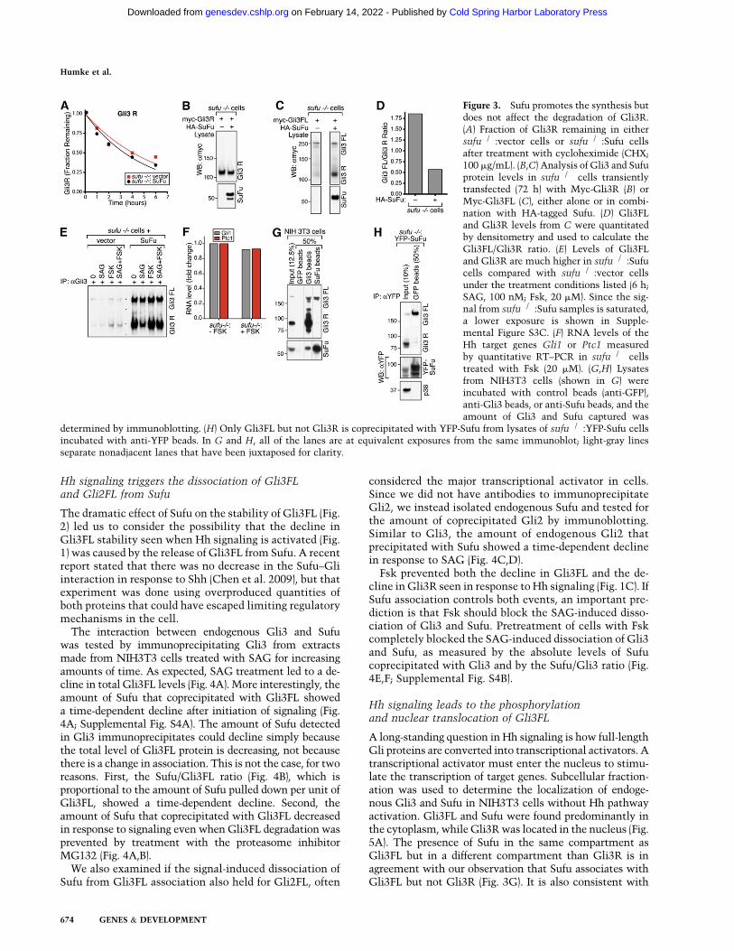
Figure 3. Sufu promotes the synthesis butdoes not affect the degradation of Gli3R.(A) Fraction of Gli3R remaining in eithersufu�/�:vector cells or sufu�/�:Sufu cellsafter treatment with cycloheximide (CHX;100 mg/mL). (B,C) Analysis of Gli3 and Sufuprotein levels in sufu�/� cells transientlytransfected (72 h) with Myc-Gli3R (B) orMyc-Gli3FL (C), either alone or in combi-nation with HA-tagged Sufu. (D) Gli3FLand Gli3R levels from C were quantitatedby densitometry and used to calculate theGli3FL/Gli3R ratio. (E) Levels of Gli3FLand Gli3R are much higher in sufu�/�:Sufucells compared with sufu�/�:vector cellsunder the treatment conditions listed (6 h;SAG, 100 nM; Fsk, 20 mM). Since the sig-nal from sufu�/�:Sufu samples is saturated,a lower exposure is shown in Supple-mental Figure S3C. (F) RNA levels of theHh target genes Gli1 or Ptc1 measuredby quantitative RT–PCR in sufu�/� cellstreated with Fsk (20 mM). (G,H) Lysatesfrom NIH3T3 cells (shown in G) wereincubated with control beads (anti-GFP),anti-Gli3 beads, or anti-Sufu beads, and theamount of Gli3 and Sufu captured was
determined by immunoblotting. (H) Only Gli3FL but not Gli3R is coprecipitated with YFP-Sufu from lysates of sufu�/�:YFP-Sufu cellsincubated with anti-YFP beads. In G and H, all of the lanes are at equivalent exposures from the same immunoblot; light-gray linesseparate nonadjacent lanes that have been juxtaposed for clarity.
Humke et al.
674 GENES & DEVELOPMENT
Cold Spring Harbor Laboratory Press on February 14, 2022 - Published by genesdev.cshlp.orgDownloaded from

a previous report that Gli3FL and Sufu are both localized inprimary cilia, but Gli3R is found only in the nucleus(Haycraft et al. 2005).
In response to Smo activation by SAG, Gli3FL shiftedfrom the cytoplasm to the nucleus, assessed by eitherabsolute levels of Gli3FL in the nucleus (Fig. 5B) or thecalculated fraction of total Gli3FL in the nucleus (Fig. 5C).Gli2FL also moved from the cytoplasm to the nucleus inresponse to SAG (Supplemental Fig. S5A), although totalGli2 levels remained stable after signal activation (Supple-mental Fig. S5C). In contrast, Sufu, Gli3R, and the standingpool of Gli1 did not show any change in subcellular lo-calization in response to SAG (Fig. 5B; Supplemental Fig.S5A). The rise in nuclear Gli3FL was caused by increasednuclear import rather than decreased nuclear export
because Leptinomycin B, an inhibitor of nuclear export,did not enhance Gli3FL accumulation in the nucleus (Sup-plemental Fig. S5B).
The activation of Hh signaling led to both the nucleartranslocation and destabilization of Gli3FL. These changesin the biochemical properties of Gli3FL may reflect itsconversion into a transcriptional activator (Gli3A), sincethe activation of transcription factors is often coupledto their degradation (Collins and Tansey 2006). To deter-mine whether the signal-induced nuclear translocationof Gli3FL is coupled to its degradation, cells were treatedwith SAG and the proteasome inhibitor MG132. MG132stabilized Gli3FL in the nucleus, but had little effect onGli3FL in the cytoplasm (Fig. 5B,C). This suggested thatGli3FL is degraded in the nucleus, after Hh signaling drivesits movement from the cytoplasm. To directly follow thedynamics and dissect the temporal order of Gli3FL nucleartranslocation and degradation, levels of Gli3FL in thenucleus and cytoplasm were assessed at various timesafter SAG addition (Fig. 5D,E). The rapid decline in cy-toplasmic Gli3FL was accompanied by a concomitantincrease in nuclear Gli3FL, showing that the initial eventwas translocation from the cytoplasm to the nucleus. Thiswas followed by the degradation of the nuclear pool ofGli3FL. Notably, nuclear translocation of Gli3FL (<30 min)occurs before target gene transcripts can be detected (>2 h)(Fig. 1B).
The above studies support the view that the activatorform of Gli3 is a labile species localized in the nucleus.This instability may be the reason that it has been difficultto detect endogenous Gli3 (and Ci155) in the nucleus afteractivating the pathway, especially when looking at latetime points after signal initiation. In searching for a bio-chemical mark that might distinguish Gli3A from Gli3FL,we observed that Gli3FL extracted from the nucleus aftertreatment with SAG consistently migrated more slowly onSDS-PAGE gels compared with either cytoplasmic Gli3FLor nuclear Gli3FL extracted in the absence of SAG (Fig.5B,F). The gel migration difference suggested that nuclearGli3FL was phosphorylated selectively in a signal-regulatedmanner.
The phosphorylation of Gli3FL induced by Smo activa-tion was unexpected because previous work had suggestedthat GliFL (and Ci155) proteins are dephosphorylated inresponse to signaling (Chen et al. 1999). Since we observedthe opposite, Gli3FL was analyzed carefully using twodifferent techniques to establish that the gel shift ob-served in the nuclear fraction was indeed caused by phos-phorylation rather than a different post-translational mod-ification. First, we used phosphate affinity SDS-PAGE(Kinoshita et al. 2009), a method in which gels incorpo-rate Mn+2-Phos-tag. This dinuclear metal complex bindsselectively to phosphomonoesters, and thus enhancesonly those gel shifts caused by phosphorylation eventson proteins. The mobility of Gli3FL extracted from thenuclei of SAG-treated cells was significantly retarded ongels containing Phos-tag when compared with identicalgels lacking Phos-tag (Fig. 5G). The mobility of nuclearGli3FL from SAG-treated cells was much slower than themobility of cytoplasmic Gli3FL or the mobility of nuclear
Figure 4. Hh signaling promotes the dissociation of Sufu fromGli3FL and Gli2FL. (A) A time-dependent decrease in the amountof Sufu that coprecipitates with Gli3 from lysates of NIH3T3 cellstreated with SAG alone (100 nM) or SAG + MG132 (25 mM).MG132 was added 30 min before SAG. (B) A time-dependentdecrease in the amount of Sufu pulled down per unit of Gli3FL,estimated using the ratio of the Sufu signal to the Gli3FL signalfrom A. (C,D) A time-dependent decrease in the amount ofGli2FL that coprecipitates with anti-Sufu beads from lysates ofNIH3T3 cells treated with SAG (100 nM) for the indicated periodsof time. (E,F) Fsk (20 mM) prevents the SAG-induced dissociation(100 nM; 6 h) of Sufu from Gli3, assayed by determining theamount of Sufu that coprecipitated with anti-Gli3 beads fromNIH3T3 lysates (shown in E). (F) The ratio of the Sufu signal tothe Gli3FL signal from E was plotted.
Regulation of Gli proteins by Sufu
GENES & DEVELOPMENT 675
Cold Spring Harbor Laboratory Press on February 14, 2022 - Published by genesdev.cshlp.orgDownloaded from
Gli3FL in the absence of SAG (Fig. 5G), demonstratingthat Gli3FL found in the nucleus in response to signalinghas a unique pattern of phosphorylation. Second, thereduced mobility of nuclear Gli3FL seen in response tosignaling could be reversed by l phosphatase treatment(Fig. 5H), providing independent evidence for phosphory-lation. Plots of Gli3FL mobility (Fig. 5H) suggested thatcytoplasmic Gli3FL was also likely phosphorylated; how-ever, the mobility, and hence the phosphorylation pattern,of nuclear Gli3FL was clearly different after SAG addition.In summary, conversion of Gli3 into a transcriptionalactivator was associated with its nuclear translocation,differential phosphorylation, and destabilization.
PKA is thought to inhibit both mammalian and Dro-sophila Hh signaling primarily by promoting formation ofGli3R (Wang et al. 2000; Smelkinson et al. 2007). However,PKA activation stabilized Gli3FL (Fig. 1C), so we consid-ered the possibility that PKA may also inhibit conversionof Gli3FL into Gli3A. Treatment of cells with Fsk inhibitedthe SAG-induced nuclear translocation (Figs. 6A; Supple-mental Fig. S6A), phosphorylation (Supplemental Fig.
S6A), and degradation of Gli3FL (Supplemental Fig. S4B).These results are consistent with the ability of Fsk toprevent the signal-induced dissociation of Sufu fromGli3FL (Fig. 4). To test whether the ability of PKA to in-hibit formation of Gli3A influenced target gene transcrip-tion, cells were treated simultaneously with Shh and Fsk.Under these conditions, Gli1 was transcribed in a shortpulse, presumably because the rapid activation of Gli1transcription by Shh was followed by the slower kinetics ofFsk action (Fig. 6B). This pulse was mirrored by changesin the level of Gli3FL, with unstable Gli3FL associatedwith high transcription and stable Gli3FL associated withlow transcription (Fig. 6B). During this experiment, Fskinhibited Gli1 and Ptc1 transcription without increasingthe levels of Gli3R. These experiments confirm that theunstable form of Gli3FL is a transcriptional activator, andthat PKA can inhibit target gene induction by preventingthe conversion of Gli3FL into Gli3A.
To dissect the relationship between Sufu dissociation,nuclear translocation, phosphorylation, and degradation,we took advantage of the observation that disruption of
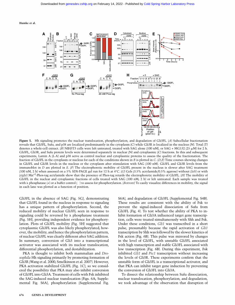
Figure 5. Hh signaling promotes the nuclear translocation, phosphorylation, and degradation of Gli3FL. (A) Subcellular fractionationreveals that Gli3FL, Sufu, and p38 are localized predominantly in the cytoplasm (C) while Gli3R is localized in the nucleus (N). Total (T)denotes a whole-cell extract. (B) NIH3T3 cells were left untreated, treated with SAG alone (100 nM), or SAG + MG132 (25 mM) for 2 h.Gli3FL, Gli3R, and Sufu protein levels were determined separately in nuclear (N) and cytoplasmic (C) fractions. In this and subsequentexperiments, Lamin A (L-A) and p38 serve as control nuclear and cytoplasmic proteins to assess the quality of the fractionation. Thefraction of Gli3FL in the cytoplasm or nucleus for each of the conditions shown in B is plotted in C. (D,E) Time courses showing changesin Gli3FL and Gli3R levels in the nucleus or the cytoplasm after stimulation with SAG (100 nM). Gli3FL and Gli3R levels from theimmunoblot in D are plotted in E. (F) The electrophoretic mobility of Gli3FL present in the nucleus is slower after SAG treatment(100 nM, 2 h) when assessed on a 5% SDS-PAGE gel run for 12 h at 4°C. (G) Gels (3.5% acrylamide/0.5% agarose) without (left) or with(right) Mn+2-Phos-tag acrylamide show that the presence of Phos-tag retards the electrophoretic mobility of Gli3FL. (H) The mobility ofGli3FL in the nuclear and cytoplasmic fractions of cells treated with SAG (100 nM; 2 h) or left untreated. Each sample was treatedwith l phosphatase (+) or a buffer control (�) to assess for phosphorylation. (Bottom) To easily visualize differences in mobility, the signalin each lane was plotted as a function of position.
Humke et al.
676 GENES & DEVELOPMENT
Cold Spring Harbor Laboratory Press on February 14, 2022 - Published by genesdev.cshlp.orgDownloaded from

cytoplasmic microtubules blocks nuclear translocation ofGli2 (and target gene transcription) in response to Hhsignaling (Kim et al. 2009). As for Gli2, the SAG-inducednuclear translocation of Gli3FL could be blocked by themicrotubule-disrupting agent nocodazole (Fig. 6C,D; Sup-plemental Fig S6B). While nocodazole treatment alsoblocked the SAG-induced phosphorylation (Fig. 6C) anddestabilization (Fig. 6E) of Gli3FL, it did not prevent thedissocation of Sufu from Gli3FL (Fig. 6E,F). Thus, cyto-plasmic microtubules appear to be required at a step afterSufu–Gli3 dissociation, but before the coupled processes ofnuclear translocation, phosphorylation, and degradation.
Sufu inhibits conversion of Gli3FLinto a transcriptional activator
To show that Sufu works by preventing the nuclear trans-location and activation of Gli3FL, we examined the sub-cellular localization of Gli3FL and Gli3R in sufu�/� cells.While sufu�/� cells have low total levels of Gli3FL (Fig. 2),a large fraction (>50%) of the Gli3FL is localized in thenucleus, consistent with the model that activated Gliproteins are highly unstable and are located in the nucleus(Fig. 7A,B). Gli3FL in nuclei of sufu�/� cells also migratedmore slowly on SDS-PAGE gels than the Gli3FL present inthe cytoplasm, suggestive of differential phosphorylation(Supplemental Fig. S7A). When Gli3FL degradation wasinhibited with MG132 in sufu�/� cells, all of the additional
Gli3FL accumulated in the nucleus (Fig. 7A,B). Thissupports the idea that Gli3A was degraded in the nucleus,either when Sufu protein was missing (Fig. 7A) or whenGli3FL was released from Sufu in response to Hh signaling(Fig. 5). The reintroduction of Sufu into sufu�/� cells ex-tinguished target gene transcription and stabilized Gli3FL(Fig. 2A). Importantly, Sufu restoration into sufu�/� cellsdramatically reduced the fraction of Gli3FL in the nucleus,despite the fact that the total level of Gli3FL was muchhigher in the presence of Sufu (Fig. 7C,D). Unlike Gli3FL,Gli3R was localized predominantly in the nucleus in thepresence or absence of Sufu (Fig. 7C).
In summary, Gli3FL in sufu�/� cells has the samebiochemical characteristics—nuclear location, instability,and differential phosphorylation—as wild-type fibroblaststreated with SAG, which both activates signaling andtriggers the dissociation of Gli3 from Sufu. This findingprovides an explanation for how sufu�/� cells can attainhigh target gene transcription despite containing lowlevels of Gli2 and Gli3 (Chen et al. 2009). Thus, Sufu in-hibits Hh signaling in the cytoplasm by preventing con-version of Gli3FL into a transcriptional activator.
Dissociation of the Sufu–Gli3 interaction dependson the ciliary motor Kif3a
Cells and embryos with defective primary cilia are im-paired in the production of activator and repressor forms
Figure 6. PKA activation blocks the nuclear transloca-tion of Gli3FL. (A) Fsk (20 mM) blocks the SAG-inducedtranslocation of Gli3FL from the cytoplasm to the nu-cleus. The plots are derived from immunoblots shown inSupplemental Figure S6A (B) NIH3T3 cells were treatedwith Fsk (10 mM) + Shh at t = 0, Gli3FL and Gli3Rproteins levels were measured by immunoblotting (left),and target gene transcription was assessed by Gli1 andPtc1 mRNA levels measured by quantitative RT–PCR(right). Nocodazole (15 mM) blocks SAG-induced (100 nM,3 h) phosphorylation (C), nuclear translocation (C,D), anddegradation (E) of Gli3FL. However, nocodazole cannotprevent the SAG-triggered dissociation of Sufu fromGli3FL, assayed with an anti-Gli3 IP (E, right panel) andquantitated as the amount of Sufu pulled down per unitof Gli3FL (F).
Regulation of Gli proteins by Sufu
GENES & DEVELOPMENT 677
Cold Spring Harbor Laboratory Press on February 14, 2022 - Published by genesdev.cshlp.orgDownloaded from

of the Gli proteins (Huangfu and Anderson 2006). Therequirement for cilia was established by genetic studies inmice, and a large decrease in Gli3R was observed in lysatesof embryos with ciliary defects (Huangfu and Anderson2005; Liu et al. 2005).
Elucidation of the means by which Sufu regulates theformation of Gli3A and Gli3R provided an opportunity tolearn which biochemical step is influenced by primarycilia. We used a mouse fibroblast cell line made from em-bryos lacking the anterograde IFT motor Kif3a (kif3a�/�
cells), which has been used previously to study the roleof primary cilia in Hh and Wnt signaling (Corbit et al.2008; Chen et al. 2009). Kif3a�/�mice have severe defectsin primary cilia and have phenotypes consistent withimpaired Hh signaling (Huangfu et al. 2003; Huangfu andAnderson 2005; Liu et al. 2005). The steady-state level ofGli3FL was similar in both kif3a�/� and kif3a+/+ cells, butkif3a�/� cells had a significantly lower level of Gli3R (Fig.8A). However, Gli3FL failed to translocate to the nucleusin kif3a�/� cells treated with SAG (Fig. 8A,B; Supplemen-tal Fig. S8A). In addition, SAG treatment of kif3a�/� cellsdid not induce the phosphorylation of nuclear Gli3FLtypically seen in wild-type cells (Supplemental Fig. S8A).In control kif3a+/+ cells, SAG-induced nuclear transloca-tion and phosphorylation of Gli3FL was maintained (Figs.8A; Supplemental Fig. S8A). These data provide directbiochemical evidence that Kif3a is required for theactivation of full-length Gli proteins, and support geneticexperiments showing that mouse embryos with damagedcilia cannot produce activator forms of the Gli proteins(Huangfu and Anderson 2005; Liu et al. 2005).
We next tested the association between Gli3 and Sufuin both kif3a+/+ and kif3a�/� cells. In the absence of path-way stimulation, the Gli3–Sufu interaction was readilydetected in cells from both genotypes (Fig. 8C), and theamount of Sufu precipitated per unit of Gli3FL (the Sufu/Gli3 ratio) derived from Gli3 immunoprecipitates wassimilar in both cell types (Fig. 8D). This supports the idea,presented in two recent studies, that primary cilia are notrequired for Sufu to inhibit Gli proteins (Chen et al. 2009;Jia et al. 2009). We next analyzed how the Gli3–Sufuinteraction changed after Hh pathway activation. SAGcaused the dissociation of Gli3FL from Sufu in kif3a+/+
cells, but SAG was unable to induce the dissociation ofGli3FL from Sufu in cells lacking kif3a�/� (Fig. 8C,D).Thus, the ability of Hh signaling to disrupt the Sufu–Gli3interaction depends on Kif3a. Indeed, this is likely to be themajor function of Kif3a and primary cilia in Hh signaling,because Kif3a is dispensable for target gene transcription insufu�/� cells (Supplemental Fig. S8B; Chen et al. 2009).
Discussion
The main goal of Hh signaling is to alter the transcrip-tional program of the cell by influencing the balancebetween the activator and repressor functions of theGli proteins. The dissociation of Sufu from Gli3 co-ordinately accomplishes the two main tasks of Hhsignaling: inhibition of Gli3R formation, and promotionof Gli3A formation. We present our model for how theSufu–Gli interaction, likely regulated by biochemical
Figure 7. Sufu prevents the nuclear translocation of Gli3FL.Immunoblot (A) and graph (B) of Gli3FL from the subcellularfractionation of sufu�/� cells untreated or treated with MG132(25 mM; 4 h). (C,D) Immunoblot (C) and graph (D) showing levelsof total (T), cytoplasmic (C), and nuclear (N) Gli3FL in sufu�/�:vector cells or sufu�/�:Sufu cells. While the Gli3FL stabilized byMG132 in A was located in the nucleus, the Gli3 stabilized bySufu readditon is located in the cytoplasm.
Figure 8. Kif3a is required for Hh-induced dissociation of theSufu–Gli3FL complex and nuclear translocation of Gli3FL. (A,B)Subcellular fractions generated from kif3a+/+ or kif3a�/� cellsleft untreated or treated with SAG (100 nM; 2 h) were assayedfor Gli3FL, Gli3R, and Kif3a levels. (C,D) The amount of Sufudetected in Gli3 immunoprecipitates is unaffected by SAGtreatment (100 nM; 2 h) in kif3a�/� cells, but drops significantlywith SAG treatment in kif3a+/+ cells, seen both in the immu-noblots (C) and by changes in the Sufu/Gli3FL ratio (D).
Humke et al.
678 GENES & DEVELOPMENT
Cold Spring Harbor Laboratory Press on February 14, 2022 - Published by genesdev.cshlp.orgDownloaded from

events at primary cilia, controls the formation of GliRand GliA.
Gli3R formation
In the absence of Hh signaling, the majority of Gli3FL isprocessed into Gli3R (Fig. 9A). In mammals, the efficientproduction of Gli3R depends on the association betweenSufu and Gli3FL (Fig. 3). While the association of Gli3FLwith Sufu appears to be independent of primary cilia, thesubsequent processing of Gli3FL into Gli3R depends on
intact cilia (Fig. 8; Huangfu and Anderson 2005). Sufumight promote Gli3 processing by recruiting it to primarycilia, although a recent report showed that overproducedGli3 localized to cilia in sufu�/� cells (Chen et al. 2009).Alternatively, the Gli3FL–Sufu complex may be a bettersubstrate than Gli3FL for the biochemical reactions re-quired for processing (Kise et al. 2009).
Once GliR is produced, its activity is independent ofboth Sufu and the primary cilium. Gli3R and Sufu pro-teins do not associate with each other in cells, and residein separate subcellular compartments. The nuclear local-ization and half-life of Gli3R are unaffected by the loss ofSufu. The mechanism by which processing of Gli3FL intoGli3R leads to its dissociation from Sufu remains un-known, especially because prior work identified a Sufu-binding region in the N terminus of Gli3 (Pearse et al.1999; Dunaeva et al. 2003); however, our data suggest thatendogenous Gli3R no longer has affinity for Sufu.
Activation of Hh signaling leads to a decrease in thelevels of Gli3R, but the mechanism by which this occurshas been unclear in mammals. Our results support themodel (Fig. 9B) that the dissociation of Sufu from Gli3FLdrives the reduction in Gli3R synthesis seen in response toHh signaling.
Gli3A formation
By studying Gli3 in the few hours after SAG treatment, wedefined three properties that are associated with an increasein the transcriptional activity of Gli3FL: rapid nuclear trans-location, phosphorylation, and destabilization (Fig. 9B). Theactivity of many transcription factors is often inverselyrelated to their stability, in some cases to limit the durationof transcription, and in other cases because proteolysisis required for efficient transcription (Collins and Tansey2006). In flies, Hh has been proposed to promote theconversion of Ci155 into a labile transcriptional activator(Ohlmeyer and Kalderon 1998). Thus, the destabilization ofGli3FL in response to signaling might reflect the conversionof Gli3FL to a transient but potent Gli3A. In terms of themechanism of Gli3 degradation, work in flies and mammalshas suggested that Gli3 or Ci155 stability may be regulatedby Cul3-based E3 ubiquitin ligases that have a BTB domain-containing substrate recognition module (HIB in flies, SPOPin mammals) (Zhang et al. 2006; Chen et al. 2009).
The phosphorylation of Gli3FL seen after activation ofsignaling could control degradation, transcriptional activa-tion, or both processes. Differential phosphorylation asso-ciated with Gli3A formation is seen only in the nucleusand occurs prior to degradation of Gli3 (Fig. 5D), suggestingthat it may be controlled by a nuclear kinase. DYRK1A isa nuclear kinase that has been implicated in the activationof Gli1, but the addition of Harmine, an inhibitor ofDYRK1A, had no effect on either nuclear translocation orphosphorylation of Gli3FL (Supplemental Fig. S5B; Maoet al. 2002; Seifert et al. 2008).
Sufu restrains Hh signaling in the cytoplasm
Our study suggests that the main locus for Sufu functionin the cell is the cytoplasm, not the nucleus. The majority
Figure 9. A model for how Sufu association controls the for-mation of Gli3R and Gli3A. (A) Gli3FL is complexed with Sufu inthe cytoplasm and is maintained in a neutral state. Without theHh signal (Hh OFF), the Sufu–Gli3 complex is recruited to cilia (1),leading to the efficient processing of Gli3FL into Gli3R (2). Gli3Rformation leads to its dissociation from Sufu (3), allowing Gli3R totranslocate into the nucleus (4), and repress Hh target genes (5). (B)When Hh signaling is initiated (Hh ON), Sufu dissociates fromGli3FL (3). This has two consequences. First, Gli3R production ishalted (2). Second, free Gli3FL translocates to the nucleus (4),where it is phosphorylated, destabilized (6), and converted toa transcriptional activator (5). The level of PKA activity in thecilium may control the relative flux between pathways leading toGli3R or Gli3A formation.
Regulation of Gli proteins by Sufu
GENES & DEVELOPMENT 679
Cold Spring Harbor Laboratory Press on February 14, 2022 - Published by genesdev.cshlp.orgDownloaded from
of Sufu is present in the cytoplasm, and Sufu does notundergo any change in localization in response to Hhsignaling. Experiments in sufu�/� cells, along with theobservation that the association between Gli3 and Sufu isdisrupted by Hh signaling, suggest that the main factorrestraining the production of Gli3A is Sufu (Fig. 9B).
The mechanism by which Hh promotes this dissocia-tion of Gli3 from Sufu is a major unresolved question.Some insights into this key step come from the findingthat activation of PKA can prevent dissociation of theGli3FL–Sufu complex, leaving Gli3FL stranded in thecytoplasm and unable to activate target genes. So, a de-crease in PKA activity in response to Hh signaling maytrigger dissociation of the Sufu–Gli3FL complex. Thereis some evidence that Smo activation can reduce PKAactivity through the inhibitory Gai class of heterotrimericG proteins (Riobo et al. 2006; Ogden et al. 2008). The dualinvolvement of Kif3a and PKA also suggests the interestinghypothesis that Hh signaling regulates Gli3–Sufu associa-tion by locally controlling the activity of PKA at theprimary cilium (Barzi et al. 2010).
Future prospects
Our study provides a parsimonious model for control ofthe final, committed step in Hh signal transduction: ac-tivation of the Gli family of transcription factors. Unrav-eling the biochemical details of this activation process isan important goal. In addition, the reactions underlyingGli activation, such as phosphorylation or nuclear trans-location, provide novel assays for both basic studies andsmall molecule and RNAi screens directed against thisimportant pathway.
Materials and methods
Constructs and cell lines
All constructs described in the study were made from mousegenes. NIH3T3 cells were obtained from American Type CultureCollection, sufu�/� mouse embryonic fibroblasts (MEFs) werekindly provided by Rune Toftgard (Svard et al. 2006), and kif3a+/+
and kif3a�/� MEFs were kindly provided by Pao-Tien Chuang(Corbit et al. 2008; Wilson et al. 2009). To make sufu�/� stablecells with Sufu or YFP-tagged Sufu added back, cDNAs encodingSufu or Sufu tagged at its N terminus with YFP were cloned intothe pRetroX-PTuner retroviral expression vector (Clontech).Retroviral production and infection of sufu�/� fibroblasts wasperformed according to established protocols (Pear et al. 1993).The pRetroX-PTuner vectors include a ligand-responsive desta-bilization domain; however, this domain could not control Sufuor YFP-Sufu levels, and thus this feature was not used.
Full-length Gli3 (amino acids 1–1583) and Gli3 repressor (aminoacids 1–740) were tagged on the N terminus with a 6-myc tagby cloning them into pCS2 + MT, and Sufu was tagged at theN terminus with a 33 HA tag by cloning it into the pCS2 + HAvector.
Cell culture
Cells were grown to confluence in medium (high-glucose DMEM,0.05 mg/mL penicillin, 0.05 mg/mL streptomycin, 2 mM gluta-max, 1 mM sodium pyruvate, 0.1 mM MEM nonessential amino
acid supplement) containing 10% FBS (Hyclone, defined grade), andthen switched to medium containing 0.5% FBS for 12 h prior to allexperiments. Cells were transfected using FugeneHD (Roche) perthe manufacturer’s instructions.
Small molecules and recombinant proteins
SAG was obtained from Alexis; forskolin was obtained fromBIOMOL; puromycin, cycloheximide, and nocodazole were ob-tained from Sigma; and Leptomycin B was obtained from EMD.The 293 EcR Shh cells (Taipale et al. 2000) were used to produceconditioned media containing processed and lipidated Shh (Sup-plemental Material). This media was used at a dilution of one tofour, with a final FBS concentration of 0.5%.
Antibodies
Anti-Gli1 (mouse monoclonal) was from Cell Signaling Tech-nologies (catalog no. L42B10); anti-Gli2 (goat polyclonal) wasfrom R&D systems (catalog no. AF3635); anti-Gli3 (goat poly-clonal) was from R&D systems (catalog no. AF3690); anti-p38,anti-Kif3a, anti-LaminA, and anti-GFP (all rabbit polyclonal) wasfrom Abcam (catalog nos. ab7952, ab11259, ab26300, and ab290);and anti-Myc (9E10, mouse monoclonal) was from Roche. Theanti-Sufu polyclonal antibody was produced (Josman Laborato-ries) in rabbits against full-length mouse Sufu protein andaffinity-purified before use. Secondary antibodies conjugated tothe horseradish peroxidase enzyme were purchased from JacksonLaboratories. The anti-Ptc1 rabbit polyclonal antibody, producedagainst a fragment of mouse Ptc1 from amino acids 1169 to 1435,has been described previously (Rohatgi et al. 2007).
Lysate production, immunoprecipitation,
and Western blot analysis
For the production of whole-cell lysates, cells were lysed in BufferA (50 mM Tris at pH 7.4, 300 mM NaCl, 2% NP-40 [v/v], 0.25%Deoxycholate [w/v], 10 mM N-ethyl maleimide, 1 mM DTT,a protease inhibitor cocktail [13 EDTA-free protease inhibitorsfrom Roche]). When cells were treated with MG132 or cyclohex-imide, these drugs were maintained in the lysis buffers.
For immunoprecipitation, antibodies were coupled covalentlyto Protein A- or Protein G-coated magnetic beads (Dynal). Anti-Gli3 or anti-Sufu-coupled beads were added to the clarified lysate.After overnight binding at 4°C, the beads were washed with BufferA and eluted with 23 SDS sample buffer.
Subcellular fractionation
All subcellular fractionation experiments were performed onice with freshly harvested cells. Cells were washed twice withphosphate-buffered saline (PBS) and twice with 10 mM HEPES (pH7.4), and then were incubated for 10 min in 10 mM HEPES (pH7.4). The HEPES buffer was removed and SEAT buffer (10 mMtriethanolamine/acetic acid at pH 7.4, 250 mM sucrose, 13 EDTAprotease inhibitor cocktail [Roche]) was added. Cells were lysed by15 passages through a 25-G needle. Nuclei were separated fromthe post-nuclear supernatant (PNS) by centrifugation at 900g for5 min and then washed once in SEAT buffer. The PNS wasalso respun at 900g for 5 min and then brought to 13 Buffer A,extracted for 1 h, and clarified by centrifugation at 20,000g for 1 h.The nuclei were extracted (same conditions as PNS) with 20 mMHEPES (pH 7.9), 1 mM MgCl2, 0.5 M NaCl, 0.2 mM EDTA, 20%glycerol, 1% Triton X-100, 1 mM DTT, benzonase, and a proteaseinhibitor cocktail. Lysates used for phosphorylation analysisincluded a phosphatase inhibitor cocktail (13 PhosphoSTOP
Humke et al.
680 GENES & DEVELOPMENT
Cold Spring Harbor Laboratory Press on February 14, 2022 - Published by genesdev.cshlp.orgDownloaded from

[Roche]) except in cases where lysates were treated with l
phosphatase. Extraction volumes for each fraction were equalizedbefore taking samples for gel electrophoresis to ensure that equalamounts of each fraction were loaded on the gel; however, thereare always unaccounted losses during fractionation due to themultiple washing and centrifugation steps.
Quantitative real-time PCR
Total cell RNA was isolated using Trizol reagent (Invitrogen) andreverse-transcribed using SuperScript III First Strand Kit (Invitro-gen). Real-time PCR (Applied Biosystems 7500) was used toquantify transcript levels. TaqMan gene expression probes (AppliedBiosystems) used were Mm00494645_m1 (gli1), Mm00970977_m1(ptc1), Mm00492333_m1 (gli3), and Mm99999915_g1 (gapdh tonormalize the samples).
Data analysis
Films were scanned on the Epson Perfection V700 photo scannerinto Photoshop at 600 dpi as grayscale TIFF files. Quantitativeanalysis of band intensities was performed with Total Lab100(Nonlinear Dynamics), and the data were transferred to Graph-Pad Prism for normalization, graphing, and curve fitting. Allcurves shown for the cycloheximide chase experiments repre-sent best-fit single exponential decay curves. To calculate thefraction of Gli3FL in the nucleus or cytoplasm, we calculated theratios N/N + C and C/N + C, where N is the intensity of the nu-clear band and C is the intensity of the cytoplasmic band.
The data presented in the figures are representative of at leastthree independent experiments, and multiple independent gelsare included showing results that support each of the main con-clusions in this study. All quantitative comparisons were per-formed on data obtained within one experiment, because abso-lute Gli3FL and Gli3R levels were variable from one experimentto another due to differences in cell confluence, cell passage num-ber, and efficiency of immunoblotting. For comparisons, all sam-ples were run on the same SDS-PAGE gel and imaged at identicalexposures after immunoblotting.
Phosphorylation analysis
For phosphate affinity electrophoresis, Phos-tag gels containing3.5% acrylamide (37.5:1 ratio of acrylamide:bis-acrylamide), 0.5%w/v SeaKem Gold agarose (Lonza), 20 mM Phos-tag acrylamide(a kind gift from Nicholas Tilmans and Pehr Harbury), and 40 mMMnCl2 were prepared and run according to a recently publishedprotocol (Kinoshita et al. 2009). For l phosphatase digestion, cy-tosolic and nuclear fractions were treated with 1 mL (400 U) of l
protein phosphatase, 13 l protein phosphatase buffer, and 1 mMMnCl2 for 1 h at room temperature. A 6% (100:1 acrylamide:bis-acrylamide) gel was used for the analysis of l phosphatase-sensitive gel shifts in Figure 6C.
Acknowledgments
We thank Rune Toftgard for sufu�/� cells, Pao-Tien Chuang forkif3a�/� and kif3a+/+ cells, Tyler Hillman and Eunice Lee foradvice on Gli3 constructs, Trina Schroer for Kif3a constructs,Maika Deffieu and Suzanne Pfeffer for advice on subcellularfractionation, and Nicholas Tilmans and Pehr Harbury for Phos-tag acrylamide. M.P.S is an Investigator of the Howard HughesMedical Institute; R.R. is supported by a NCI Pathway to In-dependence award (K99/ROO CA129174), a V Foundation Scholaraward, a SU2C Innovation Award (AACR), and the March ofDimes Basil O’Connor award; and E.W.H is supported by the
PanCAN-AACR fellowship, a pilot grant from the Stanford Di-gestive Disease Center, and the Amgen Hematology-OncologyFellowship.
References
Aza-Blanc P, Ramirez-Weber FA, Laget MP, Schwartz C, KornbergTB. 1997. Proteolysis that is inhibited by hedgehog targetsCubitus interruptus protein to the nucleus and converts it toa repressor. Cell 89: 1043–1053.
Barzi M, Berenguer J, Menendez A, Alvarez-Rodriguez R, Pons S.2010. Sonic-hedgehog-mediated proliferation requires thelocalization of PKA to the cilium base. J Cell Sci 123: 62–69.
Chen CH, von Kessler DP, Park W, Wang B, Ma Y, Beachy PA.1999. Nuclear trafficking of Cubitus interruptus in thetranscriptional regulation of Hedgehog target gene expres-sion. Cell 98: 305–316.
Chen JK, Taipale J, Young KE, Maiti T, Beachy PA. 2002. Smallmolecule modulation of Smoothened activity. Proc Natl
Acad Sci 99: 14071–14076.Chen MH, Wilson CW, Li YJ, Law KK, Lu CS, Gacayan R, Zhang
X, Hui CC, Chuang PT. 2009. Cilium-independent regulationof Gli protein function by Sufu in Hedgehog signaling isevolutionarily conserved. Genes & Dev 23: 1910–1928.
Cheng SY, Bishop JM. 2002. Suppressor of Fused represses Gli-mediated transcription by recruiting the SAP18–mSin3 co-repressor complex. Proc Natl Acad Sci 99: 5442–5447.
Collins GA, Tansey WP. 2006. The proteasome: A utility tool fortranscription? Curr Opin Genet Dev 16: 197–202.
Cooper AF, Yu KP, Brueckner M, Brailey LL, Johnson L,McGrath JM, Bale AE. 2005. Cardiac and CNS defects ina mouse with targeted disruption of suppressor of fused.Development 132: 4407–4417.
Corbit KC, Aanstad P, Singla V, Norman AR, Stainier DY, ReiterJF. 2005. Vertebrate Smoothened functions at the primarycilium. Nature 437: 1018–1021.
Corbit KC, Shyer AE, Dowdle WE, Gaulden J, Singla V, ChenMH, Chuang PT, Reiter JF. 2008. Kif3a constrains b-catenin-dependent Wnt signalling through dual ciliary and non-ciliary mechanisms. Nat Cell Biol 10: 70–76.
Ding Q, Fukami S, Meng X, Nishizaki Y, Zhang X, Sasaki H,Dlugosz A, Nakafuku M, Hui C. 1999. Mouse suppressor offused is a negative regulator of sonic hedgehog signaling andalters the subcellular distribution of Gli1. Curr Biol 9: 1119–1122.
Dunaeva M, Michelson P, Kogerman P, Toftgard R. 2003.Characterization of the physical interaction of Gli proteinswith SUFU proteins. J Biol Chem 278: 5116–5122.
Haycraft CJ, Banizs B, Aydin-Son Y, Zhang Q, Michaud EJ, YoderBK. 2005. Gli2 and Gli3 localize to cilia and require theintraflagellar transport protein polaris for processing andfunction. PLoS Genet 1: e53. doi: 10.1371/journal.pgen.0010053.
Huangfu D, Anderson KV. 2005. Cilia and Hedgehog responsive-ness in the mouse. Proc Natl Acad Sci 102: 11325–11330.
Huangfu D, Anderson KV. 2006. Signaling from Smo to Ci/Gli:Conservation and divergence of Hedgehog pathways fromDrosophila to vertebrates. Development 133: 3–14.
Huangfu D, Liu A, Rakeman AS, Murcia NS, Niswander L,Anderson KV. 2003. Hedgehog signalling in the mouserequires intraflagellar transport proteins. Nature 426: 83–87.
Jia J, Kolterud A, Zeng H, Hoover A, Teglund S, Toftgard R, LiuA. 2009. Suppressor of Fused inhibits mammalian Hedgehogsignaling in the absence of cilia. Dev Biol 330: 452–460.
Kim J, Kato M, Beachy PA. 2009. Gli2 trafficking links Hedgehog-dependent activation of Smoothened in the primary cilium to
Regulation of Gli proteins by Sufu
GENES & DEVELOPMENT 681
Cold Spring Harbor Laboratory Press on February 14, 2022 - Published by genesdev.cshlp.orgDownloaded from
transcriptional activation in the nucleus. Proc Natl Acad Sci
106: 12666–21671.Kinoshita E, Kinoshita-Kikuta E, Koike T. 2009. Separation
and detection of large phosphoproteins using Phos-tag SDS-PAGE. Nat Protoc 4: 1513–1521.
Kise Y, Morinaka A, Teglund S, Miki H. 2009. Sufu recruitsGSK3b for efficient processing of Gli3. Biochem Biophys Res
Commun 387: 569–574.Liu A, Wang B, Niswander LA. 2005. Mouse intraflagellar
transport proteins regulate both the activator and repressorfunctions of Gli transcription factors. Development 132:3103–3111.
Lum L, Zhang C, Oh S, Mann RK, von Kessler DP, Taipale J,Weis-Garcia F, Gong R, Wang B, Beachy PA. 2003. Hedgehogsignal transduction via Smoothened association with a cyto-plasmic complex scaffolded by the atypical kinesin, Costal-2.Mol Cell 12: 1261–1274.
Mao J, Maye P, Kogerman P, Tejedor FJ, Toftgard R, Xie W, WuG, Wu D. 2002. Regulation of Gli1 transcriptional activity inthe nucleus by Dyrk1. J Biol Chem 277: 35156–35161.
Methot N, Basler K. 1999. Hedgehog controls limb developmentby regulating the activities of distinct transcriptional activatorand repressor forms of Cubitus interruptus. Cell 96: 819–831.
Methot N, Basler K. 2000. Suppressor of fused opposes hedgehogsignal transduction by impeding nuclear accumulation of theactivator form of Cubitus interruptus. Development 127:4001–4010.
Ogden SK, Fei DL, Schilling NS, Ahmed YF, Hwa J, Robbins DJ.2008. G protein Gai functions immediately downstream ofSmoothened in Hedgehog signalling. Nature 456: 967–970.
Ohlmeyer JT, Kalderon D. 1998. Hedgehog stimulates matura-tion of Cubitus interruptus into a labile transcriptionalactivator. Nature 396: 749–753.
Pastorino L, Ghiorzo P, Nasti S, Battistuzzi L, Cusano R,Marzocchi C, Garre ML, Clementi M, Scarra GB. 2009.Identification of a SUFU germline mutation in a family withGorlin syndrome. Am J Med Genet 149A: 1539–1543.
Pear WS, Nolan GP, Scott ML, Baltimore D. 1993. Production ofhigh-titer helper-free retroviruses by transient transfection.Proc Natl Acad Sci 90: 8392–8396.
Pearse RV 2nd, Collier LS, Scott MP, Tabin CJ. 1999. Vertebratehomologs of Drosophila suppressor of fused interact with thegli family of transcriptional regulators. Dev Biol 212: 323–336.
Preat T. 1992. Characterization of Suppressor of fused, a com-plete suppressor of the fused segment polarity gene ofDrosophila melanogaster. Genetics 132: 725–736.
Riobo NA, Saucy B, Dilizio C, Manning DR. 2006. Activation ofheterotrimeric G proteins by Smoothened. Proc Natl Acad
Sci 103: 12607–12612.Rohatgi R, Milenkovic L, Scott MP. 2007. Patched1 regulates
hedgehog signaling at the primary cilium. Science 317: 372–376.
Seifert A, Allan LA, Clarke PR. 2008. DYRK1A phosphorylatescaspase 9 at an inhibitory site and is potently inhibited inhuman cells by harmine. FEBS J 275: 6268–6280.
Smelkinson MG, Zhou Q, Kalderon D. 2007. Regulation ofCi–SCFSlimb binding, Ci proteolysis, and hedgehog pathwayactivity by Ci phosphorylation. Dev Cell 13: 481–495.
Stone DM, Murone M, Luoh S, Ye W, Armanini MP, Gurney A,Phillips H, Brush J, Goddard A, de Sauvage FJ, et al. 1999.Characterization of the human suppressor of fused, a negativeregulator of the zinc-finger transcription factor Gli. J Cell Sci
112: 4437–4448.Svard J, Heby-Henricson K, Persson-Lek M, Rozell B, Lauth M,
Bergstrom A, Ericson J, Toftgard R, Teglund S. 2006. Genetic
elimination of Suppressor of fused reveals an essential re-pressor function in the mammalian Hedgehog signalingpathway. Dev Cell 10: 187–197.
Taipale J, Chen JK, Cooper MK, Wang B, Mann RK, MilenkovicL, Scott MP, Beachy PA. 2000. Effects of oncogenic muta-tions in Smoothened and Patched can be reversed by cyclop-amine. Nature 406: 1005–1009.
Taylor MD, Liu L, Raffel C, Hui CC, Mainprize TG, Zhang X,Agatep R, Chiappa S, Gao L, Lowrance A, et al. 2002.Mutations in SUFU predispose to medulloblastoma. Nat
Genet 31: 306–310.Tempe D, Casas M, Karaz S, Blanchet-Tournier MF, Concordet
JP. 2006. Multisite protein kinase A and glycogen synthasekinase 3b phosphorylation leads to Gli3 ubiquitination bySCFbTrCP. Mol Cell Biol 26: 4316–4326.
Varjosalo M, Taipale J. 2008. Hedgehog: Functions and mecha-nisms. Genes & Dev 22: 2454–2472.
Wang QT, Holmgren RA. 1999. The subcellular localization andactivity of Drosophila cubitus interruptus are regulated atmultiple levels. Development 126: 5097–5106.
Wang B, Li Y. 2006. Evidence for the direct involvement ofbTrCP in Gli3 protein processing. Proc Natl Acad Sci 103:33–38.
Wang B, Fallon JF, Beachy PA. 2000. Hedgehog-regulated pro-cessing of Gli3 produces an anterior/posterior repressorgradient in the developing vertebrate limb. Cell 100: 423–434.
Wilson CW, Nguyen CT, Chen MH, Yang JH, Gacayan R, HuangJ, Chen JN, Chuang PT. 2009. Fused has evolved divergentroles in vertebrate Hedgehog signalling and motile cilio-genesis. Nature 459: 98–102.
Zhang W, Zhao Y, Tong C, Wang G, Wang B, Jia J, Jiang J. 2005.Hedgehog-regulated Costal2–kinase complexes control phos-phorylation and proteolytic processing of Cubitus interrup-tus. Dev Cell 8: 267–278.
Zhang Q, Zhang L, Wang B, Ou CY, Chien CT, Jiang J. 2006. Ahedgehog-induced BTB protein modulates hedgehog signal-ing by degrading Ci/Gli transcription factor. Dev Cell 10:719–729.
Humke et al.
682 GENES & DEVELOPMENT
Cold Spring Harbor Laboratory Press on February 14, 2022 - Published by genesdev.cshlp.orgDownloaded from
10.1101/gad.1902910Access the most recent version at doi: 24:2010, Genes Dev.
Eric W. Humke, Karolin V. Dorn, Ljiljana Milenkovic, et al. association between Suppressor of Fused and the Gli proteinsThe output of Hedgehog signaling is controlled by the dynamic
Material
Supplemental
http://genesdev.cshlp.org/content/suppl/2010/03/31/24.7.670.DC1
References
http://genesdev.cshlp.org/content/24/7/670.full.html#ref-list-1
This article cites 47 articles, 19 of which can be accessed free at:
License
ServiceEmail Alerting
click here.right corner of the article or
Receive free email alerts when new articles cite this article - sign up in the box at the top
Copyright © 2010 by Cold Spring Harbor Laboratory Press
Cold Spring Harbor Laboratory Press on February 14, 2022 - Published by genesdev.cshlp.orgDownloaded from