the opioid properties of endomorphins in isolated organs
TRANSCRIPT

Semmelweis University, School of Ph. D. Studies
The opioid properties of endomorphins in isolated
organs and rat brain slices
Ph. D. dissertation
Mahmoud Al-Khrasani
Supervisor: Susanna Fürst, M.D., Ph.D., D.Sci.
Department of Pharmacology and Pharmacotherapy
Semmelweis University, Faculty of Medicine Budapest, Hungary, 2004

2
CONTENTS
SUMMARY ...............................................................................................................4 The opioid properties of endomorphins in isolated organs and rat brain
slices
ÖSSZEFOGLALÁS .............................................................................................5 Az endomorfinok opioid tulajdonságai izolált szervekben és patkány
agyszeleteken
ABBREVIATIONS...............................................................................................6
INTRODUCTION-OVERVIEW OF THE LITERATURE.......9
AIM OF THE STUDY......................................................................................15
MATERIALS AND METHODS................................................................16
Drugs ...............................................................................................................................16
Animals ...........................................................................................................................16
Methods........................................................................................................................17
I. Isolated organs ............................................................................................................17
A.) Mouse vas deferens ......................................................................................17
B.) Guinea-pig ileum (longitudinal muscle strip/Auerbach plexus)..............17
Experimental paradigms ...................................................................................18
Mouse vas deferens and longitudinal muscle strip/Auerbach plexus
of guinea-pig ileum.................................................................................18
Evaluation...........................................................................................................19
A.) Agonist activity in mouse vas deferens and guinea-pig ileum .....19
B.) Agonist affinity in mouse vas deferens ...........................................19
1.) Theoretical part ............................................................................19
2.) Practical part ................................................................................21
II. NTS-DVN (Nucleus Tractus Solitarii-Dorsal motor Vagal Nucleus) slices.........22
Preparation, experimental paradigm...............................................................22
Evaluation...........................................................................................................23
Statistics ..........................................................................................................................23

3
RESULTS .................................................................................................................24
Analysis of opioid properties in isolated organs .........................................................24
A.) General opioid pharmacology of endomorphin-related peptides ...........24
B.) Determination of receptor constants for ? -opioid receptor agonists in
mouse vas deferens .....................................................................................33
The modulatory effects of endomorphins and DAMGO on the field stimulation-
induced 3H-norepinephrine release from adult rat nucleus tractus solitarii-
dorsal motor vagal nucleus slices......................................................................43
DISCUSSION .........................................................................................................50
Analysis of opioid properties in isolated organs .........................................................50
A.) General opioid pharmacology of endomorphin-related peptides ...........50
B.) Determination of receptor constants for ? -opioid receptor agonists in
mouse vas deferens .....................................................................................52
The modulatory effects of endomorphins and DAMGO on the field stimulation-
induced 3H-norepinephrine release from adult rat nucleus tractus solitarii-
dorsal motor vagal nucleus slices......................................................................54
CONCLUSIONS ..................................................................................................56
ACKNOWLEDGEMENTS ...........................................................................57
REFERENCES .....................................................................................................58
RELEVANT PUBLICATIONS ..................................................................76
Papers ..................................................................................................................76
Abstracts .............................................................................................................77
Oral presentations ..............................................................................................77
Posters .................................................................................................................77
IRRELEVANT PUBLICATION ...............................................................78
4
SUMMARY
The opioid properties of endomorphins in isolated organs and rat brain slices
My studies were aimed at the characterization of opioid properties of recently
discovered brain peptides, endomorphins (Tyr-Pro-Trp-Phe-NH2, endomorphin-1, EM-1 and Tyr-Pro-Phe-Phe-NH2, endomorphin-2, EM-2) and their synthetic analogs, using
different in vitro pharmacological techniques. Since natural endomorphins have been
reported to possess potent and µ-opioid receptor type-selective agonist effect and,
surprisingly, also partial agonist properties, special attention was paid to these issues.
The µ-opioid receptor-selective agonist enkephalin analog (DAMGO), morphiceptin,
morphine and normorphine were used as reference agonists. Technically two types of in vitro systems were used: µ-opioid receptor-containing, field-stimulated i) isolated
organs (mouse vas deferens, MVD hosting d, µ and ? opioid receptors and guinea-pig
ileum, GPI, µ, ?) and ii) rat brain slices (nucleus tractus solitarii-dorsal motor vagal
nucleus complex, NTS-DVN). The endomorphin analogs were modified in position 1, 2
and 4 as compared to the parent natural peptides. 3' ring hydroxylation on Tyr1 with or
without a-methylation resulted in a loss in agonist potency whereas 2’, 6’-dimethylation
(Dmt) increased potency considerably as assayed in MVD. Substitution of Pro2 by D-
Met, D-Ser or cycloSer but not by L-Ser or Hyp yielded analogs with potencies
comparable to that of parent peptide. Substitution of D- or L-Ser in position 4 in the D-or L-Ser2-substituted analogs caused further loss in agonist potency. Free carboxylic
terminus reduces potency whereas the change of amide function for an alcoholic one (-
ol-derivatives) preserves agonist activity. In addition the agonist actions were exerted at
the µ-opioid receptor type both in MVD and GPI with the exception of the derivative
with a free C-terminus. This latter tendency matches the one for morphiceptin and its
free carboxylic pair. Using the partial µ-opioid receptor pool inactivation strategy by
ß-funaltrexamine in MVD natural endomorphins, their -ol-derivatives and ?Dmt?1 -EM-
1 were found partial agonists whereas ?D-Met?2-EM-2 is a possible full agonist.
DAMGO, DAMGA and morphiceptin were full agonists, normorphine was a possible
full agonist whereas morphine was a partial µ-opioid receptor agonist. In adult rat NTS-
DVN slices the a2-adrenoceptor agonist clonidine, DAMGO and both natural
endomorphins inhibited the field stimulation- induced release of 3H-norepinephrine (3H-
NE). However, DAMGO had shown dose dependent inhibitory effect but endomorphins
did not even in the presence of dipeptidyl-aminopeptidase IV inhibitor, Diprotin A. One of the possible explanations of this phenomenon is that endomorphins behave as partial
agonists also in the NTS-DVN complex.

5
ÖSSZEFOGLALÁS
Az endomorfinok opioid tulajdonságai izolált szervekben és patkány agyszeleteken Tanulmányom célja az, hogy jellemezze a nemrég fölfedezett agyi peptidek, az
endomorfinok, (Tyr-Pro-Trp-Phe-NH2, endomorfin-1, EM-1 and Tyr-Pro-Phe-Phe-NH2, endomorfin-2, EM-2) és ezek szintetikus analógjainak tulajdonságait különbözo in vitro farmakológiai technikákkal. A természetes endomorfinokról kimutatták, hogy eros és szelektív µ-opioid agonisták, de meglepo módon, parcialis agonista tulajdonságokkal is rendelkeznek. Ezért érdemel különös figyelmet ez a téma. µ-Opioid receptor szelektív agonistákat használtunk referensként: az enkefalin származék DAMGO-t, a morficeptint, a morfint és a normorfint. Két in vitro technikát alkalmaztunk: a.) izolált szerveket – egér vas deferenst (MVD), mely d, µ és ?, valamint tengerimalac ileumot (GPI), mely µ és ? opioid receptorokat tartalmaz–, és b.) patkány agyszeleteket – melyben nucleus tractus solitarii-dorsal motor vagal nucleus complex (NTS-DVN) található.
Az eredeti endomorfinokat illetve azoknak az 1, 2 és 4 helyzetben módosított analógjait használtuk. Az elso két analóg, ahol a Tyr1 gyuru 3’ helyén csak hidroxilcsoport van illetve ezen kívül még egy a-helyzetu metilcsoport is található gyenge agonistának bizonyult ugyanakkor a 2’, 6’-dimetil szubsztitució (Dmt) eros agonista hatást eredményezett az MVD-ben. Ha a Pro2-t helyettesítjük D-Met-el, D-Ser-el vagy cycloSer-el, de nem L-Ser-nel vagy Hyp-al akkor az így kapott analógok hatáserossége az eredeti peptidéhez hasonló. Ha a D- vagy L-Ser2 analógoknál a 4 helyzetben D- vagy L-Ser-t szubsztituálunk, akkor tovább csökken az agonista hatás erossége. A C-terminális helyen a szabad karboxilcsoport csökkenti, viszont az alkohol- vagy amidcsoport megorzi az agonista aktivitását. Az általunk használt vegyületek, a C-terminális szabad karboxilcsoportot tartalmazók kivételével, az MVD-ben és GPI-ben lévo µ-receptorokon keresztül fejtették ki hatásukat. Hasonló tendencia figyelheto meg a morficeptinnél és annak C-terminális karboxilcsoportot tartalmazó analógjánál is. A részleges µ-opioid receptor inaktivációs stratégiához a ß-funaltrexamint alkalmaztuk MVD-en és azt találtuk, hogy endomorfinok és azok alkohol származékai, a ?Dmt1?-EM-1, részleges agonisták, míg statisztikailag lehetséges, hogy a ?D-Met?2-EM-2 teljes agonista. A DAMGO, DAMGA és a morficeptin teljes agonisták, a normorfin lehetséges, hogy teljes agonista, míg a morfin csak részleges µ-opioid receptor agonista. Az a2-adrenoceptor agonista (Klonidin), DAMGO és az endomorfinok gátolták a 3H-noradrenalin (3H-NA)-nak az elektromos téringerlés hatására történo felszabadulását a felnott patkányok vagus komplexének (NTS-DVN) szeletkészítményén. Míg a DAMGO hatása dózisfüggo, az endomorfinok hatásai nem, még a dipeptidyl-aminopeptidaz IV gátló diprotin A jelenlétében sem. A jelenség egyik lehetséges magyarázata az, hogy az endomorfinok részleges agonistákként viselkednek az NTS-DVN komplexben is.

6
ABBREVIATIONS
1/?A? reciprocal of equieffective agonist concentration in the
absence of the partial receptor inactivator.
1/?A’? reciprocal of equieffective agonist concentration
after the partial receptor inactivation.
?A? agonist concentration.
?A’? equieffective agonist concentration in the presence
of partial receptor inactivator.
AUC area under the curve.
?? MeDopa1?-EM-2 ?? Me,3’OH L-Tyr1?-endomorphin-2.
? -FNA beta-funaltrexamine.
?B? antagonist concentration.
Ci curie.
CNS central nervous system.
?cycloSer2?-EM-2 ?cycloSerine 2?-endomorphin-2.
DAMGA ?D-Ala2, NMePhe 4, Gly5-NH2?-enkephalin.
DAMGO ?D-Ala2, NMePhe 4, Gly5-ol?-enkephalin.
DAP-IV dipeptidyl-aminopeptidase IV(DPIV, EC 3.4.14.5).
Diprotin A IPI (Ile-Pro-Ile).
Dmt1-EM-1 ?2’, 6’-Dimethyl-L-tyrosine 1?-endomorphin-1.
Dmt1-EM-2 ?2’, 6’-Dimethyl-L-tyrosine 1?-endomorphin-2.
DR dose ratio.
DT-II deltorphin-2.

7
?D-Met2?-EM-2 ?D-Met2?-endomorphin-2.
?Dopa1?-EM-2 ?3’OH L-Tyr1?-endomorphin-2.
?D-Ser2?-EM-2 ?D-Ser2?-endomorphin-2.
?D-Ser2, D-Ser4?-EM-2 ?D-Ser2, D-Ser4?-endomorphin-2.
EM-1 endomorphin-1.
EM-1-ol endomorphin-1-ol.
EM-2 endomorphin-2.
EM-2-ol endomorphin-2-ol.
GPI Longitudinal muscle strip-Auerbach plexus of
guinea-pig ileum.
3H-NE Levo-?ring-2,5,6-3H?-norepinephrine.
?Hyp2?-EM-2 ?Hydroxyprolyl2?-endomorphin-2.
IC50 50% inhibitory concentration.
KA dissociation constant for agonist.
Ke equilibrium dissociation constant for antagonist.
?L-Leu4-OH?-EM-2 ?L-Leu4-OH?-endomorphin-2.
?L-Phe4-OH?-EM-2 ?L-Phe4-OH?-endomorphin-2.
?L-Pro4-OH?-Mor ?L-Pro4-OH?-morphiceptin.
?L-Ser2?-EM-2 ?L-Ser2?-endomorphin-2.
?L-Ser2, L-Ser4?-EM-2 ?L-Ser2, L-Ser4?-endomorphin-2.
MVD mouse vas deferens.
NTS the nucleus of the solitary tract.
NTS-DVN nucleus tractus solitarii-dorsal vagal nucleus.
Ntx naltrexone.

8
NX naloxone.
OFQ orphanin FQ.
ORL-l opioid receptor-like receptor 1.
q fraction of receptors remaining after partial receptor
inactivation.
S stimulus, in the Stephenson occupation theory, it is
proportional to the fractional receptor occupancy.
S.E.M standard error of mean.
S1 AUC above the baseline after the first electrical field
stimulation.
S2 AUC above the baseline after the second electrical field
stimulation.

9
INTRODUCTION-OVERVIEW OF THE LITERATURE
Naturally occurring opioid peptides can produce their effects through their interaction
with one or more type of opioid receptors (? , ? , ?) regardless of their sources.
On the basis of amino acid sequences at the N-terminal, two types of
mammalian endogenous opioid peptides exist, one containing Tyr-Gly-Gly-Phe as the
message domain (enkephalins, endorphins and dynorphins) and the other containing the
Tyr-Pro-Trp/Phe sequence (endomorphin-1 and -2).
The first endogenous agonists for opioid receptors, enkephalins
(?Met?enkephalin, Try-Gly-Gly-Phe-Met and ?Leu?enkepalin, Try-Gly-Gly-Phe-Leu)
were isolated and identified from mammalian brain by Hughes and his coworkers in
1975 ?63?. At a short notice enkephalins were proposed as the endogenous ligands for
opiate receptor sites because of their agonist activity both in MVD and in GPI.
However, the differences between the activities of morphine and enkephalins in in vitro
bioassays namely, MVD and GPI led to the discovery of the ? -opioid receptor ?86?.
Moreover both peptides were found to display higher affinity to ? than to ? -opioid
receptors. The second endogenous opioid peptide, ? -endorphin was discovered in 1976
?84? and found to be potent at both ? and ? -opioid receptors ?1?. Dynorphins were
discovered by Goldstein and his coworkers in 1979 ?45?. They represent the last of the
three currently known families of endogenous opioid peptides. In contrast to
enkephalins and ? -endorphin, dynorphins preferentially bind to ?-opioid receptors ?23?.
Recently two peptides, endomorphin-1 and endomorphin-2 have been isolated from
bovine ?181? then from human brain ?50? and they had shown high affinity and
selectivity toward ? -opioid receptors. They were proposed to be the fourth family of
mammalian endegenous opioid peptides.
In 1995 endogenous opioid- like peptide named nociceptin by one group ?99? and
orphanin FQ (OFQ) by other ?133? was isolated and had shown a significant sequence
homology to dynorphin A with the similar length of 17 amino acids, identical amino
acids residues at C-terminal and slight modification at the N-terminus (Phe-Gly-Gly-
Phe instead of Tyr-Gly-Gly-Phe). The presence of phenylalanine in the N-terminal is
sufficient to abolish interaction of this peptide with the three classical opioid-peptide

10
receptors. Nociceptin interacts with so called opioid receptor like (ORL1) receptor
which was accepted as a member of the family of opioid receptors on the basis of its
structural homology towards the classical opioid receptor types though the discrepancy
in the pharmacological effect is still present ?108?. In addition the inhibitory effect of
this peptide on electrically stimulated contractions of MVD and GPI was insensitive to
the opioid receptor antagonist naloxone ?182?.
Naturally occurring opioid peptides (dermorphin, dermenkephalin and
deltorphins) have been isolated from amphibian skin. They have an amino acid
sequence of Tyr- (D-Ala/D-Met)-Phe at their N-terminus. These are the non mammalian
amphibian skin peptides dermorphin (Tyr-D-Ala-Phe-Gly-Tyr-Pro-Ser-NH2) and
dermenkephalin (Tyr-D-Met-Phe-His-Leu-Met-Asp-NH2) which was also named as
deltorphin ?35? because of its high affinity and selectivity for ? -opioid receptors. They
were extracted from the skin of the Argentinean frog Phyllomedusa sauvagei by
Montecucchi ?106? and Kreil and their coworkers ?75? respectively. Dermorphin is a ? -
selective agonist without significant affinity at ? - and ?-opioid receptors ?4; 13; 140?.
The first highly selective, potent, natural ? -opioid receptor agonist peptides were named
as ?D-Ala2?-deltorphin-I (Tyr-D-Ala-Phe-Asp-Val-Val-Gly-NH2) and ?D-Ala2?-
deltorphin-II (Tyr-D-Ala-Phe-Glu-Val-Val-Gly-NH2) to different iate them from
deltorphin ?35?.
The selective interaction of dermorphin with ? -opioid receptors through its
N-terminal Tyr-D-Ala-Phe-Gly sequence ?107? led to the synthesis of many potent
analogs such as DALDA (Tyr-D-Arg-Phe-Lys-NH2) and TAPS (Tyr-D-Arg-Phe-Sar,
Sar= N-methylglycine) ?143; 145?.
Before the discovery of endomorphins naturally occurring opioid peptides with
similar structures to that of endomorphins were found. They preferentially bound to
? -opioid receptors. These were ? -casomorphin (Tyr-Pro-Phe-Pro-Gly), hemorphin (Tyr-
Pro-Trp-Thr), Tyr-MIF-1 (Tyr-Pro-Leu-Gly-NH2) and Tyr-W-MIF-1 (Tyr-Pro-Trp-Gly-
NH2). Their structures were based also on Tyr-Pro-X (where X is Phe, Trp or aliphatic
amino acid) sequence in their N-terminal. ? -casomorphin ?54? and hemorphin ?12? were
isolated from heptapeptide fragment of bovine beta-casein (Tyr-Pro-Phe-Pro-Gly-Pro-
Ile) and from digests of hemoglobin respectively. Tyr-MIF-1 (Tyr-Pro-Leu-Gly-NH2,

11
MIF= melanocyte-stimulating hormone release inhibiting factor= Pro-Leu-Gly-NH2)
and Tyr-W-MIF-1 (Tyr-Pro-Trp-Gly-NH2) were isolated from bovine hypothalamus
and human brain cortex ?33; 49; 58; 59?. Interestingly Tyr-MIF-1 and Tyr-W-MIF-1
were found to act as agonists as well as antagonists at ? -opioid receptors ?34; 180?.
? -Casomorphin-derived opioid tetrapeptide morphiceptin (Tyr-Pro-Phe-Pro-NH2) was
the first representative of Tyr-Pro group displaying morphine like activities and
selectivity for ? -opioid receptors ?22?. It should be recalled that in the enkephalin-
endorphin-dynorphin-related group of endogenous opioid peptides even a moderate
preference for the ? -opioid receptor type was rather exceptional whereas a
selectivities/preferences for the ? or ? type were obviously present (for review see ?62?.)
In addition no endogenous ligand has been discovered in mammals with preferential
binding to ? -opioid receptor until 1997, though the plant- derived alkaloid, morphine
and related compounds which are clinically very important in relieving pain were
reported to act primarily through ? -opioid receptors ?95?. However, on the one hand
morphiceptin was effective only in the higher nanomolar or low micromolar
concentration range and on the other hand no morphiceptin-related peptide could be
detected in mammalian brain.
The real breakthrough came in 1997 with the discovery of endomorphin-1 (Tyr-
Pro-Trp-Phe-NH2; EM-1) and endomorphin-2 (Tyr-Pro-Phe-Phe-NH2; EM-2), the first
mammalian brain opioid peptides having high agonist potency and selectivity for µ-
opioid receptor ?50; 181?. These endogenous opioid tetra peptides were not only highly
potent and selective ? -opioid receptor agonists ?181? but their N-terminal amino acid
sequence differed from those of previously known endogenous mammalian opioid
peptides. Another distinctive feature is that while the precursors pro-opiomelanocortin
(the precursor of ? -endorphin), pro-enkephalin (the precursor of ?Met5?- and
(?Leu5?enkephalin), pro-dynorphin (the precursor of dynorphins) ?66; 109; 116? are well
known no such a precursor for endomorphin-1 and -2 has hitherto been detected.
Furthermore some other items are also missing to consider these peptides as
neurotransmitters, neurohormones or hormones, such as the solid proof of the Ca2+
dependent release ?60?.

12
Results obtained by use immunocytochemical and in situ hybridization
demonstrate that enkephalins and dynorphins are widely distributed throughout the CNS
including spinal trigeminal nucleus, the spinal cord dorsal gray laminae (in which
terminate the first-order somatosensory neurons carrying signals including of
nociceptive type) as well as NTS (nucleus tractus solitarii) (where viscerosensory
signals of autonomic character relayed or terminate) whereas the existence of ß-
endorphin is relatively limited (mainly in the arcuate nucleous and NTS ?10; 68; 83;
147; 178?. Enkephalins, ß-endorphin, dynorphins were also detected in the peripheral
tissues including the vas deferens ?71; 152? as well as in the enteric nervous system of
guinea pigs ?30; 31; 79?. Recent studies reveal that endomorphin-1 widely distributed
throughout the brain ?94? whereas endomorphin-2 is more prevalent in the spinal cord in
particular in superficial laminae of spinal dorsal horn and in nucleus of spinal trigeminal
tract ?93; 174?.
The concept of the multiple receptors was proposed first by Martin and his
coworkers ?92?. After two decades of hard work carried out in many laboratories, now it
is clear that there are three well defined or classical types of opioid receptors namely, ? ,
? and ?. Cloning of these receptors ?25; 36; 40; 69; 85; 97; 102; 179? have led to farther
understanding the function of opioid receptors at the molecular level. In addition, the
sequence analysis of these cloned receptors revealed that opioid receptors belong to the
superfamily of G-protein-coupled receptors. Moreover these receptors are highly
homologous in their protein structure ?70?. Opioid receptors are coupled through
G-proteins of the pertussis toxin-sensitive Gi/Go family ?78?. The effectors include
adenylyl cyclase ?25; 36; 40; 69; 85; 97; 102; 179?, N- and L-type Ca2+ channels ?124;
125; 168?, phospholipase C ?65; 158? and inwardly rectifying K+ channels ?53?. Opioid
receptor activation have been reported to inhibit adenylyl cylase ?149? and Ca2+
channels ?55; 162? and activate K+ channels ?117?, though increase of intracellular Ca2+
level has also been reported ?64?. Both the limitation of Ca2+ entry and the
hyperpolarization of the cells may give tenable explanation for the opioid blockade of
transmitter release.
At early stage, the G-protein activation can be measured by the binding of the
nonhydrolyzable GTP analog, guanosine-5’-O-(3-?35S?thio) triphosphate (?35S?GTP?S)

13
to isolated membranes in the presence of excess guanosine diphosphate (GDP) ?57; 87;
153; 171?. At cellular and tissue level the agonist efficacy can be measured by the
ability of agonists to induce maximal ?35S?GTP?S binding to the cell or tissue
membranes upon their interaction with G-protein coupled receptors ? 32; 88; 148; 171?.
Endomorphins stimulate the binding of 35S-GTP-?-S to the membranes of
? -opioid receptor-containing cells or tissues ?3; 51; 105; 154?. Endomorphins inhibit
calcium channels ?56; 101? following their interaction with ? -opioid receptors. This
inhibition of Ca2+ current in periaqueductal grey (PAG) failed to take place in ? -opioid
receptor-deficient mice ?26?. It has been shown that endomorphins activate inwardly
rectifying potassium channels of Xenopus oocytes co-expressing ? -opioid receptors
with G-protein-activated K+ channels ?46?. These effects of endomorphins were found
to be surmounted by ? -opioid receptor antagonists ?3; 67; 104; 110; 111; 159?, but not
of ? - or ?-antagonists ?67; 110?. Endomorphins inhibit electrically induced muscle
contractions in GPI preparations ?96; 139; 170; 181? by depressing acetylcholine release
from the myenteric plexus ?115?, similarly to DAMGO ?74?, another ? -opioid receptor
selective agonist ?115?. Natural endomorphins have shown to produce spinal and
supraspinal, ? -opioid receptor-mediated analgesia ?44; 139; 157; 161; 181?. Systemic
administration of endomorphins has been reported to cause vasodilatation in rat ?16; 17;
27; 28?, rabbit ?18? and mice ?19?. However, there is an apparent controversy about the
mechanism of this property ?19; 20; 134?.
One of the major tasks of medicinal chemistry is to produce therapeutically
useful drugs by proper, inventious modification of the structure of biologically active
natural substances. To do this first the pharmacodynamic properties of natural
substances should be characterized to find leads as to the tests relevant to the desired as
well as the unwanted actions of the substance. Then by suitable synthetic procedures
novel structures should be produced and, in turn, by studying the biological properties
of resulting substances the structure-activity relationships must be established to set up
a design rationale for further development.
One of my tasks was to characterize the opioid effects of novel endomorphin
congeners where the synthetic modifications, in part, followed the strategies which were
successful in other opioid peptides.

14
For the general characterization of opioid properties of natural endomorphins
and their novel analogs, we used primarily the mouse vas deferens (MVD) bioassay. We
chose this tissue for this purpose because it is the isolated organ that contains all three
major opioid receptor types i.e. ? , ? and ? ?82; 86?. However, for prominently
interesting novel analogs besides MVD we used another bioassay, the longitudinal
muscle stripe of guinea-pig ileum (GPI) which contains opioid receptors only of the
? - and ?-types ?82; 86; 175?.
Some reports indicated that endomorphins acted as partial agonists in the
?35S?GTP?S-binding assay ?3; 61; 104; 112; 154?, which is unusual feature in a first
messenger. We decided to investigate this issue by using a pharmacological approach.
To obtain receptor constants for the agonists ?42? in the mouse vas deferens, the strategy
of partial, irreversible inactivation of ? -opioid receptors by the alkylating analog of
naltrexone, ? -funaltrexamine ?129? was used. ? -funaltrexamine has been characterized
mainly as an irreversible ? -opioid receptor antagonist and reversible ?-opioid receptor
agonists ?39; 52; 163; 176; 177?. These receptor constants enabled us to assess the
partial properties of these peptides and also of nonpeptides.
The NTS-DVN (nucleus tractus solitarii-dorsal vagal nucleus) is a complex
circuitry of local interneurons and projection neurons signaling via biogenic amine,
amino acid and peptide neuroregulators and their respective receptors ?120?. It has been
reported that opioids in the NTS influence cardiovascular effects and catecholamine
release ?6; 29?. In addition, NTS contains abundant stores of endogenous
catecholamines (A2 and C2 cell groups). The NTS-DVN has a prominent ? -opioid
receptor density among the brainstem areas ?90?. Endomorphins have a characteristic
distribution pattern in the NTS-DVN ?94?. Furthermore the ? -opioid receptor selective
agonist enkephalin analog DAMGO has been reported to inhibit stimulation- induced 3H-norepinephrine (3H-NE) release in rat NTS-DVN slices ?7?. These finding indicated
the modulatory effect of µ-opioid receptors in norepinephrine release from NTS in vivo
as well as in vitro. For these reasons we extended our interest to analyze the effect of
endomorphins on the electrically stimulated release of 3H-norepinephrine from NTS-
DVN slices of rat in vitro.

15
AIM OF THE STUDY
The present study was designed to:
1.) Explore the structure-activity relationships of novel endomorphins analogs in
isolated organs.
2.) Determine the affinities and efficacies of endomorphins, ?2’,6’-Dimethyl-L-
tyrosine1?- endomorphin-1, endomorphin-1- and –2-ol, ?D-Ser2? and ?D-Met2?-
endomorphin-2, morphiceptin, ?D-Ala2, NMePhe4, Gly5-ol?-enkephalin
(DAMGO), ?D-Ala2, NMePhe4, Gly5-NH2?-enkephalin (DAMGA) as well as
morphine and normorphine as non peptide µ-opioid agonists in mouse vas
deferens.
3.) Measure the inhibitory effect of endomorphin-1 and endomorphin-2 on field
electrical stimulus induced 3H-norepinephrine release from rat NTS-DVN slices.

16
MATERIALS AND METHODS Drugs
Endomorphin-1, endomorphin-2, endomorphin-1-ol, endomorphin-2-ol, ?L-Leu4-OH?-
endomorphin-2, ?L-Phe4-OH?-endomorphin-2, ?D-Ser2?-endomorphin-2, ?D-Met2?-
endomorphin-2, ?L-Ser2?-endomorphin-2, ?D-Ser2, D-Ser4?-endomorphin-2, ?L-Ser2, L-
Ser4?-endomorphin-2, ?cycloSer2?-endomorphin-2, ?Hydroxyprolyl2?-endomorphin-2,
?? MeDopa1?-endomorphin-2, ?Dopa1?-endomorphin-2, morphiceptin, ?L-Pro4-OH?-
morphiceptin, ?D-Ala2, NMePhe4, Gly5-ol?-enkephalin (DAMGO), ?D-Ala2, NMePhe4,
Gly5-NH2?-enkephalin (DAMGA) and diprotin A were synthetised by the Research
Group of Peptide Chemistry of Hungarian Academy of Sciences at Eötvös University
Budapest. The details of the synthesis have been given elsewhere ?2; 11; 72; 119; 139?.
Deltorphin-II was kindly supplied by G. Tóth (Biological Research Center of
Hungarian Academy of Sciences, Szeged) as well as ?2’,6’-Dimethyl-L-tyrosine1?-
endomorphin-1 and ?2’,6’-Dimethyl-L-tyrosine1?- endomorphin-2.
Naltrexone hydrochloride and naloxone hydrochloride were a gifts from Du Pont
Pharmaceuticals, Geneva, Switzerland. ? -funaltrexamine hydrochloride was purchased
from Tocris Cookson Ltd. (Bristol, UK). Morphine sulfate and normorphine base were
obtained from ICN Alkaloida Ltd. (Tiszavasvári, Hungary). 3H-norepinephrine (2.1
TBq= 56.3 Ci/mmol) was purchased from NEN (Boston, USA). Clonidine
hydrochloride was obtained from Sigma-Aldrich (St. Louis, USA), ascorbic acid from
Reanal (Budapest, Hungary). All the other substances used were of analytical grade and
purchased either from the Sigma-Aldrich Co (St. Louis, USA) or Reanal (Hungary).
Drugs were dissolved in bidistilled water and in some cases with the addition of
drops of 0.1n HCl or ethanol. Krebs solution was used for subsequent dilutions.
Animals
Male CFLP mice, non-albino guinea-pigs and Wistar/ Wistar rats were used
through out the experiments. Animals were purchased from LATI or Charles River,
Hungary. They were kept in groups in temperature- and humidity-controled room at a
12-h light/ dark cycle. The conditions of animal housing and experimentation followed
17
ethical guidelines set by the Ethical Board of Semmelweis University, based on EC
Directive 86/609/EEC.
Every efforts were made to minimize the number and any suffering of animals used in
the experiments from which the data of this thesis were obtained.
Methods
I. Isolated organs
A.) Mouse vas deferens. Vasa deferentia removed from CFLP (Carworth Europe
Farm, Lanne-Patter, ICI Alderly Park I. stock) mice weighing 35-45 g were prepared
and used as previously described ?135?. Briefly, after decapitation, the abdominal
viscera were exposed with a mid- line incision and the vasa deferentia were rapidly
removed from the animals. The organs (a single vasa/bath) were mounted under an
initial tension of 0.1 g in Mg2+-free Krebs’ solution of the following compisition
(mM/L): NaCl, 118.0; NaHCO3, 25.0; KCl, 4.7; KH2PO4, 1.2; CaCl2, 2.5; glucose, 11.0
aerated with carbogen (O2: CO2= 95: 5) at 31oC. Field electrical stimulation (upper ring,
lower straight wire electrode arrangement) was used. The parameters of stimulation
were as follows: pairs (100 ms pulse distance) of rectangular impulses (1 ms pulse
width, 9 V/cm i.e. supramaximal intensity) were repeated by 10s. The contractions were
monitored by a.transducer connected to amplifier and recorder.
B.) Longitudinal muscle strip/Auerbach plexus of guinea-pig ileum. Male,
non-albino guinea-pigs weighing 400-500g were used. The strips were prapared
according to Paton and Vizi ?121? and the experimental conditions were the same as
used previously ?135?. In brief, the animals were killed by decapitation and a segment
of ileum not including the 10 cm nearest the ileo-cecal junction was taken. The
longitudinal muscles were carefully removed from circular muscles by cotton balls
wetted in Krebs. 25-40 mm long muscle strips were mounted under an initial tension of
0.8g in Krebs’ solution of the following composition (mM/L): NaCl, 118.0; NaHCO3,
25.0; KCl, 4.7; KH2PO4, 1.2; glucose, 11.0; CaCl2, 2.5; MgSO4, 1.2 aerated with
carbogen at 36?C. The parameters of field stimulation were as follows: supramaximal
(1 ms pulse width, 9V/cm intensity) rectangular impulses delivered at 0.1 Hz frequency.

18
The muscle contractions were monitored by a.transducer connected to amplifier and
recorder.
Experimental paradigms
Mouse vas deferens and longitudinal muscle strip/Auerbach plexus of guinea-
pig ileum In the isolated organ series, 30-40 min equilibration was used for mouse vas
deferens, 45-60 min for guinea-pig ileum under stimulation. The dose-response curves
for the agonists were constructed in non-cumulative manner; the drug exposure was less
than 2 min with the exception of Dmt1-endomorphins, where it was 10-25 min. Then
tissues were washed and allowed to regain their pre-drug twitch height before
subsequent durg administration. The administration cycle was 12-18 min, with 3-5
interim washes with the exception of Dmt1-endomorphins, where it was 40-60 min with
8-12 washes. The preparations were equilibrated with naltrexone for 20 min; the single-
dose method ?73? was used for assessing the antagonism in guinea-pig ileum, whereas
complete dose-response curves were taken with the agonists also in the presence of
antagonists in mouse vas deferens. Based on the results of pilot experiments, a 30 min
exposure time to 5x10⁻⁷ M ? -funaltrexamine was chosen to carry out experiments for
determining the receptor constans of opioid agonists in mouse vas deferens. In the
control period, always in paired arrangement, endomorphin-1, endomorphin-1-ol,
DAMGA and DAMGO dose response curves were constructed from four to sex pre-set
concentrations, followed by a single concentration of deltorphin-II. The incubation
conditions with ? -funaltrexamine were the same when we extended the analyses to
?2’, 6’-dimethyl-L-tyrosine1?-endomorphin-1, endomorphin-2, endomorphin-2-ol,
?D-Ser2?-endomorphin-2, ?D-Met2?-endomorphin-2, morphiceptin, morphine and
normorphine with the exception that we did not give a single dose of deltorphin-II. In
one paired experiment, a crossover design was used, i.e. the same preparation received
alternately endomorphin-1 and the -ol derivative; these results were not pooled with the
others. Exposure to ? -funaltrexamine was followed by a 40-60 min washout period with
8-12 washes. A present criterion for inclusion, which has been recommended by Ward
and his coworkers ?176?, was at least 80% recovery. After recovery a selected dose of
19
agonists was repeated until the responses became stabilized; it took 2-4 repetitions.
Thereafter the agonist effects were tested at four-six further, pre-set concentration
levels. At the end of administration cycle, deltorphin-II was added in the series of
experiment planed for the determination of receptor constants for endomorphin-1,
endomorphin-1-ol, DAMGA and DAMGO. In the series of experiments design for
determination the receptor constants of ?2’, 6’-Dimethyl-L-tyrosine1?-endomorphin-1,
endomorphin-2 and 2-ol, ?D-Ser2?-endomorphin-2, ?D-Met2?-endomorphin-2,
morphiceptin, morphine and normorphine the same circumences were kept with
exception we carried out the experiments without deltorphin-II addition.
Evaluation
A.) Agonist activity in mouse vas deferens and guinea-pig ileum.
In isolated organs, the IC5O values were calculated from the logarithmic
regressions of individua l dose-response curves. For determining the parameters of
antagonism (dose ratio, DR and equilibrium dissociation constant, Ke) by the
competitive opioid receptor antagonist naltrexone, either the single-dose method (in
guinea-pig ileum, ?73?) was used or they were calculated from complete dose-response
curves taken in the absence and presence of antagonist (in mouse vas deferens, ?8?).
Both in MVD and GPI Ke values were caculated as:
Ke = ?B?/DR-1, where ?B? is naltrexone concentration. For the pooled IC50 and Ke
values, the geometric means and the 95% confidence intervals ?38? were calculated.
B.) Agonist affinity in mouse vas deferens.
1.) Theoretical Part (quoted from Furchgott and Bursztyn 1967)
The theoretical basis for the procedure for obtaining dissociation constants of
receptors-agonist complexes has been previously discussed in detail ?41?, but it is
appropriate to consider briefly here the formulations used and the assumptions inherent
in the theory.
The following relationship (modified from Stephenson, ?160?) is assumed to apply for
steady-state conditions:
EA/Em=f(S) =f (??RA?)= f???Rt??A?/KA+?A?? (1),

20
where EA is the measured response and Em is the potential maximal response of the
responding tissue or effector; EA/Em is some function of the stimulus, S, and approaches
1 as S becomes very high; S equals the product of intrinsic efficacy, ?, times the
concentration of receptor-agonist complex, ?RA?, of the effector; and ?Rt? is the total
concentration of active receptors; ?A? is the concentration of free agonist in the region
of and in equilibrium with the receptors, and KA is the dissociation constant of RA. The
equality of ?RA? with ?Rt??A?/ (KA+?A?) comes from the low of mass action. It should
be noted that ??Rt? is equivalent to Stephenson’s efficacy term, e.
After irreversible inactivation of a fraction of the receptors, leaving a fraction, q, still
active, one has
EA’/Em’=f (S’) =f (??RA’?)= f??q?Rt??A’?/ KA+?A’?? (2),
where EA’, S’, ?RA’? and ?A’? are equivalent to EA, S, ?RA? and ?A? respectively,
following reduction of ?Rt? to q?Rt?. When EA’ equals EA, then S’ equals S, and ?RA’?
equals ?RA?, and from the equations 1 and 2 it follows that:
1/?A?= (1-q) /q KA +1/q*1/?A’? (3).
A plot of 1/?A? against 1/?A’? (reciprocals of equiactive concentrations of the agonist
before and after inactivation) should fall on a straight line.
The value of the q should be equal to 1/(slope), and that of KA should be
(slope-1)/(intercept).
In developing the theory outlined above and in applying it to the analysis of actual
experimental data, the following assumptions are made:
(a) That the agonist elicits a response of only one type in the effector and does so by
reacting directly with only one type of receptor.
(b) That the population of this type of receptor is uniform with respect to KA.
(c) That both before and after irreversible inactivation of a fraction of the receptors, EA
is the same function of S, and S has the same relationship to ?RA? (i.e., ? remains the
same).
(d) That when EA is measured, ?A? is essentially equal to the concentration of the
agonist in solution bathing or perfusing the effector.
(e) That when EA is measured, a steady state exists which is governed by the mass
action law relating ?RA?/?Rt? to ?A? and KA.

21
(f) That the agent used to inactivate the receptor irreversibly alters the sensitivity of the
effector to the agonist only by reducing the concentration of active receptors for the
agonist.
(g) That during the period over which the agonist is tested to obtain concentration-
response data following washout of the inactivating agent, the fraction of receptors
inactivated neither decreases nor increases significantly.
(h) That the formation of RA does not lead to inactivation (even transiently), so that when
RA dissociates, the R released has the same potential for contributing to response as it had
before complex formation (i.e., there is no desensitization due to reactions with A).
As will be apparent, precautions must often be taken in actual experiments to try to
satisfy the conditions of the assumptions given above.
2.) Practical Part
In order to assess the receptor constants from the dose-response relationships
obtained before and after ? -funaltrexamine exposure, sigma plot program was used for
curve fitting to the respective set of points. For the „double reciprocal” plot (1/A versus
1/A’ ?42; 164; 165? first the equiactive concentrations of agonist were taken at the
suitable segment of dose-response curve before (A) and after (A’) ? -funaltrexamine
with 5 or 10% ordinate increments. For understanding see Fig. 1 panel A. Then the plot
of 1/A against 1/A’ (the reciprocals of equiactive concentrations of the agonist before
and after the receptor irreversible partial inactivation) was performed. From the linear
regression of double reciprocal plot, the active receptor fraction after ? -funaltrexamine
(q) is given as 1/slope; for convenience, it was expressed in percent. The apparent
dissociation constants of agonist (KA) is given by (slope-1)/y intercept (Fig 1, panel B).
The efficacy of drugs was calculated as the ratio of KA over IC50. For the presentation of
pooled results geometric means and 95 % confidence intervals ?38? were calculated.

22
Fig. 1. The theoretical method of partial irreversible blockade for determining the agonist dissociation constant
II. NTS-DVN SLICES
Preparation, experimental paradigm
Male Wistar/Wistar rats weighing 180-260g were used. NTS-DVN slices were
prepared and the experiments were carried out by a slightly modified version of the
procedure described by Rónai et al.(in press), based on the method originally devised by
Arakawa et al. ?7?. In brief, approximately 500 ? m thick coronal slice was cut below, at
the level of and above the obex. From each slice a midline triangle (?3.5 mm sides,
apex down) was dissected and the apical parts were removed. The resulting trapezoids
were divided in the midline and were cut into 200 ? m thick prisms with a MacIlwain
chopper. The pooled prisms prepared from the two sides were passed to parallel loading
and superfusion chambers, respectively. After equilibration for 30 min in 2.5 ml Krebs'
solution containing 10-6 M ascorbic acid, aerated with carbogen at 36 ?C, the slices were
loaded with 2.5-2.5 ?Ci 3H-norepinephrine (56.3 Ci/mmol specific activity, NEN,
Boston) for 15 min. The slices transferred to the superfusion chambers were superfused
before collection with carbogen-saturated Krebs' containing no additives at 36 ?C for
50 min at a rate of 1.0 ml/min and for 10 min at 0.5 ml/min. Throughout the experiment
A B
A 1 A 2 A 3 A 1 A 2 A 3 AA 1 A 2 A 3 A 1 A 2 A 3
E
A 1 A 2 A 3 A 1 A 2 A 3 AA 1 A 2 A 3 A 1 A 2 A 3
E
K=Slope -1Intercept
Slope =1q
1/A
K=Slope -1Intercept
K=Slope -1Intercept
Slope
1/A
K=Slope -1Intercept
Slope
1/A
K=Slope -1Intercept
K=Slope -1Intercept
Slope
K=Slope -1Intercept
Slope
1/A
K=Slope -1Intercept
K=Slope -1Intercept
Slope
1/A
K=Slope -1Intercept
K=Slope -1Intercept
Slope
K=Slope -1Intercept
Slope
1/A
K=Slope -1Intercept
K=Slope -1Intercept
Slope
K=Slope -1Intercept
Slope
1/A
K=Slope -1Intercept
K=Slope -1Intercept
Slope
K=Slope -1Intercept
Slope =1q
1/A
K=Slope -1Intercept
K=Slope -1Intercept
Slope
1/A
K=Slope -1Intercept
Slope
1/A
K=Slope -1Intercept
K=Slope -1Intercept
Slope
K=Slope -1Intercept
Slope
1/A
K=Slope -1Intercept
K=Slope -1Intercept
Slope
1/A
K=Slope -1Intercept
K=Slope -1Intercept
Slope
K=Slope -1Intercept
Slope
1/A
K=Slope -1Intercept
K=Slope -1Intercept
Slope
K=Slope -1Intercept
Slope
1/A
K=Slope -1Intercept
K=Slope -1Intercept
Slope

23
the rate of 0.5 ml/min was maintained with 3-min fractions; stimulation took place
during fractions 7 and 17. The parameters were as follows: field stimulation, rectangular
pulses of 2 ms width, 25 mA intensity, 2 Hz for 3 min. Drugs were added from fraction
12 with the exception of diprotin A which, in some experiments, was present in the
superfusate from the last 10 min of pre-perfusion. At the end of experiments
radioactivity remaining in the tissue was extracted with 1.0 ml 0.1 n HCl.
Evaluation
Release rate was expressed as percent of tissue content/fraction. Stimulation-
induced release (S1 and S2) was characterized by the areas under the curve after
stimulation, the arithmetic mean of last three samples before stimulation being taken as
baseline. S2/S1 ratios were calculated and the drug actions were assessed by the
alterations in S2/S1 ratios. The geometric mean and the 95% confidence intervals are
given for the ratios ?38?. To characterize the effect of diprotin A preincubation (added
58 min before S1) on the stimulation-induced release, comparison by student’s t-test for
grouped samples was used.
STATISTICS
Both in isolated organs and brain slices analysis of variance (ANOVA) followed
by Least Significant Differences (LSD) test ?156? for multiple comparisons was used for
evaluation the significance of differences. All the results were transformed to logarithmic
values before carrying out the statistical analysis. Student’s t-test for grouped samples was
applied. A probability of p? 0.05 was considered statistically significant. Throughout the
thesis the molar concentrations of drugs were given (M or nM).

24
RESULTS
ANALYSIS OF OPIOID PROPERTIES IN ISOLATED ORGANS
A.) General opioid pharmacology of endomorphin-related peptides
Since one of the major aims of the study was to establish the opioid receptor
type preference and agonist potency of a series of endomorphin-related, mostly novel
peptide analogs, the following strategy was followed. First, the inhibitory concentration-
response curves were constructed for the substances in isolated organs where ? -opioid
receptors are present then the interaction between the ? -opioid receptor-preferring
competitive antagonist naltrexone and the opioid agonist was determined. The paradigm
whereby the interaction between an opioid agonist (here, DAMGO) and naltrexone is
determined is demonstrated in Fig. 2 (upper panel, A). The inhibitory dose-response
curves of agonist are constructed in the absence and in the presence of at least three
different concentrations of antagonist. From the rightward shifts of dose-response
curves by the antagonist (dose ratios, DR) a Schild-plot is constructed Fig. 2 (lower
panel, B). A Schild slope not significantly different from unity (here from -1.00)
denotes competitive type of antagonism; the intercept with the abscissa (pA2) is a
characteris tic measure of antagonist affinity. For a more accurate comparison, not the
pA2 but the Ke values were calculated for the antagonists according to the equation
given in the “method” section. The Schild plots for natural endomorphins are given in
Fig. 3.

25
Fig. 2/A. The rightward shifts of DAMGO dose response curve in MVD caused by 1, 3 and 10 nM naltrexone (Ntx). Blue (control); green, cyan and red in the presence of 1, 3 and 10nM Ntx respectively (n=6)
B
- Log Ntx
7 8 9 10
Lo
g (D
R-1
)
0
1
2
3
Fig. 2/B. Schild regressions in MVD, data points for naltrexone antagonism of responses to DAMGO. Ordinates: Logarithms of dose ratios-1. Abscissa: - logarithms of molar concentrations of naltrexone. Linear regressions were constructed from 6 animals.
1e-8 1e-7 1e-6 1e-50
20
40
60
80
100
Concentration (M)
Inh
ibiti
on
(%)
A
1e-8 1e-7 1e-6 1e-50
20
40
60
80
100
Concentration (M)
Inh
ibiti
on
(%)
A

26
A
- Log N t x7 8 9 10
Lo
g (D
R-1
)
0
1
2
3
B
- Log N t x7 8 9 10
Lo
g (D
R-1
)
0
1
2
3
Fig. 3. Schild regressions in MVD, data points for naltrexone
antagonism of responses to EM-1 (panel A) and EM-2 (panel B). Ordinates: logarithms of dose ratios-1. Abscissa: - logarithms of molar concentrations of naltrexone. Linear regressions were constructed from 6 animals for each compound.

27
In Table 1 and 4 it is shown how naltrexone antagonism can be taken as
indicator of opioid receptor type preference. The naltrexone Ke in the range of 0.2-0.6
nM indicates agonist action at ? -opioid receptors whereas higher Ke values
(characteristically, above 4) indicate agonism at other (in the case ?L-Phe4-OH?-
endomorphin-2 and ?D-Ala2?Leu-enkephalin, delta) opioid receptor type. Intermediate
Ke values, of course, reflect a mixed receptor type spectrum.
In the series of tetrapeptides having Tyr-Pro motif in their N-terminus the rank
order of agonist activity in the mouse vas deferens was endomorphin-2 = endomorphin-
1 ? endomorphin-2-ol ? endomorphin-1-ol ? Morphiceptin ? ?L-Phe4-
OH?endomorphin-2 ??L-Pro4-OH?-morphiceptin ? ?L-Leu4-OH?-endomorphin-2
(Table 1). A similar rank order of potency was obtained in the ginea-pig ileum
(Table 1). The parent (amidated) endomorphin-1 and endomorphin-2 proved to be
slightly potent than their -ol derivatives namely endomorphin-1-ol and endomorphin-2-
ol both in guinea-pig ileum and in mouse vas deferens (Table 1). ?L-Phe4-
OH?endomorphin-2 was found to be 272 and 433 times less active than its parent
compound in mouse vas deferens and ginea-pig ileum respectively. To a lesser extent,
similar pattern was observed between the ?L-Pro4-OH?-morphiceptin and the parent
(amidated) morphiceptin, which was approximately 12 times potent than it carboxyl
derivative in both organs (Table 1). The dramaticaly loss in potency was observed with
?L-Leu4-OH?-endomorphin-2 in mouse vas deferens and in the ginea-pig ileum where
the IC50 values were over the millimolar and micromolar range respectively (Table 1).
However, both endomorphin-1-ol and endomorphin-2-ol had shown an equipotent
agonist activity in both isolated organs.

28
TABLE 1. The opioid characteristics of peptides with Tyr-Pro sequence in their N-
terminal in longitudinal muscle strip of guinea-pig ileum (GPI) and mouse vas
deferens (MVD)
GPI MVD
Peptide
IC50 (nM)a
Ntx Ke (nM)b
IC50 (nM)a
Ntx Ke (nM)b
H-Tyr-Pro-Phe -Pro-NH2 (Morphiceptin)
1033.5 (673.1-1586.9)
n=6
0.50 (0.46-0.55)
n=6
858.4 (584.6-1260.4)
n=6
0.14 (0.11-0.19)
n=6 H-Tyr-Pro-Phe -Pro-OH (?L-Pro4-OH?-Mor)
13,115 (11,258-15,278)
n=4
1.12 (0.86-1.45)
n=4
10,497 (7,935-13,885)
n=6
0.26 (0.21-0.33)
n=6 H-Tyr-Pro-Phe -Phe -NH2 (EM-2)
11.0 (7.3-16.7)
n=6
0.41 (0.27-0.61)
n=4
21.36 (14.79-30.85)
n=6
0.27 (0.24-0.29)
n=4 H-Tyr-Pro-Phe-Phe -ol (EM-2-ol)
48.2 (25.8-90.1)
n=8
0.40 (0.27-0.60)
n=7
36.13 (22.16-58.9)
n=12
0.29 (0.20-0.42)
n=6
H-Tyr-Pro-Phe -Phe -OH (?L-Phe 4-OH?-EM-2)
4,760 (4,180-5,430)
n=4
0.31 (0.24-0.41)
n=4
5,807 (4,444-7,587)
n=8
5.38 (3.06-9.47)
n=4 H-Tyr-Pro-Phe-Leu-OH (?L-Leu4-OH?-EM-2)
44,654 (23,654-85,182)
n=6
1.174 (0.743-1.856)
n=6
?1,000,000 (?10-3M)
H-Tyr-Pro-Trp-Phe-NH2 (EM-1)
14.7 (9.4-23.2)
n=9
0.20 (0.17-0.23)
n=4
31.9 (19.4-52.3)
n=14
0.21 (0.19-0.24)
n=4
H-Tyr-Pro-Trp-Phe-ol (EM-1-ol)
61.2 (45.1-83.0)
n=8
0.31 (0.25-0.37)
n=8
80.6 (57.1-113.6)
n=16
0.42 (0.33-0.53)
n=6
Footnotes: a 50% inhibitory concentration; geometric means and 95% confidence intervals
are listed
b Equilibrium dissociation constant; geometric means and 95% confidence
intervals
Table 2 illustrates the effects of endomorphin-related peptides with modifications
in positions 1, 2 and 4 tested in mouse vas deferens. ?Dopa1?-endomorphin-2 had shown
an agonist potency of 20 times less than of the parent compound in the mouse vas
deferens (Table 2). In addition, in the same organ, ?? MeDopa1?-endomorphin-2 produced
an agonist activity of more than 10 ?M (Table 2).

29
On the other hand, Dmt1-endomorphins had shown an agonist activity
approximately 15-25 times higher than of parent peptides either in mouse vas deferens
or in ginea-pig ileum (Tables 1, 2, 3). In addition, Dmt1-endomorphins were equipotent
in both isolated organs (Table. 3). It should be kept in mind that even natural
endomorphins rank among the most potent ? receptor agonists; a further increase in
potency provided powerful agonists with IC50 values falling into the subnanomolar
range. The agonist effect of Dmt1-endomorphins was not only strong but also durable;
in contrast to the average washout-recovery cycle characteristic of the majority of
opioid agonist peptides in isolated organs, this cycle was as long as 30-50 minutes.
?D-Ser2?-endomorphin-2 was found to display an agonist potency which is
equipotent with that of the parent compound in mouse vas deferens (t-test, P=0.690)
(Table 2). On the other hand, in the ginea-pig ileum the agonist activity of this peptide
was approximately 6 times less than of the parent peptide (Table1, 3) (t-test, p=?0.001).
In contrast to ?D-Ser2?-endomorphin-2, ?D-Met2?-endomorphin-2 was less potent than
the parent compound either in the mouse vas deferens (t-test, p=0.002) or in the ginea-
pig ileum (t-test, p=0.010). In addition this peptide produced a similar agonist activity in
both isolated organs (Table1 and 3). ?cycloSer2?-endomorphin-2 was three times less
active than the parent peptide wherase ?L-Ser2?-endomorphin-2 was apparently weak
agonist in the mouse vas deferens with IC50 of two order less than of the parent peptide
(Table 2 and 1).
?D-Ser2, D-Ser4?-endomorphin-2 and ?L-Ser2, L-Ser4?-endomorphin-2 displayed
an agonist activity of 66 to 187-times less potent than the parent peptide (Table 1 and
2). Moreover, ?Hyp2?–endomorphin-2 had shown an agonist activity of 133 times less
than of the parent compound (Table 1 and 2).

30
TABLE 2. The opioid characteristics of endomorphin-2 and -1 derivative in mouse
vas deferens (MVD)
Peptide
IC50 (nM)a
Ntx Ke (nM)b
(2’,6’ Me)Tyr-Pro-Phe-Phe-NH2 Dmt1-EM-2
0.89 (0.60-1.31, n=5)
0.52 (0.36-0.76, n=5)
H-Dopa-Pro-Phe-Phe-NH2 ?3’OH L-tyr1?-EM-2
440.8 (387.0-502.1, n=4)
0.37 (0.34-0.40, n=4)
H-? MeDopa-Pro-Phe-Phe-NH2 ?? Me, 3’OH L-tyr1?-EM-2
?? 10.000, n= 4
H-Tyr-D-Ser-Phe-Phe-NH2 ?D-Ser2?-EM-2
32.69 (14.88-71.78, n=8)
0.23 (0.20-0.26, n=4)
H-Tyr-D-Ser-Phe-D-Ser-NH2 ?D-Ser2, D-Ser4?-EM-2
1,395 (1,211-1,607, n=4)
0.39 (0.32-0.47, n=3)
H-Tyr-Ser-Phe-Phe-NH2 ?L-Ser2?-EM-2
2,336 (1,719-3,176, n=4)
0.28 (0.24-0.32, n=4)
H-Tyr-Ser-Phe-Ser-NH2 ?L-Ser2, L-Ser4?-EM-2
3,938 (3,593-4,316, n=4)
0.26 (0.22-0.30, n=4)
H-Tyr-cycloSer-Phe-Phe-NH2 ?cycloSer2?-EM-2
67.2 (58.8-77.1, n=4)
0.35 (0.27-0.44, n=4)
H-Tyr-HyPro-Phe-Phe-NH2 ?Hyp2?-EM-2
2,840 (2,251-3,582, n=4)
0.25 (0.18-0.34, n=4)
H-Tyr-D-Met-Phe-Phe-NH2 ?D-Met2?-EM-2
81.35 (38.66-171.17, n=7)
0.38 (0.34-0.42, n=4)
(2’,6’Me)Tyr-Pro-Trp-Phe-NH2 Dmt1-EM-1
0.87 (0.49-1.57, n=12)
0.61 (0.41-0.90, n=5)
Footnotes: a 50% inhibitory concentration; geometric means and 95% confidence intervals
are listed
b Equilibrium dissociation constant; geometric means and 95% confidence
intervals
31
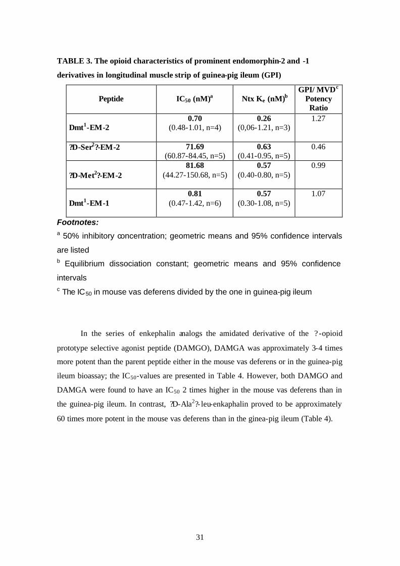
TABLE 3. The opioid characteristics of prominent endomorphin-2 and -1
derivatives in longitudinal muscle strip of guinea-pig ileum (GPI)
Peptide
IC50 (nM)a
Ntx Ke (nM)b
GPI/ MVDc Potency Ratio
Dmt1-EM-2
0.70 (0.48-1.01, n=4)
0.26 (0,06-1.21, n=3)
1.27
?D-Ser2?-EM-2 71.69 (60.87-84.45, n=5)
0.63 (0.41-0.95, n=5)
0.46
?D-Met2?-EM-2
81.68 (44.27-150.68, n=5)
0.57 (0.40-0.80, n=5)
0.99
Dmt1-EM-1
0.81 (0.47-1.42, n=6)
0.57 (0.30-1.08, n=5)
1.07
Footnotes: a 50% inhibitory concentration; geometric means and 95% confidence intervals
are listed
b Equilibrium dissociation constant; geometric means and 95% confidence
intervals
c The IC50 in mouse vas deferens divided by the one in guinea-pig ileum
In the series of enkephalin analogs the amidated derivative of the ? -opioid
prototype selective agonist peptide (DAMGO), DAMGA was approximately 3-4 times
more potent than the parent peptide either in the mouse vas deferens or in the guinea-pig
ileum bioassay; the IC50-values are presented in Table 4. However, both DAMGO and
DAMGA were found to have an IC50 2 times higher in the mouse vas deferens than in
the guinea-pig ileum. In contrast, ?D-Ala2?- leu-enkaphalin proved to be approximately
60 times more potent in the mouse vas deferens than in the ginea-pig ileum (Table 4).

32
TABLE 4. The opioid characteristics of enkephalin derivatives, (D-Ala2, Leu-
enkephalin, DAMGO and its Gly5-NH2 congener) in longitudinal muscle strip of
guinea-pig ileum (GPI) and mouse vas deferens (MVD)
GPI MVD
Peptide
IC50 (nM)a
Ntx Ke (nM)b
IC50 (nM)a
Ntx Ke (nM)b
?D-Ala2?Leu-enkephalin 72.8 (57.6-91.4)
n=4
0.54 (0.35-0.83)
n=4
1.18 (0.97-1.43)
n=6
6.20 (4.96-7.74)
n=6 DAMGO 29.2
(17.5-48.6) n=12
0.30 (0.27-0.33)
n=5
63.96 (46.45-88.07)
n=9
0.33 (0.29-0.37)
n=6 DAMGA
7.99 (4.95-12.9)
n=6
0.31 (0.25-0.39)
n=4
18.32 (14.11-23.79)
n=13
0.40 (0.26-0.59)
n=6 Footnotes: a 50% inhibitory concentration; geometric means and 95% confidence intervals
are listed
b Equilibrium dissociation constant; geometric means and 95% confidence
intervals
33
B.) Determination of receptor constants for ? -opioid receptor agonists in
mouse vas deferens.
From the pilot experiments, the chosen concentration of ? -funaltrexamine was
5x10⁻⁷ M. It caused 34.1 ? 1.4 (n=56, arithmetic mean ? S.E.M.) inhibition, with a
moderate tendency of recovery throughout the 30 min exposure at the endpoint. After
40-60 minutes and 8-12 washes there was a recovery to 94.9 ? 1.9% of control; no
exclusion was necessary since recovery was higher than 80% in each experiment.
Figure 4 illustrates the dose response curves before and after exposure to 5x10⁻⁷ M
? -FNA for endomorphin-1 and endomorphin-1-ol (panel A), DAMGA and DAMGO
(panel B) in MVD. Concentration-effect curves for certain compounds such as
endomorphin-2 and endomorphin-2-ol, morphine and normorphine obtained in the same
preparation and under the same conditions are presented in fig. 5, 6 respectively.

34
Fig. 4. The inhibitory dose-response curves of endomorphin-1 (? , ? ) and
endomorphin-1-ol (?, ¦ ) (panel A, n=6); DAMGA (? , ? ) and DAMGO (?, ¦ ) (panel B, n=4 and 5 respectively) in the mouse vas deferens. Points represent the arithmetic mean, vertical lines the S.E. Symbols: open: values obtained before ?-FNA (control); dark: values obtained after 30 min exposure to 5x10⁻⁷ M ?-FNA.
Concentration (M)
1e-9 1e-8 1e-7 1e-6 1e-5 1e-4
Inh
ibit
ion
(%
)
0
20
40
60
80
100 A
Concentration (M)1e-9 1e-8 1e-7 1e-6 1e-5 1e-40
20
40
60
80
100
Inh
ibit
ion
(%
)
B

35
A
Concentration (M)
1e-9 1e-8 1e-7 1e-6 1e-5
Inh
ibiti
on
(%
)
0
20
40
60
80
100
B
Concentration (M)
1e-9 1e-8 1e-7 1e-6 1e-5
Inh
ibiti
on
(%
)
0
20
40
60
80
100
Fig. 5. The inhibitory dose-response curves of ? - opioid receptor agonists
in mouse vas deferens before and after ? -funaltrexamine treatment.
Panels: A: endomorphin-2 (n=5); B: endomorphin-2-ol (n=6). Points represent the arithmetic mean, vertical lines the S.E. Symbols: open: values obtained before ?-FNA (control); dark: values obtained after 30 min exposure to 5x10⁻⁷ M ?-FNA.

36
A
Concentration (M)
1e-8 1e-7 1e-6 1e-5 1e-4
Inh
ibiti
on
(%)
0
20
40
60
80
100
B
Concentration (M)
1e-8 1e-7 1e-6 1e-5
Inh
ibiti
on
(%)
0
20
40
60
80
100
Fig. 6. The inhibitory dose-response curves of ? - opioid receptor agonists in mouse vas deferens before and after ? -funaltrexamine treatment. Panels: A: Morphine (n=4); B: Normorphine (n=4). Points represent the arithmetic mean, vertical lines the S.E. Symbols: empty before (control) and full after 5x10⁻⁷ M ?-FNA treatment.

37
Pretreatment of the vasa with 5x10⁻⁷ M ? -FNA caused a rightward shift, slope
reduction and in some cases a considerable Emax reduction of agonist dose response
curves. Nevertheless, the scope of these changes was different for the various subsets of
agonists.
To obtain receptor constants for the agonists according to the method described
by Furchgott and Bursztyn ?42?, first equieffective concentrations were chosen and
plotted as reciprocals for individual, paired experiments (for details see Methods); such
plots are shown in figure 7 for endomorphin-1 (panel A), endomorphin-1-ol (panel B)
and in figure 8 for DAMGO (panel A) and DAMGA (panel B).
From the double reciprocal plots and their dose response curves the control IC50 values,
KA, q and KA/ IC50 ratio were obtained (Table 5).

38
Fig. 7. Double reciprocal plots of equieffective concentrations before
and after treatment the tissue with ? -FNA for endomorphin-1 (panel A), endomorphin-1-ol (panel B) in MVD (single experiment). 1/A and 1/A’ are the reciprocal of equieffective agonist concentrations before and after ?-FNA treatment respectively.
1/A'
0,000 0,005 0,010 0,015 0,020 0,0250,00
0,02
0,04
0,06
0,08
0,10
0,12
0,14
The method of partial irreversible blockade (by b-FNA) for determining the dissociation
A1/A
1/A'
0,0000 0,0005 0,0010 0,0015 0,0020 0,00250,000
0,005
0,010
0,015
0,020
0,025
0,030
0,035
0,040 B1/A

39
Fig. 8. Double reciprocal plots of equieffective concentrations before and after treatment the tissue with ? -FNA for DAMGO (panel A) and DAMGA (panel B) in the MVD (single experiment). 1/A and 1/A’ are the reciprocal of equieffective agonist concentrations before and after ?-FNA treatment respectively.
A
1/A'
0,0000 0,0001 0,0002 0,0003 0,0004 0,0005
1/A
0,0000
0,0005
0,0010
0,0015
0,0020
0,0025
0,0030
0,0035
0,0040
0,0045
0,0050
B
1/A'0,000 0,002 0,004 0,006 0,008 0,010 0,012 0,014
1/A
0,00
0,02
0,04
0,06
0,08
0,10
0,12
0,14
0,16
0,18

40
TABLE 5. The receptor constants of some ? -receptor agonists in isolated MVD
No Compound (n)a IC50(nM)b KA (nM)c q (%)d KA/IC50 1 Normorphine 4 178.3
(139.6-227.8) 1,748
(1,542-1,981) 14.4
(11.8-18.9) 9.80
(8.11-11.8) 2 Morphine 4 219.3
(158.4-280.9) 685.4
(323.6-1,452) 12.7
(7.94-20.3) 3.25
(1.93-5.46) 3 DAMGO 5 50.1
(41.5-60.6) 728
(489-1,220) 16.9
(12.5-22.8) 15.4
(10.3-23.2) 4 DAMGA 4 18.50
(16.66-20.54) 355
(274-461) 14.1
(11.0, 13.5) 19.2
(14.6-25.2) 5 Morphiceptin 4 2,636
(2,330-2,983) 36,245
(28,967-45,350) 12.5e
(11.6-13.5) 13.8
(9.82-19.3) 6 EM-2 5 17.5
(15.3-20.0) 117.2
(74.8-183.5) 17.5
(15.3-20) 6.69
(4.21-10.6) 7 EM-2-ol 6 26.3
(20.4-34.0) 92.6
(68.2-125.8) 27.9
(22.2-35.2) 3.51
(2.53-4.86) 8 ?D-Ser2?-EM-2 4 71.1
(60.2-83.9) 2,220
(1,462-3,370) 3.6
(2.6-4.9) 29.7
(22.3-39.6) 9 ?D-Met2?-EM-2 4 40.7
(21.8-76.0) 465.3
(319.5-677.5) 11.2
(7.68-16.3) 11.2
(9.07-13.8) 10 EM-1 6 23.7
(19.0-29.6) 38.4
(19.2-77.0) 45.1
(33.2-61.1) 1.62
(0.94-2.81) 11 EM-1-ol 6 105.31
(80.52-137.74)
632 (465-859)
14.0 (10.3-18.9)
5.58 (4.66-6.68)
12 Dmt1-EM1-1 6 1.41
(1.08-1.84) 4.83
(2.66-7.09) 30.0f
(19.9-45.5) 3.01g
(1.57-6.08) Footnotes aNumber of experiments b50% inhibitory concentrations; geometric means and 95% confidence intervals cApparent dissociation constant of agonist; geometric means and 95% confidence intervals dActive residual receptor fraction; geometric means and 95% confidence intervals STATISTICS q: for compound "10" q is different at p<0.01 from all the others for compound "12" q is different from 4,8,9,11 (p<0.01) and 1,2,3,5 (p<0.05) for compound "7" q is different from 4,9,11 (p<0.05) and 8 (p<0.01) for compound "8" q is different from 7,10,12 (p<0.01) KA/IC50: for compound "8" is different at p<0.01 from all but "4" for compound "3" is different from 2,7,8,10,11,12 (p<0.01) and 6 (p<0.05) for compound "4" is different from 2,7,10,11,12 (p<0.01) and 6 (p<0.05) for compound "5" is different from 8,10,12 (p<0.01) and 2,7 (p<0.05)

41
Likewise, we determined the receptor constants for the rest of agonists in Table
5. The efficacies of drugs were calculated as the ratio of KA over IC50. The IC50 were
extended between 1.41 and 2,632 nM whereas the KA values between 4.83 and 36,245
nM (Table 5); these sets of data were not analyzed statistically. The efficacy values
were ranged between 1.62 and 29.7 (Table 5). For most of agonists, the fractions of
viable receptors left in the tissues after ? -FNA treatment (q) fall into the 11.2-17.5%
range whereas significantly higher values for three agonists and very low value for one
(Table 5).
?D-Ser2?-endomorphin-2 had apparently a prominent high efficacy value among
the other agonists. This tendency prompted us to check its validity in the rat vas
deferens, which contains a ? -opioid receptor- like receptor supply with very low
receptor reserve ?155?. Therefore, only high-efficacy ? -opioid receptor agonists can be
expected to be effective in this preparation. DAMGO was found to be highly effective
agonist whereas ?D-Ser2?-endomorphin-2 was a weak agonist in isolated rat vas
deferens (Al-Khrasani and Rónai, unpublished). The possible reasons for this difference
will be treated in detail in the discussion part. Thus, when we assessed the rank order of
agonist efficacies, ?D-Ser2?-endomorphin-2 was excluded from the evaluation. Taking
the efficacy of DAMGO as 100, the rank order of the relative efficacies of the agonists
were DAMGA? DAMGO ? morphiceptin? ?D-Met2?-EM-2 ? normorphine ? EM-2 ?
EM-1-ol ? Dmt1-EM-1 ? morphine ? EM-2-ol ? EM-1
(Table 6). Statistically, the results of this analysis suggest that DAMGO, DAMGA,
morphiceptin behave as the full agonists, ?D-Met2?-EM-2 and normorphine are possibly
full agonists whereas EM-2, EM-1-ol, Dmt1-EM-1, morphine, EM-2-ol and EM-1 as the
partial agonists.

42
TABLE 6. The relative efficacies of some ? -receptor agonists in the mouse vas deferense, based on the KA/IC50 ratios
Agonist Relative efficacya (nb)
DAMGO 100c
DAMGA 118.6 ? 15.9 4 Morphiceptin 86.9? 15.3 4
?D-Met?2-EM-2 68.1? 7.7 4
Normorphine 59.5? 5.8 4 EM-2 44.1? 8.7? 5
EM-1-ol 36.8? 3.1?? 4
?Dmt1?-EM-1 23.1? 6.3?? 6 Morphine 22.1? 6.1?? 4
EM-2-ol 22.1? 3.2?? 6
EM-1 11.1? 2.1?? 6 Footnotes aThe efficacy of DAMGO was taken as 100. Arithmetic mean ? S.E.M bNumber of experiments cThe KA/IC50 value for DAMGO was 16.8 ? 2.8 and n=4 ? p?0.05 vs DAMGA; ? ? p?0.05 vs. both DAMGO and DAMGA by ANOVA followed by LSD test.
The reduction in the effectiveness of ? -opioid receptor agonist deltorphin-II was
moderate though statistically significant (88.3 ? 1.5 % inhibition before vs 67.7 ? 2.4 %
after ? -funaltrexamine exposure at 10-9 M concentration of agonist; p?0.05, n=16,
paired “t” test).
43
THE MODULATORY EFFECTS OF ENDOMORPHINS AND DAMGO ON
THE FIELD STIMULATION-INDUCED 3H-NOREPINEPHRINE RELEASE
FROM ADULT RAT NUCLEUS TRACTUS SOLITARII-DORSAL MOTOR
VAGAL NUCLEUS SLICES
Applying two electrical field stimulations 30 min apart (S1 and S2) to induce 3H-NE release from adult rat nucleus tractus solitarii-dorsal motor vagal nucleus (NTS-
DVN) slices results in S2 over S1 ratio of 1.05 value for control
(Table.7 and fig 9, 10).
Fraction number
2 3 4 5 6 7 8 9 10 11 12 13 14 15 16 17 18 19 20 21
3 H-n
ore
pin
eph
rin
e re
leas
e(%
of
tiss
ue
con
ten
t / f
ract
ion
)
0
1
2
3
S1 S2
Fig. 9. The effects of field electrical stimulation on 3H-NE release from rat NTS-DVN slices (control, n=4). Stimulations indicated by S1 and S1 (red bars). Points represent the mean ? S.E.M.
The ? 2-adrenoceptor receptor agonist clonidine (10-6M) reduced this ratio to
0.34 % (Table 7 and fig 10).

44
DAMGO caused a dose dependent and naloxone-reversible inhibitory effect on
electrical stimulus induced release of the 3H-NE from (NTS-DVN) slices
(fig.10 and 11, Table 7).
Fig. 10. The effects of clonidine and DAMGO on the stimulation-
induced 3H-NE release from adult rat NTS-DVN. Data are expressed as
mean? S.E.M. * p?0.05; ** p?0.01.
S2/S1: the ratio of area under the curve after stimulation S2 and S1,
respectively.
a- compared to the control (white column)
b- compared to 10-6 DAMGO (3d, red column)
0
0.2
0.4
0.6
0.8
1
1.2
CONTROL
CLONIDINE
10-6 M DAMGO
10-5 M DAMGO
10-5 M DAMGO+1
0-6 M NX
**a
*b
**a
n=3
n=4
n=4
n=3
n=3
S2/S1
10-6 M

45
Fraction number
2 3 4 5 6 7 8 9 10 11 12 13 14 15 16 17 18 19 20 21
3 H-n
ore
pin
eph
rin
e re
leas
e(%
of t
issu
e co
nte
nt /
frac
tio
n)
0
1
2
3
S1 S2
A
Fraction number
2 3 4 5 6 7 8 9 10 11 12 13 14 15 16 17 18 19 20 21
3H
-no
rep
inep
hri
ne
rele
ase
(% o
f tis
sue
con
ten
t / fr
acti
on
)
0
1
2
3
4
S1 S2
B
Fig. 11. The naloxone-reversible inhibitory effect of DAMGO on the stimulation-induced 3H-norepinephrine release from rat nucleus tractus solitarii-dorsal motor vagal nucleus slices. Panel A: 10-5 M DAMGO (open bar), panel B: 10-5 M DAMGO+ 10-6 M NX (open bar + dark blue bar). Points represent the arithmetic mean, vertical lines, the S.E.values obtained in three independent experiments. Stimulation cycles indicated by S1 and S2 (red bars).

46
In the absence of dipeptidyl aminopeptidase (DAP-IV) inhibitor (Diprotin A)
endomorphin-1 at the concentrations of 10-6 M and 10-5 M caused S2/S1 ratios of 0.81
and 0.64 respectively whereas these figures for endomorphin-2 0.71 and 0.59. The
inhibitory effect of both peptides at the concentration of 10-6M was enhanced in the
presence of diprotin A (fig.12 and Table 7). However the presence of diprotin A did not
influence the inhibitory effect of endomorphins at the concentration of 10-5M. This
effect appeared to have a ceiling at an S2/S1 of 0.60 (Table 7).

47
A
2 3 4 5 6 7 8 9 10 11 12 13 14 15 16 17 18 19 20 210
1
2
3
S1 S2
3 H-n
ore
pin
eph
rin
e re
leas
e(%
of
tissu
e co
nte
nt
/ fra
ctio
n)
Fraction number
B
2 3 4 5 6 7 8 9 10 11 12 13 14 15 16 17 18 19 20 210
1
2
3
4
S1 S2
3H
-no
rep
inep
hri
ne
rele
ase
(% o
f ti
ssu
e co
nte
nt
/ fra
ctio
n)
Fraction number Fig.12 The inhibitory effect of endomorphins on the stimulation-induced
3H-norepinephrine release from adult rat nucleus tractus solitarii-dorsal motor vagal nucleus slices in the presence of DAP-IV enzyme inhibitor diprotin A (green bar). Panel A: 10-5M endomorphin-1 (black bar); panel B: 10-5M endomorphin-2 (pink bar). Slices were stimulated at S1 and S2 (red bars). Points represent the arithmetic mean, vertical lines, the S.E. Values obtained in four independent experiments.

48
Diprotin A slightly but significantly enhanced the stimulation- induced 3H-NE
release, the AUC (S1) values being 2.26? 0.18 (n=32) in the absence and
3.42?0.41(n=16) in the presence of enzyme inhibitor (p<0.01). The presence of diprotin
A added 12 min before the second stimulation did not effect the S2/S1 ratio.
Fig. 13. Summary of the S2/S1 ratios in the presence and absence of
Diprotin A in adult rat NTS-DVN slices.
Data are presented as mean ?S.E.M obtained from 4 animals with exception 10-
6M EM-1 (n=6). ** p?0.01;
S2/S1: the ratio of area under the curve after stimulation S2 and S1,
respectively.
a- compared to the control (white column)
b- compared to diprotin A (2nd, hatched column)
0
0.5
1
1.5
CONTROL
10-4 M
dipro
tin A
10-6 M EM
-2
10-5 M EM
-2
10-6 M EM
-1
10-6 M EM
-1+ 10
-4 M di
protin
A
10-5 M EM
-1
**a**b**a **a
**b**a
**a**b
**a**a**b
S2/S12
10-5 M EM
-1+ 10
-4 M di
protin
A
10-6 M EM
-2+ 10
-4 M di
protin
A
10-5 M EM
-2+ 10
-4 M di
protin
A

49
TABLE 7.The effect of ? -opioid receptor agonists and clonidine on the stimulation- induced release of 3H-norepinephrine from adult rat nucleus tractus solitarii-
dorsal vagal nucleus slices No Druga Conc.
(M) N S2/S1
b Statistics LSDc-test
1 None ---- 4 1.047 (0.946-1.159)
-------
2 Clonidine 10-6 3 0.336 (0.230-0.490)
p?0.01 vs. 1
3 DAMGO 10-6 4 0.726 (0.590-0.892)
p?0.01 vs. 2
4 DAMGO 10-5 3 0.483 (0.355-0.657)
P?0.01 vs. 1; p?0.05vs. 3
5 DAMGO +Naloxone
10-5
10-6 3 0.950
(0.840-1.073) p?0.01 vs. 4
6 Diprotin A 10-4 4 1.249 (0.922-1.816)
7 EM-1 10-6
6 0.806
(0.576-1.258) p?0.01 vs. 2
8 EM-1 +Diprotin A
10-6
10-4 4 0.613
(0.594-0.683) p?0.01 vs. 1; p?0.01 vs. 6;
p?0.01 vs. 2
9 EM-1 10-5 4 0.642 (0.512-805)
p?0.01 vs. 1; p?0.01 vs. 2
10 EM1 +Diprotin A
10-5
10-4 4 0.675
(0.547-0.837) p?0.01 vs. 1; p?0.01 vs. 6;
p?0.01 vs. 2 11 EM-2 10-6
4 0.710
(0.465-1.085) p?0.01 vs. 1; p?0.01 vs. 2
12 EM-2 +Diprotin A
10-6
10-4 4 0.665
(0.500-0.886) p?0.01 vs. 1; p?0.01 vs. 6;
p?0.01 vs. 2
13 EM-2 10-5
4 0.590
(0.571-0.609) p?0.01 vs. 1; p?0.05vs. 2
14 EM-2 +Diprotin A
10-5
10-4 4 0.557
(0.309-1.009) p?0.01 vs. 1; p?0.01 vs. 6;
p?0.05vs. 2 Footnotes aDrugs were added 15 min before S2 with the exception of diprotin A which was added 28 min before S1. bThe ratio of area under the curve after stimulation S2 and S1, respectively, geometric means and 95% confidence intervals are listed. cANOVA followed by Least Significant Differences test.

50
DISCUSSION
ANALYSIS OF OPIOID PROPERTIES IN ISOLATED ORGANS
A.) General opioid pharmacology of endomorphin-related peptides
Since one of the major aims was to determine the opioid characteristics of novel
endomorphin derivatives in isolated organs, the principle of measurement and the
"thumb rules" of characterization should be discussed first. The assay systems were
neurally (field) stimulated smooth muscle preparations containing presynaptically
located inhibitory ? , ? or ? type of opioid receptors. The MVD contains all the three
major opioid receptor types whereas GPI contains functional ? and ? types ?9; 82; 86;
175?. Agonist potencies can be characterized by the presynaptic inhibitory effects,
expressed most conveniently in terms of 50 % inhibitory concentrations (IC50). The
receptor types where the agonists exert their actions could be determined by analyzing
the interaction with the moderately ? receptor type-preferring competitive opioid
antagonist naltrexone. The affinity of naltrexone to the preferred i.e. ? receptor type can
be characterized by a Ke falling into the 0.2-0.6 nM range ?48; 139?. The affinity of
antagonist towards both the ? and ? opioid receptors is 20-30 times lower ?48; 136?.
Therefore, if an agonist-naltrexone interaction in MVD where all the three opioid
receptor types are present yields a naltrexone Ke in the 0.2-0.6 nM range it can be
declared that the agonist exerted its action at the ? -opioid receptors in the concentration
range used in the interaction analyses. If the antagonist Ke is conspicuously and also
statistically different from the indicated range then the agonist is likely to act also at
other (i.e. ? or ?) opioid receptor types.
Since the MVD contains a dominant ? receptor pool with a considerable spare
fraction whereas GPI lacks functional ? receptors, ? -receptor-preferring agonists tend to
be more potent in MVD than in GPI ?86; 82?. On the other hand, the spare ? and ?
receptor pool is much higher in GPI than in MVD ?100?. Consequently, ? receptor-
preferring agonists ?89? and ? partial agonists are likely to be more potent in GPI than
in MVD.
The novel endomorphin derivatives were modified in positions 1, 2 and at the
C-terminus. C-terminal free carboxylic function in the endomorphin series, similarly to
enkephalin-related peptides ?137; 138? and morphiceptin, reduced agonist potency and

51
also the selectivity at ? -opioid receptors. In the case of EM-2 with free carboxylic
terminus, in contrast to the morphiceptin-related analogous pair, there was a clear
indication of ? receptor type selection. One should recall that
Tyr-Tic-Phe-Phe-OH (TIPP, ?114; 146?) has been reported to possess selective ? -opioid
receptor antagonist properties. Aromatic moiety in position 4 is more advantageous for
opioid activity than an aliphatic one (Leu, Ser, D-Ser) or proline. Alcoholic C-terminal
function, similarly to the previous experience acquired in the enkephalin-analogue
series, provides almost identical advantages in potency and selectivity at ? -opioid
receptors as compared to C-terminal amidation.
Introduction of a hydroxyl into position 3' of tyrosine1 resulted in moderate loss
in potency. If, however, ? -methylation was accompanied by the introduction of a
hydroxyl into position 3' of aromatic ring, the reduction in agonist potency was
approximately of 500-times. One may surmise that ? -methylation may cause an
unfavorable change in the steric position of one of phenolic hydroxyl in the aromatic
ring.
2', 6'-dimethylation on Tyr1, presumably partly due to an increased
hydrophobicity, partly due to an induced conformational constraint on the side chain is
known to enhance the affinity of resulting structures to more than one type of opioid
receptors ?5; 14; 21; 47; 113; 126; 141; 142?. In the endomorphin series, it resulted in
analogues with subnanomolar IC50 values and, as judged from the slow washout pattern,
in slow dissociation kinetics.
Substitution of Pro2 by Hyp or Ser cause a 100-fold loss of potency. On the
other hand, cycloserine, D-Ser or D-Met substitution causes no or only a minor loss in
potency. One should realize that the tetrapeptides containing an aliphatic D-amino acid
in position 2 cannot be designated as endomorphin analogues in the strictest sense; the
opioid structural motif bears a much closer resemblance to amphibian skin opioid
peptides.
In general and for the purposes of next discussion section in particular, it should
be pointed out that, as judged from their interaction with naltrexone, all endomorphin
analogues containing C-terminal amide or alcohol function, preserved the ? -opioid
receptor type-selectivity of parent natural peptides. Because of the slow association-

52
dissociation kinetics of Dmt1-derivatives, there is an ambiguity as far as the extent of
their ? receptor type selectivity is concerned.
B.) Determination of receptor constants for ? -opioid receptor agonists in
mouse vas deferens.
When studying E/(A) relationships (commonly referred to as dose-response
relationships) of agonists in isolated tissues it is done with the understanding that the
determined agonist potency is the result of a complex chain of receptor- and tissue-
related functions ?160; 165?. It is a long-held wish of pharmacologis ts to devise
methods suitable for determine agonist affinities by pharmacological means for all sets
of agonists and also to characterize the agonist property referred to as "intrinsic
efficacy" ?43; 167?. Methods for determining the affinity of pure, competitive receptor
antagonists are readily available ?8; 73; 144; 166; 175?; it is also feasible to calculate the
affinities of a subset of partial agonists ?73?. A method, based on the fractional
inactivation of the receptor pool in a given tissue has been devised by Furchgott and
Bursztyn ?42? and was used successfully thereafter to establish the affinities and
efficacies of a wide range of agonists ?37; 131; 132; 151?.
In the opioid field, the fumaramate derivative of naltrexone, ? -funaltrexamine
has been reported as a potentially useful tool for the irreversible inactivation of ? -opioid
receptors ?163?. It is an irreversibly inactivating antagonist at ? , to a much lesser extent
at ? opioid receptors and also a reversible ?-opioid receptor agonist ?39; 52; 176; 177?.
The agent has been used previously to determine receptor constants of a limited number
of opioid agonists ?15; 24; 80; 127; 128; 150?.
My aim was to analyze pharmacologically the partial agonist properties of
endomorphins, substantiated recently by biochemical means ?3; 61; 81; 154? and also to
create a comprehensive receptor constant data set for a number of ? -opioid receptor
agonists with particular reference to Tyr-Pro-related agonists.

53
When I decided to use ? -FNA as a research tool, the choice fell on MVD as the
assay system for the following reasons:
1- The MVD has been reported to possess a lower ? -opioid receptor reserve as
compared to GPI ?82?. This factor was assumed to facilitate the functional reflection of
receptor inactivation.
2- The MVD is known to possess a much lower ? receptor pool than the GPI ?82?; this
factor was expected to diminish interference from ?-receptor agonism by ? -FNA.
The delicate issue in MVD is the presence of a highly effective set of ? -opioid
receptors. Therefore, comparable data set could be obtained only for those agonists
where the extent of ? -opioid receptor selectivity is satisfactorily high. This criterion,
with one possible exception, appeared to have been met for the agonists studied.
? -FNA treatment caused a rightward shift, slope reduction and, in some cases, a
marked Emax reduction in agonist dose-response curves but the extent of these changes
was different for the different substes of agonists. After having calculated the receptor
constants (KA and residual receptor fraction "q"), as an indirect validation attempt, we
sought correlation between the displacing IC50 against 3H-naloxone in the rat brain
receptor binding assay in the presence of Na+ and the KA values determined by
pharmacological means (Rónai et al., submitted). For the purposes of correlation we
collected receptor-binding data issued from the same laboratory (Institute of
Biochemistry, Biological Research Center, Szeged). With the exception of data pair for
?D-Ser2?-EM-2 the correlation was excellent; taking into account even the deviant data
pair for correlational analysis, the correlation was found significant.
The residual receptor fraction after ? -FNA treatment fell into the 11-18 % range
for the majority of agonists. If ? -FNA would have affected uniformly the accessibility
of binding sites, the same residual binding capacity range could be expected for all ? -
opioid receptor agonists; this, apparently, was not the case. A high residual receptor
fraction was calculated for a number of endomorphin derivatives, whereas, apparently, a
low residual fraction for ?D-Ser2?-EM-2.
When the efficacy-related ratios (KA/IC50) were calculated, setting up a rationale
for subdivision of agonists into full- and partial subsets was hindered by the apparently
very high efficacy obtained for ?D-Ser2?-EM-2. Since in the case of this analogue

54
ambiguity arose even in the context of KA we decided to extend to the rat vas deferens
(Al-Khrasani and Rónai, in preparation) where a highly dominant ? -opioid receptor
population is thought to be present ?155? with a very low receptor reserve. In this
preparation DAMGO and DAMGA behaved as full agonists whereas ?D-Ser2?-EM-2
had a very low efficacy. The Ke determined for ?D-Ser2?-EM-2 in this preparation was
rather close to the KA calculated in MVD. To give full explanation to these phenomena
will require more extensive analyses. It appears, however, justified to exclude the
KA/IC50 ratio calculated for the peptide in MVD when setting up the subdivision of
agonists into full and partial agonist subsets. By using the statistical comparisons of
KA/IC50 ratios, the proposed subsets are as follows: DAMGO and its amide, DAMGA
are clearly full agonists. This rating is likely to hold also for morphiceptin. ?D-Met2?-
EM-2 and normorphine are also possible full agonists although to decide more firmly
their status, further refinement might be required. All the other agonists fall into the
partial agonist category.
It is also apparent that subsets may exist even among the partial agonists. The
most striking feature is the high residual receptor fraction for a number of partial agonist
endomorphin derivatives. It has been suggested that different sets of ? -opioid agonists
may use distinct "binding pockets" or binding site combinations on the opioid receptor
molecule ?76; 77; 103; 118; 130?. The high residual receptor fraction may indicate that
this might be the case also for some endomorphin-derivatives.
THE MODULATORY EFFECTS OF ENDOMORPHINS AND DAMGO ON
THE FIELD STIMULATION-INDUCED 3H-NOREPINEPHRINE RELEASE
FROM ADULT RAT NUCLEUS TRACTUS SOLITARII-DORSAL MOTOR
VAGAL NUCLEUS SLICES
This area of rat lower brainstem has been shown to contain several endogenous
opioid peptide sets (enkephalins, dynorphins, ? -endorphin and endomorphins) ?1; 93;
94; 123? as well as all the three major opioid receptor types ?91?.
Arakawa et al. ?7? have reported the inhibition of stimulation- induced norepinephrine
release by DAMGO in this preparation.

55
My results fully confirmed the naloxone-reversible inhibitory effect of
DAMGO. EM-1 and EM-2 displayed also inhibitory actions; however, in contrast to
DAMGO, the inhibition by endomorphins appeared to level off between 10-6-10-5 M
concentration. It should be mentioned that none of these opioid agonists was as potent
inhibitor as the ? 2-adrenoceptor agonist clonidine. The norepinephrine released from
this preparation may originate from local A2 neurons but also from projection neurons
of locus coeruleus and rostral ventrolateral medulla ?120; 173?. It is possible all the
processes releasing norepinephrine, irrespective of their source, are endowed by
inhibitory ? 2 autoreceptors whereas only a subset contains ? -opioid receptors.
One of the possible reasons for the flattening of the concentration-response
curve of a drug, particularly if the agent is a peptide, is the potential inactivation by
tissue enzymes. This possibility appeared to be rather unlikely in the light of recent
reports on the remarkable resistance of endomorphins against various peptidases ?169?.
A characteristic potential cleaving site in endomorphins is splitting after Pro2 by
dipeptidyl-aminopeptidase IV (DAP-IV, EC 3. 4. 14. 5) ?98; 122; 139?. Therefore, I
tested the effect of endomorphins in the presence of enzyme inhibitor diprotin A (Ile-
Pro-Ile, ?172?. As expected, the enzyme inhibitor was only marginally effective at lower
endomorphin concentration and not at all at 10-5M of peptide concentration. Although
further experiments are needed to fully justify the conclusion, there is a probability that
the "flattening" of endomorphin action at the higher concentration may be due to the
partial agonism also in NTS-DVN slice preparation.

56
CONCLUSIONS
All endomorphin analogs as well as DAMGA are ? -opioid receptor-selective
agonists with exception the derivative with a free C-terminus.
The methylation of Tyr1 at position 2’ and 6’ enhanced both the activity and
affinity of endomorphins without interfering with the selectivity.
Methylation at the ? -carbon atom accompanied by the introduction of a
hydroxyl group into position 3' of aromatic ring of Tyr1 is apparently unfavorable for
the opioid receptors.
Substitution of Pro2 by D-Ser, D-Met or cycloSer in endomorphin-2 results in
analogs with agonist activity comparable to that of the parent peptide whereas
substitution by L-Ser or Hyp caused a marked decrease in the agonist activity.
Amidated Phe in position 4 of endomorphin-2 is requirable both for the agonist
activity and selectivity of the peptide.
Alcohol function at the C-terminal of na tural endomorphins cause only a minor
reduction in the agonist activity and preserves the selectivity of parent peptides.
Irreversible partial inactivation of ? -receptor pool revealed that DAMGO,
DAMGA and morphiceptin are full agonists whereas EM-1, EM-2, EM-1-ol, EM-2-ol
and morphine are partial agonists but ?D-Met?2-EM-2 and normorphine may be a full
agonists. It appears that the presence of Pro in position 2 and the aromatic amino acid
(Phe) in position 4 are prerequisites of partial agonism.
For some partial agonist endomorphins a significantly higher residual receptor
fraction remained available after ß-FNA treatment than either for the morphinan-based
agonists, enkephalin-based agonists or another Tyr-Pro – based agonist, morphiceptin.
This may be taken as a possible indication that these peptides act, at least in part, at
different region(s) of the receptor molecule as compared to the other agonists.
At the central level, electrical field stimulus evoked 3H-NE release from adult
rat NTS-DVN revealed that DAMGO is a full agonist but endomorphins might behave
differently. Additionally, these results confirm the presence of the functional ? -opioid
receptors in this region of CNS.

57
ACKNOWLEDGEMENTS
This study was carried out at the department of Pharmacology and Pharmacotherapy,
Semmelweis University Budapest, Hungary.
I would like to express my thanks to my supervisor Professor Susanna Fürst, Head of
the Department of Pharmacology and Pharmacotherapy for the possibility to
perform my Ph. D. dissertation work and for here continuous support during my
work.
Further, I wish to thank deeply Dr. András Rónai, for introducing me into the field of
experimental pharmacology and for his permanent and valuable theoretical and
practical advices.
In addition, my thanks go to Dr. Tamas Friedmann and Professor Dr. Klára Gyires
for their professional advices.
I would also like to thank:
My colleagues Dr. Katalin Müllner, Katalin Fülöp, Jeno Balogh, Antal Gulyás and
Lilla Gabriel.
For all those individuals who have kindly given help to me during my work.
Finally, I wish to thank my family for their patience and continuos support.
This study was supported by ETT 182/97 KO, 262/2001 KO, OTKA TD-32736, ETT
156/2000.
58
REFERENCES
1. Akil, H., Watson, SJ., Young, E., Lewis, ME., Khachaturian, H. and Walker, JM.,
1984. Endogenous opioid: biology and function. Ann. Rev. Neurosci. 7, 223-255.
2. Al Khrasani,M., Orosz,G., Kocsis,L., Farkas,V., Magyar,A., Lengyel,I., Benyhe,S.,
Borsodi,A., Ronai,A.Z., 2001. Receptor constants for endomorphin-1 and
endomorphin-1-ol indicate differences in efficacy and receptor occupancy. Eur.J
Pharmacol 421, 61-67.
3. Alt, A., Mansour, A., Akil, H., Medzihradsky, F., Traynor, J. R., Woods, J. H.,
1998. Stimulation of guanosine-5'-o- (3-[35S] thio) triphosphate binding by
endogenous opioid acting at a cloned mu receptor. J. Pharmacol. Exp. Ther. 286,
282-288.
4. Amiche, M., Defour, A., Nicolas, P., 1988. Structural requirements for dermorphin
opioid receptor binding. Int. J. Pept. Protein Res. 32, 28-34.
5. Amodeo, P., Balboni, G., Crescenzi, O., Guerrini, R., Picone, D., Salvadori, S.,
Tancredi, T. and Temussi P. A., 1995. Conformational analysis of potent and very
selective ? opioid dipeptide antagonists. FEBS Lett. 377, 363-367.
6. Appel, NM., Kiritsy-Roy, JA., van Loon, GR., 1986. Mu receptors at discrete
hypothalamic and brainstem sites mediate opioid peptide- induced increases in
central sympathetic outflow. Brain Res. 378, 8-20.
7. Arakawa, K., De Jong, W., Mulder, A. H., Versteeg, D. H. G., 1991. The
electrically stimulated release of 3H-noradrenaline from nucleus tractus solitarii
slices in vitro is modulated via µ-opioid receptors. Eur. J. Pharmacol. 192, 311-
316.
8. Arunlakshana, O., Schild, H.O., 1959. Some quantitative uses of drug antagonists.
Br. J. Pharmacol. 33,48-58.
9. Berzetei-Gurske,I.P., Toll,L., 1992. The mu-opioid activity of kappa-opioid
receptor agonist compounds in the guinea pig ileum. Eur. J. Pharmacol 212, 283-
286.
10. Bloom,F., Battenberg,E., Rossier,J., Ling,N., Guillemin,R., 1978. Neurons
containing beta-endorphin in rat brain exist separately from those containing
enkephalin: immunocytochemical studies. Proc.Natl.Acad.Sci.U.S.A 75, 1591-
1595.

59
11. Bódi, J., Süli-Vargha, H., Ludányi, K., Vékey, K., Orosz, G., 1997. New strategy
for the synthesis of large peptides as applied to the C-terminal Cysteine-rich 41
amino acid fragment of the mouse agouti protein. Tetrahedron Lett. 38, 3293-3296.
12. Brantl, V., Gramsch, C., Lottspeich, F., Mertz, R., Jaeger, K. H., Herz, A., 1986.
Novel opioid peptides derived from hemoglobin: hemorphins. Eur. J. Pharmacol.
125, 309-310.
13. Broccardo, M., Erspamer, V., Falconieri Erspamer, G., Improta, G., Linari, G.,
Melchiorri, P., Montecucchi, PC., 1981. Pharmacological data on dermorphins, a
new class of potent opioid peptides from amphibian skin. Br. J. Pharmacol. 73,
625-631.
14. Bryant, S. D., Salvadori, S., Cooper, P.S., Lazarus, L.H., 1998. New delta-opioid
antagonists as pharmacological probes. Trends. Pharmacol. Sci. 19, 42-46.
15. Carroll, J. A., Shaw, J. S., Wickenden, A. D., 1988. The physiological relevance of
low agonist affinity binding at opioid mu-receptors. Br. J. Pharmacol 94, 625-631.
16. Champion, HC., Zadina, JE., Kastin, AJ., Hackler, L., Ge, LJ., Kadowitz, PJ.
1997a. Endomorphin 1 and 2, endogenous ligands for the mu-opioid receptor,
decrease cardiac output, and total peripheral resistance in the rat. Peptides. 18,
1393-1397.
17. Champion, HC., Zadina, JE., Kastin, AJ., Kadowitz, PJ., 1997b. The endogenous
mu-opioid agonists, endomorphin 1 and 2, have vasodilator activity in the
hindquarters vascular bed of the rat. Life Sci. 61 (26), PL 409-PL 415.
18. Champion, HC., Zadina, JE., Kastin, AJ., Hackler, L., Ge, LJ., Kadowitz, PJ.
1997c. The endogenous mu-opioid receptor agonists endomorphins 1 and 2 have
novel hypotensive activity in the rabbit. Biochem. Biophys. Res. Commun. 235 (3),
567-570.
19. Champion, HC., Zadina, JE., Kastin, AJ., Kadowitz, PJ. 1998a. Endomorphin 1 and
2 have vasodepressor activity in the anesthetized mouse. Peptides. 19 (5), 925-929.
20. Champion, HC. and Kadowitz, PJ. 1998b. D-[Ala2]endomorphin 2 and
endomorphin 2 have nitric oxide-dependent vasodilator activity in rats. Am. J.
Physiol. 274, H1690-H1697.
21. Chandrakumar, NS., Stapelfeld, A., Beardsley, PM., Lopez, OT., Drury, B.,
Anthony, E., Savage, MA., Williamson, LN., Reichman, M.,1992. Analogs of the

60
delta opioid receptor selective cyclic peptide [2-D-penicillamine, 5-D-
penicillamine]-enkephalin: 2’, 6’-dimethyltyrosine and Gly3-Phe4 amide bond
isostere substitutions. J. Med. Chem. 1992 35, 2928-2938.
22. Chang, KJ., Lillian, A., Hazum, E., Cuatrecasas, P., Chang, JK., 1981.
Morphiceptin (-NH4Tyr-Pro-Phe-Pro-COH2): a potent and specific agonist for
morphin (mu) receptors. Sci. 212, 75-77.
23. Chavkin, C., James, I.F. and Goldstein, A., 1982. Dynorphin is a specific
endogenous ligand of the ?-opioid receptor. Science 215, 413-415.
24. Chavkin,C., Goldstein, A., 1984. Opioid receptor reserve in normal and morphine-
tolerant guinea pig ileum myenteric plexus. Proc.Natl.Acad.Sci.U.S.A 81, 7253-
7257.
25. Chen, Y., Mestek, A., Liu, J., Hurley, JA., Yu, L., 1993. Molecular cloning and
functional expression of a mu-opioid receptor from rat brain. Mol. Pharmacol. 44,
8-12.
26. Connor, M., Schuller, A., Pintar, JE., Christie, MJ. 1999. Mu-opioid receptor
modulation of calcium channel current in periaqueductal grey neurons from
C57B16/J mice and mutant mice lacking MOR-1. Br. J. Pharmacol. 126, 1553-
1558.
27. Czapla, MA., Champion, HC., Zadina, JE., Kastin, AJ., Hackler, L., Ge, LJ.,
Kadowitz PJ. 1998. Endomorphin 1 and 2, endogenous mu-opioid agonists,
decrease systemic arterial pressure in the rat. Life Sci. 62, PL 175-PL 179.
28. Czapla, MA., Gozal, D., Alea, OA., Beckerman, RC., Zadina, JE., 2000.
Differential cardiorespiratory effects of endomorphin 1 and endomorphin 2,
DAMGO and morphine. Am. J. Respir. Crit. Care Med. 162: 994-999.
29. De Jong, W., Petty, MA., Sitsen, JM., 1983. Role of opioid peptides in brain
mechanisms regulating blood pressure. Chest. 83, 306-308.
30. Elde, R., Hokfelt, T., Johansson,O., Terenius,L., 1976. Immunohistochemical
studies using antibodies to leucine-enkephalin: initial observations on the nervous
system of the rat. Neuroscience 1, 349-351.
31. Elde, R. and Hökfelt, T., 1993. Coexistence of Opioid Peptides with Other
Neurotransmitters. In Handbook of Experimental Pharmacology. (ed.: Herz, A.),
Vol. 104/I. pp 585-624.

61
32. Emmerson, PJ., Clark, MJ., Mansour, A., Akil, H., Woods, JH., Medzihradsky, F.,
1996. Characterization of opioid agonist efficacy in a C6 glioma cell line
expressing the mu opioid receptor. J. Pharmacol. Exp. Ther. 278, 1121-1127.
33. Erchegyi, J., Kastin, AJ., Zadina, JE., 1992. Isolation of a novel tetrapetide with
opiate and antiopiate activity from human brain cortex: Tyr-Pro-Trp-Gly-NH2
(Tyr-W-MIF-1). Peptides 13, 623-631.
34. Erchegyi,J., Zadina,J.E., Qiu,X.D., Kersh,D.C., Ge,L.J., Brown,M.M., Kastin,A.J.,
1993. Structure-activity relationships of analogs of the endogenous brain peptides
Tyr-MIF-1 and Tyr-W-MIF-1. Pept. Res. 6, 31-38.
35. Erspamer, V., Melchiorri, P., Falconier-Erspamer, G., Negri, L., Corsi, C.,
Severini, C., Barra, D., Simmaco, M., Kreil, G., 1989. Deltorphins: a family of
naturally occuring peptides with high affinity and selectivity for ? -opioid binding
sites. Natl. Acad. Sci. USA 86, 5188-5192.
36. Evans, CJ., Keith, DE Jr., Morrison, H., Magendzo, K., Edwards, RH., 1992.
Cloning of a delta opioid receptor by functional expression. Science. 258, 1882-
1884.
37. Flavahan, N. A., Vanhoutte, P. M., 1986. Alpha-1 and alpha-2 adrenoceptor:
response coupling in canine saphenous and femoral veins. J Pharmacol Exp Ther
238, 131-138.
38. Fleming, W.W., Westfall, D.P., De la Lande, I.S., Jellett, L.B., 1972. Log-normal
distribution of equieffective doses of norepinephrine and acetylcholine in several
tissues. J. Pharmacol. Exp. Ther. 181, 339-345.
39. Franklin, T.G., Traynor, J.R., 1991. Alkylation with ß-funaltrexamine suggests
differences between µ-opioid receptor systems in guinea-pig brain and myenteric-
plexus. Br. J. Pharmacol. 102, 718-722.
40. Fukuda, K., Kato, S., Mori, K., Nishi, M., Takeshima, H., 1993. Primary structures
and expression from cDNAs of rat opioid receptor delta- and mu-subtypes. FEBS
Lett. 327, 311-314.
41. Furchgott, R. F., 1966. The use of ? -haloalkylamines in the differentiation of
receptors and in the determination of dissociation constants of receptor-agonist
complexes. In Advances in Drug Research. N. J. Harper, and A. B. Simmonds,
Eds. 3: 21-55. Academic Pess, Inc. New York, N. Y.

62
42. Furchgott, R. F., Bursztyn, P., 1967. Comparison of dissociation constants and of
relative efficacies of selected agonists acting on parasympathetic receptors. Ann. N.
Y. Acad. Sci. 144, 882-899.
43. Furchgott, R.F., 1978. Pharmacological characterization of receptors: its relation to
radioligand-binding studies. Fed Proc 37: 115-120.
44. Goldberg, IE., Rossi, GC., Letchworth, SR., Mathis, JP., Ryan-Moro, J., Leventhal,
L., Su, W., Emmel, D., Bolan, EA., Pasternak, GW., 1998. Pharmacological
characterization of endomorphin-1 and endomorphin-2 in mouse brain. J.
Pharmacol. Exp. Ther. 286, 1007-1013.
45. Goldstein, A., Tachibana, S., Lowney, L.I., Hunkapillar, M. and Hood, L., 1979.
Dynorphin-(1-13), an extraordinarily potent opioid peptide. Proc. Natl. Acad. Sci.
USA 76, 6666-6670.
46. Gong, J., Strong, JA., Zhang, S., Yue, X., DeHaven, RN., Daubert, JD., Cassel,
JA., Yu, G., Mansson, E., Yu, L. 1998. Endomorphins fully activate a cloned
human mu opioid receptor. FEBS Lett. 439, 152-156.
47. Guerrini, R., Capasso, A., Sorrentino, L., Anacardio, R., Bryant, SD., Lazarus, LH.,
Attila, M., Salvadori, S., 1996. Opioid receptor selectivity alteration by single
residue replacement: synthesis and activity profile of [Dmt1]deltorphin B. Eur. J.
Pharmacol. 302, 37-42.
48. Gyires, K., Ronai, A.Z., Toth, G., Darula, Z., Furst, S., 1997. Analysis of the role
of delta opioid receptors in gastroprotection in the rat. Life Sci. 60, 1337-1347
49. Hackler, L., Kastin, AJ., Erchegyi, J., Zadina, JE., 1993. Isolation of Tyr-W-MIF-1
from bovine hypothalami. Neuropeptides 24,159-164.
50. Hackler, L., Zadina, JE., Ge, LJ., Kastin, AJ.,1997. Isolation of relatively large
amounts of endomorphin-1 and endomorphin-2 from human brain cortex.
Peptides.18, 1635-1639.
51. Harrison, L.M., Kastin, AJ., Zadina, JE., 1998. Differential effects of
endomorphin-1, endomorphin-2, and Tyr-W-MIF-1 on activation of G-proteins in
SH-SY5Y human neuroblastoma membranes. Peptides. 19, 749-753.
52. Hayes, A.G., Sheehan, M.J., Tyers, M.B., 1985. Determination of the receptor
selectivity of opioid agonist in the guinea-pig ileum and mouse vas deferens by use
of ß-funaltrexamine. Br. J.Pharmacol. 86, 899-904.

63
53. Henry, DJ., Grandy, DK., Lester, HA., Davidson, N., Chavkin, C., 1995. Kappa-
opioid receptors couple to inwardly rectifying potassium channels when
coexpressed by Xenopus oocytes. Mol. Pharmacol. 47, 551-557.
54. Henschen, A., Lottspeich, F., Brantl, V., Teschemacher, H., 1979. Novel opioid
peptide derived from casein (beta-casomorphins). II. Structure of active
components from bovine casein peptone. Hoppe-Seyler’s Z. Physiol. Chem. 360,
1217-1224.
55. Hescheler, J., Rosenthal, W., Trautwein, W., Schultz, G., 1987. The GTP-binding
protein, Go, regulates neuronal calcium channels. Nature. 325, 445-447.
56. Higashida, H., Hoshi, N., Knijnik, R., Zadina, JE., Kastin, AJ. 1998. Endomorphins
inhibit high-threshold Ca2+ channel currents in rodent NG108-15 cells
overexpressing mu-opioid receptors. J. Physiol. 15; 507 (Pt 1): 71-75.
57. Hilf, G., Gierschik, P., Jakobs, KH., 1989. Muscarinic acetylcholine receptor-
stimulated binding of guanosine 5’-O-(3-thiotriphosphate) to guanine-nucleotide-
binding proteins in cardiac membranes. Eur. J. Biochem. 186, 725-731.
58. Horvath, A., Kastin, AJ., 1989. Isolation of tyrosine-melanocyte-stimulating
hormone release- inhibiting factor 1 from bovine brain tissue. J. Biol. Chem. 264,
2175-2179.
59. Horvath, A. and Kastin, A.J., 1990. Evidence for presence of Tyr-MIF-1(Tyr-Pro-
Leu-Gly-NH2) in human brain cortex. Int. J. Pept. Protein Res. 36, 281-284.
60. Horvath, G., 2000. Endomorphin-1 and endomorphin-2: pharmacology of the
selective endogenous mu-opioid receptor agonists. Pharmacol Ther 88, 437-463.
61. Hosohata, K., Burkey, TH., Alfaro-Lopez, J., Varga, E., Hruby, VJ., Roeske, WR.,
Yamamura, HI., 1998. Endomorphin-1 and endomorphin-2 are partial agonists at
the human mu-opioid receptor. Eur. J. Pharmacol. 346, 111-114
62. Hruby, V. J., Gehrig, C. A., 1989. Recent developments in the design of receptor
specific opioid peptides. Med. Res. Rev. 9, 343-401.
63. Hughes, J., Smith, T. W., Kosterlitz, L.H., Fothergill, L. A., Morgan, B. A., Morris,
H. R., 1975. Identification of two related pentapeptides from the brain with potent
opiate agonist activity. Nature, 258, 577-580.

64
64. Jin, W., Lee, NM., Loh, HH., Thayer, SA., 1992. Dual excitatory and inhibitory
effects of opioids on intracellular calcium in neuroblastoma x glioma hybrid
NG108-15 cells. Mol. Pharmacol. 42, 1083-1089.
65. Johnson, PS., Wang, JB., Wang, WF., Uhl, GR., 1994. Expressed mu opiate
receptor couples to adenylate cyclase and phosphatidyl inositol turnover.
NeuroReport. 5, 507-509.
66. Kakidani, H., Furutani, Y., Takahashi, H., Noda, M., Morimoto, Y., Hirose, T.,
Asai, M., Inayama, S., Nakanishi, S., Numa, S., 1982. Cloning and sequence
analysis of cDNA for porcine beta-neo-endorphin/ dynorphin precursor. Nature.
298, 245-249.
67. Kakizawa, K., Shimohira, I., Sakurada, S., Fujimura, T., Murayama, K., Ueda, H.
1998. Parallel stimulations of in vitro and in situ [35S]GTPgammaS binding by
endomorphin 1 and DAMGO in mouse brains. Peptides. 19, 755-758.
68. Khachaturian, H., Schaefer, M. K. H. and Lewis, M. E., 1993. Anatomy and
Function of the Endogenous Opioid System. In Handbook of Experimental
Pharmacology. (ed.: Herz, A.) Vol. 104/I. pp 471-497.
69. Kieffer, BL., Befort, K., Gaveriaux-Ruff, C., Hirth, CG., 1992. The delta-opioid
receptor: isolation of a cDNA by expression cloning and pharmacological
characterization. Proc. Natl. Acad. Sci. U S A. 89, 12048-12052.
70. Kieffer, BL.,1995. Recent advances in molecular recognition and signal
transduction of active peptides: receptors for opioid peptides. Cell. Mol. Neurobiol.
15, 615-635.
71. Kilpatrick, D. L., 1993. Opioid Peptide Expression in Peripheral Tissues and Its
Functional Implications. In Handbook of Experimental Pharmacology. (ed.: Herz,
A.), Vol. 104/II., pp 551-570.
72. Kocsis, L., Benyhe, S., Borsodi, A., Rónai, AZ., AL-Khrasani, M., Magyar, A.,
Orosz, G., 2001. Endomorphin related peptide alcohols: structure-activity studies.
In Peptides 2000. (eds.: Jean Martinez and Jean-Alain Fehrentz), EDK, Paris,
France 2001. pp 823-824.
73. Kosterlitz, H.W., Watt, A.J., 1968. Kinetic parameters of narcotic agonists and
antagonists with particular reference to N-allylnoroxymorphone (naloxone). Br. J.
Pharmacol. 33, 266-276.

65
74. Kosterlitz, H.W., Paterson, S.J., 1981. Tyr-D-Ala-Gly-MePhe-NH(CH2)2OH is a
selective ligand for the µ-opiate binding site. Br. J. Pharmacol. 73,299.P.
75. Kreil, G., Barra, D., Simaco, M., Erspamer, V., Erspamer, GF., Negri, L., Severini,
C., Corsi, R., Melchiorri, P., 1989. Deltorphin, a novel amphibian skin peptide with
high selectivity and affinity for ? -opioid receptors. Eur. J. Pharmacol. 162, 123-
128.
76. Law, P.Y., Loh, H. H., 1999. Regulation of opioid receptor activities. J Pharmacol
Exp Ther 289, 607-624.
77. Law, P. Y., Wong, Y. H., Loh, H. H., 1999. Mutational analysis of the structure
and function of opioid receptors. Biopolymers 51, 440-455.
78. Law, P. Y., Wong, Y. H., Loh, H. H., 2000. Molecular mechanisms and regulation
of opioid receptor signaling. Annu. Rev. Pharmacol Toxicol. 40, 389-430.
79. Leander, S., Ekman, R., Uddman, R., Sundler, F., Hakanson, R., 1984. Neuronal
cholecystokinin, gastrin-releasing peptide, neurotensin, and beta-endorphin in the
intestine of the guinea pig. Distribution and possible motor functions. Cell Tissue
Res. 235, 521-531.
80. Leff, P., Dougall, I. G., 1988. Estimation of opioid receptor agonist dissociation
constants with beta-chlornaltrexamine, an irreversible ligand which also displays
agonism. Br.J Pharmacol 95, 234-240.
81. Lengyel, I., Orosz, G., Biyashev, D., Kocsis, L., Al Khrasani, M., Ronai, A.,
Tomboly, C., Furst, Z., Toth, G., Borsodi, A., 2002. Side chain modifications
change the binding and agonist properties of endomorphin 2. Biochem. Biophys.
Res. Commun. 290, 153-161.
82. Leslie, F. M., 1987. Methods used for the study of opioid receptors. Pharmacol
Rev. 39, 197-249.
83. Lewis, J., Mansour, A., Khachaturian, H., Watson, S. J., Akil, H., 1987. Opioids
and pain regulation. Pain Headache 9, 129-159.
84. Li, C. H. and Chung, D., 1976. Isolation and structure of an untriakontapeptide
with opiate activity from camel pituitary glands. Proc. Natle. Acad. Sci. USA 73,
1145-1148.

66
85. Li, S., Zhu, J., Chen, C., Chen, YW., Deriel, JK., Ashby, B., Liu-Chen, LY.,1993.
Molecular cloning and expression of a rat kappa opioid receptor. Biochem. J. 295,
629-633.
86. Lord, J.A.H., Waterfield, A.A., Hughes, J. and Kosterlitz, H.W., 1977. Endogenous
opioid peptides: multiple agonists and receptors. Nature 267, 495-499.
87. Lorenzen, A., Fuss, M., Vogt, H, Schwabe, U., 1993. Measurement of guanine
nucleotide-binding protein activation by A1 adenosine receptor agonists in bovine
brain membranes: stimulation of guanosine-5’-O-(3-[35S]thio)triphosphate
binding. Mol. Pharmacol. 44, 115-123.
88. Lorenzen, A., Guerra, L., Vogt, H., Schwabe, U., 1996. Interaction of full and
partial agonists of the A1 adenosine receptor with receptor/G protein complexes in
rat brain membranes Mol. Pharmacol. 49, 915-926.
89. Mako, E., Ronai, A.Z., 2001. Characterization of kappa and delta opioid receptors
in isolated organs by using type/subtype selective agonists and antagonists. Med.
Sci. Monit. 7, 350-356.
90. Mansour, A., Khachaturian, H., Lewis, M.E., Akil, H., Watson, S.J., 1988.
Anatomy of CNS opioid receptors. Trends Neurosci. 11, 308-314.
91. Mansour, A., Fox, CA., Burke, S., Meng, F., Thompson, RC., Akil, H., Watson, SJ.
1994. Mu, delta, and kappa opioid receptor mRNA expression in the rat CNS: an in
situ hybridization study. J. Comp. Neurol. 350, 412-438.
92. Martin, WR., Eades, CG., Thompson, JA., Huppler, RE., Gilbert, PE., 1976. The
effects of morphine- and nalorphine- like drugs in the nondependent and morphine-
dependent chronic spinal dog. J. Pharmacol. Exp. Ther. 197, 517-532.
93. Martin-Schild, S., Zadina, J.E., Gerall, S., Vigh, S., Kastin, A.J., 1997. Localization
of endomorphin-2 like immunoreactivity in the rat medulla and spinal cord.
Peptides. 18, 1641-1649.
94. Martin-Schild, S., Gerall, A.A., Kastin, A.J., Zadina, J.E., 1999. Differential
distribution of endomorphin-1 and endomorphin-2- like immunoreactivities in the
CNS of the rodent. J. Comp. Neurol. 405, 450-471.
95. Matthes, HW., Maldonado, R., Simonin, F., Valverde, O., Slowe, S., Kitchen, I.,
Befort, K., Dierich, A., Le Meur, M., Dolle, P., Tzavara, E., Hanoune, J., Roques,
BP., Kieffer, BL. 1996. Loss of morphine- induced analgesia, reward effect and
67
withdrawal symptoms in mice lacking the mu-opioid-receptor gene. Nature. 31;
383 (6603): 819-23.
96. McConalogue, K., Grady, EF., Minnis, J., Balestra, B., Tonini, M., Brecha, NC.,
Bunnett, NW., Sternini, C. 1999. Activation and internalization of the mu-opioid
receptor by the newly discovered endogenous agonists, endomorphin-1 and
endomorphin-2. .Neuroscience. 90, 1051-1059.
97. Meng, F., Xie, GX., Thompson, RC., Mansour, A., Goldstein, A., Watson, SJ.,
Akil, H., 1993. Cloning and pharmacological characterization of a rat kappa opioid
receptor. Proc. Natl. Acad. Sci. U S A. 90, 9954-9958.
98. Mentlein, R., 1999. Dipeptidyl-peptidase IV (CD26)-role in the inactivation of
regulatory peptides. Regul. Pept. 85: 9-24.
99. Meunier, J. C., Mollereau, C., Toll, L., Suaudeau, C., Moisand, C., Alvinerie, P.,
Butour, J. L., Guillemot, J. C., Ferrara, P., Monsarrat, B., 1995. Isolation and
structure of the endogenous agonist of opioid receptor- like ORL1 receptor. Nature
377, 532-535.
100. Miller, L., Shaw, J. S., Whiting, E. M., 1986. The contribution of intrinsic activity
to the action of opioids in vitro. Br. J. Pharmacol. 87, 595-601.
101. Mima, H., Morikawa, H., Fukuda, K., Kato, S., Shoda, T., Mori K. 1997. Ca2+
channel inhibition by endomorphins via the cloned mu-opioid receptor expressed in
NG108-15 cells.
102. Minami, M., Toya, T., Katao, Y., Maekawa, K., Nakamura, S., Onogi, T., Kaneko,
S., Satoh, M., 1993. Cloning and expression of a cDNA for the rat kappa-opioid
receptor. FEBS. Lett. 329, 291-295.
103. Minami, M., Nakagawa,T., Seki,T., Onogi,T., Aoki,Y., Katao,Y., Katsumata,S.,
Satoh,M., 1996. A single residue, Lys108, of the delta-opioid receptor prevents the
mu-opioid-selective ligand [D-Ala2, N-MePhe4, Gly-ol5]enkephalin from binding
to the delta-opioid receptor. Mol. Pharmacol 50, 1413-1422.
104. Mizoguchi, H., Wu, HE., Narita, M., 2001. Partial agonistic action of
endomorphins in the mouse spinal cord. Neurosci. Lett. 310, 66-68.
105. Monory, K., Bourin, MC., Spetea, M., Tomboly, C., Toth, G., Matthes, HW.,
Kieffer, BL., Hanoune, J., Borsodi, A. 2000. Specific activation of the mu opioid

68
receptor (MOR) by endomorphin 1 and endomorphin 2. Eur. J. Neurosci. 12(2),
577-584.
106. Montecucchi, PC., de Castiglione, R., Erspamer, V., 1981a. Identification of
dermorphin and Hyp6-dermorphin in skin extracts of the Brazilian frog
Phyllomedusa rhodei. Int. J. Pept. Protein. Res. 17, 316-321.
107. Montecucchi, PC., de Castiglione, R., Piani, S., Gozzini, L., Erspamer, V., 1981b.
Amino acid composition and sequence of dermorphin, a novel opiate- like peptide
from the skin of Phyllomedusa sauvagei. Int. J. Pept. Protein. Res. 17, 275-283.
108. Monteillet-Agius, G., Fein, J., Anton, B., Evans, C. J., 1998. ORL-1 and mu opioid
receptor antisera label different fibers in areas involved in pain processing. J. Comp
Neurol. 399, 373-383.
109. Nakanishi S, Inoue A, Kita T, Nakamura M, Chang AC, Cohen SN, Numa S.,
1979. Nucleotide sequence of cloned cDNA for bovine corticotropin-beta-
lipotropin precursor. Nature. 278, 423-427.
110. Narita, M., Mizoguchi, H., Oji, G.S., Tseng, E.L., Suganuma, C., Nagase, H.,
Tseng, L.F., 1998. Characterization of endomorphin-1 and –2 on (35S) GTPgamma
S binding in the mouse spinal cord. Eur. J. Pharmacol. 351, 383-387.
111. Narita, M., Mizoguchi, H., Narita, M., Sora, I., Uhl, GR., Tseng, LF. 1999.
Absence of G-protein activation by mu-opioid receptor agonists in the spinal cord
of mu-opioid receptor knockout mice. Br. J. Pharmacol. 126, 451-456.
112. Narita, M., Mizoguchi, H., Narita, M., Dun, NJ., Hwang, BH., Endoh, T., Suzuki,
T., Nagase, H., Suzuki, T., Tseng, LF., 2000. G protein activation by endomorphins
in the mouse periaqueductal gray matter. J Biomed Sci. 7, 221-225.
113. Neilan, C. L., Nguyen, T. M., Schiller, P. W., Pasternak, G. W., 2001.
Pharmacological characterization of the dermorphin analog [Dmt (1)] DALDA, a
highly potent and selective mu-opioid peptide. Eur.J Pharmacol 419, 15-23.
114. Nevin, S. T., Toth, G., Nguyen, T. M., Schiller, P. W., Borsodi, A., 1993. Synthesis
and binding characteristics of the highly specific, tritiated delta opioid antagonist
[3H]TIPP. Life Sci. 53, L57-L62. Schiller, P.W., Weltrowska,G., Nguyen,T.M.,
Wilkes,B.C., Chung,N.N., Lemieux,C., 1993. TIPP[psi]: a highly potent and stable
pseudopeptide delta opioid receptor antagonist with extraordinary delta selectivity.
J Med. Chem. 36, 3182-3187.

69
115. Nishiwaki, H., Saitoh, N., Nishio, H., Takeuchi, T. and Hata, F. 1998. relationship
between inhibitory effect of endogenous opioid via mu-receptors and muscarinic
autoinhibition in acetylcholine release from myenteric plexus of guinea pig ileum.
Jpn. J. Pharmacol. 77, 279-286.
116. Noda, M., Furutani, Y., Takahashi, H., Toyosato, M., Hirose, T., Inayama, S.,
Nakanishi, S., Numa, S., 1982. Cloning and sequence analysis of cDNA for bovine
adrenal preproenkephalin. Nature. 295, 202-206.
117. North, RA., Williams, JT., Surprenant, A., Christie, MJ., 1987. Mu and delta
receptors belong to a family of receptors that are coupled to potassium channels
Proc. Natl. Acad. Sci. U S A. 84, 5487-5491.
118. Onogi, T., Minami, M., Katao, Y., Nakagawa, T., Aoki, Y., Toya, T., Katsumata,
S., Satoh, M., 1995. DAMGO, a mu-opioid receptor selective agonist, distinguishes
between mu- and delta-opioid receptors around their first extracellular loops. FEBS
Lett. 357, 93-97.
119. Orosz, G., Kiss, L. P., 1998. Simple and efficient synthesis of 2-chlorotritylchloride
resin. Tetrahedron Lett. 39,3241-3242.
120. Palkovits, M., 1999. Interconnections between the neuroendocrine hypothalamus
and the central autonomic system. Frontiers in Neuroendocrinology 20, 270-295.
121. Paton, W.D.M., Vizi, E.S., 1969. The inhibitory action of noradrenaline and
adrenaline on acetylcholine output by guinea-pig longitudinal muscle strips. Br. J.
Pharmacol.35, 10-28.
122. Peter, A., Tóth, G., Tömböly, C., Laus, G., Tourwe, D., 1999. Liquid
chromatographic study of the enzymatic degradation of endomorphins, with
identification by electrospray ionization mass spectrometry. J. Chromatogr. A.
846: 39-49.
123. Pierce, TL., Wessendorf, MW. 2000. Immunocytochemical mapping of
endomorphin-2-immunoreactivity in rat brain. J. Chem. Neuroanat. 18, 181-207.
124. Piros, ET., Prather, PL., Loh, HH., Law, PY., Evans, CJ., Hales, TG. 1995. Ca2+
channel and adenylyl cyclase modulation by cloned mu-opioid receptors in GH3
cells.
70
125. Piros, ET., Prather, PL., Law, PY., Evans, CJ., Hales, TG., 1996. Voltage-
dependent inhibition of Ca2+ channels in GH3 cells by cloned mu- and delta-
opioid receptors. Mol. Pharmacol. 50, 947-956.
126. Pitzele, BS., Hamilton, RW., Kudla, KD., Tsymbalov, S., Stapelfeld, A., Savage,
MA., Clare, M., Hammond, DL., Hansen, DW. Jr., 1994. Enkephalin analogs as
systemically active antinociceptive agents: O- and N-alkylated derivatives of the
dipeptide amide L-2,6-dimethyltyrosyl-N-(3-phenylpropyl)-D-alaninamide. J. Med.
Chem. 37, 888-896.
127. Porreca, F., Burks, T. F., 1983. Affinity of normorphine for its pharmacologic
receptor in the naive and morphine-tolerant guinea-pig isolated ileum. J Pharmacol
Exp Ther 225, 688-693.
128. Porreca, F., LoPresti, D., Ward, SJ., 1990. Opioid agonist affinity in the guinea-pig
ileum and mouse vas deferens. Eur. J. Pharmacol. 179, 129-139.
129. Portoghese, P. S., Larson, D. L., Sayre, L. M., Fries, D. S., Takemori, A. E., 1980.
A novel opioid receptor site directed alkylating agent with irreversible narcotic
antagonistic and reversible agonistic activities. J Med. Chem. 23, 233-234.
130. Quock, R. M., Burkey, T. H., Varga, E., Hosohata, Y., Hosohata, K., Cowell, S.
M., Slate, C. A., Ehlert, F. J., Roeske, W. R., Yamamura, H. I., 1999. The delta-
opioid receptor: molecular pharmacology, signal transduction, and the
determination of drug efficacy. Pharmacol Rev. 51, 503-532.
131. Raffa, R. B., Tallarida, R. J., Gero, A., 1979. Determination of the stimulus-
response relation for three alpha-adrenergic agonists on rabbit aorta.
Arch.Int.Pharmacodyn. Ther 241, 197-207.
132. Rama Sastry, B.V., Cheng, H.C., 1972. Dissociation constants of D- and L-
lactoylcholines and related compounds at cholinergic receptors. J Pharmacol Exp
Ther 180, 326-339.
133. Reinscheid, R. K., Nothacker, H. P., Bourson, A., Ardati, A., Henningsen, R. A.,
Bunzow, J. R., Grandy, D. K., Langen, H., Monsma, F. J. Jr., Civelli, O., 1995.
Orphanin FQ: a neuropeptide that activates an opioidlike G protein-coupled
receptor. Science 270, 792-794.
134. Rialas, C. M., Fimiani, C., Bilfinger, T. V., Salzet, M., Stefano, G. B., 1998.
Endomorphin-1 and -2 inhibit human vascular sympathetic norepinephrine release:

71
lack of interaction with mu 3 opiate receptor subtype. Zhongguo Yao Li Xue. Bao.
19, 403-407.
135. Rónai, A. Z., Gráf, L., Székely, J. I., Dunai-Kovács, Zs., Bajusz, S.,1977a.
Differential behaviour of LPH-(61-91)-peptide in different model systems:
comparison of the opioid activities of LPH-(61-91)-peptide and its fragments.
FEBS Lett. 74, 182-184.
136. Ronai, A. Z., Berzetei, I., Bajusz, S., 1977b. Differentiation between opioid
peptides by naltrexone. Eur. J Pharmacol 45, 393-394.
137. Ronai, A. Z., Szekely, J. I., Berzetei, I., Miglecz, E., Bajusz, S., 1979.
Tetrapeptide-amide analogues of enkephalin: the role of C-terminus in determining
the character of opioid activity. Biochem. Biophys. Res. Commun. 91, 1239-1249.
138. Ronai, A. Z., Berzetei, I. P., Bajusz, S., Szekely, J. I., 1981. The in vitro
pharmacology of D-Met2, Pro5-enkephalinamide. J Pharm. Pharmacol 33, 534-
535.
139. Rónai, A. Z., Timar, J., Makó, E., Erdo, F., Gyarmati, Zs., Tóth, G., Orosz, G.,
Fürst, S., Székely, J. I., 1998/99. Diprotin A, an inhibitor of dipeptidyl
aminopeptidase IV (EC 3.4.14.5) produces naloxone-reversible analgesia in rats.
Life Sci. 64: 145-152.
140. Rossi, A. C., De Castiglione, R., Perseo, G., 1986. Opioid receptor binding profile
of selected dermorphin- like peptides. Peptides 7, 755-759.
141. Salvadori, S., Attila, M., Balboni, G., Bianchi, C., Bryant, S. D., Crescenzi, O.,
Guerrini, R., Picone, D., Tancredi, T., Temussi, P. A., 1995. Delta opioidmimetic
antagonists: prototypes for designing a new generation of ultraselective opioid
peptides. Mol. Med. 1, 678-689.
142. Sasaki, Y., Suto, T., Ambo, A., Ouchi, H., Yamamoto, Y., 1999. Biological
properties of opioid peptides replacing Tyr at position 1 by 2,6-dimethyl-Tyr.
Chem Pharm. Bull. (Tokyo). 47, 1506-1509.
143. Sato, T., Sakurada, S., Sakurada, T., Furuta, S., Chaki, K., Kisara, K., Sasaki, Y.,
Suzuki, K., 1987. Opioid activities of D-Arg2-substituted tetrapeptides. J
Pharmacol Exp Ther 242, 654-659.
144. Schild, H. O., 1957. Drug antagonism and pAx. Pharmacol Rev. 9, 242-246.

72
145. Schiller, P. W., Nguyen, T. M., Chung, N. N., Lemieux, C., 1989. Dermorphin
analogues carrying an increased positive net charge in their "message" domain
display extremely high mu opioid receptor selectivity. J Med.Chem. 32, 698-703.
146. Schiller, P. W., Weltrowska, G., Nguyen, T. M., Wilkes, B. C., Chung, N. N.,
Lemieux, C., 1993. TIPP[psi]: a highly potent and stable pseudopeptide delta
opioid receptor antagonist with extraordinary delta selectivity. J Med.Chem. 36,
3182-3187.
147. Schwartzberg, D. G., Nakane, P. K., 1983. ACTH-related peptide containing
neurons within the medulla oblongata of the rat. Brain Res. 276, 351-356.
148. Selley, DE., Sim, LJ., Xiao, R., Liu, Q., Childers, SR., 1997. mu-Opioid receptor-
stimulated guanosine-5’-O-(gamma-thio)-triphosphate binding in rat thalamus and
cultured cell lines: signal transduction mechanisms underlying agonist efficacy.
Mol. Pharmacol. 51, 87-96.
149. Sharma, SK., Klee, WA., Nirenberg, M., 1977. Opiate-dependent modulation of
adenylate cyclase. Proc. Natl. Acad. Sci. U S A. 74, 3365-3369.
150. Sheehan, M. J., Hayes, A. G., Tyers, M. B., 1986. Pharmacology of delta-opioid
receptors in the hamster vas deferens. Eur.J Pharmacol 130, 57-64
151. Sheys, E. M., Green, R. D., 1972. A quantitative study of alpha adrenergic
receptors in the spleen and aorta of the rabbit. J Pharmacol Exp Ther 180, 317-325.
152. Shu-Dong, T., Phillips, D. M., Halmi, N., Krieger, D., Bardin, C. W., 1982. Beta-
endorphin is present in the male reproductive tract of five species. Biol. Reprod.
27, 755-764.
153. Sim, LJ., Selley, DE., Childers, SR., 1995. In vitro autoradiography of receptor-
activated G proteins in rat brain by agonist-stimulated guanylyl 5’-[gamma-
[35S]thio]-triphosphate binding. Proc. Natl. Acad. Sci. U S A. 92, 7242-7246.
154. Sim, LJ., Liu, Q., Childers, SR., Selley, DE.,1998. Endomorphin- stimulated
[35S]GTPgammaS binding in rat brain: evidence for partial agonist activity at mu-
opioid receptors. J Neurochem. 70, 1567-1576.
155. Smith, C. F., Rance, M. J., 1983. Opiate receptors in the rat vas deferens. Life Sci.
33 Suppl 1, 327-330.
156. Snedecor G W, Cochran W G. Statistical methods. Eighth edition, fifth printing.
Lowa State University Press, Ames 1994.

73
157. Soignier, RD., Vaccarino, AL., Brennan, AM., Kastin, AJ., Zadina, JE. 2000.
Analgesic effects of endomorphin-1 and endomorphin-2 in the formalin test in
mice. Life Sci. 67, 907-912.
158. Spencer, RJ., Jin, W., Thayer, SA., Chakrabarti, S., Law, PY., Loh, HH., 1997.
Mobilization of Ca2+ from intracellular stores in transfected neuro2a cells by
activation of multiple opioid receptor subtypes. Biochem. Pharmacol. 54, 809-818.
159. Spetea, M., Monory, K., Tomboly, Cs., Toth, G., Tzavara, E., Benyhe, S.,
Hanoune, J., Borsodi, A., 1998. In vitro binding and signaling profile of the novel
mu opioid receptor agonist endomorphin 2 in rat brain membranes. Biochem
Biophys Res Commun. 250, 720-725.
160. Stephenson, R. P., 1956. A modification of receptor theory. Br.J Pharmacol 11,
379-393.
161. Stone, LS., Fairbanks, CA., Laughlin, TM., Nguyen, HO., Bushy, TM.,
Wessendorf, MW., Wilcox, GL. 1997. Spinal analgesic actions of the new
endogenous opioid peptides endomorphin-1 and -2. Neuroreport. 8, 3131-3135.
162. Surprenant, A., Shen, KZ., North, RA., Tatsumi, H., 1990. Inhibition of calcium
currents by noradrenaline, somatostatin and opioids in guinea-pig submucosal
neurones.
163. Takemori, A.E., Larson, D.L., Portoghese, P.S., 1981. The irreversible narcotic
antagonistic and reversible agonistic properties of the fumarate methyl ester
derivative of naltrexone. Eur. J. Pharmacol. 70, 445-451.
164. Tallarida, R.J., Jacob, L.S., 1979. The dose-response relation in pharmacology.
Springer, New York, NY.
165. Tallarida, R.J., 1982. The use of drug-receptor affinity measures in the
differentiation of receptors. Fed. Proc.41, 2323-2327.
166. Tallarida, R.J., Robinson, M. J., Porreca, F., Cowan, A., 1982. Estimation of the
dissociation constant of naloxone in the naive and morphine-tolerant guinea-pig
isolated ileum: analysis by the constrained Schild plot. Life Sci. 31, 1691-1694.
167. Tallarida, R. J., 1988. Pharmacologic methods for identification of receptors. Life
Sci. 43, 2169-2176.

74
168. Tallent, M., Dichter, M. A., Bell, G. I., Reisine, T., 1994. The cloned kappa opioid
receptor couples to an N-type calcium current in undifferentiated PC-12 cells.
Neuroscience 63, 1033-1040.
169. Tomboly, C., Peter, A., Toth, G., 2002. In vitro quantitative study of the
degradation of endomorphins. Peptides 23, 1573-1580.
170. Tonini, M., Fiori, E., Balestra, B., Spelta, V., D'Agostino, G., Di Nucci, A., Brecha,
NC., Sternini, C. 1998. Endomorphin-1 and endomorphin-2 activate mu-opioid
receptors in myenteric neurons of the guinea-pig small intestine. Naunyn
Schmiedebergs Arch Pharmacol. 358(6),686-689.
171. Traynor, JR., Nahorski, SR., 1995. Modulation by mu-opioid agonists of
guanosine-5’-O-(3-[35S]thio)triphosphate binding to membranes from human
neuroblastoma SH-SY5Y cells. Mol. Pharmacol. 47, 848-854.
172. Umezawa, H., Aoyagi, T., Ogawa, K., Naganawa, H., Hamada, M., Takeuchi, T.,
1984. Diprotins A and B, inhibitors of dipeptidyl aminopeptidase IV, produced by
bacteria. J Antibiot.(Tokyo) 37, 422-425.
173. Van Giersbergen, PLM., Palkovits, M., De Jong, W., 1992. Involvement of
neurotransmitters in the nucleus tractus solitarii in cardiovascular regulation.
Physiol Rev. 72, 789-824.
174. Wang, Q. P., Zadina, J. E., Guan, J. L., Shioda, S., 2002. Morphological studies of
the endomorphinergic neurons in the central nervous system. Jpn.J.Pharmacol. 89,
209-215.
175. Ward, A. W. and Takemori, A. E., 1976. Studies on the narcotic receptor in the
guinea-pig ileum. J. Pharmac. Exp. Ther. 199, 117-123.
176. Ward, S. J., Portoghese, P. S., Takemori, A. E., 1982a. Pharmacological profiles of
ß-funaltrexamine (ß-FNA) and ß-chlornaltrexamine (ß-CNA) on the mouse vas
deferens preparation. Eur. J. Pharmacol. 80, 377-384.
177. Ward, S. J., Portoghese, P. S., Takemori, A. E., 1982b. Pharmacological
characterization in vivo of the novel opiate, ß- funaltrexamine. J. Pharmacol.Exp
Ther.220, 494-498.
178. Watson, S. J., Akil, H., 1980. alpha-MSH in rat brain: occurrence within and
outside of beta-endorphin neurons. Brain Res. 182, 217-223.
75
179. Yasuda, K., Raynor, K., Kong, H., Breder, CD., Takeda, J., Reisine, T., Bell, GI.,
1993. Cloning and functional comparison of kappa and delta opioid receptors from
mouse brain. Proc. Natl. Acad. Sci. U S A. 90, 6736-6740.
180. Zadina, J. E., Kastin, A. J., Kresh, D. and Wyatt, A., 1992. Tyr-MIF-1 and
hemorphin can act as opiate agonists as well as antagonists in guinea pig ileum.
Life Sci. 51, 869-885.
181. Zadina, J. E., Hackler, L., Ge, L. J., Kastin, A. J., 1997. A potent and selective
endogenous agonist for the mu-opiate receptor. Nature 386, 499-502.
182. Zhang,G., Murray,T.F., Grandy,D.K., 1997. Orphanin FQ has an inhibitory effect
on the guinea pig ileum and the mouse vas deferens. Brain Res. 772, 102-106.
76
RELEVANT PUBLICATIONS
Papers
1. Al-Khrasani, M., Elor, G., Yusuf, A. M., Ronai, A. Z., 2003. The effect of
endomorphins on the release of 3H-norepinephrine from rat nucleus tractus solitarii
slices. Regul Pept. 111, 97-101.
2. Al-Khrasani, M., Orosz, G., Kocsis, L., Farkas, V., Magyar, A., Lengyel, I.,
Benyhe, S., Borsodi, A., Ronai, A. Z., 2001. Receptor constants for endomorphin-1
and endomorphin-1-ol indicate differences in efficacy and receptor occupancy. Eur.J
Pharmacol 421, 61-67.
3. Lengyel, I., Orosz, G., Biyashev, D., Kocsis, L., Al-Khrasani, M., Ronai, A. Z,
Tomboly, C., Furst, Z., Toth, G., Borsodi, A., 2002. Side chain modifications
change the binding and agonist properties of endomorphin-2. Biochem. Biophys.
Res. Commun. 290, 153-161.
4. Kocsis, L., Benyhe, S., Borsodi, A., Rónai, A. Z, Al-Khrasani, M., Magyar, A.,
Orosz, G. Endomorphin-related peptide alcohols: structure-activity studies.
In: Peptides 2000 (Martinez J, Fehrentz J-A, eds.) EDK, Paris, pp. 823-824, 2001.
5. Rónai, A. Z., Kató, E., Al-Khrasani, M., Hajdú, M., Müllner, K., Elor, G., Gyires,
K., Fürst, S., Palkovits, M. Age and Monosodium Glutamate Treatment Cause
Changes in the Stimulation-Induced [3H]-Norepinephrine Release From Rat
Nucleus Tractus Solitarii-Dorsal Vagal Nucleus Slices. Life Sci., accepted.
6. Tömböly, C., Kövér, K., Péter, A., Tourwé, D., Biyashev, D., Borsodi, A.,
Al-Khrasani, M., Rónai, A. Z., Tóth, G. Structure-activity study on the Phe side-
chain arrangement of endomorphins using conformationally constrained analogues.
J. Med. Chem., accepted.

77
Abstracts
a.) Oral presentations
1. AL-Khrasani, M., Orosz, G., Tóth, G., Benyhe, S., Rónai, A. Z. The in vitro
analysis of the opioid properties of potent µ receptor type agonist endomorphins and
(D-Ala2, MePhe4, Gly5-ol) –enkephalin (DAMGO) and their analogs. Yemeni
Scientifc Research Foundation Science conference 2000, abstract vol., p. 13.
2. AL-Khrasani, M., Kocsis, L., Orosz, G., Tóth, G., Rónai, A. Z. Partial and full
agonism in Tyr-Pro-related opioid peptides and non-peptide opioids. YSRF Science
conference, Sanaa, Yemen, October 11-13, 2001, abstract vol., p. 97
3. AL-Khrasani, M., Orosz, G., Kocsis, L., Farkas, V., Magyar, A., Benyhe, S.,
Borsodi, A., Rónai, A. Z. Endomorphins and novel endomorphin analogs: general in
vitro pharmacology. In Scientific Bulletin International Multidisciplinary
Conference, organized by the North University of Baia Mare, Romania, 4th
EDITION, 16-22. May 25-26, 2001
4. Al-Khrasani, M., Elor, G., Kató, E., Rónai, A. Z. The effect of endomorphins and
DAMGO on the release of 3H-norepineprine from rat brain slices in vitro. 2003.
május 26-28.,Balatonszemes, Hungary
b.) Posters 1. Al-Khrasani, M., Elor, G., Abbas, M. Y., Rónai, A. Z. The effect of endomorphins
on 3H-NE release from rat nucleus tractus solitarii-dorsal vagal nucleus slices: a
further possible indication of partial agonism. European Opioid Conference (EOC),
Uppsala, Sweden, April 7-9, 2002, Opioid in drug dependence, behaviour and pain.
Abstract No (p37)
2. Kocsis, L., Lengyel, I., Borsodi, A., Rónai, A. Z., Al-Khrasani, M., Magyar, A.,
Orosz, G. Endomorphin analogue peptide. European Opioid Conference (EOC),
Uppsala, Sweden, April 7-9, 2002, Opioid in drug dependence, behaviour and pain.
Abstract No (p38)

78
IRREVELENT PUBLICATION
Borvendeg, S. J., Al-Khrasani, M., Rubini, P., Fischer, W., Allgaier, C., Wirkner, K.,
Himmel, H. M., Gillen, C., Illes, P., 2003. Subsensitivity of P2X but not vanilloid 1
receptors in dorsal root ganglia of rats caused by cyclophosphamide cystitis. Eur.J
Pharmacol 474, 71-75.