the open ophthalmology journal · anterior segment optical coherence tomography analysis the open...
TRANSCRIPT

Send Orders for Reprints to [email protected]
110 The Open Ophthalmology Journal, 2018, 12, 110-120
1874-3641/18 2018 Bentham Open
The Open Ophthalmology Journal
Content list available at: www.benthamopen.com/TOOPHTJ/
DOI: 10.2174/1874364101812010110
RESEARCH ARTICLE
Anterior Segment Optical Coherence Tomography Analysis of IrisMorphometric Changes Induced by Prostaglandin AnaloguesTreatment in Patients with Primary Open Angle Glaucoma or OcularHypertension
R Mancino1,*, E Di Carlo1, D Napoli1, A Martucci 1, A Mauro2, Sorge RP3, M Cesareo 1 and CNucci1
1Ophthalmology Unit, Department of Experimental Medicine and Surgery, University of Rome Tor Vergata, Rome, Italy2Department of Ophthalmology, “San Giovanni Evangelista” Hospital, Tivoli (RM), Italy3Laboratory of Biometry, Department of Systems Medicine, University of Rome Tor Vergata, Rome, Italy
Received: March 25, 2018 Revised: May 3, 2018 Accepted: June 4, 2018
Abstract:
Background:
The study aimed to evaluate iris thickness changes in patients with Primary Open Angle Glaucoma (POAG) or Ocular Hypertension(OHT) under treatment with Prostaglandin Analogues (PG).
Objectives:
Primary outcome measures were iris thickness at the region of Dilator Muscle Region (DMR) and Sphincter Muscle Region (SMR).DMR/SMR ratio was also evaluated. The secondary outcome was the correlation between PG treatment length and iris parameters.
Methods:
The charts of patients with POAG or OHT who underwent Visante OCT were retrospectively selected. The patients were divided in agroup using PG for at least 6 months and a group using hypotensive drops not including PG or alpha-adrenergic agonists. A thirdgroup included healthy subjects.
Result:
98 subjects were selected. Patients with POAG or OHT using PG eyedrops showed a significant iris thickness reduction at DMRcompared to healthy subjects and to patients using hypotensive eyedrops not containing PG. Significantly higher SMR thicknessvalues were found in PG group compared to both control groups. DMR/SMR ratio significantly reduced in PG group. No correlationwas found between PG treatment length and iris parameters.
Conclusion:
The present data indicate that PG treatment induced DMR thickness reduction and an increase in SMR thickness. These changeswere not related to the duration of PG exposure.
Keywords: Prostaglandin analogues, Ocular Hypertension, Glaucoma, Visante OCT, Iris thickness, Dilator and sphincter muscleregion.
* Address correspondence to this author at the Ophthalmology Unit, Department of Experimental Medicine and Surgery, University of Rome TorVergata, Via Montpelier 1, 00133, Rome, Italy, Tel: +390620903572, Fax: +390620903571; E-mail: [email protected].

Anterior Segment Optical Coherence Tomography Analysis The Open Ophthalmology Journal, 2018, Volume 12 111
1. INTRODUCTION
Glaucoma is an optic neuropathy characterized by progressive loss of retinal ganglion cells that can conduct tostructural optic nerve head changes and visual field alterations [1 - 3]. Intraocular Pressure (IOP) is the only modifiablerisk factor and, until now, represents the only target of antiglaucomatous therapy. The aim of this therapy is to reduceIOP and maintain visual function [4].
A 20-25% reduction of intraocular pressure has been proven to be effective in preventing glaucomatous diseasedevelopment in the eyes affected by Ocular Hypertension (OHT) and, furthermore, in slowing or halting diseaseprogression in eyes with Primary Open Angle Glaucoma (POAG) [5].
Glaucoma medical therapy is composed of several drugs, both topical eye drops and tablets, with differentcharacteristics of efficacy and tolerability. Five main classes of topical drugs are available: they include prostaglandinanalogues and Prostanoids (PG), beta-blockers, topical carbonic anhydrase inhibitors, adrenergic agonists andparasympathomimetics [6].
Prostaglandin analogues are the most effective drugs at lowering IOP and can be suggested as initial medicaltherapy unless other considerations such as contraindications, cost, side effects, intolerance, or patient refusal precludethis [7].
A recent meta-analysis of randomized clinical trials showed that prostaglandin analogues are effective in reducingintraocular pressure in a range from 31% to 33% compared to maximum IOP measured before starting treatment [8].
The different side effects of prostaglandin analogues include increased lengthening of eyelashes around the eyes anddarkening of the iris. This process is not reversible on discontinuation of treatment [9, 10]. The mechanism of irisdarkening was largely investigated to exclude the risk to develop cancerous and precancerous lesions induced by celldivision [11]. Both clinical [12] and histological [13] studies prove that no proliferative events occurred on iris tissuesduring PG therapy. Several evidences indicate that the pathophysiological process underlying iris darkening is theincrease of melanogenesis within the iris stroma induced by PG treatment [14], but the exact mechanism remainscontroversial.
The effect of topical prostaglandin analogues on iris structures was investigated in vivo, with the use of anteriorsegment optical coherence tomography, in beagle dogs to evaluate their effects on modifying pupil size and iridocornealangle anatomy [15].
Anterior segment optical coherence tomography (Visante OCT, Carl Zeiss, Dublin, CA) allows to obtain in vivoimaging of the anterior segment structures of the eye [16].
Visante OCT captures and analyzes the cross-sectional images of cornea, anterior chamber, iris and the centralportion of the lens using a non-invasive technique based on low coherence interferometry. This tool is particularlyuseful to visualize narrow angles and to provide accurate measurements of anterior chamber depth [17].
Visante OCT, furthermore, provides fast and high-resolution images, which can be analyzed and measured bycalipers.
The aim of the study was to evaluate with the use of anterior segment optical coherence tomography, the occurrenceof iris morphometric changes in patients with POAG or OHT under treatment with PG.
2. MATERIALS AND METHODS
This pilot, retrospective, case-control, monocentric study adhered to the tenets of Declaration of Helsinki and wasapproved by the Ethics Committee of Tor Vergata University Hospital, Rome, Italy.
We retrospectively analyzed the charts of patients who underwent fully ophthalmic examination and Visante OCTbetween 2011 and 2015 at Glaucoma and General Ophthalmology Clinics of the Tor Vergata University Hospital,Rome, Italy.
The inclusion criteria, for each eye of all patients, were: above 18 years of age; diagnosis of POAG or OHT in botheyes according to the criteria of the European Glaucoma Society [18]; intraocular pressure measured by Goldmannapplanation tonometry should have been below or equal to 18 mmHg; iridocorneal angle should have been greater orequal to 30° calculated with Visante OCT; previous anterior segment imaging acquired with Visante OCT.

112 The Open Ophthalmology Journal, 2018, Volume 12 Mancino et al.
Exclusion criteria were: diagnosis of secondary glaucoma; treatment with alpha-2 adrenergic agonists or othertopical non antiglaucomatous eyedrops; previous history of uveitis, rubeoisis iridis, diabetes, tumors and congenitalabnormalities of iris, neuro-ophthalmological pathologies, previous episode of acute angle-closure glaucoma, oculartrauma; previous laser or surgical management of glaucoma (iridotomy, trabeculoplasty, trabeculectomy); systemic useof alpha-1-adrenergic receptor antagonists drugs; history of neurological diseases.
Patients fulfilling the inclusion and exclusion criteria were divided into three groups:
Patients with the diagnosis of POAG or OHT in both eyes treated with single eye drop containing PG for at least1.6 months;Control group formed by patients with diagnosis of POAG or OHT in both eyes treated with ocular hypotensive2.drops not containing PG for at least 6 months;Control group with healthy subjects, which do not take any kind of eye drops and are not affected by any ocular3.disease.
The third group was retrospectively selected by the chart of subjects, which spontaneously were gone to theOphthalmology department in order to perform a screening visit for the prevention of glaucoma. These subjects haveundergone a complete examination including the study of the anterior chamber by Visante OCT.
The study analyzed, among all subjects, the data obtained from an accurate ophthalmic examination includinggeneral and ophthalmic clinical history, visual acuity, slit-lamp biomicroscopy of the anterior segment, Goldmannapplanation tonometry, fundoscopic exam and anterior segment data using Visante OCT.
For all subjects, the anterior segment optical coherence tomography should have been conducted by a singlespecialist in a room under standardized light conditions (300 lux). Iris cross-sectional images should have been acquiredusing the “anterior segment single mode” of the Visante OCT. Visante is a Time-Domain OCT characterized by asuperluminescent LED with a wavelength of 1,310 nm that allows axial resolution of 18 µm, transverse resolution of 60µm and a 16 mm x 6 mm scanning field. The anterior segment mode permits to obtain 256 A-scans per line with anacquisition time of 0,125 seconds per single line [19].
To acquire images in a non-accommodative state, the patient’s distance refraction was always used to adjust thefixation target. Visante OCT allows manipulation of the head and image plane to ensure a better visualization of theanterior segment features analyzed. The examiner observed a real-time image of the subject’s eye on the monitor, thusallowing easier manipulation and precision of alignment. Quality control parameters were defined as follows: well-centred image, clearly defined scleral spur and absence of artifacts. Iris crypts were avoided. For each patient, only thecross-sectional images were selected according to the quality control parameters.
The analysis considered only the images with the scanning acquisition plane set at 180° through the centre of thepupil in order to avoid interference with the lid margin. The investigation examined the iris thickness in 2 differentpositions for all patients: in the Dilator Muscle Region (DMR) and in the Sphincter Muscle Region (SMR), according tothe methodology described by Prata et al. [20]. Briefly, the study considered the pupillary margin as the medial edgeand the temporal periphery of the iris as the temporal landmark.
The region of the dilator muscle was measured at half of the distance between the scleral spur and the pupillarymargin and the region of the sphincter muscle at 0,75 mm distance from the pupillary margin Fig. (1a-c). For thestatistical analysis, we considered the mean value of the measurements calculated at two sides of the pupil.
The ratio between DMR and SMR (DMR/SMR) was also calculated to compensate for possible intersubjectvariability.
All data analyses were initially listed in an EXCEL database (Microsoft, Redmond, Washington – United States)and the analysis was performed using the Statistical Package for the Social Sciences Windows, version 15.0 (SPSS,Chicago, Illinois, USA). Descriptive statistics consisted of the mean and standard deviation for parameter with Gaussiandistributions (after confirmation with histograms and the Kolgomorov-Smironov test). Comparison of continuousvariables among groups was performed with ANOVA one-way with Bonferroni correction. Pearson correlation analysiswas used to assess the influence of PG treatment and age on iris thickness parameters. A p-value less than 0.05 wasconsidered statistically significant.

Anterior Segment Optical Coherence Tomography Analysis The Open Ophthalmology Journal, 2018, Volume 12 113
Fig. (1a). Iris thickness measurements in a patient treated with prostaglandin analogues DMR, dilator muscle region; SMR, sphinctermuscle region.
Fig. (1b). Iris thickness measurements in a patient treated with ocular hypotensive drops not containing PG.

114 The Open Ophthalmology Journal, 2018, Volume 12 Mancino et al.
Fig. (1c). Iris thickness measurements in a healthy subject.
3. RESULTS
A total of 98 subjects (196 eyes) homogeneous for sex and age were considered for the analysis. 64 patients (128eyes; 65,30%) were diagnosed with POAG or OHT in both eyes and were divided into two subgroups. The first groupincluded 34 patients (68 eyes; 34,69%) under treatment with PG for at least 6 months Table 1. 14 of them (41,17%)were in therapy with Latanoprost; 9 with Travoprost (26,47%); 9 used Bimatoprost (26,47%); 2 in therapy withTafluprost (5,89%). The mean duration of PG treatment was 50,32 ± 32,38 months.
Table 1. Descriptive statistics and iris thickness values measured in all three groups.
VariablePG Group NOPG Group CTRL Group ANOVA
(n=34, 68 eyes) (n=30, 60 eyes) (n=34, 68 eyes) TESTp-value
Age (years) 66,60 ± 14,50 67,10 ± 10,76 64 ± 13,76 0,082
Sex22 males 17 males 19 males
0,07012 females 13 females 15 females
Iris thickness at DMR0,51 ± 0,067 0,56 ± 0,057 0,55 ± 0,055 <0,0001
(mm)Iris thickness at SMR
0,59 ± 0,074 0,52 ± 0,049 0,52 ± 0,064 <0,0001(mm)
DMR/SMR ratio 0,88 ± 0,085 1,07 ± 0,099 1,07 ± 0,092 <0,0001DMR, Dilator Muscle Region; SMR, Sphincter Muscle Region.CTRL GROUP, healthy subjects; PG GROUP, patients treated with prostaglandin analogues.NOPG GROUP, patients treated with hypotensive eyedrops different from prostaglandin analogues and alpha-adrenergic agonists.
The second group included 30 patients (60 eyes; 30,61%) affected by POAG or OHT treated topically with drugsdifferent from PG or alpha-adrenergic agonists (beta-blockers, topical carbonic anhydrase inhibitors or association)(Table 1).
The healthy subjects group included 34 subjects (68 eyes; 34,69%) (Table 1).
Descriptive statistics and iris thickness measurements are shown in (Table 1).
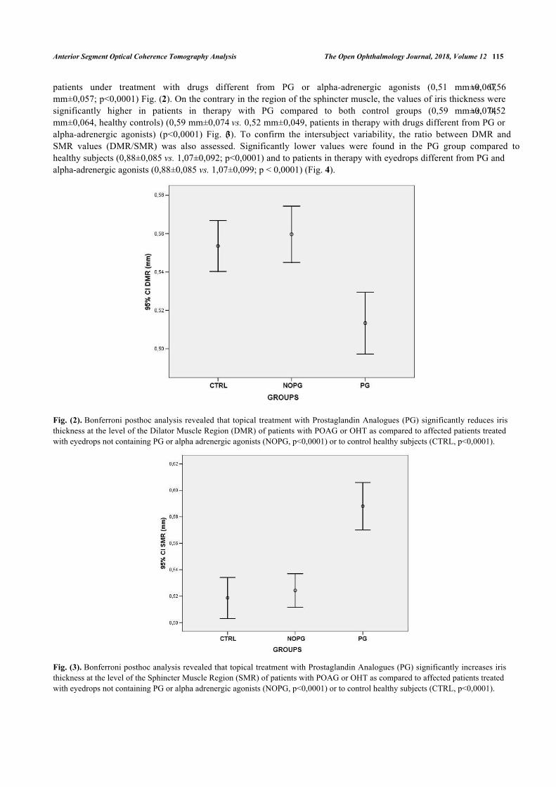
At the level of the dilator muscle region, the iris thickness significantly reduced in patients with POAG or OHTunder treatment with PG compared to both healthy subjects (0,51 mm±0,067 vs. 0,55 mm±0,055; p<0,0001) and
Anterior Segment Optical Coherence Tomography Analysis The Open Ophthalmology Journal, 2018, Volume 12 115
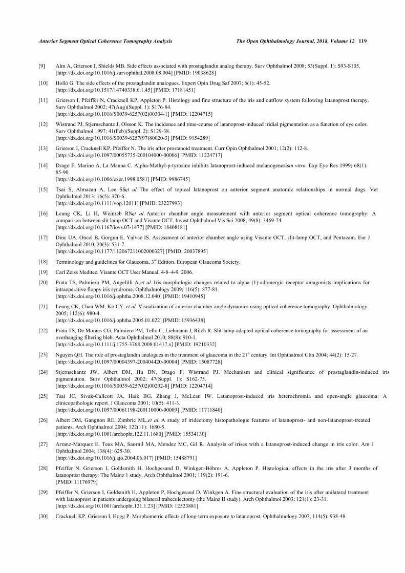
patients under treatment with drugs different from PG or alpha-adrenergic agonists (0,51 mm±0,067 vs. 0,56mm±0,057; p<0,0001) Fig. (2). On the contrary in the region of the sphincter muscle, the values of iris thickness weresignificantly higher in patients in therapy with PG compared to both control groups (0,59 mm±0,074 vs. 0,52mm±0,064, healthy controls) (0,59 mm±0,074 vs. 0,52 mm±0,049, patients in therapy with drugs different from PG oralpha-adrenergic agonists) (p<0,0001) Fig. (3). To confirm the intersubject variability, the ratio between DMR andSMR values (DMR/SMR) was also assessed. Significantly lower values were found in the PG group compared tohealthy subjects (0,88±0,085 vs. 1,07±0,092; p<0,0001) and to patients in therapy with eyedrops different from PG andalpha-adrenergic agonists (0,88±0,085 vs. 1,07±0,099; p < 0,0001) (Fig. 4).
Fig. (2). Bonferroni posthoc analysis revealed that topical treatment with Prostaglandin Analogues (PG) significantly reduces iristhickness at the level of the Dilator Muscle Region (DMR) of patients with POAG or OHT as compared to affected patients treatedwith eyedrops not containing PG or alpha adrenergic agonists (NOPG, p<0,0001) or to control healthy subjects (CTRL, p<0,0001).
Fig. (3). Bonferroni posthoc analysis revealed that topical treatment with Prostaglandin Analogues (PG) significantly increases iristhickness at the level of the Sphincter Muscle Region (SMR) of patients with POAG or OHT as compared to affected patients treatedwith eyedrops not containing PG or alpha adrenergic agonists (NOPG, p<0,0001) or to control healthy subjects (CTRL, p<0,0001).

116 The Open Ophthalmology Journal, 2018, Volume 12 Mancino et al.
Fig. (4). Bonferroni posthoc analysis showed a significant reduction in DMR/SMR ratio in patients with POAG or OHT in therapywith Prostaglandin Analogues (PG) compared to affected patients treated with eyedrops not containing PG or alpha adrenergicagonists (NOPG, p<0,0001) and to control healthy subjects (CTRL, p<0,0001).
Comparison of iris thickness values between the group of patients treated with eyedrops not containing PG andhealthy subjects did not reveal significant differences (p>0,05) in the DMR region (0,56 mm±0,057 vs. 0,55 mm±0,055)and in the SMR region (0,52 mm±0,0049 vs. 0,52 mm±0,064). Also the DMR/SMR ratio was not significantly differentin these two groups (1,07±0,099 vs. 1,07±0,092).
Pearson correlation analysis showed no significant correlation between the duration of prostaglandin analogues eyedrops treatment and iris thickness parameters, at the level of DMR (r= 0,007), SMR (r= 0,156) and for DMR/SMR ratio(r= -0,191). Furthermore, there was no significant correlation between age and iris thickness values: DMR (r= -0,154),SMR (r= -0,088) and DMR/SMR ratio (r= -0,047).
4. DISCUSSION
The data reports, for the first time, evidence supporting the occurrence of iris morphological alterations, measuredwith the aim of Visante OCT, in patients affected by primary open-angle glaucoma or ocular hypertension in therapywith prostaglandin analogues.
Visante OCT is a non-invasive method to analyze iris structural changes in treated patients. It allows to measurevarious anterior chamber parameters such as corneal thickness, anterior chamber depth, anterior chamber angle, and hasbeen used to evaluate corneal scars and ulcers, blebs, ciliary body cleft and iris lesions [21, 22].
In particular, Visante OCT scans revealed a significant reduction in iris thickness at the level of the dilator muscleand a significant increase in the region of the sphincter muscle of patients under treatment with prostaglandin analoguescompared to both control groups (healthy subjects and patients under treatment with antiglaucoma drops non containingPG or alpha-adrenergic agonists). These alterations seem to be not influenced by age and not related to the length oftreatment. Moreover, the study documented a reduction in DMR/SMR ratio in patients with POAG or OHT using PGcompared to the other two groups, strengthening the previous findings that indicate a relevant modification of the irismorphology observed only in the PG group.
Furthermore, no significant differences in iris OCT values were found between eyes with primary open angleglaucoma or ocular hypertension in therapy with hypotensive eye drops not containing prostaglandin analogues oralpha-adrenergic agonists and healthy eyes. Altogether, these data support the hypothesis that the iris changes observedin our study are induced by the use of PG and are not related to glaucoma pathophysiology.
The mechanisms underlying our observations are not known. Many in vitro and histopathological studies on PG

Anterior Segment Optical Coherence Tomography Analysis The Open Ophthalmology Journal, 2018, Volume 12 117
treatment dealt with the pathogenesis of iris darkening [23 - 27]. Mainz 1 [28] and Mainz 2 [29] studies assert thatshort-term therapy with Latanoprost does not produce morphological or cellular proliferation changes in the irisspecimens obtained during glaucoma surgery. However, Cracknell et al. [30] demonstrated that iris darkeningassociated with long-term therapy with Latanoprost was due to a small increase in the size of mature melanin granules,especially in the anterior border region.
Moreover, Marquez et al. [27] demonstrated that iris specimens obtained from eyes with the previous history oflong-term therapy with Latanoprost showed an increased thickness of the anterior limiting layer of iris compared tountreated irises. Therefore, we can hypothesize, with caution, that the increased iris thickness in the Sphincter MuscleRegion (SMR) observed in eyes treated with prostaglandin analogues eye drops could be related to thishistopathological alteration. However, we should underline that the increased thickness of the anterior limiting layerwas homogeneous for the entire iris size and not located in a specific point as we found at the level of the sphinctermuscle [27].
Albert et al. [31] confirmed previous studies showing increased thickness of the anterior border layer of iris inLatanoprost-treated specimens. Moreover, they found that the number of nuclear invaginations and prominent nucleolisignificantly decreased in iris specimens obtained by eyes treated chronically with Latanoprost. These cytologicalalterations could explain, in part, the significant iris thickness reduction that we found in the region of dilator muscle(DMR) in the eyes in therapy with prostaglandin analogues. However, the results are not clear whether the reduction innuclear invaginations and prominent nucleoli was concentrated at the level of dilator muscle, thence these data need tobe considered with caution.
Few reports focused on the effects in vivo of Prostaglandin analogues on the anterior segment structures. Tsai et al[15], investigated the prophylactic efficacy of Latanoprost treatment for primary angle closure glaucoma in normalfemale beagle dogs with the use of anterior segment OCT. They demonstrated a decrease in pupil diameter, crowding ofanterior chamber structures and narrowing of iridocorneal angle. In our study, we have not reported cases of narrowingof the anterior chamber angle. However, studies on humans receiving topical latanoprost have shown no effect or mildeffect in accommodation and only minimal changes in pupil’s diameter [32, 33]. Further studies are needed tothoroughly investigate the possible relation between prostaglandin analogues eyedrops and the modification of theanterior chamber structures. The altered iris thickness observed in patients using prostaglandin analogues eye dropsneeds to be further evaluated in order to find a possible relation between iris morphology and pupil dynamics.
Iris is an extremely mobile structure. We may speculate that muscular alteration may be a contributing cause topupillary response to light alterations previously described in patients with glaucoma [34]. It has been shown that linearranges for constriction and for dilatation have sharply defined limits in each individual’s eye [35]. Mechanical factors,such as muscular mass reduction, may influence the extent and speed of pupillary movements.
Prata et al. [20] demonstrated iris structural alterations associated with intraoperative floppy iris syndrome inpatients with prostatic disease in therapy with systemic alpha-1-adrenergic receptor antagonists (alpha-1-ARA) usingSL-OCT. They found a significant reduction in iris thickness in the region of Dilator Muscle Region (DMR) in patientsusing alpha-1-ARA compared to healthy and untreated controls. The research study also showed the same significantchanges iris thickness in the region of DMR in eyes treated with prostaglandin analogues eye drops.
Pathogenesis of Intraoperative Floppy Iris Syndrome (IFIS) seems to be related to progressive loss of iris muscletone caused by atrophy of iris dilator muscle [36, 37].
Based on the data, the eyes treated with prostaglandin analogues could show a “floppy iris” effect during cataractsurgery. Assessing patients with Visante preoperatively, may be helpful to establish a more appropriate pre-operativeapproach. Nevertheless, further studies are required to establish a possible connection between prostaglandin analoguestreatment and Floppy Iris Syndrome during phacoemulsification.
To the best of our knowledge, this is the first report to detect iris morphometric changes obtained with Visante OCTin patients with primary open-angle glaucoma or ocular hypertension using prostaglandin analogues eye drops.
Our study has some limitations; it is retrospective in nature and was conducted on relatively small number ofsubjects in each subgroup. Particularly, the study did not analyze the effects of any single type of prostaglandinanalogues eye drops in order to avoid reducing the overall sample size. These limitations make it difficult to establishany evidence and a cause-effect relationship between prostaglandin analogues treatment and morphological changes iniris thickness. We acknowledge that each group of patients was not classified based on the severity of glaucoma. We

118 The Open Ophthalmology Journal, 2018, Volume 12 Mancino et al.
supposed to overcome these limitations considering a second control group composed of people affected by primaryopen-angle glaucoma or ocular hypertension in therapy with eye drops not containing prostaglandin analogues eyedrops.
Our observations should further be confirmed by prospective studies including larger sample groups to betterunderstand and characterize the effects of prostaglandin analogues in patients with glaucoma.
CONCLUSION
The present data suggest a possible modification of the iris morphology in patients affected by OH or POAG undertreatment with PG eyedrops. Specifically, the use of Visante OCT has revealed reduction in iris thickness at the level ofDMR and an increase in iris thickness at SMR in patients using PG.
The study has demonstrated that these changes are not related to PG time exposure.
ETHICAL APPROVAL AND CONSENT TO PARTICIPATE
The study was approved by the Ethics Committee of Tor Vergata University Hospital, Rome, Italy.
HUMAN AND ANIMAL RIGHTS
No Animals were used in this research. All human research procedures followed were in accordance with the ethicalstandards of the committee responsible for human experimentation (institutional and national), and with the HelsinkiDeclaration of 1975, as revised in 2013.
CONSENT FOR PUBLICATION
Registration Number: PROTOCOLLO DI STUDIO REGISTRO SPERIMENTAZIONI 137/15.
CONFLICT OF INTEREST
The authors declare no conflict of interest, financial or otherwise.
ACKNOWLEDGEMENTS
Decleared none.
REFERENCES
[1] Nucci C, Martucci A, Cesareo M, et al. Links among glaucoma, neurodegenerative, and vascular diseases of the central nervous system. ProgBrain Res 2015; 221: 49-65.[http://dx.doi.org/10.1016/bs.pbr.2015.04.010] [PMID: 26518072]
[2] Scuderi GL, Cesareo M, Perdicchi A, Recupero SM. Standard automated perimetry and algorithms for monitoring glaucoma progression.Prog Brain Res 2008; 173: 77-99.[http://dx.doi.org/10.1016/S0079-6123(08)01107-2] [PMID: 18929103]
[3] Perdicchi A, Abdolrahimzadeh S, Cutini A, Ciarnella A, Scuderi GL. Evaluation of the progression of visual field damage in patientssuffering from early manifest glaucoma. Clin Ophthalmol 2016; 10: 1647-51.[http://dx.doi.org/10.2147/OPTH.S113995] [PMID: 27601881]
[4] Leske MC, Heijl A, Hyman L, Bengtsson B. Early manifest glaucoma Trial: Design and baseline data. Ophthalmology 1999; 106(11):2144-53.[http://dx.doi.org/10.1016/S0161-6420(99)90497-9] [PMID: 10571351]
[5] HeiJl AH. Reduction of intraocular pressure and glaucoma progression. Results from the early manifest glaucoma trial. Arch Ophthalmol2002; 120: 1268-79.[http://dx.doi.org/10.1016/S0161-6420(99)90497-9]
[6] Vass C, Hirn C, Sycha T, Findl O, Bauer P, Schmetterer L. Medical interventions for primary open angle glaucoma and ocular hypertension.Cochrane Database Syst Rev 2007; 17(4)[http://dx.doi.org/10.1002/14651858.CD003167.pub3]
[7] Stewart WC, Konstas AG, Nelson LA, Kruft B. Meta-analysis of 24-hour intraocular pressure studies evaluating the efficacy of glaucomamedicines. Ophthalmology 2008; 115(7): 1117-1122.e1.[http://dx.doi.org/10.1016/j.ophtha.2007.10.004] [PMID: 18082886]
[8] van der Valk R, Webers CA, Schouten JS, Zeegers MP, Hendrikse F, Prins MH. Intraocular pressure-lowering effects of all commonly usedglaucoma drugs: A meta-analysis of randomized clinical trials. Ophthalmology 2005; 112(7): 1177-85.[http://dx.doi.org/10.1016/j.ophtha.2005.01.042] [PMID: 15921747]
Anterior Segment Optical Coherence Tomography Analysis The Open Ophthalmology Journal, 2018, Volume 12 119
[9] Alm A, Grierson I, Shields MB. Side effects associated with prostaglandin analog therapy. Surv Ophthalmol 2008; 53(Suppl. 1): S93-S105.[http://dx.doi.org/10.1016/j.survophthal.2008.08.004] [PMID: 19038628]
[10] Holló G. The side effects of the prostaglandin analogues. Expert Opin Drug Saf 2007; 6(1): 45-52.[http://dx.doi.org/10.1517/14740338.6.1.45] [PMID: 17181451]
[11] Grierson I, Pfeiffer N, Cracknell KP, Appleton P. Histology and fine structure of the iris and outflow system following latanoprost therapy.Surv Ophthalmol 2002; 47(Aug)(Suppl. 1): S176-84.[http://dx.doi.org/10.1016/S0039-6257(02)00304-1] [PMID: 12204715]
[12] Wistrand PJ, Stjernschantz J, Olsson K. The incidence and time-course of latanoprost-induced iridial pigmentation as a function of eye color.Surv Ophthalmol 1997; 41(Feb)(Suppl. 2): S129-38.[http://dx.doi.org/10.1016/S0039-6257(97)80020-3] [PMID: 9154289]
[13] Grierson I, Cracknell KP, Pfeiffer N. The iris after prostanoid treatment. Curr Opin Ophthalmol 2001; 12(2): 112-8.[http://dx.doi.org/10.1097/00055735-200104000-00006] [PMID: 11224717]
[14] Drago F, Marino A, La Manna C. Alpha-Methyl-p-tyrosine inhibits latanoprost-induced melanogenesis in vitro. Exp Eye Res 1999; 68(1):85-90.[http://dx.doi.org/10.1006/exer.1998.0581] [PMID: 9986745]
[15] Tsai S, Almazan A, Lee SS, et al. The effect of topical latanoprost on anterior segment anatomic relationships in normal dogs. VetOphthalmol 2013; 16(5): 370-6.[http://dx.doi.org/10.1111/vop.12011] [PMID: 23227993]
[16] Leung CK, Li H, Weinreb RN, et al. Anterior chamber angle measurement with anterior segment optical coherence tomography: Acomparison between slit lamp OCT and Visante OCT. Invest Ophthalmol Vis Sci 2008; 49(8): 3469-74.[http://dx.doi.org/10.1167/iovs.07-1477] [PMID: 18408181]
[17] Dinc UA, Oncel B, Gorgun E, Yalvac IS. Assessment of anterior chamber angle using Visante OCT, slit-lamp OCT, and Pentacam. Eur JOphthalmol 2010; 20(3): 531-7.[http://dx.doi.org/10.1177/112067211002000327] [PMID: 20037895]
[18] Terminology and guidelines for Glaucoma, 3rd Edition. European Glaucoma Society.
[19] Carl Zeiss Meditec. Visante OCT User Manual. 4-8–4-9. 2006.
[20] Prata TS, Palmiero PM, Angelilli A, et al. Iris morphologic changes related to alpha (1)-adrenergic receptor antagonists implications forintraoperative floppy iris syndrome. Ophthalmology 2009; 116(5): 877-81.[http://dx.doi.org/10.1016/j.ophtha.2008.12.040] [PMID: 19410945]
[21] Leung CK, Chan WM, Ko CY, et al. Visualization of anterior chamber angle dynamics using optical coherence tomography. Ophthalmology2005; 112(6): 980-4.[http://dx.doi.org/10.1016/j.ophtha.2005.01.022] [PMID: 15936438]
[22] Prata TS, De Moraes CG, Palmiero PM, Tello C, Liebmann J, Ritch R. Slit-lamp-adapted optical coherence tomography for assessment of anoverhanging filtering bleb. Acta Ophthalmol 2010; 88(8): 910-1.[http://dx.doi.org/10.1111/j.1755-3768.2008.01417.x] [PMID: 19210332]
[23] Nguyen QH. The role of prostaglandin analogues in the treatment of glaucoma in the 21st century. Int Ophthalmol Clin 2004; 44(2): 15-27.[http://dx.doi.org/10.1097/00004397-200404420-00004] [PMID: 15087728]
[24] Stjernschantz JW, Albert DM, Hu DN, Drago F, Wistrand PJ. Mechanism and clinical significance of prostaglandin-induced irispigmentation. Surv Ophthalmol 2002; 47(Suppl. 1): S162-75.[http://dx.doi.org/10.1016/S0039-6257(02)00292-8] [PMID: 12204714]
[25] Tsai JC, Sivak-Callcott JA, Haik BG, Zhang J, McLean IW. Latanoprost-induced iris heterochromia and open-angle glaucoma: Aclinicopathologic report. J Glaucoma 2001; 10(5): 411-3.[http://dx.doi.org/10.1097/00061198-200110000-00009] [PMID: 11711840]
[26] Albert DM, Gangnon RE, Zimbric ML, et al. A study of iridectomy histopathologic features of latanoprost- and non-latanoprost-treatedpatients. Arch Ophthalmol 2004; 122(11): 1680-5.[http://dx.doi.org/10.1001/archopht.122.11.1680] [PMID: 15534130]
[27] Arranz-Marquez E, Teus MA, Saornil MA, Mendez MC, Gil R. Analysis of irises with a latanoprost-induced change in iris color. Am JOphthalmol 2004; 138(4): 625-30.[http://dx.doi.org/10.1016/j.ajo.2004.06.017] [PMID: 15488791]
[28] Pfeiffer N, Grierson I, Goldsmith H, Hochgesand D, Winkgen-Böhres A, Appleton P. Histological effects in the iris after 3 months oflatanoprost therapy: The Mainz 1 study. Arch Ophthalmol 2001; 119(2): 191-6.[PMID: 11176979]
[29] Pfeiffer N, Grierson I, Goldsmith H, Appleton P, Hochgesand D, Winkgen A. Fine structural evaluation of the iris after unilateral treatmentwith latanoprost in patients undergoing bilateral trabeculectomy (the Mainz II study). Arch Ophthalmol 2003; 121(1): 23-31.[http://dx.doi.org/10.1001/archopht.121.1.23] [PMID: 12523881]
[30] Cracknell KP, Grierson I, Hogg P. Morphometric effects of long-term exposure to latanoprost. Ophthalmology 2007; 114(5): 938-48.
120 The Open Ophthalmology Journal, 2018, Volume 12 Mancino et al.
[http://dx.doi.org/10.1016/j.ophtha.2006.10.025] [PMID: 17292473]
[31] Albert DM, Gangnon RE, Grossniklaus HE, Green WR, Darjatmoko S, Kulkarni AD. A study of histopathological features of latanoprost-treated irides with or without darkening compared with non-latanoprost-treated irides. Arch Ophthalmol 2008; 126(5): 626-31.[http://dx.doi.org/10.1001/archopht.126.5.626] [PMID: 18474771]
[32] Dinslage S, Diestelhorst M, Kühner H, Krieglstein GK. The effect of latanoprost 0.005% on pupillary reaction of the human eye.Ophthalmologe 2000; 97(6): 396-401.[http://dx.doi.org/10.1007/s003470070087] [PMID: 10916381]
[33] Kurtz S, Leibovitch I, Shemesh G, Rothkoff L, Loewenstein A. The effect of latanoprost on accommodation in young patients with ocularhypertension. J Glaucoma 2003; 12(1): 54-6.[http://dx.doi.org/10.1097/00061198-200302000-00011] [PMID: 12567113]
[34] Martucci A, Cesareo M, Napoli D, et al. Evaluation of pupillary response to light in patients with glaucoma: A study using computerizedpupillometry. Int Ophthalmol 2014; 34(6): 1241-7.[http://dx.doi.org/10.1007/s10792-014-9920-1] [PMID: 24550056]
[35] Loewenfeld IE, Newsome DA. Iris mechanics. I. Influence of pupil size on dynamics of pupillary movements. Am J Ophthalmol 1971; 71(1Pt 2): 347-62.[http://dx.doi.org/10.1016/0002-9394(71)90410-7] [PMID: 5100474]
[36] Flach AJ. Intraoperative floppy iris syndrome: Pathophysiology, prevention, and treatment. Trans Am Ophthalmol Soc 2009; 107: 234-9.[PMID: 20126500]
[37] Shtein RM, Hussain MT, Cooney TM, Elner VM, Hood CT. Effect of tamsulosin on iris vasculature and morphology. J Cataract Refract Surg2014; 40(5): 793-8.[http://dx.doi.org/10.1016/j.jcrs.2013.10.031] [PMID: 24631201]
© 2018 Mancino et al.
This is an open access article distributed under the terms of the Creative Commons Attribution 4.0 International Public License (CC-BY 4.0), acopy of which is available at: (https://creativecommons.org/licenses/by/4.0/legalcode). This license permits unrestricted use, distribution, andreproduction in any medium, provided the original author and source are credited.