the of chemistry vol. 266, no. 25, issue pp. … · a. identification, purification, and...
TRANSCRIPT
THE JOURNAL OF BIOLOGICAL CHEMISTRY Vol. 266, No. 25, Issue of September 5, pp. 16363-16369.1991 Printed in U. S. A.
Identification, Purification, and Characterization of Major Antigenic Proteins of Cumpylobucter jejuni”
(Received for publication, April 5, 1991)
Zhiheng PeiS, Richard T. Ellison, IIIOll, and Martin J. Blaser$II** From the $Department of Medicine and Department of Microbiology and Immunology, Diuision of Infectious Diseases, Vanderbilt Uniuersity School of Medicine, Nashuille, Tennessee 37232, the 1) Department of Veterans Affairs Medical Center, Nashville, Tennessee 37212, the §Medical and Research Seruices, Department of Veterans Affairs Medical Center, and the VDepartment of Medicine, Diuision of Infectious Diseases, University of Colorado School of Medicine, Denuer, Colorado 80220
Evidence from developing countries and volunteer studies indicates that immunity to Campylobacter je- juni and Campylobacter coli may be acquired, but the antigenic basis for this protection is poorly defined. We have purified to homogeneity four proteins with molecular weights of 28,000 (PEBl), 29,000 (PEBB), 30,000 (PEB3), and 31,000 (PEB4) from epidemic C. jejuni strain 81-176 using acid extraction and sequen- tial ion-exchange, hydrophobic interaction, and gel fil- tration chromatography. The relative amino acid com- positions of these four proteins are similar. NH2-ter- minal sequence analysis indicates that all four proteins are different, although the first 35 amino acids of PEB2 and PEB3 are 51.4% homologous. Isoelectric focusing showed that all four are basic proteins with PI of 8.5 for PEBl protein and >9.3 for the others. Use of the purified proteins as antigens in an IgG enzyme- linked immunosorbent assay (ELISA) found that sero- conversion to the PEBl or PEB3 proteins occurred in 15 of 19 patients with sporadic C. jejuni or C. coli infection. In comparison, only two, six, and 14 of these patients seroconverted to PEB2, PEB4, or the acid extract antigen. In an ELISA with whole bacterial cells as antigens, antiserum to the acid-extracted antigens showed broad recognition of C. jejuni, C. coli, C. fetus, C. lari, and Helicobacter pylori. Antiserum to PEBl recognized all 35 C. jejuni and all 15 C. coli strains but none of the isolates of the other three bacterial species. The PEBl and PEB3 proteins appear to be candidate antigens for both a Campylobacter vaccine and for serological assays for the pathogen.
Campylobacter jejuni and the closely related species Cam- pylobacter coli are now recognized as important causes of acute diarrheal disease in humans throughout the world (1-3). C. jejuni and C. coli are frequently present in the intestinal tracts of the animals we use for food processing, and transmission to humans from consumption of undercooked poultry and raw milk is common (4-7). Control of these pathogens in both human and animal populations would be desirable.
Several lines of evidence suggest that protective immunity
*This work was supported in part by the United States Army Medical Research and Development Command, the Medical Research Service of the Department of Veterans Affairs, and the Thrasher Research Fund. The costs of publication of this article were defrayed in part by the payment of page charges. This article must therefore be hereby marked “aduertisement” in accordance with 18 U.S.C. Section 1734 solely to indicate this fact.
** To whom reprint requests should be addressed Dept. of Medi- cine, Division of Infectious Diseases, Vanderbilt University School of Medicine, A-3310 Medical Center North, Nashville, TN 37232.
can be developed against these pathogens. The attack rate for diarrheal disease with C. jejuni is decreased in adults in developing countries (8, 9) and in chronic drinkers of raw milk in the United States (10) suggesting that immunity is induced by recurrent reexposure. Studies in human volunteers have shown that experimental infection with C. jejuni induces serum and intestinal antibodies directed against the pathogen, and also protects against subsequent illness (but not infec- tion) upon rechallenge with the same strain (11).
However, while exposure to these bacteria may induce a protective host response (8-ll), the pathogenicity of live C. jejuni and C. coli limits the utility of a whole cell vaccine. A vaccine containing C. jejuni antigens that were presented to the recipients in a non-virulent manner would be preferable. This could be accomplished using a vaccine composed solely of purified C. jejuni antigens. However, there is considerable heterogeneity of both heat-stable and heat-labile antigens (12). Nevertheless, serum antibody response to C. jejunilcoli in infected persons has been assessed using crude materials extracted with glycine at low pH (8, 13), but the precise antigenic basis of this reactivity is poorly understood.
For both development of vaccines and serological testing, characteristics of the common antigen present in this mixture would be useful. The flagella of C. jejuni are present in this extract and have been found to contain major antigens, but these proteins are heterogeneous and subject to phase varia- tion (14, 15). Several studies have suggested that a C. jejuni protein migrating at approximately M , = 30,000 by SDS- PAGE’ is common, cell surface-exposed, and antigenic to humans (16-19). Because several C. jejuni proteins migrate at approximately this molecular weight, purification of the proteins is necessary to determine if a single common antigen truly exists. We have now purified four C. jejuni proteins between M , = 28,000 and 31,000 and have shown that while they are similar in amino acid compositions, they differ in amino-terminal sequence. Two of the four proteins are com- monly recognized by convalescent sera from patients with sporadic C. jejunilcoli enteritis. These two proteins may be candidates for a vaccine against Campylobacter enteritis and also may be of value in serological assays for the diagnosis of this infection.
EXPERIMENTAL PROCEDURES
Bacterial Strains and Growth Conditions-A C. jejuni strain 81- 176, originally isolated from an outbreak of C. jejuni diarrhea and shown to be virulent in human volunteers and non-human primates
The abbreviations used are: SDS-PAGE, sodium dodecyl sulfate- polyacrylamide gel electrophoresis; HPLC, high performance liquid chromatography; ELISA, enzyme-linked immunosorbent assay; HEPES, 4-(2-hydroxyethyl)-l-piperazineethanesulfonic acid.
16363
16364 Major Antigens in C. jejuni and C. coli
(9, 20), was used as the source for antigen preparation. This strain was maintained frozen at -70 "C in brucella broth (BBL Microbiology Systems, Cockeysville, MD) containing 15% glycerol and has been deposited in the American Type Culture Collection (ATCC 55026). For purification of C. jejuni proteins, the bacterial strain was grown on trypticase soy agar with 5% sheep blood (PASCO, Wheat Ridge, CO) in a microaerobic atmosphere (5% oxygen, 10% carbon dioxide, and 85% nitrogen) at 37 "C for 24 h for three generations. Other strains including 38 C. jejuni, 20 C. coli, five Campylobacter fetus, five Campylobacter lari, and five Helicobacter pylori strains studied were from either the Campylobacter strain library of Vanderbilt University or from the Centers for Disease Control (CDC) Campylobacter labo- ratory provided by Charlotte Patton. Of the CDC strains, nine of 11 C. jejuni, all nine C. coli, and all five C. lari strains have been identified to the species level by DNA hybridization. These strains had also been maintained at -70 "C in brucella broth containing 15% glycerol and for studies were also grown on trypticase soy agar with 5% sheep blood in a microaerobic atmosphere at 37 "C.
Purification of C. jejuni Proteins with HPLC-For isolation of the antigens, bacterial cells were harvested and washed with cold distilled water by centrifugation at 8000 X g for 10 min. A crude mixture of surface proteins was extracted with 0.2 M glycine hydrochloride buffer, pH 2.2, as previously described (21). The preparation was lyophilized and reconstituted with distilled water and desalted using a Sephadex G-15 (Pharmacia LKB Biotechnology Inc.) column with distilled water as running buffer. For final purification of proteins, the crude mixture was separated by hydrophobic interaction chro- matography performed on a phenyl-Superose column (Pharmacia), ion-exchange chromatography on a Mono S column (Pharmacia), or gel filtration chromatography on a Superose 12 column (Pharmacia) using a fast protein liquid chromatography system (Pharmacia) (Fig. 1). The column eluates were monitored for UV absorbance at 280 nm to define protein peaks. Fractions were checked for presence and purity of specific antigens using SDS-PAGE analysis performed with 0.75-mm thick gels in a mini-Protean I1 dual slab cell (Bio-Rad) a t 250 mA for about 40 min.
Analytic Procedures-Protein concentrations were measured using a BCA protein assay kit (Pierce Chemical Co.) for crude proteins, and using Quantigold (Diversified Biotech, Newton Center, MA) for the purified proteins. SDS-PAGE was performed in a modified Laem- mli gel system as described by Ames (22). Proteins were resolved using the modified silver stain of Oakley et al. (23). Molecular mass standards (Bio-Rad) and their weight in daltons were: phosphorylase b (97,000), bovine serum albumin (66,200), ovalbumin (45,000), car- bonic anhydrase (31,000), soybean trypsin inhibitor (21,500), and lysozyme (14,000). The isoelectric point of the proteins was deter- mined by isoelectric focusing in Resolve@ thin layer agarose gels with a pH range of 3 to 10 (IsoLab, Inc., Akron, OH). Protein samples (0.5 pg) were focused under native conditions and resolved using the silver stain of Willoughby and Lambert (24). An experient formula of PI versus migration distance was generated by using four standard proteins of known PI between 8.3 and 9.3 as follows: L-lactic dehy- drogenase (8.3, 8.4, and 8.6) and trypsinogen (9.3) (Sigma). Analysis for the presence of disulfide bonds in the purified proteins was performed by treating the proteins with 0.9% dithiothreitol a t 37 "C for 30 min in SDS sample buffer (1.57% Tris base, 4.0% SDS, 20% glycerol, and 0.0025% bromphenol blue, pH 6.8), and monitoring molecular weight change by SDS-PAGE in comparison to untreated proteins.
Western blotting was performed by the method of Towbin et al. (25). After SDS-PAGE separation, proteins were transferred to nitro-
bated for 30 min in Tris/saline blotting buffer (TSBB) (10 mM Tris cellulase paper by electroblotting, the nitrocellulose paper was incu-
base, pH 8.0, 0.5 M NaC1, 0.5% Tween 20, 0.02% NaN3), then incubated for 60 min with primary rabbit antiserum at a 1:2000 dilution in TSBB. After three washes in TSBB, the nitrocellulose paper was incubated for 60 min with 1:2000 dilution of alkaline phosphatase-conjugated anti-rabbit IgG (Amersham Corp.). After washing, the nitrocellulose paper was developed in substrate solution containing 9 ml of 3 mM MgC12 in 50 mM Tris, pH 10.0, 1 ml of 0.1% nitroblue-tetrazolium and 0.1 ml of 0.5% of 5-bromo-4-chloro-3- indoxylphosphate (Sigma) in dimethyl formamide.
Production of Antiserum-Antisera to the crude acid extract mix- ture and the M, 28,000 antigen (PEB1) of strain 81-176 were raised in adult New Zealand White female rabbits by three subcutaneous injections of 5 pg of protein as previously described (26).
Detection of Antibody Response in Human Patients-The purified antigens were compared with the previously described crude acid-
extracted preparation for utility as antigens for serodiagnosis of Campylobacter infection (13). A standardized enzyme-linked immu- nosorbent assay (ELISA) with 20 ng of protein/ELISA well was used to detect human anti-Campylobacter antibodies. Sera obtained from patients who sought attention at medical facilities in Denver, Colo- rado (27) for acute diarrheal illness were studied. Acute phase sera had been obtained within 7 days of illness onset and convalescent sera had been obtained 11-40 days later. Seroconversion was defined as an optical density value in convalescent serum that was at least 50% greater than that in the paired acute phase serum.
Digestion of PEBl Antigen with Proteases-For exoprotease diges- tion with proteinase K (Boehringer Mannheim) 24-h cultures of Campylobacter strains on blood agar plates were harvested in sterile distilled water (5 ml/plate). The cells were pelleted at 3500 X g for 10 min, resuspended in water, and protein concentration determined using the BCA protein assay kit and adjusted to 240 pg/ml with water. 100 pl of bacterial suspension was incubated for 60 min at 37 "C with Proteinase K (2.4 pg) (or water alone as control), 100 p1 of SDS-sample buffer added, the samples boiled for 5 min, and then electrophoresed on SDS-PAGE with 15% acrylamide. For digestion with endoprotease protease V8 (Boehringer Mannheim), the crude acid extract mixtures were dialyzed against water to remove glycine, denatured by boiling in 2% SDS for 5 min, and then diluted 1 : l O with 0.1 M NaHC03. Either Protease V8 or water was added to these denatured samples at ratios up to 1:4 of enzyme/bacterial proteins (w/w), and the samples incubated for up to 48 h at 37 "C. The samples then underwent SDS-PAGE and Western blotting with rabbit anti- serum to PEB1.
Determination of Native Molecular Weight of PEBl Antigen-The native molecular weight of PEBl antigen was determined in a Super- ose 12 (Pharmacia) gel filtration column using gel-filtration molecular weight markers (Sigma) as follows: horse spleen apoferritin (443,000), sweet potato @-amylase (200,000), yeast alcohol dehydrogenase (150,000), bovine erythrocyte carbonic anhydrase (29,000), and horse heart cytochrome c (12,400). Blue dextran (2,000,000) was used to determine the void volume. Individual protein standards were dis- solved in an equilibration buffer containing 50 mM Tris-HC1,lOO mM KCl, pH 7.5. Crude acid-extract mixtures of C. jejunilcoli proteins were dialyzed against water to remove glycine and concentrated using Centricon-10 microconcentrators (Amicon, Danvers, MA). The first water extracts of C. jejunilcoli strains were concentrated in the same way. Each of these samples were diluted 1:l with the Tris-KC1 buffer, then either 50 p1 of sample or a molecular weight protein standard was loaded onto the column. Elution volume of the standards was individually determined by the position of the absorption peak at 280 nm. A standard curve for molecular weight determination was gen- erated by a semilog regression of the elution volumes uersus the log,, molecular weights of the individual protein standards. The elution volume of PEBl antigen was determined by assaying for the presence of the M, = 28,000 band in each fraction using SDS-PAGE and Western blotting with rabbit anti-PEB1.
Identification and Differentiation of Isolates of Campylobacter and Helicobacter by ELISA-35 C. jejuni, 15 C. coli, 10 C. fetus, five C. lari, and five H. pylori strains were used as antigens in this study. The bacteria were grown overnight, harvested in distilled water, and protein concentrations adjusted to 1 pg/pl. Whole bacterial cells (0.5 pg protein/well) were then used in an IgG ELISA (13). The specific antisera were absorbed with whole Escherichia coli cells to remove any antibodies to E. coli that may cross-react with Campylobacter and diluted (1:500 for antisera to PEBl protein and for the acid-extracted preparation) prior to use in the assay. An optical density value greater than 0.1 was defined as positive.
Identification of Campylobacter by Western blot with Antiserum to PEBI-Whole bacterial cells were prepared as described above, and SDS-PAGE performed on a 15% acrylamide gel using 0.5 pg of bacterial protein/lane, and then Western blot analysis performed as described above.
Amino Acid Analysis and Amino-terminal Sequencing-Purified proteins were prepared by dialysis against water and lyophilization. Amino acid analysis was performed using the method of Jones (28). Amino-terminal sequencing was performed on an Applied Biosystems 470 A Protein Sequencer equipped with a 120A autoanalyzer using the 03RPTH program, as previously described (26).
RESULTS
Purification of C. jejuni Antigens-Whole bacterial cells and crude acid-extracted mixtures from four C. jejuni strains

Major Antigens in C. jejuni and C. coli 16365
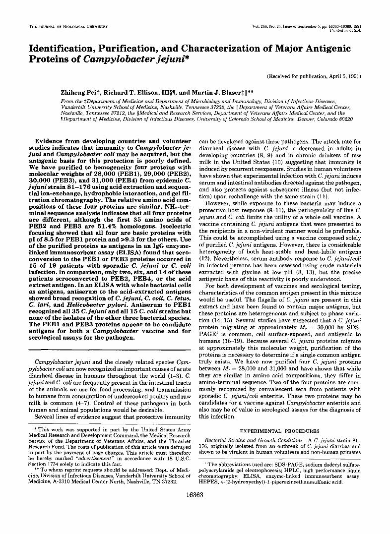
isolated from 1978 to 1987 were compared (data not shown). Major proteins in the acid-extracted preparations migrated at about 62, 30, 28, 24, and 21 kDa. The protein profiles of both whole cells and acid-extracted mixtures were very similar, although the strains had been isolated over a 10-year period. Because of its well-characterized virulence despite multiple in vitro passages (11), we started our purification with the crude acid-extract mixture from strain 81-176. The purification of the PEB antigens is summarized in Fig. 1. PEBl was purified to greater than 98% homogeneity by a single passage of the acid extract preparation on a phenyl-Superose column that fractionated material into three major protein peaks (Fig. 2). Peak 1 represented hydrophilic materials that did not bind to the column at pH 9.0. Peak 2 contained an essentially pure mixture of M , = 30,000 (PEB3) and 31,000 (PEB4) antigens. Peak 3 contained purified PEBl antigen that migrated at 28,000 in SDS-PAGE. The precise elution point of peaks 2 and 3 varied with the age of the column, eluting at approxi- mately 0.3-0.5 M Na2S04 in a new column (Fig. 2) and 0.0- 0.1 Na2S04 in a much used column (data not shown). How- ever, the SDS-PAGE profile of each peak remained similar and the third peak always contained pure PEB1. The PEB2 ( M , = 29,000), PEB3 ( M , = 30,000), and PEB4 ( M , = 31,000) antigens were partially purified by cation-exchange separation on a Mono S column (Fig. 3). PEB3 eluted at approximately 180 mM NaCl (peak 3 ) , PEB4 at approximately 200-220 mM NaCl (peak 3 + peak 4 ) , and PEB2 at approximately 300 mM NaCl (peak 7). PEB3 was further purified to homogeneity on a phenyl-Superose column as originally used for PEB1. The PEB4 was purified to homogeneity by passing the partially purified material through a Superose 12 gel filtration column with 0.15 M phosphate, 0.15 M NaCl, pH 7.2, at a flow rate of 0.5 ml/min. For PEB2, the partially purified antigen from the Mono S column was further purified to homogeneity on the phenyl-Superose column as for PEB1, followed by a Superose 12 column, as for PEB4 antigen. The final purified products are shown in Fig. 4.
Seroconversion to C. jejuni Proteins in Patients with Acute
FIG. 1. Schematic purification of PEB proteins from C. je- juni strain 81-176.
I 0 - 0 4 0 . 0 5 A 0 (D
* 0 . 0 3
:: i f: 8 2 0 . 0 2
0 . 0 1
0.00
I
v 0
0 . 6 2 a
0.3
0 . 0 0 4 0 8 0
Time (minutes)
FIG. 2. Purification of PEBl antigen from C. jejuni strain 81-176 by hydrophobic interaction fast protein liquid chro- matography on a phenyl-Superose column. Elution was with 20 mM borate, pH 9.0, with a linear decrease of Na2S04 concentration from 1.5 to 0.0 M over 60 min at a flow rate of 0.25 ml/min. Fractions of eluted materials were collected every 2 min and assayed for the presence of PEBl by SDS-PAGE. Peak 3 contained purified PEB1.
Diarrheal Illness by ELISA-The purified proteins were then compared with the crude acid-extracted mixture from strain 81-176 in an ELISA to detect IgG responses in patients with sporadic cases of diarrhea (Table I). Of 19 patients with C. jejuni or C. coli diarrhea, 14 seroconverted to the acid-ex- tracted mixture, 15 to PEBl or PEB3, two to PEB2, and six to PEB4. None of patients without Campylobacter diarrhea seroconverted to any of these antigens.
Digestion of the PEBl Antigen with Proteases-Since bac- terial antigens may be polysaccharide, lipopolysaccharides, or protein in nature, we tested whether PEBs are proteins to further characterize these antigens. Although previous exper- iments showed that PEBl and PEB3 were common antigens, we were unsuccessful in purifying sufficient PEB3 due to its instability at temperatures above 0 "C. Thus, the following characterizations only focused on PEBl antigen. Whole cells of two C. jejuni and two C. coli strains and an acid extract from a third C. jejuni strain were digested with proteinase K or V8. Rabbit antiserum to PEBl was used in Western blot to monitor the sensitivity of PEBl to the protease digestions. This antiserum is monospecific and recognized a single anti- gen band migrating at M, = 28,000 in whole cell preparations, indicating that no degradation of the protein occurred during the extraction and purification processes. The M , = 28,000 band disappeared after proteinase K digestion but not after sham digestion (data not shown), indicating that the major antigenic component of PEBl is a protein. PEBl was fully resistant to digestion with protease V8 at low enzyme concen- tration (2% enzyme for 24 h at 37 "C). Continuation of diges- tion with 25% enzyme for another 24 h partially hydrolyzed PEBl into two antigenic fragments that migrate at M , = 16,000 and 12,000 (data not shown).
Determination of the Native Molecular Weight of the PEBl Antigen-Two C. jejuni (strains 81-176 and D1916) and two C. coli (strains D743 and D1035) strains were used in this
16366 Major Antigens in C. jejuni and C. coli
FIG. 3. Purification of PEBP, PEBI, and PEB4 proteins from acid extract of strain 81-176 by cation- exchange chromatography on a Mono S column with 50 mM HEPES, pH 9.0, at a flow rate of 1 ml/min and a linear gradient from 0 mM NaCl to 4 0 0 mM NaCl over a period of 20 min. Peaks contained partially purified proteins, PEB3 (peak 3), PEB4 (peak 3 + 4 ) , and PEB2 (peak 7).
WC AE 2 8 K 2 9 K
9 2 K -
6 6 K -
4 5 K -
3 1K-
21K-
0.10 -
0.08 -
!! 0.06 - 0 0 N
Q
2 0 .04 SI
0
-
0.02 -
30K
0
3 1 K
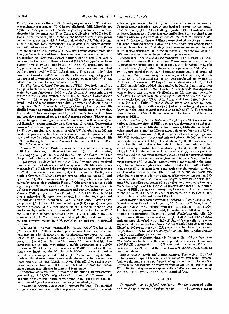
FIG. 4. SDS-PAGE (with 12% acrylamide) of purified C. jejuni proteins from strain 81-176. The lanes are whole bacterial cells ( W C ) , acid extract (AE) , PEBl (28K), PEB2 (29K), PEB3 (30K), and PEB4 (31K). Molecular weight markers are shown at left.
study. Crude acid-extract mixtures were chromatographed on the Superose 12 column. PEBl consistently eluted from the column immediately after the molecular weight standard pro- tein carbonic anhydrase (Mr = 29,000) (data not shown), and had a calculated molecular weight of 28,900. To compare the effect of extraction conditions on polymerization of PEB1, we included water extract preparations in this study. Only trace amounts of PEBl was extracted in water, so the water extract was concentrated 50-fold before use. PEBl extracted in water was also found to have a molecular weight of 28,900 f 1,000 for each of four strains tested. The similarity in the calculated molecular weight of PEBl as defined by SDS-PAGE and gel filtration chromatography indicates that the native form of PEBl is a monomer of M , = 28,900 f 1,000. PEBl in C. jejuni and C. coli strains have the same native molecular weights.
10 20
Time (minutes)
1
I
-
J - C 30
1.0
0.8
0 . 6 I X
0.4
$
0.2
) . O
Identification of Campylobacter and Helicobacter Strains by Antiserum to C. jejuni by ELISA-We next examined the potential application of antibodies to C. jejuni proteins for identification and diagnosis of Campylobacter and/or Helico- bacter species. Rabbit antisera to the acid-extract mixture of strain 81-176 and to PEBl were used in this study. By IgG ELISA we tested for the presence of cross-reactive antigens in 35 C. jejuni strains, 15 C. coli, 10 C. fetus, five C. lari, and five H. pylori strains. The control normal rabbit serum did not recognize any of the isolates tested. Antiserum to the acid-extract mixture recognized all 35 C. jejuni, all 15 C. coli, nine of 10 C. fetus, all five C. lari, and three of five H. pylori strains. In contrast, the antiserum to PEBl recognized all 35 C. jejuni and all 15 C. coli isolates but none of the isolates of the other three bacterial species (Fig. 5). Thus, antiserum to PEBl appeared to have exceptional discriminatory power having both 100% sensitivity and specificity for C. jejuni and C. coli.
Detection of PEBl in Whole Bacterial Cells by Western Blot-To further confirm the specificity of recognition of antiserum to PEB1, we performed Western blotting to deter- mine the bands recognized in preparations of whole cells of various Campylobacter and Helicobacter species. In total, 18 C. jejuni strains, 14 C. coli, three C. fetus, four C. lari strains, and one H. pylori strain were tested. A M, = 28,000 band was found in all 18 C. jejuni and all 14 C. coli strains but was not present in any of the C. fetus, C. lari, or H. pylori strains tested (Fig. 6). C. jejuni strains consistently had higher OD values in ELISA than C. coli (Fig. 5) and denser bands at M , = 28,000 in the Western blot analysis (Fig. 6). This Western blot experiment provided physical evidence that PEBl from various c. jejunilcoli strains are all conserved in size, antigen- ically related, and can be recognized by antiserum to PEBl antigen from a single strain, 81-176 (ATCC55026). However, the results also suggest that either more PEBl is present in C. jejuni, or that there are slight antigenic differences between PEBl of C. jejuni and C. coli. The amino-terminal residues of PEBl from C. jejuni and C. coli are 70% identical (Fig. 7); the amino acid differences are consistent with antigenic differ- ences.
Determination of PI-Prior to isoelectric focusing, the pu- rified antigens were dialyzed against water and concentrated

Major Antigens in C. jejuni and C. coli 16367 TABLE I o b c d e f g h i j k l m n o
Seroconversion to C. jejuni proteins as determined by ELISA of serum from Campylobacter-infected persons and persons with
other diarrheal diseases Seroconversion is defined as ODdI4 value increase by a t least 50%
in convalescent serum compared with that in acute serum and optical densitv value in convalescent serum neater than 0.200 in ELISA. 28- "
Patient Isolate AE PEBl PEBB PEBB PEB4
1. 2. 3. 4. 5. 6. 7. 8. 9.
10. 11. 12. 13. 14. 15. 16.
C. jejunilcoli C. coli + + - + - C. coli C. jejuni C. coli C. jejuni C. jejuni C. jejuni C. coli C. jejuni C. jejuni C. coli C. jejuni C. jejuni C. jejuni C. jejuni C. jejuni
"
+ + + + + + + + + + + + + + + + + + + +
+ -0
0 - + + + + + + + + + + + +
+ + -0
+ -
17. C. jejuni + + + + + 18. C. jejuni + + - - -
0 - 19. C. jejuni -a - - - % seroconversion 73.7 78.9 10.5 78.9 31.6
Other pathogens 1. Shigella 2. Salmonella 3. Shigella 4. Yersinia 5. Salmonella
" " - " - " " - " " " - " " -
% seroconversion 0 0 0 0 0
No pathogens identified
1. 2.
" " _ " " _ 3. 4.
" " _ - " "
5. " " - % seroconversion 0 0 0 0 0
a Optical density value greater than 1.0 in both acute and conva- lescent serum. Patients with acute serum OD value greater than 1.0 were not considered to show seroconversion, regardless of OD values in convalescent serum.
0.800 0 C. jejuni (13-35) bs9 C. coli (n-15) [x] C. fetus (n-10) m C. laridis (17-5) m H. pylori (n=5)
g- 0.600
z a W v ,
0.400 ax g E + 0 a 0.200
0.000
ANTI-ACID EXTRACT ANTI-PEE1
FIG. 5. Recognition of Campylobacter and Helicobacter cells by antisera to C. jejuni proteins by ELISA. Whole bacterial cells were used as antigens. First antibodies were rabbit anti-acid extract or rabbit anti-PEB1 from strain 81-176. An optical density value a t 414 nm greater than 0.1 was defined as positive (mean f S.E. of two experiments).
using centricon-10 microconcentrators (Amicon). All four proteins were found to be basic with a PI of 8.5 for PEB1, and PI greater than 9.3 for PEB2, PEB3, and PEB4.
Amino Acid Composition and Amino-terminal Sequence-
FIG. 6. Western blot of rabbit anti-PEB1 with representa- tive Campylobacter and Helicobacter strains. The antigens used are whole cells prepared as described in the text. Bacterial strains of C. jejuni (except strains 81-176, 81-93, and 81-94), C. coli, and C. lari had been identified by DNA hybridization to the species level. The C. fetus strains were identified by the presence of high molecular weight surface array proteins detected by SDS-PAGE and Western blot (25). The method for the Western blot is as described in the text. The arrow indicates bands migrating a t M, = 28,000, which were found in all C. jejuni strains (81-176,81-93,81-95, D996, and D1916, lanes a-e) and all C. coli strains (D743, D1035, D130, D126, and D115, lanes f-j) but not found in any of C. lari strains (D459 and D1014, lanes k and l), C. fetus strains (84-32 and 80-109, lanes m and n ) or H. pylori strain (16-11A, lane 0 ) .
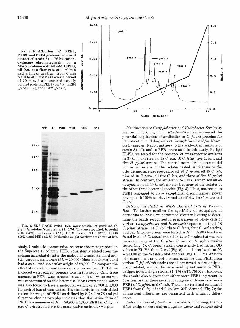
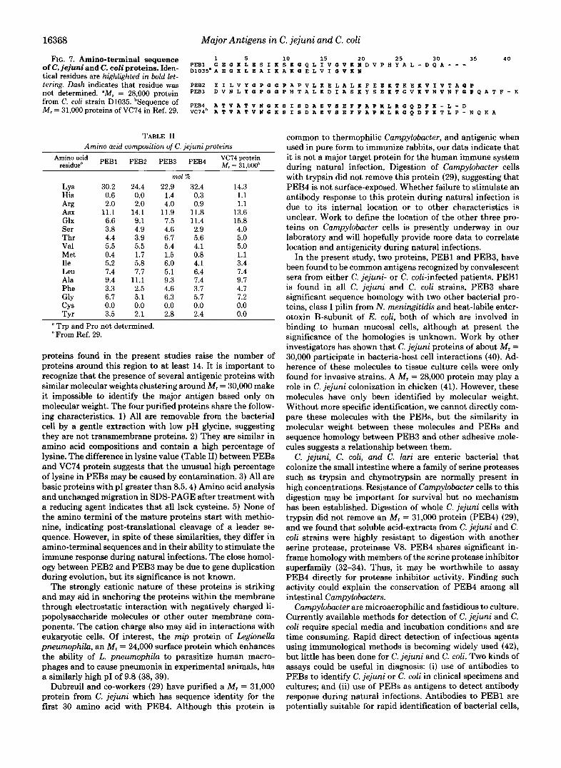
The amino acid composition of the four proteins was found to be similar (Table 11). Not surprisingly, basic amino acids (Lys, His, and Arg) predominate, ranging from 26.4 to 33.6%. Cysteine was not found in any of the four proteins. Treatment with dithiothreitol did not change the electrophoretic migra- tion of the four proteins (data not shown), confirming the absence of intermolecular disulfide bonds. A comparison of the NH2-terminal amino acid sequences of the four proteins indicated that each is unique, although there is 51.4% identity in the NH2-terminal 35 amino acids of PEBB (MI = 29,000) and PEB3 (Mr = 30,000) (Fig. 7). None of the NH, termini of these mature proteins starts with methionine, suggesting that a leader sequence was cleaved during maturation. The four amino acid sequences were compared with several GeneBank databases including PIR version 25.0, Swiss-Prot 14, EMBL 24, and unique GeneBank sequences 65 24. The NH2 termini of PEBl or PEB2 had no significant in-frame homology with other known proteins. For PEB3, we identified a potential in-frame homology with the deduced protein se- quences of two bacterial structures related to adherence to eukaryotic cells, class I pili from Neisseria meningitidis (30) and E. coli heat-labile enterotoxin B-subunit (31). PEB4 was found to share an identical NH2-terminal sequences through the first 30 amino acids with an M, = 31,000 protein purified from a C. jejuni strain VC74 (29) (Fig. 7). PEB4 also exhibits partial homology with two proteins related to the serine protease inhibitor superfamily including human corticoste- roid-binding globulin (32), serine proteinase inhibitor I of vaccinia virus (33, 34), and with two mammalian proteins involved in cell-to-cell interaction including mouse neural cell adhesion molecule (35) and human MHC class I1 lymphocyte antigen Dpw4-0-1 (36).
DISCUSSION
We have developed methods for the purification of four basic proteins from C. jejuni strain 81-176 and demonstrated the existence of common antigens, migrating between MI = 28,000 and 31,000 by SDS-PAGE. Although no more than four protein bands of whole cells and acid-extract of C. jejuni are distinguishable between M , = 28,000 and 31,000 by one- dimensional SDS-PAGE, more than 10 protein spots are resolved around MI = 30,000 by two-dimensional gel electro- phoresis with PI between 4.0 and 6.8 (37). The four basic PEB
Major Antigens in C. jejuni and C. coli
FIG. 7. Amino-terminal sequence 1 5 10 15 2 0 2 5 30 35 4 0 ofC.jejuniand c. coliproteins. iden- PEBl G E Q K L E S I K S K Q Q L I V Q V K N D V P H Y A L - D Q A - - - D 1 0 3 5 ~ A E G K L E A I K A K Q E L V I Q V K N
tical residues are hi.qhli.qhted in bold let- tering. Dash indicates that residue was PEBZ E I L v Y Q P G Q P A P v L K E L A L K F E E K T K E K v I v T A Q P not determined. ZIM. = 28.000 ,,rotein PEB3 D V N L Y 0 P Q 0 P H T A L K D I A S K Y S E K T G V K V N V N F G P Q A T F - K from strain bSequence Of PEB4 A T V A T V N Q 8 1 8 D A E V 8 E F P A P N L R Q Q D F K - L - D
I -
M,=31,000proteinsofVC74inRef.29. ~ ~ 7 4 ~ A T v A T v N G K 8 I s D A E v 8 E F F A P n L R Q Q D P K T L P - N Q K A
TABLE I1 Amino acid commsition of C. ieiuni Droteins
Amino acid PEBl PEBP PEBB PEB4 E::$$ residue"
LY 5 30.2 24.4 His 0.6 0.0 Arg 2.0 2.0 Asx 11.1 14.1 Glx 6.6 9.1 Ser 3.8 4.9 Thr 4.4 3.9 Val 5.5 5.5 Met 0.4 1.7 Ile 5.2 5.8 Leu 7.4 7.7 Ala 9.4 11.1 Phe 3.3 2.5 GlY 6.7 5.1 CY5 0.0 0.0 Tvr 3.5 2.1
mol % 22.9 32.4
1.4 0.3 4.0 0.9
11.9 11.8 7.5 11.4 4.6 2.9 6.7 5.6 5.4 4.1 1.5 0.8 6.0 4.1 5.1 6.4 9.3 7.4 4.6 3.7 6.3 5.7 0.0 0.0 2.8 2.4
14.3 1.1 1.1
13.6 15.8 4.0 5.0 5.0 1.1 3.4 7.4 9.7 4.7 7.2 0.0 0.0
" Trp and Pro not determined. From Ref. 29.
proteins found in the present studies raise the number of proteins around this region to at least 14. It is important to recognize that the presence of several antigenic proteins with similar molecular weights clustering around M, = 30,000 make it impossible to identify the major antigen based only on molecular weight. The four purified proteins share the follow- ing characteristics. 1) All are removable from the bacterial cell by a gentle extraction with low pH glycine, suggesting they are not transmembrane proteins. 2) They are similar in amino acid compositions and contain a high percentage of lysine. The difference in lysine value (Table 11) between PEBs and VC14 protein suggests that the unusual high percentage of lysine in PEBs may be caused by contamination. 3) All are basic proteins with PI greater than 8.5.4) Amino acid analysis and unchanged migration in SDS-PAGE after treatment with a reducing agent indicates that all lack cysteine. 5) None of the amino termini of the mature proteins start with methio- nine, indicating post-translational cleavage of a leader se- quence. However, in spite of these similarities, they differ in amino-terminal sequences and in their ability to stimulate the immune response during natural infections. The close homol- ogy between PEBB and PEB3 may be due to gene duplication during evolution, but its significance is not known.
The strongly cationic nature of these proteins is striking and may aid in anchoring the proteins within the membrane through electrostatic interaction with negatively charged li- popolysaccharide molecules or other outer membrane com- ponents. The cation charge also may aid in interactions with eukaryotic cells. Of interest, the n i p protein of Legionella pneumophila, an M, = 24,000 surface protein which enhances the ability of L. pneumophila to parasitize human macro- phages and to cause pneumonia in experimental animals, has a similarly high PI of 9.8 (38, 39).
Dubreuil and co-workers (29) have purified a M , = 31,000 protein from C. jejuni which has sequence identity for the first 30 amino acid with PEB4. Although this protein is
common to thermophilic Campylobacter, and antigenic when used in pure form to immunize rabbits, our data indicate that it is not a major target protein for the human immune system during natural infection. Digestion of Campylobacter cells with trypsin did not remove this protein (29), suggesting that PEB4 is not surface-exposed. Whether failure to stimulate an antibody response to this protein during natural infection is due to its internal location or to other characteristics is unclear. Work to define the location of the other three pro- teins on Campylobacter cells is presently underway in our laboratory and will hopefully provide more data to correlate location and antigenicity during natural infections.
In the present study, two proteins, PEBl and PEB3, have been found to be common antigens recognized by convalescent sera from either C. jejuni- or C. coli-infected patients. PEBl is found in all C. jejuni and C. coli strains. PEB3 share significant sequence homology with two other bacterial pro- teins, class I pilin from N. meningitidis and heat-labile enter- otoxin B-subunit of E. coli, both of which are involved in binding to human mucosal cells, although at present the significance of the homologies is unknown. Work by other investigators has shown that C. jejuni proteins of about M, = 30,000 participate in bacteria-host cell interactions (40). Ad- herence of these molecules to tissue culture cells were only found for invasive strains. A M, = 28,000 protein may play a role in C. jejuni colonization in chicken (41). However, these molecules have only been identified by molecular weight. Without more specific identification, we cannot directly com- pare these molecules with the PEBs, but the similarity in molecular weight between these molecules and PEBs and sequence homology between PEB3 and other adhesive mole- cules suggests a relationship between them.
C. jejuni, C. coli, and C. lari are enteric bacterial that colonize the small intestine where a family of serine proteases such as trypsin and chymotrypsin are normally present in high concentrations. Resistance of Campylobacter cells to this digestion may be important for survival but no mechanism has been established. Digestion of whole C. jejuni cells with trypsin did not remove an M, = 31,000 protein (PEB4) (29), and we found that soluble acid-extracts from C. jejuni and C. coli strains were highly resistant to digestion with another serine protease, proteinase V8. PEB4 shares significant in- frame homology with members of the serine protease inhibitor superfamily (32-34). Thus, it may be worthwhile to assay PEB4 directly for protease inhibitor activity. Finding such activity could explain the conservation of PEB4 among all intestinal Campylobacters.
Campylobacter are microaerophilic and fastidious to culture. Currently available methods for detection of C. jejuni and C. coli require special media and incubation conditions and are time consuming. Rapid direct detection of infectious agents using immunological methods is becoming widely used (4'4, but little has been done for C. jejuni and C. coli. Two kinds of assays could be useful in diagnosis: (i) use of antibodies to PEBs to identify C. jejuni or C. coli in clinical specimens and cultures; and (ii) use of PEBs as antigens to detect antibody response during natural infections. Antibodies to PEBl are potentially suitable for rapid identification of bacterial cells,
Major Antigens in C. jejuni and C. coli 16369
and PEBl and PEB3 appear to be good candidate proteins to detect antibody responses.
Clinical and epidemiologic studies suggest it may be possible to develop an effective vaccine for C. jejuni and C. coli (8-11). Since diseases caused by C. jejuni and C. coli are self-limited, and recovery is usually associated with development of anti- bodies to specific antigens on the bacterial cell surface (13), one approach to vaccine development is to identify and char- acterize these cell surface molecules. We have found two proteins, PEBl and PEB3 that are targets for the human immune system during natural infection. Whether or not the immune response to PEBl and PEB3 is protective is unknown at present. Further study needs to focus on localization of the PEBs on the bacterial cell surface, and their role in bacterial adherence and pathogenesis, and the efficiency of PEBl or PEB3 for active immunization or their antibodies for passive protection of animals experimentally infected with C. jejuni or C. coli.
Acknowledgment-We thank Charlotte Patton for providing de- fined CamDvlobacter strains.
1.
2. 3.
4.
5.
6.
7.
8.
9.
10.
11.
12.
13.
14.
. _
REFERENCES Blaser, M. J., and Reller, L. B. (1983) N . Engl. J. Med. 305,
Chowdhury, M. N. H. (1984) Trop. Geogr. Med. 36,215-222 Walker, R. I., Caldwell, M. B., Lee, E. C., Guerry, P., Trust, T.
J., and Ruiz-Palacios, G. M. (1986) Microbiol. Reu. 50,81-94 Harris, N. V., Thompson, D., Martin, D. C., and Nolan, C. M.
(1986) Am. J. Public Health 7 6 , 401-406 Harris, N. V., Weiss, N. S., and Nolan, C. M. (1986) Am. J. Public
Health 76,407-411 Deming, M. S., Tauxe, R. V., Blake, P. A., Dixon, S. E., Fowler,
B. S., Jones, T. S., Lockamy, E. A., Patton, C. M., and Sikes, R. 0. (1987) Am. J. Epidemiol. 126,525-534
Blaser, M. J., Cravens, J., Powers, B. W., LaForce, F. M., and Wang, W. L. (1979) Am. J. Med. 67, 715-718
Blaser, M. J., Taylor, D. N., and Echeverria, P. (1986) J. Inject. Dis. 153,249-254
Blaser, M. J., Black, R. E., Duncan, D. J., and Amer J. (1985) J. Clin. Microbiol. 21 , 164-67
Blaser, M. J., Sazie, E., and Williams, L. P. Jr. (1987) J. Am. Med. Ass. 257,43-46
Black, R. E., Levine, M. M., Clements, M. L., Hughes, T. P., and Blaser, M. J. (1988) J. Inject. Dis. 157,472-479
Patton, C. M., Barret, T. J., and Morris, G. K. (1985) J. Clin. Microbiol. 2 2 , 558-565
Blaser, M. J., and Duncan, D. J. (1984) Inject. Immun. 44 , 292- 298
Caldwell, M. B., Guerry, P., Lee, E. C., Burans, J . P., and Walker, R. I. (1985) Inject. Immun. 50, 941-943
1444-1452
15. Harris, L. A., Logan, S. M., Guerry, P., and Trust, T. J. (1987)
16. Blaser, M. J., Hopkins, J . A., Berka, R. M., Vasil, M. L., and
17. Blaser, M. J., Hopkins, J. A., and Vasil, M. L. (1984) Inject.
18. Logan, S. M., and Trust, T. J. (1983) Inject. Immun. 42, 675-
19. Newell, D. G., Mcbride, H., and Pearson, A. D. (1984) J. Gen.
20. Russell, R. G., Blaser, M. J., Sarmiento, J. I., and Fox, J. (1989)
21. McCoy, E. C., Doyle, D., Burda, K., Corbeil, L. B., and Winter,
22. Ames, G. F-L. (1974) J. Biol. Chem. 249 , 634-644 23. Oakley, B. R., Kirsch, D. R., and Morris, N. R. (1980) Anal.
24. Willoughby, E. W., and Lambert, A. (1983) Anal. Biochem. 130 ,
25. Towbin, H. T., Staehelin, and Gordon, J. (1979) Proc. Natl. Acad. Sci. U. S. A. 76,4350-4354
26. Pei, Z., Ellison, R. T., 111, Lewis, R. V., and Blaser, M. J. (1988) J. Biol. Chem. 263,6416-6420
27. Perez-Perez, G. I., Cohn, D. L., Guerrant, R. L., Patton, C. M., Reller, L. B., and Blaser, M. J. (1989) J. Infect. Dis. 160,460- 468
28. Jones, B. N., Paabo, S., and Stein S. (1981) J. Liq. Chromatogr.
29. Dubreuil, J . . D., Kostrzynska, M., Logan, S. M., Harris, L. A., Austin, J. W., and Trust, T. J. (1990) J. Clin. Microbiol. 28,
30. Potts, W. J., and Saundars, J. R. (1988) Mol. Microbiol. 2, 647-
31. Yamamoto. T.. Goiobori. T., and Yokota, T. (1987) J. Bacterwl.
J . Bacteriol. 169 , 5066-5071
Wang, W. L. (1983) Inject. Immun. 42 , 276-284
Immun. 43,986-993
682
Microbiol. 130, 1201-1208
Inject. Immun. 57, 1438-1444
A. J. (1975) Inject. Zmmun. 11, 517-525
Biochem. 105, 361-363
353-358
4,565-586
1321-1328
653 . .
169,1352-1357" 32. Harlev. M. G.. Reventos. J.. Musto. N. A.. Gunsalus. G. L.. and
Bardin, C. W. (1987) Proc. Natl. Acad. Sci. U. S. A: 8 4 , 5153- 5157
33. Smith, G. L., Howard, S. T., and Chan, Y. S. (1989) J. Gen Virol. 70,2333-2343
34. Kotwal, G. J., and Moss, G. (1989) J. Virol. 63. 600-606 35. Barbas, J. A., Chaix, J. C., Steinmetz, M., and Goridis, C. (1988)
EMBO J. 7,625-632 36. Gustafsson, K., Widmark, E., Jonsson, A. K., Servenius, B.,
Sachs, D. H., Larhammar, D., Rask, L., and Peterson, P. A. (1987) J . Biol. Chem. 262,8776-8786
37. Dunn, B. E., Blaser, M. J., and Snyder, E. L. (1987) Inject. Immun. 5 5 , 1564-1572
38. Cianciotto, N. P., Eisenstein, B. I., Mody, C. H., and Engleberg, N. C. (1990) J. Infect Dis. 162, 121-126
39. Engleberg, N. C., Carter, C., Weber, D. R., Cianciotto, N. P., and Eisenstein, B. I. (1989) Inject Immun. 57 , 1263-1270
40. Melo, M. A,, and Pechere, J-C. (1990) Inject. Immun. 5 8 , 1749- 1756
41. Meinersmann, R. T., Stern, N. J., and Blankenship, L. C. (1990) Curr. Microbiol. 2 1 , 17-21
42. Miotti, P. (1987) Eur. J. Epidemiol. 3, 356-364