the occurrence of oxidative stress during reperfusion in experimental animals and men
TRANSCRIPT

Cardiovascular Drugs and Therapy 1991; 5:277-288 © Kluwer Academic Publishers, Boston. Printed in U.S.A.
The Occurrence of Oxidative Stress During Reperfusion in Experimental Animals and Men
Roberto Ferrari, Claudio Ceconi, Salvatore Curello, A n n a Cargnoni, Evasio Pasini, Odoardo Visioli Cattedra di Cardiologia, Universita" degli Studi di Brescia, Brescia, Italy
Summary. Reperfusion is the prerequisite for the ischemic myocardium to recover its metabolic and mechanical func- tion. However, reperfusion after a prolonged period of isch- emia in the experimental animal may exacerbate, or at least accelerate, the occurrence of ischemic injury, whilst in hu- mans at the least it is not beneficial. This entity has been called reperfusion damage, since much of the damage is be- lieved to be caused by events occurring at the moment of reperfusion rather than by changes occurring during isch- emia. The existence of reperfusion damage, however, has been questioned, and evidence in favour of the concept is sparse. At the moment the molecular events occurring at the time of reperfusion are not completely understood, and the relative importance of several proposed deleterious mecha- nisms is not yet established. One of the most fashionable ideas for the cause of reperfusion damage is that the function of cell membrane is modified by oxygen radicals generated at the moment of reperfusion. Evidence in favour of and against this hypothesis is described in detail in the present article.
Cardiovasc Drugs Ther 1991;5:277-288
Key Words. oxygen, oxygen free radicals, ischemia, reperfu- sion, oxidative stress
the onset of chest pain [3]. Left ventricular function, however, does not always improve [4]. Benefits of later thrombolysis are fewer and, in any case, are present to approximately 4 to 6 hours [3]. The molecu- lar events occurring on reperfusion may be responsi- ble for the ventricular impairment present after early reperfusion or for the lack of benefits after late reper- fusion [5,6].
A rapid increase of oxygen tension occurs at the time of reperfusion, and recent data indicate that the toxic effects of oxygen, mainly via reactive oxygen reduction intermediates, might contribute to the re- perfusion damage.
The present article will first give a brief review of the occurrence of and protection against the oxygen intermediates in the heart. Evidence for the involve- ment of the oxygen intermediates in the molecular events occurring during reperfusion in experimental animals and in humans will then be discussed.
Reperfusion of heart muscle, after more than 60 min- utes of ischemia, is associated with the release of the intracellular component, reduction of contractility, in- flux of calcium, disruption of the cell membrane, and eventual necrosis of at least a portion of the tissue [1]. This entity has been called reperfusion damage and the early literature has been reviewed by Hearse [2]. Since then, the existence of reperfusion damage has been questioned and doubts exist as to whether reper- fusion causes further injury or just an acceleration of the manifestation of the damage caused by ischemia.
The cause of this reaction is apparently multifacto- rial and with the advent of angioplasty and thrombo- lytic treatment for acute myocardial infarction has be- come a key issue with important clinical implications.
It is clear that the greatest benefit of thrombolysis in terms of mortality is observed in those patients who receive thrombolytic t reatment within 1 to 2 hours of
Occurrence o f Reac t i ve O x y g e n
R e d u c t i o n I n t e r m e d i a t e s
A free radical is an atom, ion, or molecule with one or more unpaired electrons: This configuration leads to an increased reactivity with other molecules. The amount of reactivity depends on the facility with which a species can accept electrons (is reduced) or donate them (is oxidized).
Molecular oxygen is relatively nonreactive because of its unusual structure, including two unpaired elec- trons with parallel electrons spin. The majority of or- ganic compounds that might react with oxygen con- tain paired electrons. The simultaneous insertion of two such paired electrons into a molecule of oxygen
Address for correspondence and reprint requests: Prof. Roberto Ferrari, Cattedra di Cardiologia, Universita' degli Studi, c/o Spedali Civili--P.le Spedali Civili, 1, 25100 Brescia, Italy
277

278 Ferrari, Ceconi, Curello, Cargnoni, Pasini and Visioli
would violate the rules of quantum mechanics. This restriction explains why ordinary molecular oxygen is relatively nonreactive.
Incoming electrons prefer to enter the orbitals one at a time, thus leading to the formation of reduced oxygen intermediates or oxygen free radicals.
The reduction of oxygen to water in the myocar- dium proceeds by two pathways. The mitochondrial cytochrome oxidase reduces 95% of oxygen to water by tetravalent reduction without the production of any intermediates. The remaining 5% of oxygen pro- ceeds by a univalent pathway in which several inter- mediates are produced, such as superoxide anions (0~), hydrogen peroxides (H202), and hydroxyl radical (.OH), where the dot (.) represents an unpaired elec- tron and signifies a free radical.
The superoxide anion is comparatively unreactive and in the physiologic condition is converted by dismu- tation to hydrogen peroxide. This is less reactive, longer lived, and more lipophilic than the superoxide anion, and it can diffuse considerable distances from its site of generation. The major danger of increased tissue concentration of hydrogen peroxide is the pro- duction of hydroxyl radical by the Haber Weiss or Fenton reaction. The hydroxyl radical has a short half-life, but is extremely reactive and will rapidly interfere with unsaturated fatty acid side chains, re- sulting in lipid peroxidation.
In addition, some compounds react spontaneously with oxygen, they autoxidize. Virtually all autoxi- dations result in the formation of reactive oxygen re- duction intermediates. Autoxidation of adrenaline [8], pyrogallol [9], and many other compounds leads to the formation of the superoxide radical. The superoxide radical is released when the methemoglobin is formed from oxyhemoglobin [10]. Some oxidases also form su- peroxide, the most important in the heart being xan- thine oxidase, which oxidizes hypoxanthine and xan- thine to uric acid [11]. The microsomal cytochrome P4~0 system releases superoxide [12]. There are also indications that superoxide is formed as a byproduct during prostaglandin synthesis [13]. Upon activation of leukocytes (polymorphonuclears, monocytes, mac- rophages, eosinophils) large amounts of superoxide are released [14,15]. It follows that the superoxide and secondary products subsequently formed are of great importance for the killing ability of the cells, but might also lead to damage in surrounding tissue.
M y o c a r d i a l De fense M e c h a n i s m s
A g a i n s t Oxygen Free Rad ica l s
Nature has provided the heart with a number of sys- tems protecting against oxygen toxicity and has orga-
nized its metabolism so as to minimize the formation of oxygen intermediates. These mechanisms are sche- matically illustrated in Figure 1. The superoxide radi- cal is dismutated to hydrogen peroxide by superoxide dismutases (SOD) localized either in the mitochondrial matrix (Mn-SOD) or in the cytosol (CuZn-SOD) [11]. Intracellular concentrations of hydrogen peroxide are also kept low by its divalent reduction to water, driven mainly by glutathione peroxidases (the cata- lase activity in the heart being very low), which cata- lyzes the reaction between reduced glutathione and hydrogen peroxide, forming oxidized glutathione [16].
The hydroxyl radical reacts so rapidly with com- pounds that no direct enzymic scavenging is possible. Low-molecular weight compounds like ascorbate and reduced glutathione will provide protection by re- acting with hydroxyl radical [17].
In membranes and lipoproteins, the lipid-soluble alpha-tocopherol (vitamin E) protects against peroxi- dation of polyunsaturated fatty acids, acting as a chain breaker [18]. Finally, free iron concentrations are kept low by binding to proteins. Intracellular iron is bound to ferritin, and extracellular iron is bound to transferrin and neutrophil-derived lactoferrin. In the pathologic condition, however, the buffering effect of these proteins may be altered, causing the release of free iron.
Interestingly, we have shown [17,19,20] that isch-
02
2H20
Fig. 1. Schematic representation of the myocardial defense mechanisms against oxygen free radical.

Oxidative Stress During Reperfusion 279
emia causes a severe reduction of SOD activity and a depauperation of GSH, thus making the myocardium more vulnerable to the deleterious effects of oxygen free radicals that might be produced on reperfusion. During ischemia there is also a depletion of naturally occurring antioxidants, such as alpha-tocopherol and ascorbate [21].
Sources of Free Radicals During Myocardial Ischemia and Reperfusion
Oxygen free radicals are normally produced even in the aerobic myocardium, which is able to handle and neutralize them by the activity of the above- mentioned defense mechanisms. During ischemia, and particularly during reperfusion, the production of oxy- gen free radicals might increase above the neutraliz- ing capacities of the myocardium that are impaired. Under these conditions, radicals could be generated from mitochondria, myocardial cell membrane, endo- thelial cells, and white cells.
During ischemia, the components of the mitochon- drial electron transport chain become reduced, allowing an increase of electron leakage from the res- piratory chain, which, in turn, will react with residual molecular oxygen, leading to the formation of super- oxide radicals. Reperfusion will reenergize the mito- chondria, but electron egress through cytochrome oxi- dase will be reduced because of the lack of ADP, again causing the formation of oxygen free radicals. There is evidence of increased production of reduced oxygen intermediates from heart mitochondria harvested after ischemia, and particularly after reperfusion [22-25]. Interestingly, production from the mitochon- dria is the only source of oxygen free radicals that has never been questioned.
Free radicals may be generated within membranes, in association with the arachidonic acid cascade and with autooxidation of catecholamines. During isch- emia, the calcium-mediated activation of phospholi- pases increases the release of arachinodate [26]. Dur- ing this period, noradrenaline is accumulated in the extracellular space of the ischemic area, and there is a large, sudden release of noradrenaline on reperfu- sion of the myocardium. The precise role of these two systems in oxygen free radicals production, however, is not yet known.
In the capillary endothelial cell, the enzyme xan- thine dehydrogenase during ischemia is converted in the oxidase form, which catalyzes the conversion of hypoxanthine and xanthine to uric acid, using oxygen as an electron acceptor. On reperfusion, the delivered oxygen can be reduced by this system, producing oxy- gen free radicals. In addition, there is evidence that
allopurinol, an inhibitor of xanthine oxidase, protects the myocardium against reperfusion damage [27-29]. However, allopurinol could be protective by mecha- nisms other than inhibition of the enzyme [30], and not all the studies with this compound are positive [31-33]. Recently it has been shown that distribution of xanthine oxidase varies widely, the rabbit, pig, and human myocardium having essentially no activity [34-36], and yet, these species are not immune to re- perfusion injury. In addition, conversion into the true oxidase form in the heart is rather low [37], making it very unlikely that xanthine oxidase is an important source of free radicals during early reperfusion, when the majority of damage occurs.
Neutrophils, when activated, generate several types of oxygen free radicals, which are relevant to the defense against bacterial infection and inflamma- tory reactions, such as acute myocardial infarction [38]. Thus the recruited neutrophils to the infarct site might damage the myocardium, producing oxygen free radicals. Removal of white cells from the blood does reduce infarct size [39], but neutrophils are ab- sent from many preparations in which oxidative dam- age during reperfusion has been demonstrated. Fur- thermore, doubts exist as to whether neutrophils are activated at the time of reperfusion or relatively later, when damage has already occurred [6].
A schematic representation of the possible sources of oxygen free radicals during myocardial ischemia fol- lowed by reperfusion is shown in Figure 2.
Demonstration of Oxygen Free Radical Production During Ischemia and Reperfusion
Direct measurement of free radical species has been limited primarily by the instability of these oxygen metabolites. One primary technique has been used, electron paramagnetic resonance (EPR) spectros- copy, a system employed for many years to identify and characterize free radicals in simple chemical sys- tems with or without the use of spin trap agents. Di- rect spectroscopy (EPR) has been used to analyze fro- zen myocardium, whilst EPR spectroscopy with spin adducts has been used to analyze coronary effluent.
Conflicting results have been obtained. Zweier et al. [40] identified a spectral signal similar to superox- ide oxygen-centered free radicals, which increased significantly during 10 minutes of ischemia, and even more significantly during the first 10 seconds of reper- fusion. However, Luber and associates [41], using the same EPR technique in a similar model of ischemia and reperfusion, failed to confirm the presence of such

280 Ferrari, Ceconi, Curello, Cargnoni, Pasini and Visioli
02
ADRENALINE ADRENOCFIOME
C~T(3P~gM • o;
SUBSTRATE CATABOLISM
H
~ m ~ m m m
ADP AMP
P
~ MrrGC~N~
ADENOSINE
UPASE A2
ARACHINODATE
L E UKO1RIENE$ PFIOSTAGLANDIN. c
o2"-
XANTHINE
0 ,oo
COFIC~IARY CAPIL.~FIY
URIC ACfD NEUTROPHILS
Fig. 2. Schematic representation of the sources of oxygen free radicals during reperfusion of the ischemic myo- cardium.
oxygen-derived free radical species and doubted the importance of free radical generation during postisch- emic reperfusion. Following the original report of Zweier et al. [40], many other authors using different spin trap agents have reported that superoxide or its derivative radicals can be demonstrated in the reper- fused isolated hearts [42-44] or in vivo in animals as much as 3 hours after reperfusion [45]. However, elec- tron spin resonance spectra reported by other investi- gators did not produce the same conclusion [46,47]. Additionally, Nakazawa et al. [48], performing experi- ments with either isolated rat and rabbit hearts, or open-chest canine hearts subjected to ischemia and reperfusion, have pointed out that direct-ERP spec- troscopy results need to be analyzed with caution, since artifactual radicals are misleading problems that are common to this method. In particular, the super- oxide and nitrogen-centered radicals that are com- monly detected have been shown to be artificially produced by pulverization of the frozen samples. Interestingly, these authors identified the radicals ha- tive to the myocardium as coenzyme Q10, suggesting
that the mitochondria might be an important site of production of these radicals. Therefore, the studies employing the EPR technique tend to suggest, but not univocally, that free radicals are produced in sig- nificant amounts during reperfusion. The presence of blood in the system is not a prerequisite for oxygen free radical production, suggesting a possible direct myocardial formation of these toxic species. At the moment, however, it is not possible to determine which radicals are produced under these circum- stances.
Evidence of oxygen free radical production also comes from experiments in which the occurrence of oxidative stress has been measured. This will be the subject of the next section.
Occurrence of Oxidative Stress During Ischemia and Reperfusion in the Experimental Setting
Oxidative stress is a condition in which oxidant metab- olites can exert their toxic effect because of increased production of, an altered cellular mechanism of protec- tion, or both [49-51]. Oxidative stress is evidenced by an increase of the product of lipid peroxidation and by an increase of tissue content and myocardial release of oxidized glutathione (GSSG) [52,53].
The most frequently utilized assay of lipid peroxi- dation is the measurement of malondialdehyde with thiobarbituric acid. Several positive results employing this method have provided evidence for the occur- rence of oxidative stress during reperfusion in experi- mental animals [54-56]. However, this method lacks specificity, and many doubts remain about the inter- pretation of these results [57]. Lipid peroxidation does not necessarily prove that peroxidation itself is a pri- mary mechanism of damage, as it may be a secondary phenomenon in tissue already damaged by other means [58]. When more accurate technology (HPLC) has been employed for malondialdehyde measure- ments, no sign of lipid peroxidation could be detected, after either early or late reperfusion of the severely ischemic heart [59]. In addition, we have been unable to demonstrate an increase in tissue content of the other products of lipid peroxidation, such as conju- gated dienes, hydroperoxides, and Shift's bases dur- ing reperfusion of the irreversibly damaged myocar- dium [60].
More precise evidence of oxygen free radical dam- age comes from experiments in which oxidative stress has been measured by determining glutathione status. The scheme illustrated in Figure 3 represents the glutathione reduction-oxidation cycle in the heart.

Oxidative Stress During Reperfusion 281
~HMS NADP~
G6P (NADPH
GRD = Glutathione Reductase HMS = Hexose Monophosphate Shunt
GRD
GSSG ~_ J , Prot-SSG
GPD / "I-F/ t-",N GSH ~ --Prot-SH
GPD = Glutathione Peroxidase "IF = Thiol Transferase
Fig. 3. Schematic representation of glutathione metabolism at myocardial level.
The hexose monophosphate shunt (HMPS) produces, through glucose-6-phosphate oxidation, the reducing equivalents (NADPH) for the action of glutathione re- ductase. Reduced glutathione (GSH) is then utilized by GSH peroxidases, but it is also in dynamic equilib- rium with all cellular sulfhydryl groups. In fact, gluta- thione-mixed disulfides with proteins constitute an im- portant part of total cellular glutathione, and the entire equilibrium is regulated by thiol transferases. There is much evidence proving that glutathione plays an important role in myocardial metabolism [61,63]. In the heart, glutathione predominantly occurs intra- cellularly in concentrations of 1.1 ~M [64]. More than 95% of cardiac glutathione is in the form of GSH, the GSH/GSSG ratio of aerobic myocardium being over 50 [18]. Among other functions, glutathione is a key factor in the detoxification of electrophilic metabolites and reactive oxygen intermediates. As the determi- nant of the sulfhydryl/disulfide ratio [65], glutathione modulates the activity of a number of enzymes, and it may be involved in the transport of amino acids across the cell membrane [66]. Furthermore, GSH as a co- substrate of glutathione peroxidase (Figure 1) plays an essential protective role against oxygen free radi- cals and prevents peroxidation of membrane lipids, the activity of superoxide dismutase in the heart being nearly four times less than in the liver, and the cata- lase activity being extremely low [65]. This protective mechanism results in an increased formation of intra- cellular oxidized glutathione (GSSG) that is actively transported across the cell membrane, so that its in- tracellular concentration is kept low.
Thus, changes of glutathione status and the in- creased formation and release of GSSG from the cell reflects glutathione peroxidase activity and indicates an inability of the cell to produce reducing equivalents for GSH resynthesis, and it is considered a sensitive and specific index of myocardial oxidative stress [16,17,66-70].
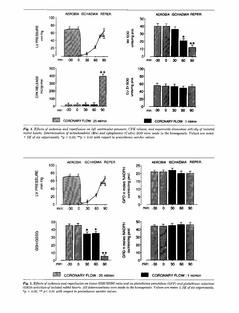
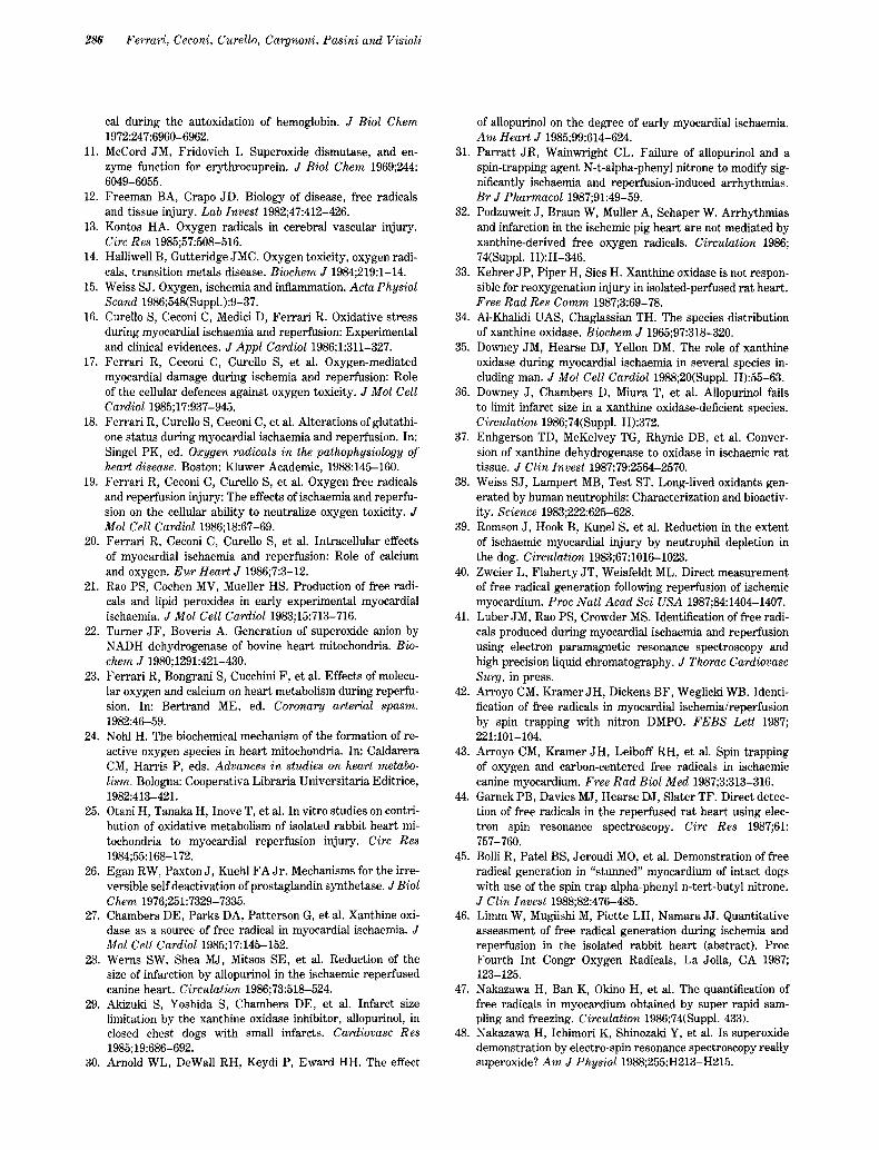
To investigate the possible role of oxygen free radi- cals in reperfusion injury, we have determined the effects of ischemia on the activity of mitochondrial and cytosolic superoxide dismutase, and of glutathione peroxidase and glutathione reductase, the two major lines of defense against oxygen free-radical produc- tion. In addition, we have also measured the tissue GSH/GSSG ratio as an index of oxidative stress. These experiments have been performed using iso- lated and perfused rabbits hearts, as previously de- scribed [17,19,20], and the data are shown in Figures 4-6. Figure 4 shows that the reduction of coronary flow to 1 ml/min induced a rapid decline of developed pressure, with contractile activity completely ceasing 9 minutes after the onset of ischemia. Resting pres- sure began to rise progressively 20 minutes after the onset of ischemia. Ninety minutes of ischemia specifi- cally reduced the activity of mitochondrial Mn-SOD, whilst the same period of ischemia did not affect the activity of the cytosolic CuZn-SOD, or of glutathione peroxidase or glutathione reductase (Figure 5). We have no information on mitochondria function in these experiments. However, it is possible that the residual flow of 1 ml/min maintained some degree of mitochon- drial respiration. Figure 5, however, shows that isch- emia induced a reduction in the myocardial GSH/ GSSG ratio. This was mainly due to a consistent re- duction of tissue content of GSH, GSSG being un- changed (Figure 6).
Reperfusion induced a further increase of diastolic pressure, with almost no recovery of developed pres- sure. As indicated in Figure 6, on reperfusion there was also a significant increase of tissue GSSG from the ischemic value of 0.18 _+ 0.02 nM/mg protein to 0.55 -+ 0.02 nM/mg protein (Figure 6), resulting in a further decline of GSH/GSSG ratio (Figure 5). Fig- ures 4 and 5 show that the readmission of coronary flow did not modify significantly the SOD, GRD, and GPD activities.
The alterations of mitochondrial SOD and of the GSH/GSSG ratio during reperfusion were coincident with important changes in the rate of release of GSH and GSSG, as shown in Figure 6. Ischemia did not significantly alter the rate of GSH or GSSG release, but on reperfusion there was a marked and sustained release of GSH and GSSG from the heart.
These results suggest that ischemia induces meta- bolic alterations capable of reducing the defense mech- anisms against oxygen toxicity. The prime alteration seems to lie at the level of mitochondrial SOD, its activity being reduced by 50%. Under these condi- tions the readmission of molecular oxygen is likely to stimulate the production of oxygen radicals above the neutralizing capacity of mitochondrial SOD. Conse-
UJ n-
n,- E 0,.
.J
LU .c_
u J ~
v E Q. (,.)
100
80'
60"
40'
20'
0"
500'
400'
300
200"
100"
AEROBIA ISCHAEMIA REPER.
min: -30 0 30 60 90
0" v..T.. ...... THe, T T rain: -30 0 30 60
AEROBIA ISCHAEMIA REPER.
40
t a 30 O~
¢n ~ 20
10
0 rain: -30 0 30 60 90
0 90 min: -30 0 30
CORONARY FLOW : 25 ml/min
/ IW 6O 9O
CORONARY FLOW : 1 ml/min
Fig. 4. Effects of ischemia and reperfusion on left ventricular pressure, CPK release, and superoxide dismutase activity of isolated rabbit hearts. Determination of mitochondrial (Mn) and cytoplasmic (CuZn) SOD were made in the homogenate. Values are mean +- SE of six experiments. *p < 0.05; **p < 0.01 with respect to preischemic aerobic values.
uJ G~ 3 03 031
E Q. > _J
100
80
60
40
20
0
AEROBIA ISCHAEMIA REPER 2 5 T
~ 15' ~ E
10' C
D ~ 5'
~ 0' min: -30 0 30 60 90
AEROBIA
min: -30 C)
ISCHAEMIA REPER.
(.9 03 03 (.9
03
50
40
30
20'
10'
0" min:
IF'7
-30 0
-I- 50" n
. • 4o • 1" "1" z n
@@
30 60 90
CORONARY FLOW : 25 ml/min
_~ ~ 30'
c c° ~ 20'
Q ~) 10" n" £.9
O" min:
m
-30 0 30
m
60 9O
CORONARY FLOW : 1 ml/min
FO. 5. Effects of ischemia and reperfusion on tissue GSH/GSSG ratio and on glutathione peroxidase (GPP) and glutathione reductase (GRD) activities of isolated rabbit hearts. Al l determinations were made in the homogenate. Values are mean +- SE of six experiments. *p < 0.05; ** p< 0.01 with respect to preischemic aerobic values.

Oxidative Stress During Reperfusion 283
-I- 2 Or) O. (.9 o~
CD o o0"6 F - E
t -
10"
8"
6"
4"
2"
0"
AEROBIA ISCHAEMIA REPER•
min: -30 0
"T"
/ 9O
10"
6. ~ 'r: 4-
2-
O.
AEROBIA ISCHAEMIA REPER.
~ o
min: -30 0 30 60 90
Or) (.9
I-"- ¢,-
0.6"
0 . 4 "
0.2"
0" ,,,=/l min: -30 0 30 60 90
CORONARY FLOW : 25 ml/min
C 0:•~
.. o.s] o'
rain: -30 0 30 60 go
CORONARY FLOW : 1 ml/min
Fig. 6. Effects of ischemia and reperfusion on tissue content and release of reduced (GSH) and oxidized (GSSG) glutathione of iso- lated and perfused rabbit hearts. Values are mean +- SE of six experiments, *p < 0.05; **p < 0.01 with respect to preischemic aerobic values.
quently, the second line of defense against oxygen toxicity, GPD, is likely to be highly stimulated. We found a severe alteration of the glutathione status, indicating that this was the case and that myocardial oxidative damage had occurred and that it had proba- bly been counteracted at that level•
Studies on animals show that the oxidative stress on reperfusion is correlated with the duration of the ischemic period [16]. Reperfusion after a short period of ischemia (30 to 60 minutes) does not result in oxida- tive stress, probably because the defense mechanisms are still able to protect the myocardial cells against the burst of oxygen free radicals generated by read- mission of oxygen. Reperfusion after more prolonged period of ischemia, when the defense mechanisms are likely to be reduced, results in further damage, with no recovery in function.
O c c u r r e n c e o f O x i d a t i v e S t r e s s A f t e r
I s c h e m i a a n d R e p e r f u s i o n i n M a n
Clinical investigations are hampered by the impossi- bility of directly measuring the ephemeral free radi- cals and by the difficulties in following the molecular changes occurring during early phases of reperfusion.
In addition, in the clinical setting it is almost impossi- ble to standardize the onset, severity, and duration of ischemia and reperfusion.
We attempted to resolve this problem by measur- ing the arterial and coronary sinus difference of GSH and GSSG of coronary artery disease (CAD) patients subjected to different periods of global ischemia fol- lowed by reperfusion during coronary artery bypass grafting. Because of the high rate of glutathione auto- oxidation and its disappearance in the blood, we deter- mined plasma levels of GSH and GSSG using a method modified by us [68] in which the blood is treated imme- diately after collection with thiol, stabilizing agents.
In Figures 7 and 8 are reported the arterial- coronary sinus differences for GSH (Figure 7) and GSSG (Figure 8) of CAD patients subjected to aortic cross-clamping in which the mean clamping period was as long as 25 -+ 3 minutes (group 1, 12 patients) and 55 _+ 5 minutes (group 2, 10 patients), followed in both groups by 30 minutes of reperfusion. In all pa- tients, before clamping there was a small positive ar- teriovenous difference for GSH and GSSG. During the following 25 (group 1) or 55 (group 2) minutes of global ischemia, it was not possible to sample from the coro- nary sinus because of the abolition of coronary flow.

uJ +100 0 z L U --.- , nr" '-o I.U --. 0 I..L. IJ..
D o > E 4: ~ -lOO . . j I
rr n" -200 < O O i L O >-
-300
m
I I
PRECLAMPING
minutes 0
DECLAMPING
I !
10 20
Group 1 (mean clamping = 26')
Group 2 (mean clamping = 53')
OUT OF THE PUMP
30
Fig. 7. Myocardial arterio-coronary sinus difference for reduced glutathione (GSH) of CAD patients subjected to open-heart surgery. *p < 0.05; **p < 0.01 with respect to group 1.
PRECLAMPING +10
,,._~ 0
>' ~ -lO
o , ,
- 30
DECLAMPING
]:
! !
minutes 0 10 20
OUT OF THE PUMP
30
I Group 1 (mean clamping = 26')
I Group 2 (mean clamping = 53')
Fig. 8. Myocardial arterio-coronary sinus difference for oxidized glutathione (GSSG) of CAD patients subjected to open-heart surgery. *p < 0.05; **p < 0.01, with respect to group 1.

Oxidative Stress During Reperfusion 285
During reperfusion, after the short period of ischemia (group 1), there was a small and transient release of GSH (Figure 7) and GSSG (Figure 8) into the coro- nary sinus, reaching a peak 3 minutes after declamp- ing; the GSH and GSSG concentrations in the coro- nary sinus then star ted to decline and fell below the arterial values. In contrast, reperfusion of patients of group 2 after a more prolonged period of ischemia resulted in more pronounced and sustained release of GSH and GSSG from the myocardium. GSSG produc- tion was still continuing after 30 minutes of declamp- ing, the arteriocoronary sinus difference remaining negative, even after the patients were disconnected from the pump (Figure 8).
The results obtained in the two groups of patients show that, as in animal studies, reperfusion results in an oxidative stress, depending on the duration of the ischemic period. When the period of clamping was re- duced to 25 minutes, reperfusion resulted in a small and transient rise of GSH and GSSG in the coronary sinus. I t is probable that this represented a washout process. Reperfusion reinstated after 55 minutes of ischemia led to a significant release of GSH and GSSG, which was still on at the end of the procedure. This is similar to the effects of reperfusion after prolonged ischemia in the isolated heart and presumably implies oxidative stress. I t is interesting to note that in pa- tients of group 2, there was a direct correlation be- tween the duration of the clamping period and the release of GSSG into the coronary sinus and an in- verse correlation between the degree of GSSG release in the coronary sinus and the recovery of hemody- namic function after surgery [71]. Thus these cases indicate that reperfusion of CAD patients might in- duce oxidative damage after a prolonged period of ischemia, and oxygen free radicals may be involved in reperfusion damage.
C o n c l u s i o n
Without doubt, reperfusion is the most effective way to t reat the ischemic myocardium. Later reperfusion may cause, or at least accelerate, myocardial damage, the extent of which can be modified, although the un- derlying mechanism is still not understood and it is likely that it has multifactorial origin. Myocardial pro- duction of oxygen free radicals above the neutralizing capacity of the myocytes may be an important cause of the deleterious effects of reperfusion. This possibility, however, is by no means proven.
There is evidence that prolonged ischemia reduces the naturally occurring defense mechanisms of the heart against oxygen free radicals. Although each method had limitations, when several different meth-
ods have been compared, there is also evidence, albeit preliminary, that the formation of these toxic species is enhanced after ischemia, and particularly so after reperfusion. I t seems that the presence of blood is not a prerequisite for oxygen free radical formation and that the mitochondria are likely to be an important site of production. Experimental work does indicate that the oxygen free radical-mediated component of the reperfusion damage can be circumvented, provid- ing optimism for pharmacologic treatment. However, several controversies exist regarding the meaning of studies in which agents known to interfere with oxy- gen free radicals have provided protection, and con- clusions derived from such studies should be consid- ered with caution.
Almost no data are available on the role of oxygen free radicals in reperfusion injury in humans. This, in our opinion, is the goal to achieve in the very near future, before antioxidants are blindly and irrationally combined with reperfusion therapy in the t reatment of acute myocardial infarction in humans.
A c k n o w l e d g m e n t s
This work was supported by the Italian C.N.R. grant 087432. We thank Miss Ornella Del Ciello and Miss Roberta Bonetti for secretarial assistance in preparing the manuscript.
R e f e r e n c e s
1. Ferrari R, Curello S, Cargnoni A, et al. Metabolic changes during post-ischaemic reperfusion. J Mol Cell Cardiol 1988;20, Suppl II:119-133.
2. Hearse DJ. Reperfusion of ischaemic myocardium. J Mol Cell Cardiol 1977;9:607-616.
3. GISSI Study. Effectiveness of intravenous thrombolytic treatment in acute myocardial infarction. Lancet 1986;1: 397-401.
4. YusufS, Collins R, Peto R, etal. Intravenous and intracoro- nary fibrinolytic therapy in acute myocardial infarction: Overview of results on mortality, reinfarction and side ef- fects from 33 controlled randomized trials. Eur Heart J 1984;6:556-585.
5. Braunwald E, Kloner RA. Myocardial reperfusion: A dou- ble-edged sword. J Clin Invest 1985;76:1713-1719.
6. Poole-Wilson PA. Reperfusion damage in heart muscle: Still unexplained but with new clinical relevance. Clin Physiol 1987;7:439-453.
7. Thompson JA, Hess ML. The oxygen free radical system: A fundamental mechanism in the production of myocardial necrosis. Prog Cardiovasc Dis 1986;6:449-462.
8. Misra HP, Fridovich I. The role of superoxide anion in the autooxidation of epinephrine and a simple assay for superox- ide dismutase. J Biol Chem 1972;247:3170-3175.
9. Marklund SL, Grankvis K, Taijeda F. Oxy-radicals in the toxicity of cellular toxins. In: Greenvald RA, Goh G, eds. Oxy-radicals and their scavenger system, Amsterdam: Elsevier, 1983:6-104.
10. Misra HP, Fridovich I. The generation of superoxide radi-
286 Ferrari, Ceconi, Curello, Cargnoni, Pasini and Visioli
cal during the autoxidation of hemoglobin. J Biol Chem 1972:247:6960-6962.
11. McCord JM, Fridovich I. Superoxide dismutase, and en- zyme function for erythrocuprein. J Biol Chem 1969;244: 6049-6055.
12. Freeman BA, Crapo JD. Biology of disease, free radicals and tissue injury. Lab Invest 1982;47:412-426.
13. Kontos HA. Oxygen radicals in cerebral vascular injury. Circ Res 1985;57:508-516.
14. Halliwell B, Gutteridge JMC. Oxygen toxicity, oxygen radi- cals, transition metals disease. Biochem J 1984;219:1-14.
15. Weiss SJ. Oxygen, ischemia and inflammation. Acta Physiol Scand 1986;548(Suppl.):9-37.
16. Curello S, Ceconi C, Medici D, Ferrari R. Oxidative stress during myocardial ischaemia and reperfusion: Experimental and clinical evidences. J Appl Cardiol 1986;1:311-327.
17. Ferrari R, Ceconi C, Curello S, et al. Oxygen-mediated myocardial damage during ischemia and reperfusion: Role of the cellular defences against oxygen toxicity. J Mol Cell Cardiol 1985;17:937-945.
18. Ferrari R, Curello S, Ceconi C, et al. Alterations ofglutathi- one status during myocardial ischaemia and reperfusion. In: Singel PK, ed. Oxygen radicals in the pathophysiology of heart disease. Boston: Kluwer Academic, 1988:145-160.
19. Ferrari R, Ceconi C, Curello S, et al. Oxygen free radicals and reperfusion injury: The effects of ischaemia and reperfu- sion on the cellular ability to neutralize oxygen toxicity. J Mol Cell Cardiol 1986;18:67-69.
20. Ferrari R, Ceconi C, Curello S, et al. Intracellular effects of myocardial ischaemia and reperfusion: Role of calcium and oxygen. Eur Heart J 1986;7:3-12.
21. Rao PS, Cochen MV, Mueller HS. Production of free radi- cals and lipid peroxides in early experimental myocardial ischaemia. J Mol Cell Cardiol 1983;15:713-716.
22. Turner JF, Boveris A. Generation of superoxide anion by NADH dehydrogenase of bovine heart mitochondria. Bio- chem J 1980;1291:421-430.
23. Ferrari R, Bongrani S, Cucchini F, et al. Effects of molecu- lar oxygen and calcium on heart metabolism during reperfu- sion. In: Bertrand ME, ed. Coronary arterial spasm. 1982:46-59.
24. Nohl H. The biochemical mechanism of the formation of re- active oxygen species in heart mitochondria. In: Caldarera CM, Harris P, eds. Advances in studies on heart metabo- lism. Bologna: Cooperativa Libraria Universitaria Editrice, 1982:413-421.
25. Otani H, Tanaka H, Inove T, et al. In vitro studies on contri- bution of oxidative metabolism of isolated rabbit heart mi- tochondria to myocardial reperfusion injury. Circ Res 1984;55:168-172.
26. Egan RW, Paxton J, Kuehl FA Jr. Mechanisms for the irre- versible self deactivation of prostaglandin synthetase. J Biol Chem 1976;251:7329-7335.
27. Chambers DE, Parks DA, Patterson G, et al. Xanthine oxi- dase as a source of free radical in myocardial ischaemia. J Mol Cell Cardiol 1985;17:145-152.
28. Werns SW, Shea MJ, Mitsos SE, et al. Reduction of the size of infarction by allopurinol in the ischaemic reperfused canine heart. Circulation 1986;73:518-524.
29. Akizuki S, Yoshida S, Chambers DE, et al. Infarct size limitation by the xanthine oxidase inhibitor, allopurinol, in closed chest dogs with small infarcts. Cardiovasc Res 1985;19:686-692.
30. Arnold WL, DeWall RH, Keydi P, Eward HH. The effect
of allopurinol on the degree of early myocardial ischaemia. A m Heart J 1985;99:614-624.
31. Parratt JR, Wainwright CL. Failure of allopurinol and a spin-trapping agent N-t-alpha-phenyl nitrone to modify sig- nificantly ischaemia and reperfusion-induced arrhythmias. Br J Pharmacol 1987;91:49-59.
32. Podzuweit J, Braun W, Muller A, Schaper W. Arrhythmias and infarction in the ischemic pig heart are not mediated by xanthine-derived free oxygen radicals. Circulation 1986; 74(Suppl. II):II-346.
33. Kehrer JP, Piper H, Sies H. Xanthine oxidase is not respon- sible for reoxygenation injury in isolated-perfused rat heart. Free Rad Res Comm 1987;3:69-78.
34. A1-Khalidi UAS, Chaglassian TH. The species distribution of xanthine oxidase. Biochem J 1965;97:318-320.
35. Downey JM, Hearse D J, Yellon DM. The role of xanthine oxidase during myocardial ischaemia in several species in- cluding man. J Mol Cell Cardiol 1988;20(Suppl. II):55-63.
36. Downey J, Chambers D, Miura T, et al. Allopurinol falls to limit infarct size in a xanthine oxidase-deficient species. Circulation 1986;74(Suppl. II):372.
37. Enhgerson TD, McKelvey TG, Rhynie DB, et al. Conver- sion of xanthine dehydrogenase to oxidase in ischaemic rat tissue. J Clin Invest 1987;79:2564-2570.
38. Weiss S J, Lampert MB, Test ST. Long-lived oxidants gen- erated by human neutrophils: Characterization and bioactiv- ity. Science 1983;222:625-628.
39. Romson J, Hook B, Kunel S, et al. Reduction in the extent of ischaemic myocardial injury by neutrophil depletion in the dog. Circulation 1983;67:1016-1023.
40. Zweier L, Flaherty JT, Weisfeldt ML. Direct measurement of free radical generation following reperfusion of ischemic myocardium. Proc Natl Acad Sci USA 1987;84:1404-1407.
41. Luber JM, Rao PS, Crowder MS. Identification of free radi- cals produced during myocardial ischaemia and reperfusion using electron paramagnetic resonance spectroscopy and high precision liquid chromatography. J Thorac Cardiovasc Surg, in press.
42. Arroyo CM, Kramer JH, Dickens BF, Weglicki WB. Identi- fication of free radicals in myocardial ischemia/reperfusion by spin trapping with nitron DMPO. F E B S Lett 1987; 221:101-104.
43. Arroyo CM, Kramer JH, Leiboff RH, et al. Spin trapping of oxygen and carbon-centered free radicals in ischaemic canine myocardium. Free Rad Biol Med 1987;3:313-316.
44. Garnck PB, Davies MJ, Hearse DJ, Slater TF. Direct detec- tion of free radicals in the reperfused rat heart using elec- tron spin resonance spectroscopy. Circ Res 1987;61: 757-760.
45. Bolli R, Patel BS, Jeroudi MO, et al. Demonstration of free radical generation in "stunned" myocardium of intact dogs with use of the spin trap alpha-phenyl n-tert-butyl nitrone. J Clin Invest 1988;82:476-485.
46. Limm W, Mugiishi M, Piette LH, Namara JJ. Quantitative assessment of free radical generation during ischemia and reperfusion in the isolated rabbit heart (abstract). Proc Fourth Int Congr Oxygen Radicals, La Jolla, CA 1987; 123-125.
47. Nakazawa H, Ban K, Okino H, et al. The quantification of free radicals in myocardium obtained by super rapid sam- pling and freezing. Circulation 1986;74(Suppl. 433).
48. Nakazawa H, Ichimori K, Shinozaki Y, et al. Is superoxide demonstration by electro-spin resonance spectroscopy really superoxide? A m J Physiol 1988;255:H213-H215.
Oxidative Stress During Reperfusion 287
49. Ceconi C, Curello S, Cargnoni A, et al. The role of glutathi- one status in the protection against ischaemic and reperfu- sion damage: Effects of N-acetyl cysteine. J Mol Cell Cardiol 1988;20:5-13.
50. Ferrari R, Ceconi C, Curello S, et al. Oxygen utilization and toxicity at myocardial level. In: Benzi G, Packer L, Sili- prandi N, eds. Biochemical aspects of physical exercise. Amsterdam: Elsevier, 1986:145-156.
51. Meister A. Methods for selective modification of glutathione metabolism and study of glutathione transport. In: Meister A, ed. Methods in enzymology. New York: Academic Press, 1985;571-585.
52. Meister A, Anderson ME. Glutathione (review). Ann Rev Biochem 1983;52:711-760.
53. Chance B, Sies M, Boveris A. Hydroperoxide metabolism in mammalian organs (review). Physiol Rev 1979;59:527-605.
54. Gaudel Y, Duvelleroy MA. Role of oxygen radicals in car- diac injury due to reoxygenation. J Mol Cell Cardiol 1984;16:459-470.
55. Guarnieri C, Flamigni F, Caldarera CM. Role of oxygen in the cellular damage induced by re-oxygenation of hypoxic heart. J Mol Cell Cardiol 1980;12:797-808.
56. Meerson FZ, Kagan VE, Kozlov Yu P, et al. The role of lipid peroxidation in pathogenesis of ischemic damage and the antioxidant protection of the heart. Basic Res Cardiol 1982;77:465-485.
57. Gutteridge JMC, Quinlan GJ. Malondialdehyde formation from lipid peroxides in the thiobarbituric acid test: The role of lipid radicals, iron salts, and metal chelators. J Appl Bio- chem 1983;5:293-299.
58. Dormandy TL. Free radical oxidation and antioxidants. Lancet 1978;1:647-650.
59. Ceconi C, Cargnoni A. Pasini E, et al. Evaluation of phos- pholipid peroxidation as malondialdehyde during myocardial ischaemic and reperfusion injury. Am J Physiol, in press.
60. Ceconi C, Pasini E, Benigno M, et al. Heart lipid peroxida-
tion: HPLC versus TBA for determination of malondialde- hyde. International Teach-in for Promoting Scientific Basis of Cardiology, Roma, 23-25 October, 1989.
61. Curello S, Ceconi C, Bigoli C, et al. Change in the cardiac glutathione status after ischaemia and reperfusion. Expe- rientia 1985;41:42-43.
62. Ferrari R, Ceconi C, Curello S, et al. Molecular events oc- curring during post-ischaemic reperfusion. In: Dhalla NS, Innes IR, Beamish RE, eds. Myocardial ischaemia, Boston: Nijhoff Publishing, 1987:67-84.
63. Larsson A, Orrenius S, Holmegren A, eds. Functions of glutathione: Biochemical, physiological, toxicological and clinical aspects, New York: Raven Press, 1983.
64. Mc Intyre TM, Curthoys NP. The interorgan metabolism of glutathione. Int J Biochem 1980;12:545-551.
65. Mannervik B, Axelsson K. Role of cytoplasmic thioltransfer- ase in cellular regulation by thiol-disulphide interchange. Biochem J 1980;190:125-130.
66. Meister A, Tate SS. Glutathione and related-glutamil com- pounds: Biosynthesis and utilization. Ann Rev Biochem 1976;45:559-564.
67. Ferrari R, Visioli O, Caldarera CM, Nayler WG. Vitamin E and the heart: Possible role as antioxidants. Acta Vitamin et Enzymol 1982;5:11-22.
68. Curello S, Ceconi C, Cargnoni A, et al. Improved procedure for determining glutathione plasma as an index of myocar- dial oxidative stress. Clin Chem 1987;33/8:1448-1449.
69. Adams JP, Lauterburg BM, Mitchell JR. Plasma glutathi- one and glutathione disulfide in rat: Regulation and response to oxidative stress. J Pharmacol Exp Ther 1983;227:749- 754.
70. Ishikawa H, Sies H. Cardiac transport of glutathione disul- fide and s-conjugate. J Biol Chem 1984;259:333-342.
71. Ferrari R, Alfieri O, Curello S, et al. Occurrence of oxida- tive stress during reperfusion of the human heart. Circula- tion 1990;81(1):201-211.