the nucleic acids friedrich miescher in 1869 isolated what he called nuclein from the nuclei of pus...
TRANSCRIPT
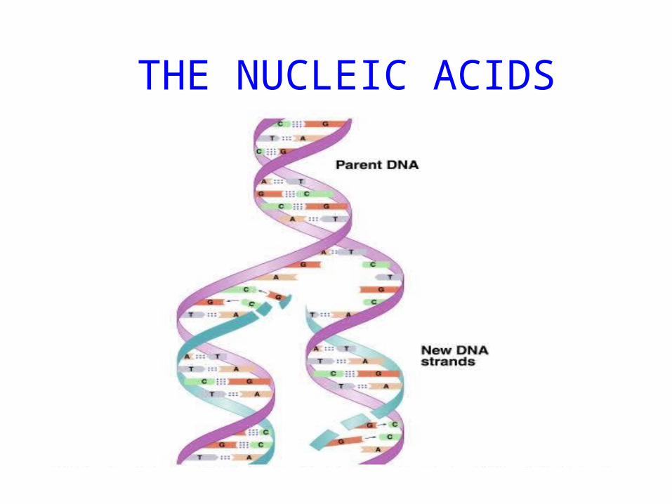
THE NUCLEIC ACIDS
Friedrich Miescher in 1869
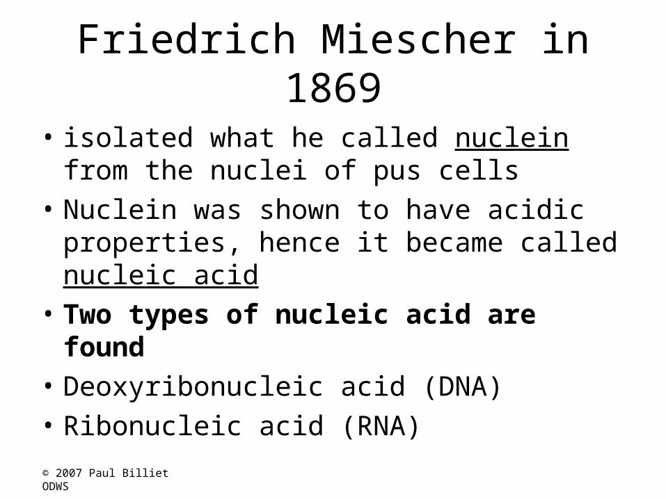
• isolated what he called nuclein from the nuclei of pus cells
• Nuclein was shown to have acidic properties, hence it became called nucleic acid
• Two types of nucleic acid are found• Deoxyribonucleic acid (DNA)• Ribonucleic acid (RNA)
© 2007 Paul Billiet ODWS

The distribution of nucleic acids in the eukaryotic cell
• DNA is found in the nucleus with small amounts in mitochondria and chloroplasts
• RNA is found throughout the cell
© 2007 Paul Billiet ODWS

DNA as genetic material: The circumstantial evidence
1. Present in all cells and virtually restricted to the nucleus2. The amount of DNA in somatic cells (body cells) of any
given species is constant (like the number of chromosomes)
3. The DNA content of gametes (sex cells) is half that of somatic cells. In cases of polyploidy (multiple sets of chromosomes) the DNA content increases by a proportional factor
4. The mutagenic effect of UV light peaks at 253.7nm. The peak for the absorption of UV light by DNA

NUCLEIC ACID STRUCTURE
• Nucleic acids are polynucleotides• Their building blocks are nucleotides

NUCLEOTIDE STRUCTUREPHOSPATE SUGAR
Ribose or Deoxyribose
NUCLEOTIDE
BASE
PURINES PYRIMIDINES
Adenine (A)Guanine(G)
Cytocine (C)Thymine (T)Uracil (U)
© 2007 Paul Billiet ODWS

Ribose is a pentose
C1
C5
C4
C3 C2
O
© 2007 Paul Billiet ODWS

RIBOSE DEOXYRIBOSE
CH2OH
H
OH
C
C
OH OH
C
O
H HH
C
CH2OH
H
OH
C
C
OH H
C
O
H HH
C
Spot the difference
© 2007 Paul Billiet ODWS

THE SUGAR-PHOSPHATE BACKBONE
• The nucleotides are all orientated in the same direction
• The phosphate group joins the 3rd Carbon of one sugar to the 5th Carbon of the next in line.
P
P
P
P
P
P
© 2007 Paul Billiet ODWS

ADDING IN THE BASES
• The bases are attached to the 1st Carbon
• Their order is important It determines the genetic information of the molecule
P
P
P
P
P
P
G
C
C
A
T
T© 2007 Paul Billiet ODWS

DNA IS MADE OF TWO STRANDS OF POLYNUCLEOTIDE
P
P
P
P
P
P
C
G
G
T
A
A
P
P
P
P
P
P
G
C
C
A
T
T
Hydrogen bonds
© 2007 Paul Billiet ODWS
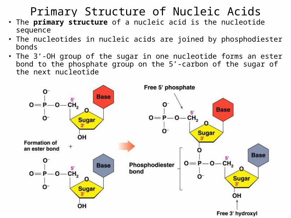
Primary Structure of Nucleic Acids• The primary structure of a nucleic acid is the nucleotide sequence• The nucleotides in nucleic acids are joined by phosphodiester bonds• The 3’-OH group of the sugar in one nucleotide forms an ester bond
to the phosphate group on the 5’-carbon of the sugar of the next nucleotide
Reading Primary Structure
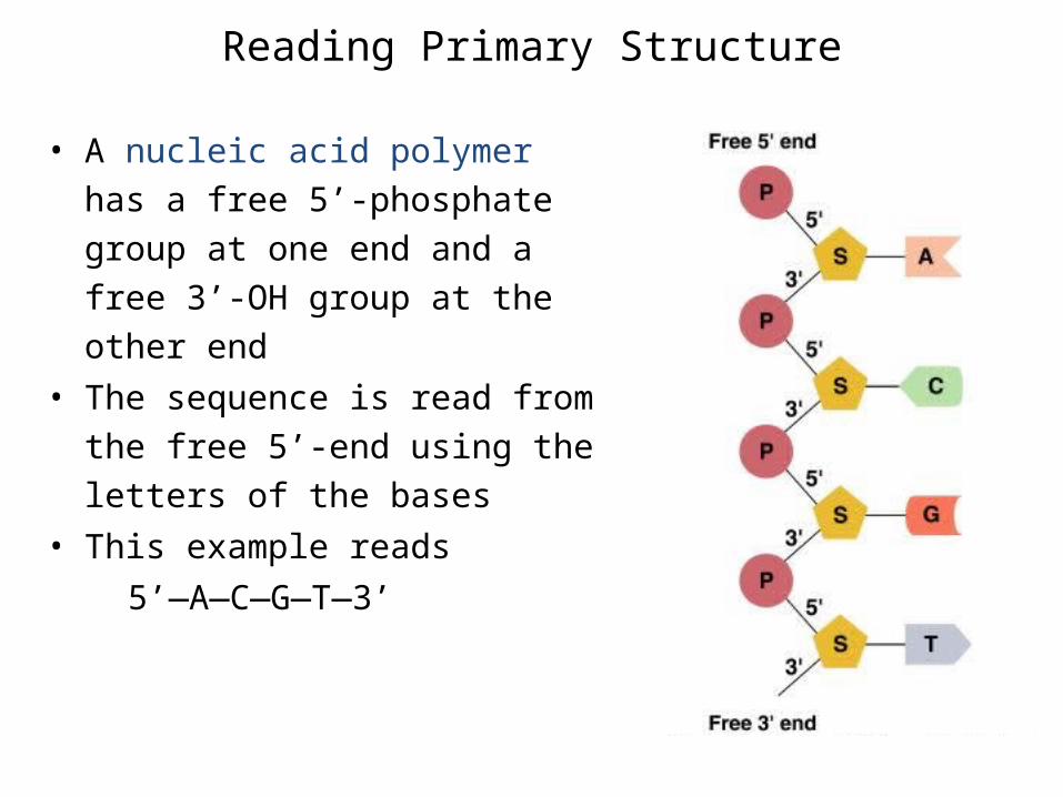
• A nucleic acid polymer has a free 5’-phosphate group at one end and a free 3’-OH group at the other end
• The sequence is read from the free 5’-end using the letters of the bases
• This example reads 5’—A—C—G—T—3’

DNA IS MADE OF TWO STRANDS OF POLYNUCLEOTIDE
• The sister strands of the DNA molecule run in opposite directions (antiparallel)
• They are joined by the bases• Each base is paired with a specific partner:A is always paired with T G is always paired with CPurine with Pyrimidine• This the sister strands are complementary but not
identical• The bases are joined by hydrogen bonds, individually
weak but collectively strong
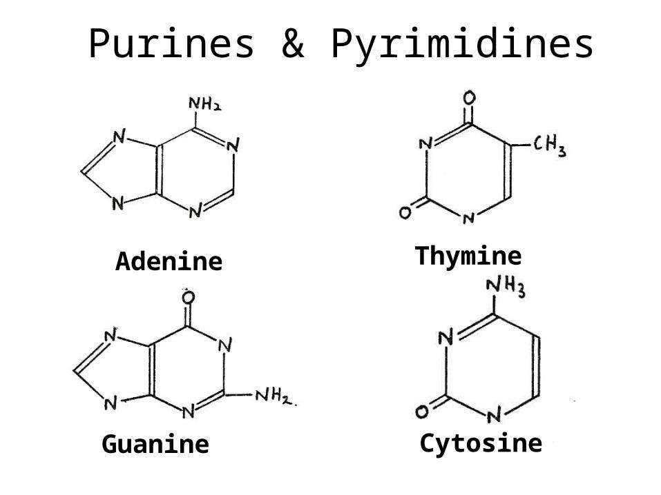
Purines & Pyrimidines
Adenine
CytosineGuanine
Thymine

Watson & Crick Base pairing

AMP, ADP and ATP• Additional phosphate groups can be added to the nucleoside
5’-monophosphates to form diphosphates and triphosphates• ATP is the major energy source for cellular activity

Example of RNA Primary Structure• In RNA, A, C, G, and U are linked by 3’-5’ ester bonds
between ribose and phosphate

Base Pairing in the DNA Double Helix
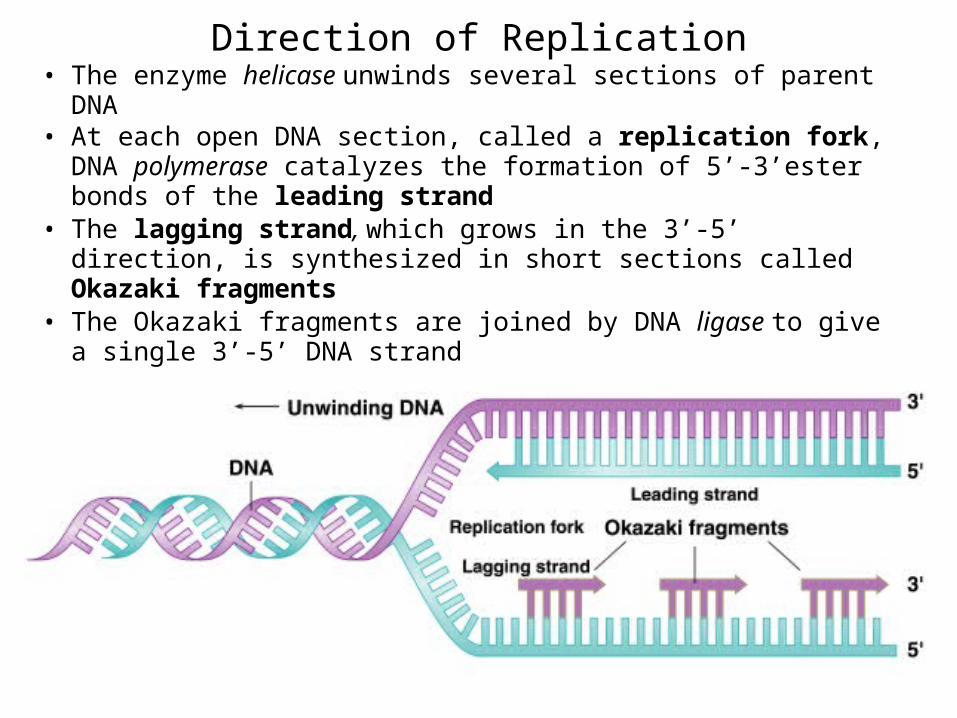
Direction of Replication• The enzyme helicase unwinds several sections of parent DNA • At each open DNA section, called a replication fork, DNA
polymerase catalyzes the formation of 5’-3’ester bonds of the leading strand
• The lagging strand, which grows in the 3’-5’ direction, is synthesized in short sections called Okazaki fragments
• The Okazaki fragments are joined by DNA ligase to give a single 3’-5’ DNA strand

Enzymes and Proteins Involved in DNA Replication

Types of RNA
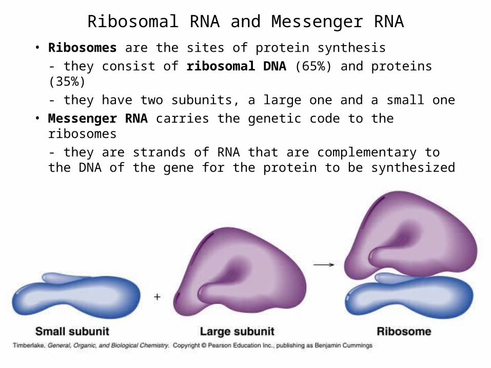
Ribosomal RNA and Messenger RNA
• Ribosomes are the sites of protein synthesis- they consist of ribosomal DNA (65%) and proteins (35%)- they have two subunits, a large one and a small one
• Messenger RNA carries the genetic code to the ribosomes- they are strands of RNA that are complementary to the DNA of the gene for the protein to be synthesized

Transfer RNA• Transfer RNA translates the genetic code from the messenger RNA
and brings specific amino acids to the ribosome for protein synthesis• Each amino acid is recognized by one or more specific tRNA• tRNA has a tertiary structure that is L-shaped
- one end attaches to the amino acid and the other binds to the mRNA by a 3-base complimentary sequence

Protein Synthesis• The two main processes involved in protein synthesis are
- the formation of mRNA from DNA (transcription)- the conversion by tRNA to protein at the ribosome (translation)
• Transcription takes place in the nucleus, while translation takes place in the cytoplasm
• Genetic information is transcribed to form mRNA much the same way it is replicated during cell division

Transcription• Several steps occur during transcription:
- a section of DNA containing the gene unwinds
- one strand of DNA is copied starting at the initiation point, which has the sequence TATAAA
- an mRNA is synthesized using complementary base pairing with uracil (U) replacing thymine (T)
- the newly formed mRNA moves out of the nucleus to ribosomes in the cytoplasm and the DNA re-winds

RNA Polymerase• During transcription, RNA polymerase moves along
the DNA template in the 3’-5’direction to synthesize the corresponding mRNA
• The mRNA is released at the termination point

Processing of mRNA
• Genes in the DNA of eukaryotes contain exons that code for proteins along with introns that do not
• Because the initial mRNA, called a pre-RNA, includes the noncoding introns, it must be processed before it can be read by the tRNA
• While the mRNA is still in the nucleus, the introns are removed from the pre-RNA
• The exons that remain are joined to form the mRNA that leaves the nucleus with the information for the synthesis of protein

Removing Introns from mRNA

Regulation of Transcription
• A specific mRNA is synthesized when the cell requires a particular protein
• The synthesis is regulated at the transcription level:
- feedback control, where the end products speed up or slow the synthesis of mRNA
- enzyme induction, where a high level of a reactant induces the transcription process to provide the necessary enzymes for that reactant
• Regulation of transcription in eukaryotes is complicated and we will not study it here
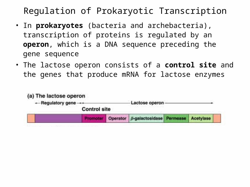
Regulation of Prokaryotic Transcription
• In prokaryotes (bacteria and archebacteria), transcription of proteins is regulated by an operon, which is a DNA sequence preceding the gene sequence
• The lactose operon consists of a control site and the genes that produce mRNA for lactose enzymes
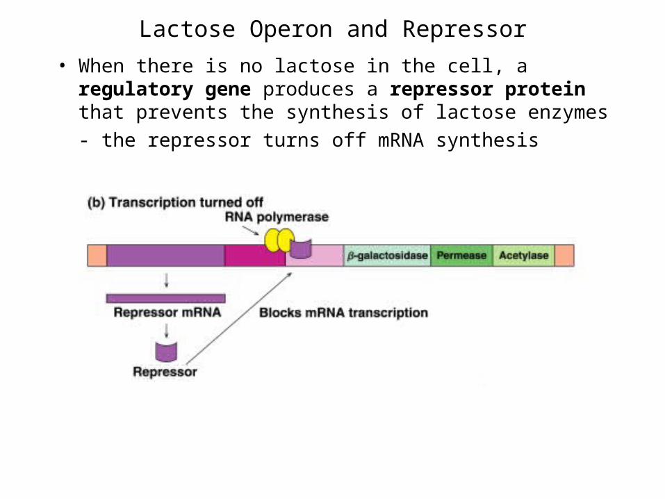
Lactose Operon and Repressor
• When there is no lactose in the cell, a regulatory gene produces a repressor protein that prevents the synthesis of lactose enzymes- the repressor turns off mRNA synthesis

Lactose Operon and Inducer
• When lactose is present in the cell, some lactose combines with the repressor, which removes the repressor from the control site
• Without the repressor, RNA polymerase catalyzes the synthesis of the enzymes by the genes in the operon
• The level of lactose in the cell induces the synthesis of the enzymes required for its metabolism
RNA Polymerase
The Genetic Code• The genetic code is found in the sequence of nucleotides in
mRNA that is translated from the DNA• A codon is a triplet of bases along the mRNA that codes for a
particular amino acid• Each of the 20 amino acids needed to build a protein has at least
2 codons• There are also codons that signal the “start” and “end” of a
polypeptide chain• The amino acid sequence of a protein can be determined by
reading the triplets in the DNA sequence that are complementary to the codons of the mRNA, or directly from the mRNA sequence
• The entire DNA sequence of several organisms, including humans, have been determined, however,- only primary structure can be determined this way- doesn’t give tertiary structure or protein function
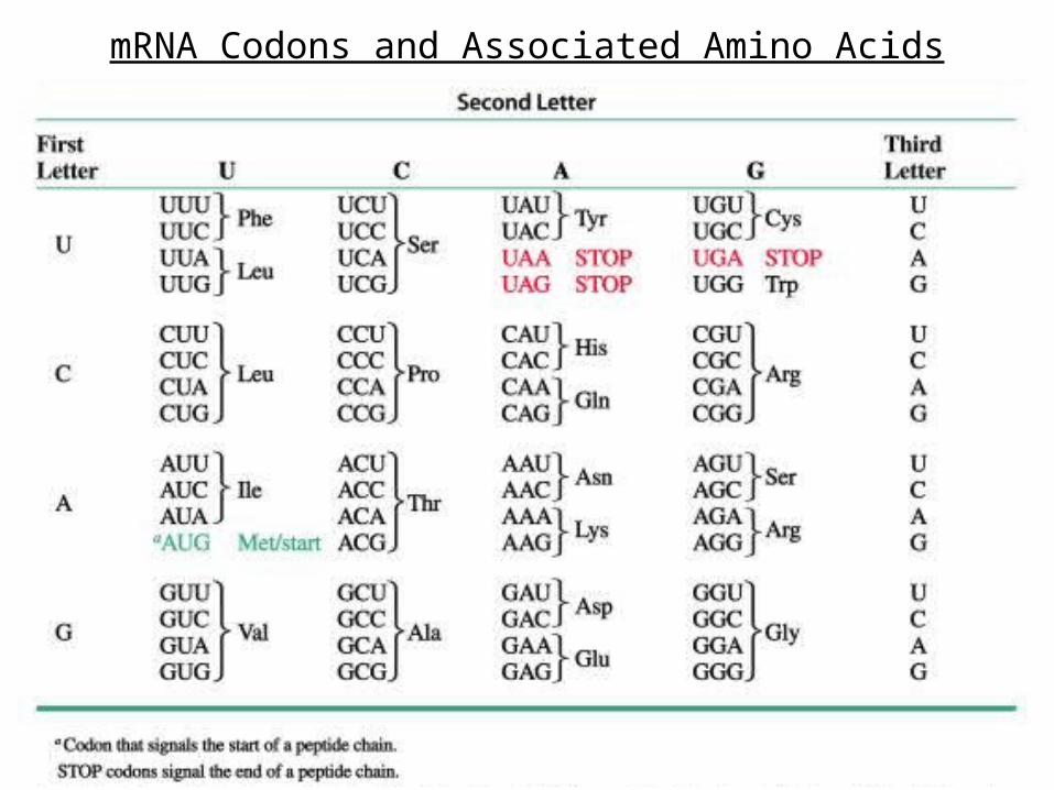
mRNA Codons and Associated Amino Acids

Reading the Genetic Code• Suppose we want to determine the amino acids coded
for in the following section of a mRNA
5’—CCU —AGC—GGA—CUU—3’
• According to the genetic code, the amino acids for these codons are:
CCU = Proline AGC = Serine GGA = Glycine CUU = Leucine
• The mRNA section codes for the amino acid sequence of Pro—Ser—Gly—Leu
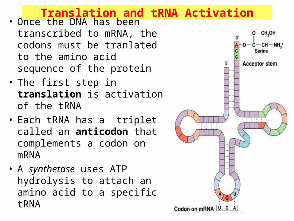
Translation and tRNA Activation• Once the DNA has been
transcribed to mRNA, the codons must be tranlated to the amino acid sequence of the protein
• The first step in translation is activation of the tRNA
• Each tRNA has a triplet called an anticodon that complements a codon on mRNA
• A synthetase uses ATP hydrolysis to attach an amino acid to a specific tRNA

Initiation and Translocation• Initiation of protein synthesis occurs when a mRNA attaches to a
ribosome• On the mRNA, the start codon (AUG) binds to a tRNA with
methionine• The second codon attaches to a tRNA with the next amino acid• A peptide bond forms between the adjacent amino acids at the
first and second codons• The first tRNA detaches from the ribosome and the ribosome shifts
to the adjacent codon on the mRNA (this process is called translocation)
• A third codon can now attach where the second one was before translocation

Termination• After a polypeptide with all the amino acids for a protein
is synthesized, the ribosome reaches the the “stop” codon: UGA, UAA, or UAG
• There is no tRNA with an anticodon for the “stop” codons
• Therefore, protein synthesis ends (termination)
• The polypeptide is released from the ribosome and the protein can take on it’s 3-D structure
(some proteins begin folding while still being synthesized, while others do not fold up until after being released from the ribosome)