the nervous system nervous system links sensory receptors … sys… · · 2011-01-19the nervous...
TRANSCRIPT

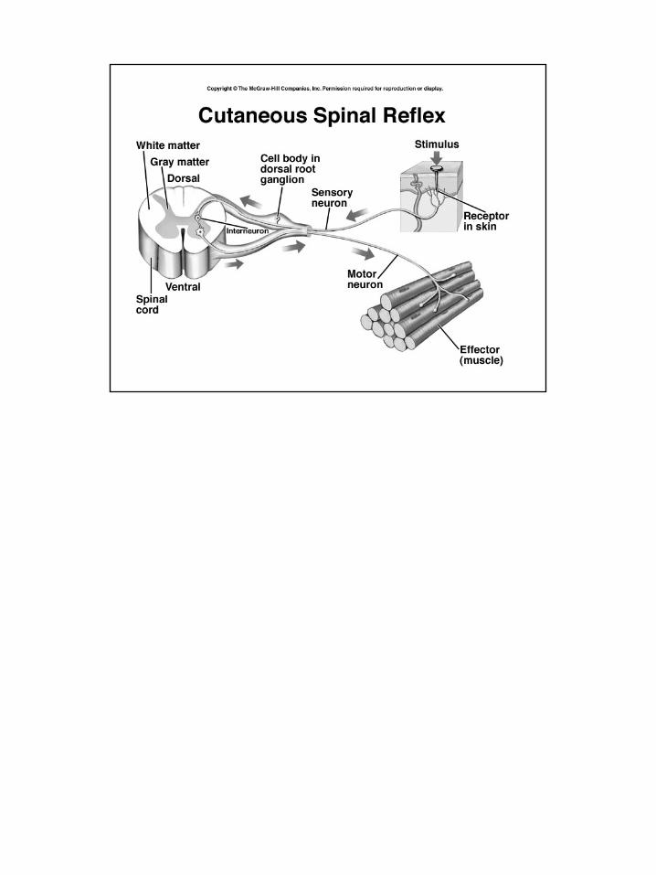
The Nervous SystemNervous system links sensory receptors and motor effectorsSensory (afferent) neurons carry impulses from receptorsMotor (efferent) neurons carry impulses to effectors - muscles
and glands
Association neurons(interneurons)are present inmost nervoussystems -found in brainand spinal cord(CNS)
Association neurons allow for integration of information, reflexes and associative functions (decision making)
Peripheral nervous system (PNS) is composed of sensory and motorneuronsSomatic motor neurons stimulate skeletal muscle contractionAutonomic motor neurons regulate smooth and cardiac muscle,
glands
Autonomic system hassympathetic and parasympathetic divisions
Act antagonistically.

Neuron Structure and FunctionCell body - an enlarged region containing nucleusDendrites extend from cell body -
branched cytoplasmic extensionsMotor and association neurons have many branched dendrites
Axon - a longextension of the cellfor signal transmission- usually only oneper cell
Cell can receive input from many sourcesSurface of cell body integrates information from dendritesIf input is sufficient an nerve impulse is sent along axonWave of depolarization travels outward from cell body
Neurons have supporting cellscalled neuroglia in CNSinclude Schwann cells and oligodendrocytesEither cell can envelop axon, forming myelin sheathNodes of Ranvier - gaps between cells
Schwann cells produce myelin in PNSOligodendrocytes produce myelin in
CNSMyelin sheath insulates neuron in
layers of neuroglial cell membranesMyelination increases speed of nerve
impulseIn brain -
white matter is myelinatedgray matter is not myelinated

Nerve impulses are produced on the axon membraneThere is a charge difference across the plasma membrane in all cells
called the “membrane potential”Interior of cell is negative relative to extracellular side
Resting membrane potential - the charge difference in a cell at rest about 70 millivolts (-70 mV)
Membrane potential due to threefactorssodium-potassium pumps
(3 Na+ out for 2 K + in)different permeability for
different ionsmost important is the
presence of fixed anions (-)proteins and organic phosphates
Each ion has its own equilibrium potential - influenced byconcentration and charge differencesFor K+ - there is 30x more inside cell than outside - K+ will diffuseout due to a concentration difference - but it is also attracted to thenegative charges inside the cell - if not held by negative charges itwould move (out) until the membrane potential was -90 mVAt rest, the concentration differences of all ions across the cellmembrane, and differences in membrane permeability, result in anoverall charge difference of -70 mV

Cell membrane potential can change in response to stimulationDepolarization - becomes less negative (-70 mV to -50 mV)Hyperpolarization - becomes more negative (-70mV to -85 mV)
Nerve and muscle cells have Na+ and K+ channels that are sensitiveto change in the membrane potential - “voltage-gated channels”Protein pore opens or closes with change in membrane potentialAt rest, all Na + gates are closed, some K + gates are open
Different types ofstimulation can cause achannel to open or close

Excitatory stimulation causes depolarization (less negative) - dueto some Na+ gates opening
Inhibitory stimulation causes hyperpolarization (more negative) -due to some K+ gates closing, or Cl- gates opening
The effects of multiple simultaneous stimuli are summed
Small scale stimulation - causing a slight change in membranepermeability and membrane potential is called a “gradedpotential” - followed by a return to the resting membrane potential
If a graded potential reaches the “threshold potential” then asequences of changes in membrane permeability and membranepotential begin - called an “action potential”
The thresholdpotential is usuallyabout -55 mV
Action potential -• large depolarization• repolarization• hyperpolarization• return to resting
only seen in neurons,muscle cells, and senseorgan cells

At rest (1) Na+ channels are closed and some K+ channels are open
When stimulated (2) some Na+ channels open causing small depolarization
If threshold depolarization (3) is reached all Na+ channels open causing large depolarization
At maximum depolarization (4) Na+ channels closeK+ flowing out starts repolarization and opening of all K+ channelscauses complete repolarization (5) and then hyperpolarizationAfter hyperpolarization, some K channels close and cell returns toresting membrane potential (6)
The entiresequence ofchanges requiresabout 0.003 sec.
Following theaction potentialthe Na+/K+pumprestores theconcentrationgradients

Action potentials are always of the same intensity - “all or none”
Action potentials can’t be summed - each one is a separate sequence of events
Immediately following an action potential the cell enters a “refractory period” when Na+ channels can’t be opened again -corresponds to the time in which the Na+/K+ pump reestablishesthe original concentration gradients
Nerve impulses are action potentials that are propagated along theaxonAn action potential begins withthreshold depolarization at the base ofthe axon
An action potential in one region causesthreshold depolarization the nextadjacent region of the axon
A uniform action potential wave travelsalong the axon - a nerve impulse
All nerve impulses are of the sameintensity and magnitude - “all or none”
After a nerve impulse the axon enters arefractory period when it can’t transmitanother impulse
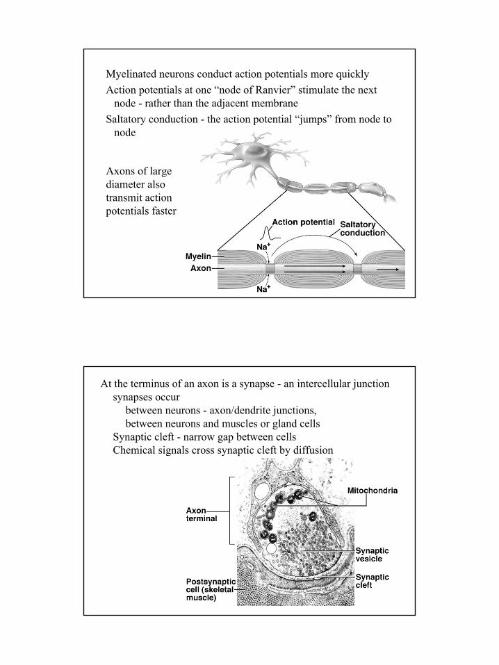
Myelinated neurons conduct action potentials more quicklyAction potentials at one “node of Ranvier” stimulate the next
node - rather than the adjacent membraneSaltatory conduction - the action potential “jumps” from node to
node
Axons of largediameter alsotransmit actionpotentials faster
At the terminus of an axon is a synapse - an intercellular junctionsynapses occur
between neurons - axon/dendrite junctions,between neurons and muscles or gland cells
Synaptic cleft - narrow gap between cellsChemical signals cross synaptic cleft by diffusion

Axon terminus has synaptic vesicles - contain neurotransmittersWhen an action potential arrives at terminus - voltage-gated Ca++
channels openedStimulates fusion of synaptic vesicle membrane with plasma
membrane - contents released via exocytosisMore action potentials cause more vesicles to release contents
Neurotransmitters diffuseacross cleft, bind toreceptor proteins - produce change in receiving cell
Neurotransmitters are then degraded by enzymes in the synaptic cleft and recycled into the axon
At neuromuscular junctions the neurotransmitter isacetylcholine (ACh)
ACh causes depolarization of muscle cell membrane throughthe opening of ion channels in the muscle cell membrane
Depolarization is transmitted via T-tubules to the sarcoplasmicreticulum and ultimately causes muscle contraction

Different neurotransmitters have different effectsExcitatory neurotransmitters cause depolarization of receiving cellInhibitory neurotransmitters cause hyperpolarization of receiver
Glutamate, Glycine and GABAGlutamate - excitatory transmitter in vertebrate CNS
Normal amounts produce physiological stimulationExcessive amounts cause neurodegradation
Glycine and GABA (gamma aminobutyric acid)Inhibitory neurotransmitters - Cause hyperpolarizationOpens chemically-regulated gated Cl- channelInward diffusion of Cl- makes inside more negativeImportant for control of body movements, brain functionsAssociated with sedation effects of Valium (diazepam)

Biogenic Amines - chemicals derived from amino acidsEpinephrine, dopamine, norepinephrine, serotonin
Epinephrine (adrenaline) is a hormoneDopamine is a neurotransmitter found in the brain
Controls body movement and other functionsDegeneration of dopamine-releasing neurons causes Parkinson's
diseaseSchizophrenia associated with excessive dopamine activity
Norepinephrineneurotransmitter in the brain and some autonomic neuronsComplements actions of epinephrine hormone
Serotoninhelps regulate sleep, implicated in various emotional statesInsufficient activity of serotonin-producing neurons results in
clinical depressionSome antidepressant drugs block elimination of serotonin from
synaptic cleft - e.g. Prozac, Paxil, Zoloft