the n-terminus of htert contains a dna-binding domain...
TRANSCRIPT

The N-terminus of hTERT contains a DNA-bindingdomain and is required for telomerase activityand cellular immortalizationDavid C. F. Sealey1,2,3, Le Zheng1,3, Michael A. S. Taboski1,2,3, Jennifer Cruickshank2,3,
Mitsuhiko Ikura1,3 and Lea A. Harrington1,2,3,4,*
1Department of Medical Biophysics, University of Toronto, 2Campbell Family Institute for Breast CancerResearch, 3Ontario Cancer Institute, University Health Network, Toronto, Ontario, M5G 2C1, Canada and4Wellcome Trust Centre for Cell Biology, University of Edinburgh, Edinburgh, EH9 3JR, UK
Received August 6, 2009; Revised November 20, 2009; Accepted November 24, 2009
ABSTRACT
Telomerase defers the onset of telomeredamage-induced signaling and cellular senescenceby adding DNA onto chromosome ends. The abilityof telomerase to elongate single-stranded telomericDNA depends on the reverse transcriptase domainof TERT, and also relies on protein:DNA contactsoutside the active site. We purified the N-terminusof human TERT (hTEN) from Escherichia coli, andfound that it binds DNA with a preference fortelomeric sequence of a certain length andregister. hTEN interacted with the C-terminus ofhTERT in trans to reconstitute enzymatic activityin vitro. Mutational analysis of hTEN revealed thatamino acids Y18 and Q169 were required fortelomerase activity in vitro, but not for the interac-tion with telomere DNA or the C-terminus. Thesemutants did not reconstitute telomerase activity incells, maintain telomere length, or extend cellularlifespan. In addition, we found that T116/T117/S118, while dispensable in vitro, were required forcellular immortalization. Thus, the interactions ofhTEN with telomere DNA and the C-terminus ofhTERT are functionally separable from the roleof hTEN in telomere elongation activity in vitroand in vivo, suggesting other roles for the proteinand nucleic acid interactions of hTEN within, andpossibly outside, the telomerase catalytic core.
INTRODUCTION
Telomeres are specialized nucleoprotein structures thatform the ends of linear chromosomes in most eukaryotes.
Telomere DNA, which ranges in length from 100 to300 bp in ciliates and yeasts, 5–15 kbp in humans, andup to 100 kbp in mice, is comprised by a short repeatingduplex sequence (50-TTAGGG-30/30-AATCCC-50 inhumans) that terminates in a short G-rich single-strandedoverhang (1). The G-overhang can invade the duplex at anupstream position to create a large telomeric loop, orT-loop, in a manner that is facilitated by TRF2 (2,3).TRF1 and TRF2 homodimers bind to the telomereduplex and form a platform for other components of theshelterin complex: RAP1, TIN2, TPP1 and POT1. Inaddition to binding TPP1, POT1 interacts with, and reg-ulates access to the G-overhang (4). TRF2 and POT1protect the telomere by limiting the activation of ATMand ATR pathways, respectively, and downstreamend-processing activities that normally accompany theDNA damage response (5).Telomeres shorten with every cycle of DNA replication
due to the positioning and degradation of the terminalRNA primer involved in generating the daughter laggingstrand, and the nucleolytic resection involved ingenerating the G-overhang (6–9). Human telomeresshorten by �100 bp/cell division (10). In cells thatundergo many cell divisions, telomeres can reach aminimum length that elicits a DNA damage response(11,12). Consequently, cells enter a usually irreversiblenon-proliferative state termed senescence. The onset ofsenescence can be indefinitely postponed by the mainte-nance of telomere length by telomerase (see below).Telomerase, originally discovered in Tetrahymena
thermophila by Greider and Blackburn (13), is a reversetranscriptase formed by the telomerase reversetranscriptase (TERT) and the associated telomeraseRNA (TR) which bears the template for synthesis of thetelomeric G-strand (14–19). The mutation of telomerasecomponents and premature telomere shortening are
*To whom correspondence should be addressed. Tel: +44 131 650 7113; Fax: +44 131 650 5379; Email: [email protected]
Published online 23 December 2009 Nucleic Acids Research, 2010, Vol. 38, No. 6 2019–2035doi:10.1093/nar/gkp1160
� The Author(s) 2009. Published by Oxford University Press.This is an Open Access article distributed under the terms of the Creative Commons Attribution Non-Commercial License (http://creativecommons.org/licenses/by-nc/2.5), which permits unrestricted non-commercial use, distribution, and reproduction in any medium, provided the original work is properly cited.
at University H
ealth Netw
ork - Health S
ciences Library on May 3, 2010
http://nar.oxfordjournals.orgD
ownloaded from

associated with syndromes such as dyskeratosis congenitaand idiopathic pulmonary fibrosis that may involve theexhaustion of stem cell compartments (20). In manyhuman somatic cell types, hTERT is transcriptionallyrepressed (15,16,21). In these cells, telomere erosion andthe eventual activation of a DNA damage checkpoint actas barriers to tumorigenesis (22). In cells capable ofovercoming senescence, further telomere shortening canresult in telomere instability, chromosome end-to-endfusion and tumorigenic conversion (23). Notably, cancercells can divide indefinitely due to the maintenanceof telomeres by the activation of hTERT transcriptionand telomerase activity (16,21,24) [in exceptional cases,telomeres in some cell types can be maintained by therecombination-based alternative lengthening of telomeres(ALT) (25)]. As proof of concept, primary cells transducedwith hTERT cDNA gain the ability to maintain telomeresand divide indefinitely (26,27). Thus, telomerase is anattractive target for the development of anti-cancertherapeutics.The recruitment of telomerase to telomeres is coordina-
ted with DNA replication (28) and is regulated by theshelterin complex. Telomere length is ‘counted’ by thecomplement of telomere duplex binding factors and theireffectors, such that the probability of elongation of a giventelomere is inversely correlated with its length (29–33). Inhuman cells, POT1 negatively regulates telomerase actionat the telomere by binding directly to, and possibly con-cealing the 30 DNA terminus (34–37). POT1 may also pos-itively regulate telomerase action at the telomere; whenpositioned on DNA upstream of a 30 end in vitro, POT1stimulates telomerase activity (36,38).TERT contains several evolutionarily conserved
domains. Domains in the C-terminal half of hTERT[amino acids (aa) 601–936] share homology with otherreverse transcriptases and form the active site of theenzyme (15–17). Domains in the central region of theprotein (CP, QFP, T; aa 397–594) bind the TR (39). Aconserved N-terminal domain (GQ), identified by multiplesequence alignment of mammalian, yeast, ciliate and plantTERT sequences (40), is separated from the CP domain bya non-conserved region that varies in length betweenorganisms (39). Telomerase exhibits nucleotide additionprocessivity by adding one nucleotide at a time onto the30-end of a DNA primer, up to the 50-end of the RNAtemplate. While Saccharomyces cerevisiae telomerase, forexample, has the ability to add only a limited number oftelomeric repeats onto a DNA primer in vitro (41), humanand T. thermophila telomerases exhibit an ability to addmultiple telomeric repeats without dissociating from theprimer (42,43). Observations that the 50-end of the primercan influence substrate utilization led to the notion that aso-called ‘anchor site’ outside of the reverse transcriptasedomain of TERT contacts the DNA primer upstream ofthe active site and may facilitate iterative copying andrepositioning of the RNA template (44–49). Mountingevidence suggests that the N-terminus of TERT containsthis anchor site for DNA (50) that is important for bothtelomerase activity and repeat addition processivity.Indeed, subsequent structural determination of the
N-terminus of T. thermophila TERT revealed a groove
on one side of the domain that may accommodatesingle-stranded DNA (51). The domain can formcrosslinks to single-stranded DNA primers, and removalof the domain or mutation of exposed residues along thegroove reduces primer crosslinking and impairs telomereDNA elongation in vitro (51–53). The N-termini ofS. cerevisiae and human TERTs also interact with DNA(40,54,55), and mutations in the region have beenidentified that impair telomerase activity/processivityand, in some cases, the ability of telomerase to immortal-ize cells (40,54–63).
In this study, we found that the N-terminus of humanTERT (hTEN), expressed and purified from bacteria,exhibited a length- and sequence-dependent affinity fortelomeric DNA in an electrophoretic mobility shift assay(EMSA). Human TEN also interacted with, and restoredcatalytic potential to an hTERT truncation mutantlacking the N-terminus in trans. We identified point muta-tions in hTEN that strongly impaired telomerase activityand the ability of telomerase to immortalize cells inculture, but did not impair the interaction with telomericDNA or the hTERT C-terminus.
MATERIALS AND METHODS
Expression and purification of recombinant hTERT
To express hTERT in E. coli, the DNA sequence encodinghTERT(aa 1–200) was optimized by correcting for E. colicodon bias and minimizing mRNA secondary structure(GenScript Corp.). The custom DNA sequence wassynthesized (Blue Heron Biotechnology) and subclonedinto BamHI and XhoI sites of a modified pET32a vectorto create a Thioredoxin(Trx)-HIS6-hTERT(1–200) codingsequence. The fusion protein of �36 kDa was expressed inBL21(DE3) codon plus E. coli (Stratagene). Cells weregrown at 37�C to an OD (600) of 1.0 and expressionwas induced with 0.2mM IPTG at 15�C overnight. Cellswere harvested by centrifugation and frozen at �80�C.Cell pellets were resuspended in lysis buffer (50mMTris–Cl pH7.5, 25% v/v glycerol, 500mM NaCl, 0.2%v/v NP40, 10mM imidazole, 1mM DTT, 0.2mMTCEP, Roche Complete protease inhibitor cocktail) andincubated with 0.1mg/ml lysozyme and 0.05mg/mlDNaseI at 4�C. Cell debris was removed by centrifugationat 26 000g for 30min. Soluble lysate was incubated withNi–NTA resin (Qiagen) at 4�C. After washing with 20column volumes of wash buffer (50mM Tris–Cl pH 7.5,25% v/v glycerol, 1.5M NaCl, 20mM imidazole, 1mMDTT, 0.2mM TCEP), bound proteins were eluted in50mM Tris–Cl pH7.5, 25% v/v glycerol, 500mM NaCl,300mM imidazole (or 50mM imidazole in SupplementaryFigure S1), 1mM DTT and 0.2mM TCEP. Trx-HIS6-hTERT(1–200) was further purified by HiTrap SPcation exchange (GE Healthcare) in a buffer of 50mMTris–Cl pH 7.5, 25% v/v glycerol, 5mM DTT, 0.2mMTCEP with a 0.08–1.0M NaCl gradient. The proteincould not be dialyzed to a low-salt buffer or concentratedwithout undergoing precipitation. The control Trx-HIS6protein was expressed from pET32a and purified overNi–NTA resin.
2020 Nucleic Acids Research, 2010, Vol. 38, No. 6
at University H
ealth Netw
ork - Health S
ciences Library on May 3, 2010
http://nar.oxfordjournals.orgD
ownloaded from
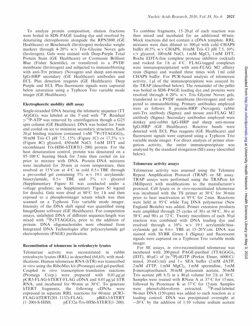
To analyze protein composition, elution fractionswere boiled in SDS–PAGE loading dye and resolved bydenaturing eletrophoresis alongside the RPN5800 (GEHealthcare) or Benchmark (Invitrogen) molecular weightmarkers through 4–20% w/v Tris–Glycine Novex gels(Invitrogen). Gels were stained with Deep-Purple TotalProtein Stain (GE Healthcare) or Coomassie BrilliantBlue (Fisher Scientific), or transferred to a PVDFmembrane (Invitrogen) and subjected to immunoblottingwith anti-Trx primary (Novagen) and sheep anti-mouseIgG-HRP secondary (GE Healthcare) antibodies andECL Plus detection reagents (GE Healthcare). DeepPurple and ECL Plus fluorescent signals were capturedbelow saturation using a Typhoon Trio variable modeimager (GE Healthcare).
Electrophoretic mobility shift assay
Single-stranded DNA bearing the telomeric sequence (TTAGGG)3 was labeled at the 50-end with 32P. Residualg-32P-ATP was removed by centrifugation though a G25spin column (GE Healthcare). DNA was boiled for 5minand cooled on ice to minimize secondary structures. Each20 ml binding reaction contained 1 nM 32P-(TTAGGG)3,50mM Tris–Cl pH 7.5, 15% (Figure 1C) or 17.5% v/v(Figure 4C) glycerol, 450mM NaCl, 5mM DTT andrecombinant Trx-HIS6-hTERT(1–200) protein. For theheat denaturation control, protein was denatured on a95–100�C heating block for 5min then cooled on iceprior to mixture with DNA. Protein–DNA mixtureswere incubated for 30min at room temperature, thenresolved at 15V/cm at 4�C in cold 0.5�TBE througha pre-cooled gel containing 5% w/v 19:1 acrylamide:bisacrylamide, 0.5� TBE and 5% v/v glycerol(Supplementary Figure S1 was conducted under avoltage gradient; see Supplementary Figure S1 legendfor details). Gels were dried at 80�C for 1 h and thenexposed to a phosphorimager screen which was thenscanned on a Typhoon Trio variable mode imager.Intensity of the DNA shift signal was quantified usingImageQuant software (GE Healthcare). For competitionassays, unlabeled DNA of different sequence/length wasmixed with 32P-(TTAGGG)3 prior to the addition ofprotein. DNA oligonucleotides were obtained fromIntegrated DNA Technologies after polyacrylamide gelelectrophoresis (PAGE) purification.
Reconstitution of telomerase in reticulocyte lysates
Telomerase activity was reconstituted in rabbitreticulocyte lysates (RRL) as described (64,65), with mod-ifications. Human telomerase RNA (hTR) was transcribedin vitro using the RiboMax kit (Promega) and gel-purified.Coupled in vitro transcription–translation reactions(Promega Corp.) were prepared with 0.01mg/mlpCR3-FLAG-hTERT-FLAG cDNA and 0.01mg/ml hTRRNA, and incubated for 90min at 30�C. To generatehTERT fragments, the following cDNAs wereexpressed in separate RRL reactions for 60min: pCR3-FLAG-hTERT(201–1132)-FLAG; pBiEx3-hTERT(1–200)-S-HIS8; pET32a-Trx-HIS6-hTERT(1–200).
To combine fragments, 15–20 ml of each reaction wasthen mixed and incubated for an additional 60min.Mock reactions did not contain a cDNA template. RRLmixtures were then diluted to 500 ml with cold CHAPSbuffer (0.5% w/v CHAPS, 10mM Tris–Cl pH 7.5, 10%v/v glycerol, 100mM NaCl, 1mM MgCl2 1mM DTT,Roche EDTA-free complete protease inhibitor cocktail)and rocked for 1 h at 4�C. FLAG-tagged complexeswere immunoprecipitated with anti-FLAG M2 affinityresin (Sigma) and washed three times with 1ml coldCHAPS buffer. For PCR-based analysis of telomeraseactivity, 1 ml of the immunoprecipitate was assayed bythe TRAP (described below). The remainder of the pelletwas boiled in SDS–PAGE loading dye and proteins wereresolved through 4–20% w/v Tris–Glycine Novex gels,transferred to a PVDF membrane (Invitrogen) and sub-jected to immunoblotting. Primary antibodies employedwere as follows: S protein-HRP (Novagen); rabbitanti-Trx antibody (Sigma); and mouse anti-FLAG M2antibody (Sigma). Secondary antibodies employed weredonkey anti-rabbit IgG-HRP and sheep anti-mouseIgG-HRP (GE Healthcare). Reactive bands weredetected with ECL Plus reagents (GE Healthcare) andfluorescent signals were captured using a Typhoon Triovariable mode imager. For direct analysis of primer elon-gation activity, the entire immunoprecipitate wasanalyzed by the standard elongation (SE) assay (describedbelow).
Telomerase activity assays
Telomerase activity was assessed using the TelomereRepeat Amplification Protocol (TRAP) or SE assay.TRAP assays were performed using the TRAPeze kit(Millipore) with modifications to the manufacturer’sprotocol. Cell lysate or in vitro-reconstituted telomerasewas incubated with the TS primer at 30�C for 30minprior to heat inactivation at 95�C for 2min. Reactionswere held at 95�C while Taq DNA polymerase (NewEngland Biolabs) was added. Primer extension productswere amplified through 25 cycles of 30 s at 94�C, 30 s at50�C and 90 s at 72�C. Twenty microliters of each 50 mlreaction was combined with DNA loading dye andresolved through a 10% w/v 19:1 acrylamide:bisa-crylamide gel in 0.6� TBE at 15–20V/cm. DNA wasstained with SYBR Green I (Sigma) and fluorescentsignals were captured on a Typhoon Trio variable modeimager.For SE assays, in vitro-reconstituted telomerase was
incubated with 200 pmol PAGE-purified (TTAGGG)3(IDT), 40 mCi of [a-32P]-dGTP (Perkin Elmer, 6000Ci/mmol, 20mCi/ml) and 1� SEA buffer (2mM dATP,2mM dTTP, 1mM MgCl2, 1mM spermidine, 5mMb-mercaptoethanol, 50mM potassium acetate, 50mMTris–acetate pH 8.5) in a 40 ml volume for 2 h at 30�C.Samples were treated with RNase A at 37�C for 15min,followed by Proteinase K at 37�C for 15min. Sampleswere phenol:chloroform extracted. 32P-end-labeled96-mer DNA (150 cpm) was added to each reaction as aloading control. DNA was precipitated overnight at�20�C by the addition of 1/10 volume sodium acetate
Nucleic Acids Research, 2010, Vol. 38, No. 6 2021
at University H
ealth Netw
ork - Health S
ciences Library on May 3, 2010
http://nar.oxfordjournals.orgD
ownloaded from

pH 5.2, two volumes cold ethanol and 5 mg GenElutelinear polyacrylamide. DNA was pelleted by centri-fugation and resuspended in 3 ml gel loading buffer(100% v/v formamide, 0.6� TBE). Samples were boiledfor 3min and resolved by electrophoresis in 0.6� TBEthrough a 10% w/v denaturing polyacrylamide gel(29:1, acrylamide:bisacrylamide, 7M urea, 0.6� TBE).The gel was dried at 80�C for 1 h, and then exposed toa phosphorimager screen which was then scanned ona Typhoon Trio variable mode imager.
Mutagenesis of hTERT(1–200)
The structure-based sequence alignment of the N-terminusof TERT (51) was used as a guide to select residues inhTERT(1–200) that may be exposed to the surface ofthe protein (refer to Supplementary Figure S4). Thedesign of PCR primers for site-directed mutagenesis wasaided by The Primer Generator (66). Primers wereobtained from Operon and IDT. The incorporation ofintended mutations into the cDNA (and the absence ofunwanted mutations) was confirmed by DNA sequencing.
205
M 10 40 65 80 Trx-HIS6- hTERT(1-200)
32P-(TTAGGG)3
:BWDeep Purple αTrx
Trx-HIS6
Trx-HIS6- hTERT(1-200) : probe
(TTAGGG)3 : probe
670 Trx-HIS6 : probe
200
10 40 65 80
200
14 56 90 110 110 110 110 110110b10 102 103
1 2 3 4 5 6
1 2 3 4 5 6 7 8 9 10 11
2 3 4 5 6
116978066
5545
30
21
147
180
115
82
644937
26
19
15
6
*
**
*
**
A
C
Bng
ng
Figure 1. Recombinant hTERT(1–200) interacts with telomeric DNA. (A) Analysis of Trx-HIS6-hTERT(1–200) expressed in E. coli and purified asdescribed in ‘Materials and Methods’ section. Proteins were boiled in SDS–PAGE loading dye, resolved through a 4–20% w/v Tris–Glycine Novexgel and stained with Deep Purple. The mass (ng) of Trx-HIS6-hTERT(1–200) present in lanes 2–5 was determined by comparing the band intensityof the full length fusion protein (Single asterisk) to the average intensity of bands in the RPN5800 molecular size marker (M, lane 1, 30 ng/band).Molecular mass (kDa) is indicated at the left. Lane 6, Trx-HIS6 (Double asterisk) purified as described in ‘Materials and Methods’ section.(B) Proteins prepared as in (A) were transferred to PVDF and immunoblotted with anti-Trx (Novagen) and anti-mouse IgG antibodies. Lane 1(not shown) contains Benchmark molecular size marker. [Supplementary Figure S3, lanes 7–18 and Figure 3B, lanes 9–10 were probed with adifferent anti-Trx antibody (Sigma)]. (C) EMSA of telomeric DNA. 32P-(TTAGGG)3 was mixed with the following components as described in‘Materials and Methods’ section: lane 1, DNA alone; lane 2–5, recombinant Trx-HIS6-hTERT(1–200) [same amounts as in (A) and (B), expressed asmolar ratio to radiolabeled probe]; lane 6, pre-boiled (b) Trx-HIS6-hTERT(1–200); lane 7, Trx-HIS6 [same amount as in (A) and (B),expressed as molar ratio to radiolabeled probe]; lanes 9–11, Trx-HIS6-hTERT(1–200) in the presence of unlabeled specific competitor oligonucleotide(at the indicated molar ratio to radiolabeled probe). Lanes 5 and 8 are duplicates. Complexes were resolved by native polyacrylamidegel electrophoresis.
2022 Nucleic Acids Research, 2010, Vol. 38, No. 6
at University H
ealth Netw
ork - Health S
ciences Library on May 3, 2010
http://nar.oxfordjournals.orgD
ownloaded from

Generation and passaging of stable cell lines
pcDNA3.1(hyg)-hTERT vectors were linearized withSspI, treated with calf intestinal phosphatase (NewEngland Biolabs), and then gel-purified. Human embry-onic kidney HA5 cells were transduced with eitherwild-type or mutant hTERT DNA using FuGENE6transfection reagent (Roche). Cells were grown inalpha-Minimal Essential Medium (a-MEM) containing10% v/v fetal bovine serum and 2mML-glutamine, andthen selected with 200 mg/ml (Supplementary Figure S5) or100 mg/ml hygromycin (Figure 5). Cells undergoing mocktransfection (with no DNA) did not survive selection.After 4–5 weeks of selection, colonies were treated withTrypLE Express (Invitrogen) and pooled. Polyclonal pop-ulations were passaged in media containing hygromycinby plating 2� 105 cells in a 10 cm plate at regular intervals.The number of cumulative population doublings at eachpassage was determined by the formula as described (67).The experiment was performed with late-passage cellsapproaching crisis (Supplementary Figure S5), and alsowith cells at an earlier passage (Figure 5). Apoptosis wasassessed using the TiterTACS assay (R&D Systems) andby visual inspection of cellular morphology bymicroscopy.
Protein extraction
Cells were washed with ice-cold PBS and dislodged fromtissue culture plates using a cell scraper. Cells wereresuspended and lysed in 4–5 volumes of ice-coldCHAPS buffer (0.5% w/v CHAPS, 10mM Tris–ClpH 7.5, 10% v/v glycerol, 1mM MgCl2, 5mMb-mercaptoethanol, Roche EDTA-free Completeprotease inhibitor and 400U Roche RNase Inhibitor)for 30min on ice. Insoluble material was pelleted bycentrifugation at 13 000 r.p.m. for 30min. The proteinconcentration of the cleared whole cell extract was deter-mined by the Bradford Assay (Bio-Rad).
Analysis of mRNA by RT–PCR
Total RNA was isolated from cells using TRIzol reagent(Invitrogen), and then treated with DNase I (Roche).cDNA was generated from RNA using Superscript IIreverse transcriptase (Invitrogen) and random hexamerprimers (Invitrogen). Residual RNA was removed fromcDNA products using RNase H (Invitrogen). cDNAswere amplified by PCR using Taq DNA polymerase(New England Biolabs) under the following conditions:94�C for 5min, followed by 32 cycles of 94�C for 45 s,57�C for 45 s and 72�C for 1min, followed by a final exten-sion of 72�C for 4min. The following gene-specific primerswere used: GAPDH, 50-CGGAGTCAACGGATTTGGTCGTAT-30 and 50-TGCTAAGCAGTTGGTGGTGCAGGA-30; hTERT, 50-AAGTTCCTGCACTGGCTGATGAG-30 and 50-TCGTAGTTGAGCACGCTGAACAG-30;hygromycin resistance (HYG-R), 50-CGCAAGGAATCGGTCAATAC-30 and 50-ACATTGTTGGAGCCGAAATC-30. DNA was resolved through a 0.8% w/v agarosegel, stained with ethidium bromide, and imaged on aTyphoon Trio variable mode imager.
Telomere length analysis
Genomic DNA was isolated from cells using the DNeasykit (Qiagen) and digested with RsaI and HinfI. Restrictionfragments were resolved through a 0.5% w/v agarose gelat 45V (2V/cm) for 24 h. DNA was denatured in buffercontaining 0.5M NaOH, 1.5mM NaCl for 30min, andneutralized in buffer containing 1.5M NaCl, 0.5mMTris–Cl pH 7.5 for 30min. DNA was transferredto Hybond N+ membrane in 20� SSC. Followingtransfer, DNA was UV-crosslinked to the membrane,which was then rinsed in 2� SSC. Telomeric DNA washybridized to a 32P 50-end-labeled (CCCTAA)3 probe inChurch buffer (0.5M NaPO4 pH 7.2, 1% w/v BSA, 7%w/v SDS, 1mM EDTA), then washed with 1� SSC, 0.1%w/v SDS. The membrane was exposed to a phosphori-mager screen which was then scanned using a TyphoonTrio variable mode imager. The weighted mean oftelomere length in each lane was calculated accordingthe following formula, as previously described (68):�(ODi)/�(ODi/Li), where ODi is the signal intensity andLi is the length in nucleotides of DNA at position ias determined by comparison to the molecular massstandards.
RESULTS
Production of recombinant hTERT(1–200) in a bacterialexpression system
The N-terminus of T. thermophila TERT contains aDNA-binding domain (51–53). Mutations in this regionimpair telomerase activity, as do mutations in the equiva-lent region of hTERT (55,58–63). Thus, this region hasbeen dubbed the telomerase essential N-terminal (TEN)domain (51). Although the N-terminus of TERTcontains a region of homology (GQ), the sequence conser-vation between human and T. thermophila TERTs in thisregion is especially low. Unpurified hTERT(aa 1–300)and hTERT(1–350) generated in reticulocyte lysatescan be captured onto biotinylated telomeric DNA ina neutravidin pull-down assay [(55), D.C.F. Sealey andL.A. Harrington, unpublished results]. To clarify thisapparent hTERT–DNA interaction, we developed astrategy to produce recombinant hTERT in bacteria.We expressed Trx-HIS6-hTERT(1–200) in E. coli and,
despite a low expression level and limited solubility (<5%of expressed protein was recovered in the soluble fractionof the cell lysate), we purified the protein over two chro-matographic steps to a modest degree of homogeneity (see‘Materials and Methods’ section). The fusion protein wasdetected at the expected size of 36 kDa (Figure 1A). Thepresence of Trx fused at the N-terminus was confirmed bywestern blotting (Figure 1B) and the identity of TERT wasconfirmed by tandem mass spectrometry (MS) (T. Gohand T. LeBihan, data not shown). Minor bands athigher molecular weights were identified as the bacterialchaperones DNaK and DNaJ and the translation elonga-tion factor Tu (T. Goh and D.C.F. Sealey data notshown). The majority of peptides derived from the lowermolecular weight bands, some of which were reactive to
Nucleic Acids Research, 2010, Vol. 38, No. 6 2023
at University H
ealth Netw
ork - Health S
ciences Library on May 3, 2010
http://nar.oxfordjournals.orgD
ownloaded from

anti-Trx antibody, corresponded to fragments ofTrx-HIS6-hTERT(1–200) based on MS analysis (Figure1B and data not shown). The purity of full lengthTrx-HIS6-hTERT(1–200) in a typical purification wasdetermined to be 75% based on Coomassie staining(data not shown). Based on Deep Purple staining (whichis more sensitive than Coomassie), purity was determinedto range from 20% (Figure 1A) to 40% (Figure 4A).
hTERT(1–200) interacts with telomeric DNA in vitro
To determine whether hTERT(1–200) interacts withDNA, we performed EMSAs. Recombinant Trx-HIS6-hTERT(1–200) was mixed with radiolabeledsingle-stranded DNA oligonucleotides containing threefull telomeric repeats and subjected to nativepolyacrylamide gel electrophoresis. A slower mobilitycomplex contained labeled DNA, and this complexincreased in intensity upon addition of increasingamounts of protein (Figure 1C). The DNA shift reacheda maximum when the binding mixture contained 450mMNaCl, and persisted in 1M NaCl (data not shown). Heatdenaturation of the protein preparation inactivated theapparent DNA-binding activity (Figure 1C, lane 6). Thegel shift observed with radiolabeled DNA was, asexpected, competed by an excess molar ratio of unlabeled,specific oligonucleotide (Figure 1C, lanes 9–11). Anextract prepared from cells expressing Trx-HIS6 andpurified over Ni–NTA resin (in the same manner asTrx-HIS6-hTERT) did not interact with telomeric DNA(Figure 1C, lane 7). Furthermore, the electrophoreticmobility of telomeric DNA in complex withTrx-HIS6-hTERT [or another hTERT(1–200)-S-HISfusion protein] was altered upon the addition ofanti-HIS antibody but not other antibodies. Specifically,a slower mobility complex disappeared upon incubationwith increasing concentrations of anti-HIS antibody, con-comitant with the enrichment or appearance of fastermobility complexes (Supplementary Figure S1).Antibodies specific to their cognate protein often elicit a‘supershift’ (i.e. retardation of mobility) upon binding tothe DNA:protein complex. Although not as common,antibody recognition can perturb the intact nucleoproteincomplex leading to the disappearance of complexes, orappearance of faster mobility complexes, owing to analteration of relative DNA:protein stoichiometry(69–71). These data show that hTEN was present in thetelomeric DNA complex, and argue for a specific interac-tion between the N-terminus of hTERT and telomericDNA.
hTERT(1–200) interacts preferentially with telomericDNA of a particular register and sufficient length
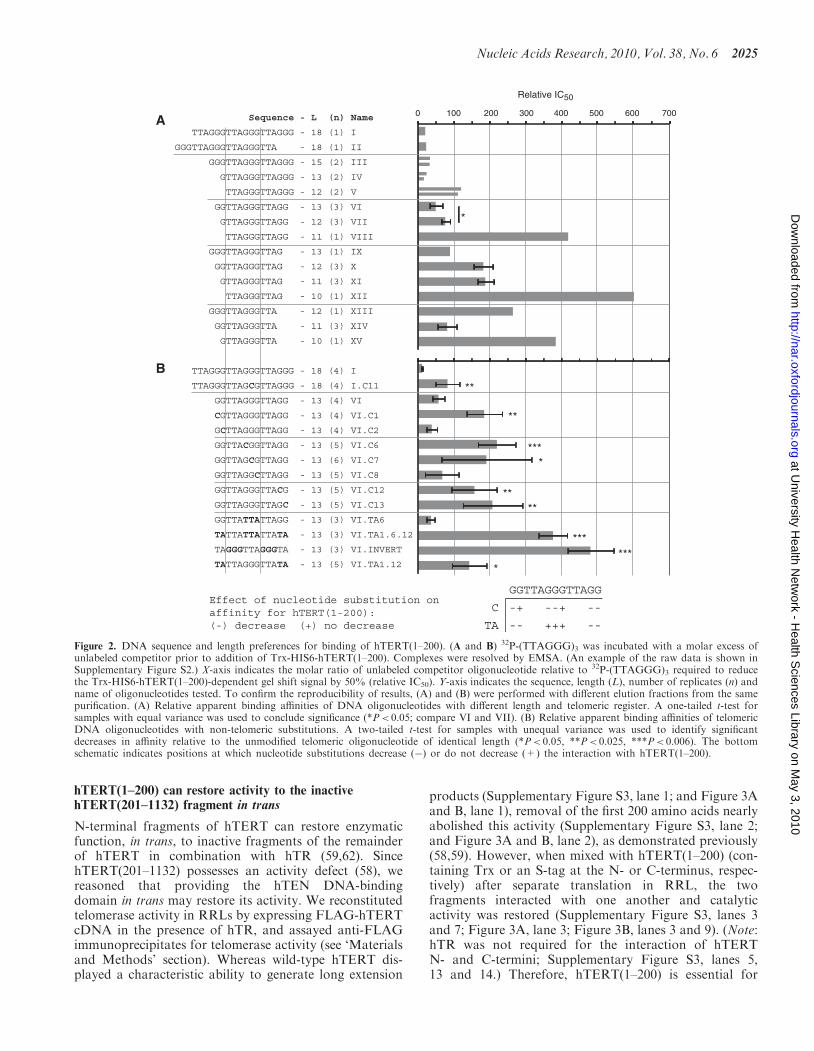
To determine the DNA-binding specificity of the hTENcomplex, we tested the ability of unlabeled competitorDNA oligonucleotides of different length and/or telomericregister to compete with labeled (TTAGGG)3 for bindingto Trx-HIS6-hTERT(1–200), and expressed the rela-tive apparent binding affinities as the amount of
oligonucleotide required to reduce the signal intensity ofthe mobility shift by 50% (Figure 2, SupplementaryFigure S2 and data not shown). Single-strandedoligonucleotides 18 nucleotides (nt) in length did notrequire a specific 50- or 30-end in the telomeric register tofacilitate binding (Figure 2A, compare oligonucleotides I,II). Oligonucleotides ending in GGG required at least13 nt of telomeric sequence to bind comparably to the18 nt (TTAGGG)3 probe (I, III, IV, V). Foroligonucleotides 13 nt in length, a specific telomericregister was preferred: in decreasing order of apparentaffinity, oligonucleotides that terminated in GGG, GGand G (IV, VI, IX). Reducing the length to 12 nt in anyregister reduced the apparent binding affinity (compare IVand V; VI and VII; IX and X). Oligonucleotides 12 nt inlength were recognized when containing, in decreasingorder of apparent affinity, terminal GG, GGG, G andno G (VII, V, X, XIII). Reducing oligonucleotide lengthto 11 nt reduced the apparent affinity for DNA ending inGG (VII and VIII); when ending in G, a further decreasein apparent affinity was observed only upon decreasing thelength to 10 nt (X, XI, XII). Surprisingly, foroligonucleotides ending in TTA, 11 nt (but not 10 nt)bound with a greater apparent affinity than did 12 nt(XIII, XIV, XV). From this dataset, we conclude thatsingle-stranded telomeric DNA 13 nt in length andterminating in GGG or GG was a preferred substratefor hTEN binding (IV, VI), whereas oligonucleotides ofthis length with different telomeric registers, or shorteroligonucleotides, did not bind with a comparableapparent affinity.
To determine the sequence specificity of the DNA inter-action, we performed another set of competition experi-ments with oligonucleotides containing non-telomericsubstitutions (Figure 2B). Telomeric DNA 18 nt inlength in which the middle G at position 11 wasreplaced with C had a 7-fold lower apparent affinity forhTEN (compare I and I.C11). For telomeric DNA 13 nt inlength and ending in a GG register (VI), replacing G withC significantly reduced apparent affinity at some positions(VI.C1, C6, C7, C12, C13) but not others (VI.C2, C8).Interestingly, replacing the central GGG with TTA didnot reduce apparent affinity (VI.TA6), whereas replacingthe GG with TA at the 50- and 30-ends did reduce apparentaffinity (VI.TA1.12). Combining these central, 50 and 30
GG-to-TA replacements reduced apparent affinity(VI.TA1.6.12), suggesting that GG residues form criticalcontacts with hTEN and that the central GGG, while notrequired, does stabilize the interaction in the absence ofother G contacts. Inverting the GGG and TA positionsacross the 13 nt also reduced relative binding affinity(VI.INVERT), again suggesting that GG ‘bookends’ facil-itate the hTEN:DNA interaction, and that the relativepositions of internal G residues are also important.Taken together, these data indicate that the interactionof hTEN with oligonucleotides 13 nt in length dependson G-rich character at certain positions—a demonstrationof telomere sequence specificity (Figure 2B, lower), and inkeeping with previous observations on the preferredsubstrate composition for elongation by telomerase (see‘Discussion’ section).
2024 Nucleic Acids Research, 2010, Vol. 38, No. 6
at University H
ealth Netw
ork - Health S
ciences Library on May 3, 2010
http://nar.oxfordjournals.orgD
ownloaded from
hTERT(1–200) can restore activity to the inactivehTERT(201–1132) fragment in trans
N-terminal fragments of hTERT can restore enzymaticfunction, in trans, to inactive fragments of the remainderof hTERT in combination with hTR (59,62). SincehTERT(201–1132) possesses an activity defect (58), wereasoned that providing the hTEN DNA-bindingdomain in trans may restore its activity. We reconstitutedtelomerase activity in RRLs by expressing FLAG-hTERTcDNA in the presence of hTR, and assayed anti-FLAGimmunoprecipitates for telomerase activity (see ‘Materialsand Methods’ section). Whereas wild-type hTERT dis-played a characteristic ability to generate long extension
products (Supplementary Figure S3, lane 1; and Figure 3Aand B, lane 1), removal of the first 200 amino acids nearlyabolished this activity (Supplementary Figure S3, lane 2;and Figure 3A and B, lane 2), as demonstrated previously(58,59). However, when mixed with hTERT(1–200) (con-taining Trx or an S-tag at the N- or C-terminus, respec-tively) after separate translation in RRL, the twofragments interacted with one another and catalyticactivity was restored (Supplementary Figure S3, lanes 3and 7; Figure 3A, lane 3; Figure 3B, lanes 3 and 9). (Note:hTR was not required for the interaction of hTERTN- and C-termini; Supplementary Figure S3, lanes 5,13 and 14.) Therefore, hTERT(1–200) is essential for
A
B
Sequence - L (n) Name
TTAGGGTTAGGGTTAGGG - 18 (1) I
GGGTTAGGGTTAGGGTTA - 18 (1) II
GGGTTAGGGTTAGGG - 15 (2) III
GTTAGGGTTAGGG - 13 (2) IV
TTAGGGTTAGGG - 12 (2) V
GGTTAGGGTTAGG - 13 (3) VI
GTTAGGGTTAGG - 12 (3) VII
TTAGGGTTAGG - 11 (1) VIII
GGGTTAGGGTTAG - 13 (1) IX
GGTTAGGGTTAG - 12 (3) X
GTTAGGGTTAG - 11 (3) XI
TTAGGGTTAG - 10 (1) XII
GGGTTAGGGTTA - 12 (1) XIII
GGTTAGGGTTA - 11 (3) XIV
GTTAGGGTTA - 10 (1) XV
TTAGGGTTAGGGTTAGGG - 18 (4) I
TTAGGGTTAGCGTTAGGG - 18 (4) I.C11
GGTTAGGGTTAGG - 13 (4) VI
CGTTAGGGTTAGG - 13 (4) VI.C1
GCTTAGGGTTAGG - 13 (4) VI.C2
GGTTACGGTTAGG - 13 (5) VI.C6
GGTTAGCGTTAGG - 13 (6) VI.C7
GGTTAGGCTTAGG - 13 (5) VI.C8
GGTTAGGGTTACG - 13 (5) VI.C12
GGTTAGGGTTAGC - 13 (5) VI.C13
GGTTATTATTAGG - 13 (3) VI.TA6
TATTATTATTATA - 13 (3) VI.TA1.6.12
TAGGGTTAGGGTA - 13 (3) VI.INVERT
TATTAGGGTTATA - 13 (5) VI.TA1.12
0 100
Relative IC50
200 300 400 500 600 700
**
**
****
***
******
*
*
*
GGTTAGGGTTAGG
-+ --+ --
-- +++ --
C
TA
Effect of nucleotide substitution on affinity for hTERT(1-200):(-) decrease (+) no decrease
Figure 2. DNA sequence and length preferences for binding of hTERT(1–200). (A and B) 32P-(TTAGGG)3 was incubated with a molar excess ofunlabeled competitor prior to addition of Trx-HIS6-hTERT(1–200). Complexes were resolved by EMSA. (An example of the raw data is shown inSupplementary Figure S2.) X-axis indicates the molar ratio of unlabeled competitor oligonucleotide relative to 32P-(TTAGGG)3 required to reducethe Trx-HIS6-hTERT(1–200)-dependent gel shift signal by 50% (relative IC50). Y-axis indicates the sequence, length (L), number of replicates (n) andname of oligonucleotides tested. To confirm the reproducibility of results, (A) and (B) were performed with different elution fractions from the samepurification. (A) Relative apparent binding affinities of DNA oligonucleotides with different length and telomeric register. A one-tailed t-test forsamples with equal variance was used to conclude significance (*P< 0.05; compare VI and VII). (B) Relative apparent binding affinities of telomericDNA oligonucleotides with non-telomeric substitutions. A two-tailed t-test for samples with unequal variance was used to identify significantdecreases in affinity relative to the unmodified telomeric oligonucleotide of identical length (*P< 0.05, **P< 0.025, ***P< 0.006). The bottomschematic indicates positions at which nucleotide substitutions decrease (�) or do not decrease (+) the interaction with hTERT(1–200).
Nucleic Acids Research, 2010, Vol. 38, No. 6 2025
at University H
ealth Netw
ork - Health S
ciences Library on May 3, 2010
http://nar.oxfordjournals.orgD
ownloaded from
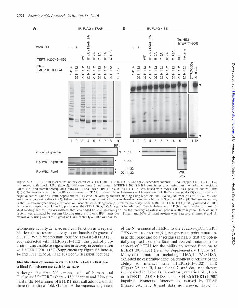
telomerase activity in vitro, and can function as a separa-ble domain to restore activity to an inactive fragment ofhTERT. While recombinant, purified Trx-HIS-hTERT(1–200) interacted with hTERT(201–1132), this purified prep-aration was unable to regenerate in activity in combinationwith hTERT(201–1132) (Supplementary Figure S3, lanes 8,14 and 17; Figure 3B, lane 10) (see ‘Discussion’ section).
Identification of amino acids in hTERT(1–200) that arecritical for telomerase activity in vitro
Although the first 200 amino acids of human andT. thermophila TERTs share <15% identity and 25% sim-ilarity, the N-terminus of hTERT may still adopt a similarthree-dimensional fold. Guided by the sequence alignment
of the N-terminus of hTERT to the T. thermophila TERTTEN domain structure (51), we generated point mutationsin acidic, basic and polar residues in hTEN that are poten-tially exposed to the surface, and assayed mutants in thecontext of hTEN for the ability to restore function tohTERT(201–1132) (refer to Supplementary Figure S4).Many of the mutations, including T116A/T117A/S118A,exhibited no discernible effect on telomerase activity or theability to interact with hTERT(201–1132)+hTR(Figure 3A and B, lanes 5 and 7, and data not shown;summarized in Table 1). In contrast, mutation of Q169Ain hTERT(1–200)-S-HIS8 or Trx-HIS6-hTERT(1–200)impaired telomerase function as assayed by TRAP(Figure 3A, lane 8 and data not shown; Table 1).
A B
1-11
32
201-
1132
201-
1132
201-
1132
201-
1132
CH
AP
S
201-
1132
201-
1132
201-
1132
hTR + FLAG-hTERT-FLAG
hTERT(1-200)-S-HIS8 WT
H17
A/Y
18A
/R19
A
H17
A
Y18
A
R19
A
Q16
9A
WT
H17
A/Y
18A
/R19
A
H17
A
Y18
A
R19
A
Q16
9A
bact
eria
RR
L
mock RRL
RRL
+ +
1 2 3 4 5 6 7 8 9 1 2 3 4 5 6 7 8 9 121110
1-200In > WB: S protein
IP > WB1: S protein
IP > WB2: FLAG
IP: FLAG > TRAP
Trx-HIS6- hTERT(1-200)
IP: FLAG > SE
1-11
32
201-
1132
201-
1132
201-
1132
201-
1132
201-
1132
201-
1132
96 n
t LC
(TTA
GG
G) 3
201-
1132
201-
1132
201-
1132
+ +
1-200
1-1132201-1132 WB:
αTrx
Figure 3. hTERT(1–200) rescues the activity defect of hTERT(201–1132) in a Y18- and Q169-dependent manner. FLAG-tagged hTERT(201–1132)was mixed with mock RRL (lane 2), wild-type (lane 3) or mutant hTERT(1–200)-S-HIS8 containing substitutions at the indicated positions(lanes 4–8) and immunoprecipitated onto anti-FLAG resin (IP). FLAG-hTERT(1–1132) was mixed with mock RRL as a positive control (lane1). (A) Telomerase activity in the IPs was assessed by TRAP. Irrelevant lanes between 8 and 9 were removed. Buffer alone (CHAPS) was assayed as anegative control (lane 9). Immunoprecipitates (IP) were analyzed by western blotting using S protein-HRP (WB1), followed by anti-FLAG M2 andanti-mouse IgG antibodies (WB2). Fifteen percent of input protein (In) was analyzed on a separate blot with S protein-HRP. (B) Telomerase activityin the IPs was analyzed using a radioactive, linear standard elongation (SE) telomerase assay. Lane 9, 10, Trx-HIS-hTERT(1–200) produced in RRLor bacteria, respectively. Lane 11, position of the (TTAGGG)3 DNA oligonucleotide upon 50-end-labeling with 32P (bottom arrowhead). Lane 12,96 nt loading control (top arrowhead) that was added to each reaction prior to the recovery of extension products. Bottom panel: 15% of inputprotein was analyzed by western blotting using S protein-HRP (lanes 3–8). Fifteen and 60% of input protein were analyzed in lanes 9 and 10,respectively, using anti-Trx (Sigma) and anti-rabbit IgG-HRP antibodies.
2026 Nucleic Acids Research, 2010, Vol. 38, No. 6
at University H
ealth Netw
ork - Health S
ciences Library on May 3, 2010
http://nar.oxfordjournals.orgD
ownloaded from

Also, Q169A rendered telomerase unable to catalyzenucleotide addition in the SE assay—an unequivocal indi-cation that Q169 is required for catalytic activity(Figure 3B, lane 8 and data not shown; Table 1) similarto the requirement for Q168 in T. thermophila TERT (51).The H17A/Y18A/R19A mutant displayed a similar inabil-ity to elongate telomeric DNA, although the defect byTRAP was not as pronounced (Figure 3A and B, lane 4;Table 1). The defect of the triple mutant was attributed toa single amino acid change at Y18, but not H17 or R19(Figure 3A and B, lanes 5–7). None of the mutationsdescribed above disrupted the interaction withhTERT(201–1132)+hTR (Figure 3A).
Y18 or Q169 are dispensable for the interaction ofhTERT(1–200) with DNA
To further study the effect of hTEN mutation on the bio-chemical activities of telomerase, we produced hTENmutants in bacteria alongside wild-type hTEN in thesame manner as described above. Purifications ofTrx-HIS6-hTERT(1–200) wild-type, Y18A, T116A/
T117A/S118A and Q169A contained a prominent bandat the expected size that was recognized by anti-Trxantibody (Figure 4A and B). Lower molecular weightfragments of the fusion protein co-purified with theintact protein, as in our purification of wild-type hTEN(see also Figure 1B). Only Q169A contained a noticeabletruncation product at 31 kDa that was recognizedby anti-Trx antibody and was present in equal abun-dance to the full-length 36 kDa fusion protein(Figure 4A and B).To determine whether residues in hTEN that are
required for activity are required for DNA binding, weperformed EMSAs with hTEN mutants. As before,Trx-HIS6-hTERT(1–200) shifted the single-strandedtelomeric DNA oligonucleotide in a native gel in aconcentration-dependent manner (Figure 4C, lanes 2–4).The T116A/T117A/S118A mutant also bound telomericDNA (Figure 4C, lanes 8–10), which is consistent withthe predicted position of these residues on the surface ofthe domain opposite the putative DNA-binding groove,and an apparent lack of involvement in the catalytic
Table 1. Summary of hTERT mutant phenotypes
Allele Telomerase activitya DNA bindingb Cell lifespan extensionc Predicted surfaced
TRAP SE
Normal activity, binds DNA, extends cellular lifespanWT + (17, 4e) + (7, 1e) + + (2)
Normal activity, binds DNA, does not extend cellular lifespanT116A/T117A/S118A + (3) + (1) + � (2) A/B
Defective activity, binds DNA, does not extend cellular lifespanH17A/Y18A/R19A � (8) � (4) � (3) A/B.GY18A � (3) � (4) + ? (1) A/B.GQ169A � (10, 2e) � (3) + � (3) A.G
Normal activity (not tested: DNA binding, cellular lifespan extension)R15A/S16A + (3) + (1) A/B.GH17A + (5) + (4) . . . . . . . . . . . . A/BR19A + (5) + (4) A/B.GR29A/R30A + (3) + (2) BQ34A/R37A + (5) + (1) . . . . . . . . . . . . BR48A + (3) + (1) A/BQ53A + (3) + (1) A/BS70A/R72A/Q73A + (3) + (1) . . . . . . . . . . . . BD105A + (3) + (1) BE113A + (3) + (1) AS121A/Y122A + (3) + (1) . . . . . . . . . . . . AD129A + (3) + (1) A.GS134A + (3) + (1) AR155A + (3) + (1) . . . . . . . . . . . . BT182A/Q183A/R185A + (5) + (1) A/B.GH189A/S191A + (3) + (2) A/B.G
‘+’ no defect or ‘�’ defect. Phenotype representative of (n) experimental replicates.aTelomerase was reconstituted by mixing RRL-expressed hTERT(1–200)-S-HIS8 with FLAG-hTERT(201–1132)-FLAG in the presence of hTR.bTrx-HIS6-hTERT(1–200), purified from bacteria, was mixed with (TTAGGG)3 and complexes were analyzed by electrophoretic mobility shift assay(EMSA).cHA5 cells stably expressing hTERT(1–1132) were passaged under selection. (?) indicates that expression of the Y18A mutant was lost during theexperiment, precluding a conclusion regarding loss of function.dSee Supplementary Figure S4. Predicted surface position based on alignment of the hTERT sequence to the structure of the T. thermophila TERTTEN domain (51). Side ‘A’ includes the putative DNA-binding groove. Side ‘B’ faces opposite Side ‘A.’ A/B represents an edge between both sides.G indicates a location in/near the putative DNA-binding groove. Refer to ‘Materials and Methods’ section for additional details.eTelomerase was reconstituted by mixing RRL-expressed Trx-HIS6-hTERT(1–200) with FLAG-hTERT(201–1132)-FLAG in the presence of hTR.Anti-FLAG immunoprecipitates were assayed for telomerase activity by TRAP or SE protocols.
Nucleic Acids Research, 2010, Vol. 38, No. 6 2027
at University H
ealth Netw
ork - Health S
ciences Library on May 3, 2010
http://nar.oxfordjournals.orgD
ownloaded from

reaction cycle (Table 1, Supplementary Figure S4).Interestingly, mutation of Y18 or Q169, each of whichimpaired telomerase activity, did not affect the ability ofhTEN to bind telomeric DNA (Figure 4C, lanes 5–7,11–13). One could infer that these resides do not contactDNA, or that these residues may contact DNA (based ontheir predicted positions in/near the putativeDNA-binding groove) but that neither Y18 nor Q169 isthe sole determinant of a DNA interaction—at least not toan extent that can be resolved by the EMSA assay. Theability of hTEN(Q169A) to bind telomeric DNA in anEMSA differs from the reduced primer crosslinking
ability of T. thermophila TERT(Q168A) (51) (see‘Discussion’ section).
Residues in hTERT(1–200) are required fortelomerase-mediated extension of cellular lifespan
To determine whether Y18 and Q169, as determinants oftelomerase activity in vitro, are also required fortelomerase function in cells, we performed a cellularimmortalization experiment. We created polyclonal HA5cell lines [HA5 is a mortal, SV40-transformed humanembryonic kidney cell line that bypasses senescence and
205
M 20
28 35 63 28 42 77 28 35 70 28 35 70
25 45 20 30 55 20 25 50 20 25 50 20 25 45 20 30 55 20 25 50 20 25 50 ng
Trx-HIS6- hTERT(1-200)WT Y18A
T116AT117AT118A Q169A
WT Y18A
T116AT117AT118A Q169A
WT Y18A
T116AT117AT118A Q169A
32P-(TTAGGG)3
Deep Purple WB: αTrx
1 2 3 4 5 6 7 8 9 10 11 12 13 2 3 4 5 6 7 8 9 10 11 12 13
1 2 3 4 5 6 7 8 9 10 11 12 13
1169780665545
30
21
14
7
A
C
B
* **
Trx-HIS6- hTERT(1-200) : probe
Figure 4. Selected mutations in hTERT(1–200) do not abrogate DNA-binding activity. (A) Analysis of Trx-HIS6-hTERT(1–200) (wild-type ormutant, as indicated) expressed in E. coli and purified as described in ‘Materials and Methods’ section. Proteins were boiled in SDS–PAGEloading dye, resolved through a 4–20% w/v Tris–Glycine Novex gel and stained with Deep Purple. The mass (ng) of Trx-HIS6-hTERT(1–200)present in lanes 2–13 was determined by comparing the band intensity of the full-length fusion protein (*) to the average intensity of bands in theRPN5800 molecular size marker (M, lane 1, 30 ng/band). Molecular mass indicated at left (kDa). (B) Proteins prepared as in (A) were transferred toPVDF and immunoblotted with anti-Trx (Novagen) and anti-mouse IgG-HRP antibodies. (C) Electrophoretic mobility shift assay of telomericDNA. 32P-(TTAGGG)3 was mixed with the following components as described in ‘Materials and Methods’ section: lane 1, DNA alone; lanes 2–13,increasing amounts of wild-type or mutant Trx-HIS-hTERT(1–200) [same amounts as in (A) and (B); expressed as molar ratio to radiolabeledprobe]. Complexes were resolved by native polyacrylamide gel electrophoresis.
2028 Nucleic Acids Research, 2010, Vol. 38, No. 6
at University H
ealth Netw
ork - Health S
ciences Library on May 3, 2010
http://nar.oxfordjournals.orgD
ownloaded from

A
Days in culture
0 20 40 60 80 100 120 140 160 180Cum
ulat
ive
Pop
ulat
ion
Dou
blin
gs (
PD
)0
10
20
30
60
70 pcDNA3.1WTH17A/Y18A/R19AY18AT116A/T117A/S118AQ169AD868A/D869A
B
C
12
1098
7
6
5
4
3
12
1098
7
6
5
4
3
55.
35.
05.
55.
64.
84.
74.
55.
55.
05.
65.
1
5.7
5.1
5.2
5.7
5.4
5.2
5.7
5.5
5.6
5.4
PD
TRF
16163
4131 62
511
314
165 10 5
17914
114 16
31 2 54 6 7 8 9 1110 1213
14 171516
19 2118 20
pcD
NA
3.1
WT
H17
A/Y
18A
/R18
A
Y18
A
T11
6A/T
117A
/S11
8A
Q16
9A
D86
8A/D
869A
D hTERT
IC
pcD
NA
3.1
hTERT
WT
H17
A/Y
18A
/R18
A
Y18
A
T11
6A/T
117A
/S11
8A
Q16
9A
D86
8A/D
869A
CH
AP
S
1 2 3 4 5 6 7 8
HeL
a
9
GAPDH
hTERT
HYG-R
3PD 5316 3 107 3 22 8 812 9 13 15 H2O
Pre
-RTpc
DN
A3.
1
WT
H17
A/Y
18A
/R18
A
Y18
A
T11
6A/T
117A
/S11
8A
Q16
9A
D86
8A/D
869A
hTERT
1 2 3 4 5 6 7 8 9 10 11 12 13 14 15 16
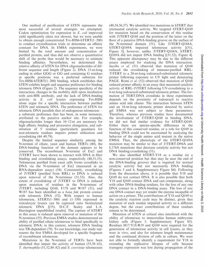
Figure 5. Selected mutations in the N-terminus of hTERT interfere with the ability of telomerase to extend the replicative lifespan of primary human(HA5) cells. (A) Polyclonal cell lines containing wild-type or mutant hTERT cDNA (or pcDNA3.1 as a negative control) were derived from HA5cells (at earlier passage than cells used in Supplementary Figure S5). Cells were passaged under selection with hygromycin. Y-axis contains a breakspanning population doublings (PD) 35–55 in order to display the uninterrupted growth of cells expressing wild-type hTERT. (B) Lysates ofpolyclonal cell lines containing wild-type or mutant hTERT cDNA were prepared in CHAPS buffer at Day 35 and assayed for telomeraseactivity by TRAP. CHAPS buffer was assayed as a negative control (lane 8). HeLa cell lysate was assayed as a positive control (lane 9). IC indicatesTRAP internal control product. Lanes containing irrelevant samples were omitted. (C) Analysis of hTERT mRNA expression level at early and latepassage by RT–PCR. Hygromycin resistance (HYG-R, upper), hTERT (middle) and GAPDH cDNAs were amplified using gene-specific primers(refer to ‘Materials and Methods’ section). Lanes containing irrelevant samples were omitted. (D) Analysis of the terminal telomere restrictionfragments (TRF) at the indicated population doubling (PD) of polyclonal HA5 cell lines receiving wild-type or mutant hTERT. Genomic DNA wasisolated, digested with RsaI and HinfI, and subjected to Southern blotting using a (CCCTAA)3 probe (refer to ‘Materials and Methods’ section).Molecular size (kbp) is shown at left according to the migration of 1 kbp+DNA ladder (not shown). Left, middle and right panels were analyzedseparately and aligned according to duplicate samples contained on individual blots (not shown). The weighted mean of telomere length (kbp) isindicated at the bottom of each lane. The presence of an interstitial, cross-hybridizing band at 3 kbp can be used to estimate the relative DNAloading of each lane.
at University H
ealth Netw
ork - Health S
ciences Library on May 3, 2010
http://nar.oxfordjournals.orgD
ownloaded from

encounters crisis—characterized by telomere instabilityand apoptosis—and does not undergo spontaneousimmortalization (72)] stably expressing wild-type ormutant hTERT, and monitored population doublinglevels at regular intervals. Cells that received wild-typehTERT cDNA continued to divide through the durationof the experiment, whereas cells receiving empty vector(pcDNA3.1) or hTERT(D868A/D869A) (mutations thatinactivate reverse transcriptase activity) succumbed toapoptosis within �10 population doublings (Supple-mentary Figure S5, data not shown), which is consistentwith previous observations (17,73). Cells receivinghTERT(H17A/Y18A/R19A) (n=2), Q169A (n=2), orT116A/T117A/S118A mutants (n=1) were also unableto divide beyond 10 population doublings.To confirm these results, we derived a second set of
stable lines from HA5 cells at an earlier passage. Asbefore, cells receiving wild-type hTERT survivedthrough the duration of the experiment. In contrast,cells receiving mutant hTERT cDNA failed to survivebeyond 15–20 population doublings (Figure 5A). Thisdelay in reaching crisis relative to HA5 cells transducedat a later passage (Supplementary Figure S5) is in keepingwith the different initial telomere lengths of these two pop-ulations (10). We assayed telomerase activity in lysatesprepared from cells at day 35 in the experiment(Figure 5B). Lysates from cells containing H17A/Y18A/R19A, Y18A, Q169A, or D868A/D869A mutants did notdisplay telomerase activity, which is consistent with thereduced activity of these mutants in reticulocyte lysates(Figures 5B, 3A and B, Table 1) (17,65). AlthoughT116A/T117A/S118A reconstituted telomerase activityin vitro, cells containing this mutant did not display elon-gation activity (Figure 5B, Table 1).Analysis of mRNA transcript levels confirmed expres-
sion of the vector-encoded hygromycin resistance gene atearly and late time points in the experiment (Figure 5C).Expression of hTERT(WT), H17A/Y18A/R19A, T116A/T117A/S118A and D868A/D869A alleles was maintainedat later population doublings. Expression of Y18A wasnot maintained (Figure 5C, lanes 7 and 8); thus, wecould not formally conclude that this mutant cannotimmortalize cells. Notably, Y18A phenocopied H17A/Y18A/R19A in vitro and H17A/Y18A/R19A-transducedcells did not survive (Table 1). Q169A was expressed ata lower level than wild-type hTERT in the second exper-iment, but at a comparable level in the first experiment(Figure 5C, lanes 11 and 12 versus 3 and 4; SupplementaryFigure S6, lanes 3 and 4, and 5 and 6 versus lane 2).Regardless of the level of expression, Q169A did notreconstitute activity in lysates (Supplementary Figure S6,lanes 3–5). Therefore, we conclude that hTERT-Q169Acannot immortalize cells (Table 1). Although none of theaforementioned mutants immortalized cells, we dididentify mutants that enabled the reconstitution oftelomerase activity in lysates and the extension ofcellular lifespan (D.C.F. Sealey and M. Taboskiunpublished results).To determine whether mutations in hTEN interfere with
the ability of telomerase to maintain telomeres, weanalyzed telomere length changes in HA5 stable cell
lines by terminal restriction fragment (TRF) analysis.Average telomere lengths in cells expressing wild-typehTERT were maintained up to population doubling 16(Figure 5D, lane 4), whereas telomeres in cells expressingH17A/Y18A/R19A, T116A/T117A/S118A, Q169A orD868A/D869A mutants became shorter as the popula-tions reached crises (Figure 5A and D). The averagetelomere length of cells expressing wild-type hTERTbecame shorter beyond population doubling 16(Figure 5D, lanes 5–7), but the population did not encoun-ter crisis (Figure 5A). This result is consistent with thefindings of other studies that the maintenance ofminimal telomere DNA by hTERT is sufficient forlifespan extension (74,75). Analysis of telomere lengthchanges in cells from the first immortalization experiment(Supplementary Figure S5) revealed similar results (datanot shown). Therefore, the mutations in hTEN that wehave described render telomerase unable to maintaintelomeres and extend cellular lifespan.
DISCUSSION
In this study, we described the ability of the N-terminusof hTERT (hTEN, a.a. 1–200) to interact withtelomeric DNA, and complement the activity defect ofhTERT(201–1132)+hTR as a separable domain.Mutational analysis identified that Y18 and Q169residues were required for primer extension activityin vitro. These residues, as well as T116/T117/S118, wererequired for telomere length maintenance andtelomerase-mediated extension of cellular lifespan, butnot for the interactions of hTEN with DNA orhTERT(201–1132)+hTR.
Many previous studies have used telomerase activity asa read-out for investigating telomerase-primer interac-tions, in part due to the difficulty of producing sufficientquantities of purified hTERT to measure DNA bindingdirectly. By EMSA (Figures 1C and 2), we demonstratedthat the interaction between purified Trx-HIS6-hTERT(1–200) and telomeric DNA was independent of hTR,other domains of hTERT and telomerase activity. Theinteraction was reproducible across independent purifica-tions of protein and many experimental replicates. Therecombinant protein also interacted with hTERT(201–1132) (Supplementary Figure S3). Despite theseinteractions, Trx-HIS6-hTERT(1–200) produced inbacteria did not regenerate telomerase activity in combi-nation with RRL-hTERT(201–1132)+hTR (Figure 3,Supplementary Figure S3), suggesting that E. coli maynot provide the folding pathways, modification activities,or binding partners required for telomerase activity that aeukaryotic system can provide (65,76–78). This inability tosupport catalysis was not due to interference of theN-terminal Trx-HIS6 tag, since Trx-HIS6-hTERT(1–200) produced in RRL conferred a comparable levelof telomerase activity as did RRL-produced hTERT(1–200)-S-HIS8 (Supplementary Figure S3). Attempts toactivate bacterial Trx-HIS6-hTERT(1–200) with RRLcomponents were unsuccessful (data not shown).
2030 Nucleic Acids Research, 2010, Vol. 38, No. 6
at University H
ealth Netw
ork - Health S
ciences Library on May 3, 2010
http://nar.oxfordjournals.orgD
ownloaded from
Our method of purification of hTEN represents themost successful of several strategies we attempted.Codon optimization for expression in E. coli improvedyield significantly (data not shown), but we were unableto obtain enough concentrated Trx-HIS6-hTERT(1–200)to conduct crystallization trials or determine an affinityconstant for DNA. In EMSA experiments, we werelimited by the total amount and concentration ofpurified protein, and thus did not observe a quantitativeshift of the probe that would be necessary to estimatebinding affinities. Nevertheless, we determined therelative affinity of hTEN for DNA oligonucleotides of dif-ferent sequence and length (Figure 2). DNA 13-nt-longending in either GGG or GG and containing G residuesat specific positions was a preferred substrate forTrx-HIS6-hTERT(1–200) binding, which establishes thathTEN exhibits length and sequence preference for bindingtelomeric DNA (Figure 2). The sequence specificity of theinteraction, changes in the mobility shift upon incubationwith anti-HIS antibody, and the lack of DNA-bindingactivity in E. coli lysates expressing Trx-HIS6alone argue for a specific interaction between purifiedhTEN and telomeric DNA. The preference of hTEN fortelomeric DNA parallels that of endogenous telomerase—a substrate preference that has been, in some instances,attributed to the putative anchor site. For example,oligonucleotides longer than 10–12 nt are necessary forhigh affinity binding and processive elongation, and sub-stitution of 50 residues (particularly guanines) fornon-telomeric residues impairs primer utilization andcrosslinking (44–49,79).
Despite the limited sequence similarity among theN-termini of ciliate, yeast and human TERTs (40), theDNA-binding function of the domain appears to beconserved. The recombinant N-termini of Est2 andTtTERT have been shown to interact with DNA in filterbinding and crosslinking assays, respectively (40,51,53).Telomerase purified from yeast cells forms crosslinks toDNA via the N-terminus of Est2 (measured in anRNA-dependent assay) (54). Consistently, crosslinkingof TtTERT (purified from RRL) to DNA is reducedupon removal of the N-terminus (51,53). Also, theextent of crosslinking of TtTERT to DNA is reducedupon mutation of residues in the N-terminus ofTtTERT, including Q168, F178 and W187 (51), andW187 has been identified as a bona fide protein contactwith telomeric DNA (52). With respect to humantelomerase, hTERT(1–300) and (1–350) expressed inreticulocyte lysates can be captured onto biotinylatedtelomeric DNA [(55), D.C.F. Sealey and L.A.Harrington unpublished results]. The capture of hTERTin this assay is reduced upon removal or mutation of theN-terminus (55). Previous EMSA studies demonstrated anability of purified ciliate telomerase to bind specifically totelomeric DNA; in this context, telomere DNA bindingwas TR-dependent (79). To our knowledge, our study rep-resents the first EMSA developed for a specific fragmentof recombinant telomerase.
Mutations in the N-termini of TERTs have beenidentified that impair the activity of human (55,58–63),T. thermophila (51,52,80–82) and S. cerevisiae telomerases
(40,54,56,57). We identified two mutations in hTERT thatattenuated catalytic activity. We targeted hTERT-Q169for mutation based on the conservation of this residuewith TtTERT-Q168 and the position of the latter on thefloor of a putative DNA-binding groove on the surface ofthe N-terminal domain (51). Like TtTERT-Q168A,hTERT-Q169A impaired telomerase activity [(51),Figure 3]; however, unlike TtTERT-Q168A, hTERT-Q169A did not impair DNA binding [(51,52), Figure 4].This apparent discrepancy may be due to the differentassays employed for studying the DNA interaction.Jacobs et al. (51) observed that mutation of Q168reduced the crosslinking of immuno-purified RRL-TtTERT to a 20-nt-long iodouracil-substituted telomericprimer following exposure to UV light and denaturingPAGE. Romi et al. (52) observed that mutation of Q168reduced primer affinity in an assay that measured catalyticactivity of RRL-TtTERT following UV-crosslinking to a6-nt-long iodouracil-substituted telomeric primer. The for-mation of TERT:DNA crosslinks in these experimentsdepends on the proximity of iodouracil to reactiveamino acid side chains. The interaction between hTENand an 18-nt-long telomeric primer detected by nativegel EMSA was not subject to these parameters.Therefore, whereas previous studies have documentedthe involvement of TtTERT-Q168 in binding DNA,we did not find similar evidence for hTERT-Q169.Either there are species-specific differences in thefunction of this conserved residue, or a role for Q169 inbinding DNA could not be ascertained by analyzing thebehavior of the single amino acid mutant in the nativeEMSA. Thus, the phenotype of the hTERT-Q169Amutation may be similar to that of TtTERT-D94A andL174A mutations that decrease catalytic activity but notDNA binding/crosslinking (51).We also identified hTERT-Y18 as a residue (at a
non-conserved position but that may lie near the end ofthe DNA-binding groove) that is required for normalcatalytic activity but not necessarily DNA binding(Figures 3 and 4, Supplementary Figure S4). Followingfrom the discussion above, it is possible that Y18 andQ169 do not contact DNA. It is also possible that bothY18 and Q169 contact DNA and can compensate, alongwith other DNA-binding residues, for the loss of any oneDNA contact in a DNA-binding assay. The loss of anyone DNA contact may yet render the enzyme incapable ofaction on a primer. The contributions of Y18 and Q169 tothe catalytic reaction cycle may be distinct, given thatmutation of each residue impaired activity to a differentdegree, but the exact contributions of these residuesremain to be determined.Mutation of hTEN at critical sites interfered with the
ability of telomerase to immortalize human embryonickidney cells (Figure 5, Supplementary Figure S5).Residues H17/Y18/R19 and Q169 were required for thegeneration of telomerase activity in cell lysates, as theywere in vitro, and also for telomere length maintenanceand the continued division of cells in culture. We werenot able to formally identify a requirement for Y18 inextending the replicative lifespan of cells becausetransgene expression was lost during propagation of the
Nucleic Acids Research, 2010, Vol. 38, No. 6 2031
at University H
ealth Netw
ork - Health S
ciences Library on May 3, 2010
http://nar.oxfordjournals.orgD
ownloaded from

culture (Figure 5C, lanes 7 and 8). Interestingly, theT116A/T117A/S118A triple mutant, which did notexhibit deficits in DNA binding or activity in vitro(Figure 4, Table 1), did not regenerate telomeraseactivity in cell lysates and did not immortalize cells(Figure 5, Supplementary Figure S5). Moriarty et al.showed that an hTERT mutant lacking residues 110–119displays normal activity in vitro on telomeric primers 18 ntin length (although repeat addition processivity wasreduced on shorter primers), but reduced activity in celllysates and an inability to immortalize HA5 cells (62,63).Collectively, these results suggest that residues 116–118may interact with cellular factors that influencerecruitment to the telomere or telomerase activityin vivo. This possibility is consistent with our predictionthat residues 116–118 may be exposed to the non-DNA-binding surface of hTEN (Supplementary Figure S4). Bythe criteria of Armbruster et al. hTERT(T116A/T117A/S118A) fulfills the characteristics of a so-called ‘DAT’mutation that dissociates the in vitro and in vivo activitiesof telomerase (60). The human POT1/TPP1 complex,which interacts with, and can stimulate telomerase, is acandidate modulator of telomerase in vivo (38,83), butwhether the complex interacts with the N-terminus ofhTERT is not known. Notably, A. thaliana POT1A inter-acts with telomerase activity in cells and the N-terminus ofAtTERT in vitro (84,85). Several hTERT mutants thatdisplayed no telomerase activity defect in vitro were nottested for the ability to immortalize cells (Table 1). It willbe interesting to determine if additional mutations in thisset have a ‘DAT’ phenotype, which might further definethe residues that are required for the in vivo functions oftelomerase (60).In addition to binding DNA, hTR and presumably
other cellular factors, the N-terminus of hTERT interactswith the C-terminal portion of hTERT in complex withhTR [(59), Figure 3]. Although N- and C-terminalportions of hTERT each contact hTR, hTR or othernucleic acids are not required to bridge this interaction(60,62,86) (Supplementary Figure 3). Co-expression ofthe N-terminus of hTERT with the hTERT C-terminus–hTR complex reconstitutes active telomerase [(59), Figure3]. There is also evidence to suggest that C-terminalregions of TERT interact with DNA [(51–53,55,87)D.C.F. Sealey and L.A. Harrington unpublished results],and that C-terminal regions are required for normaltelomerase function in vitro and in vivo (58,61,87–89).Thus, it is possible that N- and C-terminal regions ofTERT combine to form one DNA-binding site.Alternatively, N- and C-terminal regions of TERT maybind DNA separately, but in a cooperative manner. In thelatter scenario, the N-terminus of TERT, which has beendescribed as a low affinity DNA-binding domain (53), maynot recruit telomerase to primers, but once DNA is boundto another site in hTERT, the local concentration ofDNA available to the N-terminus may be sufficientlyhigh for a stable interaction to occur. In turn, the N-terminus may contribute specificity (Figure 2), alongwith the RNA template in hTR, to the primer sequencesthat can be bound and elongated efficiently by telomerase.This hypothesis is consistent with the crosslinking of
E. aediculatus telomerase to primers at both 50- and30-ends (49), and the observation that telomerase cannotextend short primers or primers with non-G-rich 50-endsefficiently (44–48,90). Finally, whether the functionalmultimerization of N- and C-terminal regions of hTERTreflects an intra- or inter-molecular interaction in thecontext of native telomerase remains to be determined.
The exact telomerase reaction mechanism has yet to befully elucidated. Structure–function analysis is beginningto shed light on how various domains of TERT, TR, otherproteins and telomeric DNA associate and work together.For example, TtTERT-W187 can crosslink to the samenucleotide in telomeric primers with different registers,suggesting that N-terminus of TERT may form a staticinteraction with DNA during each round of RNAtemplate copying (52). The sequence-specificity of theDNA interaction with the N-terminus of hTERT at dif-ferent nucleotide positions may reflect this property(Figure 2). Also, mutation of TtTERT-L14 impairstelomere repeat addition processivity but not DNAbinding, which led Zaug et al. (82) to propose models ofhow the N-terminus may behave during primertranslocation. These findings, along with the knowledgethat TtTERT-Q168, the N-terminus of Est2, and nowhuman TERT-Q169 and Y18 are required for productiveelongation of DNA [(51,52,57), Figure 3], are beginning toinform an understanding of the molecular events of thetelomerase reaction cycle. Deciphering exactly how theN-terminus of hTERT enables telomerase function mayprovide an additional target for inhibiting telomerasefunction in cancer.
NOTE ADDED IN REVISION
While this article was under review, Wyatt et al. (91)reported that mutation of hTERT-Q169 impairstelomerase catalytic activity in vitro. The Q169Amutation did not reduce the interaction of RRL-expressedhTERT(1–300) with (TTAGGG)3 in a biotinylated DNApull-down assay, but did reduce the interaction with (TTAGGG)2 and TTAGGG primers. Further, hTERT(Q169A)did not restore telomerase activity, or confer lifespanextension or telomere maintenance to telomerase-negativecells. Thus, these findings are consistent with the presentstudy.
SUPPLEMENTARY DATA
Supplementary Data are available at NAR Online.
ACKNOWLEDGEMENTS
The authors thank Dr Tom Cech, Dr Elaine Podell, DrArt Zaug, Dr Chris Marshall, Carol Liu, Dr LindaHolland, Dr Susan McCracken, Dr Fiona Pryde, DrLaura Gardano, Dr Steve Innocente, Jen Dorrens andDr Rob Laister for technical assistance, advice, orsharing unpublished data. They thank Theo Goh and DrThierry LeBihan for mass spectrometry analysis ofpurified, recombinant hTERT.
2032 Nucleic Acids Research, 2010, Vol. 38, No. 6
at University H
ealth Netw
ork - Health S
ciences Library on May 3, 2010
http://nar.oxfordjournals.orgD
ownloaded from

FUNDING
National Cancer Institute of Canada (NCIC15072 toL.H.); the National Institutes of Health (AG02398-05 toL.H.); the Wellcome Trust (WT84637 to L.H.); theCanadian Institutes of Health Research (MT-13611 toM.I.); and the Canada Foundation for Innovation. Wethank T.W. Mak for transitional funding from theCampbell Family Institute for Breast Cancer Research.Funding for open access charge: Wellcome Trust.
Conflict of interest statement. None declared.
REFERENCES
1. De Lange,T., Lundblad,V. and Blackburn,E.H. (2006) Telomeres,2nd edn. Cold Spring Harbor Laboratory Press, Cold SpringHarbor, NY.
2. Griffith,J.D., Comeau,L., Rosenfield,S., Stansel,R.M., Bianchi,A.,Moss,H. and de Lange,T. (1999) Mammalian telomeres end in alarge duplex loop. Cell, 97, 503–514.
3. Nikitina,T. and Woodcock,C.L. (2004) Closed chromatin loops atthe ends of chromosomes. J. Cell Biol., 166, 161–165.
4. Palm,W. and de Lange,T. (2008) How shelterin protectsmammalian telomeres. Annu. Rev. Genet., 42, 301–334.
5. Denchi,E.L. and de Lange,T. (2007) Protection of telomeresthrough independent control of ATM and ATR by TRF2 andPOT1. Nature, 448, 1068–1071.
6. Watson,J.D. (1972) Origin of concatemeric T7 DNA.Nat. New Biol., 239, 197–201.
7. Olovnikov,A.M. (1973) A theory of marginotomy. The incompletecopying of template margin in enzymic synthesis ofpolynucleotides and biological significance of the phenomenon.J. Theor. Biol., 41, 181–190.
8. Jacob,N.K., Kirk,K.E. and Price,C.M. (2003) Generation oftelomeric G strand overhangs involves both G and C strandcleavage. Mol. Cell, 11, 1021–1032.
9. Sfeir,A.J., Chai,W., Shay,J.W. and Wright,W.E. (2005)Telomere-end processing the terminal nucleotides of humanchromosomes. Mol. Cell, 18, 131–138.
10. Harley,C.B., Futcher,A.B. and Greider,C.W. (1990) Telomeresshorten during ageing of human fibroblasts. Nature, 345, 458–460.
11. d’Adda di Fagagna,F., Reaper,P.M., Clay-Farrace,L., Fiegler,H.,Carr,P., Von Zglinicki,T., Saretzki,G., Carter,N.P. andJackson,S.P. (2003) A DNA damage checkpoint response intelomere-initiated senescence. Nature, 426, 194–198.
12. Herbig,U., Jobling,W.A., Chen,B.P., Chen,D.J. and Sedivy,J.M.(2004) Telomere shortening triggers senescence of human cellsthrough a pathway involving ATM, p53, and p21(CIP1), but notp16(INK4a). Mol. Cell, 14, 501–513.
13. Greider,C.W. and Blackburn,E.H. (1985) Identification of aspecific telomere terminal transferase activity in Tetrahymenaextracts. Cell, 43, 405–413.
14. Feng,J., Funk,W.D., Wang,S.S., Weinrich,S.L., Avilion,A.A.,Chiu,C.P., Adams,R.R., Chang,E., Allsopp,R.C., Yu,J. et al.(1995) The RNA component of human telomerase. Science, 269,1236–1241.
15. Nakamura,T.M., Morin,G.B., Chapman,K.B., Weinrich,S.L.,Andrews,W.H., Lingner,J., Harley,C.B. and Cech,T.R. (1997)Telomerase catalytic subunit homologs from fission yeast andhuman. Science, 277, 955–959.
16. Meyerson,M., Counter,C.M., Eaton,E.N., Ellisen,L.W., Steiner,P.,Caddle,S.D., Ziaugra,L., Beijersbergen,R.L., Davidoff,M.J., Liu,Q.et al. (1997) hEST2, the putative human telomerase catalyticsubunit gene, is up-regulated in tumor cells and duringimmortalization. Cell, 90, 785–795.
17. Harrington,L., Zhou,W., McPhail,T., Oulton,R., Yeung,D.S.,Mar,V., Bass,M.B. and Robinson,M.O. (1997) Human telomerasecontains evolutionarily conserved catalytic and structural subunits.Genes Dev., 11, 3109–3115.
18. Kilian,A., Bowtell,D.D., Abud,H.E., Hime,G.R., Venter,D.J.,Keese,P.K., Duncan,E.L., Reddel,R.R. and Jefferson,R.A. (1997)Isolation of a candidate human telomerase catalytic subunit gene,which reveals complex splicing patterns in different cell types.Hum. Mol. Genet., 6, 2011–2019.
19. Nakayama,J., Tahara,H., Tahara,E., Saito,M., Ito,K.,Nakamura,H., Nakanishi,T., Ide,T. and Ishikawa,F. (1998)Telomerase activation by hTRT in human normal fibroblasts andhepatocellular carcinomas. Nat. Genet., 18, 65–68.
20. Armanios,M. (2009) Syndromes of telomere shortening.Annu. Rev. Genomics Hum. Genet, doi:10.1016/j.ajhg.2009.10.028.
21. Kim,N.W., Piatyszek,M.A., Prowse,K.R., Harley,C.B.,West,M.D., Ho,P.L., Coviello,G.M., Wright,W.E., Weinrich,S.L.and Shay,J.W. (1994) Specific association of human telomeraseactivity with immortal cells and cancer. Science, 266, 2011–2015.
22. Campisi,J. (2005) Senescent cells, tumor suppression, andorganismal aging: good citizens, bad neighbors. Cell, 120,513–522.
23. Artandi,S.E. and DePinho,R.A. (2000) A critical role fortelomeres in suppressing and facilitating carcinogenesis.Curr. Opin. Genet. Dev., 10, 39–46.
24. Shay,J.W. and Bacchetti,S. (1997) A survey of telomerase activityin human cancer. Eur. J. Cancer, 33, 787–791.
25. Cesare,A.J. and Reddel,R.R. (2008) Telomere uncapping andalternative lengthening of telomeres. Mech. Ageing Dev., 129,99–108.
26. Bodnar,A.G., Ouellette,M., Frolkis,M., Holt,S.E., Chiu,C.P.,Morin,G.B., Harley,C.B., Shay,J.W., Lichtsteiner,S. andWright,W.E. (1998) Extension of life-span by introduction oftelomerase into normal human cells. Science, 279, 349–352.
27. Vaziri,H. and Benchimol,S. (1998) Reconstitution of telomeraseactivity in normal human cells leads to elongation of telomeresand extended replicative life span. Curr. Biol., 8, 279–282.
28. Sealey,D., Zakian,V. and Harrington,L. (2006) In DePamphilis,M.(ed.), DNA Replication and Human Disease. Cold Spring HarborLaboratory Press, Cold Spring Harbor, pp. 561–591.
29. Kyrion,G., Boakye,K.A. and Lustig,A.J. (1992) C-terminaltruncation of RAP1 results in the deregulation of telomere size,stability, and function in Saccharomyces cerevisiae.Mol. Cell Biol., 12, 5159–5173.
30. Wotton,D. and Shore,D. (1997) A novel Rap1p-interacting factor,Rif2p, cooperates with Rif1p to regulate telomere length inSaccharomyces cerevisiae. Genes Dev., 11, 748–760.
31. van Steensel,B. and de Lange,T. (1997) Control of telomerelength by the human telomeric protein TRF1. Nature, 385,740–743.
32. Smogorzewska,A., van Steensel,B., Bianchi,A., Oelmann,S.,Schaefer,M.R., Schnapp,G. and de Lange,T. (2000) Control ofhuman telomere length by TRF1 and TRF2. Mol. Cell Biol., 20,1659–1668.
33. Teixeira,M.T., Arneric,M., Sperisen,P. and Lingner,J. (2004)Telomere length homeostasis is achieved via a switch betweentelomerase- extendible and -nonextendible states. Cell, 117,323–335.
34. Loayza,D. and De Lange,T. (2003) POT1 as a terminaltransducer of TRF1 telomere length control. Nature, 423,1013–1018.
35. Lei,M., Podell,E.R. and Cech,T.R. (2004) Structure of humanPOT1 bound to telomeric single-stranded DNA provides a modelfor chromosome end-protection. Nat. Struct. Mol. Biol., 11,1223–1229.
36. Lei,M., Zaug,A.J., Podell,E.R. and Cech,T.R. (2005) Switchinghuman telomerase on and off with hPOT1 protein in vitro.J. Biol. Chem., 280, 20449–20456.
37. Kelleher,C., Kurth,I. and Lingner,J. (2005) Human protection oftelomeres 1 (POT1) is a negative regulator of telomerase activityin vitro. Mol. Cell Biol., 25, 808–818.
38. Wang,F., Podell,E.R., Zaug,A.J., Yang,Y., Baciu,P., Cech,T.R.and Lei,M. (2007) The POT1-TPP1 telomere complex is atelomerase processivity factor. Nature, 445, 506–510.
39. Kelleher,C., Teixeira,M.T., Forstemann,K. and Lingner,J. (2002)Telomerase: biochemical considerations for enzyme and substrate.Trends Biochem. Sci., 27, 572–579.
Nucleic Acids Research, 2010, Vol. 38, No. 6 2033
at University H
ealth Netw
ork - Health S
ciences Library on May 3, 2010
http://nar.oxfordjournals.orgD
ownloaded from

40. Xia,J., Peng,Y., Mian,I.S. and Lue,N.F. (2000) Identification offunctionally important domains in the N-terminal region oftelomerase reverse transcriptase. Mol. Cell Biol., 20, 5196–5207.
41. Bosoy,D. and Lue,N.F. (2004) Yeast telomerase is capable oflimited repeat addition processivity. Nucleic Acids Res., 32,93–101.
42. Greider,C.W. (1991) Telomerase is processive. Mol. Cell Biol., 11,4572–4580.
43. Prowse,K.R., Avilion,A.A. and Greider,C.W. (1993) Identificationof a nonprocessive telomerase activity from mouse cells.Proc. Natl Acad. Sci. USA, 90, 1493–1497.
44. Morin,G.B. (1989) The human telomere terminal transferaseenzyme is a ribonucleoprotein that synthesizes TTAGGG repeats.Cell, 59, 521–529.
45. Morin,G.B. (1991) Recognition of a chromosome truncation siteassociated with alpha-thalassaemia by human telomerase.Nature, 353, 454–456.
46. Harrington,L.A. and Greider,C.W. (1991) Telomerase primerspecificity and chromosome healing. Nature, 353, 451–454.
47. Collins,K. and Greider,C.W. (1993) Tetrahymena telomerasecatalyzes nucleolytic cleavage and nonprocessive elongation.Genes Dev., 7, 1364–1376.
48. Lee,M.S. and Blackburn,E.H. (1993) Sequence-specific DNAprimer effects on telomerase polymerization activity.Mol. Cell Biol., 13, 6586–6599.
49. Hammond,P.W., Lively,T.N. and Cech,T.R. (1997) The anchorsite of telomerase from Euplotes aediculatus revealed byphoto-cross-linking to single- and double-stranded DNA primers.Mol. Cell Biol., 17, 296–308.
50. Lue,N.F. (2004) Adding to the ends: what makes telomeraseprocessive and how important is it? Bioessays, 26, 955–962.
51. Jacobs,S.A., Podell,E.R. and Cech,T.R. (2006) Crystal structureof the essential N-terminal domain of telomerase reversetranscriptase. Nat. Struct. Mol. Biol., 13, 218–225.
52. Romi,E., Baran,N., Gantman,M., Shmoish,M., Min,B., Collins,K.and Manor,H. (2007) High-resolution physical and functionalmapping of the template adjacent DNA binding site incatalytically active telomerase. Proc. Natl Acad. Sci. USA, 104,8791–8796.
53. Finger,S.N. and Bryan,T.M. (2008) Multiple DNA-binding sitesin Tetrahymena telomerase. Nucleic Acids Res., 36, 1260–1272.
54. Lue,N.F. (2005) A physical and functional constituent oftelomerase anchor site. J. Biol. Chem., 280, 26586–26591.
55. Wyatt,H.D., Lobb,D.A. and Beattie,T.L. (2007) Characterizationof physical and functional anchor site interactions in humantelomerase. Mol. Cell Biol., 27, 3226–3240.
56. Friedman,K.L. and Cech,T.R. (1999) Essential functions ofamino-terminal domains in the yeast telomerase catalytic subunitrevealed by selection for viable mutants. Genes Dev., 13,2863–2874.
57. Lue,N.F. and Li,Z. (2007) Modeling and structure functionanalysis of the putative anchor site of yeast telomerase.Nucleic Acids Res., 35, 5213–5222.
58. Beattie,T.L., Zhou,W., Robinson,M.O. and Harrington,L. (2000)Polymerization defects within human telomerase are distinct fromtelomerase RNA and TEP1 binding. Mol. Biol. Cell, 11,3329–3340.
59. Beattie,T.L., Zhou,W., Robinson,M.O. and Harrington,L. (2001)Functional multimerization of the human telomerase reversetranscriptase. Mol. Cell Biol., 21, 6151–6160.
60. Armbruster,B.N., Banik,S.S., Guo,C., Smith,A.C. andCounter,C.M. (2001) N-terminal domains of the humantelomerase catalytic subunit required for enzyme activity in vivo.Mol. Cell Biol., 21, 7775–7786.
61. Lee,S.R., Wong,J.M. and Collins,K. (2003) Human telomerasereverse transcriptase motifs required for elongation of a telomericsubstrate. J. Biol. Chem., 278, 52531–52536.
62. Moriarty,T.J., Marie-Egyptienne,D.T. and Autexier,C. (2004)Functional organization of repeat addition processivity and DNAsynthesis determinants in the human telomerase multimer.Mol. Cell Biol., 24, 3720–3733.
63. Moriarty,T.J., Ward,R.J., Taboski,M.A. and Autexier,C. (2005)An anchor site-type defect in human telomerase that disruptstelomere length maintenance and cellular immortalization.Mol. Biol. Cell, 16, 3152–3161.
64. Weinrich,S.L., Pruzan,R., Ma,L., Ouellette,M., Tesmer,V.M.,Holt,S.E., Bodnar,A.G., Lichtsteiner,S., Kim,N.W., Trager,J.B.et al. (1997) Reconstitution of human telomerase with thetemplate RNA component hTR and the catalytic protein subunithTRT. Nat. Genet., 17, 498–502.
65. Beattie,T.L., Zhou,W., Robinson,M.O. and Harrington,L. (1998)Reconstitution of human telomerase activity in vitro. Curr. Biol.,8, 177–180.
66. Turchin,A. and Lawler,J.F. Jr (1999) The primer generator: aprogram that facilitates the selection of oligonucleotides forsite-directed mutagenesis. Biotechniques, 26, 672–676.
67. Hayflick,L. (1973) In Kruze,P.F. and Patterson,M.K. (eds), TissueCulture: Methods and Applications. Academic Press, New York,pp. 220–223.
68. Chai,W., Shay,J.W. and Wright,W.E. (2005) Human telomeresmaintain their overhang length at senescence. Mol. Cell Biol., 25,2158–2168.
69. Sinquett,F.L., Dryer,R.L., Marcelli,V., Batheja,A. and Covey,L.R.(2009) Single nucleotide changes in the human Igamma1 andIgamma4 promoters underlie different transcriptional responses toCD40. J. Immunol., 182, 2185–2193.
70. Dempsey,L.A., Hanakahi,L.A. and Maizels,N. (1998) A specificisoform of hnRNP D interacts with DNA in the LR1heterodimer: canonical RNA binding motifs in asequence-specific duplex DNA binding protein. J. Biol. Chem.,273, 29224–29229.
71. Hanakahi,L.A., Dempsey,L.A., Li,M.J. and Maizels,N. (1997)Nucleolin is one component of the B cell-specific transcriptionfactor and switch region binding protein, LR1. Proc. Natl Acad.Sci. USA, 94, 3605–3610.
72. Counter,C.M., Avilion,A.A., LeFeuvre,C.E., Stewart,N.G.,Greider,C.W., Harley,C.B. and Bacchetti,S. (1992) Telomereshortening associated with chromosome instability is arrested inimmortal cells which express telomerase activity. EMBO J., 11,1921–1929.
73. Kim,M., Xu,L. and Blackburn,E.H. (2003) Catalytically activehuman telomerase mutants with allele-specific biologicalproperties. Exp. Cell Res., 288, 277–287.
74. Zhu,J., Wang,H., Bishop,J.M. and Blackburn,E.H. (1999)Telomerase extends the lifespan of virus-transformed human cellswithout net telomere lengthening. Proc. Natl Acad. Sci. USA, 96,3723–3728.
75. Ouellette,M.M., Liao,M., Herbert,B.S., Johnson,M., Holt,S.E.,Liss,H.S., Shay,J.W. and Wright,W.E. (2000) Subsenescenttelomere lengths in fibroblasts immortalized by limiting amountsof telomerase. J. Biol. Chem., 275, 10072–10076.
76. Bachand,F. and Autexier,C. (1999) Functional reconstitution ofhuman telomerase expressed in Saccharomyces cerevisiae.J. Biol. Chem., 274, 38027–38031.
77. Holt,S.E., Aisner,D.L., Baur,J., Tesmer,V.M., Dy,M.,Ouellette,M., Trager,J.B., Morin,G.B., Toft,D.O., Shay,J.W. et al.(1999) Functional requirement of p23 and Hsp90 in telomerasecomplexes. Genes Dev., 13, 817–826.
78. Masutomi,K., Kaneko,S., Hayashi,N., Yamashita,T., Shirota,Y.,Kobayashi,K. and Murakami,S. (2000) Telomerase activityreconstituted in vitro with purified human telomerase reversetranscriptase and human telomerase RNA component.J. Biol. Chem., 275, 22568–22573.
79. Harrington,L., Hull,C., Crittenden,J. and Greider,C. (1995) Gelshift and UV cross-linking analysis of Tetrahymena telomerase.J. Biol. Chem., 270, 8893–8901.
80. Miller,M.C., Liu,J.K. and Collins,K. (2000) Template definitionby Tetrahymena telomerase reverse transcriptase. EMBO J., 19,4412–4422.
81. Jacobs,S.A., Podell,E.R., Wuttke,D.S. and Cech,T.R. (2005)Soluble domains of telomerase reverse transcriptase identified byhigh-throughput screening. Protein Sci., 14, 2051–2058.
2034 Nucleic Acids Research, 2010, Vol. 38, No. 6
at University H
ealth Netw
ork - Health S
ciences Library on May 3, 2010
http://nar.oxfordjournals.orgD
ownloaded from

82. Zaug,A.J., Podell,E.R. and Cech,T.R. (2008) Mutation in TERTseparates processivity from anchor-site function. Nat. Struct. Mol.Biol., 15, 870–872.
83. Xin,H., Liu,D., Wan,M., Safari,A., Kim,H., Sun,W.,O’Connor,M.S. and Songyang,Z. (2007) TPP1 is a homologue ofciliate TEBP-beta and interacts with POT1 to recruit telomerase.Nature, 445, 559–562.
84. Rossignol,P., Collier,S., Bush,M., Shaw,P. and Doonan,J.H.(2007) Arabidopsis POT1A interacts with TERT-V(I8), anN-terminal splicing variant of telomerase. J. Cell Sci., 120,3678–3687.
85. Surovtseva,Y.V., Shakirov,E.V., Vespa,L., Osbun,N., Song,X. andShippen,D.E. (2007) Arabidopsis POT1 associates with thetelomerase RNP and is required for telomere maintenance.EMBO J., 26, 3653–3661.
86. Arai,K., Masutomi,K., Khurts,S., Kaneko,S., Kobayashi,K. andMurakami,S. (2002) Two independent regions of humantelomerase reverse transcriptase are important for its
oligomerization and telomerase activity. J. Biol. Chem., 277,8538–8544.
87. Hossain,S., Singh,S. and Lue,N.F. (2002) Functional analysisof the C-terminal extension of telomerase reverse transcriptase.A putative ‘‘thumb’’ domain. J. Biol. Chem., 277, 36174–36180.
88. Banik,S.S., Guo,C., Smith,A.C., Margolis,S.S., Richardson,D.A.,Tirado,C.A. and Counter,C.M. (2002) C-terminal regions of thehuman telomerase catalytic subunit essential for in vivo enzymeactivity. Mol. Cell. Biol., 22, 6234–6246.
89. Huard,S., Moriarty,T.J. and Autexier,C. (2003) The C terminusof the human telomerase reverse transcriptase is a determinant ofenzyme processivity. Nucleic Acids Res., 31, 4059–4070.
90. Greider,C.W. and Blackburn,E.H. (1987) The telomere terminaltransferase of Tetrahymena is a ribonucleoprotein enzyme withtwo kinds of primer specificity. Cell, 51, 887–898.
91. Wyatt. et al. (2009) Human telomerase reverse transcriptase(hTERT) Q169 is essential for telomerase function in vitro and invivo. PLoS ONE, 4, e7176.
Nucleic Acids Research, 2010, Vol. 38, No. 6 2035
at University H
ealth Netw
ork - Health S
ciences Library on May 3, 2010
http://nar.oxfordjournals.orgD
ownloaded from