the microbiota-gut-brain axis. a study in zebrafish … · used as a probiotic in mammals to adult...
TRANSCRIPT
THE MICROBIOTA-GUT-BRAIN AXIS.
A STUDY IN ZEBRAFISH (DANIO RERIO)
by
Luca Borrelli
PhD in
Model Organisms in Biomedical and Veterinary Research
Cycle XXVII
a.a. 2012-2013/2014-2015
A thesis submitted to Università degli Studi di Napoli Federico II for the degree of
Doctor of Philosophy
Napoli, Italy
March, 2015
Department of Veterinary Medicine and Animal Productions

ii
THE MICROBIOTA-GUT-BRAIN AXIS. A STUDY IN ZEBRAFISH
(DANIO RERIO)
Author: Luca Borrelli
Major Advisor: Professor Alessandro Fioretti
Copyright statement
This copy of the thesis has been supplied on the condition that anyone who consults it
is understood to recognize that its copyright rests with its author and that no
quotation from the thesis and no information derived from it may be published
without the author’s prior consent.

iii
Alla curiosità, eterna espressione di liberta e
al silenzio assordante di ogni ispirazione

iv
THE MICROBIOTA-GUT-BRAIN AXIS. A STUDY IN ZEBRAFISH (DANIO RERIO)
Abstract
The microbiota is essential in the host’s physiology, development, reproduction,
immune system, nutrient metabolism, in brain chemistry and behavior. The gut
microbiota plays a crucial role in the bidirectional gut–brain axis, a communication
that integrates the gut and central nervous system (CNS) activities, and thus, the
concept of microbiota–gut–brain axis is emerging where the microbes have
considered as signaling components in the gut-brain axis. Animal studies reveals,
in particular, that gut bacteria influence the brain-derived neurotrophic factor
(BDNF) levels, and behavior specially after probiotic administration. How this
alterations in brain chemistry are related to specific behavioral changes is unclear
but it will likely be a focus of future research efforts. Among these animal studies,
to our knowledge, no studies on the microbiota–gut–brain axis in zebrafish (Danio
rerio) have been carried out. We hypothesized that a continuous administration of
an exogenous probiotic might also influence the host’s behavior and
neurochemical gene expression. The purpose of this study was to determine
whether probiotic strain can modulate gut commensal bacteria influencing brain
neurochemistry and behavior in zebrafish. Thus, we treated adult zebrafish for 28
days with Lactobacillus rhamnosus, a probiotic strain which is one of the main
components of the commensal microflora of human intestinal tract and it is widely
used as a probiotic in mammals to adult male and female AB wild tipe zebrafish.
We established differences between treated with probiotic strain and control

v
group in shoaling behavior pattern, using a Video Tracker software; we quantified
brain-derived neurotrophic factor (BDNF) gene expression by using RT-qPCR; we
at last analyzed the microbiota profiles within two experimental groups by sing the
culture-independent methods such as Denaturing Gradient Gel Electrophoresis
(DGGE) and Next-Generation Sequencing (NGS). The probiotic treated group,
compared to the control group, showed a statistically significant near two-fold
increase in BDNF gene expression, different shoaling behavioural pattern and a
shift in microbiota composition with a significant increase of Firmicutes and a
reduction of Proteobacteria.
The results of each approach may support the existence of a microbiota–gut–brain
axis, in adult zebrafish and in line with numerous animal studies we can speculate
that microbiota manipulation could influence behavior and brain expression of
BDNF.

vi
TABLE OF CONTENTS
page
Abstract iv
Table of Contents vi
List of Abbreviation ix
CHAPTER 1 13 INTRODUCTION AND LITERATURE REVIEW
1.1 Microbiota-Gut-Brain axis and its impact 14
1.2 Danio rerio: The translational opportunity 15
1.3 How zebrafish can influence the current understanding 20 of host-interacion in gut-brain axis
1.4 Probiotics manipulate the microbiota 24
CHAPTER 2 28 GENERAL METHODS
2.1 Overview 28
2.2 Experimental fishes and husbandry 29
2.3 Probiotic administration 30
2.4 Water quality 30
2.5 Euthanasia and fish dissection 31
CHAPTER 3 34 CAN PROBIOTICS MODULATE ZEBRAFISH BEHAVIOR?
3.1 Abstract 34
3.2 Introduction 35
3.2.1 Automated behavioral analysis 36
3.3 Materials and methods 38
3.3.1 Animals and husbandry 38
3.3.2 Apparatus and Behavioral testing 39

vii
3.3.3 Statistical analysis 42
3.4 Results 43
3.5 Discussion and Conclusion 45
CHAPTER 4 48 CULTURE INDEPENDENT METHODS FOR MICROBIOTA EVALUATION OF ZEBRAFISH TREATED WITH LACTOBACILLUS RHAMNOSUS AND CONTROL GROUP: DENATURING GRADIENT GEL ELECTROPHORESIS (DGGE) AND NEXT GENERATION SEQUENCING (NGS)
4.1 Abstract 48
4.2 Introduction 49
4.3 Materials and methods for DGGE 52
4.3.1 Animals and husbandry 52
4.3.2 Experimental fish and feeding 53
4.3.3 DNA extraction 53
4.3.4 16S rRNA amplification, Polymerase Chain Reaction (PCR) 54
4.3.5 Denaturing Gradient Gel Electrophoresis (DGGE) 56
4.3.6 Excision of DGGE bands for sequence analysis 57
4.3.7 16S rRNA amplification for excised bands 58
4.3.8 Purification of PCR product and Sequence analysis 58
4.3.9 Statistical analysis 60
4.4 Materials and methods for Ion Torrent PGM technology NGS 61
4.4.1 Samples 61
4.4.2 16S rRNA amplification 61
4.4.3 Ion Torrent PGM sequencing of 16S rRNA gene-
based amplicons 61
4.4.4 Sequence-based microbiota analysis 62
4.4.5 Statistical analysis 63

viii
4.5 Results 63
4.5.1 DGGE analysis of gut bacterial community 63
4.5.2 NGS analysis of gut bacterial community 71
4.5.3 OTUs comparision of probiotic treated and control group 73
4.6 Discussion and Conclusion 77
CHAPTER 5 84 EXPRESSION OF BRAIN DERIVIDED NEUROTROPHIC FACTOR (BDNF) MRNA IN ZEBRAFISH TREATED WITH A PROBIOTIC STRAIN, LACTOBACILLUS RHAMNOSUS
5.1 Abstract 84
5.2 Introduction 85
5.3 Materials and Methods 89
5.3.1 Animals and husbandry 89
5.3.2 Probiotic administration 89
5.3.3 RT-qPCR analysis of BDNF 90
5.3.4 Statistical analysis 91
5.3.5 Immunohistochemistry 91
5.4 Results 92
5.4.1 RT-qPCR 92
5.4.2 Immunohistochemistry 93
5.5 Discussion Conclusion 95
CHAPTER 6 98 FINAL DISCUSSION
REFERENCES 103
ACKNOLEDGEMENT 121

ix
LIST of ABBREVIATION
5-HT: norepinephrine
A: Adenine
AD: Average Distance
ANOVA: Analysis of variance
ANS: autonomic nervous system
APS: ammonium persulphate
B: Bacillus
BDNF: Brain-derived neurotrophic factor
BLA: basolateral amygdale
C: Cytosine
CNS: changes in the central nervous system
CP: (Water) column position
CT: threshold cycle
CTRL: control group
D:N: Day:Night light cycle
DA: dopamine
DGGE: denaturing gradient gel electrophoresis
Dl: lateral domains
Dm: medial domains
DNA: Deoxyribonucleic acid
DR: disease resistance
DV: Distance Variance
EDTA: Ethylene diamine tetra acetic acid

x
ENS: enteric nervous system
F: fecundity/gonadal development/spawning rates etc
FISH: fluorescence in situ hybridization
G: guanine
GABA: gamma-aminobutyric acid
GALT: gut-associated lymphoid tissue
GI: gastrointestinal
GIT: gastrointestinal tract
GM: gut microbiota
GP: growth performance
HPA: hypothalamus–pituitary–adrenal
IBD: inflammatory bowel disease
IBS: irritable bowel syndrome (IBS)
IMS: Industrial Methylated Spirits
LAB: Lactic acid bacteria
Lb: lactobacillus
MCT: micro centrifuge tubes
MDS: multidimensional scaling
MGB: microbiota–gut–brain
MS-222: Ethyl 3-aminobenzoate methanesulfonic acid or “Tricaine” mesylate
mRNA: messenger ribonucleic acid
NaHCO3: sodium bicarbonate
ND: Nearest Distance
NGF: Nerve Growth Factor
NGS: Next-Generation Sequencing

xi
NH3: Ammonia
NH4+: Ammonium
NO2-: Nitrite
NO3-: Nitrate
NT-3: Neurotrophin-3
NT-4: Neurotrophin-4
NT-6: Neurotrophins-6
OA: Occupied Area (or Shoal size area)
OSP: Open Source Physics
OTUs: observed taxonomical units
pH: potential of Hydrogen
PA: pathogen antagonism
PCoA: Principal Coordinates Analysis
PCR: Polymerase Chain Reaction
PGM: Personal Genome Machine®
Pro-BDNF: pro-protein Brain-derived neurotrophic factor
PROBIO: probitic treated group
QIIME: Quantitative Insights Into Microbial Ecology
qPCR: quantitative PCR
RAPD: (PCR) random amplified polymorphic DNA
rER: relative expression ratio
RIN: RNA integrity number
RNA: Ribonucleic acid
rRNA: Ribosomal ribonucleic acid
T: thymine

xii
TAE: Tris-acetate-EDTA
TEMED: Tetramethylethylindiamine
TGGE: temperature gradient gel electrophoresis
Trk: tropomyosin-related kinase
TrkB: receptor tropomyosin-related kinase
UPGMA: Unweighted Pair Group Method with Arithmetic mean
VAN: vagal afferent nerves

13
CHAPTER 1
INTRODUCTION AND LITERATURE REVIEW
“The manner in which the secretions of the alimentary canal and of certain other
organs … are affected by strong emotions, is another excellent instance of the direct
action of the sensorium on these organs, independently of the will or of any
serviceable associated habit.”
“The Expression of the Emotions in Man and Animals” (Charles Darwin, 1872)
Microorganisms have long been recognised as fundamental to the cause and
prevention of human disease, as demonstrated by the early work of Lister, Koch
and overall, Louis Pasteur wrote:
“The role of the infinitely small in nature is infinitely great”
This is particularly true of the microbial communities present in the
gastrointestinal tract (GIT) of mammals and other vertebrates, termed the gut
microbiota, which has received significant interest over the last decade. There is an
increasing understanding of the role of the gut microbiota in maintaining health
through immunomodulation, protection, nutrition and metabolism, disease and
behavior.
14
1.1 Microbiota–Gut–Brain axis and its Impact
Vertebrates microbiota is a dynamic co-existing microorganism ecosystem, which
has evolved in a mutualistic relationship with its host. This micro-ecosystem plays
several crucial roles serving the host in development, facilitation and functionality
of the innate and adaptative immune response by protecting it against invasive
pathogens; in nutrition, metabolizing complex lipids and polysaccharides that
otherwise would be inaccessible nutrients. It also neutralizes drugs and
carcinogens, modulating intestinal motility; microbiota regulates the intestinal
barrier homeostasis and it makes visceral perception possible (Cryan and Dinan,
2012; Montiel-Castro et al., 2013; Wang et al., 2013; Clements et al., 2014). The gut
microbiota has played a crucial role in the bidirectional gut–brain axis that
integrates the gut and central nervous system (CNS) activities, and thus the
concept of microbiota–gut–brain axis is emerging. The microbiota - gut - brain axis
represent a bi-directional communication system, comprised of neural pathways,
such as the enteric nervous system (ENS), vagus, sympathetic and spinal nerves,
and humoral pathways, which include cytokines, hormones, and neuropeptides as
signaling molecules. Recent studies from animal models, supports a role of
microbes as signaling components in the gut-brain axis. This communication has a
bottom-up or top-bottom pathways (Bercick et al., 2012). CNS can influence gut
microbiome (the constituent genome, protein and metabolites of the microbiota)
through neural and endocrine pathways in both direct and indirect manners. The
autonomic nervous system (ANS) and hypothalamus–pituitary–adrenal (HPA) axis
that liaise the CNS and viscera can modulate gut physiology such as motility,
secretion and epithelial permeability as well as systemic hormones, which in turn
15
affects the niche environment for microbiota and also host-microbiome interaction
at the mucosae (Cryan and Dinan, 2012). The bottom-up regulation of the CNS by
microbiome can be achieved through neural, endocrine, metabolic and
immunological mechanisms. The neural pathway is operational through the
enteric nervous system (ENS), a main division of the ANS that governs the GI
functions, and vagal afferent nerves (VAN) that convey sensory information from
viscera to the CNS (Wang et al., 2013). Interesting reviews report the recognition
that the gut microbiota influences several signaling pathways led to the suggestion
of the concept of a microbiota–gut–brain (MGB) axis (Rhee et al., 2009; Cryan and
Dinan, 2012; Forsythe et al., 2012). The proposal of a MGB axis suggests that
through a dynamic alignment, microbiota inhabiting the intestinal lumen affects its
host’s CNS activity (including vegetative and cognitive functions), and vice versa
brain activity impacts microbiota development and composition.
1.2 Danio rerio: the translational opportunity
The zebrafish (Danio rerio; superorder Ostariophysi, order Cypriniformes) native
to Southeast Asia is an omnivorous freshwater teleost fish indigenous to the inland
waters of Pakistan, India, Bangladesh, Nepal and Burma (Engeszer et al., 2007).
Over the last 40 years, the zebrafish has emerged as a pre-eminent vertebrate
becoming a popular model organism for biomedical research (Figure 1.1).
Historically, it was Dr George Streisinger at the University of Oregon who brought
zebrafish into the laboratory setting in the late 1960s to develop the forward

16
genetic techniques that would ultimately establish zebrafish as a robust research
model (Roeselers et al., 2011).
It has multiple advantages in biomedicine research. Zebrafish is an in vivo model
and a vertebrate species with common conserved cell types, organs, and
physiological systems (e.g., stress endocrine axis), it has sufficient physiological
complexity and high physiological homology to humans and other vertebrates,
genetically tractable organism with fully sequenced genome and easily
manipulated genetically. Zebrafish also has a fast and abundant reproduction (e.g.,
a single female lays several hundred eggs each week), rapid development
(hatching in <3 days and becoming mature by day 90) from ‘transparent’ eggs and
transparent embryos (enables monitoring organ development and manipulating it
in vivo – e.g., by injecting drugs or genes) All factors that make it easy of genetic
and other experimental manipulations. High space/cost-efficiency and excellent
potential for high-throughput screens. Various zebrafish strains are available with
over 1000 transgenic and mutant zebrafish strains. As a lower vertebrate, it
respects the 3R principles (replacement, refinement, reduction) of the Directive
2010/63/EU of the European parliament and of the council (Kalueff et al., 2014;
Stewart et al., 2014).
Zebrafish being physiologically homologous to mammals, it possesses also all
major neurotransmitters, including neurotransmitter receptors, transporters, and
enzymes of synthesis and metabolism, similar to those observed in humans and
rodents and it is a relatively complex vertebrate species (Kalueff et al., 2014a).
Zebrafish are currently used to study a wide range of neurobehavioral domains,
including anxiety and sociality (Gerlai et al., 2009; Gerlai 2014). Rose et al. (2007),

17
described fish behavior as simple and stereotyped. Recent studies demonstrate a
complex behavioral patterns in zebrafish (Gerlai, 2010; Gerlai, 2014;). For
example, affective disorders, such as exposed to stimuli that evoke fear or anxiety,
zebrafish display a range of clear-cut quantifiable behaviors, including markedly
reduced exploration, increased scototaxis (dark preference), geotaxis
(diving/bottom dwelling), thigmotaxis (preference of peripheral areas), freezing
(immobility) and erratic movements (sudden bouts of high-velocity darting with
rapid successive turns) (Kalueff et al., 2013; Cachat et al., 2010; Wong et al., 2010;
Egan et al., 2009;). These behavioral phenotypes are strikingly analogous to those
of both rodents and humans. Anxiety is currently one of the most common human
brain disorders, affecting millions worldwide. Zebrafish display well-developed
functional neuroendocrine systems, generally homologous to those established in
mammals. Similar to humans, stress responses in zebrafish are mediated by
cortisol activated by the cascade of hypothalamo-putuitary hormones and acting
via glucocorticoid receptors.

18
Fig. 1.1 Utility of zebrafish in biomedical research in 2004–2013. The number of PubMed publications (pie diagram) was assessed in December 2013 for various model organisms, yielding more than 532 000 publications for mice, 361 000 for rats, 54 000 for dogs, 34 000 for fruit flies, 15 000 for zebrafish, and 13 000 for nematodes (Caenorhabditis elegans). Line diagram shows normalized (expressed as % of total) number of publications per respective species (note that zebrafish publications display the sharpest increase compared with other animal models. Bottom left shows zebrafish in the phylogenetic tree and bottom right shows the comparative analyses of zebrafish brain versus other model organisms; note generally similar brain characteristics in zebrafish and mammals, including humans. (Stewart et al., 2014)
It should be interesting to follow the zebrafish research in a historical perspective.
Kalueff et al. (2014) in a recent paper wrote that “The history of Science can be
both encouraging and ironic. 110 years ago, Ivan Pavlov won the Nobel Prize for
his groundbreaking study of the physiology of digestion. This line of research has
later contributed to his theory of conditioned reflexes (Pavlovian conditioning), for
which Pavlov remains one of the world's most renowned and influential

19
physiologists. Back then, all “serious”science was performed in dogs, prompting
Pavlov to acknowledge “man's best friend” in his 1904 Nobel lecture. He further
expressed his gratitude by commissioning the world's first Monument to the Dog
in 1935. One can only imagine what would happen if someone told the fiery Pavlov
that “primitive” rats and mice will replace his beloved dogs, becoming the
neuroscience's most popular model organisms for decades. Perhaps, Pavlov would
have been even more surprised to learn that zebrafish are widely used today to
study conditioning and other related complex CNS phenomena” (Kalueff et al.,
2014). It is also interesting to know that the reciprocal impact of the
gastrointestinal tract on brain function has been recognized since the middle of the
nineteenth century just through work of Ivan Pavlov, Claude Bernard, William
Beaumont, William James and Carl Lange (Dinan and Cryan 2012).
“Translational” concept is becoming crucial in biomedicine. It links human
disorders to animal models and biomarkers using the “bench to bedside” approach
(Kalueff et al., 2014).
Animal models, in fact, are revealing how host genes impact the microbiome and
how the microbiome regulates host genetic programs. Model systems are
revealing roles for the microbiome and its modulation in host physiology ranging
from mate selection to skeletal biology (Kostic et al., 2013; Maradonna et al.,
2013) lipid metabolism (Wang et al. 2011; Semova et al. 2012) hepatic stress and
immunity (Gioacchini et al., 2014) and others studies presented above in this work.
Furthermore, increasingly, data are showing that the gut microbiome has played a
crucial role in the bidirectional gut–brain axis that integrates the gut and central
nervous system (CNS) activities, with psychotropic effects, controlling canonical
20
aspects of CNS, immunity, neurochemistry and behavior in health and disease
(Bercik and Collins, 2014; Wang et al., 2013; Dinan et al., 2013; Dinan and Cryan,
2013; Savignac et al., 2013; O’Manhony et al., 2014;). Furthermore, the main goal of
laboratory animal models is to recapitulate the mechanistic features of human
diseases and health and to allow the intervention methods that could modify these
mechanismsin the desired direction.
The zebrafish (Danio rerio) in this case, with its microbiota, still among the
simplest vertebrate models, is emerging as a powerful model system for studying
the complexities of host–microbiota interactions (Kostic et al., 2013).
1.3 How zebrafish can influence the current understanding of host-
microbiota interaction and gut-brain axis
The relationship between gut microbiota and host physiology is an interesting
translational area of zebrafish digestive system research. There is a high degree of
homology between zebrafish and mammals not only in the adaptive immune
system, but also in the digestive system. Zebrafish have a pancreas, gall bladder,
liver, and intestine. The cells of the intestinal epithelium include absorptive
enterocytes, goblet cells, and enteroendocrine cells similar to mammals. A lot of
study support that the zebrafish could be used as an experimentally malleable
system for modeling host–microbiota interactions in humans and animals (Rawls
et al. 2006;. He et al., 2013; Rawls et al., 2004, 2006; Kanther, 2010; Semova et al.,
2012; Brugman et al., 2009). Zebrafish also has innate and adaptive immune
21
systems similar to higher vertebrates and it is studied for host-bacterial
interactions too (Trede et al., 2004).
In the freshwater zebrafish (Danio rerio), an experimentally induced lack of
microbiota arrests the development of the species’ gut at specific points of
differentiation, an effect than can, nevertheless, be reversed by the introduction of
bacteria (Bates et al., 2006). An experiment by Rawls and collaborators (2006),
revealed differences between mammalian and teleost microbiota where a
reciprocal gut microbiota transplant was performed between GF zebrafish and
mice (Rawls et al., 2006). The gut microbes of the zebrafish microbe-transplanted
mice resembled the gut microbes of conventional mice, rather than that of the
mouse gut microbe-transplanted zebrafish. Similarly, the gut microbes of
conventional zebrafish resembled the gut microbes of mouse gut microbe-
transplanted zebrafish. These experiments demonstrate that the gut sculpts the
community it has to work with into a predefined shape heavily influenced by the
host. This comparative metagenomic profiling of zebrafish and mouse gut
microbiota revealed that they share six bacterial divisions, including
Proteobacteria, Firmicutes, Bacteroidetes, and in lower abundance,
Verrucomicrobia, Actinobacteria and Planctomycetes divisions. The observations
of Rawls et al. (2006), raise the question of what host factors perform this
sculpting and suggest that zebrafish will be a very useful model system to identify
such factors.
Danio rerio, has had their microbiota scrutinized via either culture dependent or
independent techniques (Table 1.1). Roeselers et al. (2011) revealed a “core
microbiome” among this species, dominated by γ-Proteobacteria and enriched

22
with a diverse assemblage of Fusobacteria species. Striking similarities were
observed between the microbiomes of domesticated and wild individuals, implying
a role for host selection on microbiota, and to an extent validating the conclusions
of previous laboratory studies. γ-Proteobacteria and Fusobacteria classes were
the most dominant constituents of the microbiota and were shared by all fish,
despite the relatively large geographical and generational distances that separated
them. The selective pressures of the zebrafish intestinal environment appear to
favor a highly specific collection of microbes influenced by host anatomy,
physiology, nutrient availability, and immunology (referred to as gut habitat
effects) much more strongly than the effects of dietary differences or environment
which may be expected to be important factors in mammals (Kostic et al., 2013).
The intestinal microbiota dysbiosis in zebrafish with inflammatory bowel disease
(IBD)-like colitis was characterized by an increased proportion
of Proteobacteria and a decreased of Firmicutes. This condition is present at the
same time in human gut microbiota associated with IBD and in chronic
inflammatory diseases. (He et al., 2013). There is also increasing evidence that
dysbiosis modulates peripheral and central nervous system function, leading to
alterations in brain signalling and behaviour (Bercik et al. 2011; Collins et al.
2013;). Inflammatory bowel disease can be modeled in zebrafish also using a
chemical called oxazolone, which induces intestinal inflammation (Brugman et al.
2009). In zebrafish treated with the antibiotic vancomycin, Fusobacteria became
the dominant phyla in the gut microbiota, and the inflammatory response
observed in response to oxazolone was markedly decreased. Treatment with
colistin sulfate increased γ-Proteobacteria in the gut microbiota, and these

23
zebrafish developed intestinal inflammation in response to oxazolone treatment.
These results demonstrate that certain members of the microbiota, such as the γ-
Proteobacteria, may help drive intestinal inflammation in an experimental model
of colitis and may increase propensity for inflammatory responses in the
gastrointestinal tract. Animal studies in general, have demonstrated that the early
phase of enteric infection is accompanied by anxiety-like behavior, which is
mediated through vagal ascending pathways. Chronic infection alters gut function,
including motility and visceral sensitivity, as well as feeding patterns, anxiety and
depression-like behavior. (Bercik and Collins 2014). The high co-morbidity
between stress-related psychiatric symptoms such as anxiety with gastrointestinal
(GI) disorders including irritable bowel syndrome (IBS) and inflammatory bowel
disorder (IBD) is further evidence of the importance of the gut-brain or brain-gut
axis. Thus, modulation of the brain-gut axis is being seen as an attractive target for
the development of novel treatments for a wide variety of disorders ranging from
obesity, mood, and anxiety disorders to GI disorders such as IBS (Dinan and Cryan,
2013). These studies, associated with those conducted on zebrafish, revealed this
widely used cyprinid fish, as a valuable vertebrate developmental model,
interesting to study gut microbiota ontogenesis, host-microbiota and host-
pathogen interactions by a multidisciplinary approach to the study of both health
and disease. In this regard, we can understand the bidirectional signaling between
the microbiota, gut and brain, in zebrafish underlie potential and significant
impacts on human and animal health, opening new research prospective and
preventive and therapeutic opportunities.

24
Table 1.1 Studies evaluating the diversity of zebrafish associated microbial communities
1.4 Probiotics manipulate the microbiota
Probiotic studies are among the most commonly carried out to support a
relationship between gut microbiota and brain and behavior and data is now
emerging using different models to support the contention that a variety of other
potential probiotics can exert psychotropic potential (Dinan and Cryan, 2013).
Probiotics, from the Greek, meaning “for life”, are live organisms that, when
ingested in adequate quantities, exert a health benefit on the host. They have been
reported to have a widerange of effects in both human and animal studies (Cryan
and Dinan, 2012). The first formal description of a probiotic was provided by Elie
Metchnikoff in 1908, based on his observation that individuals who lived in a
certain region of Bulgaria had a longer life span than those in other parts of the
country, a fact that he related to the regular consumption of a fermented milk
product. In 1912, in a special contribution to Cosmopolitan, Metchnikoff wrote “In
Study Organ Tecnique Phyla (in order of
abundance)
Semova et al., 2012
Hindgut 16S/454
Pyrosequencing
Firmicutes, Proteobatceria,
Bacteriodetes + minor phyla
Roeselers et al., 2011
Intestinal mucosa
16S/454 Pyrosequencing, Sanger
sequence, TRFLP profiling
Proteobacteria, Fusobacteria,
Firmicutes, Actinobateria
Cantas et al., 2012
Intestinal contents
13 16S/Culture + Sanger Sequencing
Gamma-proteobacteria, beta-
proteobatceria, alpha-proteobatceria,
firmicutes
Merrifield et al., 2013
Hindgut 16S/DGGE + Sanger
sequencing
(no order) Fusobacteria,
Gammaproteobacteria

25
effect, we fight microbe with microbe…there seems hope that we shall in time be able
to transform the entire intestinal flora from a harmful to an innocuous one…the
beneficent effect of this transformation must be enormous” , (Bested et al., 2013).
The idea of manipulating gut microbiota of fish developed as a consequence of the
fact that potentially beneficial bacterial communities such as lactic acid bacteria
(LAB) naturally constitute only a minor proportion of intestinal microbiota of fish
or shellfish (Ringø et al., 2010). LAB are a group of Gram-positive rods and cocci
that are non-sporing, lacking catalase and oxidase (cytochrome c), and are
fermentative in Hugh–Leifson medium (Merrifield and Ringø, 2014). The group of
LAB represents a large part of the microbiota of vertebrates and their beneficial
effects on the immune system, gastrointestinal tract, and reproduction, have been
widely reported. (Avella et al., 2012). In a recent study, Lyte (2011) hypothesizes
the ability of probiotics to synthesize neuroactive compounds and these probiotics
have the potential to act as psychotropic agents. Furthermore the ability of certain
probiotic bacteria, such as Lb. rhamnosus (JB-1), to influence emotional behavior in
mice has been shown to be mediated via GABA receptors (Bravo et al., 2011).
Certain strains of Lactobacillus and Bifidobacterium secrete gamma-aminobutyric
acid (GABA). This is the main inhibitory neurotransmitter in the brain regulating
many physiological and psychological processes, with dysfunction in the system
implicated in anxiety and depression. Other essential neurotransmitters such as
serotonin (5-HT), norepinephrine and dopamine (DA) are also produced by
microbes. For example, certain Lactobacillus and Bifidobacterium species produce
gamma-aminobutyric acid; Escherichia, Bacillus and Saccharomyces spp. produce
noradrenaline; Candida, Streptococcus, Escherichia and Enterococcus spp. produce

26
5-HT; Bacillus produces DA; and Lactobacillus produces acetylcholine (Dinan et al.,
2014). Serotonin functions as a key neurotransmitter at both terminals of the gut-
brain axis and emerging data implicates the gut microbiota influence on
tryptophan metabolism and the serotonergic system and therefore, on behavioral
effects (O’Mahony et al., 2015). A recent studies have shown that fatty acid
concentrations in the brain (including arachidonic acid and docosahexaenoic acid)
are elevated in mice whose diets were supplemented with the Bifidobacterium
breve strain NCIMB 702258. Arachadonic acid and docosahexaenoic acid are
known to play important roles in neurodevelopmental processes, including
neurogenesis, can alter neurotransmission and protect against oxidative stress.
Moreover, their concentrations in the brain influence anxiety, depression and
learning and memory. Further study present in different reviews showing that
certain probiotic strains can modulate various aspects of brain function and
behaviour, some of which are vagus dependent (Cryan and Dinan, 2012).
Genetic, nutritional and environmental parameters affect the abundance and
diversity of gut microbiota in fish. The manipulation of fish gut microbiota will
result in elevation of resistance against pathogens, growth enhancement, improved
lipid metabolism, stimulation of immune response and better physiological status
for the gut (Llewellyn et al.,2014). Thus, strategies for the manipulation of gut
microbiota of fish toward beneficial communities are developing (e.g., lactic acid
bacteria) (Ringø et al., 2014). Although the mechanisms by which probiotics exert
their beneficial effects on the host are largely unknown, probiotic administration
showed promising results on growth performance and health of teleost fish
(Llewellyn et al., 2014). The zebrafish has become an important model for

27
assessing the gut microbiota of vertebrates (Rawls et al. 2004; 2006; Carnevali et
al. 2013) and this model has also been used to assess the efficacy of potential
probiotic colonization and GI microbial modulation. Two further studies have also
verified that Lb. rhamnosus (strain IMC 501®) has good capacity to populate the GI
tract of zebrafish at multiple life stages (Avella et al. 2012; Gioacchini et al. 2012).
The administration of Lactobacillus strains in teleosts has shown varying degrees
of success. Studies which have successfully modulated the GI microbiota of fish
with Lactobacillus strains have demonstrated that these changes can often lead to
the improvement of general animal welfare in terms of survival, immune status,
growth performance and/or stress response (Dimitroglou et al. 2011). Probiotics
benefit the host by improving either disease resistance, health status, growth
performance, feed utilization, stress response, which is achieved at least in part via
improving the hosts or the environmental microbial balance.
The potential consequences of modulation of gut microbiota are here emphasized,
considering overall the communication pathways between the gut microbiota and
the brain. To our knowledge numerous putative studies were conducted on
probiotics and, in general, on microbiota in zebrafish but no one was found to
describe the demonstrable impact of modulation of microbiota on behavior and
neurochemistry expression. We conclude by providing same prospective
considering, zebrafish a potential animal model to understand this bidirectional
communication, to continue to provide mechanistic insight and proof-of concept
studies. This study could be translated in human and animal in future.

28
CHAPTER 2
GENERAL METHODS
2.1 Overview
All housing, feeding and behavioural experiments were carried out at the
University of Napoli Federico II, Stabulario di pesci rettili ed anfibi, Department of
Biology; Real time PCR was conducted in the laboratory of Genetic, Department of
Biology University of Napoli Federico II; DGGE and NGS were conducted at
Laboratories of the School of Biological Science, Plymouth University, UK. The
general procedures and analytical techniques, which were used in the present
study, are listed in this chapter. Further methods and techniques specific to
individual experiments are described in their respective methodology sections in
the relevant experimental chapters. All fishes were treated in accordance with the
Directive of the European Parliament and of the Council on the Protection of
Animals Used for Scientific Purposes (directive 2010/63/EU) and in agreement
with the Bioethical Committee of University of Napoli Federico II. All experimental
works involving fish were conducted in accordance with the Ethic Committee,
under authorization with protocol number 47339-2013.

29
2.2 Experimental fish and husbandry
During the present research project 72 zebrafish (Danio rerio) wild-tipe AB were
used to conduct the experimental analysis, divided in three biological identical
replicates.
Adult 4–6-month-old male and female zebrafish (~ 30:70%) of heterozygous “wild
type” strain were obtained from local commercial distributors (Carmar sas,
Napoli) (photo 2.1). All fish were given at least 14 days to acclimate to the
laboratory environment and housed in groups of 12 fish per 30-L tank. All tanks
were filled with deionized water before introducing the fish. Fishes were fed two
times daily with commercial food (SERA Vipagran®, Germany). The fish were fed
the diets at 1.5%–2% of bodyweight per day automatically using Rondomatic 400
(Grässlin, Germany). Two experimental groups were evaluated: a control group
(CTRL), which was fed twice with a commercial diet only and a probiotic-treated
group (PROBIO), which was fed twice the commercial diet and twice with the
lyophilized probiotic strain Lactobacillus rhamnosus. The room and water
temperatures were maintained at 25–27 °C. Illumination (1010 ± 88 lx) was
provided by ceiling-mounted fluorescent light tubes on a 14-h cycle

30
(D:N=14h:10h) consistent with the standards of zebrafish care (Westerfield,
2000). All fish used in this study were experimentally naïve.
2.3 Probiotic administration
The probiotic strain used was Lb. rhamnosus IMC 501, provided by Synbiotec s.r.l.
at a final concentration of 106 colony-forming units/g (0,01 g/l) for 28 days. The
fish were fed twice per day with the lyophilized probiotic strain Lactobacillus
rhamnosus, automatically, using Rondomatic 400 feeder (Grässlin, Germany).
(Figure 2.2)
2.4 Water quality
During the trials, water quality parameters such as temperature, oxygen and pH in
the system were measured daily. The water temperature was maintained at a
suitable temperature (25 – 27,5 °C) throughout the experiments with a
thermostatically controlled chiller (Askoll, 50 Watt). The system pH was adjusted
with sodium bicarbonate (NaHCO3) as necessary to maintain the level within the
desired range (pH 6.5 - 7.5) and the dissolved oxygen levels were maintained
above 80% with additional aeration provided by a side supply of compressed air.
Water conducibility was 300-600 microsiemens.
Total ammonia, nitrite and nitrate were measured weekly by using commercial
kits and cuvettes for ammonia, nitrite and nitrate (Askoll Test, Italy). The following
31
levels of nitrogenous compounds were considered acceptable: NH3/NH4+ = 0
mg/L; NO2- = <0.25 mg/L; NO3- = <0.25 mg/L These levels were controlled three
times/week with partial changes of water when necessary.
2.5 Fish euthanasia and dissection
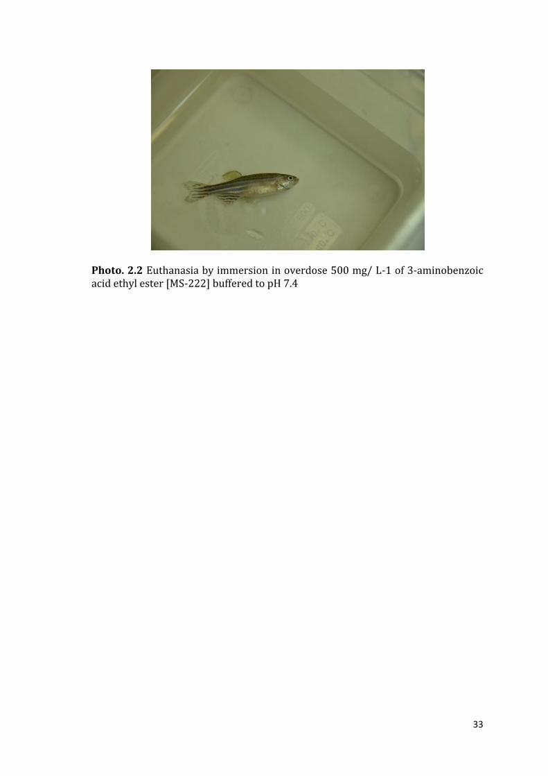
Fishes were euthanized by immersion in overdose 500 mg/ L-1 of 3-aminobenzoic
acid ethyl ester (MS-222) buffered to pH 7.4 (Sigma–Aldrich, USA) (Photo 2.2). To
avoid possible external contamination while removing the intestine, the surface of
each fish was cleaned using 70% Industrial Methylated Spirits (IMS). Under aseptic
conditions, under a light source, fish were dissected, with sterilized micro surgical
blade and forceps, where brain and the GI tract were entirely excised. Each tissue
was replace into individual sterile 1.5 mL micro centrifuge tubes (MCT) with 1 ml
of RNAlater® sterile solution (Life Technologies, USA) and stored at -80°C until
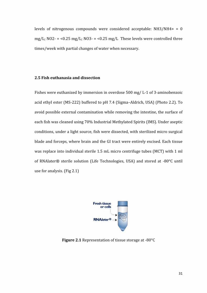
use for analysis. (Fig 2.1)
Figure 2.1 Representation of tissue storage at -80°C

32
Figure 2.3 Representation of the automatic feeder and probiotic administration
Photo 2.1 Adult zebrafish ( Danio rerio) AB wild type strain used.
33
Photo. 2.2 Euthanasia by immersion in overdose 500 mg/ L-1 of 3-aminobenzoic acid ethyl ester [MS-222] buffered to pH 7.4

34
CHAPTER 3
CAN PROBIOTICS MODULATE ZEBRAFISH BEHAVIOR?
3.1 Abstract
The zebrafish (Danio rerio) is a well known model organism in translational
neuroscience and behavioural research. It is increasingly utilized in biomedical
and psychopharmacological research aimed at modeling human brain disorders.
Abnormal social behavior represents the core symptom of several
neuropsychiatric and neurodevelopmental disorders. The zebrafish is a highly
social species and has been proposed for modeling such disorders. Behavioral
paradigms that can induce zebrafish social behavior are of importance. It has some
advantages over other vertebrate species used in biomedical research that stem
from its prolific nature, preference to form tightly packed groups (shoals), and the
fact that it has been a preferred subject of geneticists for the past few decades
(Gerlai, 2014). A growing body of data supports the hypothesis that probiotics can
exert psychotropic effects. Recently, it has been demonstrated that in mice the
probiotic Lactobacillus rhamnosus impacts behavior and produces neuroactive
substances such as GABA and serotonin, which act on the brain-gut axis (Dinan et
al., 2013). Here the putative link between the enteric microbiota and brain
function was tested by analyzing the effects of L. rhamnosus on behavioural
swimming pattern (movement in space and time) (Gerlai, 2014) in zebrafish. In
this study probiotic fed group and control one shoal differently. These measures
was determined by using a 2D video tracking analysis and modeling tool.
35
3.2 Introduction
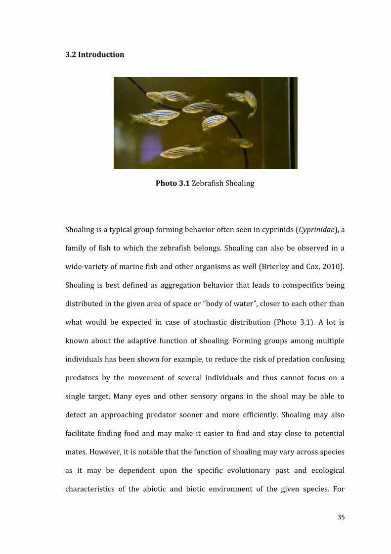
Photo 3.1 Zebrafish Shoaling
Shoaling is a typical group forming behavior often seen in cyprinids (Cyprinidae), a
family of fish to which the zebrafish belongs. Shoaling can also be observed in a
wide-variety of marine fish and other organisms as well (Brierley and Cox, 2010).
Shoaling is best defined as aggregation behavior that leads to conspecifics being
distributed in the given area of space or “body of water”, closer to each other than
what would be expected in case of stochastic distribution (Photo 3.1). A lot is
known about the adaptive function of shoaling. Forming groups among multiple
individuals has been shown for example, to reduce the risk of predation confusing
predators by the movement of several individuals and thus cannot focus on a
single target. Many eyes and other sensory organs in the shoal may be able to
detect an approaching predator sooner and more efficiently. Shoaling may also
facilitate finding food and may make it easier to find and stay close to potential
mates. However, it is notable that the function of shoaling may vary across species
as it may be dependent upon the specific evolutionary past and ecological
characteristics of the abiotic and biotic environment of the given species. For

36
example, shoaling has been found to enhance (and not reduce) predation risk in
some marine fish. Yet in other species, specific environmental constraints, e.g.
oxygen depletion in the middle of large swarms of krills or shoals of sardines, may
also influence shoaling behavior while in other species such factors may play no
role (Brierley and Cox, 2010). Whatever the actual adaptive function of shoaling
may be in zebrafish, it has been observed both in nature (Engeszer et al., 2007) and
in the laboratory (Buske and Gerlai, 2011 and Saverino and Gerlai, 2008) as one of
the most robust and consistent behavioral features of this species (Gerlai, 2014).
Probiotic studies are among the most commonly carried out to support a
relationship between gut microbiota and brain and behavior. The impact of
Lactobacillus rhamnosus on behavior is also evaluated in mice. Animals fed Lb.
rhamnosus demonstrated reduced anxiety on a variety of behavioral measures. The
study provided compelling evidence to indicate that the vagus mediates the
behavioral effects of Lb. rhamnosus. A growing body of data is emerging using
different models to support the contention that a variety of other potential
probiotics can exert psychotropic potential (Dinan and Cryan, 2013). To our
knowledge this is the first study focused on the correlation among dietary
supplementation of probiotics and behavioral pattern changing in zebrafish. This
study is focused on zebrafish shoaling behavior with the aim to compare the main
differences between the group fed with the probiotic strain, Lactobacillus
rhamnosus and the control one as described in general chapters 2.2 and 2.3.
3.2.1 Automated behavioral analyses
Zebrafish prefer to swim in shoals and the disruption of this group-forming
behavior by various environmental, pharmacological, or genetic factors can be

37
easily assessed by using video tracking tools. This “part of aggregation behavior”
has an oscillating dynamic, and this behavior can be quantified manually or using
automated video-tracking systems, assessing several endpoints, including the
average inter-fish distance; shoal area size; proximity (time each member of the
shoal spent within a specified distance from each other); nearest and farthest
neighbor distances; time spent in shoal; time spent away from shoal; number of
animals leaving the shoal and polarization (reflecting the uniformity of heading)
(Kalueff et al., 2013). Behavioral phenotypes are the most complex product of CNS
activity, and the availability of reliable video tracking techniques markedly
empowers neurobehavioral analyses in zebrafish (Stewart et al., 2014). Video-
tracking has been broadly applied to fish research including zebrafish focusing on
swimming mechanics and detection of multiple subjects in shoaling studies
(Cachat 2010). For example, both commercial and custom-made video tracking
systems are used to assess larval and adult zebrafish behavior. Such automated
observations are particularly suitable for measuring loco-motor responses (e.g.,
distance traveled or speed/velocity, turning, etc.) These software systems often
have modular structure and are standardized, user-friendly, and coupled with
thoughtfully designed hardware. Although not inexpensive, these packages are
also validated by multiple international users, and typically come with regular
upgrades and technical support, which becomes especially useful from a practical
point of view. Offering a free alternative, the custom-made tracking systems are
also available from different laboratories worldwide and can be useful for various
specific neurophenotyping tasks and experimental set-ups in zebrafish. (Stewart et
al., 2014). Same reports have either applied 2D (one camera) video-tracking
methods to assess fish stress-related behaviors or used 3D (two cameras) video-

38
tracking as well as high sampling rate to characterize fish swimming, including
assessment of zebrafish neurotoxic phenotype (Cachat 2010). Gerlai, (2014)
dimonstred that three-dimensional presentation of the shoal stimulus is not really
required to evaluate shoaling response, making sufficient zebrafish images moving
back and forth on a 2D flat surface (the computer monitor).
3.3 Materials And Methods
3.3.1 Animals and housing
Adult 4–6 month-old male and female zebrafish (~ 30:70%) of heterozygous (AB)
“wild type” short-fin strain were obtained from local commercial distributors
(Carmar sas, Napoli). The AB strain is frequently used in behavioral neuroscience
(Nowicki et al., 2014). All fish were given at least 10 days to acclimate to the
laboratory environment and housed in groups of 12 fish per 30-L tank. All tanks
were filled with deionized water before introducing the fish. The fish were fed at
1.5%–2% of bodyweight per day automatically. Two experimental groups were
evaluated: a control group (CTRL), which was fed twice per day with a commercial
diet (SERA Vipagran®, Germany) and a probiotic-treated group (PROBIO), which
was fed twice per day the commercial diet and twice per day with the lyophilized
probiotic at a final concentration of 106 colony-forming units/g for 28 days. The
probiotic strain used was L. rhamnosus IMC 501, (provided by Synbiotec s.r.l.
Camerino, Italy) The room and water temperatures were maintained at 25–27 °C.
Illumination (1010 ± 88 lx) was provided by ceiling-mounted fluoreshent light
tubes on a 14-h cycle consistent with the standards of zebrafish care (Westerfield

39
M., 2000). All fish used in this study were experimentally naïve. The experiment
was performed in biological duplicates.
3.3.2 Apparatus and behavioral testing
Two identical experimental setups were run in parallel (Figure 3.4). Twelwe
zebrafish were evaluated for each tank. Fish swimming behavior was video-
recorded between 14:00 and 15:00 h on the first day of probiotic treatment (T0)
and successively at 7 days intervals (T1-T4). Recording was performed next the
tanks with a Nikon D7000 camera for 6 min, acquaried and analysed with 2D video
tracking analysis and modeling tool (Tracker, California, USA) built on the Open
Source Physics (OSP) Java framework (www.cabrillo.edu/~dbrown/tracker/)
(Figure 3.2). This software allows to analyze a video clip or an image in order to
determine multiple variables. The program is designed to be used in physics
experiments in order to easily estimate the acceleration and velocity and distance
of a certain object. In line with Gerlai (2014), the video the tracking data were used
to determine following behavioral measure: Average Distance (AD); Distance
Variance (DV); Nearest Distance (ND); Occupied Area (or Shoal size area) (OA)
and Water column position (CP). In particular AD represents the Inter-
individual distance and it defines and calculates the average of all distances
between a focal fish and its shoal members. Each focal fish within a shoal thus will
get an inter-individual distance value and this value is calculated for any given
moment of time sampled. The disadvantage of this measure, however, is that it is
dependent upon the size of the shoal, i.e. the number of individuals that make up
the shoal. The larger the number of such individuals and larger the inter-individual
distance value will be. ND calculates the nearest distance of each single fish from

40
its neighbor. Thus again, each fish of the shoal will receive a nearest neighbor
distance (Gerlai, 2014). Contrary to AD, ND is independent from shoal size. DV or
the variability of inter-individual distance is the variance of the distances between
the focal fish and all of its shoal members. Thus again, each focal fish gets a
variance of inter-individual distance value for any given moment of time. Notably,
this variability represents the relative position of the given focal fish within a
shoal. The mean of variances of inter-individual distances when calculated for the
entire shoal represents the homogeneity of the distribution of fish within that
shoal. The less uniformly the shoal members are distributed the larger the
variance will be. It is important to note that the inter-individual distance takes the
position of every fish in the shoal into account and thus it is the most informative
measure of shoal cohesion. OA is an additional measure in this study and it
calculates the occupied area of all animals in a temporal unit on a two-dimensional
plane. CP is another measure added in this study (after the differences showed by
two group. It represents the water column position and indicates the preference of
animals to occupy the upper or the lower (part) half of the tank. It was estimated
as the % number of animals that stay in the lower part of the tank during the
period of observation.
For AD, DV and ND measurement each fish was tracked on six randomly selected
20 frame (2fps) intervals of each video. For OA and CP analysis it was used ImageJ
1.49 software (National Institute of Mental Health, Bethesda, Maryland, USA) to
select the areas occupied and the side of water column preferred by each fish in all
video collected (Figure 3.3). Ten images were randomly selected from each video.

41
ImageJ is a public domain Java image processing program inspired by NIH Image
for the Macintosh (http://imagej.nih.gov/ij/docs/intro.html).
Figure 3.2 representative illustration of the typical set up of the shoaling test (left) and application of video tracking tool (Tracker) (right), to quantify zebrafish behavior.
Figure 3.3 illustrates ImageJ softwere used to select the areas occupied and the side of water column preferred by each fish in all video collected.

42
Figure 3.4 Screenshots of a video recordeding at different time of experimentation: a, PROBIO T0; a1, CTRL T0; b, c, d and e represent respectively T1, T2, T3 and T4 of experimental observation, left tank is PROBIO group and right tank is CTRL group.
3.3.3 Statistical Analysis
Results are reported as means ± SE. Two-way ANOVA with Sidak’s post-hoc test
was used to evaluate the significance of the effect of the probiotic treatment, using
GraphPad Prism version 6.00 for Windows (GraphPad Software, La Jolla California
USA, www.graphpad.com).
43
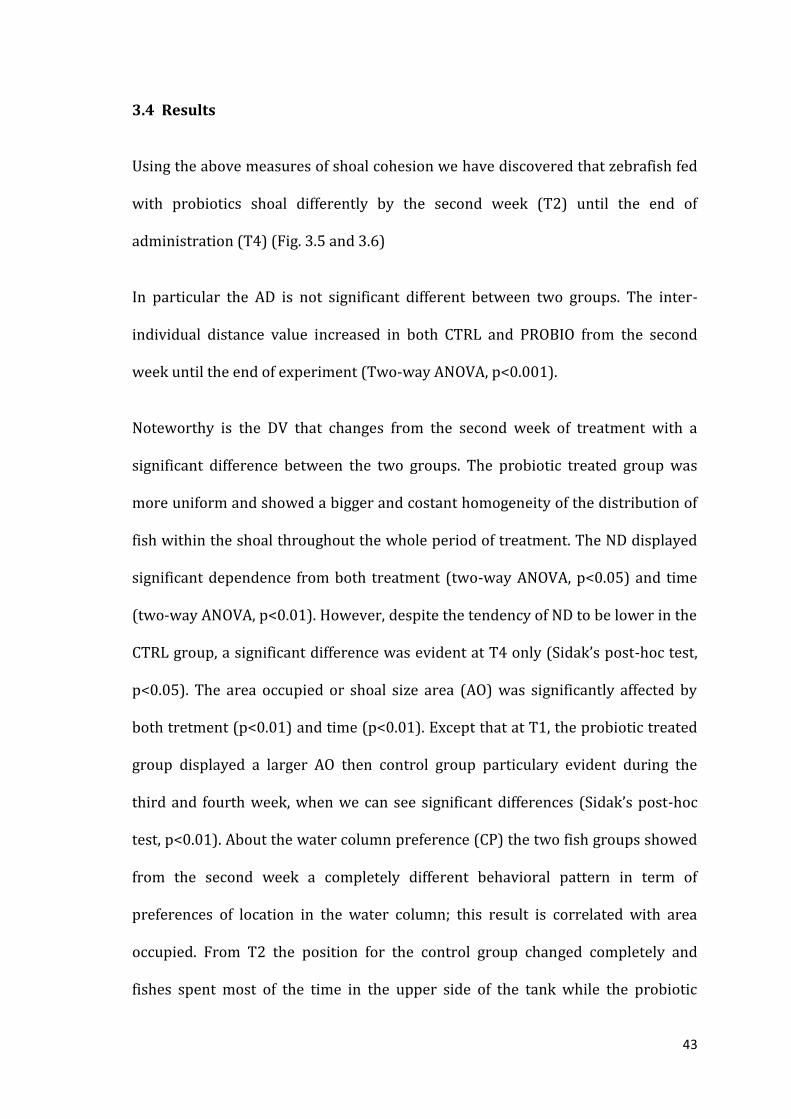
3.4 Results
Using the above measures of shoal cohesion we have discovered that zebrafish fed
with probiotics shoal differently by the second week (T2) until the end of
administration (T4) (Fig. 3.5 and 3.6)
In particular the AD is not significant different between two groups. The inter-
individual distance value increased in both CTRL and PROBIO from the second
week until the end of experiment (Two-way ANOVA, p<0.001).
Noteworthy is the DV that changes from the second week of treatment with a
significant difference between the two groups. The probiotic treated group was
more uniform and showed a bigger and costant homogeneity of the distribution of
fish within the shoal throughout the whole period of treatment. The ND displayed
significant dependence from both treatment (two-way ANOVA, p<0.05) and time
(two-way ANOVA, p<0.01). However, despite the tendency of ND to be lower in the
CTRL group, a significant difference was evident at T4 only (Sidak’s post-hoc test,
p<0.05). The area occupied or shoal size area (AO) was significantly affected by
both tretment (p<0.01) and time (p<0.01). Except that at T1, the probiotic treated
group displayed a larger AO then control group particulary evident during the
third and fourth week, when we can see significant differences (Sidak’s post-hoc
test, p<0.01). About the water column preference (CP) the two fish groups showed
from the second week a completely different behavioral pattern in term of
preferences of location in the water column; this result is correlated with area
occupied. From T2 the position for the control group changed completely and
fishes spent most of the time in the upper side of the tank while the probiotic

44
treated group preferred the medium/deeper part of the tank occupying most of
area of tank.
Figure 3.5 AD, DV and ND measurement at different time of experimental observation obtained with Tracker software and analysed statistically. DV The ND displayed significant dependence from both treatment (two-way ANOVA, p<0.05) and time (two-way ANOVA, p<0.01) and at T4 Sidak’s post-hoc test, p<0.05
45
Figure 3.6 OA (up) and CP (down) measurement at different time of experimental observation obtained with ImageJ software and analysed statistically Two-way ANOVA, p<0.001 and (Sidak’s post-hoc test, p<0.01).
3.5 Discussion and Conclusion
Video-tracking of zebrafish yields objective analysis of behavioral endpoints and
therefore provides researchers with an important tool for the investigation of
behavior in this animal model. Furthermore, such standardization promotes
reproducibility in experimental design, strengthening the investigator’s ability to
draw valid conclusions from zebrafish study data and results. Research on the

46
molecular biology and genomics of probiotics has focused on the interaction of gut
microbiota with the immune system, brain development, potential as an anticancer
agent and potential as a biotherapeutic agent in many diseases. In a study rats fed
Lb. rhamnosus demonstrated reduced anxiety based on a variety of behavioral
measures. The study provided compelling evidence to indicate that the vagus
mediates the behavioral effects of Lb. rhamnosus (Dinan and Cryan 2013). Here in
our study probiotic fed group and control one shoal differently from the second
week until the end of observation. In particular the lower value of distance
variance (DV) in the probiotic treated group, meaning more homogeneity of the
distribution of fish within that shoal (Gerlai et al.,2014), suggest us a different
signal in the shoal behavior. With regard to the nearest distance (ND), the fishes in
control group swam closer together then probiotic treated group. In this case the
shoal pattern may be associated to more anxiety/fear that causes the shoal to
“tighten” (the fish swim closer together) (Kalueff et al.,2013b). The larger occupied
shoal area (OA) by probiotic treated group suggests an increased exploration area
with a preference in the middle/deeper part of the water column of the tank. In
contrast, the control group showed a reduced shoaling area occupying most of
time to the top of the tank. This different behavior might be explained with an
increasing of attention or possibly alert in the probiotic treated group. Blaser and
Goldsteinholm (2012) supposed that aerial predation provides selective pressure
on defensive behavior in zebrafish, making avoidance of the water surface more
adaptive than seeking cover near the bottom. Further study will be needed to
determine the mechanisms as well as the ontogeny of this behavioral differences. A
lot of individual study on zebrafish were performed and individual differences in
activity were found in zebrafish behavior (Tran and Gerlai, 2013) and although

47
there is currently a relatively small group of highly trained zebrafish
neuroscientists and pathologists, the field is expanding rapidly (Kalueff et al.,
2014). Testing grouped zebrafish produces more homogeneous data and preserves
their behavioral repertoire, but requires more laborious or sophisticated data
acquisition and analysis. Such conditions may favor the study of more complex
behaviors such as those involved in sophisticated cognitive and social interactions
that are often challenging to investigate in rodents (Pagnussat et al., 2013).
Overall this study represents the first video tracking shoaling analysis of adult
zebrafish group treated with a probiotic strain such as Lb. rhamnosus. This study
has shown how a probiotic strain can modulate the behavior of zebrafish in term of
shoaling. Therefore it provides a basis for further studies on the gut-brain axis in
Danio rerio. Although human behavior will never be similar to fish responses (and
vice versa), the evolutionarily conserved nature of complex CNS traits suggests
that many human and zebrafish phenotypes share common genetic and
physiological factors, representing an exciting emerging field for further
translational studies in neuroscience (Stewart et al., 2014). The same concept is
valid for other vertebrates. Taken together, these results confirm zebrafish as a
valid, reliable, and efficacious model for basic translational research to understand
with further studies the microbiota-gut-brain axis.

48
CHAPTER 4
CULTURE-INDEPENDENT METHODS FOR MICROBIOTA EVALUATION
IN ZEBRAFISH (DANIO RERIO) TREATED WITH L. RHAMNOSUS AND
CONTROL GROUP: DENATURING GRADIENT GEL ELECTROPHORESIS
(DGGE) AND NEXT-GENERATION SEQUENCING (NGS)
4.1 Abstract
Methods of measuring bacterial communities are rapidly improving. The earliest
and most traditional technique is the culture-dependent method. In recent
decades, microbiologists have developed new culture-independent techniques to
obtain a better representation of bacterial communities present in host organisms,
for example denaturing gradient gel electrophoresis (DGGE) and temperature
gradient gel electrophoresis (TGGE) (Sevellac et al., 2014), PCR-random amplified
polymorphic DNA (RAPD) (Spanggaard et al., 2000), fluorescence in situ
hybridization (FISH) (Huber et al., 2004) and clone libraries (Kim et al., 2007).
These approaches are useful in that they offer new opportunities for detection and
identification of the microbiota, leading to a broader understanding of the
microbial composition in the gastrointestinal tract (GIT) of fish. In contrast few
studies have applied Next-Generation Sequencing (NGS) methods to investigate
the microbiome of vertebrates in their natural environment and in freshwater
fishes in particularly (Sevellec et al., 2014). The capability of high through-put
sequencing of 16S rRNA gene sequences by means of Next Generation Sequencing
(NGS) technologies has been pivotal in facilitating the discovery of gut microbiota
biodiversity. The Ion Torrent PGM instrument represents a recently
commercialized bench-top NGS platform and is marketed as being less costly and

49
with a faster turnaround as compared to other NGS techniques such as the 454 and
Illumina platforms (Milani et al., 2013). Therefore, the aim of this study was to
gain a better overall understanding about differences in the GIT microbiota
between a group of zebrafish (Danio rerio) treated with L. rhamnosus IMC 501 and
control groups by using DGGE and NGS technologies. Here we use the Ion Torrent
PGM (Personal Genome Machine) technology to allow a more complete description
of complex bacterial communities and biodiversity of the zebrafish (Danio rerio)
gut. The average of observed taxonomical units (OTUs) detectable in the probiotic
group increased compared with control group, therefore this study indicates that
dietary supplementation of Lb. rhamnosus modulates intestinal microbial
communities of zebrafish. Feeding zebrafish probiotic Lb. rhamnosus showed a
significant increase of Firmicutes phylum and, although not significant, a reduction
of Proteobacteria the greather microbiota present in dysbiosis, supporting the
antagonistic activity role of this probiotic strain.
4.2 Introduction
Vertebrate species host a considerable bacterial diversity, which may influence
their development, physiology, immune system and nutrition. The relationships
between bacteria and their hosts consists in four types. The first two types are
commensal bacteria, which may either have beneficial or neutral effects on the
host. The second type has a symbiotic obligatory relationship with the host, thus
allowing a mutual benefit between symbiotic bacteria and host. The third type is
opportunistic bacteria, which are facultative pathogenic bacteria that may become

50
actively pathogenic when the host immune system is impaired and unable to fight
off infection. The fourth type of relationship pertains to pathogenic bacteria which
are responsible for infectious diseases (Sevellec et al., 2014). The group of Lactic
acid bacteria (LAB) represents a large part of the microbiota of vertebrates and
their beneficial effects on the immune system, gastrointestinal tract and
reproduction, have been widely reported. Lactobacillus rhamnosus, is one of the
main LAB components of the commensal microflora of human intestinal tract and
it is widely used as a probiotic in mammals (Avella et al., 2012). A number of
recent studies have evidenced the positive role of Lb. rhamnosus on zebrafish
gamete quality, spawning rates, oocyte growth and maturation, larval
development, fecundity, backbone calcification and the expression of genes which
regulate growth, development and immunity (Table 4.1) (Carnevali et al., 2014a).

51
Tab. 4.1: Parameters investigated of administration of Lb. rhamnosus on zebrafish
Genera abbreviations: B. = Bacillus, Lb. = Lactobacillus. Parameters investigated: DR = disease resistance, GM = gut microbiota (inclusive of GI probiont recovery), F= fecundity/gonadal development/spawning rates etc., GP = growth performance, IR =immunological/haematological response, PA = pathogen antagonism (Carnevali et al., 2014 10 Probiotic Applications in Temperate and Warm Water Fish Species.)
The intestinal microbial communities and their metabolites play an integral role in
the ontogeny of teleosts. (Cerf-Bensussan and Gaboriau-Routhiau, 2010; Merrifield
et al., 2010; Sekirov et al., 2010; Llewellyn et al., 2014). Intestinal microbial
communities consist of allochthonous (digesta-associated, transient) and
autochthonous (mucosa-associated, indigenous) microbiota (Ringø and Birkbeck,
1999; Ringø et al., 2003). The microbiota play important roles such as assembling
of the gut-associated lymphoid tissue (GALT), it helps the immune system,
influences the integrity of the intestinal mucosal barrier, modulates proliferation
and differentiation of its epithelial lineages, regulates angiogenesis, modifies the
Potential probiont Parameters investigated
Lb. rhamnosus IMC 501 F
Lb. rhamnosus IMC 501 F
Lb. rhamnosus IMC 501 F
Lb. rhamnosus IMC 501 GM , F
Lb. rhamnosus IMC 501 GM , F
Lb. rhamnosus IMC 501 F
Lb. rhamnosus IMC 501 F
Lb. rhamnosus IMC 501 GP
Lb. rhamnosus IMC 501 and Lb. casei F, GH, GM, IR
37 commensal or probiotic Gram-positive and Gram-negative bacteria often used as probiotic strains in the food industry and/or aquaculture
DR, IR
Lactobacilli (multiple species) DR, GM, PA
B. coagulans DR, IR, PA

52
activity of the enteric nervous system and plays a key role in extracting and
processing nutrients consumed in the diet (Rawls et al., 2004). The mechanisms by
which the mammalian gut microbial community influences host biology remain
almost, despite these important effects, entirely unknown. Deciphering the
pathways through which microbial signals operate promises to provide new
chemical entities and host targets for enforcing health, and perhaps treating
diseases affecting both the intestine and extra-intestinal tissues. The zebrafish, has
several unique features that make it an attractive model organism for analyzing
these pathways (Rawls et al., 2004).
Here we sought to determine whether gut microbiota composition varies between
zebrafish treated with dietary supplementation of Lb. rhamnosus and a control
group. We used the DGGE analysis at first and Next Generation Sequencing with
Ion Torrent PGM profiles to assess individual variation in gut microbial
communities.
4.3 Materials and Methods for DGGE
4.3.1 Animal Housing
The experiment was conducted as described under section 2.2 in Stabulario of fish
anphybians and reptiles a of the University of Napoli Federico II and water quality
was monitored accordingly the section described in 2.3

53
4.3.2 Experimental fish and feeding
24 (twelve PROBIO and twelve CTRL) of 72 zebrafish (Danio rerio) were randomly
selected, in this experiment. All fishes was carefully acclimatized and feeded as
described under the section 2.2 and 2.3. In particular four zebrafish treated and
four zebrafish control for each experimentation were euthanized and dissected as
described in 2.4 of general methods.
4.3.3 DNA extraction
DNA was extracted from the zebrafish (Danio rerio) gut samples using a
combination of QIAamp® Stool Mini Kit (QUIAGEN, West Sussex, UK) with minor
modifications to the manufacturer's instructions, as described in Appendix 1. and
phenol-chloroform method. Gut samples were prepared in a sterilized Eppendorf
tube, and DNA extracted by the following five phases:
1. Lysis: 60-80 mg of samples were macerate with sterile macerators
and mixed with 500 μl of fresh lysozyme solution (50mg/ml TE buffer).
Then, the samples were incubated at 37 ºC for 30 minutes. 700 μl of buffer
ASL was added and mixed for 1 minute. The mixture was placed on a hot
plate at 90 ºC for 10 minutes and vortexed for 5 seconds with centrifugation
for 1 min at 14000 rcf.
2. Inhibitor removal: Half an inhibitor tablet was added to 800 μl of
the supernatant and vortexed for 1 min immediately, then, centrifuged for 3
min at 14000 rcf. All of supernatant was pipette into a new Eppendorf tube.
The supernatant was centrifuged for other 3 minutes.

54
3. Protein removal: 400μl of the supernatant was mixed with 20 μl of
proteinase K and 400μl of buffer AL was added and mixed for 15 seconds,
then incubated at 70ºC for one hour.
4. Phenol Chloroform Clean-up: The entire samples were poured into
a 15 ml falcon tubes carefully, and added an equal volume of ice cold Tris-
buffered phenol solution. The samples were mixed by hand and left on ice
for 10 minutes. An equal volume of chloroform/isoamyl alcohol (24:1) was
added and mixed, then centrifuged for 5 minutes at 6000 rcf. The aqueous
layer was pipette off carefully and placed in new 1.5 ml Eppendorf tube.
5. Precipitation: 400 µl of ice-cold isopropanol was added. The
samples were vortexed and placed in -20 °C freezer for overnight. Then,
samples were centrifuged at 14000 rcf for 30 minutes at 4 °C. The
supernatant were pipette carefully and discarded. 500 µl of 70% molecular
grade ethanol was added slowly, and discarded. The addition of 70%
ethanol was repeated and discarded again. The pellets were dried for 5
minutes maximum. Finally, the DNA extracted was resuspended overnight
at 4 °C by adding 30 µl of molecular grade water. The concentration of DNA
and purity were determined using a Nanodrop-100 Spectrophotometer.
4.3.4 16S rRNA amplification, Polymerase Chain Reaction (PCR)
PCR was conducted to amplify the V3 region of the 16S rRNA gene using PCR with
the forward primer P3 with a GC clamp on its 5’-end (5'-CGC CCG CCG CGC GCG
GCG GGC GGG GCG GGG GCA CGG GGG GCC TAC GGG AGG CAG CAG-3') and the
reverse primer P2 (5'- ATTACCGCGGCTGCTGG-3') (Muyzer et al., 1993). Each

55
single PCR reaction consisted of 25 μl ReadyMixTM Taq PCR Reaction Mix with
MgCl2 (Sigma-Aldrich Company Ltd., Gillingham, England), 1 μl each of primer P2
and P3 (50 pmol/μl Eurofins MWG Operon, Ebersberg, Germany), 1 μl of DNA
template and sterile, molecular grade water to adjust the final volume of the
reaction to 50 μl. Touchdown thermal cycling was conducted using a GeneAmp®
PCR System 9700 (Perkin-Elmer, CA, USA), under the following conditions: 94 °C
for 10 min, then 30 cycles starting at 94 °C for 1 min, 65 °C for 2 min, 72 °C for 3
min as described by Muyzer et al. (1993). The annealing temperature decreased by
1 °C every second cycle until 55 °C and then remained at 55 °C for the remaining
cycles. In order to check the purity and molecular weight characteristics of PCR
products, PCR products (6 μL) were loaded onto a 1.5% agarose gel (Lonza,
Rockland ME, USA), made with 1x Tris-acetate-EDTA (TAE) buffer prestained with
4 μL of SYBR® Safe™ DNA Gel Stain (Life TechnologiesTM UK) per 100 mL of
agarose (Fisher Scientific) and run with 1x TAE buffer in a Pharmacia
electrophoresis tank at 90 volts for 60 min. Five μL of Hyper Ladder IV (Bioline)
was run alongside the PCR products to assess the size of DNA products. Viewing of
agarose gels was achieved under UV light using a Bio-Rad universal hood 11 (Bio-
Rad laboratories, Italy). 18 positive PCR products samples (9 belonging to PROBIO
and 9 to CTRL) were chosen for DGGE and stored at 4 °C until use (Fig 4.1 ).

56
Fig. 4.1 Touchdown thermal cycling PCR of DNA samples extraction (1-24). Eighteen samples positive were chosen (9 PROBIO in the upper line and 9 CTRL down ).
4.3.5 Denaturing gradient gel electrophoresis (DGGE)
The resulting 18 PCR products were used to obtain DNA fingerprints of the
bacterial community present in the gut by DGGE using a Bio-Rad DGGE system
(DCode™ System, Italy). DGGE was carried out by loading 15 μL of PCR products
onto 10% acrylamide gels with a denaturing gradient of 40 - 60% (where the
denaturants were 5.6M urea (Sigma, UK) and 40% formamide (Sigma, UK).
Made using the following stock solutions; an 80% denaturant polyacrylamide
solution consisted of 25 mL of 40% acrylamide mix (high purity acrylamide), 2mL
of 50x TAE buffer (pH 8.3), 32 mL of molecular grade formamide (Sigma, UK), 34 g
of 5.6M ultrapure urea (Sigma, UK) and volume of MilliQ H2O yielding a total
volume of 100 mL. Stock 0% denaturant polyacrylamide solution consisted of 25
mL of 40% acrylamide mix (high purity acrylamide), 2mL of 50x TAE buffer (pH
8.3) and 73 mL of MilliQ H2O. One-hundred and fifty μL of 10% ammonium

57
persulphate (APS, electrophoresis grade, Sigma, UK) and 17.5 mL of
Tetramethylethylindiamine (TEMED) were added to the high and low denaturant
solutions. Twenty one mL of each acrylamide solution was added to separate 30
mL syringes and these were mounted onto a Bio-Rad gradient delivery system
(model 475, Bio-Rad laboratories). The major steps of DGGE are presented in
Figure 4.2. This was then used to pour the gel between gel plates and the gel was
left to polymerize for two hours. Additionally, PCR products from L.rhamnosus
pure colonies were loaded to the gel as a reference species to aid probiotic
identification. The gel was run at 65 V for 17h at 60 °C in 1 x TAE buffer. Viewing of
the DGGE bands was accomplished after SYBR® gold staining. Briefly, the gel was
incubated for 20 min at room temperature in 200 mL tank buffer containing 20 μL
of 10000x SYBR® gold nucleic acid gel stain (Invitrogen™, UK) with shaking on an
IKAO VIBRAX VXR basic shaking platform at 100 rpm/ min. The gel was scanned in
a Bio-Rad universal hood 11 (Bio-Rad Laboratories, Italy) and optimized for
analyses by enhancing contrast and greyscale.
4.3.6 Excision of DGGE bands, for sequence analysis
After DGGE, bands (or ‘operational taxonomic units’, OTU) of interest (those
showing clear and consistent specialization either to intestinal regions or dietary
treatments, or those clearly unaffected) were excised from the gel using sterile
pipette tips and DNA was eluted overnight at 4 °C in 1.5 mL Tube containing 20 μL
Molecular Grade Water (Photo 4.1)

58
Photo 4.1 Excision of DGGE bands (or ‘operational taxonomic units’, OTU), for sequence analysis
4.3.7 16S rRNA amplification of excised DGGE bands
From the eluate, 5 μL was used as the template for reamplification using the
forward primers P1 (5-CCTACGGGAGGCAGCAG-3; essentially P3 without the GC
clamp at its 5’ end) and the reverse primer P2 under the same conditions as
previously described (Section 4.2). Six μL was loaded onto a pre-stained agarose
gel (1.5%) to check the PCR product size.
4.3.8 Purification of the PCR products and sequence analysis
The PCR products were cleaned using a QIAquick PCR Purification Kit (Qiagen),
according to the manufacturer’s instructions and PCR yields (the concentration
and purity of DNA) were checked using a Nanodrop® 1000 spectrophotometer.
Protein purity (A260/A280) and humic acid purity (A260/ A230) were checked.

59
The PCR products were sequenced by GATC Biotech Ltd. (Germany) and
sequenced by GATC laboratories (GATC-biotech laboratories, Germany).
Nucleotide sequences were then submitted to a BLAST search in GenBank
(http://blast.ncbi.nlm.nih.gov/Blast.cgi) to retrieve the closest known alignment
identities for the partial 16 S rRNA sequences. (Figure 4.3)
Fig. 4.2 Schematic representation of the principle steps of the denaturation gradient gel electrophoresis (DGGE) process.
60
Fig. 4.3 BLAST searching in GenBank screenshot
4.3.9 Statistical analysis
Data was transformed where necessary and statistical analysis was carried out
using a One-Way ANOVA Minitab v.16 statistical software (Minitab, Plymouth, UK).
Tukey’s Multiple Comparison test was used to determine significant differences
between means and significance was accepted at the P < 0.05 level. Primer V6
(Clarke and Gorley, 2006) was used to calculate species richness, evenness and
diversity of the PCR-DGGE fingerprints according to the following formulae:
Margalef's species richness: d = (S − 1)/log(N); and Shannons diversity
index: H′ = − Σ(pi(lnpi)). Where N = total number of individuals (total intensity
units),S = number of observed taxonomical units (presumed species) and pi = the
proportion of the total number of individuals in the ith species.

61
4.4 Materials and Methods for Ion Torrent PGM technology NGS
4.4.1 Samples
With regard to the facility conditions, experimental fishes and DNA extraction the
same conditions apply as described in 4.3.1, 4.3.2 and 4.3.3. PROBIO and CTRL
DNA extraction were analyzed as follow
4.4.2 16S rRNA Gene Amplification
Partial 16S rRNA gene sequences were amplified from extracted DNA using primer
pair 27F (5'-AGA GTT TGA TCM TGG CTC AG-3'), 338RI (5'-GCW GCC TCC CGT
AGG AGT-3') and 338RII (5'-GCW GCC ACC CGT AGG TGT -3') (Roeselers et al
2011). which targets the V1-V2 region of the 16S rRNA gene sequences. The PCR
conditions used were 7 min at 95°C, 10 Cycles of 30 sec at 94 °C, 30 sec at 63 ° C,
30 sec at 72 °C and 25 Cycles of 30sec at 94°C, 30 sec at 53 °C and 30sec at 72 °C
followed by 10 min at 72 °C. Amplification was carried out by using a Verity
Thermocycler (Applied Biosystems). The integrity of the PCR amplicons was
analyzed by electrophoresis
4.4.3 Ion Torrent PGM Sequencing of 16S rRNA Gene-based Amplicons
Twenty PCR products derived from amplification of specific 16S rRNA gene
hypervariable regions were purified by electrophoretic separation on an 1.5%
agarose gel and the use of QIAquick PCR Purification Kit (Qiagen) in order to
remove primer dimers. Libraries for sequencing were prepared using the Ion Plus
Fragment Library Kit (Life Technologies) and quantified using the Ion Library
Quantitation Kit (Life Technologies). A proportion of the amplicon libraries were

62
also run on the Agilent 2100 Bioanalyzer using the High Sensitivity DNA Kit
(Agilent Technologies) in order to confirm adapter ligation and average size of
each library. Libraries for each run were diluted to 26pM and pooled, then
emulsion PCR was carried out using the Ion PGM Template OT2 400 Template Kit
(Life Technologies) according to the manufacturer’s instructions. Sequencing of
the amplicon libraries was carried out on a 318 chip using the Ion Torrent PGM
system and employing the Ion Sequencing 400 kit (Life Technologies) according to
the supplier’s instructions. After sequencing, the individual sequence reads were
filtered by the PGM software to remove low quality and polyclonal sequences.
Sequences matching the PGM A and P1 adapters were also automatically trimmed.
All PGM quality-approved, trimmed and filtered data were exported as FASTQ files.
4.4.4 Sequence-based Microbiota Analysis
The FASTQ files were converted to FASTA format and processed using QIIME
(Quantitative Insights Into Microbial Ecology). Quality control retained sequences
with a length between 250 and 400 bp, mean sequence quality score >25, with
truncation of a sequence at the first base if a low quality rolling 10 bp window was
found. Presence of homopolymers >6 bp, and sequences with mismatched primers
were omitted. In order to calculate downstream diversity measures (alpha and
beta diversity indices, Unifrac analysis), 16S rRNA Operational Taxonomic Units
(OTUs) were defined at >97% sequence homology. All reads were classified to the
lowest possible taxonomic rank using QIIME and a reference dataset from the
Ribosomal Database Project. OTUs were assigned using uclust. The hierarchical
clustering based on population profiles of most common and abundant taxa was
performed using UPGMA clustering (Unweighted Pair Group Method with

63
Arithmetic mean, also known as average linkage) on the distance matrix of OTU
abundance.
4.4.5 Statistical Analysis
Ecological metrics for richness (Chao 1) and diversity (Shannon) were calculated
in accordance with the bioinformatics of Plymouth University, UK, using Mothur
Microbial community profiles generated from amplicon sequence data were
compared using Quantitative Insights into Microbial Ecology (QIIME) v1.8.0 using
total read counts for all assembled OTU sequences as input. Taxonomy of intestinal
bacterial communities data are presented as means ± standard error of the mean.
Statistical analysis was performed using the two-tailed t test. A p value of less than
0.05 was considered significant.
4.5 Results
4.5.1. DGGE analysis of gut bacterial community
Figure 4.4 shows the PCR–DGGE bacterial profiles from the gut of zebrafish. Many
different bands are shown in the DGGE image and the gel bands which are called
operative taxonomy units (OTU) in each sample.

64
The similarity of bacterial population within and between the treatments were
measured by nonmetric multidimensional scaling (MDS) and cluster analyses of
DGGE fingerprints as shown in Figure 4.5. The half matrix similarity showed in
Table 4.2.
The both analyses of gut bacteria populations showed more similarity within
samples from same treatments than those from other groups. The half matrix
similarity of gut DGGE fingerprints is shown in Table 2 indicates the average
similarity within the control ad treatment: 70.54% in the 1st probiotics treated
group (P1), 68.70% in the respective control group (C1); 65.95% in the 2nd
probiotic treated group (P2) and 51.43% in the respective control group (C2);
82.52% in the 3rd probiotic treated group (P3) and 74.90% in the respective
control group (C3). The average DNA bands detectable in the probiotic group
increased compared with control group, being 16.88% and 14.77% respectively
(Fig 4.6), but it was not significant. However significant differences was observd in
P3 (probiotic group) compared with the control group C3 (Table 4.3).

65
Figure 4.4 DGGE fingerprints of whole intestine of treated and control group of zebrafish. Numbers are represents the bands or operative taxonomy unite (OTU) in each sample
which refers to richness in the samples.

66
Table 4.2 The half matrix similarity of bacterial population of DGGE fingerprints of gut showing the similarities between the replicates treatment.
Group P11 P12 P13 C11 C12 C13 P21 P22 P23 C21 C22 C23 P31 P32 P33 C31 C32 C33
P11 100
P12 78.57 100
P13 70.97 62.07 100
C11 59.26 72.00 64.29 100
C12 75.86 66.67 60.00 76.92 100
C13 60.61 70.97 58.82 66.67 62.50 100
P21 64.29 76.92 55.17 64.00 59.26 70.97 100
P22 64.29 53.85 55.17 64.00 66.67 64.52 53.85 100
P23 51.85 64.00 50.00 75.00 61.54 66.67 64.00 80.00 100
C21 66.67 58.06 64.71 53.33 62.50 77.78 64.52 64.52 60.00 100
C22 70.59 56.25 74.29 58.06 60.61 70.27 62.50 68.75 58.06 86.49 100
C23 34.78 57.14 41.67 60.00 45.45 46.15 66.67 47.62 60.00 30.77 37.04 100
P31 57.14 66.67 55.56 50.00 52.94 68.42 66.67 60.61 56.25 63.16 66.67 50.00 100
P32 60.00 63.16 58.54 59.46 61.54 74.42 57.89 68.42 64.86 69.77 72.73 42.42 80.00 100
P33 55.00 57.89 63.41 64.86 61.54 83.72 57.89 63.16 64.86 74.42 72.73 36.36 75.56 92.00 100
C31 64.52 48.28 43.75 50.00 66.67 64.71 62.07 62.07 50.00 70.59 68.57 33.33 66.67 68.29 73.17 100
C32 50.00 53.85 48.28 72.00 74.07 64.52 53.85 61.54 64.00 58.06 56.25 57.14 54.55 63.16 68.42 75.86 100
C33 60.00 64.29 58.06 59.26 62.07 72.73 71.43 64.29 66.67 72.73 70.59 43.48 68.57 75.00 75.00 77.42 71.43 100
Note: C = control, P = probiotic 1-3 refers to replicate number in each case, (n=18)

67
Figure 4.5: (A) Cluster analysis (B) non-metric multidimensional scaling (MDS) analysis based on the PCR-DGGE DNA fingerprints showing percentage and relative similarity of bacterial communities between control and treatment groups in zebrafish gut.

68
Fig. 4.6 DNA bands average
Table 4.3 Band numbers of bacterial community based on the PCR-DGGE DNA fingerprinting and similarity within treatments.
Treatment Band number Similarity
P1 14.66±1.52 b 70.54±8.26 a
C1 14.66±3.05 b 68.70±7.42 a
P2 12.66±0.57 b 65.95±13.18 a
C2 15.00±6.08 ab 51.43±30.52 a
P3 23.33±2.88 a 82.52±8.50 a
C3 14.66±1.52 b 74.90±3.11 a
P value 0.018 0.268
a,b Means with the different superscript in the same column are significantly different (P<0.05). Results are mean values from three replications ± standard deviations.

69
Diversity analysis of gut microflora showed in table 4.4. The Shannon index and
Margalef index indicate respectively the diversity, and richness of gut microflora of
animals. (Hill et al., 2003) These indexes were used to display the microbial
population diversity and richness in the gut, data showed in Table 4.4. The
diversity index of bacterial community based on the PCR-DGGE DNA fingerprinting
indicated that: at probiotic group had greater Shannon index and Margalef index
than control group. However, no significant differences in Shannon index, but good
significant in the Margalef index was observed in zebrafish fed with probiotics ( L.
rhamnosus).
Table 4.4: Diversity index of bacterial community in gut based on the PCR-DGGE DNA fingerprinting.
Treatment Shannon index1 Margalef index2
P1 2.68±0.10 a 5.08±0.37 ab
C1 2.67±0.20 a 5.07±0.73 ab
P2 2.53±0.04 a 4.59±0.14 b
C2 2.63±0.48 a 5.12±1.52 ab
P3 3.14±0.12 a 7.08±0.64 a
C3 2.68±0.10 a 5.08±0.37 ab
P value 0.085 0.026
Results are mean values from three replications ± standard deviations. a,b Means with the same superscript in the same column are not significantly different (P<0.05). 1 Shannon diversity index: H‟ = -SUM(pi٭Log(pi)). 2 Margalef species richness: d = (S -1) ⁄ log (N). (S: Total species, N: Total individuals)

70
A number of 25 bands were excised from the PCR-DGGE gel and were subjected to
sequence and BLAST analysis (Tab 4.5), after purification as recommended by
GATC company around (20-80 ng/µl).
Table 4.5: Summary of the 16S rRNA gene sequence analysis results generated from bands excised from DGGE gel of zebrafish gut samples.
Band Number
NCBI Accession number
Max.
Identity NCBI BLAST matches
1 NR_025533.1 97% Cetobacterium somerae strain WAL 14325 2 NR_114236.1 96% Shewanella algae strain NBRC 103173 3 NR_117686.1 98% Klebsiella pneumoniae strain DSM 30104 4 NR_025533.1 93% Cetobacterium somerae strain WAL 14325 9 KC010472.1 94% Uncultured bacterium
10 GU430248.1 83% Streptococcus sp. 11 KJ804042.1 85% Streptococcus sp. K-72-13-7 12 LK392937.1 94% Uncultured bacterium 13 NR_024951.1 97% Pseudomonas thivervalensis strain SBK26 18 JN866573.1 97% Uncultured bacterium 19 KF256027.1 99% Uncultured bacterium 21 JQ815676.1 95% Uncultured bacterium 22 KF256014.1 100% Uncultured bacterium 23 NR_029216.1 97% Propionigenium modestum strain Gra Succ 2 25 NR_026243.1 97% Propionigenium maris strain 10succ1
Twenty-five bands or OTUs were excised from the PCR-DGGE gel and all bands
were sequenced but unfortunately only 15 samples were returned and subjected
to BLAST analysis and the others were below the required standard and
sequencing data was zero. The results of the trial sequence analysis shown in Table
4.4. The most family BLAST results in zebrafish gut were related to Cetobacterium
spp., Shewanella spp., Klebsiella spp., Streptococcus spp., Pseudomonas spp.,
Propionigenium spp. strains. Otherwise, the sequences of the band numbers 9, 12,
18, 19, 21 and 22 were related to uncultured bacteria. In particular were isolated
71
Cetobacterium somerae, (band n. 1 and 4) 66,6% of PROBIO samples and 88,8% of
CTRL samples; Shewanella algae (band n. 2) was isolated in all PROBIO and CTRL
samples; Klebsiella pneumonia (band n. 3) in 55,5% of PROBIO group and 66,6 % of
related CTRL; Streptococcus sp. (band n. 11) 66,6% of PROBIO group and 22,2% of
CTRL one.
4.5.2 NGS analysis of gut bacterial community
At NGS analysis alpha diversity metrics, which describe the richness and/or
evenness of taxa in a single sample, (QIIME, ) was at first evaluated. By default,
QIIME calculates three metrics: Chao1 (chao1), Observed OTUs (previously known
as Observed Species), and Phylogenetic Diversity (Phylogenetic tree). In addition,
in the alpha parameters we added the Shannon Index (shannon) to the list of alpha
diversity measures that we calculated here. The comparison of OTUs derived from
zebrafish intestines revealed that the Chao1 richness and Shannon–Weaver
diversity estimates of the intestinal microbiotas from PROBIO and CTRL zebrafish
were not statistically different, indicating that Probiotic treatment with Lb.
Rhamnosus have not particular influence on the overall richness or diversity of the
zebrafish gut bacterial community.
In addition to alpha (or within-sample) diversity, community ecologists are often
interested in computing beta (or the between-sample) diversity between all pairs
of samples in their study. Beta diversity represents the explicit comparison of
microbial (or other) communities based on their composition. Beta diversity
metrics thus assess the differences between microbial communities. The
fundamental output of these comparisons is a square, hollow matrix where a
72
“distance” or dissimilarity is calculated between every pair of community samples,
reflecting the dissimilarity between those samples. (QIIME, ). The data in this
distance matrix can be visualized with Principal Coordinates Analysis (PCoA).
Here, we have calculated beta diversity between intestinal bacterial communities
sampled and we analyzed our 16S rRNA gene sequence data sets using the default
beta diversity metrics of weighted and unweighted UniFrac, which are
phylogenetic measures used extensively in recent microbial community
sequencing projects (Caporaso et al., 2011).
To further understand and to compare the composition of the gut microbiotas in
zebrafish PROBIO and CTRL one, we subjected these 16S rRNA gene sequences to
Principal Coordinates Analysis (PCoA). PCoA plots derived from both unweighted
(an assessment of community composition) and weighted (an assessment of
community structure which measures the distance between communities based on
their phylogenetic lineages) algorithms (Lozupone et al., 2005). PCoA is a
technique that helps to extract and visualize a few highly-informative components
of variation from complex, multidimensional data. This is a transformation that
maps the samples present in the distance matrix to a new set of orthogonal axes
such that a maximum amount of variation is explained by the first principal
coordinate, the second largest amount of variation is explained by the second
principal coordinate, etc. The principal coordinates can be plotted in two or three
dimensions to provide an intuitive visualization of differences between samples
(http://qiime.org/tutorials) (Fig 4.7 and 4.8).
73
Fig. 4.7 The unweighted Unifrac distances, is an assessment of community composition (OTUs presence/absence). PCoA plot shows the spatial distribution of samples of the groups (control samples are red and the probiotic treated samples are colored blue).
Fig. 4.8 Weighted Unifrac distances (OTUs abundance and weight branches) shows the similarity between phylogenetic lineages of bacteria communities between PROBIO (blue colored) and CTRL group (red colored); this measure also shows the degree of similarity inter-PROBIO group, clustering closer togheter.

74
All distances were computed according to the Bray-Curtis similarity index. Bray-
Curtis dissimilarity demonstrated that 16S rRNA profiles from the the majority of
CTRL groups clustered together. Four of the PROBIO samples clustered closer to
CTRL whereas the other remaining part of probio samples formed independent
groups. Also, five of the PROBIO samples clustered far from all each other formed
independent groups. CTRL groups were closer than those PROBIO.
The alpha and beta diversity analysis indicated that these samples were not
significantly different from others in their respective group. The unweighted an
weighted two-dimensional PCoA showed that PROBIO zebrafish samples clustered
together with CTRL zebrafish group establishing a similarity in composition and
structure of these gut bacterial communities.
4.5.3 OTUs Comparison of probiotic treated group (PROBIO) and control (CTRL)
Here to provide perspective on the observed relationships between gut microbiota
from two different zebrafish group investigated, we compared the taxonomy of
intestinal bacterial communities in PROBIO and CTRL zebrafish.

75
Fig. 4.9 Percentage of OTUs observed (Bar stack plots). Main differences in Phyla of bacteria within the guts of CTRL and PROBIO zebrafish group treated with
Lb rhamnosus per 28 days.
Tab 4.6 Main representative absolute composition of OTUs analyzed per Phyla of bacteria
within the guts of CTRL and PROBIO zebrafish group treated with Lb rhamnosus per 28
days. Absolute values are calculated as Mean ± SD (standard deviation)
0
10
20
30
40
50
60
70
80
90
100
CTRL PROBIO
%
Phyla
Verrucomicrobia
Proteobacteria
Fusobacteria
Firmicutes
Cyanobacteria
Bacteroidetes
Actinobacteria
PHYLA CTRL PROBIO
Mean ± SD Mean ± SD
Fusobacteria 3743 ± 1976 3363 ± 2367
Proteobacteria 850,7 ± 1187,8 495,5 ± 666,8
Firmicutes 20,2 ± 30,4 158,0 ± 193
Bacteroidetes 0,00 ± 0,00 4,3 ± 9,1
Actinobacteria 91,7 ± 249,2 0,18 ± 0,4
Cyanobacteria 48,1 ± 75,4 10,6 ± 4,6

76
Fig. 4.10 Percentage of OTUs observed (Bar stack plots). Main differences in Genera of bacteria within the guts of CTRL and PROBIO zebrafish group treated with Lb rhamnosus per 28 days.
Tab 4.7 Main representative absolute composition of OTUs analyzed per Genera of bacteria within the guts of CTRL and PROBIO zebrafish group treated with Lb rhamnosus per 28 days. Absolute values are calculated as Mean ± SD (standard deviation)
GENERA CTRL PROBIO
Mean ± SD Mean ± SD
Bacillus 3,0 4,8 3,1 4,6
Lactobacillus 74,8 150,2 458,8 806,8
Leuconostoc 11,9 5,5 12,4 8,2
Streptococcus 4,7 9,9 161,5 202,3
Cetobacterium 3742 1975,7 3363,5 2367,2
Plesiomonas 22,5 40,7 3,6 3,2
Pseudomonas 7 5,9 62,9 139,2
Vibrio 372,6 1172,7 0,9 1,3
Mycobacterium 28,2 89,3 0,04 0,09
Staphylococcus 0,01 0,03 2,7 8,5
Enterococcus 8,2 26 0,01 0,03
Pediococcus 0,01 0,03 2,95 9,2
Acinetobacter 3,1 9,6 12,8 40,6
Flavobacterium 12,2 33,8 43,1 91,1
Others 579 847,1 1350 2022,6
0
10
20
30
40
50
60
70
80
90
100
CTRL PROBIO
%
Genus
Other
Flavobacterium
Acinetobacter
Pediococcus
Enterococcus
Staphylococcus
Mycobacterium
Vibrio
Pseudomonas
Plesiomonas
Cetobacterium
Streptococcus
Leuconostoc
Lactobacillus
Bacillus

77
Total OTUs retrieved from PROBIO and CTRL zebrafish were binned into six main
phyla respectively (Figure 4.9 and Tab 4.6). Fusobacteria ribotypes dominated
both groups, accounting for 75,83% (CTRL) and 61,18% (PROBIO) of the OTUs
retrieved. Ribotypes representing Proteobacteria, Firmicutes, and Cyanobacteria
were present in both groups with relative abundances (Figure 4.9). In order of
abundance the CTRL group showed: Fusobacteria (75,83%), Proteobacteria
(17,24%), Firmicutes (4,10%), Actinobacteria (1,86%) , and Bacteroidetes
(0,00%). In PROBIO group, fed with Lb rhamnosus IMC 501 administration:
Fusobacteria (61,18%), Firmicutes (28,75 %), Proteobacteria (9,01%),
Bacteroidetes (0,78%) and Actinobacteria (0,00%).
Three bacterial class, Proteobacteria and Fusobacteria and Firmicutes appeared
consistently in the gut microbiotas of zebrafish analyzed in this study.
Proteobacteria and Fusobacteria classes as common members of the gut
microbiota in adult zebrafish. Members of these bacterial classes are especially
well adapted to conditions in the fish intestine or their surrounding aquatic
environment (Rawls et al., 2011).
The group treated with probiotic showed at genus level, the significant abundance
of Firmicutes genera (28,75 %), (Cetobacterium, Streptococcus, Lactobacillus )
compared to control group (4,10%) (Tab 4.6 and 4.7); in PROBIO group although
not significant the aboundance of Lactobacillus (8,3%) is representative of the
dietary administration of Lb rhamnosus. All Proteobacteria, instead, (Vibrio,
Aeromonas, Pseudomonas, Plesiomonas ) decreased. In agreement with Roeselers et

78
al. (2011), Proteobacteria were detected in the intestines of each fish in our
analysis. Although not significant, in the CTRL we found 17.84% of Proteobacteria
clones compared with PROBIO group that showed lower percentage of 3.31%.
Our phylogenetic analysis revealed a diverse set of Fusobacteria sequences
isolated from the intestines of zebrafish, most of which were related to
Cetobacterium somerae. The difference among two group was not significant
(Figure 4.10 and Tab 4.7 ).

79
4.6 Discussion
The zebrafish gut microbiota is numerically dominated at all stages of the zebrafish
life cycle by members of the bacterial phylum Proteobacteria, Firmicutes and
Fusobacteria also prevalent during larval and adult stages respectively. (Roeselers
et al., 2011) In this study DGGE analyses, revealed that dietary Lb. Rhamnosus
administration increased the number of OTUs. At DGGE, the average DNA bands of
OTUs detectable in the fed probiotic group were increased compared with control
group, being 16.88 and 14.77 respectively. In addition to the abundance of taxa, it
is also recognised that the microbial community ecology contributes to the
function of the GIT, potentially supporting positive adaptation to changing
conditions, and therefore measures of microbial community ecology are also useful
indicators of microbial community modulations (Nayak, 2010).
We use the PCR-DGGE as a first step of detection of bacterial community. These
method, although reliable for the analysis of the microbiota, is limited by the
resolution of band detection with complex bacterial communities and microbes of
low abundance may easily be missed (Sevellac et al., 2014). In contrast NGS
showed although limits in the detection of species, showed greater sensitivity.
The DGGE, at 16S rRNA gene sequence analysis results generated from bands
excised from DGGE gel of zebrafish gut samples we found Cetobacterium somerae,
in PROBIO and in CTRL samples. This data is completely in line with Roeselers et
al. (2013), where they found it constantly in wild and domesticated zebrafish.
Cetobacterium somerae has been shown to be indigenous to the digestive tract of
multiple freshwater fish species. C. somerae (ex Bacteroides type A) is a non-spore-

80
forming, rod-shaped, microaerotolerant, vitamin B12 (cobalamin) producing
Fusobacterium that has been shown to be indigenous to the digestive tract of
multiple freshwater fish species that do not require dietary supplements of
vitamin B12 (Sugita et al., 1991). C. somerae was not detected in the digestive tract
of two freshwater fish species which show deficiency symptoms when fed vitamin
B12-depleted diets suggesting that C. somerae may be involved in determining the
vitamin B12 requirements of freshwater fish (Roeselers et al., 2011). The isolation
of Streptococcus sp. in 66,6% of PROBIO OTUs group and 22,2% of CTRL one is in
line with a study reported by Gioacchini et al.,(2014) where the abundance of
Streptococcus spp. in zebrafish fed Lb. Rhamnosus was clearly stimulated by
probiotic administration. It is interesting the isolation of Klebsiella pneumonia in of
PROBIO and in related CTRL DNA samples, To our knowledge, this report
comprises the first study on isolation of this bacterium in zebrafish. Microbial
studies revealed that Klebsiella pneumoniae isolated from N. japonicus is the most
common pathogen causing ulcers and fin erosions, it cause fin and tail disease in
Rainbow trout. Klebsiella species were also isolated from gills and intestine of
Tilapia zilli from creeks around Port Harcourt, Nigeria, from African catfish, from
skin mucus of eel fish, Anguilla Anguilla (Diana et al., 2012).
One notable exception was the isolation of Shewanella algae in all samples
(PROBIO and CTRL). The genus Shewanella is currently composed of more than 50
species that inhabit a range of marine environs and ecosystems. (Janda et al.,
2014). Shewanella spp. is Gram-negative bacteria, saprophytes and widely
distributed worldwide. It belongs to the microflora of the marine environment but
it habits all forms of water and soil, but it has also been isolated from diverse

81
sources including dairy products, oil, and carcasses. (Vignier et al., 2013). At
present, it is unclear exactly how many Shewanella species are truly occasional
human pathogens. Recent advanced study in the taxonomy and phylogenetic
relatedness of members of this genus, show that most human infections are caused
by a single species, S. algae. (Janda et al., 2014). The pathogenicity of these species
remains unclear, partly because they are found in polymicrobial infections, but
there is now enough evidence to conclude that some Shewanella spp. are
pathogenic for humans. Shewanella infections are sometimes acquired after
exposure to seawater. The most common clinical manifestations seem to be otitis,
soft tissue infection, bacteremia, and hepatobiliary infection. Some argue than S.
algae could be more virulent species. (Janda et al., 2014; Vignier et al., 2013). Here
we report the isolation of Shewanella algae in all healthy zebrafish. To our
knoledge this is the first report of isolation of this strain specie in freshwater fish
considering that isolates such as Shewanella putrefaciens or Shewanella spp. and
other pathogen bacteria are commonly seen in ornamental diseased fish (Rose et
al., 2013) well as in lake whitefish (Coregonus clupeaformis) (Sevellec et al., 2014)
and them are also common in caught and domesticated zebrafish. Most of the
clinical isolates found in literature are Shewanella putrefaciens, but recent data
suggest that many of these isolates should be classified as the genetically distinct
species Shewanella algae, a more virulent species. This misclassification is largely
caused by the use of conventional systems that are unable to identify S. algae. As
has been suggested elsewhere, some of the S. putrefaciens infections reported
during recent years were probably caused by S. algae (Vignier et al., 2013). The
presence of Shewanella algae in all our samples might suggest zebrafish as a
possible reservoir of this pathogen. This finding has a double meaning for both

82
public health considering the serious bacterial infections in human, both for
further study of this infection in an animal model as Danio rerio, for human and
animal health. This finding is important to pay more attention to animal handling
by researchers, technicians and all people in contact with fishes in the facility. A
routine environmental and microbiological monitoring program should be applied
in the major fish or aquatic animals facility to well understand the epidemiology of
Shawanella algae.
The NGS analysis supports the major presence of phyla, Fusobacteria
Proteobacteria Firmicutes and Cyanobacteria with predominance of Fusobacteria
in both PROBIO and CTRL in line with DGGE analysis. In order of abundance in
CTRL group: Fusobacteria (75,83%), Proteobacteria (17,24%), Firmicutes (4,10%),
Actinobacteria 1,86% , and Bacteroidetes (0,00%). In PROBIO group, treated with
Lb rhamnosus: Fusobacteria (61,18%), Firmicutes (28,75 %), Proteobacteria
(9,01%), Bacteroidetes (0,78%) and Actinobacteria (0,00%).
The isolation of Streptococcus in 2,94% of PROBIO group and in 0,09 % of CTRL is
statistically significant (p value=0.02). This result confirms the DGGE analysis and
with the study reported by Gioacchini et al.,(2014) where the abundance of a strain
of Streptococcus in zebrafish fed Lb. rhamnosus was clearly stimulated by probiotic
administration. It belongs to lactic acid bacteria (LAB) group including
Lactobacillus.
The zebrafish gut microbiota, like that of humans and other mammals, are
dominated by the bacterial phyla Proteobacteria, Firmicutes, and Bacteroidetes
(Rawls et al., 2004; Rawls et al., 2006; Roeselers et al., 2011; Semova et al., 2012;

83
Cantas et al., 2011). However, the impact of diet on the zebrafish gut microbiota,
and their relationship to the microbiota in the surrounding aqueous environment,
is unknown. In line with these researches, our study identified members of the
Proteobacteria and Fusobacteria and Bacteroidetes phyla as common prevalent
members of the gut microbiota in adult zebrafish.
Gastro intestinal tract (GIT) and feces can serve as an enrichment site for
pathogenic bacteria such as Aeromonas, Pseudomonas and Vibrio species, belonging
to Proteobacteria phylum. The use of probiotics with antagonistic activity may be
used to reduce or inhibit pathogens activities (Balcázar et al., 2008). In this study,
feeding zebrafish probiotic Lb rhamnosus showed a reduction of this genera
supporting the antagonistic activity role of this probiotic strain. The intestinal
microbiota dysbiosis in zebrafish with inflammatory bowel disease (IBD)-like
colitis was characterized by an increased proportion of Proteobacteria and a
decreased of Firmicutes (Lactobacillus, Streptococcus group), which were
significantly correlated with enterocolitis severity. At the same time other
researchers have documented changes in the human gut microbiota associated
with IBD, especially a dramatically reduced diversity in the phylum Firmicutes and
concomitant increase in Proteobacteria (He et al., 2013). There is also now
increasing evidence that dysbiosis modulates peripheral and central nervous
system function, leading to alterations in brain signalling and behaviour (Bercik et
al. 2011; Collins et al. 2013;). The composition of the zebrafish gut microbiota can
have direct impacts on disease pathogenesis. Inflammatory bowel disease can be
modeled in zebrafish also using a chemical called oxazolone, which induces
intestinal inflammation (Brugman et al. 2009). In zebrafish treated with the
84
antibiotic vancomycin, Fusobacteria became the dominant phyla in the gut
microbiota, and the inflammatory response observed in response to oxazolone was
markedly decreased. Treatment with colistin sulfate increased γ-Proteobacteria in
the gut microbiota, and these zebrafish developed intestinal inflammation in
response to oxazolone treatment. These results demonstrate that certain members
of the microbiota, such as the γ-Proteobacteria, may help drive intestinal
inflammation in an experimental model of colitis. Proteobacteria have been
observed in human chronic inflammatory diseases. This study in zebrafish
suggests that members of the Proteobacteria may increase propensity for
inflammatory responses in the gastrointestinal tract. Different studies have also
shown an increase of some opportunistic pathogenic Proteobacteria and a
decreased proportion of Firmicutes phylum (He et al., 2013). In our study indeed
we assisted to a significant increase of Firmicutes ( p=0,038), The zebrafish
digestive tract is similar to that of mammals in its development, organization and
function. Zebrafish are well suited for studying host-bacterial interactions as they
have innate and adaptive immune systems similar to higher vertebrates (Trade et
al., 2004)
The relationship between (gut microbiota) and host physiology is an interesting
translational area of zebrafish digestive system research. A study tested the ability
of anaerobic bacteria derived from the human intestine to colonize the zebrafish
gut, suggesting that the zebrafish could be used as an experimentally malleable
system for modeling host–microbiota interactions in humans.
Although not significant differences exist between PROBIO and CTRL group, in this
study, feeding zebrafish probiotic Lb rhamnosus showed a reduction of

85
Proteobacteria genera the greather microbiota present in dysbiosis, supporting the
antagonistic activity role of this probiotic strain. Here we demonstrated a
significant increase of Firmicutes Phyla, These results underscore the need to
identify the selective pressures governing microbial community within the
intestinal habitat and the benefit of dietary supplementation of probiotics such as
Lb rhamnosus or other strains. This information could semplify the development of
safe and effective methods for manipulating gut microbiota composition to
promote the health of humans and other animals.

86
CHAPTER 5
EXPRESSION OF BDNF MRNA IN ZEBRAFISH TREATED WITH A
PROBIOTIC STRAIN, LACTOBACILLUS RHAMNOSUS
5.1 Abstract
Several researches on human and animal models have been evaluated to assess the
role of microbiota on brain function, providing how this microcosmic word can
influence brain chemistry and behaviour. Neurotrophic factors are included in the
big brain chemistry. Alterations of gut microbiota has been associated with stress
and decreased brain neurotrophic factor expression in the CNS. The BDNF gene
expression in zebrafish has been documented in many tissues and organs.
The purpose of this study was to determine whether probiotic strain Lactocabillus
rhamnosus influences brain neurochemistry in zebrafish. Brain-derived
neurotrophic factor (BDNF) mRNA expression was evaluated in zebrafish, for the
first time, with dietary administration for 28 days of this probiotic strain. The
probiotic treated group showed a statistically significant near two-fold increase in
BDNF expression compared to the control group. Furthermore, we assessed also
the BDNF localization by immunohistochemistry. BDNF seems distributed in all
regions of the brain, without any remarkable differeces compared to control group.
87
5.2 Introduction
Progresses has already been made in understanding the bi-directional crosstalk
governing the gut-brain axis. Accordingly, not only the brain can affects gut
functions, but the gut can also induces changes in the central nervous system
(CNS) (O’Sullivan et al., 2011). Although there is now compelling evidence for a
link between the enteric microbiota and brain function, we are only just beginning
to realize the physiological impact of the microbiota on this process (Al-Asmakh et
al., 2012).
CNS can influence gut microbiome through neural and endocrine pathways in both
direct and indirect manners. The autonomic nervous system (ANS) and
hypothalamus–pituitary–adrenal (HPA) axis that liaise the CNS and viscera can
modulate gut physiology such as motility, secretion and epithelial permeability as
well as systemic hormones, which in turn affect the niche environment for
microbiota and also host-microbiome interaction at the mucosae (Cryan and
Dinan, 2012). Microbiota is also implicated in the alteration of neurotrophic
factors, which constitute of an extensive and heterogeneous class of proteins
which play roles in controlling neuronal function and maintaining cellular integrity
(O’Sullivan et al., 2011). Particularly, neurotrophins constitute a family of
structurally related proteins required for the development and function of the
vertebrate nervous system where they regulate survival, differentiation and
synaptic plasticity of specific neuronal populations. members of the family are
brain derived neurotrophic fact (BDNF), Nerve Growth Factor (NGF),
Neurotrophin-3 (NT-3), Neurotrophin 4 (NT-4) and the fish specific Neurotrophin
6 (NT-6).

88
Brain-derived neurotrophic factor (BDNF) is a neurotrophin of particular interest
to neuroscientists, due to its clearly defined impact on several aspects of brain
function and potential relevance to brain diseases. Furthermore, evolutionary
studies have demonstrated that among all neurotorphins, BDNF is the most well
conserved throughout vertebrates evolution (Tettamanti et al., 2009; Lanave et al.,
2007). The BDNF protein contains a domain shared by all neurotrophins
characterized by six strictly conserved cysteine residues, fundamental for the
correct folding of the molecule. This feature, together with the high percentage of
amino acid identity shown by vertebrate BDNF, indicates that this factor reached
an optimally functioning structure very early in vertebrate evolution, thus
hindering further variations (Götz et al., 1992), due to an increased selective
pressure on the coding region. While all neurotrophins bind to the common p75
receptor, the binding to three distinct Trk tyrosine kinase receptors mediates the
specificity in the activity of these neurotrophic polypeptides (Chao, 2003;
Reichardt, 2006). BDNF acts on neurons of the central and peripheral nervous
system (Levi-Montalcini and Calissano, 1979; Leibrock et al., 1989). In mammals,
BDNF has been extensively studied. BDNF is highly expressed in the mammalian
hippocampus and plays a major role in synaptic transmission (Berninger et al.,
1999) and plasticity (Thoenen, 1995). BDNF activating its intracellular signalling
pathways through binding to the tropomyosin-related kinase (Trk) receptor TrkB
has a relevant function in memory, learning and in the development of the nervous
system (for review, see Chao et al., 2003).
Changes in the expression and/or function of BDNF may be relevant to a range of
human psychiatric disorder conditions, such as drug addiction, (Bolanos and

89
Nestler, 2004), depression (Duman, 2002), schizophrenia (Weickert et al., 2003)
and bipolar disorder (Thome et al., 1998). Alteration in BDNF also including
Alzheimer’s disease, Parkinson’s disease, Huntington’s disease and amyotrophic
lateral sclerosis (Adachi N, 2014; Frade and Lopez-Sanchez, 2010; Ventriglia M,
2013). Several studies have reported reduced brain-derived neurotrophic factor
(BDNF) levels in the brain and in the serum and plasma of patients with
psychosocially stressed. Stress and the biological systems involved in the stress
response have been suggested to play a role in BDNF changes. High levels of both
glucocorticoid hormones and pro-inflammatory cytokines, two key players in the
response to stress, have been associated with decreased BDNF levels in first-
episode psychosis patients (Mondelli et al., 2011). In teleostean models, data
already available in literature report the expression of BDNF in the developing
brain of zebrafish (De Felice et al., 2014) and in the adult brain of different
teleostean species (D'Angelo et al., 2014; Vissio et al., 2008). BDNF has also been
observed in other organs and tissues of adult and developing zebrafish (Germanà
et al., 2010).
Several researches on human and animal models have been evaluated to assess the
role of microbiota on brain function, providing how this microcosmic world can
influence brain chemistry and behaviour. Neurotrophic factors such as BDNF are
included in the big brain chemistry. Alterations of gut microbiota has been
associated with stress and decreased brain neurotrophic factor expression in the
CNS.
Bercick et al., (2011) demonstrated that administration of oral antimicrobials to
SPF mice transiently altered the composition of the microbiota and increased

90
exploratory behaviour and hippocampal expression of BDNF. The proliferation of
the Bifidobacteria and Lactobacilli strains in the large intestine have anxiolytic and
mnemonic effects in rodents (Bravo et al., 2011) and humans (Cryan and Dinan,
2012). O’Sullivan et al.,(2011) have shown that treatment with B. breve 6330
increased levels of BDNF total mRNA in rats Bravo et al., (2011) demonstrated
antidepressant and anxiolytic-like properties of a probiotic Lactobacillus
rhamnosus in mice. Evidence for an involvement of the vagus nerve and the central
gamma-aminobutyric acid (GABA) system in the modulation of emotional behavior
by these bacteria was also provided. Strains of Lactobacillus and Bifidobacterium
secrete gamma-aminobutyric acid (GABA). This is the main inhibitory
neurotransmitter in the brain regulating many physiological and psychological
processes, with dysfunction in the system implicated in anxiety and depression.
The changes in behaviour and GABA receptor expression following Lb. rhamnousus
treatment were also in keeping with studies of GABA B1b-deficient animals,
indicating an important role for this subunit in the development of cognitive
processes, including those relevant to fear (Forsythe and Kunze, 2012).
To our knowledge no study on zebrafish has so far been conducted to describe the
impact of probiotics on neurobiological consequences. In the present investigation,
we administrated the probiotic strain Lactobacillus rhamnosus to zebrafish to
assess whether also in zebrafish L. rhamnosus has effects on the levels of mRNA
BDNF in the CNS.

91
5.3 Materials and methods
5.3.1 Animals and husbandry
Adult 4–6-month-old male and female zebrafish (Danio rerio) (~ 30:70%) of
heterozygous “wild type” strain were obtained from local commercial distributors
(Carmar sas, Napoli). All fishes were given at least 14 days to acclimate to the
laboratory environment and housed in groups of 12 fishes per 30-L tank. All tanks
were filled with deionized water before introducing the fishes. Fishes were fed two
times daily with commercial food (SERA Vipagran®, Germany). The fishes were
fed the diets at 1.5%–2% of bodyweight per day automatically using Rondomatic
400 (Grässlin, Germany). The room and water temperatures were maintained at
25–27 °C. Illumination (1010 ± 88 lx) was provided by ceiling-mounted fluorescent
light tubes on a 14-h cycle (D:N=14h:10h) consistent with the standards of
zebrafish care (Westerfield M., 2000). All fishes used in this study were
experimentally naïve. Two experimental groups were evaluated: a control group
(CTRL) (n. 16 animals) and a probiotic-treated group (PROBIO) (n. 16 animals).
5.3.2 Probiotic administration
The control group (CTRL), was fed twice per day with a commercial diet only and a
probiotic-treated group (PROBIO), was fed twice per day the commercial diet and
twice with the lyophilized probiotic strain L. rhamnosus IMC 501, provided by
Synbiotec s.r.l. (Camerino, Italy) at a final concentration of 106 colony-forming
units/g (0,01 g/l) for 28 days (as reported in general chapter at 2.3). Both groups
are constituted by animals randomly chosen from two different biological
replicates.

92
5.3.3 RT-qPCR analysis of BDNF
Fishes were euthanized by immersion in overdose 500 mg/ L-1 of 3-aminobenzoic
acid ethyl ester [MS-222] buffered to pH 7.4 (Sigma–Aldrich, USA). After death,
fishes were dissected under a light source and with sterilized micro surgical blade
and forceps, brain was entirely excised and replaced into sterile 1.5 mL micro
centrifuge tubes (MCT) with 1 ml of RNAlater® sterile solution (Life Technologies,
USA) and stored at -80°C until use for analysis. The total RNA was extracted using
the Ambion Pure Link® RNA Mini Kit (Life Technologies, Carlsbad CA, USA).
Briefly, brain samples were thawed on ice, weighed, and macerated in lysis buffer
containing β-mercaptoethanol. The RNA was applied to a silica filter, washed three
times and eluted. DNase treatment was performed using Ambion Pure Link®
DNase Set (Life Technologies, Carlsbad CA, USA). Isolated RNA was quantified on
Nanodrop® ND-2000 UV-Vis Spectrophotometer (Thermo Scientific, Wilmington,
NC, USA) and analysed for quality using the Agilent 2100 Electrophoresis
Bioanalyzer Nano- Chip (Agilent, Stockport, UK) according to the associated
protocol. All of the RNA samples had an RNA integrity number (RIN) greater than
8. Total RNA was reverse transcribed to cDNA, using the SuperScript® VILO™
cDNA Synthesis Kit and an oligo dT primer. BDNF and β-actin primers (Tab. 5.1)
were used to amplify 30 ng of the first strand cDNA. The reactions were conducted
in technical triplicates and biological duplicates with Power SYBR® Green Master
mix (Applied Biosystems) following manufacturing instructions. The reactions
were run in the 7500 ABI Thermal Cycler (Applied Biosystems) using the following
thermal cycle: 50°C, 2 min; 95°C, 10 min; 40 cycles of 95°C, 15 sec and 60°C, 1 min.
The melting curve stage was: 95°C, 15 sec; 60°C, 1 min; 95°C, 30 sec; 60°C, 15 sec.

93
The Real-Time PCR Miner online tool (Zhao and Fernald, 2005) was used to
calculate the PCR efficiency (E) and optimal threshold cycle (CT) for each well. The
mean relative expression ratio (Rn) and standard error of the BDNF transcript was
calculated using the actin gene as the endogenous control applying the formula
Rn = R0 BDNF /R0 actin = (1+E target ) –CT BDNF /(1+E control ) -CT actin.
Tab.5.1 β-actin and BDNF primers used for RT-qPCR
Danio rerio gene Primer Sequences
β-actin forward CACAGATCATGTTCGAGACC
β-actin reverse GGTCAGGATCTTCATCAGGT
BDNF forward ATAGTAACGAACAGGATGG
BDNF reverse GCTCAGTCATGGGAGTCC
5.3.4 Statistical Analysis
Data are presented as means ± standard error of the mean. Statistical analysis was
performed using the two-tailed t test. A p value of less than 0.05 was considered
significant.
5.3.5 Immunohistochemistry
4 adult zebrafish (male and female) heads were fixed by immersion in Bouin's fluid
for 24 h at room temperature (RT), dehydrated in ethanol series and embedded in
paraffin wax. Transverse 5–7 μm thick sections were cut. Microtomical sections
were serially stained by luxol fast blue, cresyl violet, and immunocytochemistry.
After dewaxing in xylene and rehydration, slides were washed in PBS and then
94
placed in target retrieval solution (Citric buffer pH 7.4) brought to boil using
microwave, and then gently boiled for 10 min at 10% power, then left in the
solution to cool for 30 min. Sections were washed with PBS and treated with 3%
H2O2 (20 min), washed with phosphate buffered saline solution (PBS) pH 7.4 and
incubated in a humid chamber for 24 h at 4 °C with anti-NGF (sc-549, Santa Cruz
Biotechnology, Santa Cruz, CA) diluted at 1:300 with PBS containing 0.2% TritonX-
100, 0.1% bovine serum albumin, and 4% normal goat serum (NGS) (cod. S1000,
VECTOR Lab, Burlingame, CA). After incubation, the sections were washed in PBS
and incubated with EnVision for 30 min at RT. The sections were washed and the
immunoreactive sites obtained were visualized using a fresh solution of 10 mg of
3,3′-diaminobenzidine tetrahydrochloride (DAB) (cod. D5905, Sigma-Aldrich) in
15 ml of a 0.5 M Tris buffer, pH 7.6, containing 1.5 ml of 0.03% H2O2. Slides were
observed and analyzed by Nikon Eclipse 90i. The digital raw images were
optimized for image resolution, contrast, evenness of illumination, and background
by using Adobe Photoshop CS5 (Adobe Systems, San Jose, CA).
5.4 Results
5.4.1 RT-qPCR
The results for BDNF expression level measured in PROBIO and CTRL group are
showed in Fig. 5.1. The PROBIO group had a statistically significant near two-fold
increase in BDNF expression compared to the CTRL group.

95
Fig. 5.1. BDNF brain expression in L. rhamnosus treated group (PROBIO) and controls. Bars represent standard error. rER, relative expression ratio.
5.4.2 Immunohistochemistry
The anatomical nomenclature used for the description of BDNF immunoreactivity
follows that from “neuroanatomy of the zebrafish brain” atlas by Wullimann
(1996). Cells displaying immunoreactivity to BDNF were observed in all encephalic
regions, although with different pattern of distribution. Immunoreactivity has been
observed either in fibers and in cytoplasm of neurons. Grouped BDNF positive
small neurons were seen in the dorsal and ventral zones of telencephalon, and
some positive cells were also recognized along the ventricle. Sparse positive
neurons were seen in the preoptic area of diencephalon, and in some thalamic
nuclei, in the dorsal hypothalamus (Fig a) and hypothalamic inferior lobe mainly in
neurons localized in the caudal zone of periventricular hypothalamus. In the
0
0,0005
0,001
0,0015
0,002
0,0025
0,003
PROBIO_ALL CTRL_ALL
rER

96
mesencephalon, positive small neurons were observed thorughout the layers of in
the optic tect and in numerous fibers of the longitudinal tori (Fig. b). Strong
positivity to BDNF was detected in the cerebellum, in recognizable Purkinje cells of
either the valvula and body (Fig c,d). Finally, immunoreactivity was seen in the
medulla oblongata, in large neurons of anterior and intermediate reticular
formation.
Fig 5.2 IHC: a. Immunoreacitivity in numerous fibers and some small neurons of dorsal hypothalamus. b. Positivity to BDNF in fibers of the longitudinal tori. c. Positivity in the citoplasm of neurons of Purkinje and in the molecular layer of cerebellum body. d. High magnification of rectagle shown in c. Scale bars: a-c= 100 µm; bd= 50 µm.

97
5.5 Discussion and Conclusion
It is known that gut microbiota impacts various aspects of host physiology
including modulation of gut–brain communication. Here we provide evidence,
using qPCR, that Lactobacillus rhamnosus dietary administration in zebrafish
increased the transcription of BDNF mRNA in brain, without distinguishing
between the pro - and mature BDNF forms. The results are consistent with those
reported in mammals (O'Sullivan et al., 2011). Furthermore, the neuroanatomical
localization of BDNF protein seems to confirm observations carried out in previous
studies (Gatta et al., manuscript in preparation). The specificity of the antibody as
already been tested in Western blot experiments (Gatta et al., manuscript in
preparation), demonstrating that BDNF antiserum identifies three bands of ~14
kD ~26kD and ~39 kD, suggesting that the antibody recognizes pro and mature
BDNF forms. The wide distribution of BDNF in the brain of adult zebrafish
underlye the key role of this molecule in the maintenance of the nervous system.
Given the increased expression of BDNF at mRNA levels, it is arguable that
probiotics do have positive effects on brain, by regulating the synthesis of BDNF.
A number of studies has demonstrated that gut bacteria influence BDNF levels.
How this alterations in brain chemistry are related to specific behavioral changes
is unclear (Forsythe and Kunze, 2012). So, what is the relationship between gut
bacteria and the brain for human and animal health? Explication in understanding
of the microbiome–gut– brain axis comes from studies of how distinct microbial
stimuli activate the vagus and the nature of the signals transmitted to the brain
that lead to differential changes in the neurochemistry of the brain (Forsythe and
Kunze, 2014). Heart, lungs, pancreas, liver, stomach, and intestines send

98
information to the brain, via sensory fibers, by the vagus nerve (Browning et al.,
2003). This communication of visceral signals from the vagus to brain, and
particularly to neural circuitry associated with mood and anxiety, suggests the role
of this nerve in the direct communication between gut bacteria and the CNS. Gut
bacteria can influence memory, mood, and cognition and are clinically and
therapeutically relevant to a range of disorders (Galland, 2014). Evidences from
animal model studies show that gut microorganisms can activate the vagus nerve
and that such activation plays a critical role in mediating effects on the brain
chemistry and, subsequently, may have effects on behavior (Forsythe and Kunze,
2014).
The study reports, for the first time, the effects of oral administration of the
probiotic Lactobacillus rhamnosus 501 IMC on BDNF brain expression in adult
zebrafish. The results showed that the intestinal microbiota can influence CNS in
terms of neurotrophins expression.
Clarification of the precise pathway of communication underlying the microbiota–
gut–brain axis in zebrafish will require vast interdisciplinary efforts because our
results suggest that the pathway may involve production of neuroactive
substances modulating the microbiota. Sheding light on the mechanisms
underlying the central effects of gut bacteria, overall the neurotrophic effects of
probiotics strain, is important because it may lead to the discovery of alternative
pathways and substrates to treat brain disorders that do not always respond to
prescribed drugs, or when used adjunctively with conventional medications.
Finally, understanding this microbiota-gut-brain axis may have important
implications for the development of microbial or nutrition based therapeutic

99
strategies for mood disorders. This first investigations on zebrafish lays the
foundation for advancements in knowledge of the microbiota–gut–brain axis,
compelling evidence for the link between gut bacteria and brain function in this
popular “striped” animal model.

100
CHAPTER 6
FINAL DISCUSSION
The results of this study may provide the evidence for a microbiota–gut–brain axis
which influences brain biochemistry and modulates behavior in adult zebrafish.
This is supported by several lines of evidence. First, the “perturbation” of the
microbiota, feeding zebrafish with the probiotic strain L. rhamnosus IMC 501,
apported change in shoaling behavior, increased significantly brain BDNF
expression and, although not highly significant it leads to shifts in gut microbial
populations. Modulation of central neurotrophin expression, thus, may play a role
in the induction of behavior changes in animal models (Bercik et al., 2011).
This study highlights that using probiotic strain, intestinal commensals may play a
critical role in behavior and central neurotrophin expression. These bacteria may
be capable of producing and delivering neuroactive substances which act on the
brain-gut axis.
Analysis of recent studies reveals that gut bacteria influence BDNF levels,
particularly in the hippocampus. How this alterations in brain chemistry are
related to specific behavioral changes is unclear (Forsythe and Kunze, 2012), but
it will likely be a focus of future research efforts.
In the hippocampus, BDNF is associated with memory and learning, but recent
evidence indicates that increases in hippocampal BDNF are associated with
anxiolytic and antidepressant behavior. The increase in hippocampal BDNF seen in
mice is therefore consistent with their gregarious behavior. The amygdala is also

101
associated with memory and mood disorders and this studies have shown that
BDNF expression is increased in the amygdala during fear learning. Amygdala
hyperactivity has also been implicated in depression and anxiety and lower levels
of BDNF in the amygdala mice are therefore consistent with the observed increase
in exploratory behavior. (Bercik et al., 2011). Von Trotha et al., (2014) observed
that homologous structures to the mammalian amygdala and hippocampus are
respectively contained in the medial (Dm) and the lateral (Dl) domains of the adult
zebrafish pallium. This finding showed Dm as a territory that shares
developmental origin, gene expression and neuronal connections with the
mammalian basolateral amygdale (BLA). Like the mammalian BLA, Dm is activated
both upon acute administration of the rewarding drug amphetamine and following
conditioning during drug-seeking behavior. These results suggest an evolutionary
conserved function of the amygdala in the processing of positive emotions and
induction of motivated behavior in zebrafish (Von Trotha et al., 2014), and
provides that further studies on behavior and neurochemical this cyprinidae might
be required to understand the microbiota-gut-brain communication axis better.
Elucidating the mechanisms underlying the central effects of gut bacteria is
important because it may lead to the discovery of alternative pathways and
substrates to treat brain disorders that do not always respond to prescribed drugs.
In this study, Firmicutes phyla increased significantly at the expense of
representatives of the the Proteobacteria fractions in PROBIO group. Feeding
zebrafish probiotic Lb rhamnosus, althought not significan showed a reduction of
Proteobacteria genera abundance, supporting the antagonistic activity role of this
probiotic strain. A higher proportion of Proteobacteria and a lower amount of

102
Firmicutes (Lactobacillus group), which were significantly correlated with
enterocolitis severity, characterized the intestinal microbiota dysbiosis in
zebrafish with inflammatory bowel disease (IBD) -like colitis. At the same time
different researchers showed evidence of changes in the human gut microbiota
associated with IBD, particularly a dramatic decrease of diversity in the phylum
Firmicutes and a related increase in Proteobacteria (He et al., 2013). The zebrafish
digestive tract is similar to that of mammals in its development, organization and
function. Zebrafish are well suited for studying host-bacterial interactions as they
have innate and adaptive immune systems similar to higher vertebrates (Trade et
al., 2004) Hi level of proteobacteria is also associated, in zebrafish, with
inflammatory bowel disease (IBD)-like colitis. At the same time other researchers
have documented changes in the human gut microbiota associated with IBD,
especially a dramatically reduced diversity in the phylum Firmicutes and
concomitant increase in Proteobacteria (He et al., 2013). Alterations in diet can
lead to marked shifts in gut microbial populations. In this study zebrafish fed a
diet containing Lb rhamnosus (PROBIO group) were found to have a greater
diversity of gut bacteria than those receiving commercial food only (CTRL group).
In this study we may speculate that a significantly increased of Firmicutes phyla at
the expense of Proteobacteria, in PROBIO group, should be the reader key of
abundance of BDNF in zebrafish. Furthermore, zebrafish receiving Lb rhamnosus
exhibited different shoaling behavior in response to the video tracker test. While
no causal relationship was established, this study provides early support for the
suggestion that, in addition to any direct effects of dietary components, diet-
induced changes in bacterial diversity and neurotrophin factors, may influence
behavior.

103
In this research, a correlation between microbiota modulation and behavioral
effects is supported by the demonstration that two groups of zebrafis showed
different behavioral patterns in terms of distance variance (DV), nearest distance
(ND), the shoaling size area occupied (OA) and water column position (CP). Here
we speculate the hypothesis that whether the behavioral changes might be
attributed to specific alterations in the microbiota after administration of Lb
rhamnosus.
More recently it was demonstrated that long-term (28- day) oral administration of
a Lb. rhamnosus strain (JB1) could alter the normal behavior of adult balb/c mice.
Chronic treatment with the bacteria reduced anxiety-like behavior as assessed in
an elevated plus maze and decreased the time spent immobile in a forced swim
test. Overall, changes induced with this strain of L. rhamnosus were indicative of
reduced anxiety and decreased depression-like behavior (Forsythe and Kunze,
2012). This study is in line with our own work proposed here.
Thus, in answer to the question” what the relationship between gut bacteria and
the brain means for human and animal health”, we could show that a lot of
interesting studies support that the composition of the gut microbiota may be
associated with psychiatric conditions, as it has been proposed in case of
schizophrenia (Dinan et al 2014) anxiety and depression and obesity (Forsythe
and Kunze, 2012) .
This study on zebrafish is at the very early stages of understanding the complex
communication systems of the microbiota-gut-brain axis. The interest in this
mechanisms will provide us with new understanding of the symbiotic relationship

104
between the gut microbiota and their host, in humans animals or fish. In
conclusion using different techniques such as video-tracking software, q-RT PCR
and NGS we are understanding the ways in which the microbiota influences the
brain. It is becoming increasingly apparent that behaviour, neurophysiology and
neurochemistry can be affected in many ways through modulation of the gut
microbiota. This communication mechanisms will be crucially important for the
development of any microbiota-based and microbiota specific therapeutic
strategies for CNS diseases. We may assert that this form of interkingdom
signaling, based on bidirectional neurochemical interactions between the host's
neurophysiological system and the microbiota should be also robust in zebrafish,
consolidating it an interesting animal model, one of main character in the theatre
of this translational momentum.
Future studies will also help us to identify the potential for microbial-based
therapeutic strategies that may aid in the treatment of mood disorders, stress, for
human and animal health.

105
REFERENCES
Adachi N, Numakawa T, Richards M, Nakajima S, Kunugi H, 2014 New insight in
expression, transport, and secretion of brain-derived neurotrophic factor:
Implications in brain-related diseases. World J Biol Chem. 2014 Nov 26;5(4):409-
28. doi: 10.4331/wjbc.v5.i4.409.
Al-Asmakh, M, Anuar, F, Zadjali F, Rafter J, Pettersson, S, 2012. Gut microbial
communities modulating brain development and function. Gut Microbes 3, 366–
373.
Avella MA, Place A, Du SJ, Williams E, Silvi S, Zohar Y, Carnevali O, 2012.
Lactobacillus rhamnosus accelerates zebrafish backbone calcification and gonadal
differentiation through effects on the GnRH and IGF systems. PLoS
One.;7(9):e45572. doi: 10.1371/journal.pone.0045572
Bolaños CA, Nestler EJ. 2004. Neurotrophic mechanisms in drug addiction.
Neuromol Med; 5(1):69-83.
Balcazar JL, Vendrell Dl, de Blas I, Ruiz-Zarzuela I, Muzquiz JL, 2009 Effect of
Lactococcus lactis CLFP 100 and Leuconostoc mesenteroides CLFP 196 on
Aeromonas salmonicida infection in brown trout (Salmo trutta). J Mol Microbiol
Biotechnol, 17 , pp. 153–157
Bates J. M., Mittge E., Kuhlman J., Baden K. N., Cheesman S. E., Guillemin K. (2006).
Distinct signals from the microbiota promote different aspects of zebrafish gut
differentiation. Dev. Biol. 297 374–386 10.1016/j.ydbio.2006.05.006

106
Bercik P, Collins SM, 2014. The effects of inflammation, infection and antibiotics on
the microbiota-gut-brain axis. Adv Exp Med Biol. 2014;817:279-89. doi:
10.1007/978-1-4939-0897-4_13.
Bercik P, Denou E. Collins, J Jackson W, Lu J, Jury J, Deng Y, Blennerhassett P, Macri
J, McCoy K D, Verdu EF and Collins SM, 2011. The intestinal microbiota affect
central levels of brain -derived neurotropic factor and behavior in mice.
Gastroenterology 141: 599-609 e593.
Berninger B, Poo M, 1999. Exciting neurotrophins. Nature. 28;401(6756):862-3.
Bested AC, Logan AC, and Selhub EM, 2013. Intestinal microbiota, probiotics and
mental health: from Metchnikoff to modern advances: Part I – autointoxication
revisited. Gut Pathog 2013, 5:5 doi:10.1186/1757-4749-5-5
Blaser RE and Goldsteinholm K, 2012. Depth preference in zebrafish, Danio rerio:
control by surface and substrate cues. An Behav; 83:953-959. doi:
10.1016/j.anbehav.2012.01.014
Bravo JA, Forsythe P, Chew MV, Escaravage E, Savignac HM, Dinan TG, Bienenstock
J, Cryan JF, 2011. Ingestion of Lactobacillus strain regulates emotional behavior
and central GABA receptor expression in a mouse via the vagus nerve. Proc Natl
Acad Sci USA 108:16050–16055.
Brierley AS, Cox MJ, 2010. Shapes of krill swarms and fish schools emerge as
aggregation members avoid predators and access oxygen. Curr Biol;20:1758–62.

107
Browning KN, Mendelowitz D (2003) Musings on the wanderer: what’s new in our
understanding of vago-vagal reflexes? II. Integration of afferent signaling from the
viscera by the nodose ganglia. Am J Physiol Gastrointest Liver Physiol 284:G8–G14
Brugman S, Liu KY, Lindenbergh Kortleve D, Samsom JN, Furuta GT, Renshaw SA,
Willemsen R, Nieuwenhuis EES, 2009. Oxazolone-induced enterocolitis in zebrafish
depends on the composition of the intestinal microbiota. Gastroenterology 137:
1757–1767
Cachat J, Stewart A, Utterback E, Hart P, Gaikwad S, et al. (2011) Three-
Dimensional Neurophenotyping of Adult Zebrafish Behavior. PLoS ONE 6(3):
e17597. doi:10.1371/journal.pone.0017597
Cantas L., Sorby J. R., Alestrom P., Sorum H. (2012). Culturable gut microbiota
diversity in zebrafish. Zebrafish 9, 26–37 10.1089/zeb.2011.0712
Caporaso JG, Kuczynski J, Stombaugh J, Bittinger K, Bushman FD, et al., 2011. QIIME
allows analysis of high-throughput community sequencing data. Nat Methods 7:
335–336.
Carnevali et al., 2014. Probiotic Applications in Temperate and Warm Water Fish
Species. In: Aquaculture Nutrition: Gut Health, Probiotics and Prebiotics. 10: 255-
289. Ed. D.Merrifield and E Ringo, Wiley & Sons, Ltd UK.
Carnevali O, Avella MA, Gioacchini G, 2013. Effects of probiotic administration on
zebrafish development and reproduction. Gen Comp Endocr, 188:297-302.

108
Castillo DV, Escobar ML, 2011. A role for MAPK and PI-3K signaling pathways in
brain-derived neurotrophic factor modification of conditioned taste aversion
retention. Behav Brain Res 217, 248–252.
Cerf-Bensussan N, and Gaboriau-Routhiau V, 2010. The immune system and the
gut microbiota: friends or foes? Nature Reviews Immunoly, 10, 735-744.
Chao CC, Ma YL, Chu KY, Lee EH, 2003.Integrin alphav and NCAM mediate the
effects of GDNF on DA neuron survival, outgrowth, DA turnover and motor activity
in rats. Neurobiol Aging; 24(1):105-16.
Choi DC, Maguschak KA, Ye K, Jang SW, Myers KM, Ressler KJ.2010) Prelimbic
cortical BDNF is required for memory of learned fear but not extinction or innate
fear. Proc Natl Acad Sci USA 107, 2675–2680.
Clarke KR and Gorley RN, 2006. PRIMER v6: User Manual/Tutorial. Plymouth, UK,
Primer-E.
Clements KD, Angert ER, Montgomery WL and Choat JH, 2014. Intestinal
microbiota in fishes: what's known and what's not. Molecular Ecology, 23: 1891–
1898. doi: 10.1111/mec.12699
Collins SM, Kassam Z & Bercik P, 2013. The adoptive transfer of behavioral
phenotype via the intestinal microbiota: experimental evidence and clinical
implications. Curr Opin Microbiol 16, 240–245.
Cryan, J.F., Dinan, T.G., 2012. Mind-altering microorganisms: the impact of the gut
microbiota on brain and behaviour. Nat Rev Neurosci 13, 701–712.

109
D'Angelo L, De Girolamo P, Lucini C, Terzibasi ET, Baumgart M, Castaldo L,
Cellerino A, 2014. Brain-derived neurotrophic factor: mRNA expression and
protein distribution in the brain of the teleost Nothobranchius furzeri. J Comp
Neurol ;522(5):1004-30. doi: 10.1002/cne.23457.
T.C. Diana and C. Manjulatha, 2012. Incidence and Identification of Klebsiella
pneumoniae in Mucosal Buccal Polyp of Nemipterus japonicus of Visakhapatnam
Coast, India. Journal of Fisheries and Aquatic Science, 7: 454-460. doi:
10.3923/jfas.2012.454.460
Dimitroglou A, Merrifield DL, Carnevali O, Picchietti S, Avella M, Daniels, CL, Güroy
D and Davies SJ, 2011. Microbial manipulations to improve fish health and
production: a Mediterranean perspective. Fish and Shellf Immunol 30, 1–16.
Dinan TG, Borre YE, and Cryan JF, 2014. Genomics of schizophrenia: time to
consider the gut microbiome? Mol Psych; 19, 1252–1257;
doi:10.1038/mp.2014.93
Dinan TG, Crayan JF, 2013. Melancholic microbes: a link between gut microbiota
and depression? Neurogastroenterol Motil; 25(9):713-9.
Dinan TG, Stanton C, and Cryan JF, 2013. Psychobiotics: A Novel Class of
Psychotropic Biol Psych 74:720–726.
http://dx.doi.org/10.1016/j.biopsych.2013.05.001
Duman RS, 2002. Pathophysiology of depression: the concept of synaptic plasticity.
Eur Psychiatry ;17 Suppl 3:306-10

110
Egan RJ, Bergner CL, Hart PC, Cachat JM, Canavello PR, Elegante MF, 2009.
Understanding behavioral and physiological phenotypes of stress and anxiety in
zebrafish. Behav Brain Res; 205:38-44.
Engeszer RE, Patterson LB, Rao AA, Parichy DM, 2007. Zebrafish in the wild: a
review of natural history and new notes from the field. Zebrafish. Spring;4(1):21-
40.
Fijan S, 2014. Microorganisms with claimed probiotic properties: an overview of
recent literature. Int J Environ Res Public Health. 5;11(5):4745-67. doi:
10.3390/ijerph110504745.
Forsythe P and Kunze WA, 2012. Voices from within: gut microbes and the CNS.
Cell. Mol. Life Sci. doi 10.1007/s00018-012-1028-z
Forsythe P, Bienenstock J, Kunze WA, 2014 Vagal pathways for microbiome-brain-
gut axis communication. Adv Exp Med Biol.;817:115-33. doi: 10.1007/978-1-4939-
0897-4_5.
Frade JM, Lopez-Sanchez N. 2010. A novel hypothesis for Alzheimer disease based
on neuronal tetraploidy induced by p75 (NTR). Cell Cycle 9, 1934–1941.
Germanà A, Laurà R, Montalbano G, Guerrera MC, Amato V, Zichichi R, Campo S,
Ciriaco E, Vega JA, 2010. Expression of brain-derived neurotrophic factor and TrkB
in the lateral line system of zebrafish during development. Cell Mol Neurobiol;
30(5):787-93. doi: 10.1007/s10571-010-9506-z.

111
Gerlai R, 2014. Fish in behavior research: Unique tools with a great promise!, J
Neurosci Met; 234: 54-58, http://dx.doi.org/10.1016/j.jneumeth.2014.04.015
Gerlai R, 2014. Social behavior of zebrafish: From synthetic images to biological
mechanisms of shoaling. J Neurosci Methods; doi:10.1016/j.jneumeth.2014.04.028
Gerlai R, Chatterjee D, Pereira T, Sawashima T, Krishnannair R, 2009. Acute and
chronic alcohol dose: population differences in behavior and neurochemistry of
zebrafish. Genes Brain Behav;8:586–99.
Gioacchini G, Giorgini E, Merrifield DL, Hardiman G, Borini A, Vaccari L, Carnevali
O, 2012
Probiotics can induce follicle maturational competence: the Danio rerio case. Biol
Reprod; 86(3):65. doi: 10.1095/biolreprod.111.094243.
Gioacchini G, Giorgini E, Olivotto I, Maradonna F, Merrifield DL, Carnevali O, 2014.
The influence of probiotics on zebrafish Danio rerio innate immunity and hepatic
stress. Zebrafish. 11(2):98-106. doi: 10.1089/zeb.2013.0932.
Götz R, Raulf F, Schartl M, 1992 Brain-derived neurotrophic factor is more highly
conserved in structure and function than nerve growth factor during vertebrate
evolution. J Neurochem59(2):432-42
He Q, Wang L, Wang F, Wang C, Tang C, Li, Q, Zhao Q. (2013). Microbial
fingerprinting detects intestinal microbiota dysbiosis in Zebrafish models with
chemically-induced enterocolitis. BMC Microbiology, 13, 289. doi:10.1186/1471-
2180-13-28.

112
Hill TCJ, Walsh KA, Harris JA, Moffeett BF, 2003. Using ecological diversity
measures with bacterial communities FEMS Microbiology Ecology 43:1-11 DOI:
http://dx.doi.org/10.1111/j.1574-6941.2003.tb01040.x
Howe K, Clark MD, Torroja CF, et al., 2013. The zebrafish reference genome
sequence and its relationship to the human genome. Nature 496, 498–503.
Huber, I., Spanggaard, B., Appel, K.F., Rossen, L., Nielsen, T. and Gram, L. (2004)
Phylogenetic analysis and in situ identification of the intestinal microbial
community of rainbow trout (Oncorhynchus mykiss, Walbaum). Journal of Applied
Microbiology 96, 117–132.
Huynh G and Heinrich G, 2001. Brain-derived neurotrophic factor gene
organization and transcription in the zebrafish embryo. Int J Dev Neurosci.
Nov;19(7):663-73.
Janda JM, Abbott SL, 2014. The genus Shewanella: from the briny depths below to
human pathogen Crit Rev Microbiol.; 40(4):293-312. doi:
10.3109/1040841X.2012.726209. Epub 2012 Oct 8.
Kalueff AV, Echevarria DJ, Stewart AM, 2014. Gaining translational momentum:
more zebrafish models for neuroscience research. Prog. Neuropsychopharmacol
Biol Psychiatry http://dx.doi.org/10.1016/j.pnpbp.2014.01.022
Kalueff AV, Stewart AM, Gerlai R, 2014a. Zebrafish as an emerging model for
studying complex brain disorders. Trends in pharmacol sci;35(2):63-75.
doi:10.1016/j.tips.2013.12.002.

113
Kalueff et al.,2013. Towards a Comprehensive Catalog of Zebrafish Behavior 1.0
and Beyond. zebrafish; 10, 1. doi: 10.1089/zeb.2012.0861
Kostic AD, Howitt MR, Garrett WS, 2013. Exploring host-microbiota interactions in
animal models and humans. Genes Dev; 27(7):701-18. doi:
10.1101/gad.212522.112.
Lanave C, Colangelo AM, Saccone C, Alberghina L, 2007. Molecular evolution of the
neurotrophin family members and their Trk receptors. Gene ;394(1-2):1-12.
Leibrock J, Lottspeich F, Hohn A, Hofer M, Hengerer B, Masiakowski P, Thoenen H,
Barde YA, 1989. Molecular cloning and expression of brain-derived neurotrophic
factor. Nature; 341(6238):149-52.
Levi-Montalcini R, Calissano P, 1979. The nerve-growth factor. Sci Am.
Jun;240(6):68-77.
Levi-Montalcini R, Meyer H, Hamburger V, 1954 In vitro experiments on the effects
of mouse sarcomas on the spinal and sympathetic ganglia of the chick embryo.
Cancer Res. 14: 49–57
Llewellyn MS, Boutin S, Hoseinifar SH, Derome N, 2014. Teleost microbiomes: the
state of the art in their characterization, manipulation and importance in
aquaculture and fisheries Front Microbiol.; 5: 207 doi: 10.3389/fmicb.2014.00207
Lozupone C, Knight R 2005 UniFrac: a new phylogenetic method for comparing
microbial communities. Appl Environ Microbiol 71: 8228–8235.

114
Lyte M, 2011. Probiotics function mechanistically as delivery vehicles for
neuroactive compounds: Microbial endocrinology in the design and use of
probiotics. Bioessays 33:574–581.
Merrifield DL and Carnevali O, 2014. “Probiotic modulation of the gut microbiota of
fish” In Aquaculture Nutrition: GutHealth, Probiotics and Prebiotics, Edn1,edsDL
Merrifield and E Ringø (Hoboken,NJ:Wiley BlackwellScientificPublication); 8:185-
222
Merrifield DL, Dimitroglou A, Foey A, Davies SJ, Baker RTM, Bogwald J, Castex M,
Ringø E, 2010. The current status and future focus of probiotic and prebiotic
applications for salmonids. Aquaculture, 302, 1-18.
Milani C, Hevia A, Foroni E, Duranti S, Turroni F, et al. (2013) Assessing the Fecal
Microbiota: An Optimized Ion Torrent 16S rRNA Gene-Based Analysis Protocol.
PLoS ONE 8(7): e68739. doi:10.1371/journal.pone.0068739
Mondelli V, Cattaneo A, Belvederi Murri M, Di Forti M, Handley R, Hepgul N,
Miorelli A, Navari S, Papadopoulos AS, Aitchison KJ, Morgan C, Murray RM, Dazzan
P, Pariante CM, 2011. Stress and inflammation reduce brain-derived neurotrophic
factor expression in first-episode psychosis: a pathway to smaller hippocampal
volume. J Clin Psychiatry (2011) 72:1677–8410.4088/JCP.10m06745
Montiel-Castro AJ, González-Cervantes RM, Bravo-Ruiseco G, Pacheco-López G,
2013. The microbiota-gut-brain axis: neurobehavioral correlates, health and
sociality. Frontiers in Integrative Neuroscience;7:70.
doi:10.3389/fnint.2013.00070.

115
Muyzer, G., deWaal, E.C. and Uitterlinden, A.G. (1993) Profiling of complex
microbial populations by denaturing gradient gel electrophoresis analysis of
polymerase chain reaction amplified genes coding for 16S rRNA. Applied and
Environmental Microbiology 59, 695–700.
Nayak SK, 2010. Probiotics and immunity: a fish perspective. Fish Shellfish
Immunol.;29(1):2-14. doi: 10.1016/j.fsi.2010.02.017.
Nowicki M, Tran S, Muraleetharana A, Markovic S, Gerlai R, 2014. Serotonin
antagonists induce anxiolytic and anxiogenic-like behavior in zebrafish in a
receptor-subtype dependent manner. Pharmacol Biochem Behav; 126 :170–180
http://dx.doi.org/10.1016/j.pbb.2014.09.022
O’Sullivan, E. Barrett, S. Grenham, P. Fitzgerald, C. Stanton, R.P. Ross1,2, E.M.M.
Quigley1,5, Cryan JF and Dinan TG, 2011. BDNF expression in the hippocampus of
maternally separated rats: does Bifidobacterium breve 6330 alter BDNF levels?
Benef Micr; 2(3): 199-207
O' Mahony SM, Clarke G, Borre YE, Dinan TG, Cryan JF, 2014. Serotonin, tryptophan
metabolism and the brain-gut-microbiome axis. Behav Brain Res; 277:32-48. doi:
10.1016/j.bbr.2014.07.027.
Pagnussat N, Piato AL, Schaefer IC, Blank M, Tamborski AR, Guerim LD, Bonan CD,
Vianna MRM and Lara DR, 2013. One for All and All for One: The Importance of
Shoaling on Behavioral and Stress Responses in Zebrafish. Zebrafish. 10(3):338-
42. doi: 10.1089/zeb.2013.0867

116
Panula P, Sallinen V, Sundvik M, Kolehmainen J, Torkko V, Tiittula A, et al., 2006.
Modulatory neurotransmitter systems and behavior: towards zebrafish models of
neurodegenerative diseases. Zebrafish;3:235–47.
Rawls JF, Mahowald MA, Ley RE, Gordon JI. Reciprocal gut microbiota transplants
from zebrafish and mice to germ-free recipients reveal host habitat selection. Cell.
2006;127(2):423–433.
Rawls JF, Samuel BS, Gordon JI, 2004. Gnotobiotic zebrafish reveal evolutionarily
conserved responses to the gut microbiota. Proc Natl Acad Sci USA.;101(13):4596–
4601.
Rhee SH, Pothoulakis C, Mayer EA, 2009 Principles and clinical implications of the
brain-gut-enteric microbiota axis. Nat Rev Gastroenterol Hepatol; 6(5):306-14. doi:
10.1038/nrgastro.2009.35.
Ringø E, and Birkbeck TH, 1999. Intestinal microflora of fish larvae and fry. Aquac
Res, 30, 73-93.
Ringø E, Dimitroglou A, Hoseinifar SH and Davies SJ, 2014.“Prebiotics in
finfish:anupdate,”in Aquaculture Nutrition: GutHealth,Probiotics and Prebiotics,
Edn1,edsD.L.Merrifield and E.Ringø (Hoboken, NJ: Wiley -
BlackwellScientificPublication).
Ringø E, Olsen RE, Mayhew TM, Myklebust R, 2003. Electron microscopy of the
intestinal microflora of fish. Aquaculture, 227, 395-415.

117
Robinson RC, Radziejewski C, Spraggon G, Greenwald J, Kostura MR, Burtnick LD,
Stuart DI, Choe S and Jones EY, 1999. The structures of the neurotrophin 4
homodimer and the brain-derived neurotrophic factor/neurotrophin 4
heterodimer reveal a common Trk-binding site. Protein Science 8: 2589-2597.
Roeselers, G., Mittge, E.K., Stephens, W.Z., Parichy, D.M., Cavanaugh, C.M., Guillemin,
K. and Rawls, J.F. (2011) Evidence for a core gut microbiota in the zebrafish. ISME
Journal. DOI 10.1038/ismej.2011.38.
Rose JD, 2007. Anthropomorphism and ‘mental welfare’ of fishes. Dis Aquat
Organ;75:139–54.
Rose S, Hill R, Bermudez LE, Miller-Morgan T, 2013. Imported ornamental fish are
colonized with antibiotic-resistant bacteria. J Fish Dis; 36(6):533-42. doi:
10.1111/jfd.12044.
Sánchez-Ramos C1, Bonnin-Arias C, Guerrera MC, Calavia MG, Chamorro E,
Montalbano G, López-Velasco S, López-Muñiz A, Germanà A, Vega JA, 2013. Light
regulates the expression of the BDNF/TrkB system in the adult zebrafish retina.
Microsc Res Tech; 76(1):42-9. doi: 10.1002/jemt.22133.
Savignac HM, Corona G, Mills H, Chen L, Spencer JP, Tzortzis G, Burnet PW, 2013.
Prebiotic feeding elevates central brain derived neurotrophic factor, N-methyl-D-
aspartate receptor subunits and D-serine. Neurochem Int. 2013 Dec;63(8):756-64.
doi: 10.1016/j.neuint.2013.10.006.
Sekirov I, Russell SL, Antunes LCM, Finlay BB, 2010. Gut Microbiota in Health and
Disease. Physiological Reviews, 90, 859-904.
118
Semova I, Carten JD, Stombaugh J, Mackey LC, Knight R, Farber SA, et al. (2012).
Microbiota regulate intestinal absorption and metabolism of fatty acids in the
zebrafish. Cell Host Microbe 12, 277–288 10.1016/j.chom.2012.08.003
Sevellec M, Pavey SA, Boutin S, Filteau M, Derome N, Bernatchez L. Microbiome
investigation in the ecological speciation context of lake whitefish (Coregonus
clupeaformis) using next-generation sequencing. J Evol Biol. 2014 Jun;27(6):1029-
46. doi: 10.1111/jeb.12374. Epub 2014 Apr 29.
Spanggaard, B., Huber, I., Nielsen, J., Nielsen, T., Appel, K.F. and Gram, L. (2000) The
microflora of rainbow trout intestine: a comparison of traditional and molecular
identification. Aquaculture 182, 1–15.
Stewart AM, Braubach O, Spitsbergen J, Gerlai R and Kalueff AV, 2014 Zebrafish
models for translational neuroscience research: from tank to bedside. Trends in
Neurosci Vol. 37, 5. http://dx.doi.org/10.1016/j.tins.2014.02.011
Stewart AM, Ullmann JF, Norton WH, Parker MO, Brennan CH, Gerlai R, Kalueff AV,
Molecular psychiatry of zebrafish. Mol Psychiatry. 2015 Feb;20(1):2-17. doi:
10.1038/mp.2014.128.
Sugita H, Miyajima C, Deguchi Y, 1991. The vitamin B12-producing ability of the
intestinal microflora of freshwater fish. Aquaculture 92: 267–276.
Tellez G, Higgins S, Donoghue A, and Hargis B, 2006. Digestive physiology and the
role of microorganisms. J. Appl Poult Res 15, 136–144.doi:10.1093/japr/15.1.136
Thoenen H, 1995. Neurotrophins and neuronal plasticity. Science 270: 593-598.

119
Tran S and Gerlai R, 2013. Individual differences in activity levels in zebrafish
(Danio rerio). Behav Brain Res. 257: 224–229. doi:10.1016/j.bbr.2013.09.040.
Trede NS, Langenau DM, Traver D, Look AT, Zon LI. The use of zebrafish to
understand immunity. Immunity. 2004;13(4):367–379. doi: 10.1016/S1074-
7613(04)00084-6
Tyler WJ, Alonso M, Bramham CR and Pozzo-Miller LD, 2002. From acquisition to
consolidation: on the role of brain-derived neurotrophic factor signaling in
hippocampal-dependent learning. Learning and Memory 9: 224-237
Tsuchiya C, Sakata T, Sugita H, 2008. Novel ecological niche of Cetobacterium
somerae, an anaerobic bacterium in the intestinal tracts of freshwater fish. Lett
Appl Microbiol 46: 43–48
Ventriglia M, Zanardini R, Bonomini C, et al. 2013. Serum Brain-Derived
Neurotrophic Factor Levels in Different Neurological Diseases. BioMed Research
International;2013:901082. doi:10.1155/2013/901082.
Vignier N, Barreau M, Olive C, Baubion E, Théodose R, Hochedez P, Cabié A, 2013.
Human infection with Shewanella putrefaciens and S. algae: report of 16 cases in
Martinique and review of the literature. Am J Trop Med Hyg.; 89(1):151-6. doi:
10.4269/ajtmh.13-0055.
Vissio PG, Cánepa MM, Maggese MC, 2008 Brain-derived neurotrophic factor
(BDNF)-like immunoreactivity localization in the retina and brain of Cichlasoma
dimerus (Teleostei, Perciformes). Tissue Cell; 40(4):261-70. doi:
10.1016/j.tice.2008.01.001

120
Von Trotha JW, Vernier P, Bally-Cuif L. Emotions and motivated behavior converge
on an amygdala-like structure in the zebrafish. The European Journal of
Neuroscience 2014;40(9):3302-3315. doi:10.1111/ejn.12692.
Wang Z, Klipfell E, Bennett BJ, Koeth R, Levison BS, Dugar B, Feldstein AE, Britt EB,
Fu X, Chung Y-M, et al., 2011. Gut flora metabolism of phosphatidylcholine
promotes cardiovascular disease. Nature 472: 57–6
Weickert CS, Hyde TM, Lipska BK, Herman MM, Weinberger DR and Kleinman JE,
2003. Reduced brain-derived neurotrophic factor in prefrontal cortex of patients
with schizophrenia. Molecular Psychiatry 8: 592-610
Westerfield M (2000) The Zebrafish Book: A Guide for the Laboratory Use of
Zebrafish (Danio Rerio). Eugene, OR: University of Oregon.
Wong K, Elegante M, Bartels B, Elkhayat S, Tien D, Roy S, et al. Analyzing
habituation responses to novelty in zebrafish (Danio rerio). Behav Brain Res
2010;208:450–7.
Yoshii A, Constantine-Paton M (2010) Postsynaptic BDNF-TrkB signaling in
synapse maturation, plasticity, and disease. Dev Neurobiol 70, 304–322.
Zhao S, Fernald RD (2005) Comprehensive algorithm for quantitative real-time
polymerase chain reaction. J Comput Biol 12: 1047–1064.

121
ACKNOLEDGMENT
I would like to dedicate this PhD thesis to my mum, dad and Emanuela for
believing in everything I have ever done. Thanks my dear family for your
immeasurable love.
I would like to thank my supervisor Prof. Alessandro Fioretti for him support and
guidance throughout my studies and all research time. I am extremely grateful for
the help, encouragement, thoughtfulness and understanding he has provided me
over theese courious years and for the formative influence on my life.
I would also like to acknowledge the members of my supervisory team, Professors.
Luciano Gaudio, Serena Aceto, Claudio Agnisola All have given very willingly of
their time and expertise to aid me in my academic pursuits and have provided me
with invaluable advice on many occasions.Thanks to Prof. Paolo de Girolamo, my
doctoral course coordinator, for all tips.
I would deeply like to thank a lot my thesis referees, Prof. Ted Dinan and Carmine
Pariante. I was really glad to recieve their their precious words opinions and
suggestions and their works and papers have amplified my curiosity encouraging
my efforts.
Many thanks to Dr. Daniel Merrifield who have offered me the opportunity of going
to the Plymouth University developing an important biomolecular part of this
research project; Thanks to all Plymouth University reaserchers of 310 MicroLab
for their support and precious expertise in molecular biology.
I want to acknowledge all my Lab and Departement mates, particularly Ludovico
Dipineto for his humour, friendship and support which have helped me through
difficult times. I’m also thankful to Francesca Menna from the beginning of my

122
research course, for believing me, for all lively discussions; thanks to Livia
D’Angelo for her nice and smiling support and professional time; thanks to
Mariarosaria Calabria for her maternal arms and I am thankful to all guys of Centro
Sperimentale di Varcaturo, especially Tamara, Fede, Antonio, il Gargiulo, Gigi,
Giovanni and Giovanni, Ilia, Daniela, Sergio, Sofia and all the others guys for their
companionship during the best and worst of daily science and lunch time.
I would also like to thank the beautiful people of Plymouth University for their
friendship, emotional support and good time spent. I am truly thankful to many
people, that have helped and supported me, there is no words to acknowledge
them all.
Finally I would thank all my new and old friends, life companions. I truly would
never have accomplished this goal without all of your brilliant eyes and all of your
seasonal colours.
Thanks to all time spent in beautiful and courious company.