the making of a more ergonomic handle - boston scientific
TRANSCRIPT

A BOSTON SCIENTIF IC PUBLICATION MAY/JUNE 2014 • ISSUE 1
ENHANCING PATIENT OUTCOMES. DELIVERING TOTAL VALUE.™
› Education materials focus on the patient.
The Making of a More Ergonomic HandleExpect™ Slimline (SL) Needle addresses physician preference.
New Slimline Handle
Original Handle

A Message From Dave Pierce
Employees at Boston Scientific’s Costa Rica
facility planted 250 trees on the property as part of a program designed
to help reduce CO2.
The idea of green means different things to different people. For some, it’s about energy – alternatives, footprint and conservation. For others, it’s about waste management and recycling. There are many definitions.
Through our association with Practice Greenhealth, we’ve learned that many health care facilities have a fairly broad definition of green – one that includes
managing waste, energy efficiency, health risks and safety. Green initiatives are not solely motivated by cost or compliance but by the potential impact to patients, employees and the community.
Kaiser Permanente, headquartered in Oakland, California, is a great example. Through its Environmentally Preferred Purchasing program, Kaiser has reduced waste by approximately 2,400 tons, saved 87,000,000 kWh, switched to using dozens of chemically safer products at its campuses as well as made a number of improvements in its supply chain.* Kudos to Kaiser for its leadership and helping move the industry in this direction.
We too are addressing sustainability. We are incorporating LEED standards into the construction of new buildings and modification of existing facilities. We’ve achieved ISO14001 certification at all 14 of our major manufacturing and distribution facilities worldwide. We are working to find ways to help our customers achieve their sustainability goals.
For example, we are taking every opportunity to look at packaging improvements and what can be done to help reduce waste, use more recyclable materials, take up less shelf space, and improve operational and procedural efficiencies (p. 9).
Tackling the challenges of sustainability is much like figuring out the changing health care landscape – it encompasses many things and there is no one right approach, no one right delivery-of-care model. Customers like Kaiser and Geisinger Health (Read about Geisinger Health System in the October 2012 issue of ACCESS magazine) and others are taking action. They see the future – the challenges, the opportunities and the possibilities – and they are making changes to be successful in the evolving health care landscape. We are proud to be working with such customers and believe we have an important role to play in helping them achieve their goals.
Although the phrase “health care reform” is very much associated with the U.S., our customers around the world face similar cost, quality and delivery-of-care challenges. Physician education to advance GI endoscopy is a key issue in many regions. That’s why we continue to focus our efforts in this area and work to create region-specific training. Read about a recent preceptorship held at St. Joseph’s Hospital in Marseille, France (p. 6), and our new endoscopy training offerings at Boston Scientific’s Institute for Advancing Science in Paris (p. 7).
Also in this issue, read about our Expect™ Slimline (SL) Needle – a version of our EUS needle with a new handle to accommodate physician preference. Early feedback from physicians is impressive. See what they have to say and how their feedback made a difference (p. 2).
Dave Pierce
Senior Vice President, Boston Scientific President, Endoscopy Division
*Source: “Rethinking Environmentally Preferred Purchasing: A case Study in Implementation at Kaiser Permanente,” Feb. 4, 2014, V. Lochner, Practice Greenhealth website (www.practicgreenhealth.org).

BO
ST
ON
SC
IE
NT
IF
IC
NE
WS
Inside This Issue
Articles2 Designing the Expect™
Slimline (SL) Handle8 An Interview with
Maria Stewart, Director of Health Economics & ReimbursementEating After
Esophageal Stent Placement
4 Improving Outcomes through Patient Education
8 Update on Endoscopy Gurukul
5 Working with the National Pancreas Foundation
9 Boston Scientific Green Initiatives – Making a Difference
6 Preceptorship Brings Master Class Training to Physicians in France
Colon Cancer Risk Assessment
Colorectal cancer affects an estimated 1 in 20 Americans.
Take the Colorectal Cancer Risk Assessment Quiz to find out.
WHAT’S YOUR LEVEL OF RISK?
9 Close the Gap Initiative Works to Improve Patient Education and Access to Care
22 News and New Devices7 Endoscopy Training Now Part of the Paris Institute for Advancing Science
7 Covered WallFlex™ Esophageal Stent Receives CE Mark in Europe for Refractory Benign Indication
Case Studies
10-11 Expect™ Slimline (SL) Needle for EUS FNA
15-16 SpyGlass™ Direct Visualization System
12-14 WallFlex™ Stents 17-20 Resolution™ Clip Device
21 TWISTER® PLUS Rotatable Retrieval Device and Captivator™ Snare
a c c e s s 1
Opt-in to receive ACCESS magazine electronically and email updates on new products, indications and resources. Visit www.bostonscientfic.com/endo-access-subscribe.

BO
ST
ON
SC
IE
NT
IF
IC
NE
WS
2 a c c e s s
Early Clinical Outcomes
With this new needle, because it’s so sharp, you immediately go exactly
where you want to go. And in small lesions, we found in the early experience,
cases where I was pretty sure it was going to be very difficult to get a diagnosis,
we actually got a diagnosis.
— Anand Sahai, M.D.
Ergonomic Comfort
Ergonomics have been sort of a lost art in endoscopy and this needle is a
step forward. The Slimline has several positions that I can assume during the
course of a fine needle aspiration that will reduce fatigue and probably yield
better samples. This is probably the most comfortable needle I’ve used.
— David Robbins, M.D.
Enhanced Tissue Feedback and Tactile Feel
There’s an intimate connection between what you feel in the handle and
what’s happening at the tip. It translates into really amazing precision.
— Anand Sahai, M.D.
Tactile feel allows you to feel the hardness of or softness of a lesion,
and gives you a clue as to what that lesion could possibly be. The ability of the
Slimline Needle to give us that nice one-to-one motion and to allow that
feel in the handle was a great improvement in the product.
— Adam Goodman, M.D.
A novel approach to physician engagement yields new options for meeting their needs
In its very first year on the market, the Expect™ Endoscopic Ultrasound Aspiration Needle commanded an impressive market share, and was widely praised for its superior needle penetration and ability to retain its sharpness and form through multiple passes.
But word came back from the marketing and field sales groups that not all customers had the same ergonomic and actuation preferences
— and some were looking for a different shape or tactile feel to accommodate their individual techniques. That set Boston Scientific off on a unique journey to uncover and meet their customers’ needs.
The company had a novel idea: Instead of only engaging the company’s satisfied customers, what if they also reached out to physicians who preferred the competition? And what if they gave them a chance not only to suggest iterative changes, but to actually
“blue sky” their idea of the perfect EUS Needle? “By engaging physicians who liked the original Expect needle, physicians who didn’t, physicians who regularly use our devices and physicians who didn’t, we hoped to get a variety of opinions and allow them to define their ideal EUS device,” explained Kurt Geitz, vice president of research and development at Boston Scientific.
A FRESH APPROACH TO PHYSICIAN ENGAGEMENT
Boston Scientific reached out to a wide range of thought-leading physicians, from the needle’s biggest fans to its toughest critics.
“Working with marketing and sales, we engaged more than 50 physicians from the U.S., Europe, Japan and Latin America to better understand what they liked and didn’t like about the current handle,” explains Brandon Alexopolous, R&D technical team lead on the Expect Slimline Handle design project.
The team promised to meet with these physicians consistently over a short period of time. “We held regular interviews and developed multiple rounds of prototypes based on their feedback, which they then used in animal labs to simulate real life experience,” Alexopolous says. “We worked with physicians in our R&D facility, in their hospitals, at conferences such as DDW and UEG, and at live courses held around the world.”
Dr. David Robbins, associate chief of endoscopy, Lenox Hill Hospital, recalls being particularly impressed with the integrity of the process.
“In the lab, we spent a lot of time trying to quantify things like resistance, tissue compliance and ergonomics,” he says. “It was a really well-designed analysis, not a one-size-fits-all proposition.”
Working with marketing and sales, we engaged more
than 50 physicians from the U.S., Europe, Japan and
Latin America to better understand what they liked
and didn’t like about the current handle. — Brandon Alexopolous
R&D Technical Team Lead
What doctors are saying about the Expect™ Slimline Needle
Designing the Expect ™ Slimline (SL) Handle
BO
ST
ON
SC
IE
NT
IF
IC
NE
WS
a c c e s s 3
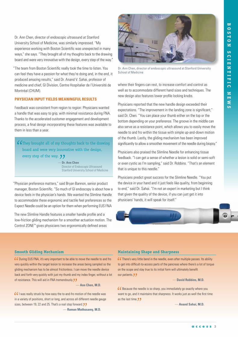
Dr. Ann Chen, director of endoscopic ultrasound at Stanford University School of Medicine, was similarly impressed. “My experience working with Boston Scientific was unexpected in many ways,” she says. “They brought all of my thoughts back to the drawing board and were very innovative with the design, every step of the way.”
“The team from Boston Scientific really took the time to listen. You can feel they have a passion for what they’re doing and, in the end, it produced amazing results,” said Dr. Anand V. Sahai, professor of medicine and chief, GI Division, Centre Hospitalier de l’Université de Montréal (CHUM).
PHYSICIAN INPUT YIELDS MEANINGFUL RESULTS
Feedback was consistent from region to region: Physicians wanted a handle that was easy to grip, with minimal resistance during FNA. Thanks to the accelerated customer engagement and development process, a final design incorporating these features was available to them in less than a year.
“Physician preference matters,” said Bryan Bannon, senior product manager, Boston Scientific. “So much of GI endoscopy is about how a device feels in the physician’s hands. We wanted the Slimline Handle to accommodate these ergonomic and tactile feel preferences so the Expect Needle could be an option for them when performing EUS FNA.”
The new Slimline Handle features a smaller handle profile and a low-friction gliding mechanism for a smoother actuation motion. The Control ZONE™ gives physicians two ergonomically defined areas
where their fingers can rest, to increase comfort and control as well as to accommodate different hand sizes and techniques. The new design also features lower profile locking knobs.
Physicians reported that the new handle design exceeded their expectations. “The improvement in the landing zone is significant,” said Dr. Chen. “You can place your thumb either on the top or the bottom depending on your preference. The groove in the middle can also serve as a resistance point, which allows you to easily move the needle to and fro within the tissue with simple up-and-down motion of the thumb. Lastly, the gliding mechanism has been improved significantly to allow a smoother movement of the needle during biopsy.”
Physicians also praised the Slimline Needle for enhancing tissue feedback. “I can get a sense of whether a lesion is solid or semi-soft or even cystic as I’m sampling,” said Dr. Robbins. “That’s an element that is unique to this needle.”
Physicians predict great success for the Slimline Needle. “You put the device in your hand and it just feels like quality, from beginning to end,” said Dr. Sahai. “I’m not an expert in marketing but I think that given the quality of the device, if you can just get it into physicians’ hands, it will speak for itself.”
They brought all of my thoughts back to the drawing
board and were very innovative with the design,
every step of the way. — Dr. Ann Chen
Director of Endoscopic Ultrasound Stanford University School of Medicine
Smooth Gliding Mechanism
During EUS FNA, it’s very important to be able to move the needle to and fro
very quickly within the target lesion to increase the areas being sampled so the
gliding mechanism has to be almost frictionless. I can move the needle device
back and forth very quickly with just my thumb and my index finger, without a lot
of resistance. This will aid in FNA tremendously.
— Ann Chen, M.D.
I was really struck by how easy the to-and-fro motion of the needle was
in a variety of positions, short or long, and across all different needle gauge
sizes, between 19, 22 and 25. That’s a real step forward.
— Raman Muthusamy, M.D.
Maintaining Shape and Sharpness
There’s very little bend in the needle, even after multiple passes. Its ability
to get into difficult-to-access parts of the pancreas where there’s a lot of torque
on the scope and stay true to its initial form will ultimately benefit
our patients.
— David Robbins, M.D.
Because the needle is so sharp, you immediately go exactly where you
want to go, and it maintains that sharpness. It works just as well the first time
as the last time.
— Anand Sahai, M.D.
Dr. Ann Chen, director of endoscopic ultrasound at Stanford University School of Medicine

BO
ST
ON
SC
IE
NT
IF
IC
NE
WS
Copies are available for health care providers from their Boston Scientific representative. Stent guides are also available at www.bostonscientific.com/endoscopy-resources.
Eating After Esophageal Stent Placement
Bo
sto
n S
cien
tifi
c, (
DFU
Tem
pla
te 5
.5in
x 8
.5in
), D
FU, B
utt
on
Pat
ien
t C
are
Gu
ide,
EN
, 907
2184
8-01
A
EndoVive™An Introduction
to Tube Feeding
with the
EndoVive Button
Gastrostomy Device
For patients undergoing endoscopic stent placement, there are often many questions about post-procedure care. As a way to help physicians respond to the growing inquiries from patients looking for information, Boston Scientific developed three guides for patients living with a gastrointestinal stent. The guides will provide physicians with additional resources with which to help educate patients as well as caregivers.
▲
Eating After Esophageal Stent Placement – Working in conjunction with a nutritionist from the University of Virginia, Boston Scientific developed a guide to provide information about diet and nutrition as well as recommendations on caring for an esophageal stent. The guide provides meal suggestions, tips and tricks for living with an esophageal stent (i.e., swallowing pills, maintaining weight, ways to avoid/reduce acid reflux, etc.) and contact information for additional third-party resources.
▲
A Guide for Patients Living with an Enteral Stent and
▲
A Guide for Patients Living with a Biliary Metal Stent – These guides are designed to help patients understand what a metal stent
is, and provide information about a stent placement procedure as well as questions to ask their physicians. Both the enteral and biliary stent guides include a plastic identification card that can be customized by the physician and staff, and is sized to fit in a wallet. The patient identification card includes information about magnetic-resonance compatibility for future medical treatment.
Patient care guides are available for patients living with a Boston Scientific tube feeding device.
▲
An Introduction to Tube Feeding with EndoVive™ Gastrostomy Tube and
▲
An Introduction to Tube Feeding with the EndoVive Button Gastrostomy Device serve as a general guideline for patients to help them with care and use of their device. These guides provide information on the basics of tube feeding as well as the necessary steps required to ensure their stoma site is clean and properly cared for. In addition, the guide provides problem solving tips as well as suggestions for general patient care such as managing common oral and stomach conditions.
▲
The Resolution™ Clip Device MR Conditional Patient Card is for patients to take home with them after receiving clip(s). The card is intended to identify the anatomical location and placement date of a clip or clips for the patient, physicians and MR technicians. The card acknowledges that the Resolution Clip Device is indicated as MR Conditional according to ASTM F2503 and patients with Resolution Clip Devices can be scanned following the guidelines listed on the card such as Static Magnetic Field Tesla and Spatial Gradient Field parameters. A copy of the card should be used in patient records as well as given to the patient as a personal pocket card for medical reference.
4 a c c e s s
Improving Outcomes through Patient Education
A Guide for Patients
Living with a
Biliary Metal Ste
ntA Guide for
Patients Living with
an Enteral Stent
Resolution ® Clip DeviceDate Placed:Anatomical Location:
Magnetic Resonance (MR) Information
Non-clinical testing has demonstrated the Resolution® Clip is MR Conditional according to
ASTM F2503. A patient with this clip(s) can be safely scanned under the following conditions:
Static Magnetic FieldStatic magnetic field of 1.5 and 3 Tesla with:
• Spatial gradient field of 2500 Gauss/cm (value extrapolated) and less
• Maximum whole body averaged specific absorption rate (SAR) of 2 W/kg in Normal Operating
Mode for a maximum scan time of 15 minutes of continuous scanning at 1.5T and at 3T.
MR Conditional per ASTM F2503

BO
ST
ON
SC
IE
NT
IF
IC
NE
WS
a c c e s s 5
Working with the National Pancreas Foundation to Provide Global Online Resources for Patients and Physicians
Boston Scientific is working with the National Pancreas Foundation to develop an interactive learning program for those seeking information on pancreatic disease states and procedures. The new program modules will be part of the foundation’s current website that works to provide easy-to-understand information for patients, and to enhance communication between patients and their physicians.
“Education is critical to helping patients manage the challenges of pancreatic disease and making informed medical decisions about their care,” said Dave Pierce, president of Boston Scientific Endoscopy. “We are proud to be leaders in helping create sections dedicated to EUS FNA and ERCP – two procedures that are playing an important role in helping diagnose pancreatic disease.”
Using a mix of animations, physician testimonials as well as downloadable information, the learning program will
help patients understand complex medical terminology, disease states, diagnostic testing, procedures and clinical implications. With easy web access, the website’s learning programs could serve as an educational resource for health care professionals working with patients in a practice setting to help explain indications for the procedures, risks associated with the procedures, and the benefits of diagnostic and therapeutic pancreatic endoscopy, EUS FNA and ERCP.
Boston Scientific is funding the project through an unrestricted education grant. The information will be co-hosted on www.pancreasfoundation.org and www.animatedpancreaspatient.com.
Education is critical to helping patients
manage the challenges of pancreatic
disease and making informed medical
decisions about their care. — Dave Pierce
President, Boston Scientific
Animations web tool help patients learn about pancreatic diseases.

BO
ST
ON
SC
IE
NT
IF
IC
NE
WS
Preceptorship Brings Master Class Training to Physicians in France
In order to meet the ever-increasing need for specialized endoscopy training, St. Joseph
Hospital in Marseille, France, implemented a preceptorship program to expand physicians’
skills in the areas of endoscopic retrograde cholangiopancreatography (ERCP) and
endoscopic ultrasound (EUS). Developed by Dr. Christian Boustière, the two-day training is
designed for physicians who are skilled endoscopists yet have limited experience in
pancreatico-biliary endoscopy.
Training in therapeutic
endoscopy and, more precisely,
in biliary and pancreatic
endoscopy requires rigorous
training and learning
which exceeds by far the
environment of academic
training in gastrointestinal
endoscopy. Creating
opportunities for continual
education such as this master
class is critical to furthering
this practice,
— Dr. Boustière
The first day includes a welcome for attendees and a review of patient cases to be treated. Each case is thoroughly reviewed, including pathologies, procedure indications and the types of devices to be used. Time is devoted for presentations, including videos to provide an update on technical innovations, guidelines or recent publications around major congresses related to the subject medical specialty. The day concludes with the schedule of cases for the next day and the pre-selected devices. The second day is fully dedicated to conducting procedures scheduled in two endoscopic rooms, one for ERCP and one for EUS. All procedures are performed under general anesthesia and intubation for ERCP.
WORKING ALONGSIDE EXPERTS
The expert starting the procedure is responsible for the case, and ensures the technique is performed adequately as discussed during the case presentation. Depending on the level of difficulty of the procedure, one of the trainees will perform all or part of the procedure under direct supervision of the expert who can regain control at any time if deemed necessary. The active participation of the trainee is a key element
of this training. It allows for trainees to test their competency levels, and at the same time improve their techniques with step-by-step guidance from the experts. Trainees benefit from the theoretical exchange as well as learning from physicians’ experiences. The variety of cases allows trainees to learn about the diagnostic and technical issues often encountered in everyday practice.
The sessions at St. Joseph Hospital are part of Boston Scientific’s preceptorship program that is designed to support ongoing education and training at select medical facilities. Training of this sort is possible in hospitals like St. Josephs where there is a high volume of endoscopic procedures, the facility is well equipped and physician expertise is readily available. Having received positive feedback from participants, the hospital plans to pursue additional master classes in ERCP and pancreatico-biliary EUS training with two additional sessions in 2014.
6 a c c e s s

BO
ST
ON
SC
IE
NT
IF
IC
NE
WS
a c c e s s 7
The development of endoscopy training at the Institute
for Advancing Science demonstrates Boston Scientific’s
continued commitment to providing industry-leading
training and education in partnership with physicians
and societies to further the practice of endoscopy.
— Paraic Curtis Vice President of Endoscopy
Europe, Boston Scientific
WallFlex™ Esophageal Stent Receives CE Mark in Europe
The WallFlex Esophageal Fully Covered Stent received CE Mark approval in Europe for the treatment of refractory benign esophageal strictures. The fully covered metal stent provides a new treatment option for patients with malignant and benign refractory esophageal strictures.
“The advantage of the WallFlex Esophageal Fully Covered Stent is its ease-of-use and now removability, which allows me to treat patients with refractory benign strictures,” said Peter D. Siersema, M.D., PH.D., professor of gastroenterology, head, Department of Gastroenterology and Hepatology at University Medical Center in Utrecht, The Netherlands.
Previously, the fully covered and partially covered WallFlex Esophageal Stents received CE Mark and FDA clearance for the palliative treatment of malignant esophageal strictures.
Endoscopy Training Now Part of the Paris Institute for Advancing Science
Endoscopy professional training and education are now part of the multi-disciplinary programs available at the Boston Scientific Institute for Advancing Science in Paris, France. The Institute offers world-class training facilities designed to meet the educational needs of endoscopists, endosonographers and pulmonologists.
Boston Scientific offers a broad curriculum of training courses from basic to advanced levels in a range of specialties and techniques including ERCP, EUS, EMR/ESD, cholangioscopy and bronchial thermoplasty.
“Hands-on training and peer-to-peer education are essential to maintaining good clinical practice and improving the quality of patient care. We hope that the endoscopy training at the Institute for Advancing Science will become an integral part of the training continuum of endoscopists and pulmonologists across Europe,” said Paraic Curtis, vice president of Endoscopy Europe, Boston Scientific.
The Institute is fully equipped to support integrated hands-on and didactic medical training in endoscopy. It contains two endoscopic towers, two endoscopic ultrasound processors, a SpyGlass™ Direct Visualization System, a full range of endoscopes, and numerous biological and non-biological models to recreate and simulate adapted clinical conditions for training and education. Physicians have access to a complete catheterization lab fully equipped with fluoroscopy capabilities. In addition, training can be conducted in multiple languages via the onsite simultaneous translation facilities.
Note: The WallFlex Esophageal Fully Covered Stent is not approved in the U.S. for the treatment of benign esophageal strictures.
The advantage of the WallFlex Esophageal Fully Covered Stent is its ease-of-use and now removability, which allows me to treat patients with refractory benign strictures. — Peter D. Siersema, M.D., Ph.D.
Professor of Gastroenterology

Look for Endoscopy Gurukul learning opportunities at conferences throughout India in 2014.
BO
ST
ON
SC
IE
NT
IF
IC
NE
WS
8 a c c e s s
AN INTERVIEW WITH Maria Stewart, director of Health Economics & Reimbursement, Boston Scientific, on changes impacting the U.S. health care industry.
Q
What do you see as the most pressing issue facing hospitals and physicians today?
Hospitals are being asked to improve quality of care and outcomes while demand is increasing and funding/payment is constrained. For example, health care reform is on the top of everyone’s mind. Hospitals are working to comply with all of the mandatory requirements associated with the value-based purchasing, hospital readmission reduction and hospital-acquired conditions programs. Hospitals must provide data on each of the specified parameters in order to receive their full payment update. In this environment, it will be critical for hospitals to increase efficiency while improving quality of care and patient satisfaction.
Q Can you tell us about the impending ICD-9 to ICD-10 coding transition?
In the United States, hospitals are preparing for the implementation of the International Classification of Disease (ICD-10) coding system. The current ICD-9 code set used to report medical diagnoses and inpatient procedures is expected to be replaced by ICD-10 in October 2014. This will be a major change for hospitals. They will be going from using a system with approximately 13,000 diagnoses codes to a system with 68,000 diagnosis codes and from approximately 3,000 inpatient procedure codes to 87,000. To help ensure accurate coding, payment and claims processing, hospitals will need to train staff and coordinate a transition plan with the support of the coding, clinical, IT and finance departments. Physicians and their staff members will also need to familiarize themselves with new ICD-10 diagnosis coding. If the transition to ICD-10 is not properly implemented, they could have denied claims, delays in processing and payments, etc.
Q How is the Boston Scientific Endoscopy Health Economics & Reimbursement team supporting
its health care partners in this changing environment?
Boston Scientific Endoscopy’s Health Economics & Reimbursement team is actively leading efforts to advocate for appropriate coverage and payment for GI and pulmonary endoscopy procedures, which we believe is critical if patients are to have access to all relevant treatment options. In addition, through discussions with hospital administrators, we have learned that education for hospital staff on the changes associated with health care reform is needed. To support the education of our healthcare partners, the Boston Scientific Health Economics & Reimbursement Endoscopy team annually sponsors a complimentary reimbursement webinar during which an expert in the field reviews the impact of health reform and upcoming changes in coding and payment. This webinar receives positive reviews and draws approximately 300 attendees each year. In addition, the team provides onsite education for hospitals, ambulatory surgery centers and physicians on these important topics.
With respect to the transition to ICD-10, Boston Scientific has worked with coding experts to develop an ICD-10 educational webinar and coding cross-reference tools. These tools will be available on the Boston Scientific website, and will be an additional resource to our health care partners as they transition from the ICD-9-CM to ICD-10 coding system.
Q How can a customer reach out to request reimbursement information or education?
To request information on reimbursement, U.S. customers may contact the Endoscopy Reimbursement Helpdesk at 1-800-876-9960 ext. 54145 or via email at: [email protected].
Since its inception in 2011, the Boston Scientific School of Endoscopy in India, known as Endoscopy Gurukul, has quickly gained momentum and established itself as a center of expertise for gastrointestinal (GI) education and training.
More than 800 medical professionals have registered with Endoscopy Gurukul to take advantage of its many learning opportunities. The school offers a variety of ways to learn, including hands on, procedural video libraries at conferences, paramedical staff training for skill enhancement, patient education and more.
“Endoscopy Gurukul is an innovative initiative from Boston Scientific that has completely changed the landscape of training endoscopists in India.”
– Dr. D. Nageshwar Reddy, chief of gastroenterology and therapeutic endoscopy at the Asian Institute of Gastroenterology, Hyderabad, India.
“The Endoscopy Gurukul is a highly positive step taken by Boston Scientific to enhance endoscopic education in our country.”
– Dr. Amit Maydeo, chairman, Baldota Institute of Digestive Sciences, Global Hospitals, Mumbai, Maharashtra
Left to right: Dr. T.S. Chandrasekar, MedIndia Hospitals (Chennai) moderates a discussion on ERCP with Dr. Randhir Sud, Medanta Institute of Digestive & Hepatobiliary Sciences (Gurgaon); Dr. D. Nageshwar Reddy; and Dr. Amit Maydeo at the Mumbai Live Endoscopy 2013 conference.
Update on Endoscopy Gurukul

BO
ST
ON
SC
IE
NT
IF
IC
NE
WS
a c c e s s 9
This 1.28 megawatt solar energy system will generate an average of 1,685,000 kilowatt hours a year, approximately 25% of the facility’s energy needs, or enough power to serve 145 average-sized American homes a year. This facility, located in Quincy, Massachusetts, is home to Boston Scientific’s international distribution center, which distributes more than 15.8 million medical device units per year to all 50 U.S. states and 46 countries.
Boston Scientific Green Initiatives – Making a Difference
As members of Practice Greenhealth, Boston Scientific is working to leverage the
information, education and tools available on the best environmental practices for health care organizations as a way to meet the needs of its customers. Here are some of Boston Scientific’s accomplishments and ongoing initiatives:
� Incorporating LEED standards into the
construction of new buildings and modification of
existing facilities – seven facilities LEED certified.
� Achieving ISO14001 certification – Certified
at all 14 major manufacturing and distribution
centers worldwide.
� Leveraging solar – A 1.28 megawatt solar energy
system will generate an average of 1,685,000
kilowatt hours a year, approximately 25 percent
of the energy needs for Boston Scientific’s
international distribution center in Quincy, Mass.
� Radial Jaw™ 4 Biopsy Forceps – Plastic packaging was made 13 percent
smaller; a box of 5 was made smaller by 35 percent. The new packaging requires
less shelf space (between 24-36% less) – helping decrease the task of restocking
and improve efficiencies when moving product from the stock room to the
procedure room. In addition, these packaging changes are expected to result in:
• A reduction in overall packaging materials by 42,452 pounds per year
• 83 fewer trucks moving product from New York to the Boston Scientific
distribution center in Quincy, Massachusetts
• 1,653 fewer pallets per year moving from Costa Rica to the Massachusetts
distribution center
� Advanix™ Biliary Stent with NaviFlex™ RX Delivery System – Reduced by
28 percent. The new fully recyclable packaging uses 23 percent less material than
the previous package.
� CRE™ Dilation Balloon – New packaging is made of fully recyclable REC-2-0
high-density polyethylene.
ENERGY AND SUSTAINABILITY
PRODUCT PACKAGING
Close the Gap Initiative Works to Improve Patient Education and Access to Care
Boston Scientific’s Close the Gap is
an ongoing program focused on initiatives to
eliminate treatment disparities in high-risk,
underserved patient populations suffering
from gastrointestinal and pulmonary diseases
by increasing awareness, sponsoring and
developing educational programs, and
improving access to care. Since its inception
in 2013, the team has worked to raise
awareness for colorectal and pancreatic
cancers through a variety of awareness-
generating and fund raising activities. The team
raised $20,000 that is being used to fund
the Colon Cancer Alliance’s new colorectal
cancer Screening Assistance Program and
more than $35,000 for The Lustgarten
Foundation’s pancreatic cancer research
initiatives. Close the Gap is also the primary
sponsor of the
Colon Cancer
Prevention
Project’s
pledge
program.
To learn more about how to get involved with colorectal and pancreatic cancer awareness activities, please visit our partners’ websites:
� The Colon Cancer Alliance: www.ccalliance.org
� The Colon Cancer Prevention Project: www.coloncancerpreventionproject.org
� The Lustgarten Foundation: www.lustgarten.org
Colon Cancer Risk Assessment
Colorectal cancer affects an estimated 1 in 20 Americans.
Take the Colorectal Cancer Risk Assessment Quiz to find out.
WHAT’S YOUR LEVEL OF RISK?
Boston Scientific has been certified to the FTSE4Good Corporate Social Responsibility index since 2004. An investment index managed by the London Stock Exchange, it measures the performance of companies that meet globally recognized standards of corporate responsibility.

GA
ST
RO
EN
TE
RO
LO
GY
CASE PRESENTED BY:
ANAND V. SAHAI, M.D., MSC (EPID), FRCPC
Professor of MedicineChief, GI Division, Centre Hospitalier de l’Université de Montréal, CANADA
A Challenging Case of Endoscopic Ultrasound Fine-Needle Aspiration
PATIENT HISTORY
A 72-year-old female presented with obstructive jaundice due to a proximal bile duct lesion suspicious for cholangiocarcinoma upon magnetic resonance cholangiopancreatography. An endoscopic ultrasound (EUS) was requested to clarify the nature of the lesion and to perform fine needle aspiration (FNA), if possible, since the patient was not a surgical candidate.
PROCEDURE AND TECHNIQUE
To visualize and biopsy hilar lesions, the echoendoscope must often be placed in a long scope position in the duodenal bulb with significant counter-clockwise torque. In this position, it may be very difficult to perform EUS-FNA because the needle may be difficult to insert into the scope, the endosonographer may be in an uncomfortable posture, and the needle may be hard to move. In this case, the lesion was particularly small. Therefore, very precise targeting was also required.
In this case, the Expect™ Slimline (SL) Endoscopic Ultrasound Aspiration Needle performed extremely well. After identifying the lesion and site to biopsy (Figure 1), the echoendoscope was withdrawn into the stomach in a more straight position. The 25g Expect SL Needle was then inserted into the operating channel and locked in place (Figure 2). (We recommend always inserting the needle into the scope when the scope is straight; this makes insertion easier and avoids trauma to the operating channel.)
The echoendoscope was then re-advanced into the bulb and the lesion was relocated. Despite the long scope position and extreme counter-clockwise torque required, the needle exited easily from the operating channel (Figures 3 and 4). The needle was easily visible, and its sharpness made penetration of the duodenal wall exceptionally easy. The Expect Slimline FNA Needle also provides a very precise tactile response. As a result, we were able to precisely target the small, intra-ductal stricture while avoiding vascular structures posterior to the duct.
OUTCOME AND CONCLUSION
As is often the case, the first pass was positive for adenocarcinoma and there were no complications. This challenging case highlights how the ease of actuation, visibility, sharpness and exceptional tactile feel of the Expect-SL needle can help ensure success in cases of EUS-FNA of small lesions with difficult scope position.
Dr. Sahai’s practice has been based
solely on EUS since 1996. The Center
Hospital, University of Montréal is
one of the busiest EUS centers in the
world, conducting approximately
2,800 cases per year and 12-14 cases
per day. Since 2000, Dr. Sahai has
conducted EUS tutorials for more than
500 physician attendees.
1 2 3 4
10 a c c e s s

GA
ST
RO
EN
TE
RO
LO
GY
EUS FNA-based Diagnosis of a Granular-cell Tumor in the Bile Duct
PATIENT HISTORY
This patient is a 24-year-old male nursing student with recurrent episodes of abdominal pain during a one-year period. In one of these episodes, he went to the emergency room of another hospital where they detected a bilirubin level of 4 mg/dL together with a high level of hepatic enzymes. The abdominal ultrasound performed showed a gallbladder with cholesterolosis without stones and a normal-sized biliary tree (Figures 1a and 1b).
The patient was referred to his regular specialist for further study. During a hospital examination, a doctor discovered conjunctival jaundice. He underwent magnetic resonance imaging that showed dilation of the intrahepatic bile duct secondary to a short stenosis of 7mm at the common hepatic duct that was suspicious for malignancy (Figures 2 and 3). The patient was referred for an endoscopic ultrasound (EUS) examination.
PROCEDURE
Under deep propofol sedation controlled by the endoscopist, EUS was performed showing dilation of the intrahepatic biliary duct. There was no presence of stones in the gallbladder and the pancreas was normal. At the level of the common bile duct (CBD) a regular thickening of the bile duct wall of a length of 13mm and 7mm was observed (Figures 1a and 1b). The distal area of the bile duct was normal. No lymphadenopathy or local hepatic lesions were observed.
A fine-needle aspiration (FNA) was performed using a 25 gauge Expect™ Slimline (SL) Needle (two passes), having previously administered a prophylactic intravenous dose of ciprofloxacino 400mg.
The cytology (Figure 4) showed the presence of a benign proliferative lesion of mesenchymal lineage consisting of sheets and plates of large round cells with granular eosinophilic cytoplasm and hyper-
chromatic oval eccentric nuclei. The biliary epithelium shows reactive hyperplasia papillary changes. No atypia, mitosis or malignancy signs were observed and an immunohistochemical study showed granular cytoplasmic positivity for S-100 protein. The cytologic findings were consistent with the existence of a granular-cell tumor.
SUMMARY AND DISCUSSION
A granular-cell tumor, or Abrikssoff’s tumor, is a benign mesenchymal tumor that is frequently located in the skeletal muscle of the tongue, skin and subcutaneous tissue. It originates in Schwann cells as evidenced by histological and immunohistochemical test results. Its location at the level of the biliary tract is rare, with only a few dozen cases reported in literature and this being the first case of cytologic diagnosis by EUS FNA.
The preoperative diagnosis is difficult because benign tumors of the biliary tract are rare and usually not taken into account in the differential diagnosis of patients with obstructive jaundice. Brush cytology obtained by ERCP is rarely diagnostic because the tumor originates from the wall thickness of the common bile duct and not in the biliary epithelium. Most patients are young, with an average age of 34 years, and more often females of African heritage.
The ability of EUS FNA to properly evaluate the biliary system and the high performance of FNA in the context of biliary strictures make it an ideal and safe method to evaluate patients with obstructive jaundice. The 25 gauge Expect SL Needle is especially useful for puncturing small lesions and transduodenal use. The excellent ultrasound visualization and low deformability make the Expect™ SL Needle a very useful tool to puncture inaccessible lesions in which the scope is in a forced position.
References:
1) Saito J et al. Granular cell tumor of the common bile duct: a Japanese case. World J Gastroenterol 2012; 18: 6324-7.
2) Fritscher-Ravens A et al. EUS-guided fine-needle aspiration of suspected hilar cholangiocarcinoma in potentially operable patients with negative brush cytology. Am J Gastroenterol 2004; 99:45-51.
3) El Chafic AH et al. Impact of preoperative endoscopic ultrasound-guided fine needle aspiration on postoperative recurrence and survival in cholangiocarcinoma patients. Endoscopy 2013; 45:883-9.
CASE PRESENTED BY:
JOSÉ RAMÓN APARICIO, M.D.
Endoscopy UnitHospital General Universitario de AlicanteAlicante, SPAIN
1a 1b 2 3 4
a c c e s s 11

GA
ST
RO
EN
TE
RO
LO
GY
Bilateral Biliary Self-expanding Metal Stenting as Treatment for a Klatskin Tumor
CASE PRESENTED BY:
PROFESSOR E.D. FEDOROV, E.V. IVANOVA, S.A. BUDZINSKIY, K.B. LUMMER, O.A. SHCHIPKOV, E.V. GORBACHEV
Russia State Medical UniversityN.I. Pirogov, Moscow University HospitalMedical Rehabilitation CenterMoscow, RUSSIA
PATIENT HISTORY
A 78-year-old female was admitted to the emergency room with complaints of jaundice, fever, skin itch and dark urine for about ten days. Laboratory data showed a total bilirubin of 12.9 mg/dl, and conjugated bilirubin 6.3 mg/dl. A CT scan and ultrasonography showed a mass lesion at the confluence of the right and left hepatic bile ducts (Bismuth-IVC). Four months prior to admission, she underwent a percutaneous transhepatic cholangiography endoscopic retrograde cholangiopancreatography (ERCP) using the rendezvous cannulation technique (Figure 1) for biliary plastic stent placement into the right hepatic duct, followed by nasobiliary drainage for reactive cholangitis, and subsequent (on the 10th day) retrograde bilateral stenting with two plastic stents.
PROCEDURE
During the ERCP, a partial obstruction of one stent and complete obstruction of the other stent was revealed as well as signs of cholangitis. The plastic stents were removed. A Dreamwire™ High Performance Guidewire 0.035" was placed via the working channel of a TJF-180 scope into the right duct, while successful placement of the second Dreamwire Guidewire into the left duct required use of an Autotome™ RX 44 Cannulating Sphincterotome. Using rotating capabilities of the Autotome Sphincterotome, we were able to
navigate the tip of the sphincterotome in the common bile duct, found the orifice of the left hepatic duct and selectively cannulated the left hepatic ducts by advancing the guidewire. Using the RX Locking Device, both guidewires were fixed in right and left hepatic ducts (Figure 2) and the sphincterotome was separated and removed. We then performed a subsequent bilateral passage of fully uncovered metal stents (WallFlex™ Biliary RX Stent 8x100mm and WALLSTENT™ RX Biliary Endoprosthesis Stent System 8x100mm) into the left and right biliary ducts (Figures 3, 4 and 5) followed by elimination of multiple stones and sludge from the left hepatic ducts.
POST-PROCEDURE
Within a short time, the patient improved and was discharged from the hospital on the fourth day.
DISCUSSION
This case represents the possibilities of endoscopic therapy using these types of devices to treat a difficult cholangiocarcinoma (Bismuth-IVC) situation. Gentle placement of self-expanding metal stents allows for adequate duct drainage, which plays a great role in effective palliation of a malignant biliary obstruction.
NOTE: Use of the WallFlex Biliary RX Fully Covered Stent for the treatment of benign strictures or stenoses has not been cleared for use in the United States.
WARNING: The safety and effectiveness of the WallFlex Biliary Stent for use in the vascular system has not been established.
3
4
21
5
12 a c c e s s

GA
ST
RO
EN
TE
RO
LO
GY
CASE PRESENTED BY:
ASHLEY T. EVANS, M.D.
Digestive Health ClinicBoise, Idaho, USA
Stenting Serves as Bridge to Surgery and Allows for Non-emergent Resection of a Malignant Colonic Neoplasm
References:
1) Leitman IM, Sullivan JD, Brams D, DeCosse JJ. Multivariate analysis of morbidity and mortality from the initial surgical management of obstruction carcinoma of the colon. Surg Gynecol Obstet 1992; 174:513.
2) Fielding LP, Wells BW. Survival after primary and after stage resection for large bowel obstruction caused by cancer. Br J Surg 1974; 61:16.
3) Targownik LE , Spiegel BM, Sack J, et al. Colonic stent vs emergency surgery for management of acute left-sided malignant colonic obstruction: a decision analysis. Gastrointes Endosc 2004;60:865.
PATIENT HISTORY
A 62-year-old man was diagnosed with a nearly obstructing rectal mass after presenting with weight loss, tenesmus and rectal bleeding. A CT scan and biopsy of a liver lesion confirmed stage-IV disease. He received five months of palliative radiation and chemotherapy with plans to proceed with a low anterior resection once chemotherapy was optimized. Unfortunately, he presented to the hospital with nausea, vomiting, abdominal pain and distension six days before Christmas. A CT scan showed distal colonic obstruction. A flexible sigmoidoscopy with enteral stent placement was planned to relieve the obstruction.
PROCEDURE
Upon intubation of the rectum with the sigmoidoscope, an obstructing rectal mass was encountered at 8cm (Figure 1). With insufflation, a pinpoint lumen was visible within the mass with a small amount of air and liquid stool passing from above (Figure 2). A cannula was used to guide a .035 x 450 Hydra Jagwire™ Guidewire into the lumen, and under fluoroscopic guidance, advance across the stricture to the upstream bowel (Figure 3). Using a felt-tipped marker, the fluoroscopic image was marked to delineate the proximal and distal edges of the obstructing mass. A 25mm x 90mm through-the-scope (TTS) WallFlex™ Colonic Stent was passed over the guidewire and the on-screen markings were used to guide the ideal placement of the stent. The stent was easily deployed and upstream air and bowel contents immediately began flowing across the stricture (Figure 4). A fluoroscopic waist was visualized with good placement of the stent across the proximal and distal margins of the tumor (Figure 5).
OUTCOME / POST-PROCEDURE
The patient tolerated the procedure well and had complete relief of his obstructive symptoms, passing multiple bowel movements within hours afterwards. He felt well enough that he asked for surgery to be delayed until after Christmas so he could spend the holiday with his family. He was discharged home on a low-residue diet. Three weeks later, the patient completed a cleansing bowel prep and non-emergent, low anterior resection with diverting ileostomy was successfully performed.
CONCLUSION
Colonic stents are an incredibly useful tool for both palliation of advanced disease and preoperative decompression. In this case, successful placement of an enteral stent across a malignant colonic stricture allowed for non-emergent resection of this patient’s tumor in a prepped bowel. In addition, decompression of the bowel allowed surgery to be postponed several weeks so that the patient could spend the holidays with his family.
There are several advantages to preoperative placement of expandable metal stents in the management of colonic obstruction. First, morbidity and mortality have been shown to be substantially lower in patients whose colonic resection can be performed on an elective basis1. In addition, patients with a primary anastomosis appear to have higher survival rates than those with emergent diverting colostomies2. It also seems that colonic stent insertion followed by elective surgery is more effective and less costly than emergent surgery3. In this patient with stage IV rectal cancer, improved quality of life and time with family was also an important benefit of placement of a colonic stent.
3
4
2
1
5
a c c e s s 13

GA
ST
RO
EN
TE
RO
LO
GY CASE PRESENTED BY:
ANTONIETTA LAMAZZA, M.D.
University of Rome La SapienzaPoliclinico Umberto IDepartment of Surgery “P. Valdoni” Digestive Endoscopy UnitRome, ITALY
A Clinical Case of a Long-term Colonic Endoscopic Stent Placement Without Complications
PATIENT HISTORY
A 74-year-old female underwent surgery in August 2005 for a left hemicolectomy caused by extrinsic neoplastic stenosis of the rectum caused by ovarian cancer for which she had undergone surgery a few years prior. In November 2008, after chemotherapy, she developed an intestinal obstruction by a new ab-extrinsic neoplastic stenosis of the colorectal anastomosis, as evidenced by an abdominal CT scan. A colonoscopy confirmed the presence of a neoplastic stenosis of the colorectal anastomosis, approximately 10-14cm from the anal verge.
A self-expanding uncovered WallFlex™ Single-Use Colonic Stent System (length 90mm - 25mm diameter) was placed that led to a complete resolution of the clinical obstruction. The stent remained patent for several years (Figure 1) and through several cycles of chemotherapy. In June 2013, after rectal bleeding, a colonoscopy showed stent neoplastic ingrowth; biopsies were then performed on the mucosa inside the stent that showed neoplastic epithelial proliferation, compatible with the diagnosis of colonic localization originating from ovarian cancer (Figure 2).
A CT scan confirmed the relapse of massive neoplastic disease, due to the presence of peritoneal carcinomatosis and liver metastases. It also showed a rectal metallic stent incorporated in the neoplastic tissue. Another colonoscopy was performed for the treatment of the neoplastic tissue within the stent using argon plasma coagulation (APC) (Figures 3 and 4).
Currently, the patient is undergoing chemotherapy without clinical signs of colonic obstruction or rectal bleeding, and her general condition is fair. The next endoscopic exam is scheduled for three months after the treatment with APC (Figure 5).
PROCEDURE
In order to treat the anastomotic stenosis, a self-expanding metal uncovered stent (WallFlex length 90mm, diameter 25mm) was used. The proper study of the stenosis occurred by performing a colonoscopy using a pediatric nasogastroscope exclusively designed for this procedure.
The pediatric nasogastroscope allowed us to traverse the stenosis, measure the length of the lesion, and helped to reduce the angle, thereby allowing the colonoscopy in the upstream part; it also allowed the positioning of an Amplatz Super Stiff™ Guidewire 70cm above, under direct endoscopic vision and radiologic control.
OUTCOME / POST-PROCEDURE
The outcome of our treatment was the recanalization of the rectum without performing the definitive colostomy, otherwise indispensable, with the risk of neoplastic cutaneous implants due to the massive peritoneal carcinomatosis. From initial stent placement until June 2013, no immediate or late complications were observed, up to and including intestinal disorders.
CONCLUSION
The interest of the clinical case described is in the long-term stent placement (five years), the absence of immediate and late complications (from November 2008 to June 2013), and the endoscopic APC treatment to resolve neoplastic infiltration within the stent (June 2013 – first late complication).
The choice of an uncovered stent has proved successful because it allowed the stent’s placement in the neoplastic stenosis without its dislocation, and when the slow growth of neoplastic tissue occurred (after five years), it was treated with a minimally invasive endoscopic technique (APC).
In our experience, the clinical case described demonstrates an optimal synergy among endoscopist, surgeon and oncologist. The choice of treatment favoring endoscopy and not surgery, the chemotherapy treatment and the biology of the tumor enabled palliation of the obstruction. The patient lived for about five years in fairly good general condition. Not having a permanent colostomy was a positive factor in the patient’s quality of life, both clinically and psychologically.
3
4
2
1
5
14 a c c e s s

GA
ST
RO
EN
TE
RO
LO
GY
PATIENT HISTORY
A 50-year-old man with hypertension and hypercholesterolemia was seen by a primary care physician in March 2012 for upper-mid abdominal pain radiating to the back. His laboratory workup was normal and he was treated for constipation, which relieved his symptoms partially. A CT scan showed his pancreas was atrophic with a dilated pancreatic duct from the neck distally toward the tail, with dilated side branches but a normal duct in the head. No obvious mass or cyst was noted and the rest of the CT was unremarkable.
The patient was referred to us for further management. He denied any past history of pancreatitis or similar abdominal pain, was completely asymptomatic and denied any weight changes or appetite loss. The laboratory work up revealed a normal hemogram, normal renal and liver function tests, amylase 49 IU/L, lipase 29 IU/L, IgG4 level 23 mg/dL, and CA19-9 < 0.8 U/ml. An MRCP was obtained which confirmed the CT findings and did not reveal any mass/cyst.
PROCEDURE
An ERCP showed a fish-mouthed ampulla (Figure 1), but no mucus was seen extruding from it. Nevertheless, it raised concern for a possible intraductal papillary mucinous neoplasm (IPMN). The pancreaticogram showed the dilated pancreatic duct and side branches with extensive filling material suspicious of mucus (Figure 2). Pancreatic fluid was aspirated for molecular analysis and carcinoembryonic antigen (CEA). A SpyScope™ Access and Delivery Catheter was used to access the pancreatic duct (Figure 3), revealing very thick globs of mucus . Multiple random biopsies were obtained using the SpyBite™ Biopsy Forceps (Figure 4 ).
OUTCOME
The pancreatic fluid CEA level was < 5 ng/ml with high DNA quantity. The SpyBite Biopsy Forceps pathology raised suspicion of dysplasia (Figure 5) and the patient was referred for surgery. Based on the above results and the fact that < 1.5cm of the normal pancreatic duct was seen in the head, the patient was deemed not a suitable candidate for subtotal pancreatectomy. He underwent a total pancreatectomy with splenectomy in May 2012 and did well post-operatively. The pathology of the resected tissue confirmed a focal invasive mucinous adenocarcinoma arising in the background of IPMN with extensive high-grade dysplasia.
The tumor size could not be assessed but pathological staging of a T1 tumor was ascertained based on the fact that the tumor was limited to the pancreas (with the invasive portion being < 2cm with negative margins absent lymphovascular or perineural invasion) there were 29 negative lymph nodes and there was no distant metastasis. The patient was then started on Gemcitabine chemotherapy which he did not tolerate beyond one cycle. Since then he is being followed by strict surveillance with a positron emission tomography/CT protocol and has remained metastasis and/or recurrence free.
CONCLUSION
This unique case illustrates the importance of the SpyGlass™ Direct Visualization System examination of highly suspicious pancreatic ductal abnormalities. With the use of SpyGlass System technology, we were able to detect pancreatic cancer at a stage when it was non-metastatic and operable, and when the patient’s overall life expectancy would be greater than otherwise expected with late-detected pancreatic adenocarcinoma. Therefore, we strongly suggest use of this available tool in detailed examination of abnormal pancreatic ducts.
1 2 3 4 5
a c c e s s 15
Early Detection of Pancreatic Cancer Nidus in a Patient with Inconspicuous IPMN Lesion
RAYBURN F. REGO, M.D.
Director of Advanced Endoscopy and Associate Professor of MedicineDivision of Gastroenterology and Hepatology Department of MedicineUniversity of Arkansas for Medical Sciences (UAMS)
Little Rock, Arkansas, USA
CASE PRESENTED BY:
MOHIT GIROTRA, M.D.
Gastroenterology and Hepatology FellowDivision of Gastroenterology and Hepatology Department of MedicineUniversity of Arkansas for Medical Sciences (UAMS)
Little Rock, Arkansas, USA

GA
ST
RO
EN
TE
RO
LO
GY
CASE PRESENTED BY:
LUIGI GHEZZO, M.D.
Santi Croce e Carle Hospital, CuneoPiemonte, ITALY
Usefulness of the SpyGlass Direct Visualization System for Difficult Stone Management
PATIENT HISTORY
In this case, we treated a 60-year-old male with a 13mm stone that clogged his cystic duct after a cholecystectomy (Figure 1). In another hospital, the patient underwent three previous endoscopic retrograde cholangiopancreatography (ERCP) procedures. During the first ERCP, physicians performed a sphincterotomy and partially removed the stones. They then placed a plastic stent. He underwent a cholecystectomy (without stone removal due to a thin common bile duct caliber) and a second ERCP that detected a big stone that was difficult to remove using a basket or extraction balloon. The patient again complained of cholangitis, so he underwent a third ERCP without success.
PROCEDURE
In December 2013, the patient was assigned to our hospital and we performed an ERCP with the SpyGlass™ Direct Visualization System and holmium laser in order to destroy the stone under direct vision and remove it. After cholangiography and removal of the plastic stent, we confirmed the presence of two stones; the biggest one (13mm) was in a difficult position, clogging the cystic duct, without proximal common bile duct dilation or inflammation.
We introduced the SpyGlass System over-the-wire that easily allowed direct visualization of the stone (Figure 2). Using the SpyGlass System, we were able to perform holmium laser therapy under vision. This allowed us to obtain a partial stone fragmentation (Figure. 3), making it easier for us to remove the fragments later with a basket (Figures 4 and 5).
CONCLUSION AND PATIENT OUTCOME
During choledocholithiasis procedures, there are often difficulties due to the number and position of stones. In these types of cases, it is not possible to remove the stones using only the standard techniques (sphincterotomy and extraction devices).
Sometimes it is not easy to capture the stone with a basket so we use external lithotripsy (extracorporeal shock wave lithotripsy); however, this technique can also be influenced by the position of the stone or the presence of fluid around the stones. These factors may decrease the wave’s energy. A good option can be the use of electrohydraulic lithotripsy or laser in order to apply the energy directly on the stone’s surface.
By using the SpyGlass Direct Visualization System, holmium laser can be applied directly on the stones. In this way, it is easier to destroy and remove the stones, managing difficult choledocholithiasis procedures in the best way.
Thanks to the SpyGlass System in combination with holmium laser, we were able to easily treat the patient and completely remove the stones. The final cholangiography showed the results (Figure 6). The patient was discharged from the hospital the day after the procedure.
3 6
2
1 4
5
16 a c c e s s

GA
ST
RO
EN
TE
RO
LO
GY
3 421 5
a c c e s s 17
PATIENT HISTORY
A 56-year-old female with a history of laryngeal cancer (status post-neck radiation and total laryngectomy approximately 20 years prior), presented with complaints of dysphagia and was found to have a severe proximal esophagus radiation-induced stricture. A percutaneous radiological gastrostomy (PRG) was subsequently placed to improve nutrition.
Over the course of 17 months, the patient underwent 18 esopha-gogastroduodenoscopies (EGD) with Tucker dilation via the gastrostomy up to 38 Fr. Successful Savary dilation was eventually achieved and Tucker dilation was no longer required. The PRG was removed 10 months after placement. Fourteen days after PRG removal the patient was noted to have persistent leakage from a gastrocutaneous fistula (GCF) at prior gastrostomy (Figure 1). The EGD revealed a hole in the anterior wall of the stomach, approximating 8mm (Figure 2). Attempted closure of the fistula using internal and external interrupted silk sutures was unsuccessful.
PROCEDURE
An EGD with endoscopic clip placement using one Resolution™ Clip to approximate the margins of the fistulous tract was performed. A decreased fistula output was noted after clip placement but leakage persisted. An EGD was repeated one week later with placement of five endoscopic clips (Figure 3) resulting in immediate closure of the gastrocutaneous fistula.
OUTCOME
Endoscopic evaluation four months after endoscopic clip placement revealed four clips in place and persistent closure of the GCF (Figure 4). A repeat EGD 16 months after endoscopic clip placement revealed one clip in place with a well-healed gastric mucosa (Figure 5).
DISCUSSION
Refractory gastrocutaneous fistulas are a known consequence following gastrostomy removal that can be very difficult to manage. Conservative management of GCFs has had limited success. Surgical laparotomy and excision of the fistulous tract is often required but results in increased morbidity and mortality, especially in debilitated patients.
Endoscopic clipping of GCFs involves grasping the mucosal and submucosal tissue layers at the fistulous tract edges and approximating the opposite walls, thus leading to macroscopic closure of the fistula opening. This case illustrates that endoscopic clip placement is a valuable modality for closing refractory gastrocutaneous fistulas and is a promising alternative to surgery.
PATRICK BRADY, M.D.
Division of Digestive DiseasesUniversity of South FloridaTampa, FL, USA
Endoscopic Clip Closure of a Gastrocutaneous Fistula in a Patient with History of Tucker Dilation via Gastrostomy
CASE PRESENTED BY:
KIMBERLY J. KOLKHORST, D.O.
Division of Digestive DiseasesUniversity of South FloridaTampa, FL, USA

GA
ST
RO
EN
TE
RO
LO
GY
CASE PRESENTED BY:
ERIC C.S. LAM, M.D., FRCPC
Clinical Associate ProfessorDivision of GastroenterologyUniversity of British ColumbiaVancouver, British Columbia, CANADA
Resolution Clip Devices Used to Close Duodenal Perforation after Endoscopic Submucosal Dissection
PATIENT HISTORY
A 56-year-old man was found to have a 10mm subepithelial lesion in the junction between the first and second part of the duodenum when undergoing an upper endoscopy to screen for Barrett’s esophagus. He had a history of diabetes mellitus that was controlled with oral hypoglycemic agents. A radial endoscopic ultrasound was performed showing a 10mm hypoechoic lesion in the submucosa consistent with a duodenal carcinoid (Figures 1 and 2). Endoscopic biopsies confirmed carcinoid tumor cells. Because of the potential risk of metastatic disease and the possibility of requiring a more extensive resection, he was referred for endoscopic submucosal dissection.
PROCEDURE
The border of the endoscopic bulge in the duodenum was marked by applying superficial cautery to the mucosal surface. A solution of 1:10,000 epinephrine and methylene blue was injected into the submucosa. Using cap assistance, a submucosal dissection was carried out around the carcinoid tumor. Upon dissecting the proximal end of the carcinoid tumor, a 5mm duodenal perforation was identified (Figure 3).
The Sensation™ Single-Use 30mm Oval Polypectomy Snare was then used to resect the carcinoid tumor immediately after the perforation was identified. Unfortunately, the intraluminal air began to enter the peritoneal space and full distension of the duodenum could not be achieved. We then applied two Resolution™ Clip devices to the 5mm duodenal perforation and this re-established retention of intraluminal air. An additional five Resolution Clip devices were then used to close the remaining 2.5cm mucosal defect (Figure 4).
OUTCOME AND POST-PROCEDURE
The pathology of the removed lesion showed complete excision of the carcinoid tumor. The patient was discharged two days post procedure. Repeat upper endoscopy three months after the resection showed a well epithelialized scar with no recurrence of tumor (Figure 5).
This case demonstrates the usefulness of Resolution Clip Devices in the closure of luminal perforations of the duodenum. The initial two Resolution Clip Devices with their wide jaws and closing pressure were able to take the opposing sides of the luminal perforation and to provide an adequate seal. By utilizing the Resolution Clip to close the perforation during the endoscopy, the patient was able to avoid surgery.
51 2 3 4
18 a c c e s s

GA
ST
RO
EN
TE
RO
LO
GY
PATIENT HISTORY AND ASSESSMENT
A 64-year-old female with serrated polyposis syndrome underwent a colonoscopy for endoscopic resection of the large proximal colon sessile serrated adenomas. This was her second colonoscopy for clearance of serrated lesions. Indigocarmine dyespray cap-fitted colonoscopy was used to facilitate detection, and a large sessile serrated adenoma/polyp (SSA/P) was identified at the hepatic flexure (Paris classification 0-IIa, 25mm, Figure 1).
PROCEDURE
Colonoscopic resection of the 25mm sessile serrated adenoma at the hepatic flexure was performed. An en bloc resection was attempted (Figure 2) following a submucosal injection of succinylated gelatin (Gelofusine) mixed with indigocarmine (80mg in 500mL solution), and using electrocautery (ERBE Vio 300D, EndocutQ effect 3). Carbon dioxide is used routinely for insufflation. After resection, a large concentric defect (approximately 10-15mm) in the muscularis propria was evident (Figure 3, mirror target sign), with corresponding target sign on the resected specimen (Figure 4, resected specimen ex-vivo, with a central white disk of muscularis propria surrounded by indigocarmine-stained blue submucosal tissue).
The defect was closed using Resolution™ Clip Devices (Figures 5
and 6). A total of 16 clips were applied sequentially from one side of the defect to the other in a “zipper” fashion. Each clip was placed immediately adjacent to the last to ensure complete closure in case of full thickness perforation.
Sequential clip closure required careful maneuvering of each clip to achieve a satisfactory position and acquire sufficient normal tissue
within the clip to tightly appose the defect margins. To achieve closure, one arm of the clip can be used to grasp and lift normal tissue before then angulating the other arm into position on other side of the defect (again grasping a generous amount of normal tissue). Gentle application of suction can help bring the margins together, while minimally advancing the catheter, often with simultaneous down angulation of the instrument tip. Excessive tension on the colon wall from forward advancement of the clip should be avoided (to encourage generous tissue capture within the clip).
PATIENT OUTCOME
Intraprocedural intravenous antibiotics were administered, and the patient was admitted to hospital for observation on a clear fluid diet. The patient reported no post-procedure abdominal pain. A CT of the abdomen excluded full thickness perforation, and the patient was discharged within 24 hours. Histology confirmed muscularis propria resection without serosa. Follow-up colonoscopy at four months demonstrated complete resection of neoplastic tissue, with a single clip remaining embedded within the scar (Figure 7).
CONCLUSIONS
Resolution Clip Devices are effective tools for closure of endoscopic perforations and deep muscle injury. During endoscopic resection, recognition of muscularis propria injury is important, and careful inspection of the base of the specimen (in vivo) and the mucosal defect for target/mirror target signs is essential. Caution with an en bloc resection of >20mm serrated lesions is warranted despite large volume submucosal injection and careful technique.
CASE PRESENTED BY:
DAVID G. HEWETT, MBBS, MSC, PH.D., FRACP
Associate Professor, University of Queensland School of MedicineDeputy Director (Endoscopy), Queen Elizabeth II Jubilee HospitalBrisbane, AUSTRALIA
Resolution Clip Devices Closure of Muscularis Propria Injury during Colonoscopic Resection of a Large Serrated Polyp
3
7
42
6
1
5
a c c e s s 19

GA
ST
RO
EN
TE
RO
LO
GY
PATIENT HISTORY AND PROCEDURE DETAILS
A 61-year-old male with past medical history significant for hypertension and diabetes mellitus was referred for screening colonoscopy (average risk). During colonoscopy, eight 5-6mm polyps were resected and retrieved from the ascending and transverse colon using a hot snare. An approximately 5cm polyp on a short, broad-based stalk measuring about 1.5 cm was seen in the sigmoid colon. An endoloop was deployed at the base of the polyp and the polyp was resected in a piecemeal manner, in antegrade and retroflexed positions. The polyp base was then elevated with a submucosal saline injection and was resected with snare cautery (Figure 1). Since the stalk was very short and broad based, the base of the polyp with the stalk was resected with the snare below the endoloop to ensure clear margins, and was submitted separately to pathology.
Immediately post-resection, significant brisk bleeding was seen from an arterial vessel in the polypectomy base. Epinephrine (1:10,000 x 20cc total) was injected at the site followed by deployment of eight Resolution™ Clip Devices (Figure 2). Direct application of the endoclips at the site of arterial bleeding (slightly less than 2mm) was done to achieve hemostasis and approximation of the edges of the 2.5cm resection site also was accomplished. Complete hemostasis was achieved at the end of the procedure (Figure 3). The patient was admitted for overnight observation. He remained stable with no further bleeding and he was discharged home the following day.
POST PROCEDURE FOLLOW UP
Two days after the procedure, the patient presented to the emergency room with hematochezia. His hematocrit had dropped from 34 to 31. He was hemodynamically stable. Flexible sigmoidoscopy revealed a small amount of persistent bleeding from the arterial site where one clip appeared to have been dislodged (Figure 4). The remaining endoclips were still in place and there was no bleeding from those parts of the polypectomy site. Epinephrine (1:10,000 x 10 cc) was injected into the bleeding area and two additional clips were placed at the site of bleeding with complete hemostasis at the end of the
procedure. The patient remained stable with no further episodes of bleeding. Pathology revealed the lesion to be a tubulovillous adenoma, completely resected with clear margins. A follow-up colonoscopy was performed three months after the initial procedure and no recurrence or residual adenoma was seen at the resection site or on surveillance biopsies of the healed polypectomy site (Figure 5).
DISCUSSION
Immediate post-polypectomy bleeding can occur in up to 1.5 to 3 percent of cases. Endoclips are being increasingly used to manage post-polypectomy bleeding. The clip can be placed across the base of the stalk of a pedunculated polyp prior to resection to minimize risk of post-polypectomy bleeding.
In our practice, we are placing endoclips for hemostasis when we feel there is an elevated risk of post-polypectomy bleeding. This could be patients with other co-morbidities and/or coagulopathy, defect sites with exposed blood vessels, or patients on anticoagulation.
When bleeding is encountered after resection of a pedunculated polyp, placing a clip across the stalk can help achieve durable hemostasis. In case of bleeding after resection of sessile polyps, the clip can be placed directly over the bleeding site, as in the above case. Additional clips can be placed to approximate the edges of the resection site in high risk situations, but the benefit of doing that has not been studied in a randomized trial design. Delayed post-polypectomy bleeding can occur from ulceration at the polypectomy site. In our patient, the relatively large caliber of the bleeding vessel, ulceration at the site and possible clip displacement led to recurrent bleeding. However, this was managed successfully with placement of additional endoclips at the site of the bleeding vessel. Our case illustrates successful management of immediate and delayed post-polypectomy arterial bleed with the use of the Resolution Clip Device.
CASE PRESENTED BY:
SHIVANGI KOTHARI, M.D. VIVEK KAUL, M.D., FACG
Center For Advanced Therapeutic EndoscopyUniversity of Rochester Medical CenterRochester, NY, USA
Successful Management of Immediate and Delayed Post-polypectomy Bleeding with the Resolution Clip Device
3 421 5
20 a c c e s s

GA
ST
RO
EN
TE
RO
LO
GY
PATIENT HISTORY
The patient was a 55-year-old male referred for non-specific gastrointestinal complaints, including irregular bowel habits and abdominal discomfort. His past medical history was significant for obesity, diabetes, hypercholesterolemia, hypertension and hypothyroidism. He previously had a cholecystectomy. Although his complaints were most likely a result of medication side effects, a colonoscopy was recommended to investigate further. This was the patient’s first colonoscopy.
PROCEDURE
The patient was sedated with midazolam 5mg and fentanyl 50 mcg IV, and monitored with the appropriate monitoring equipment. Using a video colonoscope, the scope was advanced to the ileum. The bowel was well prepared with only clear liquid remaining. A 2.5cm polyp with a central depression was identified 5cm distal to the ileoceal valve. Because of concern for malignancy, the goal was to remove the polyp en bloc. Using an Interject™ Injection Therapy Needle Catheter the polyp was lifted and injected with 10cc of normal saline (Figure 1). The polyp was then removed using a large oval Captivator™ Single Use Snare with cautery applied (Figure 2). It was removed in one piece. The polypectomy site was clean and not bleeding, but rather large and in the proximal colon (Figure 3). As such, I felt that hemostasis was required and decided to close the post-polypectomy defect (approximately 2cm in length and 5-6mm wide) using Resolution™ Clip Devices.
Three Resolution Clip Devices were placed without difficulty to close the defect. The actual polyp site was in a good position, so the clips deployed very well without any torque applied to the scope, and very little rotation of the clip was needed (Figure 4).
Before this polyp was retrieved, a second 1.0 cm polyp was identified just distal to the first polypectomy, so it was removed also with a snare cautery. Clips were not applied to this second site. Both polyps were then sitting in the lumen of the bowel.
Next, a TWISTER® PLUS Rotatable Retrieval Device was used to grab both of the large polyps together and remove them. The TWISTER PLUS Rotatable Retrieval Device fit around the two polyps without crushing them, allowing for the removal of both in one withdrawal (Figures 5
and 6). The polyps were then sent for pathology. The TWISTER PLUS Rotatable Retrieval Device retrieval net was easy to use and there was excellent visibility of the polyp tissue through the netting. The ‘spine’ (or third wire) in the TWISTER PLUS Rotatable Retrieval Device may have helped place the net over the polyps.
OUTCOME
The patient tolerated the procedure well and all the polyps were removed successfully during this single procedure. He had not experienced any post-polypectomy bleeding as discussed during his follow up more than 14 days post procedure. The largest polyp was a villous adenoma with high-grade dysplasia but no cancer. The second smaller polyp was a tubulovillous adenoma. The patient is scheduled for a follow-up colonoscopy within two weeks to be sure of complete removal given the size, pathology and saline lifting of the largest polyp, and to be sure no additional polyps were missed.
CONCLUSION
Proper polypectomy removal and retrieval is critical to performing a good-quality colonoscopy. Being able to send the polyp for pathological examination determines if the patient will or will not need resection, especially in the case of large polyps with worrisome features such as the one identified in this case: greater than 1cm, central depression, etc. Had it not been possible to remove the polyp and retrieve it properly, the patient may have had to have a resection of the area where the polyp was.
Information and in-services for the physicians and nurses in our endoscopy unit have been very helpful in using all of these Boston Scientific devices in appropriate situations.
CASE PRESENTED BY:
TARA CHALMERS-NIXON, M.D., FRCPC
Clinical Assistant ProfessorUniversity of Calgary, Rockyview HospitalCalgary, Alberta, CANADA
Polypectomy Using the Interject Needle, Captivator Snare and TWISTER PLUS Rotatable Retrieval Device
1
3
2
5
4
6
a c c e s s 21

ACCESS Magazine was produced in cooperation with several physicians. The procedures discussed in this document are those of the physicians and do not necessarily reflect the opinion, policies or recommendations of Boston Scientific Corporation or any of its employees.
Results from case studies are not predictive of results in other cases. Results in other cases may vary.
Indications, Contraindications, Warnings and Instructions for Use can be found in the product labeling supplied with each device. CAUTION: Federal (USA) law restricts this device to sale by or on the order of a physician.
WARNING: The safety and effectiveness of the WallFlex Biliary Stent for use in the vascular system has not been established.
Autotome, Advanix, Captivator, Control ZONE, CRE, DirecTip, Dreamwire, EndoVive, Expect, Jagwire, Interject, Resolution, Sensation, SpyBite, Spyscope, SpyGlass, WallFlex, and WALLSTENT are registered or unregistered trademarks of Boston Scientific Corporation or its affiliates. All other trademarks are property of their respective owners.
TWISTER® PLUS is a trademark of Horizons International Corp. Distributed by Boston Scientific Corporation, Natick, Massachusetts; manufactured by Horizons International Corp., Heredia, Costa Rica.
©2014 Boston Scientific Corporation or its affiliates. All rights reserved.
DINEND2329EA ENDO-236105-AA April 2014
› WallFlex™ RX Biliary Stents are now available in Europe in 10cm and 12cm lengths. The new stents will enable physicians to provide better treatment options for clinical indications, including anastomotic liver transplant strictures, altered anatomy and hilar strictures.
WARNING: The safety and effectiveness of the WallFlex Biliary Stent for use in the vascular system has not been established.
News and New Devices
› Expect™ Slimline (SL) Needle for EUS FNA procedures offers a smaller handle profile and a smooth, low-friction gliding mechanism utilized during tissue sampling. The Control ZONE™ on its handle has two ergonomically defined areas to optimize control during the actuation motion and to accommodate different hand sizes and techniques. All Expect Needles have a sharp needle grind and highly visible echogenic pattern, providing precise guidance and helping to maintain tip visibility. Gauge sizes are 19, 19Flex, 22 and 25 for both the Expect and Expect SL Needles.
› DirecTip™ Technology is what gives the Autotome™, TRUEtome™ and other Boston Scientific Cannulating Sphincterotomes their center-lumen design, allowing the guidewire to extend from the center of the sphincterotome to aid in positioning, and facilitate wireguided cannulation. DirecTip Technology enables the guidewire and contrast to be directed into the duct from the center of the tip, helping the physician inject contrast into the intended duct.
As Seen In
Online Global Resources For the latest news and information from Boston Scientific Endoscopy, visit www.bostonscientific.com/endo-resources