the larval chemosensory system of - rero doc · département de biologie université de fribourg...
TRANSCRIPT
Département de Biologie
Université de Fribourg (Suisse)
The larval chemosensory system of
Drosophila melanogaster:
neuroanatomy and neurotransmitter distribution
THESE
présentée à la Faculté des Sciences de l'Université de Fribourg (Suisse) pour
l'obtention du grade de Doctor rerum naturalium
François Python
de
Hauterive (Suisse)
Thèse No. 1372
Imprimerie St-Paul, Fribourg
2002
2
Acceptée par la Faculté des Sciences de l'Université de Fribourg (Suisse)
sur la proposition de:
Dr. Matthew Cobb, Université de Paris-6, France
Prof. Dr. Reinhard F. Stocker, Université de Fribourg, Suisse
Prof. Dr. Heinz Tobler, Université de Fribourg, Suisse
Prof. Dr. Dietrich Meyer, Université de Fribourg, Suisse, Président du jury
Fribourg, le 28 mars 2002
Le Directeur de thèse : Le Doyen :
Table of contents
Summary 4
Résumé 5
Introduction 6
Literature cited 10
Chapter 1 Adult-like complexity of the larval antennal lobes of Drosophila
melanogaster despite markedly low numbers of odorant receptor
neurons 13
Literature cited 32
Chapter 2 Immunoreactivity against choline acetyltransferase, GABA, histamine,
octopamine and serotonin in the larval chemosensory system of
Drosophila melanogaster 50
Literature cited 68
Remerciements 81
Curriculum vitae 82
3

SUMMARY
The goal of my Ph.D. thesis was to study the larval chemosensory system of the
fruit fly Drosophila melanogaster, both in terms of its organization and neurotransmitter
content. My studies were based on the enhancer trap technique, which provides useful
neuronal marker lines and allows ectopic expression of any transgene of interest in the
labeled cells. Four enhancer trap lines selected for their chemosensory-specific
expression pattern – together with neuron-specific and neuropil-specific antibodies –
served as markers for olfactory and gustatory receptor neurons and their target neurons
in the brain. Laser confocal microscopy in the third instar larva allowed me to establish
(1) the neuronal organization of smell and taste organs, (2) the nerves carrying the
chemosensory axons from the larval head into the central nervous system and (3) the
organization of their central target regions, the larval antennal lobe (LAL) and the
tritocerebral-suboesophageal region. My data suggest an adult-like complexity of the
LAL structure, despite drastically reduced numbers of odorant receptor neurons. To
extend the description of the larval chemosensory system, I then examined the cellular
distribution of the classical neurotransmitters acetylcholine – studied as the expression
of choline acetyltransferase – γ-aminobutyric acid, histamine, octopamine and serotonin.
My data showed an essentially similar cellular distribution of these neurotransmitters as
in the adult chemosensory system, suggesting shared mechanisms of chemosensory
information processing. In conclusion, based on this neuroanatomical and
neurochemical description, I propose the larval chemosensory system of D.
melanogaster as an alternative model system for studying smell and taste.
4

RESUME
L’objectif de mon travail de thèse a été d’étudier le système larvaire
chémosensoriel de la mouche du vinaigre Drosophila melanogaster, à la fois sur le plan
de son organisation et de son contenu de neurotransmetteurs. Mes études ont été basées
sur la technique "enhancer trap", qui fournit des lignées utiles pour le marquage
neuronal et permet l’expression ectopique d’un gène transgénique digne d’intérêt dans
les cellules désignées. Quatre lignées "enhancer trap" sélectionnées pour leur pattern
d’expression spécifique au système chémosensoriel – combinées avec des anticorps
reconnaissant le neuropile et les neurones – ont servi comme marqueurs pour les
neurones olfactifs et gustatifs et les neurones cibles dans le cerveau. La microscopie
confocale à laser dans la larve de troisième stade m’a permis d’établir (1) l’organisation
neuronale des organes de l’odorat et du goût, (2) les nerfs portant les axones
chémosensoriels de la tête de la larve jusqu’au système nerveux central et (3)
l’organisation de leurs régions cible centrales, appelé le lobe antennaire larvaire (LAL)
et la région tritocérébrale-suboesophagiale. Mes données suggèrent une complexité de la
structure du LAL semblable à celle de l’adulte, malgré des nombres drastiquement
réduits de neurones récepteurs de l’odorat. Pour élargir la description du système
larvaire chémosensoriel, j’ai ensuite examiné la distribution cellulaire de
neurotransmetteurs classiques tels que l’acetylcholine – étudiée par l’expression de la
choline acetyltransferase – l’acide γ-aminobutyrique, l’histamine, l’octopamine et la
sérotonine. Mes données ont montré principalement une distribution cellulaire de ces
neurotransmetteurs similaire au système adulte chémosensoriel. Ceci suggère des
mécanismes partagés dans le traitement de l’information chémosensorielle. Sur la base
de cette description neuroanatomique et neurochimique, je peux conclure que le système
larvaire chémosensoriel de D. melanogaster est également un système modèle pour
l’étude de l’odorat et du goût.
5
INTRODUCTION
The fruit fly Drosophila melanogaster is certainly one of the major model
systems in animal genetics, for a number of reasons. It has a small body size and a short
generation time, and each female produces hundreds of progeny. Recently, the
approximately 120-megabase euchromatic portion of the genome has been sequenced
(Adams et al., 2000). It encodes about 13'000 genes, compared to 30'000 genes for the
human genome (Lander et al., 2001; Venter et al., 2001). Lately, Drosophila has become
a focus of interest in neurobiology as well. Whereas the human brain is composed of
approximately 1014 neurons, the fly brain contains only 105 neurons, many of which are
identifiable. Various behavioral assays have been established both in the adult and in the
larva. Moreover, hundreds of mutants have been isolated showing behavioral or
neurological defects (Pflugfelder, 1998). Finally, the fruit fly disposes of powerful new
genetic and molecular tools (Rubin, 1988; Greenspan, 1996), in particular the enhancer
trap technique (O'Kane and Gehring, 1987). The P[GAL4] variant of this technique is a
versatile method allowing selective expression of any transgene in the cells of interest
(Fischer et al., 1988; Brand and Perrimon, 1993).
P[GAL4] enhancer trap flies are generated initially by the injection of a
transposable P element, the P[GAL4] construct into embryos. The construct integrates
randomly in the genome and can be remobilized subsequently in order to generate new
insertion strains. The P[GAL4] construct carries the yeast GAL4 transgene as a “primary
reporter gene”. Depending on the precise insertion site, the GAL4 gene may trap the
activity of an enhancer located in its vicinity, by expressing GAL4. In other words, the
expression of the GAL4 gene reflects the spatio-temporal expression of a nearby
Drosophila gene, because both are controlled by the same enhancer. When crossing a
given P[GAL4] strain with a second transgenic strain containing a P[UAS] construct, the
GAL4 protein will bind to the GAL4 binding site UAS (Upstream Activating Sequence).
Any gene or transgene fused to UAS will then be activated ectopically according to the
trapped enhancer (Figure). Such target genes (“secondary reporter genes”) may allow
visualization of the expression pattern, e.g. the bacterial ß-galactosidase (lacZ) gene or
the green fluorescent protein (GFP) gene from the jellyfish Aequora victoria (O'Kane
6
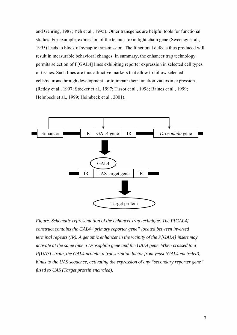
and Gehring, 1987; Yeh et al., 1995). Other transgenes are helpful tools for functional
studies. For example, expression of the tetanus toxin light chain gene (Sweeney et al.,
1995) leads to block of synaptic transmission. The functional defects thus produced will
result in measurable behavioral changes. In summary, the enhancer trap technology
permits selection of P[GAL4] lines exhibiting reporter expression in selected cell types
or tissues. Such lines are thus attractive markers that allow to follow selected
cells/neurons through development, or to impair their function via toxin expression
(Reddy et al., 1997; Stocker et al., 1997; Tissot et al., 1998; Baines et al., 1999;
Heimbeck et al., 1999; Heimbeck et al., 2001).
UAS-target gene
GAL4
Enhancer GAL4 geneIR IR
IR IR
Drosophila gene
Target protein
Figure. Schematic representation of the enhancer trap technique. The P[GAL4]
construct contains the GAL4 “primary reporter gene” located between inverted
terminal repeats (IR). A genomic enhancer in the vicinity of the P[GAL4] insert may
activate at the same time a Drosophila gene and the GAL4 gene. When crossed to a
P[UAS] strain, the GAL4 protein, a transcription factor from yeast (GAL4 encircled),
binds to the UAS sequence, activating the expression of any “secondary reporter gene”
fused to UAS (Target protein encircled).
7

The neuroanatomical basis of the adult chemosensory system in Drosophila has
been well described (Nayak and Singh, 1983; Stocker, 1994; Singh, 1997; Laissue et al.,
1999; Shanbhag et al., 1999; Shanbhag et al., 2001). Compared to vertebrate systems, it
exhibits a tremendous reduction of cell numbers, despite surprising parallels in terms of
organization (Hildebrand and Shepherd, 1997). This renders the fly an attractive model
system for analyzing olfaction and gustation. In contrast, few studies have focused on
the larval chemosensory system (Singh and Singh, 1984; Tissot et al., 1997; Cobb, 1999;
Heimbeck et al., 1999; Stocker, 2001; ). Considering its even further reduced cellular
complexity, the goal of my work was to explore the potential of the larval system as an
alternative, equally attractive chemosensory model system.
In my thesis work, I used the GAL4/UAS expression system as a tool for
studying in the confocal microscope the neuroanatomy and neurotransmitter content of
the larval chemosensory system. I chose four P[GAL4] lines that show specific reporter
expression in the olfactory and/or gustatory system, both in the periphery and in the
central nervous system (CNS). By simultaneously expressing a fluorescent GFP reporter
and applying neuronal or neuropil antibody markers, I initially focused on the
organization of the larval chemosensory system. I investigated (1) the neuronal
composition of the larval olfactory and gustatory organs, (2) the peripheral nerves used
by their afferent axons on their way to the CNS, and (3) the organization of the larval
antennal lobe (LAL), the primary target of olfactory afferents. Special attention was
given to putative subunits of the LAL, resembling adult glomeruli, and to a
corresponding compartmentation of afferent terminals and of dendritic arborizations of
target neurons. The major types of target neurons are local interneurons – intrinsic to the
LAL – and projection neurons, providing links to higher brain centers. The most
significant observation of this study was an adult-like structural complexity of the LAL
despite drastically reduced numbers of odorant receptor neurons. The paper will be
published in the “Journal of Comparative Neurology” (in press).
In the second part of my work, I extended the description of the larval
chemosensory system by analyzing in the same P[GAL4] lines the distribution of
choline acetyltransferase (ChAT) – the acetylcholine-synthesizing enzyme – and of the
neurotransmitters γ-aminobutyric acid (GABA), histamine, octopamine and serotonin. I
8
provide evidence that subsets of the olfactory and gustatory afferents as well as many of
projection neurons are strongly ChAT immunoreactive, and that perhaps the entire set of
local interneurons contain GABA as a neurotransmitter. In addition, I identified a
putative serotonergic interneuron that arborizes in the LAL neuropil. Taken together, the
cellular distribution of these classical neurotransmitters is similar as in the adult
chemosensory system, suggesting shared mechanisms of chemosensory information
processing. In summary, my thesis work proposes the larva of D. melanogaster as an
alternative model system for studying smell and taste at the functional, molecular and
developmental level.
9

LITERATURE CITED
Adams MD, Celniker SE, Holt RA, Evans CA, Gocayne JD, Amanatides PG, Scherer
SE, Li PW, Hoskins RA, Galle RF and others. 2000. The genome sequence of
Drosophila melanogaster. Science 287:2185-95.
Baines RA, Robinson SG, Fujioka M, Jaynes JB, Bate M. 1999. Postsynaptic expression
of tetanus toxin light chain blocks synaptogenesis in Drosophila. Curr Biol 9:1267-
70.
Brand AH, Perrimon N. 1993. Targeted gene expression as a means of altering cell fates
and generating dominant phenotypes. Development 118:401-15.
Cobb M. 1999. What and how do maggots smell? Biol. Rev. 74:425-59.
Fischer JA, Giniger E, Maniatis T, Ptashne M. 1988. GAL4 activates transcription in
Drosophila. Nature 332:853-56.
Greenspan RJ. 1996. Fly Pushing: The theory and Practice of Drosophila Genetics. Cold
Spring Harbor Laboratory Press, Cold Spring Harbor, NY.
Heimbeck G, Bugnon V, Gendre N, Häberlin C, Stocker RF. 1999. Smell and taste
perception in Drosophila melanogaster larva: toxin expression studies in
chemosensory neurons. J Neurosci 19:6599-609.
Heimbeck G, Bugnon V, Gendre N, Keller A, Stocker RF. 2001. A central neural circuit
for experience-independent olfactory and courtship behavior in Drosophila
melanogaster. Proc Natl Acad Sci U S A 98:15336-41.
Hildebrand JG, Shepherd GM. 1997. Mechanisms of olfactory discrimination:
converging evidence for common principles across phyla. Annu Rev Neurosci
20:595-631.
Laissue PP, Reiter C, Hiesinger PR, Halter S, Fischbach KF, Stocker RF. 1999. Three-
dimensional reconstruction of the antennal lobe in Drosophila melanogaster. J
Comp Neurol 405:543-52.
10

Lander ES, Linton LM, Birren B, Nusbaum C, Zody MC, Baldwin J, Devon K, Dewar
K, Doyle M, FitzHugh W and others. 2001. Initial sequencing and analysis of the
human genome. Nature 409:860-921.
Nayak SV, Singh RN. 1983. Sensilla on the tarsal segments and mouthparts of adult
Drosophila melanogaster Meigen (DIPTERA: DROSOPHILIDAE). Int J Insect
Morphol Embryol 12:273-291.
O'Kane CJ, Gehring WJ. 1987. Detection in situ of genomic regulatory elements in
Drosophila. Proc Natl Acad Sci U S A 84:9123-7.
Pflugfelder GO. 1998. Genetic lesions in Drosophila behavioural mutants. Behav Brain
Res 95:3-15.
Reddy S, Jin P, Trimarchi J, Caruccio P, Phillis R, Murphey RK. 1997. Mutant
molecular motors disrupt neural circuits in Drosophila. J Neurobiol 33:711-23.
Rubin GM. 1988. Drosophila melanogaster as an experimental organism. Science
240:1453-59.
Shanbhag SR, Muller B, Steinbrecht RA. 1999. Atlas of olfactory organs of Drosophila
melanogaster. 1. Types, external organization, innervation and distribution of
olfactory sensilla. Int J Insect Morphol Embryol 28:377-97.
Shanbhag SR, Park SK, Pikielny CW, Steinbrecht RA. 2001. Gustatory organs of
Drosophila melanogaster: fine structure and expression of the putative odorant-
binding protein PBPRP2. Cell Tissue Res 304:423-37.
Singh RN. 1997. Neurobiology of the gustatory systems of Drosophila and some
terrestrial insects. Microsc Res Tech 39:547-63.
Singh RN, Singh K. 1984. Fine structure of the sensory organs of Drosophila
melanogaster Meigen larva (Diptera: Drosophilidae). Int J Insect Morphol Embryol
13:255-273.
Stocker RF. 1994. The organization of the chemosensory system in Drosophila
melanogaster: a review. Cell Tissue Res 275:3-26.
Stocker RF. 2001. Drosophila as a focus in olfactory research: mapping of olfactory
sensilla by fine structure, odor specificity, odorant receptor expression, and central
connectivity. Microsc Res Tech 55:284-96.
11
Stocker RF, Heimbeck G, Gendre N, de Belle JS. 1997. Neuroblast ablation in
Drosophila P[GAL4] lines reveals origins of olfactory interneurons. J Neurobiol
32:443-56.
Sweeney ST, Broadie K, Keane J, Niemann H, O'Kane CJ. 1995. Targeted expression of
tetanus toxin light chain in Drosophila specifically eliminates synaptic transmission
and causes behavioral defects. Neuron 14:341-51.
Tissot M, Gendre N, Hawken A, Störtkuhl KF, Stocker RF. 1997. Larval chemosensory
projections and invasion of adult afferents in the antennal lobe of Drosophila. J
Neurobiol 32:281-97.
Tissot M, Gendre N, Stocker RF. 1998. Drosophila P[Gal4] lines reveal that motor
neurons involved in feeding persist through metamorphosis. J Neurobiol 37:237-50.
Venter JC, Adams MD, Myers EW, Li PW, Mural RJ, Sutton GG, Smith HO, Yandell
M, Evans CA, Holt RA and others. 2001. The sequence of the human genome.
Science 291:1304-51.
Yeh E, Gustafson K, Boulianne GL. 1995. Green fluorescent protein as a vital marker
and reporter of gene expression in Drosophila. Proc Natl Acad Sci U S A 92:7036-
40.
12
Chapter 1
Adult-like complexity of the larval
antennal lobe of Drosophila melanogaster
despite markedly low numbers of
odorant receptor neurons
This chapter will be published in The Journal of Comparative Neurology (2002), vol.
445.
13

ABSTRACT
We provide a detailed analysis of the larval head chemosensory system of
Drosophila melanogaster, based on confocal microscopy of cell-specific reporter gene
expression in P[GAL4] enhancer trap lines. In particular, we describe the neuronal
composition of three external and three pharyngeal chemosensory organs, the nerve
tracts chosen by their afferents, and their central target regions. With a total of 21
olfactory and 80 gustatory neurons, the sensory level is numerically much simpler than
that of the adult. Moreover, its design is different than in the adult, showing an
association between smell and taste sensilla. In contrast, the first order relay of the
olfactory afferents, the larval antennal lobe (LAL), exhibits adult-like features both in
terms of structure and cell number. It shows a division into approximately 30 subunits,
reminiscent of glomeruli in the adult antennal lobe. Taken together, the design of the
larval chemosensory system is a ‘hybrid’, with larval-specific features in the periphery
and central characteristics in common with the adult. The largely reduced numbers of
afferents and the similar architecture of the LAL and the adult antennal lobe, render the
larval chemosensory system of Drosophila a valuable model system, both for studying
smell and taste, and for examining the development of its adult organization.
Abbreviations: AL antennal lobe, AN antennal nerve, DO dorsal organ, DOG ganglion
of the DO, DPS dorsal pharyngeal sensilla, LAL larval antennal lobe, LBN, labial nerve,
LI local interneuron, LN labral nerve, mAb monoclonal antibody, MN maxillary nerve,
PN projection neuron, PPS posterior pharyngeal sensilla, SOG suboesophageal ganglion,
TO terminal organ, TOG ganglion of the TO, TR tritocerebrum, VO ventral organ, VOG
ganglion of the VO, VPS ventral pharyngeal sensilla
14
INTRODUCTION
Since the identification of putative olfactory and gustatory receptors in
Drosophila melanogaster (Clyne et al., 1999, 2000; Gao and Chess, 1999; Vosshall et
al., 1999) and the subsequent demonstration of receptor-specific afferent connections in
the brain (Vosshall et al., 2000; Scott et al., 2001), the fruitfly has become an attractive
focus in chemosensory research (reviews: Vosshall, 2000, 2001; Stocker, 2001; Warr et
al., 2001). The neuroanatomical basis required for the interpretation of these data has
been well established in the adult fly (Nayak and Singh, 1983; Stocker, 1994; Singh,
1997; Laissue et al., 1999; Shanbhag et al., 1999; Shanbhag et al., 2001). In contrast,
apart from a few isolated reports (Singh and Singh, 1984; Tissot et al., 1997), the larval
chemosensory system lacks a sound structural description. This is a drawback
considering the potential attractiveness of the larval system for functional and molecular
studies, due to its extremely reduced cellular complexity (see below) (Cobb, 1999;
Heimbeck et al., 1999; Stocker, 2001).
The chemosensory apparatus of the larval head is formed during late
embryogenesis (Frederik and Denell, 1982; Campos-Ortega and Hartenstein, 1997). It
consists of three organs located on the cephalic lobe, the dorsal organ (DO), the terminal
organ (TO), the ventral organ (VO), and three sets of pharyngeal sensilla (Singh and
Singh, 1984). The fine structure of the DO, TO and VO in Drosophila (Singh and Singh,
1984) is very similar to that of the housefly larva Musca domestica, which has been
meticulously described (Chu and Axtell, 1971; Chu-Wang and Axtell, 1972a; Chu-
Wang and Axtell, 1972b). In both species, 21 putative olfactory receptor neurons in the
DO establish profuse dendritic arbors in a central dome sensillum whose wall is
perforated by thousands of pore tubules. The dome is surrounded by six sensilla with
large distal pores suggesting a gustatory function. Apical pores are present also in most
of the TO sensilla, which consist of at least six different types clustered in a distal and a
dorsolateral group, as well as in one of the four sensilla comprising the much simpler
VO. Hence, the DO appears to be a mixed smell and taste organ, whereas the TO, VO
and pharyngeal sensilla may be exclusively gustatory (Chu and Axtell, 1971; Chu-Wang
and Axtell, 1972a; Chu-Wang and Axtell, 1972b; Singh and Singh, 1984; Singh, 1997).
15
This is supported by recent toxin inactivation and electrophysiological studies in
Drosophila (Heimbeck et al., 1999; Oppliger et al., 2000) which assigned an olfactory
function to the DO and a gustatory one to the TO.
Although fruitfly larvae respond to a large variety of chemicals (Ayyub et al.,
1990; Cobb et al., 1992; Cobb and Dannet, 1994; Cobb, 1999; Heimbeck et al., 1999;
Cobb and Domain, 2000; Oppliger et al., 2000), their chemosensory system is
remarkably simple in cellular terms. For example, it comprises no more than the 21
odorant receptor neurons of the DO, compared to 1300 in the adult fly. The 21 olfactory
afferents converge onto the larval antennal lobe (LAL), the precursor of the adult
antennal lobe (AL) (Tissot et al., 1997). The LAL is no bigger than a single glomerulus
of the adult lobe, and input and output fibers of the LAL – when studied as populations –
did not seem to display any obvious glomerular-like arborization (Stocker et al., 1997;
Tissot et al., 1997), which is typical for the adult AL. These observations suggest
simplicity at the central level as well. However, no description of the LAL structure or
of afferent and target neuron arborizations is available at cellular resolution. Moreover,
apart from a few casual data (Tissot et al., 1997), nothing is known about gustatory
target regions in the larval CNS, in particular for pharyngeal sensilla. This motivated us
to study the organization and connectivity of the larval chemosensory system of
Drosophila, both at the peripheral and central level. Specifically, we wanted to
determine whether and in which sense the larval system is indeed “simpler” than the
adult system. This report focuses on the larval head and will not include putative
chemosensory organs on the rest of the body.
In the present study, we made use of P[GAL4] enhancer trap lines (Brand and
Perrimon, 1993) that show expression in larval chemosensory neurons and/or their target
neurons in the brain. Analyzing their expression patterns in the confocal microscope, in
combination with neuronal or neuropil markers, allowed us to determine the cellular
composition of the larval chemosensory organs, the peripheral nerves used by their
afferents and their central target regions. In particular, we were interested in the
architecture and neuronal organization of the LAL. We show that the LAL consists of
numerous subunits, which in terms of afferent and target neuron morphology are
reminiscent of typical antennal lobe glomeruli.
16
MATERIALS AND METHODS
The P[GAL4] lines 4551 and 189Y (Osborne et al., 1997) were provided by J.-F.
Ferveur (Université de Bourgogne, Dijon) and K. Kaiser (University of Glasgow),
respectively. The lines GH146 and GH86 were isolated by G. Heimbeck (Stocker et al.,
1997; Heimbeck et al., 1999). As secondary reporter strains, we used UAS-GFP (Yeh et
al., 1995) and UAS-Tau-GFP (Murray et al., 1998), both kindly provided by A. H.
Brand (Wellcome/CRC, Cambridge). The CantonS (CS) strain served as a wildtype
reference. All strains were raised on standard cornmeal medium at 18°C or 25°C.
Antibody staining was adapted from Laissue et al. (1999). In brief, dissected
tissues from the third larval instar were fixed for 2 hours on ice in 4% paraformaldehyde
(Merck) (4 hours for the adult head), dissolved in phosphate buffered saline (pH 7.2)
containing 0.2% Triton X-100 (PBS-T, pH 7.2). Subsequent reactions took place with
gentle shaking on ice. After 3 washes of 20 minutes each in PBS-T, they were blocked
for 1-2 hours in the blocking solution (BS) containing 3% normal goat serum (Jackson
ImmunoResearch) in PBS-T. Tissues were then incubated overnight in the primary
antibody diluted in BS. Washes in PBS-T (3x 20 minutes) were followed by the
incubation of the secondary antibody diluted in BS for 5 hours. Samples were again
rinsed 3x 20 minutes in PBS-T and finally mounted in Vectashield medium (Vector
Laboratories). As primary antibodies we used mouse mAb nc82 (dilution 1:10; a gift
from A. Hofbauer, University of Regensburg), mouse anti-Synapsin (1:100; provided by
E. Buchner, University of Würzburg) and mouse anti-Elav 9F8A9 (1: 200). The anti-
Elav antibody developed by G.M. Rubin was obtained from the Developmental Studies
Hybridoma Bank developed under the auspices or the NICHD and maintained by The
University of Iowa, Dept. of Biological Sciences, Iowa City, IA 52242. The secondary
antibody utilized was Cy3-coupled goat anti-mouse IgG (Jackson ImmunoResearch),
diluted 1:100 in BS.
Preparations were studied with a BioRad MRC 1024 confocal microscope
equipped with a Kr/Ar laser. Z series of pictures were taken at intervals of 0.5 to 2 µm.
17
Image analysis was performed on a Macintosh computer using the public domain
NIH Image program (developed at the U.S. National Institutes of Health and available
on the Internet at http://rsb.info.nih.gov/nih-image/). Color selection of images was done
by the Adobe PhotoShop program.
RESULTS
The four enhancer trap lines 4551, 189Y, GH86 and GH146 which express
GAL4 in subsets of sensory neurons and their target interneurons allowed us to study the
organization of the major chemosensilla in the third larval instar, to identify the
peripheral pathways and central target regions of their afferents and to analyze the
structure and interneuron composition of the LAL (cf. Fig. 8 for a summary diagram).
The expression patterns were studied in whole mounts by confocal microscopy. The
patterns visualized by the two reporters GFP and Tau-GFP were similar, in particular
with respect to afferent paths and terminals in the CNS. However, Tau-GFP was
superior for demonstrating axonal pathways (Fig. 1A,B), due to the microtubular
association of the Tau protein. GFP and Tau-GFP patterns were confirmed by ß-
Galactosidase reporter staining in whole mounts and by Tau immunostaining in
cryosections (cf. Heimbeck et al., 1999; data not shown). Neuronal identity of cells was
established by means of the mAb anti-Elav, and the neuropil structure of the LAL was
dissected by applying the mAbs nc82 (Laissue et al., 1999) and anti-Synapsin (Klagges
et al., 1996).
18
Chemosensory Organs of the Larval Head
Reporter gene expression driven by line 4551 revealed the entire neuroanatomy
of the DO and TO (Figs. 1A,C, 2A). Their bipolar sensory neurons are assembled in two
separate ganglia, which are located well below the epidermis, in close contact to each
other. As noted previously, a small subset of neurons in the DO ganglion send their
dendrites into the TO (Kankel et al., 1980; Frederik and Denell, 1982; Tissot et al.,
1997) (Figs. 1C, 2A; see below).
Labeling with anti-Elav revealed an average of 37 sensory neurons in the dorsal
ganglion of CS wildtype larvae (n=6; males: 37-39; females: 35-37) (Table 1), which is
similar to the 35-41 neurons counted in the housefly larva (Chu and Axtell, 1971).
Likewise, the dorsal ganglion of the line 4551 comprises 34-38 neurons (n=5) (Fig. 2A),
24 of which on an average express GAL4 (n=6; 22-25) (Figs. 1C, 2A). The expression
pattern confirms that the central dome of the DO – the exclusive olfactory organ of the
larva by fine structural criteria (Singh and Singh, 1984) – is innervated by seven bundles
of dendrites (cf. Singh and Singh, 1984; Campos-Ortega and Hartenstein, 1997) (Fig.
1C). According to these reports, each bundle consists of a triplet of dendrites, yielding a
total of 21 odorant receptor neurons, whereas additional neurons of the dorsal ganglion
innervate the six putative taste sensilla surrounding the dome. Interestingly, none of the
GAL4-positive neurons of line 4551 send their dendrites into one of these six sensilla.
On the other hand, three labeled dendrites extend toward a putative gustatory sensillum
of the TO (Figs. 1C, 2A, see below). This suggests that the 24 GAL4-positive neurons in
the DOG comprise the 21 odorant receptor neurons and three taste neurons associated
with the TO (Table 1).
In the terminal ganglion of CS larvae we counted an average of 32-33 neurons
(n=6; males: 33-35; females: 30-34) (Table 1), four of which – perhaps associated with
mechanosensory scolopidia – are located more distally than the rest (Fig. 2A). This
corresponds to the 32 neurons observed in the terminal ganglion of the housefly larva
(Chu-Wang and Axtell, 1972a) and to the 30-34 neurons found in line 4551 (n=5). The
TO consists of a distal group of 11 individual sensilla and a dorsolateral group of three
sensilla, one of which – a putative gustatory papillum – is innervated by three dendrites
19
(Kankel et al., 1980; Singh and Singh, 1984) (Fig. 1C, inset). As mentioned
before, GAL4 expression in line 4551 suggests that three dendrites originating from the
dorsal ganglion extend toward the dorsolateral group of the TO, most likely into that
papillum (Fig. 1C, inset). Apart from that, line 4551 labels about 15 neurons in the
terminal ganglion (n=6; 13-17) (Figs. 1C & 2A). Most if not all of them are likely to be
gustatory, given that a large majority of the neurons of this ganglion may mediate taste
according to fine structural criteria (Chu-Wang and Axtell, 1972a). The remaining
neurons may represent mechano-, hygro- or thermoreceptors. In close vicinity to the
terminal ganglion we observe an additional cluster of up to ten neurons (Fig. 2A), which
may correspond to the “associated organ” described in the embryo (Schmidt-Ott et al.,
1994), a sensillum of unknown function.
The VO, the third external chemosensory organ, has its terminal pore located
between the third and fourth rows of spinules (Figs. 1D, 2A). Anti-Elav staining
revealed up to seven neurons in the VO ganglion (Fig. 2A, Table 1), one of which is
labeled by line 4551. Earlier studies in the VO of Musca and Drosophila reported the
presence of seven and eight neurons, respectively (Chu-Wang and Axtell, 1972b; Singh
and Singh, 1984).
The expression patterns of the lines 4551 and 189Y together with anti-Elav
tagging revealed several groups of pharyngeal sensilla. Two anterior clusters are located
in front of the cephalopharyngeal H-piece (Campos-Ortega and Hartenstein, 1997), a
dorsal and a ventral one (DPS, VPS) (Figs. 1A, 3). Moreover, a small sensillum (PPS)
sits further posterior in the lateral wall of the pharynx, in an area characterized by
longitudinal ridges (Campos-Ortega and Hartenstein, 1997) (Figs. 1A,B, 3). Due to
developmental rearrangements, the identity of these three groups compared to the
embryonic sensillum pattern is not easy to determine. According to the relative location
and associated nerve, the DPS is likely to correspond to the labral sense organ of
Campos-Ortega and Hartenstein (1997) known also as “epiphysis” (Schmidt-Ott et al.,
1994) or “dorsal pharyngeal sensilla D1-D6“ (Singh and Singh, 1984). The VPS appears
to be equivalent to the labial sensory complex (Campos-Ortega and Hartenstein, 1997;
synonyms “hypophysis”: Schmidt-Ott et al., 1994; “ventral pharyngeal sensilla V1-V4”:
Singh and Singh, 1984), whereas the PPS may correspond to the hypopharyngeal organ
20
(Schmidt-Ott et al., 1994; Campos-Ortega and Hartenstein, 1997). The ganglia of the
DPS comprise 16-17 anti-Elav stained neurons each (S. Crevoiserat, personal
communication) (Table 1), substantially more than ten neurons, as counted by Singh and
Singh (1984). Fourteen to fifteen of them express GAL4 in line 4551. The PPS ganglia
include six neurons, all marked by GAL4 (S. Crevoiserat, personal communication).
Moreover, each VPS ganglion contains 17 neurons (Singh and Singh, 1984), none of
which is labeled by 4551.
Chemosensory Head Nerves
The dorsal ganglion connects to the CNS by means of the antennal nerve (AN)
(Figs. 1A, 8). Hence, the AN is a mixed nerve comprising 21 olfactory afferents from
the dome sensillum and 12 putative gustatory afferents from other DO sensilla and the
dorsolateral TO papillum (Table 1). At about two thirds of the distance toward the brain,
the AN is joined by the labral nerve (LN) (cf. Schmidt-Ott et al., 1994), which carries at
most 22 gustatory afferents from the DPS and PPS (Fig. 1A, Table 1). The afferents
from both nerves then travel together toward the CNS. However, we were not able to
distinguish whether the two nerves have indeed fused or are just closely aligned. In
support of the second interpretation, the compound nerve bifurcates again shortly before
approaching the CNS. One branch turns toward the midline, joins the brain from
laterally and extends toward the LAL (Fig. 1E,F). The second branch enters the CNS
closer to the midline, at a site that is presumably of tritocerebral identity (TR) (Fig. 1B
inset,E). Tracing of the afferents in the line 4551 and in 189Y – whose sensory
expression is restricted to pharyngeal sensilla (Fig. 1A,B, inset, E) – suggests that the
fibers in the branch towards the LAL originate in the AN, whereas those in the branch
towards the TR derive from the LN. This further argues against a fusion of the two
nerves. However, we cannot exclude the possibility that GAL4-negative afferents
deriving from the six DO sensilla around the dome may pass into the TR branch.
Together with the mixed innervation of the TO by two distinct ganglia (see above), the
pairing of AN and LN illustrates the highly modified dipteran neuroanatomy (cf.
Strausfeld, 2001).
21

The ganglia of the TO and the VO are connected to the CNS by means of the
maxillary nerve (MN) (Fig. 1A). Hence, in contrast to the AN, the MN lacks an
olfactory component. It carries about 23 gustatory afferents from the distal TO sensilla
and the remaining dorsolateral sensilla, as well as seven gustatory afferents from the VO
(Table 1) (Chu-Wang and Axtell, 1972a; Chu-Wang and Axtell, 1972b; Campos-Ortega
and Hartenstein, 1997). The MN joins the CNS more ventrally and posteriorly and
closer to the midline than the LN branch of the compound nerve, at a location that
obviously belongs to the suboesophageal ganglion (SOG) (Fig. 1E). These two entry
sites are consistent with the supra- and suboesophageal identity of the LN and MN,
respectively (Campos-Ortega and Hartenstein, 1997). The fourth chemosensory head
nerve is the labial nerve which is chosen by the approximately 15 gustatory afferents
from the VPS (Singh and Singh, 1984; Schmidt-Ott et al., 1994) (Table 1). It joins the
SOG region of the CNS still further back than the MN.
Larval Antennal Lobe: Structure and Afferent Projections
Unlike the adult AL, the LAL is not the most anterior prominent neuropil area of
the brain. Rather, it is a small, metameric structure of 20-30 µm diameter, situated
between the mushroom bodies and the TR-SOG region (Fig. 2B-D,F) (Stocker et al.,
1995; Tissot et al., 1997). The LAL is barely visible in the unstained brain, but becomes
manifest upon the application of diverse mAbs or transgenic markers (Fig. 2B-D).
Remarkably, the mAb nc82 – which reveals the glomerular architecture of the adult AL
(Laissue et al., 1999) (Fig. 2E) – binds in a non-homogeneous manner to the LAL
neuropil as well (Fig. 2E, inset). This non-homogeneous pattern is clearly more
pronounced than that of other neuropil regions. Intensely stained areas of 5 to 10 µm
diameter, termed here subunits, are separated by clefts (Fig. 2G). The total number of
subunits may not exceed 30. More precise estimates are hampered by their ambiguous
contours, which are less distinct than in adult glomeruli. A similar patterning of the LAL
neuropil is also visible with the anti-Synapsin marker (Fig. 4), especially with respect to
two particular subunits (see below).
Remarkably, GFP staining in the afferent lines studied reveals also non-
homogeneity in the pattern of sensory terminals. Their arborizations occupy small
22
domains, most of which are of similar size and shape as the subunits mentioned before
(Fig. 2G). Inside the domains, smaller structures associated with the afferents are visible,
as reported previously for the chemosensory-specific line GH86 (Heimbeck et al., 1999)
(Fig. 5A-C). Very likely these smaller particles represent the afferent terminals proper.
Three of the sensory domains are more intensely labeled than the others (Figs. 2G, 4,
5A-C). Two large, elongated domains (E, E’) extend from lateral to anteromedial in the
posterior LAL region. They are the targets of subsets of AN afferents that segregate
from the rest of the fibers shortly after entering the LAL (Fig. 5A,B). A smaller and
heavily marked domain occupies a more dorsal position (D) and is innervated by a
single sensory axon (Fig. 5B,C). Hence, at least in these cases, individual afferents
appear to terminate in subregions of the LAL rather than distributing over its entire
volume.
Do the neuropil subunits mentioned before match to the afferent domains?
Indeed, nc82 labeling in the afferent line 4551 shows a striking correspondence between
the two elements (Fig. 2G). This is particularly evident for the intensely stained E and D
domains (Figs. 2G at 9 µm & 15 µm, 4), which are labeled by the anti-Synapsin marker
as well (Fig. 4). Such an overlap may also apply to other LAL regions, as suggested by
conspicuous parallels in the size, shape and arrangement of many sensory domains and
neuropil subunits. Thus, we propose that many of the subunits labeled by the neuropil
markers may correspond to afferent arborizations.
Larval Antennal Lobe: Interneurons and their Arborizations Similar to the adult AL, the LAL comprises two major types of interneurons, i.e.,
local interneurons (LIs) whose arborizations are restricted to the lobe, and projection
neurons (PNs), which link the lobe with higher brain centers. Line 189Y labels five to
six LIs in the LAL, which represents very likely only a fraction of their total number. As
for adult LIs (Stocker et al., 1997), larval LIs have their cell bodies posterolateral to the
lobe (Figs. 1B, inset & 2B, inset) and appear to arborize in the entire LAL neuropil.
Double labeling with nc82 or anti-Synapsin reveals highest GFP or Tau-GFP reporter
expression in the subunits mentioned before (Fig. 6A), suggesting that they are the main
sites of LI arborizations.
23
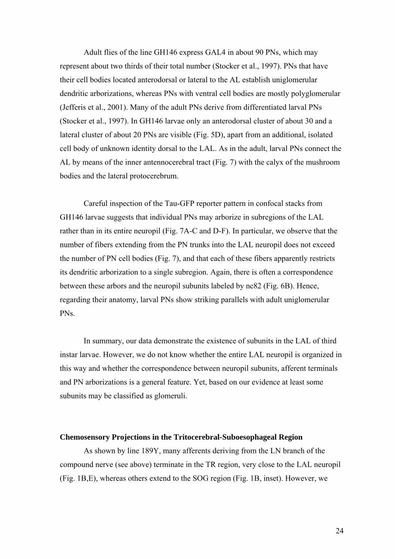
Adult flies of the line GH146 express GAL4 in about 90 PNs, which may
represent about two thirds of their total number (Stocker et al., 1997). PNs that have
their cell bodies located anterodorsal or lateral to the AL establish uniglomerular
dendritic arborizations, whereas PNs with ventral cell bodies are mostly polyglomerular
(Jefferis et al., 2001). Many of the adult PNs derive from differentiated larval PNs
(Stocker et al., 1997). In GH146 larvae only an anterodorsal cluster of about 30 and a
lateral cluster of about 20 PNs are visible (Fig. 5D), apart from an additional, isolated
cell body of unknown identity dorsal to the LAL. As in the adult, larval PNs connect the
AL by means of the inner antennocerebral tract (Fig. 7) with the calyx of the mushroom
bodies and the lateral protocerebrum.
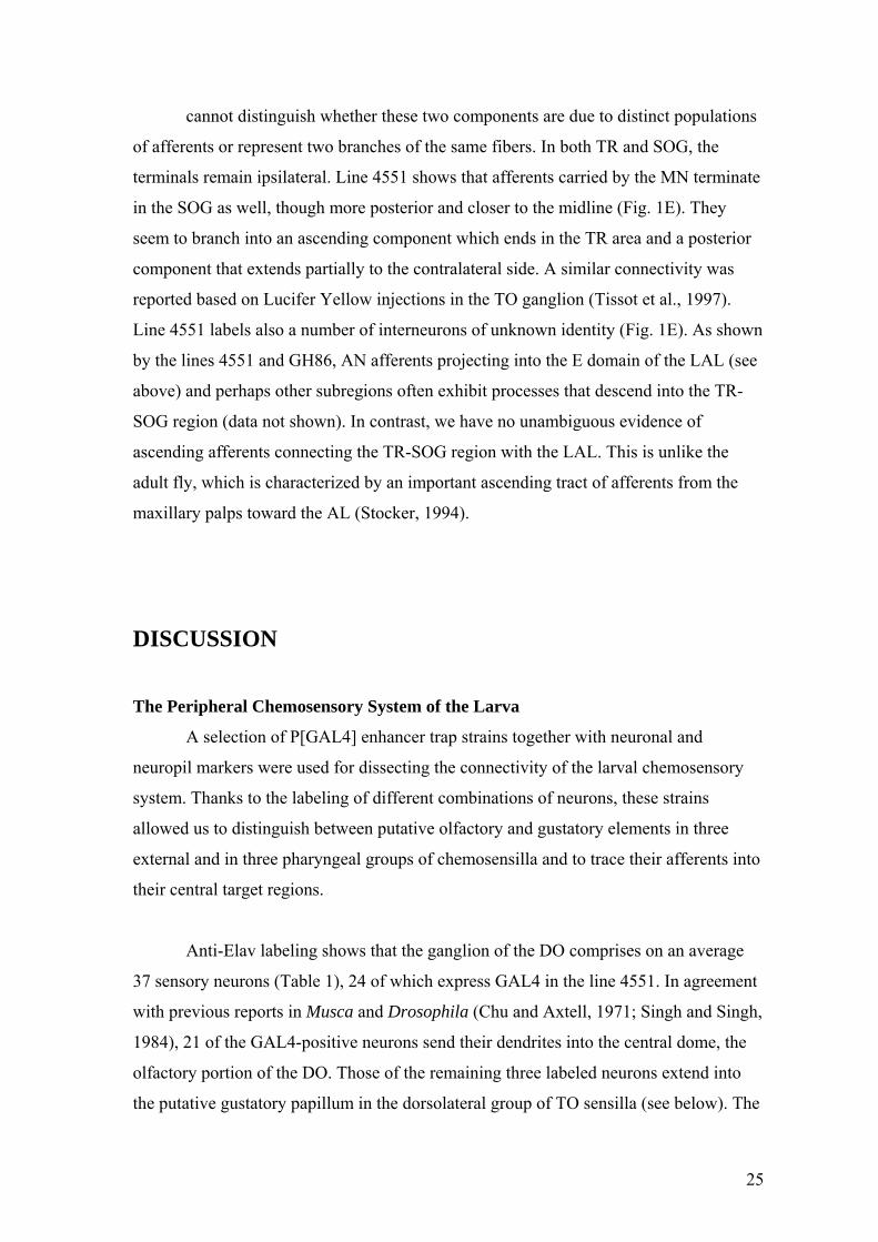
Careful inspection of the Tau-GFP reporter pattern in confocal stacks from
GH146 larvae suggests that individual PNs may arborize in subregions of the LAL
rather than in its entire neuropil (Fig. 7A-C and D-F). In particular, we observe that the
number of fibers extending from the PN trunks into the LAL neuropil does not exceed
the number of PN cell bodies (Fig. 7), and that each of these fibers apparently restricts
its dendritic arborization to a single subregion. Again, there is often a correspondence
between these arbors and the neuropil subunits labeled by nc82 (Fig. 6B). Hence,
regarding their anatomy, larval PNs show striking parallels with adult uniglomerular
PNs.
In summary, our data demonstrate the existence of subunits in the LAL of third
instar larvae. However, we do not know whether the entire LAL neuropil is organized in
this way and whether the correspondence between neuropil subunits, afferent terminals
and PN arborizations is a general feature. Yet, based on our evidence at least some
subunits may be classified as glomeruli.
Chemosensory Projections in the Tritocerebral-Suboesophageal Region
As shown by line 189Y, many afferents deriving from the LN branch of the
compound nerve (see above) terminate in the TR region, very close to the LAL neuropil
(Fig. 1B,E), whereas others extend to the SOG region (Fig. 1B, inset). However, we
24
cannot distinguish whether these two components are due to distinct populations
of afferents or represent two branches of the same fibers. In both TR and SOG, the
terminals remain ipsilateral. Line 4551 shows that afferents carried by the MN terminate
in the SOG as well, though more posterior and closer to the midline (Fig. 1E). They
seem to branch into an ascending component which ends in the TR area and a posterior
component that extends partially to the contralateral side. A similar connectivity was
reported based on Lucifer Yellow injections in the TO ganglion (Tissot et al., 1997).
Line 4551 labels also a number of interneurons of unknown identity (Fig. 1E). As shown
by the lines 4551 and GH86, AN afferents projecting into the E domain of the LAL (see
above) and perhaps other subregions often exhibit processes that descend into the TR-
SOG region (data not shown). In contrast, we have no unambiguous evidence of
ascending afferents connecting the TR-SOG region with the LAL. This is unlike the
adult fly, which is characterized by an important ascending tract of afferents from the
maxillary palps toward the AL (Stocker, 1994).
DISCUSSION
The Peripheral Chemosensory System of the Larva
A selection of P[GAL4] enhancer trap strains together with neuronal and
neuropil markers were used for dissecting the connectivity of the larval chemosensory
system. Thanks to the labeling of different combinations of neurons, these strains
allowed us to distinguish between putative olfactory and gustatory elements in three
external and in three pharyngeal groups of chemosensilla and to trace their afferents into
their central target regions.
Anti-Elav labeling shows that the ganglion of the DO comprises on an average
37 sensory neurons (Table 1), 24 of which express GAL4 in the line 4551. In agreement
with previous reports in Musca and Drosophila (Chu and Axtell, 1971; Singh and Singh,
1984), 21 of the GAL4-positive neurons send their dendrites into the central dome, the
olfactory portion of the DO. Those of the remaining three labeled neurons extend into
the putative gustatory papillum in the dorsolateral group of TO sensilla (see below). The
25
GAL4-negative neurons are likely to innervate the six peripheral DO sensilla, which
may mediate taste as well (Chu and Axtell, 1971; Singh and Singh, 1984). The average
of 32-33 neurons we have determined in the TO ganglion (Table 1) is in the same range
as the 35 neurons reported from the housefly larva (Chu-Wang and Axtell, 1972a) and
the total of 33 dendrites observed in TO sensilla of Drosophila (Singh and Singh, 1984).
Similarly, as in two previous studies (Chu-Wang and Axtell, 1972b: Musca; Singh and
Singh, 1984: Drosophila), we have counted a total of seven to eight neurons in the VO
ganglion.
Individual measurements in DO and TO ganglia of both CS and 4551 larvae
revealed some variation in neuron numbers. Because similar figures were obtained when
counted by different persons, we consider the differences to reflect real variability in
neuronal number rather than a counting artifact. How the variability relates to individual
sensilla is not known. Variations in sensory structures and the underlying neurons are
known also from the adult olfactory and gustatory systems (Nayak and Singh, 1983; de
Bruyne et al., 1999; Shanbhag et al., 2001). The minor numerical differences observed
between males and females appear to be within the range of general variability. A
significant sexual dimorphism is not to be expected given the similar feeding strategies
of male and female larvae.
Anti-Elav labeling and reporter expression in line 4551 demonstrate the
existence of three simple sets of larval pharyngeal sensilla, termed DPS, VPS and PPS.
They comprise no more than 16, 15 and 6 putative gustatory neurons, respectively
(Table 1). The first two have been studied previously (Singh and Singh, 1984); the small
PPS which is located far more posterior, is described here for the first time.
Chemosensory Target Regions in the CNS
The mixed expression of most available P[GAL4] lines in various subsets of
larval chemosensilla often prevents a precise assignment between individual receptor
neurons and their central target regions. Nevertheless, the afferent pathways shown by
the lines used strongly suggest that the fibers from the DO ganglion travelling in the AN
extend into the LAL, whereas those from the remaining chemosensilla carried by the
26
LN, MN and LBN project into successively more posterior regions of the TR-
SOG neuropil (Fig. 8). The TR-SOG as a target of putative taste neurons from the TO
and pharyngeal sensilla was confirmed by a recent study, in which reporter expression
driven by gustatory receptor gene promoters was used to trace the target regions of
single larval chemosensory neurons (Scott et al., 2001). Our data essentially confirm
Lucifer yellow injections in the DO and TO ganglia of first instar larvae (Tissot et al.,
1997), although their study had revealed the TR-SOG region as an additional target of
the AN. Judged from our data, these extra projections might either derive from the six
gustatory DO sensilla surrounding the dome – which are unlabeled by 4551 – or
correspond to the descending processes from the E domain of the LAL which we
occasionally observed (Fig. 8) (see below).
Architecture of the Larval Antennal Lobe
Perhaps our most significant observation is that the LAL consists of structural
subunits, demonstrating that the LAL architecture is less homogeneous than previously
assumed. Morphological subunits can be visualized by the application of the neuropil
markers nc82 and anti-Synapsin, and are also shown by the terminal patterns of
afferents, the dendritic arborizations of PNs and – to a lesser extent – by the branching
pattern of LIs. These different aspects of subunits show considerable overlap regarding
their size, shape and position, comparable to the glomerular architecture of the adult AL.
Although we regularly observed this type of connectivity, we do not know whether it
applies to all afferents and all PNs, and whether the subunits are the exclusive sites of
synapses as in adult glomeruli. Yet, the input and output fibers we have seen seem to be
associated with subunits of the LAL rather than with its entire neuropil. This is very
reminiscent of the adult AL and invites speculations about a functional
compartmentation of the LAL (Rodrigues, 1988; Joerges et al., 1997; Galizia et al.,
1998).
The afferent-specific LAL domains E, E’ and D are remarkable for their
prominence and peculiar shape. A domain resembling the E domain is visible also in
P[GAL4] lines that label exclusively dome-unrelated neurons in the DO ganglion
(unpublished observations), which may be gustatory. Together with the descending
27
projections of the E domain in the gustatory TR-SOG neuropil (see above), this suggests
that apart from its classical role as an olfactory target, the LAL may comprise (a)
subregion(s) associated with non-olfactory functions.
The presence of a glomerular-type or nodular-type LAL is not unique for
Drosophila, but has been reported from other holometabolan larvae, Danaus plexippus
and Manduca sexta (Nordlander and Edwards, 1970; Kent and Hildebrand, 1987;
Salecker and Malun, 1999). Structurally homogeneous LALs have been observed in the
bee (Masson and Arnold, 1984; Gascuel and Masson, 1991). However, the eventual
detection of subunits when applying tools of higher resolution would not be surprising.
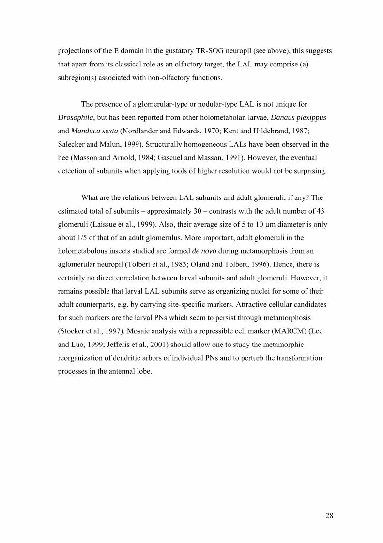
What are the relations between LAL subunits and adult glomeruli, if any? The
estimated total of subunits – approximately 30 – contrasts with the adult number of 43
glomeruli (Laissue et al., 1999). Also, their average size of 5 to 10 µm diameter is only
about 1/5 of that of an adult glomerulus. More important, adult glomeruli in the
holometabolous insects studied are formed de novo during metamorphosis from an
aglomerular neuropil (Tolbert et al., 1983; Oland and Tolbert, 1996). Hence, there is
certainly no direct correlation between larval subunits and adult glomeruli. However, it
remains possible that larval LAL subunits serve as organizing nuclei for some of their
adult counterparts, e.g. by carrying site-specific markers. Attractive cellular candidates
for such markers are the larval PNs which seem to persist through metamorphosis
(Stocker et al., 1997). Mosaic analysis with a repressible cell marker (MARCM) (Lee
and Luo, 1999; Jefferis et al., 2001) should allow one to study the metamorphic
reorganization of dendritic arbors of individual PNs and to perturb the transformation
processes in the antennal lobe.
28
Functional Implications
An important task of sensory systems is to distinguish among different
modalities. Consequently, modality-specific sensory subsystems including their central
target regions are often strictly separated. For example, in adult flies the receptor
neurons for smell and taste sit on different appendages and their afferent axons project to
spatially distinct centers. Surprisingly, the larval DO/TO complex exhibits strong ties
between smell and taste sensilla and this mix of modalities may even apply to the target
region, the LAL. These links between the two modalities may be related to the
predominant short-range orientation of larvae, which is also reflected by the dominance
of ≤ 79 gustatory over 21 olfactory receptor neurons (Table 1). For animals that live
directly on their food supply, a distinction between smell and taste stimuli may not be
very crucial. Alternatively, the links between the two senses can be explained in
developmental or evolutionary terms. For example, initially separated cephalic
structures may fuse during late embryogenesis (Schmidt-Ott et al., 1994; Campos-
Ortega and Hartenstein, 1997), as illustrated by the innervation of the dorsolateral TO
papillum from the DO ganglion (Frederik and Denell, 1982). Also, the larval system
may reflect a phylogenetically ancient state in which smell and taste systems have not
yet become fully independent.
Interestingly, recent functional and molecular data also suggest closer
relationships between smell and taste than were previously assumed. For example,
locust contact chemoreceptors can respond to certain volatile cues as well (Newland,
1998). Furthermore, the expression patterns of the newly detected family of Drosophila
gustatory receptors (Gr) (Clyne et al., 2000; Scott et al., 2001) display no clear
separation between the two modalities, in particular in the larva. Some of the Gr
members appear to be expressed in gustatory neurons, some in olfactory neurons and
some even in both. For example, Gr2B1 was found to be expressed in two dome-
associated DO neurons and in one TO neuron. These data suggest both functional and
evolutionary links between taste and smell.
In situ hybridization and Gr-driven reporter expression suggest that each
gustatory neuron expresses only one Gr type (Scott et al., 2001), similar to what had
29
been proposed before for odorant receptors (Vosshall et al., 1999; Vosshall et al., 2000).
The Gr family appears to be composed of at least 56 genes (Scott et al., 2001). So far for
seven Gr genes, promoter-driven transgene expression was detected, for five among
them in larval chemosensilla. If the remaining Gr genes are in fact expressed in the
chemosensory system, extrapolation from this 5/7 ratio suggests a total of about 40
larval Grs, i.e., functional types of neurons. This figure is compatible with the total of
about 100 chemosensory neurons determined in the larval head (Table 1) and the
observation that each of the five larval Grs is expressed in 1-3 neurons (Scott et al.,
2001). It further supports the idea that each neuron expresses a single receptor only. This
is in contrast to mammalian gustatory neurons (Hoon et al., 1999; Adler et al., 2000) and
chemosensory neurons in C. elegans (Bargmann et al., 1993; Troemel et al., 1997)
which express multiple receptors.
The Larval Olfactory System of Drosophila: a Model System?
There is emerging evidence that vertebrate and insect olfactory systems may be
organized according to common principles, in spite of largely different cell numbers
(Hildebrand and Shepherd, 1997). Hence, it is not surprising that Drosophila comprising
1300 odorant receptor neurons and less than 50 glomeruli has become a focus of
chemosensory research. Another olfactory model system, C. elegans, is attractive for an
even smaller set of 16 chemosensory neurons (Chou et al., 1996). However, expression
of multiple receptors per neuron and a unique brain organization suggest that the worm
system may operate according to different rules. Here we study the usefulness of the
larval olfactory system of Drosophila as a model, a system whose complexity in terms
of sensory neuron numbers exceeds the C. elegans system only by a factor of five. We
ask whether its central circuitry is in fact as simple as suggested by the highly reduced
number of odorant receptor neurons (Stocker, 2001). Three organizational patterns are
possible: (A) the larval system is a miniature version of the adult system, comprising
similarly reduced numbers of sensory neurons, target neurons and glomeruli, (B) the
design of the two systems is totally different, both at the peripheral and central level, or
(C) despite the reduced numbers of sensory neurons, the central target organization is as
complex as in the adult.
30
Possibility A can clearly be rejected, on the following arguments: Based on
extrapolations from the nc82 pattern and the GH146 expression pattern, the numbers of
neuropil subunits and of PNs appear to be only slightly reduced in the larva, perhaps by
a factor of 2 or 3. This is in large contrast to the reduction of olfactory receptor neurons,
which is almost two orders of magnitude lower than in the adult. In addition, the low
number of receptor neurons is not accompanied by a simpler neuroanatomy, neither in
the periphery nor in the LAL, as shown e.g. by the larval-specific association between
smell and taste. Possibility B is unlikely as well, due to the striking similarities in the
architecture of the larval and adult antennal lobe. Rather, our data support interpretation
C which states that the markedly low number of sensory neurons is not accompanied by
simplicity at the central level.
These data imply that the design of the larval chemosensory system includes
larval-specific elements in the periphery and elements shared with the adult system at
the level of the LAL. This ‘hybrid’ organization is certainly related to the fact that
during metamorphosis the sensory component of the nervous system undergoes a radical
transformation, whereas many of the central elements persist (Tissot and Stocker, 2000).
Any interpretation of functional data in the larval chemosensory system has to take into
account its specific design. Nevertheless, the observed parallels in the architecture of the
larval and adult antennal lobe may render the larval chemosensory system a very
valuable model system. The genetic and molecular tools available in Drosophila will
certainly allow to fully exploit its potential.
ACKNOLEDGEMENTS
The authors are very grateful to Bertram Gerber and Nanaë Gendre for their comments
on the manuscript.
31
LITERATURE CITED
Adler E, Hoon MA, Mueller KL, Chandrashekar J, Ryba NJ, Zuker CS. 2000. A novel
family of mammalian taste receptors. Cell 100:693-702.
Ayyub C, Paranjape J, Rodrigues V, Siddiqi O. 1990. Genetics of olfactory behavior in
Drosophila melanogaster. J Neurogenet 6:243-62.
Bargmann CI, Hartwieg E, Horvitz HR. 1993. Odorant-selective genes and neurons
mediate olfaction in C. elegans. Cell 74:515-27.
Brand AH, Perrimon N. 1993. Targeted gene expression as a means of altering cell fates
and generating dominant phenotypes. Development 118:401-15.
Campos-Ortega J, Hartenstein V. 1997. The embryonic development of Drosophila
melanogaster. Berlin Heidelberg NewYork: Springer.
Chou JH, Troemel ER, Sengupta P, Colbert HA, Tong L, Tobin DM, Roayaie K, Crump
JG, Dwyer ND, Bargmann CI. 1996. Olfactory recognition and discrimination in
Caenorhabditis elegans. Cold Spring Harb Symp Quant Biol 61:157-64.
Chu IW, Axtell RC. 1971. Fine structure of the dorsal organ of the house fly larva,
Musca domestica L. Z Zellforsch Mikrosk Anat 117:17-34.
Chu-Wang IW, Axtell RC. 1972a. Fine structure of the terminal organ of the house fly
larva, Musca domestica L. Z Zellforsch Mikrosk Anat 127:287-305.
Chu-Wang IW, Axtell RC. 1972b. Fine structure of the ventral organ of the house fly
larva, Musca domestica L. Z Zellforsch Mikrosk Anat 130:489-95.
Clyne PJ, Warr CG, Carlson JR. 2000. Candidate taste receptors in Drosophila. Science
287:1830-34.
Clyne PJ, Warr CG, Freeman MR, Lessing D, Kim J, Carlson JR. 1999. A novel family
of divergent seven-transmembrane proteins: candidate odorant receptors in
Drosophila. Neuron 22:327-38.
Cobb M. 1999. What and how do maggots smell? Biol. Rev. 74:425-59.
32
Cobb M, Bruneau S, Jallon JM. 1992. Genetic and developmental factors in the
olfactory response of Drosophila melanogaster larvae to alcohols. Proc R Soc Lond
B Biol Sci 248:103-09.
Cobb M, Dannet F. 1994. Multiple genetic control of acetate-induced olfactory
responses in Drosophila melanogaster larvae. Heredity 73:444-55.
Cobb M, Domain I. 2000. Olfactory coding in a simple system: adaptation in
Drosophila larvae. Proc R Soc Lond B Biol Sci 267:2119-25.
de Bruyne M, Clyne PJ, Carlson JR. 1999. Odor coding in a model olfactory organ: the
Drosophila maxillary palp. J Neurosci 19:4520-32.
Frederik RD, Denell RE. 1982. Embryological origin of the antenno-maxillary complex
of the larva of Drosophila melanogaster Meigen. Int J Insect Morphol Embryol
11:227-33.
Galizia CG, Nagler K, Hölldobler B, Menzel R. 1998. Odour coding is bilaterally
symmetrical in the antennal lobes of honeybees (Apis mellifera). Eur J Neurosci
10:2964-74.
Gao Q, Chess A. 1999. Identification of candidate Drosophila olfactory receptors from
genomic DNA sequence. Genomics 60:31-39.
Gascuel J, Masson C. 1991. Developmental study of afferented and deafferented bee
antennal lobes. J Neurobiol 22:795-810.
Heimbeck G, Bugnon V, Gendre N, Häberlin C, Stocker RF. 1999. Smell and taste
perception in Drosophila melanogaster larva: toxin expression studies in
chemosensory neurons. J Neurosci 19:6599-609.
Hildebrand JG, Shepherd GM. 1997. Mechanisms of olfactory discrimination:
converging evidence for common principles across phyla. Annu Rev Neurosci
20:595-631.
Hoon MA, Adler E, Lindemeier J, Battey JF, Ryba NJ, Zuker CS. 1999. Putative
mammalian taste receptors: a class of taste-specific GPCRs with distinct topographic
selectivity. Cell 96:541-51.
Jefferis GSXE, Marin EC, Stocker RF, Luo L. 2001. Target neuron prespecification in
the olfactory map of Drosophila. Nature 414:204-08.
33
Joerges J, Küttner A, Galizia CG, Menzel R. 1997. Representations of odours and odour
mixtures visualized in the honeybee brain. Nature 387:285-88.
Kankel DR, Ferrus A, Garen SH, Harte PJ, Lewis PE. 1980. The structure and
development of the nervous system. In: Ashburner M, Wright TRF, editors. The
Genetics and Biology of Drosophila. London New York San Francisco: Academic
Press. p 295-368.
Kent KS, Hildebrand JG. 1987. Cephalic sensory pathways in the central nervous
system of larval Manduca sexta (Lepidoptera : Sphingidae). Philos Trans R Soc
Lond B Biol Sci 315:1-36.
Klagges BR, Heimbeck G, Godenschwege TA, Hofbauer A, Pflugfelder GO,
Reifegerste R, Reisch D, Schaupp M, Buchner S, Buchner E. 1996. Invertebrate
synapsins: a single gene codes for several isoforms in Drosophila. J Neurosci
16:3154-65.
Laissue PP, Reiter C, Hiesinger PR, Halter S, Fischbach KF, Stocker RF. 1999. Three-
dimensional reconstruction of the antennal lobe in Drosophila melanogaster. J
Comp Neurol 405:543-52.
Lee T, Luo L. 1999. Mosaic analysis with a repressible cell marker for studies of gene
function in neuronal morphogenesis. Neuron 22:451-61.
Masson C, Arnold G. 1984. Ontogeny, maturation and plasticity of the olfactory system
in the workerbee. J Insect Physiol 30:7-14.
Murray MJ, Merritt DJ, Brand AH, Whitington PM. 1998. In vivo dynamics of axon
pathfinding in the Drosophila CNS: a time- lapse study of an identified
motorneuron. J Neurobiol 37:607-21.
Nayak SV, Singh RN. 1983. Sensilla on the tarsal segments and mouthparts of adult
Drosophila melanogaster Meigen (Diptera: Drosophilidae). Int J Insect Morphol
Embryol 12:273-91.
Newland P. 1998. Avoidance reflexes mediated by contact chemoreceptors on the legs
of locusts. J Comp Physiol [A] 183:313-24.
34

Nordlander RH, Edwards JS. 1970. Postembryonic brain development in the monarch
butterfly, Danaus plexippus plexippus L. III. Morphogenesis of centers other than
the optic lobes. W Roux’s Arch Entwicklungsmech Org 164:247-60.
Oland LA, Tolbert LP. 1996. Multiple factors shape development of olfactory
glomeruli: insights from an insect model system. J Neurobiol 30:92-109.
Oppliger FY, Guerin PM, Vlimant M. 2000. Neurophysiological and behavioural
evidence for an olfactory function for the dorsal organ and a gustatory one for the
terminal organ in Drosophila melanogaster larvae. J Insect Physiol 46:135-44.
Osborne KA, Robichon A, Burgess E, Butland S, Shaw RA, Coulthard A, Pereira HS,
Greenspan RJ, Sokolowski MB. 1997. Natural behavior polymorphism due to a
cGMP-dependent protein kinase of Drosophila. Science 277:834-36.
Rodrigues V. 1988. Spatial coding of olfactory information in the antennal lobe of
Drosophila melanogaster. Brain Res 453:299-307.
Salecker I, Malun D. 1999. Development of olfactory glomeruli. In: Hansson BS, editor.
Insect olfaction. Berlin Heidelberg NewYork: Springer. p 207-42.
Schmidt-Ott U, Gonzalez-Gaitan M, Jäckle H, Technau GM. 1994. Number, identity,
and sequence of the Drosophila head segments as revealed by neural elements and
their deletion patterns in mutants. Proc Natl Acad Sci U S A 91:8363-67.
Scott K, Brady R, Cravchik A, Morozov P, Rzhetsky A, Zuker C, Axel R. 2001. A
chemosensory gene family encoding candidate gustatory and olfactory receptors in
Drosophila. Cell 104:661-73.
Shanbhag SR, Müller B, Steinbrecht RA. 1999. Atlas of olfactory organs of Drosophila
melanogaster. 1. Types, external organization, innervation and distribution of
olfactory sensilla. Int J Insect Morphol Embryol 28:377-97.
Shanbhag SR, Park SK, Pikielny CW, Steinbrecht RA. 2001. Gustatory organs of
Drosophila melanogaster: fine structure and expression of the putative odorant-
binding protein PBPRP2. Cell Tissue Res 304:423-37.
Singh RN. 1997. Neurobiology of the gustatory systems of Drosophila and some
terrestrial insects. Microsc Res Tech 39:547-63.
35
Singh RN, Singh K. 1984. Fine structure of the sensory organs of Drosophila
melanogaster Meigen larva (Diptera: Drosophilidae). Int J Insect Morphol Embryol
13:255-73.
Stocker RF. 1994. The organization of the chemosensory system in Drosophila
melanogaster: a review. Cell Tissue Res 275:3-26.
Stocker RF. 2001. Drosophila as a focus in olfactory research: mapping of olfactory
sensilla by fine structure, odor specificity, odorant receptor expression and central
connectivity. Microsc Res Tech 55:284-96.
Stocker RF, Tissot M, Gendre N. 1995. Morphogenesis and cellular proliferation pattern
in the developing antennal lobe of Drosophila melanogaster. Roux's Arch Dev Biol
205:62-72.
Stocker RF, Heimbeck G, Gendre N, de Belle JS. 1997. Neuroblast ablation in
Drosophila P[GAL4] lines reveals origins of olfactory interneurons. J Neurobiol
32:443-56.
Strausfeld NJ. 2001. Insect Brain. In: Roth G, Wulliman MF, editors. Brain, Evolution
& Cognition. New York: Wiley. p 367-400.
Tissot M, Gendre N, Hawken A, Störtkuhl KF, Stocker RF. 1997. Larval chemosensory
projections and invasion of adult afferents in the antennal lobe of Drosophila. J
Neurobiol 32:281-97.
Tissot M, Stocker RF. 2000. Metamorphosis in Drosophila and other insects: the fate of
neurons throughout the stages. Prog Neurobiol 62:89-111.
Tolbert LP, Matsumoto SG, Hildebrand JG. 1983. Development of synapses in the
antennal lobes of the moth Manduca sexta during metamorphosis. J Neurosci
3:1158-75.
Troemel ER, Kimmel BE, Bargmann CI. 1997. Reprogramming chemotaxis responses:
sensory neurons define olfactory preferences in C. elegans. Cell 91:161-69.
Vosshall LB. 2000. Olfaction in Drosophila. Curr Opin Neurobiol 10:498-503.
Vosshall LB. 2001. The molecular logic of olfaction in Drosophila. Chem Senses
26:207-13.
36

Vosshall LB, Amrein H, Morozov PS, Rzhetsky A, Axel R. 1999. A spatial map of
olfactory receptor expression in the Drosophila antenna. Cell 96:725-36.
Vosshall LB, Wong AM, Axel R. 2000. An olfactory sensory map in the fly brain. Cell
102:147-59.
Warr C, Clyne P, de Bruyne M, Kim J, Carlson JR. 2001. Olfaction in Drosophila:
coding, genetics and e-genetics. Chem Senses 26:201-06.
Yeh E, Gustafson K, Boulianne GL. 1995. Green fluorescent protein as a vital marker
and reporter of gene expression in Drosophila. Proc Natl Acad Sci U S A 92:7036-
40.
37
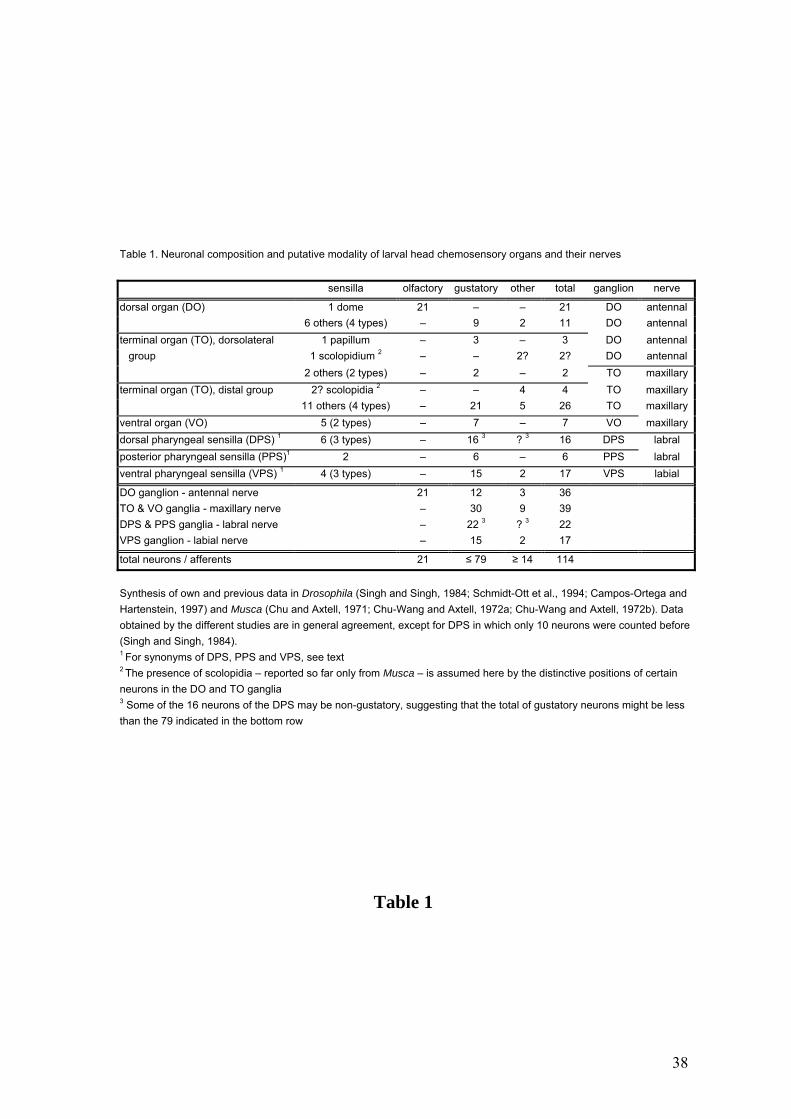
Table 1. Neuronal composition and putative modality of larval head chemosensory organs and their nerves
sensilla olfactory gustatory other total ganglion nerve
dorsal organ (DO) 1 dome 21 – – 21 DO antennal 6 others (4 types) – 9 2 11 DO antennal terminal organ (TO), dorsolateral 1 papillum – 3 – 3 DO antennal group 1 scolopidium 2 – – 2? 2? DO antennal 2 others (2 types) – 2 – 2 TO maxillary terminal organ (TO), distal group 2? scolopidia 2 – – 4 4 TO maxillary 11 others (4 types) – 21 5 26 TO maxillary ventral organ (VO) 5 (2 types) – 7 – 7 VO maxillary dorsal pharyngeal sensilla (DPS) 1 6 (3 types) – 16 3 ? 3 16 DPS labral posterior pharyngeal sensilla (PPS)1 2 – 6 – 6 PPS labral ventral pharyngeal sensilla (VPS) 1 4 (3 types) – 15 2 17 VPS labial
DO ganglion - antennal nerve 21 12 3 36 TO & VO ganglia - maxillary nerve – 30 9 39 DPS & PPS ganglia - labral nerve – 22 3 ? 3 22 VPS ganglion - labial nerve – 15 2 17
total neurons / afferents 21 ≤ 79 ≥ 14 114
Synthesis of own and previous data in Drosophila (Singh and Singh, 1984; Schmidt-Ott et al., 1994; Campos-Ortega and Hartenstein, 1997) and Musca (Chu and Axtell, 1971; Chu-Wang and Axtell, 1972a; Chu-Wang and Axtell, 1972b). Data obtained by the different studies are in general agreement, except for DPS in which only 10 neurons were counted before(Singh and Singh, 1984). 1 For synonyms of DPS, PPS and VPS, see text 2 The presence of scolopidia – reported so far only from Musca – is assumed here by the distinctive positions of certain neurons in the DO and TO ganglia 3 Some of the 16 neurons of the DPS may be non-gustatory, suggesting that the total of gustatory neurons might be less than the 79 indicated in the bottom row
Table 1
38

FIGURE LEGENDS
Fig.1. Confocal images showing the chemosensory circuitry in third instar larva of D.
melanogaster. A: Reporter expression in the P[GAL4] line 4551 reveals the dorsal and
terminal organs (DO, TO) including their ganglia (DOG, TOG) as well as dorsal and
posterior groups of pharyngeal sensilla (DPS, PPS). Afferent axons from the DOG
travel by means of the antennal nerve (AN), those from the TOG by means of the
maxillary nerve (MN), and pharyngeal afferents by means of the labral nerve (LN).
Arrowhead: contact between LN and AN. Strong expression is present in the larval
antennal lobe (LAL), the tritocerebral-suboesophageal neuropil (TR-SOG) and the
mushroom bodies (MB). B: Line 189Y labels afferents from the DPS and PPS in the
LN, as well as local interneurons (LI) of the LAL. Arrowhead: junction of nerves from
the DPS and PPS. Inset: Higher magnification of the LAL, the entrance of the LN in the
TR-SOG area and LN-derived afferent terminals (arrowheads). C: Close-up of DO and
TO expression in line 4551. Most of the labeled dendrites of the DOG extend in bundles
(BU) into the central dome (DM), except three dendrites which invade the TO
(arrowhead). They end in a papillum (P) of the dorsolateral group of the TO (DLG,
inset) (see text for details). DIG: distal group of TO sensilla. D: Cuticular
autofluorescence reveals TO, DM and ventral organ (VO) (wildtype CS). E: Line 4551
showing the entries of the AN (double arrowheads), the LN (large arrowhead) and the
MN (small arrowhead) into the CNS. Expression includes several central elements, e.g.,
an interneuron with a contralateral process (CI). F: Line GH86 reveals a characteristic
loop of the AN before joining the CNS (large arrowhead) and its separation into three
branches (small arrowheads). A-C & E: Tau-GFP reporter; C (inset) & F: GFP reporter.
The numbers of optical sections and the section thickness vary in different panels. The
CNS is oriented with anterior on top. Bars = 100 µm in A,B; 25 µm in C-F.
39
Fig. 2. Confocal images of the DO, TO and VO (A) and of the larval CNS (B-G) in the
line 4551 (except B: 189Y). A: Anti-Elav labeling (red) displays neurons in the DOG,
TOG and the VO ganglion (VOG). A VO neuron expressing Tau-GFP (arrowhead)
sends its dendrite (DE) toward the VO opening. AG: associated ganglion (see text for
details). B: Tau-GFP expression in 189Y in afferents (cf. Fig. 1B) and in local
interneurons (LI) (green) and anti-Synapsin immunostaining (red) show overlap in the
LALs (encircled, inset). C: GFP expression shows the LAL (encircled), the TR-SOG
region and the MB Kenyon cells (KC). D: Overlap of Tau-GFP (green) and neuropil-
specific nc82 staining (red) in the LAL, TR-SOG region and MBs. E: Single sections of
adult AL and LAL (inset) at the same magnification, labeled by nc82. F: Double
staining of GFP (green) and nc82 (red) yields overlap in the LAL and the MB calyx
(CX). G: Serial sections of the LAL (encircled in F) at 3 µm intervals. Afferent GFP
label (green) and neuropil nc82 label (red) and their overlap (third row) are shown.
Small arrowheads indicate LAL subunits labeled by both markers. The pattern
similarities between the two markers are particularly obvious for two subunits, E and D.
Anterior is on top. Bars = 10 µm in E,G; 25 µm in A,F; 50 µm in C; 100 µm in B,D.
Fig. 3. Entire set of pharyngeal sensilla shown by anti-Elav (A) and Tau-GFP reporter
pattern (B) in line 4551. DPS, PPS, VPS: dorsal, ventral and posterior pharyngeal
sensilla, respectively. Anterior is to the right. Bar = 100 µm.
Fig. 4. Single confocal sections of the LAL in line 4551 shown by afferent GFP reporter
labeling (left panels) and anti-Synapsin (B,F) or nc82 neuropil markers (D,H). Left and
right panels represent the same section each. Panels E,F and G,H are 6 µm apart from
the sections A,B and C,D, respectively. The subunits D and E visualized by GFP
labeling are also stained by the anti-Synapsin and anti-nc82 markers. The arrowhead
indicates a seemingly identical subunit labeled by GFP (A,C) and the two neuropil
markers (B,D). Bar = 10 µm.
Fig. 5. The LAL visualized by lines GH86 (A), 4551 (B,C) and GH146 (D). A: The AN
splits into a major and two minor branches (arrowheads). The minor branches project
into the more intensely labeled elongated domains E and E’(encircled). Inset: LAL at
40
lower magnification. B,C: Intensely labeled domains E and D including an axon
projecting into the latter (arrowheads in C). B: LAL at the entire depth. C: Assembly of
3 optical sections of the same LAL. Inset: same LAL at lower magnification. D:
Projection neurons comprise a lateral cluster (LC) and an anterodorsal cluster (AC) of
about 20 and 30 cell bodies, respectively. CB: isolated cell body of unknown identity.
A-C: GFP reporter; D: TAU-GFP reporter. Anterior is on top. Bars = 10 µm in A-C; 25
µm in D.
Fig. 6. Local interneurons shown by line 189Y (A) and projection neurons shown by
GH146 (B) both display arborizations in small domains of the LAL (green, arrowheads).
These domains overlap the neuropil subunits shown by nc82 (red) labeling, as shown in
the third column. GFP reporter. Bars = 10 µm.
Fig. 7. Successive sections through the LAL in line GH146 show two small subregions
(stippled in A-C and in D-F) which are innervated by a dendritic process (arrowheads)
extending from the PN trunk. Tracing of confocal stacks suggests restriction of
individual dendritic arbors to single subregions. The number of processes does not
exceed the number of PN cell bodies suggesting that each PN may innervate a single
subregion. iACT inner antennocerebral tract. Tau-GFP reporter. Anterior is on top. Bar
= 10 µm.
Fig. 8. Schematic diagram of head chemosensilla in the third instar larva of D.
melanogaster including peripheral nerves and central target regions. Olfactory dendrites
are shown as a W-symbol, gustatory dendrites as black boxes. Gray circles in the LAL
denote subunits, and the gray rectangle indicates the E domain (see text for details). The
dashed line represents projections from gustatory DO sensilla to the TR-SOG region
(Tissot et al., 1997) which are unlabeled by the lines used here. DIG/DLG
distal/dorsolateral group of TO sensilla, DM dome, LPR lateral protocerebrum, MB
mushroom bodies, PH pharynx. For other abbreviations, see list.
41
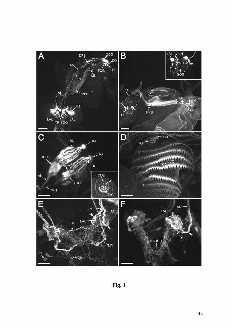
Fig. 1
42

Fig. 2
43
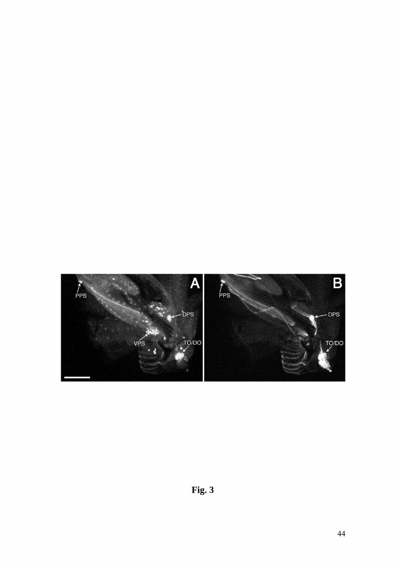
Fig. 3
44

Fig. 4
45
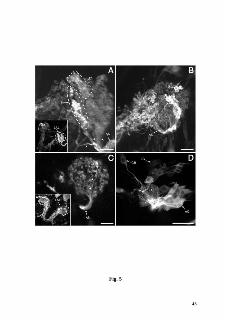
Fig. 5
46
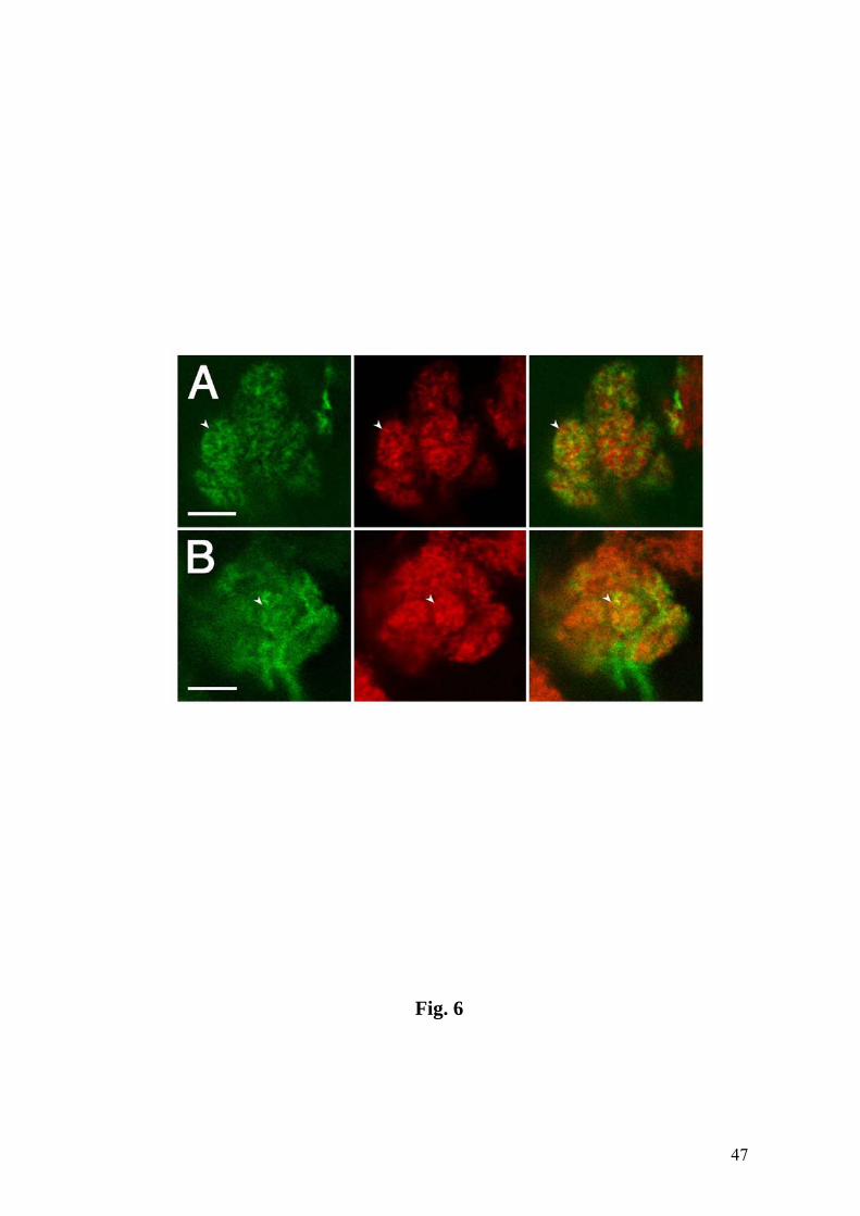
Fig. 6
47
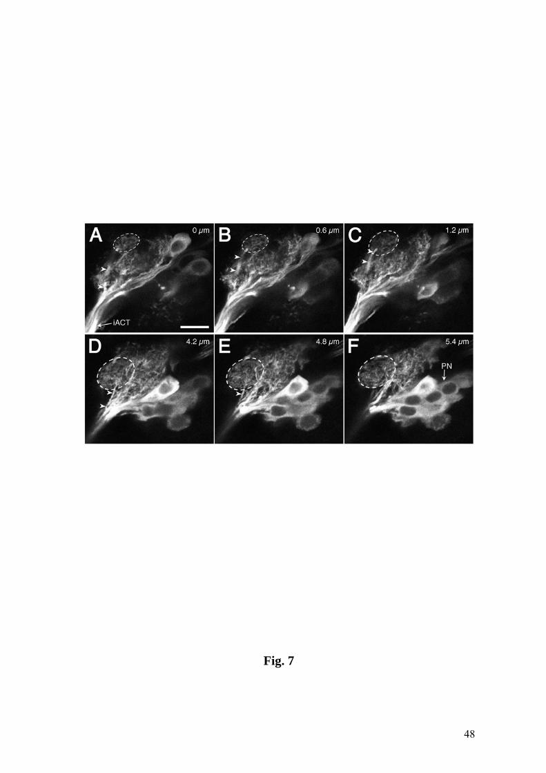
Fig. 7
48
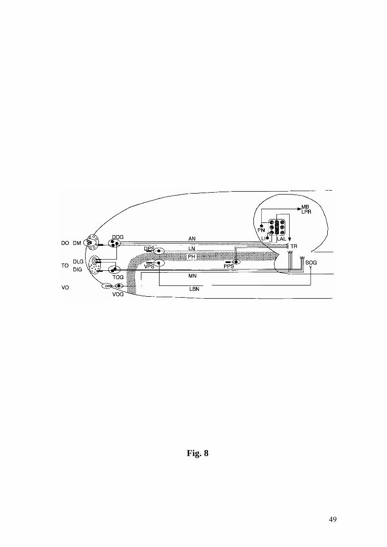
Fig. 8
49

Chapter 2
Immunoreactivity against choline acetyltransferase,
GABA, histamine, octopamine and serotonin
in the larval chemosensory system
of Drosophila melanogaster
This chapter has been submitted to The Journal of Comparative Neurology.
50

ABSTRACT
We study the distribution of choline acetyltransferase (ChAT), γ-aminobutyric
acid (GABA), histamine, octopamine and serotonin in the larval chemosensory system
of Drosophila melanogaster. Colocalization at the confocal level with GFP or Tau-GFP
reporters, expressed in selected P[GAL4] enhancer trap lines was used to identify the
cellular localization of these neurotransmitters. As in the adult fly, larval olfactory
afferents project into the (larval) antennal lobe (LAL), where they synapse onto local
interneurons and projection neurons, while gustatory afferents terminate essentially in
the tritocerebral-suboesophageal (TR-SOG) region. We demonstrate that the neuropils
of the LAL and the TR-SOG are immunoreactive to ChAT and GABA. In addition,
serotonin- and octopamine immunoreactive fibers are present in the LAL. ChAT
immunoreactivity is localized in subsets of olfactory and gustatory afferents and in many
of the projection neurons. In contrast, GABA is expressed in most, perhaps all of the
local interneurons. Finally, serotonin immunoreactivity in the LAL derives from a single
neuron that is situated close to the LAL and arborizes in additional neuropil regions.
These findings resemble the situation in the adult fly. Hence, considering the highly
reduced numbers of odorant receptor neurons in the larva as shown in our previous study
(cf. Python and Stocker, 2002), the larval system may become an attractively simple
model system for studying the roles of neurotransmitters in olfactory processing.
Abbreviations: ACh acetylcholine, AL antennal lobe, ChAT choline acetyltransferase,
DO dorsal organ, GABA γ-aminobutyric acid, IR immunoreactive, IRy
immunoreactivity, LAL larval antennal lobe, LI local interneuron, PN projection neuron,
SOG suboesophageal ganglion, TO terminal organ, TR tritocerebrum
51
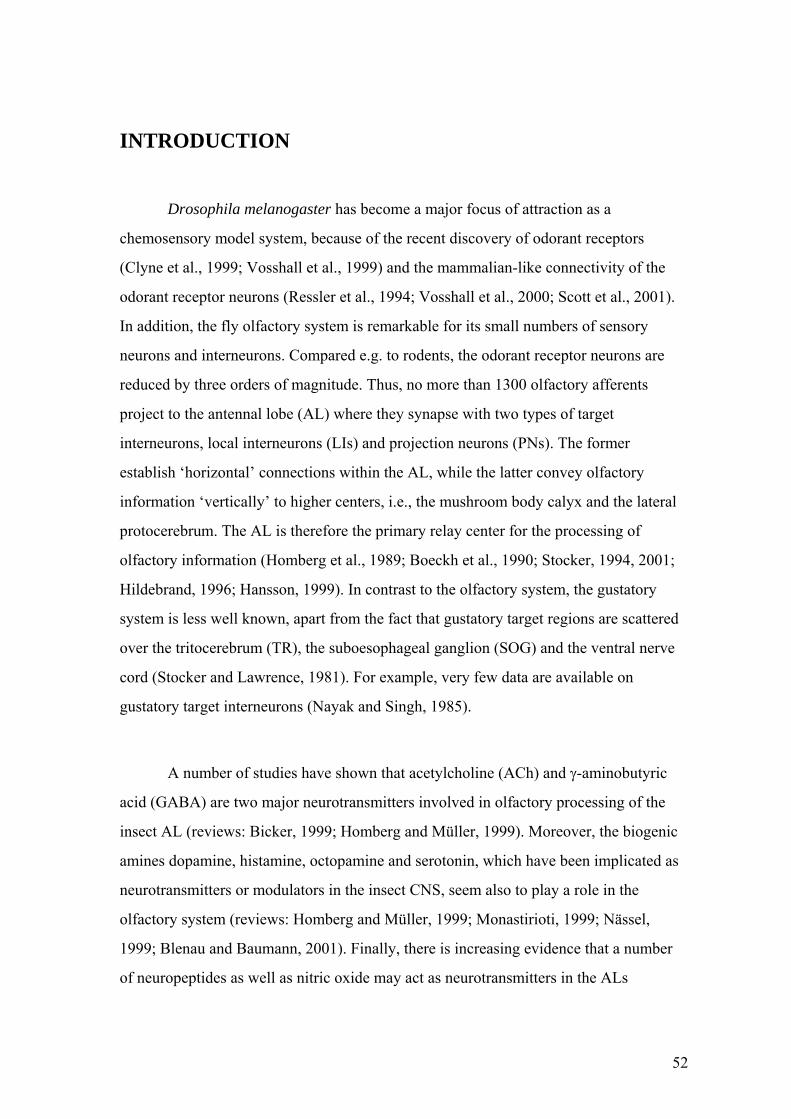
INTRODUCTION
Drosophila melanogaster has become a major focus of attraction as a
chemosensory model system, because of the recent discovery of odorant receptors
(Clyne et al., 1999; Vosshall et al., 1999) and the mammalian-like connectivity of the
odorant receptor neurons (Ressler et al., 1994; Vosshall et al., 2000; Scott et al., 2001).
In addition, the fly olfactory system is remarkable for its small numbers of sensory
neurons and interneurons. Compared e.g. to rodents, the odorant receptor neurons are
reduced by three orders of magnitude. Thus, no more than 1300 olfactory afferents
project to the antennal lobe (AL) where they synapse with two types of target
interneurons, local interneurons (LIs) and projection neurons (PNs). The former
establish ‘horizontal’ connections within the AL, while the latter convey olfactory
information ‘vertically’ to higher centers, i.e., the mushroom body calyx and the lateral
protocerebrum. The AL is therefore the primary relay center for the processing of
olfactory information (Homberg et al., 1989; Boeckh et al., 1990; Stocker, 1994, 2001;
Hildebrand, 1996; Hansson, 1999). In contrast to the olfactory system, the gustatory
system is less well known, apart from the fact that gustatory target regions are scattered
over the tritocerebrum (TR), the suboesophageal ganglion (SOG) and the ventral nerve
cord (Stocker and Lawrence, 1981). For example, very few data are available on
gustatory target interneurons (Nayak and Singh, 1985).
A number of studies have shown that acetylcholine (ACh) and γ-aminobutyric
acid (GABA) are two major neurotransmitters involved in olfactory processing of the
insect AL (reviews: Bicker, 1999; Homberg and Müller, 1999). Moreover, the biogenic
amines dopamine, histamine, octopamine and serotonin, which have been implicated as
neurotransmitters or modulators in the insect CNS, seem also to play a role in the
olfactory system (reviews: Homberg and Müller, 1999; Monastirioti, 1999; Nässel,
1999; Blenau and Baumann, 2001). Finally, there is increasing evidence that a number
of neuropeptides as well as nitric oxide may act as neurotransmitters in the ALs
52
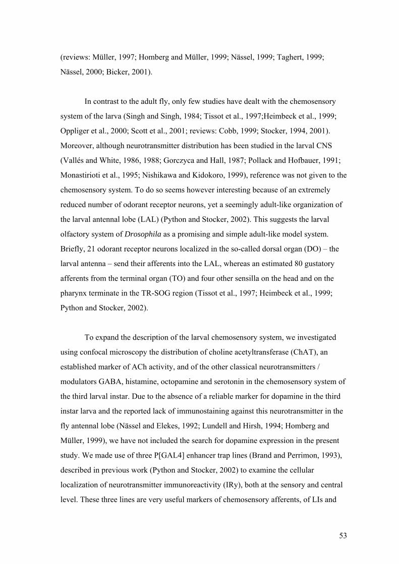
(reviews: Müller, 1997; Homberg and Müller, 1999; Nässel, 1999; Taghert, 1999;
Nässel, 2000; Bicker, 2001).
In contrast to the adult fly, only few studies have dealt with the chemosensory
system of the larva (Singh and Singh, 1984; Tissot et al., 1997;Heimbeck et al., 1999;
Oppliger et al., 2000; Scott et al., 2001; reviews: Cobb, 1999; Stocker, 1994, 2001).
Moreover, although neurotransmitter distribution has been studied in the larval CNS
(Vallés and White, 1986, 1988; Gorczyca and Hall, 1987; Pollack and Hofbauer, 1991;
Monastirioti et al., 1995; Nishikawa and Kidokoro, 1999), reference was not given to the
chemosensory system. To do so seems however interesting because of an extremely
reduced number of odorant receptor neurons, yet a seemingly adult-like organization of
the larval antennal lobe (LAL) (Python and Stocker, 2002). This suggests the larval
olfactory system of Drosophila as a promising and simple adult-like model system.
Briefly, 21 odorant receptor neurons localized in the so-called dorsal organ (DO) – the
larval antenna – send their afferents into the LAL, whereas an estimated 80 gustatory
afferents from the terminal organ (TO) and four other sensilla on the head and on the
pharynx terminate in the TR-SOG region (Tissot et al., 1997; Heimbeck et al., 1999;
Python and Stocker, 2002).
To expand the description of the larval chemosensory system, we investigated
using confocal microscopy the distribution of choline acetyltransferase (ChAT), an
established marker of ACh activity, and of the other classical neurotransmitters /
modulators GABA, histamine, octopamine and serotonin in the chemosensory system of
the third larval instar. Due to the absence of a reliable marker for dopamine in the third
instar larva and the reported lack of immunostaining against this neurotransmitter in the
fly antennal lobe (Nässel and Elekes, 1992; Lundell and Hirsh, 1994; Homberg and
Müller, 1999), we have not included the search for dopamine expression in the present
study. We made use of three P[GAL4] enhancer trap lines (Brand and Perrimon, 1993),
described in previous work (Python and Stocker, 2002) to examine the cellular
localization of neurotransmitter immunoreactivity (IRy), both at the sensory and central
level. These three lines are very useful markers of chemosensory afferents, of LIs and
53

PNs. We observe strong ChAT IRy and GABA IRy in the olfactory and gustatory target
regions, the LAL and the TR-SOG, as well as IR varicosities for octopamine and
serotonin in the LAL. Our data suggest that subsets of olfactory and gustatory afferents
and many PNs are cholinergic, and that all LIs labeled by the marker line used are
GABAergic. Finally, a serotonergic interneuron – independent of LIs or PNs – was
found to project into the LAL.
MATERIALS AND METHODS
The P[GAL4] line GH146 was generated by G. Heimbeck (Stocker et al., 1997;
Heimbeck et al., 2001), line 4551 was obtained from J.-F. Ferveur (Université de
Bourgogne, Dijon) and line 189Y (Osborne et al., 1997) was provided by K. Kaiser
(University of Glasgow). As UAS-reporter strains, we used UAS-GFP (Yeh et al., 1995)
and UAS-Tau-GPF (Murray et al., 1998), both kindly provided by A. H. Brand
(Wellcome/CRC, Cambridge). Animals were raised between 18°C and 25°C on standard
cornmeal medium. P[GAL4]/UAS-reporter heterozygotes were collected as feeding
third instar larvae.
The antibody directed against choline acetyltransferase (ChAT, # 4B1; a
generous gift from P. M. Salvaterra; Beckman Institute, City of Hope) was applied at
0.6µg/ml, anti-Histamine (kindly provided by E. Buchner, University of Würzburg,
Germany) at 1:500 or 1:1000, anti-Octopamine (Chemicon) at 1:500, anti-serotonin
(Sigma) at 1:1000, and anti-GABA (# 4TB, provided by H. Dircksen, University of
Bonn, Germany) at 1:500 or 1:1000. Cy3-coupled goat anti-mouse or anti-rabbit IgGs
were used as secondary antibodies (1:100, Jackson ImmunoResarch).
For antibody staining, the larvae were essentially processed as previously
described (Python and Stocker, 2002). However, for anti-Histamine and anti-
Octopamine, we used a fresh solution of 4% EDAC (Sigma) dissolved in phosphate
54
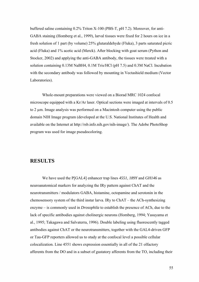
buffered saline containing 0.2% Triton X-100 (PBS-T, pH 7.2). Moreover, for anti-
GABA staining (Homberg et al., 1999), larval tissues were fixed for 2 hours on ice in a
fresh solution of 1 part (by volume) 25% glutaraldehyde (Fluka), 3 parts saturated picric
acid (Fluka) and 1% acetic acid (Merck). After blocking with goat serum (Python and
Stocker, 2002) and applying the anti-GABA antibody, the tissues were treated with a
solution containing 0.13M NaBH4, 0.1M Tris/HCl (pH 7.5) and 0.3M NaCl. Incubation
with the secondary antibody was followed by mounting in Vectashield medium (Vector
Laboratories).
Whole-mount preparations were viewed on a Biorad MRC 1024 confocal
microscope equipped with a Kr/Ar laser. Optical sections were imaged at intervals of 0.5
to 2 µm. Image analysis was performed on a Macintosh computer using the public
domain NIH Image program (developed at the U.S. National Institutes of Health and
available on the Internet at http://rsb.info.nih.gov/nih-image/). The Adobe PhotoShop
program was used for image pseudocoloring.
RESULTS
We have used the P[GAL4] enhancer trap lines 4551, 189Y and GH146 as
neuroanatomical markers for analyzing the IRy pattern against ChAT and the
neurotransmitters / modulators GABA, histamine, octopamine and serotonin in the
chemosensory system of the third instar larva. IRy to ChAT – the ACh-synthesizing
enzyme – is commonly used in Drosophila to establish the presence of ACh, due to the
lack of specific antibodies against cholinergic neurons (Homberg, 1994; Yasuyama et
al., 1995; Takagawa and Salvaterra, 1996). Double labeling using fluorescently tagged
antibodies against ChAT or the neurotransmitters, together with the GAL4-driven GFP
or Tau-GFP reporters allowed us to study at the confocal level a possible cellular
colocalization. Line 4551 shows expression essentially in all of the 21 olfactory
afferents from the DO and in a subset of gustatory afferents from the TO, including their
55
terminals in the LAL and TR-SOG region, respectively (Python and Stocker, 2002).
Line 189Y labels chemosensory afferents of the labral nerve as well as a subset of five to
six LIs, whereas line GH146 identifies about 50 PNs from an unknown total. Neither for
ChAT nor for the neurotransmitters studied here, IRy was observed in the cell bodies of
the sensory neurons. However, staining was seen in afferent terminals and in central
elements of the chemosensory pathway. Yet, pharyngeal sensilla were not studied here.
Choline acetyltransferase
Previous reports have shown strong ChAT IRy in the entire larval neuropil
including the LAL (Gorczyca and Hall, 1987; Yasuyama et al., 1995). A considerable
proportion of this IRy appears to be due to the terminals of cholinergic afferents
(Yasuyama and Salvaterra, 1999). In the present work we tried to test this notion in the
larval brain. Interestingly, the terminals of the 21 olfactory afferents which all express
GFP in line 4551 (Python and Stocker, 2002) often overlap in the LAL with ChAT-IR
elements in the LAL (Fig. 1A,B). In addition, two recently discovered subunits ‘D’ and
‘E’ inside the LAL neuropil, which are visualized by the neuropil-specific monoclonal
antibody nc82 (cf. Python and Stocker, 2002) coincide perfectly with the structures
labeled by anti-ChAT (Fig. 1C-F). These results suggest that ChAT may indeed be
expressed in afferent terminals. ChAT staining is also seen in the antennal nerve at its
entrance in the LAL (Fig. 1A,B), but neither in the peripheral parts of the nerve nor in
the sensory neurons of its origin, the DO. Lack of anti-ChAT staining in sensory neurons
was also reported from the adult olfactory system (Yasuyama et al., 1995). In contrast,
odorant receptor neurons on the antenna and maxillary palps were readily labeled by
ChAT/lacZ transformants (Kitamoto et al., 1995; Yasuyama and Salvaterra, 1999).
Hence, the absence of ChAT IRy in the peripheral part of the larval olfactory system
may not be conclusive.
Colocalization between GFP expressing afferent structures in line 4551 and
elements labeled by anti-ChAT occurs also in the gustatory target region, i.e. the TR-
SOG area (Fig. 1A,B). Minor IRy is seen in the labral nerve (not shown), but none in the
56

maxillary nerve or in its origin, the TO (Fig. 1A,B). Again, since gustatory neurons in
the adult were labeled in ChAT/lacZ transformants (Kitamoto et al., 1995) (but probably
not by anti-ChAT staining), the lack of ChAT IRy does not exclude the cholinergic
nature of larval gustatory neurons.
In addition to the colocalization of ChAT with chemosensory afferents, we
observe ChAT IRy in target neurons of the LAL. Overlap with reporter expression in
interneurons is clearly seen in the PN marker line GH146 (Fig. 1I,J), but not in the LI
marker line 189Y (Fig. 1G,H). According to their shape and their output processes in the
inner antennocerebral tract, the labeled neurons in GH146 are obviously PNs. From the
two clusters of PNs (cf. Python and Stocker, 2002), only the anterodorsal one carries
IRy, but not the lateral one (Fig. 1I,J). Moreover, only approximately half of the
anterodorsal PNs show anti-ChAT staining. In the PN region, we also observed ChAT-
positive cell bodies that do not overlap with GFP-positive PNs of the GH146 line,
indicating that the number of PNs labeled by this line is incomplete (Fig. 1I,J). In
agreement with our data, subsets of PNs in the adult olfactory pathway of Drosophila
(Buchner et al., 1986; Gorczyca and Hall, 1987), Manduca sexta (Homberg et al., 1995)
and the honeybee (Kreissl and Bicker, 1989; Bicker, 1999) were supposed to be
cholinergic. In summary, the presence of ChAT IRy in olfactory and gustatory terminals
and in subsets of PNs suggests that ACh may play a major role in larval olfactory and
gustatory processing.
GABA
Despite some background staining displayed by the anti-GABA antiserum, it has
proven useful to reveal GABAergic neurons in the locust, being identical to the IRy
pattern for the GABA-synthesizing enzyme glutamic acid decarboxylase (Homberg et
al., 1999). In the larval CNS of Drosophila we observe GABA IRy in the LAL and the
TR-SOG, but the staining pattern does not coincide with the terminals of olfactory or
gustatory afferents visualized by line 4551 (Fig. 2A,B). Moreover, neither the
chemosensory neurons in the DO and TO nor the nerves carrying their afferents show
57

any anti-GABA staining. In contrast, all the LIs labeled by line 189Y exhibit strong
GABA IRy (Fig. 2C,D). Numerous additional GABA-IR somata are present in the brain,
many of them even in close vicinity to the LAL. Yet, none of them seems to correspond
to a PN, as no colocalization was observed with any of the 50 PNs labeled by the line
GH146 (Fig. 2E,F) (Python and Stocker, 2002). This is unlike M. sexta where subsets of
PNs were reported to be GABA-IR (Hoskins et al., 1986). It is conceivable that some
among these additional GABA-IR neurons represent LIs that do not express GAL4 in
the 189Y line. However, the majority of them remain unknown. Their abundance also in
the TR-SOG cortex region even suggests that some of the gustatory interneurons – yet
unidentified – might be GABAergic as well.
These data for the first time assign GABA IRy in Drosophila positively to LIs.
Previous studies had reported higher-than-background GABA IRy in the glomeruli of
the adult AL (Aronstein and ffrench-Constant, 1995; Harrison et al., 1996). The
presence of GABA in LIs is well known from a variety of species, such as honeybees,
M. sexta, Periplaneta americana and Schistocerca gregaria (review: Homberg and
Müller, 1999). These parallels suggest that the role played by GABA in olfactory
processing might be shared in different species as well as in adult and larval systems.
Histamine
Based on the IRy of an antibody directed against histamine, this biogenic amine
was proposed as a major neurotransmitter in photoreceptor and mechanosensory bristle
neurons of adult Drosophila (Pollack and Hofbauer, 1991; Buchner et al., 1993). For
example, afferents from antennal mechanosensory bristles were labeled, but neither
olfactory afferents (nor presumably gustatory afferents from the legs). Consistent with
this view, we did not observe any histamine IRy in the chemosensory system of the larva
when applying the same antibody, neither in sensory neurons of the DO and TO nor in
the LAL and its cellular components LIs and PNs (Fig. 3A-D). This is in contrast to a
number of hemi- and holometabolous insects in which histamine IRy was detected in
subsets of LIs (Homberg and Müller, 1999). Yet, we found anti-histamine label in a
58
cluster of three neurons and two additional separate neurons in each brain hemisphere.
Most or all of them establish arborizations in the SOG region but do not show any
obvious overlap with putative gustatory terminals labeled by line 4551 (Fig. 3A,B).
Thus, histamine does not seem to be involved as a neurotransmitter in fly larval primary
chemosensory processing. We also note that none of the chemosensory head nerves –
antennal, labral or maxillary nerve – exhibits distinct histamine IRy. Hence, if histamine
in the larva shares its adult function, then neither the DO nor the TO appear to comprise
mechanosensory components.
The adult brain was reported to contain about 12 histamine-IR pairs of cells
(Pollack and Hofbauer, 1991). However, the relations between them and the five pairs
we observe in the larval brain remain unknown. Likewise, possible links between an
additional 20 histamine-IR neurons in the ventral nerve cord (Fig. 3A) and 18 histamine-
containing neurons in the thoracico-abdominal ganglia of adult Drosophila (Nässel et
al., 1990) remain in the dark.
Octopamine
The general pattern of octopamine IRy in the larval brain and SOG has been
described previously (Monastirioti et al., 1995). Accordingly, a cluster of 10-14
octopamine-IR cells in the ventral midline of the SOG exhibits profuse IR varicosities
throughout the neuropil. We confirm this pattern, despite elevated background staining
of the anti-octopamine antibody used (Fig. 3E,I). Moreover, we note that the nerves
carrying olfactory and gustatory afferents as well as their cell bodies in the DO and the
TO are devoid of labeling. Although low-density varicose IR fibers are present in the
LAL, there is no coincidence with afferent terminals as shown by line 4551 (Fig. 3E).
Similarly, there is no overlap of octopamine IRy and reporter labeling in the LI and PN
marker lines (Fig. 3F-J). This is in contrast to the adult fly in which octopamine-IR cells
were found found in the AL cortex, close to the midline (Monastirioti et al., 1995). We
believe that one of the octopamine-IR cell bodies in the ventral midline gives rise to the
arborization in the LAL but are not able to exactly identify the neuron. Very likely the
59
adult octopamine-IR cells near the midline derive from the larval midline cluster
(Monastirioti et al., 1995). Finally, no evidence of octopamine IRy is present in the
terminal region of gustatory afferents shown by line 4551. These data indicate that
octopamine might at most be involved indirectly in larval olfactory processing, perhaps
as a general neuromodulator.
Serotonin
The general pattern and development of serotonin IRy in Drosophila was
described previously (Vallés and White, 1988). In the larva, four IR clusters comprising
a total of 22 neurons were observed, whose arborizations cover most of the central brain
neuropil. However, their relations with chemosensory pathways were not studied. Using
the 4551 marker line, we note the absence of serotonin IRy in chemosensory neurons
and their afferents in the peripheral nerves. Also, we see no IR overlap with olfactory
afferents in the LAL, and no staining at all in the TR-SOG region (Fig. 4A), suggesting
that serotonin is not involved in afferent chemosensory processing. Moreover, neither
the LIs nor the PNs visualized by the two marker lines show colocalization of serotonin
IRy (Fig. 4B-D). On the other hand, the LAL displays a network of varicose serotonin-
IR fibers which derive from a single cell body close to the anterodorsal PN cluster (Fig.
4C,D). This neuron does not extend a process in the inner antennocerebral tract,
argueing against its PN identity. Instead, it appears to establish arborizations in other
neuropil regions of the brain (not shown), but the documentation of its morphology is
hampered by two accompanying neurons which are even stronger serotonin-IR. They
exhibit a contralateral branch, but do not innervate the LAL (Fig. 4C,D). Comparison
with the study of Vallés and White (1988) suggests these three neurons belong to the
SP2 cluster. Interestingly, all serotonin-IR larval neurons including the SP2 neurons
appear to persist through metamorphosis and become integrated into the adult brain
(Vallés and White, 1988). Moreover, serotonin-IR varicosities have been observed in the
adult AL, although the corresponding cell body/bodies remain(s) unknown (Vallés and
White, 1988). In M. sexta, a larval serotonergic AL interneuron is remarkable for its
persistence through metamorphosis (Kent et al., 1987). Although its anatomy is different
60
from the neuron we observe, they might share similar functions, perhaps as modulatory
neurons in the larval and adult olfactory pathway.
DISCUSSION
Here we determine the cellular localization of ChAT, GABA, histamine,
octopamine and serotonin in the chemosensory system of the third larval instar, by
applying the corresponding antibodies to three Drosophila P[GAL4] enhancer trap lines
that show reporter expression in olfactory and gustatory afferents, and/or in their central
target neurons (Python and Stocker, 2002). As shown before (Python and Stocker,
2002), the larval chemosensory system lends itself as an attractive model system, due to
a surprisingly low number of sensory neurons, and an adult-like organization of the
LAL, the primary olfactory center. Studying the neurotransmitter distribution in its
cellular elements represents an important next step in the description of this system,
which will improve our understanding of olfactory and gustatory information processing
in the larva. In the following paragraphs we discuss our data by comparison with the
system in the adult fly and with the larval and adult chemosensory systems of other
insects.
To demonstrate the distribution of ACh, we employed a monoclonal antibody
against ChAT, the enzyme catalyzing ACh synthesis (Gorczyca and Hall, 1987;
Yasuyama et al., 1995), because of the lack of useful immunoprobes against this
neurotransmitter. As recent studies have shown, the patterns of ChAT IRy, ChAT gene
transcripts and ChAT-driven transformants overlap but are not identical, the latter
yielding perhaps the highest resolution (Kitamoto et al., 1995; Yasuyama et al., 1995;
Yasuyama and Salvaterra, 1999). The lack of ChAT IRy has therefore to be interpreted
with caution. In contrast, positive labeling can be considered a reliable marker for
cholinergic neurons. For the other neurotransmitters, we used antibodies against the
61

neurotransmitters themselves providing the most straightforward way to describe their
expression pattern.
Olfactory and gustatory afferents
According to the exhaustive study by Yasuyama and Salvaterra (1999), a large
proportion of the adult sensory neurons in Drosophila appear to be cholinergic. This
seems to be true also of Manduca (Homberg and Müller, 1999) and the honeybee
(Bicker, 1999). In adult Drosophila, possibly all the olfactory and gustatory neurons as
well as certain mechanoreceptors were shown to express either ChAT or the ChAT-
driven reporter lacZ, or to exhibit a hybridization signal with a ChAT mRNA probe
(Buchner et al., 1986; Yasuyama et al., 1995; Yasuyama and Salvaterra, 1999). The
cholinergic nature of odorant receptor neurons is further supported by 3H-choline uptake
studies (Buchner and Rodrigues, 1983). In the embryo, ChAT IRy was observed in the
olfactory DO and the visual Bolwig’s organ, while in the larva expression in these
organs was seen only in ChAT/lacZ transformants (Gorczyca and Hall, 1987; Yasuyama
and Salvaterra, 1999). We confirm and extend these data and show that subsets of
afferents in the larval antennal nerve and their central target, the LAL (Tissot et al.,
1997; Python and Stocker, 2002), display ChAT-IRy. The staining pattern we observe in
the LAL neuropil is reminiscent of the reporter expression of the afferent-specific line
4551 and the monoclonal antibody nc82, which highlights the division of the LAL into
subunits (Python and Stocker, 2002). However, similar to the adult system (Yasuyama et
al., 1995), the ChAT antibody did not reveal staining of sensory cell bodies in the DO. It
is conceivable that ChAT expression is strongest in the olfactory arborizations and
diminishes in the axons at short distance from the terminals. Potential target neurons of
cholinergic olfactory afferents may be LIs, which in the adult have been shown to
express nicotinic ACh receptors (Jonas et al., 1994). In addition, subsets of adult AL
glomeruli exhibit an enriched density of muscarinic ACh receptors (Blake et al., 1993;
Harrison et al., 1995), although their cellular localization remains unknown.
62
We also note the presence of ChAT IRy in the labral nerve and its colocalization
in the TR-SOG region with afferent terminals. In contrast, the exclusively gustatory
maxillary nerve did not reveal any ChAT staining. Again, this lack of label could be due
to technical limitations. The positive evidence in the gustatory target region renders the
cholinergic nature of at least part of the taste neurons very likely.
While the specific ChAT-IR pattern described here in the LAL is likely to be
linked to olfactory afferents, other pattern components may be related to putative
cholinergic interneurons of the LAL (see below). Finally, we note that none of the other
immunoprobes applied showed any staining overlapping with the terminals of olfactory
or gustatory afferents – despite of occasional staining in the LAL (see below).
Local interneurons
The LI-specific line 189Y allowed us to clearly colocalize GABA IRy in all LIs
labeled by this line. Moreover, GABA IRy was present in other neurons close-by, which
did not overlap with PNs labeled by line GH146 (see below). This suggests that they
might represent additional, GAL4-negative LIs. Considering data from M. sexta
(Hoskins et al., 1986), the presence of GABA IRy in the entire set of LIs would not be
unusual. None of the other immunoprobes tested here showed colocalization in LIs.
While GABA immunostaining of LIs has been demonstrated in a number of
holometabolous and hemimetabolous insects (see below), the only relevant data
available in Drosophila refer to higher-than-background GABA IRy and strong IRy
against a GABA receptor subunit in the glomeruli of the adult AL (Aronstein and
ffrench-Constant, 1995; Harrison et al., 1996). However, due to the lack of cell body
staining, it was not possible to determine the cellular localization of this
neurotransmitter and its receptor. Our data suggest for the first time a GABAergic
function of LIs in Drosophila, at least at the larval stage.
63

In the honeybee, M. sexta, P. americana and S. gregaria, the GABAergic nature
of LIs has been well demonstrated both anatomically and functionally (reviews: Bicker,
1999; Homberg and Müller, 1999). In the cockroach, most of the synaptic input of
uniglomerular PNs was shown to derive from GABA-IR LI profiles (Malun, 1991),
suggesting GABAergic effects of LIs on PNs. Indeed, GABA-mediated inhibition was
shown to affect odorant-evoked synchronous activity across PNs, a process which seems
to be involved in the discrimination of similar – but not of dissimilar – odorants (Stopfer
et al., 1997).
In contrast to data from adult bees and cockroaches, we cannot confirm
histamine IR in subsets of LIs (Homberg and Müller, 1999). In fact, no obvious
histamine IRy was observed in the entire larval chemosensory system, consistent with
data from its adult counterpart (Pollack and Hofbauer, 1991; Buchner et al., 1993).
The fully-developed morphology of the LIs in the Drosophila larva (Python and
Stocker, 2002) and their neurotransmitter expression suggest that they are mature and
functional. Interestingly, GABAergic larval LIs are also known from M. sexta (Homberg
and Hildebrand, 1994). Yet, whether larval and adult LIs share the same function
remains to be shown. Interestingly, the LI marker line 189Y which we have used here
was reported to be mutant for larval foraging behavior (Osborne et al., 1997; Shaver et
al., 1998; de Belle and Kanzaki, 1999), which may suggest hidden relations between this
behavioral phenotype and a functional defect in LIs.
Projection neurons
Double-labeling in the PN marker line GH146 shows anti-ChAT IRy in many of
the PNs, suggesting that they are fully functional at the larval stage. In contrast, all other
immunoprobes tested, proved negative in PNs. The observed anti-ChAT staining is
compatible with data from the adult olfactory system of several species. In adult
Drosophila, the inner antennocerebral tract carrying the PN output fibers and the PN
terminals in the mushroom body calyx exhibit strong acetylcholinesterase staining and
64

ChAT IRy (Buchner et al., 1986; Gorczyca and Hall, 1987; Yusuyama et al., 2002). In
addition, cell bodies associated with the AL express the ChAT gene and are ChAT IR
(Yasuyama et al., 1995, 1996). Both in M. sexta and the honeybee, subsets of PNs reveal
acetylcholinesterase activity (Kreissl and Bicker, 1989; Homberg et al., 1995).
Interestingly, our data show that only PNs belonging to the anterodorsal (but not the
lateral) cluster of these neurons may be cholinergic. According to recent reports in adult
flies, the two clusters are associated with different AL glomeruli and exhibit different
output branching patterns in the mushroom body calyx and the lateral protocerebrum
(Jefferis et al., 2001; Marin et al., 2002). However, it is not known whether they differ
with respect to their ChAT IRy. Nevertheless, the neurochemical difference we observe
in the larval system strongly suggests that the two clusters may represent two
functionally distinct subpopulations.
In contrast to M. sexta (Hoskins et al., 1986; Homberg et al., 1989) and the
honeybee (Schäfer and Bicker, 1986), we did not observe GABA IRy in PNs. However,
we cannot exclude that PNs which are GAL4-negative in the GH146 line may in fact be
anti-GABA positive.
Other neurons involved in the chemosensory pathway
Apart from chemosensory afferents, LIs and PNs, at least another cellular
element appears to be involved directly in larval olfactory processing, i.e., a putative
serotonergic interneuron that establishes varicose IR terminals in the LAL and in other
brain areas. Unfortunately, serotonin IRy in two adjacent neurons prevented us from
verifying its entire morphology. According to a previous report (Vallés and White,
1988), most if not all larval serotonin-IR neurons may persist into adulthood. Indeed,
their study showed serotonin IR varicosities in the adult AL. However, whether the adult
IR element is identical to larval neuron described here remains unknown. Persistence
through metamorphosis has been demonstrated for a serotonin-IR neuron in the LAL of
M. sexta (Kent et al., 1987). Yet, a suspected role of this neuron as a template for
glomerular reorganization of the AL has not been confirmed (Oland et al., 1995). Apart
65

from M. sexta, serotonin-IR interneurons with extensive arborizations throughout the AL
have been observed in the adult honeybee and in P. americana (Rehder et al., 1987;
Salecker and Distler, 1990; Homberg and Müller, 1999). In both M. sexta and P.
americana these neurons appear to provide centrifugal input from protocerebral brain
centers, suggesting a modulatory function of olfactory processing in the AL (Homberg
and Müller, 1999).
Regarding the abundance of GABA-IR cell bodies in the TR-SOG region, we
consider it not unlikely that GABA may act as an (inhibitory) neurotransmitter in
subsets of larval gustatory target interneurons. However, evidence for this will have to
await colocalization in identified neurons. Finally, the sparse innervation of the LAL and
other neuropil areas by octopamine-IR processes, deriving from ventral midline neurons,
suggests a very general role of this biogenic amine, perhaps as a neuromodulator. An
octopaminergic neuron in the bee AL, called VUMmx1 (Kreissl et al., 1994), has
become very famous for its role in gustatory-reinforced olfactory conditioning
(Hammer, 1993).
Conclusions
We have provided evidence about the identity of some of the neurons in the
Drosophila larval chemosensory system expressing classical insect neurotransmitters.
Our data suggest that subsets olfactory and gustatory afferents and many PNs may be
cholinergic, that perhaps all the LIs may be GABAergic, and that serotonin might be
involved as a neuromodulator in a single identified neuron of the LAL. These patterns
are very similar as in the adult olfactory system of the fly and other insects, suggesting
that these neurotransmitters share similar roles in the processing of olfactory (and
perhaps gustatory) information at the larval and adult stage. Hence, together with a
number of recent studies (Cobb, 1999; Heimbeck et al., 1999; Oppliger et al., 2000;
Scott et al., 2001; Python and Stocker, 2002), these data propose the Drosophila larva as
an attractive alternative chemosensory model system.
66

ACKNOWLEDGEMENTS
We are grateful to Prof. Peter Gehr and Dr. Barbara Rothen-Rutishauser (University of
Bern, Switzerland) for providing confocal facilities, and to Drs. Bertram Gerber
(Würzburg, Germany) and Nicola Grillenzoni (Fribourg) for fruitful comments.
67
LITERATURE CITED
Aronstein K, ffrench-Constant R. 1995. Immunocytochemistry of a novel GABA
receptor subunit Rdl in Drosophila melanogaster. Invert Neurosci 1:25-31.
Bicker G. 1999. Histochemistry of classical neurotransmitters in antennal lobes and
mushroom bodies of the honeybee. Microsc Res Tech 45:174-183.
Bicker G. 2001. Sources and targets of nitric oxide signalling in insect nervous systems.
Cell Tiss Res 303:137-146.
Blake AD, Anthony NM, Chen HH, Harrison JB, Nathanson NM, Sattelle DB. 1993.
Drosophila nervous system muscarinic acetylcholine receptor: transient functional
expression and localization by immunocytochemistry. Mol Pharmacol 44:716-724.
Blenau W, Baumann A. 2001. Molecular and pharmacological properties of insect
biogenic amine receptors: lessons from Drosophila melanogaster and Apis mellifera.
Arch Insect Biochem Physiol 48:13-38.
Boeckh J, Distler P, Ernst KD, Hösl M, Malun D. 1990. Olfactory bulb and antennal
lobe. In: Schild D, editor. Chemosensory information processing. Berlin Heidelberg
NewYork: NATO ASI Ser H 39, Springer. p 201-228.
Brand AH, Perrimon N. 1993. Targeted gene expression as a means of altering cell fates
and generating dominant phenotypes. Development 118:401-415.
Buchner E, Buchner S, Burg MG, Hofbauer A, Pak WL, Pollack I. 1993. Histamine is a
major mechanosensory neurotransmitter candidate in Drosophila melanogaster. Cell
Tissue Res 273:119-125.
Buchner E, Buchner S, Crawford G, Mason WT, Salvaterra PM, Sattelle DB. 1986.
Choline acetyltransferase-like immunoreactivity in the brain of Drosophila
melanogaster. Cell Tissue Res 246:57-62.
Buchner E, Rodrigues V. 1983. Autoradiographic localization of [3H]choline uptake in
the brain of Drosophila melanogaster. Neurosci Lett 42:25-31.
Clyne PJ, Warr CG, Freeman MR, Lessing D, Kim J, Carlson JR. 1999. A novel family
of divergent seven-transmembrane proteins: candidate odorant receptors in
Drosophila. Neuron 22:327-338.
Cobb M. 1999. What and how do maggots smell? Biol. Rev. 74:425-459.
68
de Belle S, Kanzaki R. 1999. Protocerebral Olfactory Processing. In: Hansson BS,
editor. Insect olfaction. Berlin Heidelberg NewYork: Springer. p 243-281.
Gorczyca MG, Hall JC. 1987. Immunohistochemical localization of choline
acetyltransferase during development and in ChAT mutants of Drosophila
melanogaster. J Neurosci 7:1361-1369.
Hammer M. 1993. An identified neuron mediates the unconditioned stimulus in
associative olfactory learning in honeybees. Nature 366:59-63.
Hansson BS. 1999. Insect Olfaction. Berlin Heidelberg NewYork: Springer.
Harrison JB, Chen HH, Blake AD, Huskisson NS, Barker P, Sattelle DB. 1995.
Localization in the nervous system of Drosophila melanogaster of a C- terminus
anti-peptide antibody to a cloned Drosophila muscarinic acetylcholine receptor. J
Neuroendocrinol 7:347-352.
Harrison JB, Chen HH, Sattelle E, Barker PJ, Huskisson NS, Rauh JJ, Bai D, Sattelle
DB. 1996. Immunocytochemical mapping of a C-terminus anti-peptide antibody to
the GABA receptor subunit, RDL in the nervous system in Drosophila
melanogaster. Cell Tissue Res 284:269-278.
Heimbeck G, Bugnon V, Gendre N, Häberlin C, Stocker RF. 1999. Smell and taste
perception in Drosophila melanogaster larva: toxin expression studies in
chemosensory neurons. J Neurosci 19:6599-6609.
Heimbeck G, Bugnon V, Gendre N, Keller A, Stocker RF. 2001. A central neural circuit
for experience-independent olfactory and courtship behavior in Drosophila
melanogaster. Proc Natl Acad Sci U S A 98:15336-15341.
Hildebrand JG. 1996. Olfactory control of behavior in moths: central processing of odor
information and the functional significance of olfactory glomeruli. J Comp Physiol
[A] 178:5-19.
Homberg U. 1994. Distribution of neurotransmitters in the insect brain. Progress in
Zoology vol. 40. Fischer, Stuttgart
Homberg U, Christensen TA, Hildebrand JG. 1989. Structure and function of the
deutocerebrum in insects. Annu Rev Entomol 34:477-501.
69
Homberg U, Hildebrand JG. 1994. Postembryonic development of gamma-aminobutyric
acid-like immunoreactivity in the brain of the sphinx moth Manduca sexta. J Comp
Neurol 339:132-149.
Homberg U, Hoskins SG, Hildebrand JG. 1995. Distribution of acetylcholinesterase
activity in the deutocerebrum of the sphinx moth Manduca sexta. Cell Tissue Res
279:249-259.
Homberg U, Müller U. 1999. Neuroactive substances in the antennal lobe. In: Hansson
BS, editor. Insect olfaction. Berlin Heidelberg NewYork: Springer. pp 181-206.
Homberg U, Vitzthum H, Muller M, Binkle U. 1999. Immunocytochemistry of GABA
in the central complex of the locust Schistocerca gregaria: identification of
immunoreactive neurons and colocalization with neuropeptides. J Comp Neurol
409:495-507.
Hoskins SG, Homberg U, Kingan TG, Christensen TA, Hildebrand JG. 1986.
Immunocytochemistry of GABA in the antennal lobes of the sphinx moth Manduca
sexta. Cell Tissue Res 244:243-252.
Jefferis GSXE, Marin EC, Stocker RF, Luo LL (2001) Target neuron prespecification in
the olfactory map of Drosophila. Nature 414:204-208.
Jonas PE, Phannavong B, Schuster R, Schroder C, Gundelfinger ED. 1994. Expression
of the ligand-binding nicotinic acetylcholine receptor subunit D alpha 2 in the
Drosophila central nervous system. J Neurobiol 25:1494-1508.
Kent KS, Hoskins SG, Hildebrand JG. 1987. A novel serotonin-immunoreactive neuron
in the antennal lobe of the sphinx moth Manduca sexta persists throughout
postembryonic life. J Neurobiol 18:451-465.
Kitamoto T, Ikeda K, Salvaterra PM. 1995. Regulation of choline acetyltransferase/lacZ
fusion gene expression in putative cholinergic neurons of Drosophila melanogaster.
J Neurobiol 28:70-81.
Kreissl S, Bicker G. 1989. Histochemistry of acetylcholinesterase and
immunocytochemistry of an acetylcholine receptor-like antigen in the brain of the
honeybee. J Comp Neurol 286:71-84.
70
Kreissl S, Eichmuller S, Bicker G, Rapus J, Eckert M. 1994. Octopamine-like
immunoreactivity in the brain and subesophageal ganglion of the honeybee. J Comp
Neurol 348:583-595.
Lundell MJ, Hirsh J. 1994. Temporal and spatial development of serotonin and
dopamine neurons in the Drosophila CNS. Dev Biol 165:385-396.
Malun D (1991) Synaptic relationships between GABA-immunoreactive neurons and an
identified uniglomerular projection neuron in the antennal lobe of Periplaneta
americana: a double labeling electron microscopic study. Histochemistry 96:197-
207.
Marin EC, Jefferis GSXE, Komiyama T, Zhu H, Luo L (2002) Representation of the
glomerular olfactory map in the Drosophila brain. Cell (in press)
Monastirioti M. 1999. Biogenic amine systems in the fruit fly Drosophila melanogaster.
Microsc Res Tech 45:106-121.
Monastirioti M, Gorczyca M, Rapus J, Eckert M, White K, Budnik V. 1995.
Octopamine immunoreactivity in the fruit fly Drosophila melanogaster. J Comp
Neurol 356:275-287.
Müller U. 1997. The nitric oxide system in insects. Prog Neurobiol 51:363-381.
Murray MJ, Merritt DJ, Brand AH, Whitington PM. 1998. In vivo dynamics of axon
pathfinding in the Drosophila CNS: a time-lapse study of an identified motorneuron.
J Neurobiol 37:607-621.
Nässel DR. 1999. Histamine in the brain of insects: a review. Microsc Res Tech 44:121-
136.
Nässel DR. 2000. Functional roles of neuropeptides in the insect central nervous system.
Naturwiss 87:439-449.
Nässel DR, Elekes K. 1992. Aminergic neurons in the brain of blowflies and
Drosophila: dopamine- and tyrosine hydroxylase-immunoreactive neurons and their
relationship with putative histaminergic neurons. Cell Tiss Res 267:147-167.
Nässel DR, Pirvola U, Panula P. 1990. Histaminelike immunoreactive neurons
innervating putative neurohaemal areas and central neuropil in the thoraco-
abdominal ganglia of the flies Drosophila and Calliphora. J Comp Neurol 297:525-
536.
71
Nayak SV, Singh RN. 1985. Primary sensory projections from the labella to the brain of
Drosophila melanogaster Meigen (Diptera : Drosophilidae). Int J Insect Morphol
Embryol 14:115-129.
Nishikawa K, Kidokoro Y. 1999. Octopamine inhibits synaptic transmission at the larval
neuromuscular junction in Drosophila melanogaster. Brain Res 837:67-74.
Oland LA, Kirschenbaum SR, Pott WM, Mercer AR, Tolbert LP. 1995. Development of
an identified serotonergic neuron in the antennal lobe of the moth and effects of
reduction in serotonin during construction of olfactory glomeruli. J Neurobiol
28:248-267.
Oppliger FY, Guerin PM, Vlimant M. 2000. Neurophysiological and behavioural
evidence for an olfactory function for the dorsal organ and a gustatory one for the
terminal organ in Drosophila melanogaster larvae. J Insect Physiol 46:135-144.
Osborne KA, Robichon A, Burgess E, Butland S, Shaw RA, Coulthard A, Pereira HS,
Greenspan RJ, Sokolowski MB. 1997. Natural behavior polymorphism due to a
cGMP-dependent protein kinase of Drosophila. Science 277:834-836.
Pollack I, Hofbauer A. 1991. Histamine-like immunoreactivity in the visual system and
brain of Drosophila melanogaster. Cell Tissue Res 266:391-398.
Python F, Stocker RF. 2002. Adult-like complexity of the larval antennal lobe of D.
melanogaster in spite of markedly low numbers of odorant receptor neurons. J
Comp Neurol 445:374-387.
Rehder V, Bicker G, Hammer M. 1987. Serotonin-immunoreactive neurons in the
antennal lobes and suboesophageal ganglion of the honeybee. Cell Tissue Res
247:59-66.
Ressler KJ, Sullivan SL, Buck LB. 1994. Information coding in the olfactory system:
evidence for a stereotyped and highly organized epitope map in the olfactory bulb.
Cell 79:1245-1255.
Salecker I, Distler P. 1990. Serotonin-immunoreactive neurons in the antennal lobes of
the American cockroach Periplaneta americana: light- and electron-microscopic
observations. Histochemistry 94:463-473.
Schäfer S, Bicker G. 1986. Distribution of GABA-like immunoreactivity in the brain of
the honeybee. J Comp Neurol 246:287-300.
72
Scott K, Brady R, Jr., Cravchik A, Morozov P, Rzhetsky A, Zuker C, Axel R. 2001. A
chemosensory gene family encoding candidate gustatory and olfactory receptors in
Drosophila. Cell 104:661-673.
Shaver SA, Varnam CJ, Hilliker AJ, Sokolowski MB. 1998. The foraging gene affects
adult but not larval olfactory-related behavior in Drosophila melanogaster. Behav
Brain Res 95:23-29.
Singh RN, Singh K. 1984. Fine structure of the sensory organs of Drosophila
melanogaster Meigen larva (Diptera : Drosophilidae). Int J Insect Morphol Embryol
13:255-273.
Stocker RF. 1994. The organization of the chemosensory system in Drosophila
melanogaster: a review. Cell Tissue Res 275:3-26.
Stocker RF. 2001. Drosophila as a focus in olfactory research: mapping of olfactory
sensilla by fine structure, odor specificity, odorant receptor expression, and central
connectivity. Microsc Res Tech 55:284-296.
Stocker RF, Heimbeck G, Gendre N, de Belle JS. 1997. Neuroblast ablation in
Drosophila P[GAL4] lines reveals origins of olfactory interneurons. J Neurobiol
32:443-456.
Stocker RF, Lawrence PA. 1981. Sensory projections from normal and homoeotically
transformed antennae in Drosophila. Dev Biol 82:224-237.
Stopfer M, Bhagavan S, Smith BH, Laurent G. 1997. Impaired odour discrimination on
desynchronization of odour-encoding neural assemblies. Nature 390:70-74.
Taghert PH. 1999. FMRFamide neuropeptides and neuropeptide-associated enzymes in
Drosophila. Microsc Res Tech 45:80-95.
Takagawa K, Salvaterra P. 1996. Analysis of choline acetyltransferase protein in
temperature sensitive mutant flies using newly generated monoclonal antibody.
Neurosci Res 24:237-243.
Tissot M, Gendre N, Hawken A, Störtkuhl KF, Stocker RF. 1997. Larval chemosensory
projections and invasion of adult afferents in the antennal lobe of Drosophila. J
Neurobiol 32:281-297.
Vallés AM, White K. 1986. Development of serotonin-containing neurons in
Drosophila mutants unable to synthesize serotonin. J Neurosci 6:1482-1491.
73
Vallés AM, White K. 1988. Serotonin-containing neurons in Drosophila melanogaster:
development and distribution. J Comp Neurol 268:414-428.
Vosshall LB, Amrein H, Morozov PS, Rzhetsky A, Axel R. 1999. A spatial map of
olfactory receptor expression in the Drosophila antenna. Cell 96:725-736.
Vosshall LB, Wong AM, Axel R. 2000. An olfactory sensory map in the fly brain. Cell
102:147-159.
Yasuyama K, Kitamoto T, Salvaterra PM. 1995. Immunocytochemical study of choline
acetyltransferase in Drosophila melanogaster: an analysis of cis-regulatory regions
controlling expression in the brain of cDNA-transformed flies. J Comp Neurol
361:25-37.
Yasuyama K, Kitamoto T, Salvaterra PM. 1996. Differential regulation of choline
acetyltransferase expression in adult Drosophila melanogaster brain. J Neurobiol
30:205-218.
Yasuyama K, Salvaterra PM. 1999. Localization of choline acetyltransferase-expressing
neurons in Drosophila nervous system. Microsc Res Tech 45:65-79.
Yeh E, Gustafson K, Boulianne GL. 1995. Green fluorescent protein as a vital marker
and reporter of gene expression in Drosophila. Proc Natl Acad Sci U S A 92:7036-
7040.
Yusuyama K, Meinertzhagen IA, Schürmann, FW (2002) Synaptic organization of the
mushroom body calyx in Drosophila melanogaster. J Compar Neurol 445:211-226.
74
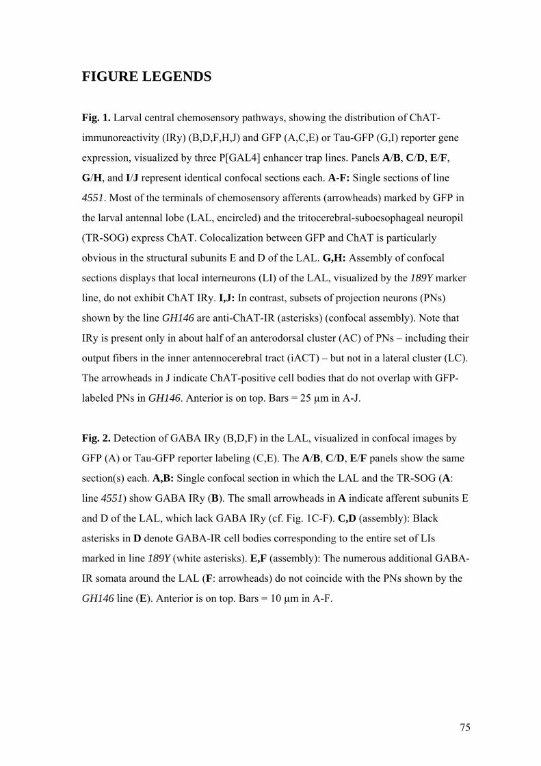
FIGURE LEGENDS Fig. 1. Larval central chemosensory pathways, showing the distribution of ChAT-
immunoreactivity (IRy) (B,D,F,H,J) and GFP (A,C,E) or Tau-GFP (G,I) reporter gene
expression, visualized by three P[GAL4] enhancer trap lines. Panels A/B, C/D, E/F,
G/H, and I/J represent identical confocal sections each. A-F: Single sections of line
4551. Most of the terminals of chemosensory afferents (arrowheads) marked by GFP in
the larval antennal lobe (LAL, encircled) and the tritocerebral-suboesophageal neuropil
(TR-SOG) express ChAT. Colocalization between GFP and ChAT is particularly
obvious in the structural subunits E and D of the LAL. G,H: Assembly of confocal
sections displays that local interneurons (LI) of the LAL, visualized by the 189Y marker
line, do not exhibit ChAT IRy. I,J: In contrast, subsets of projection neurons (PNs)
shown by the line GH146 are anti-ChAT-IR (asterisks) (confocal assembly). Note that
IRy is present only in about half of an anterodorsal cluster (AC) of PNs – including their
output fibers in the inner antennocerebral tract (iACT) – but not in a lateral cluster (LC).
The arrowheads in J indicate ChAT-positive cell bodies that do not overlap with GFP-
labeled PNs in GH146. Anterior is on top. Bars = 25 µm in A-J.
Fig. 2. Detection of GABA IRy (B,D,F) in the LAL, visualized in confocal images by
GFP (A) or Tau-GFP reporter labeling (C,E). The A/B, C/D, E/F panels show the same
section(s) each. A,B: Single confocal section in which the LAL and the TR-SOG (A:
line 4551) show GABA IRy (B). The small arrowheads in A indicate afferent subunits E
and D of the LAL, which lack GABA IRy (cf. Fig. 1C-F). C,D (assembly): Black
asterisks in D denote GABA-IR cell bodies corresponding to the entire set of LIs
marked in line 189Y (white asterisks). E,F (assembly): The numerous additional GABA-
IR somata around the LAL (F: arrowheads) do not coincide with the PNs shown by the
GH146 line (E). Anterior is on top. Bars = 10 µm in A-F.
75
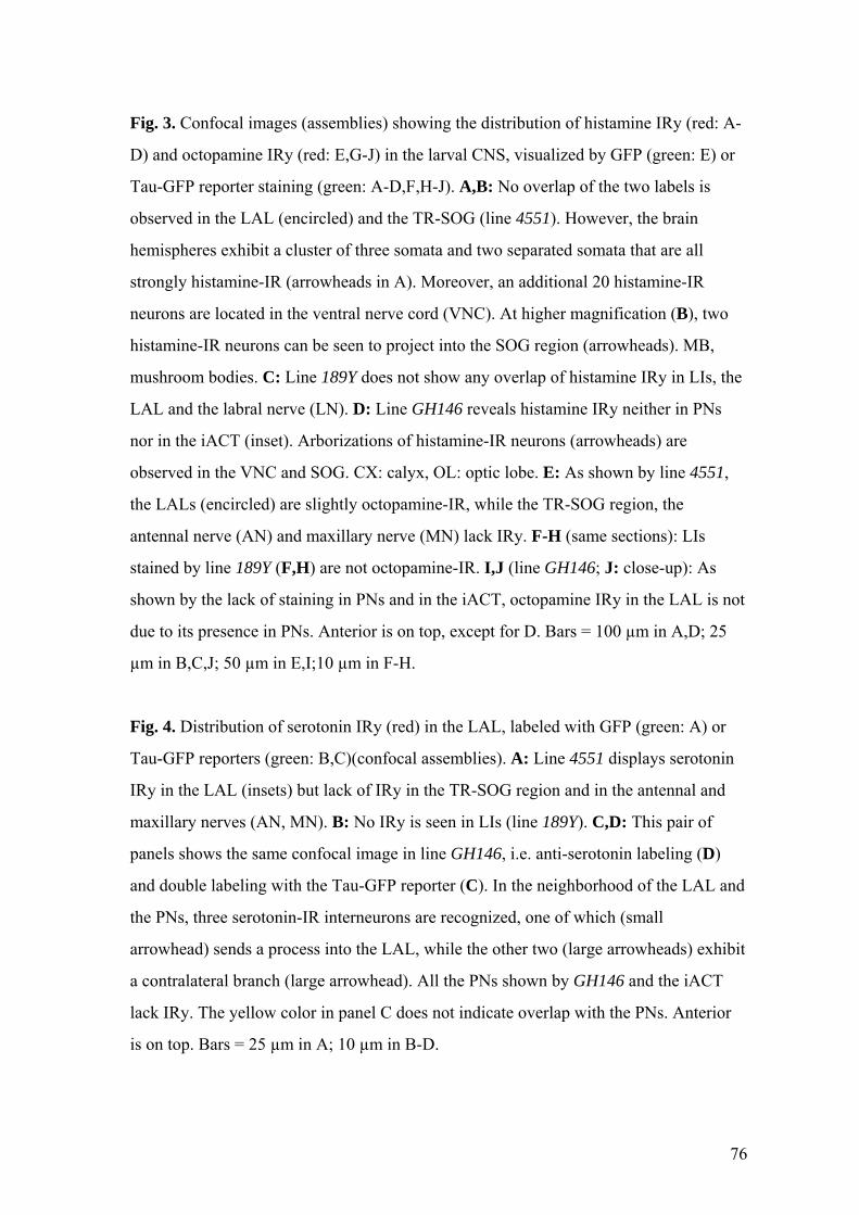
Fig. 3. Confocal images (assemblies) showing the distribution of histamine IRy (red: A-
D) and octopamine IRy (red: E,G-J) in the larval CNS, visualized by GFP (green: E) or
Tau-GFP reporter staining (green: A-D,F,H-J). A,B: No overlap of the two labels is
observed in the LAL (encircled) and the TR-SOG (line 4551). However, the brain
hemispheres exhibit a cluster of three somata and two separated somata that are all
strongly histamine-IR (arrowheads in A). Moreover, an additional 20 histamine-IR
neurons are located in the ventral nerve cord (VNC). At higher magnification (B), two
histamine-IR neurons can be seen to project into the SOG region (arrowheads). MB,
mushroom bodies. C: Line 189Y does not show any overlap of histamine IRy in LIs, the
LAL and the labral nerve (LN). D: Line GH146 reveals histamine IRy neither in PNs
nor in the iACT (inset). Arborizations of histamine-IR neurons (arrowheads) are
observed in the VNC and SOG. CX: calyx, OL: optic lobe. E: As shown by line 4551,
the LALs (encircled) are slightly octopamine-IR, while the TR-SOG region, the
antennal nerve (AN) and maxillary nerve (MN) lack IRy. F-H (same sections): LIs
stained by line 189Y (F,H) are not octopamine-IR. I,J (line GH146; J: close-up): As
shown by the lack of staining in PNs and in the iACT, octopamine IRy in the LAL is not
due to its presence in PNs. Anterior is on top, except for D. Bars = 100 µm in A,D; 25
µm in B,C,J; 50 µm in E,I;10 µm in F-H.
Fig. 4. Distribution of serotonin IRy (red) in the LAL, labeled with GFP (green: A) or
Tau-GFP reporters (green: B,C)(confocal assemblies). A: Line 4551 displays serotonin
IRy in the LAL (insets) but lack of IRy in the TR-SOG region and in the antennal and
maxillary nerves (AN, MN). B: No IRy is seen in LIs (line 189Y). C,D: This pair of
panels shows the same confocal image in line GH146, i.e. anti-serotonin labeling (D)
and double labeling with the Tau-GFP reporter (C). In the neighborhood of the LAL and
the PNs, three serotonin-IR interneurons are recognized, one of which (small
arrowhead) sends a process into the LAL, while the other two (large arrowheads) exhibit
a contralateral branch (large arrowhead). All the PNs shown by GH146 and the iACT
lack IRy. The yellow color in panel C does not indicate overlap with the PNs. Anterior
is on top. Bars = 25 µm in A; 10 µm in B-D.
76
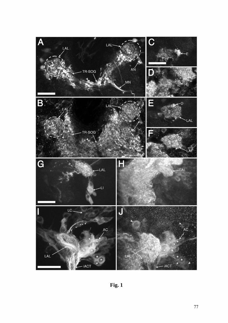
Fig. 1
77
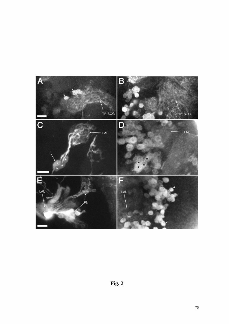
Fig. 2
78
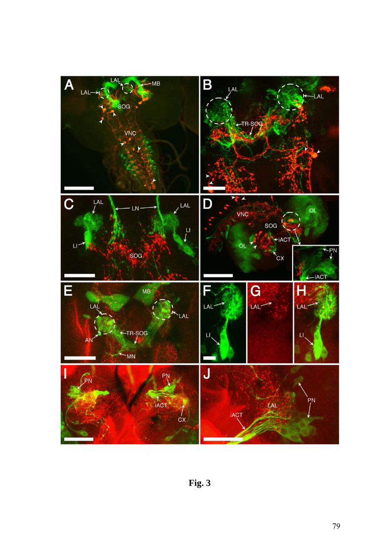
Fig. 3
79
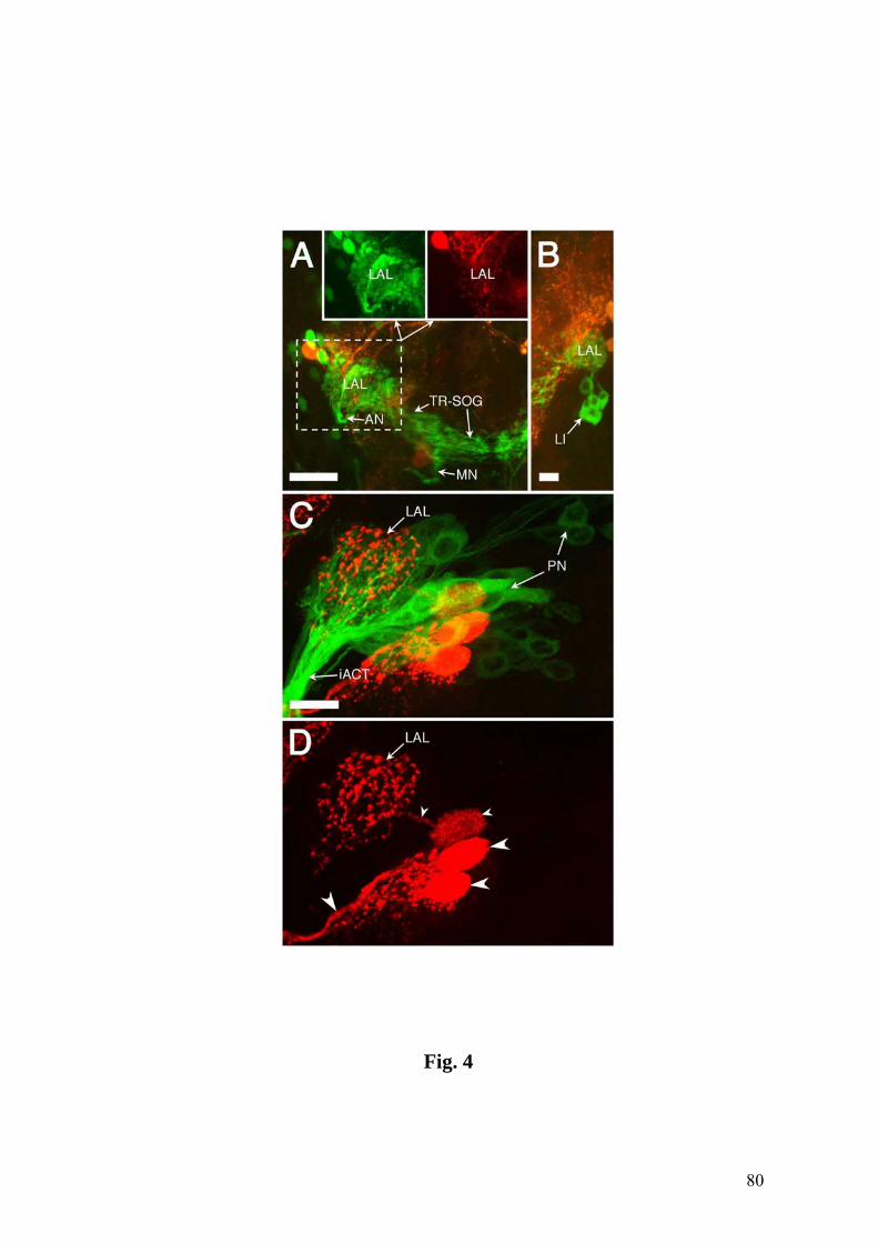
Fig. 4
80
Remerciements
Je remercie l’Unité de Biologie Animale et son directeur le prof. Heinz Tobler de
m’avoir permis d’effectuer mon travail de thèse à l’Université de Fribourg.
Ma reconnaissance va tout particulièrement à mon directeur de thèse le prof. Reinhard F.
Stocker pour sa grande disponibilité, ses conseils et pour m’avoir fait profiter de ses
compétences scientifiques.
Je tiens à remercier Dr. Matthew Cobb de Université de Paris-6 (France) pour avoir
accepté d’être expert lors de mon travail de thèse.
Je remercie chaleureusement Dr. Gertrud Heimbeck et Nanaë Gendre pour leurs
précieux conseils, leur soutien et leur bonne humeur. Ma gratitude va aussi à Stephan
Halter pour son introduction à la microscopie confocale.
Mes remerciements vont finalement à toutes les personnes qui ont contribué à la très
bonne ambiance du groupe Droso.
81
CURRICULUM VITAE
Nom PYTHON
Prénom François
Date de naissance 15 septembre 1971
Nationalité Suisse
Lieu d'origine Hauterive (FR)
Langue maternelle Français
Etat civil Marié, deux enfants
Etudes
1997-2002 Thèse de doctorat sous la direction du Prof. Reinhard F.
Stocker, Université de Fribourg (Suisse)
2000-2001 Diplôme de médiateur en technologie de l'information
Formation continue, Université de Fribourg
1991-1997 Diplôme de biologie
Université de Fribourg
1987-1991 Maturité fédérale, baccalauréat type C (scientifique)
Collège Ste-Croix, Fribourg
82