the kelly society orthopaedic journal - emory universityortho.emory.edu/documents/2016 kelly day...
TRANSCRIPT

EMORY UNIVERSITY SCHOOL OF MEDICINE
Department of Orthopaedics
The Kelly Society
Orthopaedic Journal
2016

1
Table of Contents Letter from the Chairman 2 2016 Kelly Society Visiting Professor 3 Letter from Kelly Society President 4 Kelly Day Agenda 5 PGY-5 Class 8 PGY-5 Bios, Manuscripts, and Abstracts 9-62 Elise Hiza, MD 9 Ashton Mansour, MD 12 Kyle Sweeney, MD 20 Dane Todd, MD 29 Brent Wise, MD 54
PGY-3 Manuscripts 74-97 PGY 1-4 Residency Classes 98 Emory Orthopaedics Surgical Faculty 99 Kelly Day Visiting Professors 100 Recent Publications (2015 – 2016) 102

2
LETTER FROM THE CHAIRMAN
James R. Roberson, MD
Welcome to the Kelly Society Resident Research Day. I am sure you will
find the projects reflect outstanding planning and effort on the part of
the investigators and I look forward to hearing the presentations.
This year’s Kelly Day Professor is Dr. Samir Mehta, the Chief of
Orthopedic Trauma at the University of Pennsylvania. Dr. Mehta has
become a recognized thought leader in our specialty at an early stage in
his academic career. His insightful contributions to the field of
Orthopaedic Trauma, education and clinical research are indeed
impressive. On behalf of our residents, alumni, and faculty I would like
to thank Dr. Mehta for taking the time from his very busy schedule to be
with us.
Alumni support through the Kelly Society is an integral component of the
success of our department. Thank you to all present today and for your
continued support of the Emory Orthopaedic Training Program.
Finally, I am grateful to the number of people who put a great deal of
effort into organizing this weekend’s events.
James R. Roberson, M.D.
James Roberson, MD Robert P. Kelly Professor and Chairman Adult Reconstruction Emory University School of Medicine Department of Orthopaedic Surgery

3
2016 Kelly Society Visiting Professor
Samir Mehta, MD
Dear Faculty, Residents, and Staff of the Emory University Department of
Orthopaedic Surgery:
It is truly an honour to be your guest for Kelly Day. As you can imagine, this was
an easy decision to make particularly because I can get some good baseball in
since the Braves are playing so well.
In all seriousness, for the graduating residents, this is it…this is the transition to
the “Real World” – hopefully, dissimilar for all of you to the MTV show.
I could offer deep words of wisdom, but, in all honesty, I am not sure if I myself
have really figured it out. I think that is really the beauty of medicine – it’s the
art of what we do.
One of the things I would encourage you to do tough is make a difference –
whether you choose to do so for your patients, or in your community, or
internationally – recognize that you have a skill set and an ability to adapt and
learn like no other profession. While you could nail a thousand femurs or
reconstruct three thousand ACLs, someone will come along and nail a thousand
and one or reconstruct three thousand and two.
There are several forces at work affecting, or rather afflicting our profession. I
hope each of you chooses to lead, but if not, then participate in making it better
and not accepting the status quo. Consider the idea that the “Enemy of Better
is Good” – that is accepting something that is “good enough” will limit your
ability to achieve.
I wish you all the best of luck and hope you look back with appreciation for all
that your mentors, advisors, colleagues, and friends at Emory have provided you
over the last several years.
Warmest Regards,
Samir Mehta, MD
Samir Mehta, MD Associate Professor, Department of Orthopaedic Surgery Chief, Orthopaedic Trauma and Fracture Service University of Pennsylvania

4
Letter from the Kelly Society President
James Kercher, MD
Dear fellow Emory alumni:
As my term comes to an end I wanted express my gratitude. It has been
an honor to serve as Kelly Society President. The Emory Orthopaedic
Residency has great heritage, and having the position to be continually
involved as President has been a great pleasure. I am certain that those
who follow me will have the same experience.
I would also like to thank those who have taken the time to reengage.
Since our last meeting, we have seen a significant increase in our
membership with the addition of twenty new lifetime members. This
support is greatly appreciated and necessary to keep our society
thriving. I hope that those who are currently involved continue to
remain active and help spread the word to those who are not yet
members. Because we must remember, the society is here to support
the training of future leaders.
Thank you again for allowing me the opportunity to serve as your Kelly
Society President from 2014 to 2016. I’m excited about the future and
what it holds for us as we partner to grow our membership.
James S. Kercher, MD class of 2009
James Kercher, MD Orthopaedic Surgeon, Sports Medicine Peachtree Orthopaedic Clinic

5
2016 KELLY DAY AGENDA
6:30 AM Registration & Breakfast 7:00 AM Welcoming Remarks
James Roberson, MD Resident Research Presentation: Session I 7:10 AM Biomechanical Comparison of Quadriceps and Patellar Tendon Grafts in Anterior
Cruciate Ligament Reconstruction Arthroscopy: The Journal of Arthroscopic & Related Surgery: 32(1), 71–75. 2016
Elise Hiza, MD - PGY 5 (Sports Medicine – The Southern California Orthopedic Institute) 7:20 AM Outcomes of ACL Reconstruction Using Closed Versus Variable Loop Button Fixation Brent Wise, MD - PGY 5 (Trauma – R. Adams Cowley Shock Trauma Center) 7:30 AM Utility of the Lateral Radiograph as an Adjunct to the Posteroanterior Radiograph in
Determination of the Risser Sign: Intra- and Inter- Observer Reliability and the Effect of Pelvic Tilt
Ashton Mansour, MD - PGY 5 (Hand Surgery - Vanderbilt University Medical Center) 7:40 AM Discussion - Dr. Samir Mehta and Emory Faculty Panel Discussion I 8:00 AM The Cost of Care: What is the Orthopaedic Surgeon’s Role?
Panel: Drs. Samir Mehta, William Reisman, James Roberson, Sandra Hobson Moderator: Dr. William Reisman
o Dr. Hobson: Does the implant matter? (Case presentation: DHS vs. CMN for IT fractures) o Dr. Roberson: World A versus World B: Defining Value in Orthopaedics o Dr. Mehta: Do we even know the cost of the implants? o Dr. Reisman: What can we do to contain cost?
Resident Research Presentation: Session II 9:00 AM Is Early Discharge Possible Following Posterior Spinal Fusion for Neuromuscular
Scoliosis? Laura Bellaire, MD - PGY 3 9:10 AM Humerus Fracture Fixation: Incidence, Rates and Complications as reported by
American Board of Orthopedic Surgery Part II Candidates William Carpenter, MD - PGY 3
Friday, June 3, 2016

6
9:20 AM Cellular Characterization and Modulation of Rotator Cuff Muscle Atrophy in a Small
Animal Model Jimmy Daruwalla, MD - PGY3 9:30 AM Discussion - Dr. Samir Mehta and Faculty 9:45 AM Break 2016 Kelly Day Lecture 10:00 AM Introduction of 2016 Kelly Visiting Professor
James Roberson, MD 2016 Kelly Visiting Professor Lecture Samir Mehta, MD Panel Discussion II 11:00 AM Geriatric Acetabular Fractures Panel: Drs. Samir Mehta, Tom Moore, Jr., Tom Bradbury, Rishin Kadakia Moderator: Dr. Tom Moore, Sr.
o Dr. Kadakia: Case presentation o Dr. Moore, Jr.: 10 min argue to fix o Dr. Bradbury: 10 min argue to replace o Dr. Mehta: Discussion
12:00 PM Lunch Presentation Operation Eagle Claw: The 1979 Iran Hostage Crisis (Lessons Learned and “What Ifs?”)
Carl Savory, MD - The Houston Clinic, Columbus, GA
Resident Research Presentation: Session III 1:00 PM Risk Factors for Lumbar Spinal Epidural Lipomatosis Anuj Patel, MD - PGY 3 1:10 PM Continued Delay in Diagnosis of Slipped Capital Femoral Epiphysis Robert Runner, MD - PGY 3 1:20 PM Radiographic Assessment of Guided Growth: The Correlation Between Screw
Divergence and Change in Anatomic Alignment Kyle Sweeney, MD - PGY 5 (Musculoskeletal Oncology – University of Chicago) 1: 30PM Orthopaedic Surgery Resident Training: What Procedures Are Essential? Dane Todd, MD - PGY 5 (Sports Medicine – University of Utah) 1:40 PM Discussion - Dr. Samir Mehta and Emory Faculty

7
Panel Discussion III 2:00 PM Are We Behind the Times in Resident Education?
Panel: Drs. Samir Mehta, Mara Schenker, Tom Bradbury, Brent Wise Moderator: Dr. Tom Bradbury
o Dr. Schenker: Resident selection o Dr. Borden: Shortcomings in Education – The Resident’s Perspective o Dr. Wise: What to do about duty hours? o Dr. Mehta: Planning for The Real World: Educating About Leadership and The Business
of Medicine
2:45 PM Break Faculty Presentations
3:00 PM Basic and Translational Research in Emory Orthopaedics & Regenerative Engineering Approaches for Musculoskeletal Disease and Injury Nick Willett, PhD 3:10 PM The Osteogenic Effects of a Novel Small Molecule Inhibitor of Sclerostin in Vivo Steven Presciutti, MD 3:20 PM Development of Novel Rodent Model of Ankle Arthrodesis Jason Bariteau, MD 3:30 PM The Ballistics of Orthopaedics in Atlanta Tom Moore, Sr., MD 4:00 PM Introduction of Reunion Class of 2006 and The Future of the Kelly Society 4:15 PM Closing Remarks
James Roberson, MD and Thomas Bradbury, MD
**CME credits will be issued in January 2017. Please remember to provide a PIN number to access your statement. For more information, contact Sonya Williams at

8
2015 – 2016 Orthopaedic Chief Surgery Residents
PGY-5
Elise Hiza, MD
Fellowship Match: The Southern California
Orthopedic Institute Valencia, CA
Sports Medicine
Medical School: University of
Colorado, Denver School of
Medicine
Hometown: Olney Springs, CO
Ashton Mansour, MD Fellowship Match: Vanderbilt University
Medical Center Nashville, TN
Hand & Upper Extremity
Medical School: Louisiana State
University School of Medicine
Hometown: Alexandria, LA
Kyle Sweeney, MD Fellowship Match: University of Chicago
Chicago, IL
Musculoskeletal Oncology
Medical School: Vanderbilt University
School of Medicine
Hometown: Portage, IN
Dane Todd, MD Administrative Chief
Fellowship Match: The University of Utah
Salt Lake City, UT
Sports Medicine
Medical School: Emory University
School of Medicine
Hometown: Lincoln, NE
Brent Wise, MD Administrative Chief
Fellowship Match: R. Adams Cowley Shock
Trauma Center Baltimore, MD
Trauma
Medical School: University of
Florida School of Medicine
Hometown: Harrisburg, PA

9
Elise Hiza, MD PGY-5
Sports Medicine Fellowship Southern California Orthopedic Institute Van Nuys, CA EDUCATION: University of Colorado School of Medicine – Denver, CO Doctor of Medicine, May 2011 The Colorado College, College Station, TX Bachelor of Arts, Emphasis Sports Medicine, May 2006
HONORS AND LEADERSHIP: AOA Emerging Leaders Forum 2015 1st Place Resident Research Award Georgia Orthopaedic Society 2014 M. Gage Oschner Resident Research Award 2014 2nd Place Kelly Day Resident Research Award – Emory University 2014 1st Place Atlanta International Trauma Symposium 2014 PODIUM PRESENTATIONS: Hiza E, Gottschalk M, Umpierrez E, Bush P, Reisman W. The Effect of a Dedicated Orthopaedic Advanced Practice Provider in a Level I Trauma Center: Analysis of Length of Stay and Cost
Georgia Committee on Trauma 2014
Georgia Society for the American College of Surgeons, State Meeting 2014
Georgia Orthopaedic Society 2014
Society for the American College of Surgeons, Regional Meeting 2014
Orthopaedic Trauma Association 2015 POSTER PRESENTATIONS: Hiza E, Gottschalk M, Umpierrez E, Bush P, Reisman W. The Effect of a Dedicated Orthopaedic Advanced Practice Provider in a Level I Trauma Center: Analysis of Length of Stay and Cost.
Atlanta Trauma Symposium 2014
Bellaire L, Hiza E, Moore TJ, Umpierrez, E. Musculoskeletal Mycobacterium Tuberculosis Infection as the Initial Presentation of Systemic Tuberculosis.
Southern Orthopaedic Association 2012

10
Biomechanical Comparison of Quadriceps and
Patellar Tendon Grafts in Anterior Cruciate Ligament
Reconstruction
Raj H. Shani, MD, Erica Umpierrez, MD, Michael Nasert, BA, Elise A. Hiza, MD, John Xerogeanes, MD
PURPOSE To quantify the structural and material properties of 10-mm central sections of the quadriceps and patellar tendons in the setting of anterior cruciate ligament reconstruction using cadaveric grafts and biomechanical analysis. METHODS The structural and mechanical properties of 11 boneepatellar tendonebone (BPTB) and 12 quadriceps tendonebone (QT) allografts were evaluated. Ten-millimeter-wide tendon grafts from both patellar and quadriceps tendons were harvested and subjected to biomechanical testing using the MTS servohydraulic test machine (MTS Systems, Eden Prairie, MN). The cross-sectional area was also calculated and compared between the BPTB and QT grafts. RESULTS The mean cross-sectional area was 91.2 _ 10 mm2 for the QT compared with 48.4 _ 8 mm2 for the BPTB (P ¼ .005). The mean ultimate stress was 23.9 _ 7.4 MPa for the QT and 33.4 _ 9.0 MPa for the BPTB (P ¼ .01). Ultimate strain was similar between the 2 tested groups, with a 10.7% change in the QT group and an 11.4% change in the BPTB group (P ¼ .484). The Young modulus of elasticity was 255.3 _ 64.1 MPa for the QT and 337.8. _ 67.7 MPa for the BPTB (P ¼ .006). The mean stiffness was 466.2 _ 133 N/mm for the QT and 278.0 _ 75 N/mm for the BPTB (P ¼ .005). The mean ultimate load to failure was 2,185.9 _ 758.8 N for the QT compared with 1,580.6 _ 479.4 N for the BPTB (P ¼ .045). CONCLUSIONS The cross-sectional area of the QT was nearly twice that of the BPTB. Ultimate load to failure and stiffness were also significantly higher for the QT graft. The variability in the cross-sectional area was similar in both tendon groups. CLINICAL RELEVANCE On the basis of graft predictability and biomechanical properties, our study reaffirms that the QT graft is a biomechanically sound alternative for anterior cruciate ligament reconstruction. See commentary on page 76 Over 200,000 anterior cruciate ligament reconstructions (ACLRs) are performed in the United States yearly, making it one of the most common orthopaedic procedures.1 Autograft choices for ACLR primarily include boneepatellar tendonebone (BPTB), hamstring tendon, and quadriceps tendon (QT). BPTB graft is often considered the gold standard, but each graft has its own advantages and disadvantages, making the appropriate graft choice for ACLR a subject for debate.2 The QT has been a viable option for ACLR since it was introduced by Marshall et al.3 in 1979. Yet, because of concerns raised by Noyes et al.4 in 1984 of relative graft weakness when compared with BPTB graft, the QT lost favor in clinical practice. In their testing, however, Noyes et al. used a composite QT graft composed of partialthickness QT, prepatellar tissue, and patellar tendon. This graft is significantly different from the

11
central QT-bone construct used in subsequent studies by Harris et al.5 and Staubli et al.6 in the late 1990s. These later works showed that the QT graft trended toward greater strength than the BPTB graft. However, few studies have re-examined the biomechanical properties of the QT and BPTB grafts using modern biomechanical techniques. Recent studies have advocated the use of the QT because it yields equal results in terms of anterior knee.

12
Ashton Mansour, MD PGY-5
Hand and Upper Extremity Fellowship Vanderbilt University Nashville, TN EDUCATION: Louisiana State University Health Sciences Center – New Orleans, LA Doctor of Medicine, May 2011 Texas A&M University, College Station, TX Bachelor of Science, Biological Sciences, May 2006
HONORS AND LEADERSHIP: Residency Interviewing and Selection Committee 2012-2016 PUBLICATIONS: "Inline" Axial Reconstructed Ct Scans Provide a Significantly Larger Assessment of C2 Pedicle Diameter for Screw Placement Than "Standard" Axial Scans: Implications for Surgical Planning. John Rhee, MD, Tim Borden, MD, David Vizurraga, MD, And Ashton Mansour, MD. Awaiting Final Publishing in Journal of Spinal Disorders & Technique "Iatrogenic Femoral Neck Fracture After Closed Reduction of Anterior Hip Dislocation in The Emergency Department” Michael D. Smith, MD, Ashton Mansour, MD, Michael S. Sridhar, MD, Sarah Jamieson, MSN, ANP, And Thomas J. Moore, MD. For Publication Online in The August 2015 E-Publishing Section of the American Journal of Orthopedics “The Leg Hammock for Closed Reduction of Tibial Shaft Fractures.” Colyn Watkins MD, Ashton Mansour MD, Dane Todd MD, Sarah Jamieson Np. Published in Orthopedics 2015 Feb;38(2):113-6. “Immediate Spica Casting of Femur Fractures in The Operating Room Versus the Emergency Department: Analysis of Reduction, Complications, And Cost.” Alfred A. Mansour, III MD; Jill C. Wilmoth, MD; Ashton S. Mansour, BS; Jeffery E. Martus, MD. Published in The Journal of Pediatric Orthopaedics, December 2010, Volume 30, Number 8. PRESENTATIONS: Podium Presentation at 2010 Aaos National Meeting, New Orleans, La. March 11,2010. “Femur Fracture Spica Casting in or Versus Ed: Analysis of Reduction, Complications and Cost.” Mansour A., Wilmoth J., Mansour As., Mencio G., Lovejoy S., Martus, J. Presented at The LOA 2010 Annual Meeting, New Orleans, La. April 10, 2010. “Thinking Inside the Box. The Biomechanical Response of the Distal Femur in Relation to The Size, Depth, Position, And

13
Angulation of the Box Cut When Performing Posterior Stabilized Total Knee Arthroplasty.” W Sherman, MD; R Rooney, MD; B Walton, MD; S Cook, PhD; M Kester, PhD; A Mansour, BS. Poster Presentation at 2010 POSNA National Meeting, Waikoloa, Hi. May 4, 2010. “Femur Fracture Spica Casting in or Versus Ed: Analysis of Reduction, Complications and Cost.” Mansour A., Wilmoth J., Mansour AS., Mencio G., Lovejoy S., Martus, J Poster Presentation at The 2012 “Science of GME” Projects Day at Emory University School of Medicine. “Iatrogenic Femoral Neck Fracture After Closed Reduction of Obturator Hip Dislocation in The Emergency Room: A Case Report.” Michael Smith MD, Ashton Mansour MD, Michael Sridhar MD, Sarah Jamieson BS NP, Thomas Moore, MD. “CSRS ‘Inline’ Axial Reconstructed Ct Scans Provide Significantly Larger Assessment of C2 Pedicle Diameter for Screw Placement Than ‘Standard’ Axial Scans: Implications for Surgical Planning.” David Vizurraga MD, Timothy Borden MD, Ashton Mansour MD, John Rhee MD.
American Society for Surgery of the Hand, Residents and Fellows Meeting, San Francisco, Ca Podium Presentation: 2013
Oral Presentation at The 2013 CSRS Annual in Los Angeles, Ca, December 5-7, 2013.
2014 AAOS Annual Meeting, New Orleans, Louisiana, March 11-15, 2014.
AOA/COA: Scientific Poster Display at The 2014 AOA/COA Combined Meeting, Palais Des Congrès De Montréal in Montreal, Quebec, June 18-21, 2014.

14
Utility of The Lateral Radiograph as an Adjunct to the
Posteroanterior Radiograph in Determination of the Risser
Sign: Intra- and Inter- Observer Reliability and the Effect of
Pelvic Version
Ashton Mansour, MD, Bryan J. Sirmon, MD, Shawn Duxbury, MD, Nick N. Patel, MD, Neeta Shenvi, Walter A. Carpenter, MD, Nicholas D. Fletcher, MD
PURPOSE Radiographic classification of apophyseal closure of the ilium as described by Risser is traditionally done on the frontal radiograph. The use of a lateral radiograph to augment the PA radiograph has been suggested however no prior study has accounted for individual Risser grading and pelvic version. This study sought to evaluate intra- and inter-rater reliability of Risser grading with and without the addition of a lateral radiograph and evaluate the impact of pelvic version on reproducibility. METHODS This study looked at 131 patients aged 10-18 treated for adolescent idiopathic scoliosis (AIS). Radiographs were randomized and then reviewed by four separate physicians at different clinical stages at three week intervals. Pelvic tilt and pelvic incidence was also collected. Inter and intra-observer reliability was calculated based on scoring at weeks 1 and 3. The influence of pelvic tilt was determined based on a median PT of 10° and PI of 52°. RESULTS Intra-observer agreement was moderate to substantial for all participants (K= 0.59-0.80 ± 0.05) using the PA radiograph with improvement to substantial to almost perfect agreement after the addition of the lateral radiograph (K = 0.65-0.81±0.06). Intra-observer agreement was most unreliable for Risser stage 3 and most reliable for stage 0/5 and 4 using the PA and Lateral radiographs. Inter-rater agreement was not significantly impacted by the addition of the lateral radiograph. Agreement was higher in patients with low PT (PT<10°) at all time points with addition of the lateral radiograph. The impact of the lateral radiograph in patients with lower PI (<52°) was modest but also significant. CONCLUSION The addition of the lateral radiograph can augment the frontal view for patients undergoing treatment for adolescent scoliosis. Pelvic version appears to impact the ability to adequately visualize the posterior iliac apophysis and the addition of a lateral radiograph in patients with low PI and PT may allow for easier interpretation of this region which may have significant impact on clinical decision making. We recommend that the surgeon utilize the lateral view when determining the Risser grade, especially in patients with pelvic retroversion. INTRODUCTION
The Risser grading system, described by Joseph C. Risser in 1958[16], has long been used to help guide management and treatment of adolescent idiopathic scoliosis (AIS). Two accepted grading systems based on radiographs taken in the anterior/posterior plane exist, however the American system, rather than the French classification, remains the predominant system in the United States. [2]. The U.S grading system has higher sensitivity, specificity and accuracy than the French system, when combined with years

15
since menarche, to determine growth cessation [21]. Nonetheless, intra-observer reliability reports vary widely. In an effort to improve reproducibility with Risser grading, Kotwicki [9](2008) introduced the idea that the iliac apophysis can be better visualized using the lateral spine radiograph because the sacral-iliac (SI) joint obscures the posterior-medial aspect of the iliac apophysis on a frontal view. He concluded that the current Risser grading does not consider the actual excursion of the iliac apophysis, because one-third of the apophysis cannot be observed on the frontal radiograph and that evaluation of iliac apophyseal fusion can be more accurately estimated when the lateral radiograph is used to complement the frontal radiograph. We hypothesized that the addition of the lateral radiograph would improve intra and inter-observer reliability over a purely frontal radiograph, and that this benefit would vary with pelvic morphology.
METHODS
Four surgeons (one senior attending, one first year attending, one fourth year resident, and one third year resident) reviewed 131 computer randomized spinal radiographs to determine Risser grading. Participants were blinded to demographic information regarding age or sex. Reviewers were first presented with a standard 36-inch digital posteroanterior (PA) spinal radiograph and the Risser sign was recorded. Risser grading was performed as described by Joseph Risser [16] with a Risser 0 representing no ossification of the iliac apophysis, Risser 1 with <25% ossification, Risser 2 with 50% ossification, Risser 3 with 75% ossification, and Risser 4 with 100% ossification. A patient was deemed to be a Risser 5 if complete ossification of the iliac apophysis was present with concomitant fusion to the ilium. Risser 0 and 5 classifications were combined due to the fact that the proximal femora or triradiate cartilage were often not visible for evaluation due to gonadal shielding or radiographic cropping.
After repeat randomization, the reviewers were presented with the same PA radiograph as well as a standing lateral radiograph of the spine without shielding and the Risser staging was again recorded. All patients were selected from a database of surgical patients from the senior author, however these were taken at different stages in the treatment (observation, bracing, and preoperatively) to represent the entire spectrum of disease. A single grade was given by all reviewers without separate classification of ossification on the lateral radiograph as described by Kotwicki et al[9]. Identical measurements were performed at a second time point three weeks after the first measurement, using first the PA radiograph and then both the PA and lateral views. Intra- and Inter-rater reliability assessment using Kappa statistics for multiple users was utilized. A Kappa score of <0 correlates with less than chance agreement; 0.0-0.2 shows slight agreement; 0.20-0.40 shows fair agreement; 0.40-0.60 shows moderate agreement; 0.60-0.80 shows substantial agreement and >0.80 shows almost perfect agreement [3,10].
A single observer reviewed all 131 radiographs to determine the pelvic version as defined by the pelvic tilt (PT), which was obtainable in 112 (85.5%) of standing lateral radiographs. PI was determined on a lateral radiograph by measuring the angle subtended by a line drawn between the center of the femoral head and the sacral endplate and a line drawn perpendicular to the center of the endplate of the sacrum. Pelvic incidence was divided based on the mean within the cohort (52°). PT was determined on the lateral radiograph by measuring the angle subtended by a line drawn from the midpoint of the sacral endplate to the center of the bicoxofemoral axis and a vertical plumb line extended from the bicoxofemoral axis. Post hoc analysis found a median PT of 10° and thus inter and intra-rater reliability was determined for each Risser grade while stratifying for PT and PI. RESULTS
Intra-observer agreement was moderate to substantial for all participants (κ= 0.59-0.80 ± 0.05) using the PA radiograph with improvement to substantial to almost perfect agreement after the addition of the lateral radiograph (κ = 0.65-0.81±0.06). Agreement appeared to correlate with year in training as reliability increased with seniority in nearly all cases (Table 1). Agreement varied by individual Risser

16
grade, with Risser 3 having the lowest agreement using solely the PA radiograph (κ=0.44-0.69±0.09) as well as the PA and lateral radiograph (κ=0.17-0.43±0.09). Risser 0/5 (κ=0.77-0.92 ±0.09) and Risser 1 (κ=0.50-0.83 ±0.09) had the highest intra-observer agreement using the PA radiograph only. Risser 0/5 and Risser 4 had the highest intra-observer agreement using the PA and lateral (Risser 0 (κ=0.70-0.93 ±0.09); Risser 4 (κ=0.74-0.84 ±0.09) radiograph.
Inter-observer agreement was moderate for all participants using the PA and PA + Lateral radiograph alone at the first time point (PA κ = 0.58 ±0.02; PA and Lateral κ= 0.56 ±0.02)(Table 2). Inter-observer agreement was substantial for all participants using the PA and PA + Lateral radiograph at the second time point (PA κ= 0.61 ±0.02; AP and lateral κ= 0.62 ±0.02)(Table 2). The addition of the lateral radiograph did not increase the inter-observer agreement amongst observers. Similar to the intra observer analysis, agreement was highest for Risser 0/5 and 4 and lowest for Risser 2 and 3 using both the PA and PA+lateral radiographs (Table 3).
Pelvic version parameters including PT and PI were visible in 112 (85.4%) of patients. Inter-rater agreement was calculated among all four raters by pelvic tilt and pelvic incidence (Table 4). The addition of a lateral radiograph improved reliability at both time points in patients with PT<10°. This improvement was not seen in patients with a more anteverted pelvis (PT>10°). The addition of a lateral radiograph also resulted in a modest increase in inter-rater agreement in patients with a lower PI, indicative of a more retroverted pelvis. Confounding matters was the fact that an improvement in agreement was seen at week 3 in patients with a low PI using the PA radiograph alone. The improvement was larger, however, in patients with a PA and lateral radiograph. DISCUSSION The concept that the excursion of the iliac apophysis can be used as a predictor of spinal maturity is not new. Risser concluded that the appearance and closure of the iliac apophysis closely coincides with the closure of the growth plates in the vertebra and could be used to predict remaining spinal growth. Although the Risser grading system is widely used by surgeons to predict remaining spinal growth in patients with adolescent idiopathic scoliosis (AIS), and help guide treatment, it is not without its limitations. Hacquebord et al [5] highlighted many limitations of the Risser grading system including anomalous ossification progression making staging difficult [19], and difficulty visualizing the apophasis due to overlap on the frontal view [7]. The utility of the Risser score in determining the onset of greatest peak height velocity has also been questioned with [11,12] the challenge afforded by the observation that the Risser stage 4 is an inaccurate measure of cessation of curve progression [1, 7, 13], poor correlation with the period of greatest curve progression [17,18], and the tendency for male patients to continue to grow into Risser Stage 5 [8]. In addition, Shuren et al [19](1992) showed poor intra-observer reliability when using the current Risser system and Hammond et al [6](2011) determined that the inter-observer and intra-observer reliability showed only moderate agreement when attempting to predict scoliosis progression. In contrast, Reem, et al [14] (2008) found reliability of the Risser sign to be acceptable. Others argue that the anterior to posterior radiograph more clearly shows the entire apophysis when compared to a posterior to anterior radiograph. Regardless of the challenges in using the Risser system, it continues to be used clinically due to the ease of use, the ability to visualize on standard upright spinal films, and the familiarity to most pediatric spinal deformity surgeons [4, 14, 17,18].
The present study evaluated the impact of the lateral radiograph and pelvic parameters on Risser grading using a spectrum of observers. We found that intra-observer agreement was significantly improved with the addition of the lateral radiograph. Risser scores of 0,1,4 and 5 were the most reproducible, showing higher intra-observer reliability using either the PA or PA/lateral radiographs. This likely extends from challenges visualizing the posteromedial portion of the iliac apophysis on the PA radiograph and the irregular ossification seen during the Risser 2 and 3 phases where ossification may not occur in either an anteroposterior or posteroanterior direction. It is common, as previously noted by

17
Kotwicki [9], for ossification of the midportion of the iliac apophysis to occur in a fragmented manner making differentiation between a Risser 2 and 3 more challenging. The lower agreement seen in Risser 2 and 3 is concerning as it pertains to clinical care as it is common practice to offer bracing up in children who are Risser 0-2. The fact that the lateral radiograph failed to drastically increase the reliability of radiographic evaluation suggests that perhaps other modalities for determining remaining growth should be considered. Nonetheless, the intra-observer reliability was improved in Risser 0,1,4,and 5 cases with the addition of the lateral radiograph and we feel that the ability to distinguish a child who is more immature (Risser 0 or 1) from one who is more mature is critical for both surgical and non surgical decision making in scoliosis care.
One interesting finding was that the addition of the lateral radiograph did not affect the inter-observer reliability and that this was inferior to the intra-observer reliability for all measurements. Inter-observer agreement is moderate while the intra-observer reliability is higher. As previously mentioned by multiple authors, apophyseal closure is often irregular and does not follow the traditional quartile concept introduced by Risser thus requiring some interpretation on the surgeon’s part. It is unclear the true importance of the inter-observe agreement in treating AIS because in most cases there is only one surgeon rendering treatment and the ability to radiographically establish the progression of apophyseal closure is based on a continuum of changes. One possible explanation would result from the discrepancy in years of experience and familiarity of the Risser grading of each surgeon. Our data failed to show a clear pattern or consistency between the senior/junior attending surgeons and the residents, it would be interesting to see if inter-observer agreement would significantly improve if this study was repeated with only senior level surgeons. We chose to maintain the current study design as multiple providers are often reviewing spinal radiographs and may be responsible for documenting skeletal maturity using the Risser system.
Pelvic morphology played an important role in the intra-observer reliability of certain patients. In particular, patients with more pelvic retroversion as indicated by lower pelvic tilt benefitted more from the addition of the lateral radiograph. There was no difference in agreement when a PA film was used in isolation at either time point regardless of pelvic tilt. When the lateral radiograph was added, patients with lower pelvic tilt showed better agreement than those with the PA view alone at both the 1 week and 3-week time points. As noted by Izumi [7],visualization of the posterior medial iliac apophysis is more difficult due to the overlap of the iliac and the sacrum when the pelvis is viewed from the frontal plane alone. Pelvic anteversion brings the posterior ilium more proximal and anterior allowing for better visualization of the iliac apophysis, likely explaining why the lateral radiograph did not add to the ability to visualize the apophyseal fusion. In patients with pelvic retroversion and a low PT, the lateral radiograph appears to allow the reader to better visualize the posterior apophysis and determine the extent of fusion. It is our recommendation that physicians consider obtaining a lateral spine radiograph in those patients with pelvic retroversion and PT<10° to better assist with clinical decision making. While the addition of a lateral radiograph does increase the exposure of the patient to more harmful radiation, the recent adoption of biplanar low dose radiography using the EOS machine allows for significantly lower radiation doses. We would recommend that the treating physician take advantage of the lateral radiograph afforded by such machines and utilize the closure of the posterior iliac crest apophysis for clinical decision making.
This study has several limitations. First, readers were not provided with any clinical information regarding the treated patients and thus we grouped Risser 0 and Risser 5 patients together as the visualization of other growth plates (i.e. proximal femoral physis or triradiate cartilage) was variable. The clinical difference between a patient who is a Risser 0 versus Risser 5 is typically striking as the latter is approaching adulthood however our decision to combine these two groups may have impacted our final data. Secondly, we did not control for patient body mass index (BMI) or bone mineral density (BMD), choosing to use a more representative sample of patients rather than constrain our data to a certain patient demographic. Evaluation of radiographic landmarks in patients with increased BMI or lower BMD

18
can be challenging. Though our data was looking at intra- and inter-rater reliability, however, we feel that the inclusion of a large spectrum of patients would not impact our results as no decision was being made based on the radiograph and the no radiograph was excluded from any of the analyses. The final limitation arose from our inability to control for patient hip and knee positioning or pelvic rotation during radiography as this was a retrospective review and whole body radiographs were not available. While this may affect pelvic tilt, it should not have affected pelvic incidence. It is possible that the relative pelvic version where a patient may gain apparent pelvic retroversion by extending their pelvis, is more significant with regards to Risser grading than the positional independent pelvic incidence. Further study is needed using whole body radiography to further evaluate this finding.
CONCLUSION
When using the Risser grading system to help guide management and treatment for AIS the addition of the lateral radiograph improves intra- observer agreement without hindering inter-observer agreement. The benefit of the lateral radiograph appears to be greatest in children with acetabular retroversion. We advocate for the use of the lateral radiograph in patients when the frontal radiograph does not provide adequate visualization of the posterior ilium such as those with relative pelvic retroversion.

19
REFERENCES 1. Anderson M, Hwang SC, Green WT. Growth of the normal trunk in boys and girls during the second
decade of life; related to age, maturity, and ossification of the iliac epiphyses. J Bone Joint Surg Am. 1965;47:1554–1564.
2. Bitan FD, Veliskakis KP, Campbell BC. Differences in the Risser grading systems in the United States and France. Clin Orthop Relat Res. 2005;436:190–195.
3. Fleiss J (1977) Measuring nominal scale agreement among many raters. Psychol Bull 76: 378-382 4. Goldberg MS, Poitras B, Mayo NE, Labelle H, Bourassa R, Cloutier R. Observer variation in assessing
spinal curvature and skeletal development in adolescent idiopathic scoliosis. Spine (Phila Pa 1976). 1988;13:1371–1377.
5. Hacquebord J, Leopold S. In brief: The Risser classification: a classic tool for the clinician treating adolescent idiopathic scoliosis. Clin Orthop Relat Res, 2012 vol. 470 (8) pp. 2335-2338
6. Hammond et al. Inter-observer and intra-observer reliability of the Risser sign in a metropolitan scoliosis screening program. J Pediatr Orthop, 2011 vol. 31 (8) pp. e80-4
7. Izumi Y. The accuracy of Risser staging. Spine (Phila Pa 1976). 1995;20:1868–1871. 8. Karol LA, Johnston CE 2nd, Browne RH, Madison M. Progres- sion of the curve in boys who have
idiopathic scoliosis. J Bone Joint Surg Am. 1993;75:1804–1810. 9. Kotwicki T. Improved accuracy in Risser sign grading with lateral spinal radiography. Eur Spine J,
2008 vol. 12(12) pp. 1676-1685 10. Landis JR, Koch GG (1977) The measurement of observed agreement for categorical data. Biometrics
33: 159-174. 11. Little DG, Song KM, Katz D, Herring JA. Relationship of peak height velocity to other maturity
indicators in idiopathic scoliosis in girls. J Bone Joint Surg Am. 2000;82:685–693. 12. Little DG, Sussman MD. The Risser sign: a critical analysis. J Pediatr Orthop. 1994;14:569–575. 13. Noordeen MH, Haddad FS, Edgar MA, Pringle J. Spinal growth and a histologic evaluation of the Risser
grade in idiopathic scoliosis. Spine (Phila Pa 1976). 1999;24:535–538
14. Reem J, Carney J, Stanley M, Cassidy J. Risser sign inter-rater and intra-rater agreement: is the Risser sign reliable? Skeletal Radiol. 2009;38:371–375.
15. Risser JC. Important practical facts in the treatment of scoliosis. AAOS Instructional Course Lectures. Ann Arbor 1948;5: 248–260.
16. Risser JC. The Iliac apophysis; an invaluable sign in the management of scoliosis. Clin Orthop Relat Res. 1958;11:111–119.
17. Sanders JO, Browne RH, McConnell SJ, Margraf SA, Cooney TE, Finegold DN. Maturity assessment and curve progression in girls with idiopathic scoliosis. J Bone Joint Surg Am. 2007;89:64–73.
18. Sanders JO, Khoury JG, Kishan S, Browne RH, Mooney JF 3rd, Arnold KD, McConnell SJ, Bauman JA, Finegold DN. Predicting scoliosis progression from skeletal maturity: a simplified classification during adolescence. J Bone Joint Surg Am. 2008;90:540– 553
19. Shuren N, Kasser JR, Emans JB, Rand F. Reevaluation of the use of the Risser sign in idiopathic scoliosis. Spine (Phila Pa 1976). 1992;17:359–361.
20. Tachdjian M. Pediatric Orthopedics. Philadelphia: Saunders; 1990. p. 2290. 21. Weijun Wang et al. The value of different Risser grading systems in determining growth maturity of girls
with adolescent idiopathic scoliosis. Stud Health Technol Inform, 2012 vol. 176 pp. 183-187

20
Kyle Sweeney, MD PGY-5
UPCO FELLOWSHIP TRAINING: Musculoskeletal Oncology Fellowship University of Chicago Chicago, IL EDUCATION: Vanderbilt University, Nashville, TN Doctor of Medicine, May 2011 Michigan State University, East Lansing, MI Bachelor of Science, High Honors Double Major: Physiology, Human Biology, May 2005
HONORS AND LEADERSHIP: Thomas E. Whitesides Resident’s Award – Georgia Orthopaedic Society 2014 PUBLICATIONS Sweeney KR, Shi WJ, Gottschalk MB, Fletcher N, Bruce BW. Radiographic Assessment of Guided Growth: The Correlation between Screw Divergence and Change in Anatomic Alignment. In Submission Sweeney KR, Cundiff CA, Shehata BM, Walton ZJ, Reimer NB. Intraosseous Myofibroma of the Femoral Neck: A Case Report and Review of the Literature. In Submission Sweeney KR, Slone HS, Labib SA. Role of Arthroscopy in the Treatment of Pigmented Villonodular Synovitis of the Ankle and Subtalar Joints. In Submission. Richards JE, Morris BJ, Guillamondegui OD, Sweeney KR, Tressler MA, Obremsky WT, Kregor PJ. The Effect of Body Mass Index on Posttraumatic Transfusion after Pelvic Trauma. Am Surg. 2015 Mar;81(3)239-44. Gottschalk MB, Premkumar A, Sweeney KR, Boden, SD, Heller JG, Yoon ST. Posterolateral Lumbar Arthrodesis with and Without Interbody Arthrodesis for Degenerative Spondylolisthesis: A Comparative Value Analysis. Spine. 2015 Jan;40(12):917-25. Faucher GK, Golden ML, Sweeney KR, Hutton WC, Jarrett CD. Comparison of Screw Trajectory on Stability of Oblique Scaphoid Fractures: A Mechanical Study. J Hand Surg Am. 2014 Mar;39(3):430-5. Sweeney KR, Fritz RA, Rodgers SM. Careers in Medicine at Vanderbilt University School of Medicine: An Innovative Approach to Specialty Exploration and Selection. Acad Med. 2012 Jul;87(7):942-8.

21
POSTERS/ABSTRACTS Sweeney KR, Shi WJ, Gottschalk MB, Fletcher N, Bruce BW. Radiographic Assessment of Angular Correction of Genu Varum and Genu Valgus Using Guided Growth: The Correlation Between Screw Divergence and Change in Anatomic Alignment. Pediatric Orthopaedic Society of North America Annual Meeting 2016. Gottschalk MB, Sweeney KR, Boden, SD, Heller JG, Yoon ST. Posterolateral Lumbar Arthrodesis with and Without Interbody Arthrodesis for Degenerative Spondylolisthesis: A Comparative Value Analysis. LSRS Annual Meeting 2014. Thotala DK, Sweeney KR, Leahy K, Hu R, Hallahan D. “Valproix Acid Enchanges Radiation Therapy by Protecting Normal Hippocampal Neurons and Sensitizing Malignant Glioblastoma Cells in vivo and in vitro.” Fuel and Energy Abstracts. 2011 Sep;81(2) Richards J, Morris B, Sweeney KR, Guillamondegui O, Tressler M,Obremskey W, and Kregor P. “The Relationship of Obesity and Complications Post-Trauma in High-Energy Pelvic and Acetabular Fractures.” Western Trauma Association 9/2010 Richards J, Morris B, Sweeney KR, Guillamondegui O, Tressler M,Obremskey W, and Kregor P. “Does Body Mass Index affect hemorrhage in high-energy pelvic and acetabular fractures?” AAOS 5/2010 Silverstein E, Sweeney KR, Schoonover B, McNew B, Ye I, Boustani A, and Rodgers S. “Careers in Medicine Events for MS I-III at Vanderbilt.” AAMC: Southern Group on Student Affairs Spring Meeting. 9/2009. Sweeney KR, Hallahan D, and Thotala D. “Valproic acid protects hippocampal neurons from radiotoxicity in vitro and in vivo.” AACR 100th Annual Meeting. 4/2009 PRESENTATIONS Gottschalk MB, Sweeney KR. Surgical Training Using Three-Dimensional Simulation in Placement of Cervical Lateral Mass Screws: A Blinded Randomized Control Trial. GOS Annual Meeting. 9/2014. Sweeney KR. “Bone Metabolism and Osteoporosis.” NAON Educational Meeting. 2013 Sweeney KR, Silverstein E, and Rodgers S. “Maximizing CiM Potential: A Look at the Contributions of Students as Leaders at the Vanderbilt School of Medicine” (2009) - AAMC Professional Development Conference. Savannah, GA. 2009

22
Radiographic Assessment of Guided Growth: The Correlation
Between Screw Divergence and Change in Anatomic
Alignment
Kyle Sweeney, MD, W. Jeffrey Shi, MD, Michael Gottschalk, MD, Nicholas Fletcher, MD, Robert Bruce, MD INTRODUCTION
In children, the anatomic alignment of the knee changes in predictable patterns as part of normal development. Children typically begin life with genu varum with approximately 15 degrees of varus angulation at birth,1 gradually progress to genu valgum, and eventually return to what is considered normal physiologic alignment (slight valgus) around age 5 or 6.1 Between the ages of 3 and 6 normal valgus alignment can reach up to 20 degrees in some cases.2 While most children’s alignment corrects without intervention, some children may progress to pathologic genu valgum or genu varum. These deformities can cause significant problems, including pain, disruption of normal gait, and subsequent arthritis and joint instability if the abnormal alignment is left unaddressed.3 When these deformities do not resolve spontaneously and physiological alignment is not achieved by early adolescence, surgical intervention is often indicated.4
Monitoring the changes in anatomic alignment which occur during guided growth, full length standing radiographic films have been traditionally used. This view can allow for examination of multiple angles around the knee and examination of the mechanical axis of the lower extremity.13 Specifically, the anatomic tibiofemoral angle (aTFA) has been shown to be a reliable measurement of alignment at the knee, and the anatomic lateral distal femoral angle (aLDFA) and medial proximal tibial angle can be used as adjunct measurements to better quantify change.14 As asymmetric growth occurs during hemiepiphysiodesis, the screws used in the construct diverge.15 In modern systems, the two screws have the ability to diverge up to 30 degrees.11 This study seeks to determine the relationship between a change in screw divergence (SD) and a change in knee alignment, and ultimately to provide a reliable measure for determining surgical correction. METHODS
All patients between the ages of 3 and 18 diagnosed with genu varum and genu valgum treated with hemiepiphysiodesis using a two-hole tension band plate between January 1, 2000 and January 1, 2015 were identified via a search of the medical records. For each individual patient identified, preoperative aTFA and aLDFA were recorded using full-length standing lower extremity films. Initial SD was measured using intraoperative fluoroscopic images. At each subsequent clinic follow-up, aTFA, aLDFA, and SD was measured using full-length standing lower extremity films and compared to preoperative and intraoperative measurements in order to quantify the change in anatomic alignment and screw divergence. Figure 1 illustrates the techniques used to obtain each measurement. For every patient, each subsequent postoperative image was used as an independent data point. A subset of limbs underwent simultaneous hemiepiphysiodesis of the distal femur and proximal tibia. In this subgroup of patients, only the aLDFA was used.
A subset of 15 patient films was re-examined 2 weeks after the initial review by both the original observers as well as an attending pediatric orthopedic surgeon to determine intraobserver and interobserver reliability.

23
STATISTICS
Statistical analysis was performed using JMP Pro Software 10 (Cary, NC). For demographic data descriptive statistics were calculated and reported using frequency, range, and mean. A linear regression analysis (least squares method) was performed for modeling change in screw divergence and the studied limb alignment variables. A multivariate logistic regression analysis was also performed to identify independent predictors of change in aTFA and aLDFA. Agreement correlation coefficients were calculated for intraobserver and interobserver reliability. RESULTS Demographics
A total of 31 patients and 48 limbs were identified including 13 males and 18 females. Table 1 provides details on demographic data. A subset of 12 limbs underwent simultaneous distal femoral and proximal tibial hemiepiphysiodesis. Most patients had multiple postoperative visits at which time full-length limb films were obtained. Each of these images were utilized as individual events to be compared to the preoperative imaging which resulted in a total of 107 distinct data points. Given that aTFA was not calculated for limbs that underwent simultaneous distal femoral and proximal tibial hemiepiphysiodesis, the total number of aTFA measurements utilized was 80 and the total number of aLDFA utilized was 107. Linear Regression Analysis
Results of the linear regression analysis are summarized in Figure 2. For every 1-degree change in SD there was a resultant 1.80 degrees of change in aTFA and 2.11 degrees of change in aLDFA. Change in aTFA is predicted by the equation ∆aTFA = 0.41 × |∆SD| + 1.39. The coefficient of determination (R2) value of ∆SD vs ∆FTA was 0.50. The change in aLDFA was predicted by the equation ∆aLDFA = 0.27 ×∆SD + 1.84 with a R2 of 0.31. Correlation was estimated by the Restricted Maximum Likelihood Method (REML). ∆aTFA and ∆SD had a correlation coefficient of 0.68 (95% CI 0.54-0.78.) ∆aLDFA and ∆SD had a correlation coefficient of 0.56 (95% CI 0.42-0.68). Multivariate Regression Analysis
∆SD and gender were the only two independent predictors for ∆aLDFA and ∆aTFA. Individually, age at procedure (95% CI 0.34,1.40), gender (CI 1.48, 5.26), weight (CI 0.04, 0.18), height (-0.23, -0.05), and ∆SD (0.43, 0.62) were all significant predictors of ∆aTFA (Table 2). However, only ∆SD (CI 0.23, 0.39) and gender (CI -2.78, -0.56) were predictors of ∆aLDFA (Table 3). The coefficient of determination (R2) was 0.71 for the ∆aTFA model and 0.45 for the ∆aLDFA model. Finally, there was no correlation between ∆SD and height, weight, gender, or ethnicity.
Interoberver and Intraobserver Control
Three different readers read the same films twice a week apart to obtain an inter-rater and intra-rater agreement. The inter-rater agreement was 0.97 and the intra-rater agreement was 0.94.
DISCUSSION
In the past, genu varum and valgum were often corrected with osteotomies to address the angular deformity. However, these osteotomies had significant complications and high morbidity5 prompting surgeons to seek alternative paths for angular correction. Dr. Walter Blount, a pioneer in the field of disorders affecting the growth plate,6 was the first physician to develop and employ a physeal staple which tethered the edge of the growth plate to modulate growth and correct angular deformity.7 It has since been shown that physeal modification can provide angular correction without the high morbidity and complication rate associated with traditional osteotomy.5 This procedure, along with variations that have followed, seeks to harness the Hueter-Volkmann law which states that compressive forces across the

24
physis results in growth arrest.8 Therefore, when one edge of the growth plate is tethered, growth will asymmetrically occur across the physis providing a change in angulation. Using these principles, multiple techniques have sought to use guided growth as the surgical treatment for angular deformities. Previously, like the correction of tibia vara developed by Dr. Blount, one edge of the physis was tethered using an epiphyseal staple.9 Though physeal stapling has been effective at correcting the angular deformity, it has been associated with hardware prominence and failure, with an associated risk of early physeal closure.10 As a result, the use of a two-hole tension band plate with non-locking screws to create a tension band across the physis was proposed. 11 Though results between the two procedures have been shown to largely equivalent,12 the plate construct for guided growth has become more common as implants have improved.
Normal changes in coronal plane alignment of the pediatric knee is a dynamic process, starting first with genu varum before progressing to genu valgum.1 Most children will attain normal, adult alignment by the age of eight.16 While this progression is typically a predictable process, pathologic degrees of varus and valgus are not infrequent. Long-term sequelae of coronal malalignment include an altered gait pattern, abnormal lever arm function, cosmesis, and most importantly, premature degenerative arthritis.15 The traditional surgical option for angular correction is the osteotomy. While effective in anatomic correction, osteotomies are associated with the potential for serious complications including peroneal nerve palsies, compartment syndromes, deep and superficial infections, vascular injuries, iatrogenic fractures, and physeal damage.17,18
An alternative approach commonly used today is guided growth via hemiepiphysiodesis. Surgical indications are considered to be a mechanical axis lying outside of the two central quadrants of the knee in a child with at least six months of remaining skeletal growth.19 Options for fixation include traditional staples as well as newer 2-hole or 4-hole tension band plates.1,10,11,15,20. Wiemann et al retrospectively reviewed 63 cases of angular correction about the knee using staples and tension band plates and found no difference between the two fixations with regards to the amount of correction and overall complication rates (6.7% in “normal” physes and 27.8% in “abnormal physes”).10
Rates of correction have been reported as approximately 10 degrees per year,10 but this is dependent on the site of hemiepiphysiodesis. Balil et al prospectively followed 25 children (37 legs) and found mean rates of correction to be 0.7 degrees per month for the distal femur, 0.5 degrees per month for the proximal tibia, and 1.2 degrees per month when combined.19 The total amount of achievable correction is dependent on multiple factors, most notably the amount of skeletal growth remaining. In patients who reach skeletal maturity prior to achieving full correction, osteotomy remains a viable option.
Clinically following patients undergoing guided growth requires regular radiographic assessment of coronal plane alignment. Standing full-length limb studies are generally used to assess the change in anatomic and mechanical axis of the knee.10-12,15,19,20 While this imaging technique is the gold standard for measuring angular deformity of the knee, it involves administration of radiation to the pelvis and genitalia and is typically performed multiple times during the course treatment. The present study provides an alternative strategy to reliably assess changes in anatomic alignment while minimizing radiation exposure to sensitive structures in a pediatric population. We found that for every 1.0-degree change in SD, there was 1.8 degrees in FTA and 2.11 degrees in aLDFA. The correlation between change in SD and change in anatomic alignment was not affected by height, weight, gender or age.
One limitation of the study is the possibility of changes in relationship between the rate of SD and angular correction over time. In other words, we are unable to assess whether the magnitude of change in SD is dependent on the existing magnitude of SD. It could also be argued that a limitation of the study is the use of intraoperative fluoroscopy as opposed to standard radiography to establish our initial measurements of screw divergence. However, if care is used to obtain consistent true AP imaging intraoperatively, differences in the imaging modality should not present a problem.

25
CONCLUSION
Change in coronal plane anatomic alignment in patients being treated for genu valgum or genu varum with hemiepiphysiodesis can be reasonably estimated by measuring the change in screw divergence. Therefore, when following patients postoperatively, focal radiographic imaging of the knee can be utilized in lieu of standing full-length limb radiographs in order to limit radiation to the pelvis and genitalia in this sensitive patient population.

26
REFERENCES 1. Goldman V, Green DW. “Advances in growth plate modulation for lower extremity malalignment
(knock knees and bow legs).” Curr Opin Pediatr. 2010 Feb;22(1):47-53 2. Mooney III JF. “Lower Extremity Rotational and Angular Issues in Children.” Pediatr Clin North Am.
2014 Dec;61(6):1175-83 3. Ghanem I, Karam JA, Widmann, RF. “Surgical epiphysiodesis indications and techniques: update.” Curr
Opin Pediatr. 2011 Feb;23(1):53-59 4. Ferrick MR, et al. “Correction of non-Blount’s angular knee deformity by permanent
hemiepiphysiodesis.” J Pediatr Orthop. 2004;24(4):721-402 5. Nouth F, Kuo LA. “Percutaneous epiphysiodesis using transphyseal screws (PETS): prospective case
study and review.” J Pediatr Orthop. 2004 Nov-Dec;24(6):721-725 6. Blount WP. “Tibia vara: Osteochondrosis Deformans Tibiae.” J Bone Joint Surg. 1937 Jan. 7. Blount WP, Clarke GR. “Control of Bone Growth by Epiphyseal Stapling.” J Bone Joint Surg. 1949 Jan 8. Mielke CH, Stevens PM. “Hemiepiphyseal stapling for knee deformities in children younger than 10
years: a preliminary report.” J Pediatr Orthop. 1996 Jul-Aug:16(4):423-429 9. Pistevos G, Duckworth T. “The correction of genu valgum by epiphyseal stapling.” J Bone Joint Surg
Br. 1977 Feb:59(1):72-76 10. Wiemann JM 4th, Tryon C, et al. “Physeal stapling versus 8-plate hemiepiphysiodesis for guided
correction of angular deformity about the knee.” J Pediatr Orthop. 2009 Jul-Aug;29(5):481-485. 11. Stevens PM. “Guided growth for angular correction: a preliminary series using a tension band plate.”
J Pediatr Orthop. 2007 Apr-May;27(3):253-259 12. Gottliebsen M, Rahbek O, et al. “Hemiepiphysiodesis: similar treatment time for tension-band plating
and for stapling: a randomized clinical trial on guided growth for idiopathic genu valgum.” Acta Orthop. 2013 Apr;84(2):202-206
13. Arazi M, Ogun TC, et al. “Normal development of the tibiofemoral angle in children: a clinical study of 590 normal subjects from 3 to 17 years of age.” J Pediatr Orthop. 2001 Mar-Apr;21(2): 264-267
14. Akhmedov B, Sung KH, et al. “Reliability of lower-limb alignment measurements in patients with multiple epiphyseal dysplasia.” Clin Orthop Relat Res. 2012 Dec;470(12):3566-3576
15. Cataneda P, Urguhart B, et al. “Hemiepiphysiodesis for the correction of angular deformity about the knee. “ J Pediatr Orthop. 2008 Mar;28(2):188-191
16. Salenius P, Vankka E. The development of the tibiofemoral angle in children. J Bone Joint Surg [Am] 1975;57-A:259–61.
17. Pinkowski JL, Weiner DS. Complications in proximal tibial osteotomies in children with presentation of technique. J Pediatr Orthop. 1995;15:307.
18. Steel HH, Sandrow RE, Sullivan PD. Complications of tibial osteotomy in children for genu varum or valgum. Evidence that neurological changes are due to ischemia. J Bone Joint Surg Am. 1971;53:1629.
19. Ballal MS, Bruce CE, Nayagam S. Correcting genu varum and genu valgum in children by guided growth: temporary hemiepiphysiodesis using tension band plates. J Bone Joint Surg Br 2010; 92;2:273–276.
20. Stevens PM, Maguire M, Dales MD, Robins AJ. Physeal stapling for idiopathic genu valgum. J Pediatr Orthop 1999; 19:645–649.

27
TABLE 1: Demographic Information
Gender Male : 13 Female: 18
Race Caucasian: 21 African American: 9 Mixed/Other: 1
Range Mean
Age 7 – 22 (years) 16 (years)
Height 101 (cm) - 179 (cm) 145 (cm)
Weight 16.1 (kg) - 85.4 (kg) 42.6 (kg)
# of Follow ups 1 - 4 2
Length of Follow up 0.4 (months) - 22.9 (months) 8.8 (months)
Table 1: Demographic patient data. The mean values listed are weighted averages based on total
individual data points.
Figure 1: Example of the technique used to measure aTFA, aLFDA, and SD on full-length standing lower
extremity radiographs

28
Figure 2: Linear regression using the fit model for comparison between changes in SD and changes in
ALDFA/changes in aTFA.
TABLE 2. Multivariable Logistic Regression for Predictors of ∆𝐚𝐓𝐅𝐀
Parameter Estimate SE 95% CI
Intercept -0.50 4.55
∆SD 0.52 0.05 (0.43, 0.62)
Gender (Female vs Male) 3.37 0.95 (1.48, 5.26)
Weight (Kg) 0.11 0.04 (0.04, 0.18)
Height (cm) -0.14 0.05 (-0.23, -0.05)
Age (years) at Procedure 0.87 0.27 (0.34, 1.40)
Table 2: Predictors of changes in aTFA.
TABLE 3. Multivariable Logistic Regression for Predictors of ∆𝐚𝐋𝐃𝐅𝐀
Parameter Estimate SE 95% CI Intercept 2.96 3.94
∆SD 0.31 0.04 (0.23, 0.39)
Gender (Female vs Male) -1.67 0.56 (-2.78, -0.56)
Weight (Kg) 0.06 0.03 (-0.01, 0.13)
Height (cm) -0.07 0.04 (-0.14, -0.01)
Age (years) at Procedure 0.43 0.24 (-0.03, 0.91)
Table 3: Predictors of ∆aLDFA.
y = 0.41x + 1.39
y = 0.27x + 1.84
0
0.5
1
1.5
2
2.5
3
3.5
4
0 1 2 3 4 5
aTFA
or
ALD
FA
SD
Fit Model for Predicting ∆aTFA and ∆ALDFA
∆SD and ∆aTFA
∆SD and ∆ALDFA

29
Dane Todd, MD PGY-5
Administrative Chief Resident UPCOMING FELLOWSHIP TRAINING:
Sports Medicine Fellowship The University of Utah Salt Lake City, UT EDUCATION: Emory University School of Medicine, Atlanta, GA Doctor of Medicine, May 2011 University of Nebraska, Lincoln, NE Bachelor of Science, Biological Sciences, May 2006
HONORS AND LEADERSHIP: Administrative Chief Resident, Orthopaedic Surgery Residency 2015-2016 Program Evaluation Committee Member 2012-2016 Residency Interviewing and Selection Committee 2012-2016 Outstanding PGY-2 Award Recipient - Emory University Orthopaedics, 2013 Resident Research Award Winner - Georgia Orthopaedic Society Annual Meeting 2012 Resident Research Award Winner - Southern Orthopaedic Association Annual Meeting, 2011 PEER-REVIEWED PUBLICATIONS: Xerogeanes JW, Hammond KE, Todd DC. Anatomic Landmarks Utilized for Physeal-Sparing, Anatomic Anterior Cruciate Ligament Reconstruction: an MRI-Based Study. JBJS Am 2012. Feb 1;94(3) 268-76 Hammond KE, Xerogeanes JW, Todd DC. Anatomic, Transepiphyseal Anterior Cruciate Ligament Reconstruction. JBJS Essential Surgical Techniques, 2013. Feb 13;3(1):e3 1-11 Watkins CJ, Todd DC, Jamieson S, Mansour AS. The Leg Hammock for Closed Reduction of Tibial Shaft Fractures. Orthopedics. 2015 Feb; 38(2):113-116 Todd DC, Ghasem AS, Xerogeanes JW. Height, Weight, and Age Predict Quadriceps Tendon Length and Thickness in Skeletally Immature Patients. American Journal of Sports Medicine. 2015 Apr; 43(4): 945-52 BOOK CHAPTERS: Todd DC, Umpierrez GE. 2013. Type 1 Diabetes. In Endocrine Pathophysiology. (pp 140-157) Philadelphia, PA: Lippincott Williams & Wilkins

30
ABSTRACTS AND/OR PROCEEDINGS: Gottschalk MB, Ghasem A, Todd DC, Daruwalla J, Xerogeanes JW, Karas SG. 2014. Posterior Shoulder Instability: Does Glenoid Version Predict Recurrence, Risk for Contralateral Instability, and Direction of Instability? Arthroscopy 2015 March; 31(3):488-93 Todd DC, Ghasem AS, Xerogeanes JW. Height, Weight, and Age Predict Quadriceps Tendon Length and Thickness in Skeletally Immature Patients. American Journal of Sports Medicine. 2015 Apr; 43(4): 945-52 Todd DC, Ghasem A, Xerogeanes JW. 2014. Height, Weight, and Age Predict Quadriceps Tendon Length and Thickness in Skeletally Immature Patients. American Journal of Sports Medicine. • Selected for Podium Presentation: Southern Orthopaedic Association Annual Meeting 2014 • Podium Presentation: Emory Orthopaedics Annual Research Meeting 2014 Reisman WM, Gettys FK, Montijo HE, Johnson JP, Todd DC, Karunakar MA. Angular Malalignment After Intramedullary Nailing of Infra-isthmal Femur Fractures. • Podium Presentation: Georgia Committee on Trauma Annual Meeting 2012 Xerogeanes JW, Hammond KE, Todd DC. Anatomic Landmarks Utilized for Physeal-Sparing, Anatomic Anterior Cruciate Ligament Reconstruction: an MRI-Based Study. JBJS Am 2012. Feb 1;94(3) 268-76 • Podium Presentation: American Academy of Orthopaedic Surgeons Annual Meeting 2011 • Podium Presentation: Arthroscopy Association of North America Annual Meeting 2011 • Podium Presentation: Southern Orthopaedic Association Annual Meeting 2011 • Podium Presentation: Georgia Orthopaedic Society Annual Meeting 2011

31
Orthopaedic Surgery Resident Training: What Procedures
Are Essential?
Dane Todd, MD, Ajay Premkumar, MD, Aaron Gebrelul, MD, Nicholas D. Fletcher, MD INTRODUCTION As the United States healthcare landscape changes to embrace a patient centered focus with outcomes based practices, treatments, and procedures, resident physician training must adapt. Furthermore, these adaptations must take shape in the context of new work hour restrictions and accreditation systems aimed at producing new physicians with the same level of competency and skill of their predecessors, despite fewer total hours of training. As such, the Accreditation Council for Graduate Medical Education (ACGME), in conjunction with program directors, sponsoring institutions, partner organizations, residents, and the public, is working to improve the quality of resident education and clinical competency while streamlining the process of physician training (Nasca, Philibert, Brigham, & Flynn, 2012). Although this process began in 1981, the Next Accreditation System (NAS) was created and implemented for orthopaedic surgery in July 2013. The NAS changed the prior 4-5 year assessment interval to a biannual assessment of specific milestones that residents are expected to demonstrate at established intervals during training (Nasca et al., 2012). These milestones were agreed upon by the American Board of Medical Specialties (ABMS), review committees, residents, and program-director associations. While the full details of the Orthopaedic surgery milestones are beyond the scope of this paper, the operative procedures and pathology orthopaedic surgery residents are evaluated upon are: anterior cruciate ligament (ACL) injury, ankle arthritis, ankle fractures, carpal tunnel syndrome, degenerative spinal conditions, diabetic foot, diaphyseal femur and tibia fractures, distal radius fracture, adult elbow fracture, hip and knee osteoarthritis, hip fractures, metastatic bone lesions, meniscal tear, pediatric septic hip, rotator cuff injury, and pediatric supracondylar humerus fracture (Stern, 2012). These orthopaedic surgery milestones are not intended to be a comprehensive list of all procedures and pathology that orthopaedic residents should manage when completing their training; however, by virtue of their prominence in evaluation and accreditation, they will undoubtedly be emphasized during training. Although many of these procedures and diseases are seen and managed by most orthopaedic surgeons, several are specialty specific. While it is important for graduating residents to have the capacity to evaluate, identify, provide emergent care for, and appropriately triage these specific conditions, the everyday practice of a general orthopaedist may vary greatly from that of a fellowship trained physician. The American Board of Orthopaedic Surgery(ABOS) has provided us valuable information regarding the number and variety of cases being performed by practicing surgeons early in their careers via the procedures submitted by applicants undertaking Part II of the ABOS certification examination (Garrett et al., 2006). It also shows that certain procedures, such as spine operations, are specific to a small subset of surgeons as even though 4 of the top 35 procedures were spine procedures these operations were reported by less than 25% of the reporting surgeons(Garrett et al., 2006). However, this data does not provide information regarding a history fellowship training or practice setting, i.e. academic vs. private practice, which can greatly affect the variety of surgical procedures performed. Prior studies have attempted to identify those surgical procedures essential to successful completion of surgical training. Noland et al. surveyed 10 hand fellowship trained surgeons and asked them to rank the top 10 hand surgery procedures that should be mastered by graduating orthopaedic residents. They then asked orthopaedic surgery program directors whether each technique should be

32
mastered by residents. They found agreement on 8 of the 10 procedures, however, they disagreed on Zone II flexor tendon repairs and microsurgery for nerve repairs (Noland, Fischer, Lee, & Hentz, 2013). While there was consensus on 80% of the procedures, this shows that opinions of 'essential' procedures vary between surgeons based on training and practice. An additional survey of hand fellowship program directors found that a minority of directors supported creation of additional pathways, such as an integrated residency program for training hand surgeons (Davis Sears, Larson, & Chung, 2012). While this study further identified those procedures specific to hand surgeons, it also showed that there is a majority belief among hand surgeons that certain hand specific procedures should be mastered through additional, specialized training rather than integrated into current residency training. In addition to identifying those surgical procedures essential to resident training, many researchers are attempting to identify new training models to efficiently train residents with increasingly restrictive work hours. Some of these new approaches include web based applications for self-assessment, objective structured clinical examination (OSCE) scenarios, objective structured assessments of technical skills (OSATS) for specific procedures, and the development of surgical skills simulation laboratories (Chen, Lee, Chen, & Lee, 2013; Karam, Pedowitz, Natividad, Murray, & Marsh, 2013; Van Heest et al., 2009; Wu, Dietz, Bordley, & Borgstrom, 2009). Maintaining quality resident education will require the development of novel training solutions, identification of essential procedures, and defining clear expectations to meet the demands of patients and institutions without adding additional years to resident training requirements (Grantcharov & Reznick, 2009). The purpose of this study was to identify the procedures and competencies deemed essential by practicing orthopaedic surgeons for residency completion. The secondary objective of this study was to explore potential variability in opinions among survey respondents. MATERIALS & METHODS This study was approved by our institutional review board. We inferred completion of the survey as implied consent. PARTICIPANTS Contact information for physicians who graduated from a single accredited United States Orthopaedic Surgery Residency Program from 1964-2014 was obtained from administrative records. A total of 201 physicians were identified, and e-mail addresses were available for 171 physicians. Alumni were contacted via e-mail and asked about a willingness to complete a web-based survey focusing on which surgical procedures they believe should be mastered by graduates upon completion of an orthopaedic surgery residency program. 110 practicing surgeons responded and agreed to complete the survey. SURVEY An initial pilot version of the survey was sent to nineteen faculty members of the same institution in which these alumni trained for residency. This survey included 211 procedures, and faculty members were asked to answer “Yes”—a surgeon should be able to perform this procedure autonomously after five years of residency training—or “No”— a surgeon should not be able to perform this procedure autonomously after five years of residency training. As done in prior studies of this nature, a procedure was deemed “essential” if 50% or more of respondents answered “Yes.” Additionally, faculty members were asked to provide recommendations for additional procedures to be added to the list for the final survey. There were 19 procedures for which all faculty members answered “Yes” (Table 1). These 19 procedures were excluded from the final version of the survey under the assumption that orthopaedic surgeons would nearly universally endorse these procedures. After removing these procedures, adding 18 additional procedures that were suggested by faculty members, and consolidating 3 redundant procedures that

33
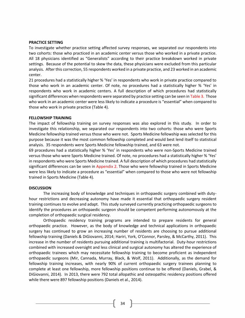
appeared in two different sections, the final survey had 207 procedures. The final survey was created and e-mailed to physicians via an online survey application. In addition to questions regarding essential procedures, demographic questions were also included at the end of each survey. Alumni were asked to indicate the number of years they have been practicing orthopaedic surgeons, as well as what fellowship(s), if any, they completed. Information regarding their practice site was also ascertained, such as the percent breakdown of patient care they deliver by subspecialty (eg. 50% Sports, 50% Spine) as well as their practices’ size and setting. Alumni were also asked to indicate whether they are primary instructors of residents and/or fellows. After stating willingness to participate, surveys were sent directly to graduates’ e-mail addresses. Graduates were sent reminder emails every 10 days if they had yet to completing the survey to encourage participation. STATISTICAL ANALYSIS Statistical analyses were performed using the R:A language and environment for statistical computing (R Foundation for Statistical Computing, http://www.R-project.org). Medians and interquartile ranges (IQRs) were reported for continuous data and percentages were reported for categorical data. Medians were compared using the Mann-Whitney U test and proportions were compared using chi2 or Fisher exact tests, as appropriate. Two-tailed p values <0.05 were considered statistically significant. RESULTS Out of the 110 graduates who agreed to participate in this study, 98 completed the survey, for a response rate of 89.1%. Table 2 summarizes the characteristics of those who responded. A majority of the respondents practice in urban settings in the southeastern United States. The most common fellowship completed was Sports Medicine, and nearly one-third of respondents work in a large-private group type practice. A majority of the respondents were not primary educators of residents or fellows. Figure 1 lists the 207 procedures in decreasing order of percent “Yes” responses, while Firgures 2 through 10 break down the procedures according to subspecialty. According to our criteria, 123 procedures were classified as “essential,” while the remaining 84 procedures were classified as “non-essential.” Of the 11 procedures that were unanimously deemed “essential” by all 98 respondents, eight were trauma-related while the other three were sports medicine procedures. Only 2 procedures received zero ‘yes’ responses, osteotomy for adult spinal deformity correction and revision total ankle arthroplasty. WORK-HOUR RESTRICTIONS In order to gain insight on how work-hour restrictions could affect survey responses, graduates were separated into two cohorts: those who were trained mostly during the age of work-hour restrictions (completed residency in 2005 or later) versus those who trained mostly before the introduction of work-hour restrictions (completed residency before 2005). Fifty-five of the respondents were trained before work-hour restriction, and 43 were trained after the introduction of work hour restrictions. 61 procedures had a statistically higher % ‘Yes’ in respondents who trained before work-hour restrictions compared to those who trained after their implementation. Of note, no procedures had a statistically significant higher % ‘Yes’ in respondents who trained after work hour restrictions. A full description of which procedures had statistically significant differences when respondents were separated by their timing of training and the implementation of work-hour restrictions can be seen in Appendix 1. Those who trained with work hour restrictions were less likely to indicate a procedure as “essential” when compared to those who trained before the advent of work-hour restrictions (Table 4).
34
PRACTICE SETTING To investigate whether practice setting affected survey responses, we separated our respondents into two cohorts: those who practiced in an academic center versus those who worked in a private practice. All 18 physicians identified as “Generalists” according to their practice breakdown worked in private settings. Because of the potential to skew the data, these physicians were excluded from this particular analysis. After this correction, 55 respondents worked in a private practice, and 23 worked in an academic center. 21 procedures had a statistically higher % ‘Yes’ in respondents who work in private practice compared to those who work in an academic center. Of note, no procedures had a statistically higher % ‘Yes’ in respondents who work in academic centers. A full description of which procedures had statistically significant differences when respondents were separated by practice setting can be seen in Table 3. Those who work in an academic center were less likely to indicate a procedure is “essential” when compared to those who work in private practice (Table 4). FELLOWSHIP TRAINING The impact of fellowship training on survey responses was also explored in this study. In order to investigate this relationship, we separated our respondents into two cohorts: those who were Sports Medicine fellowship trained versus those who were not. Sports Medicine fellowship was selected for this purpose because it was the most common fellowship completed and would best lend itself to statistical analysis. 35 respondents were Sports Medicine fellowship trained, and 63 were not. 69 procedures had a statistically higher % ‘Yes’ in respondents who were non-Sports Medicine trained versus those who were Sports Medicine trained. Of note, no procedures had a statistically higher % ‘Yes’ in respondents who were Sports Medicine trained. A full description of which procedures had statistically significant differences can be seen in Appendix 2. Those who were fellowship trained in Sports Medicine were less likely to indicate a procedure as “essential” when compared to those who were not fellowship trained in Sports Medicine (Table 4). DISCUSSION
The increasing body of knowledge and techniques in orthopaedic surgery combined with duty-hour restrictions and decreasing autonomy have made it essential that orthopaedic surgery resident training continues to evolve and adapt. This study surveyed currently practicing orthopaedic surgeons to identify the procedures an orthopaedic surgeon should be competent performing autonomously at the completion of orthopaedic surgical residency.
Orthopaedic residency training programs are intended to prepare residents for general orthopaedic practice. However, as the body of knowledge and technical applications in orthopaedic surgery has continued to grow an increasing number of residents are choosing to pursue additional fellowship training (Daniels & DiGiovanni, 2014; Hariri, York, O'Connor, Parsley, & McCarthy, 2011). This increase in the number of residents pursuing additional training is multifactorial. Duty-hour restrictions combined with increased oversight and less clinical and surgical autonomy has altered the experience of orthopaedic trainees which may necessitate fellowship training to become proficient as independent orthopaedic surgeons (Mir, Cannada, Murray, Black, & Wolf, 2011). Additionally, as the demand for fellowship training increases, with nearly 90% of current orthopaedic surgery trainees planning to complete at least one fellowship, more fellowship positions continue to be offered (Daniels, Grabel, & DiGiovanni, 2014). In 2013, there were 792 total allopathic and osteopathic residency positions offered while there were 897 fellowship positions (Daniels et al., 2014).

35
In our study 83.7% of respondents completed at least one fellowship. This is slightly lower than the current trend of 90% of graduates planning to complete a fellowship (Daniels et al., 2014). However, our study respondents included 55 surgeons who completed most or all of their training prior to the implementation of work hour restrictions and 41 of the respondents had been in practice for 16 years or more. Furthermore, the nature of the residency training program being centered at a large academic institution may self-select residents more likely to pursue additional training and specialization than enter general practice directly from residency.
For the purposes of this study a procedure was deemed ‘essential’ for residents to master prior to completion of residency training if 50% of respondents believed the procedure was essential. One hundred twenty-three of the 207 procedures sent out in the final survey were deemed essential. Combining this with the pilot survey results, 142 procedures were considered essential for residency training. While this list is by no means comprehensive, it represents the majority of cases one would expect to see throughout residency training and practice. The only subspecialty that is obviously underrepresented in the survey is musculoskeletal oncology as the great variation in pathology, tumor location, and unique procedural variables encountered in performing oncologic procedures is impossible to accurately identify and assess in this format.
While each subspecialty had procedures rated as essential and non-essential, only 4 of the 16 surveyed spine procedures were considered essential. Furthermore, only 2 of these procedures were invasive: lumbar laminectomy and lumbar decompression. This finding is consistent with the ABOS Part II procedures evaluation by Garrett et al. which found that the majority of reported spine procedures were being performed by spine fellowship trained orthopedic surgeons (Garrett et al., 2006). These results should encourage residency programs to evaluate the amount of time devoted to spine training if the goal is to train general orthopaedic surgeons.
An examination of the adult reconstructive procedures reveals an interesting dichotomy. Primary total knee arthroplasty and primary total hip arthroplasty were unanimously chosen as essential in the pilot study, however, only one quarter of respondents believed that revision total knee and hip arthroplasty should be mastered during residency. Pour et al. showed that adult reconstruction fellowship trained surgeons had a greater number of primary and revision total hip and knee cases on their ABOS Part II examination than non-adult reconstruction fellowship trained surgeons (Eslam Pour et al., 2016; Pour et al., 2016). However, surgeons performing a larger volume of knee and hip arthroplasties reported fewer complications regardless of fellowship training (Eslam Pour et al., 2016; Pour et al., 2016). While the number of arthroplasties performed is the main determinant of complication frequency, adult reconstruction trained surgeons are likely to perform more arthroplasties and more complex primary and revision arthroplasties over the course of their careers. As such, our study findings that residents should be expected to perform primary hip and knee arthroplasties, but not revision arthroplasties upon residency completion is in line with the findings by Pour et al (Eslam Pour et al., 2016; Pour et al., 2016).
There are currently more fellowship positions available in sports medicine than any other subspecialty of orthopaedics (Daniels & DiGiovanni, 2014). This is reflected in our respondents, of whom 45 (36.8%) had completed a sports medicine fellowship. When we compared the responses of sports medicine trained physicians to all other surgeons zero procedures had a statistically significant higher ‘yes’ response from the sports medicine trained group than all other surgeons. In fact, there were 69 procedures that were significantly more likely to receive a ‘yes’ response than non-sports medicine trained physicians (p<0.05, Appendix 2). This is an interesting finding which we do not have an explanation for. Future studies should have a greater number of respondents and will allow for comparisons between each subspecialty.
While the majority of respondents worked in a large private group (32.99%, Table 2) or in private practice settings (74.5%), a substantial proportion of surgeons identified themselves as working in an academic center (23.7%). Our survey identified 21 procedures that surgeons working in private practice

36
were more likely to label ‘essential’ than those working at academic centers (p<0.05, Table 3). Additionally, there were no procedures that surgeons working at academic centers rated more ‘essential’ than surgeons working in private practice. While the reasons for these findings are undoubtedly multifactorial, it is possible that many of the surgeons working in private practice groups may not have all subspecialties covered by fellowship trained surgeons. As such, they may be more likely to identify a procedure as ‘essential’ because they do not have the ability to easily refer it on to a fellowship trained physician.
Since the implementation of resident duty hour restrictions in 2003 residency programs have been adapting to train new physicians with the same competency and skill of their predecessors with fewer total hours of training (ACGME, 2016). Fifty-five respondents (56%) completed all or a majority of their training prior to the implementation of duty hour restrictions. These surgeons identified 61 more procedures as ‘essential’ than those surgeons who completed the majority of their training after the implementation of duty hour restrictions (p<0.05, Appendix 1). This may reflect a difference in approach toward sub-specialization for more recently trained orthopaedic surgeons. As orthopaedic surgery has evolved there is increased orthopaedic knowledge, more orthopaedic procedures, and a larger volume of information and procedures to master within each individual subspecialty. As such, newer surgeons are more likely to limit their scope of practice to the procedures they consider themselves ‘experts’ in.
There are several limitations to this study. First, the respondents are limited to currently practicing orthopaedic surgeons who trained at the same academic institution. This may have led to a bias toward surgeons more likely to pursue a career in academic medicine or seek further fellowship training. Future studies should expand the target group to include surgeons who trained at a variety of community based and academic institutions in different regions of the country. Second, a larger number of respondents would have allowed for identification of trends between different subspecialties. However, we did achieve a nearly 90% response rate to the survey which would be difficult to maintain with increasing the number of surveys distributed. Third, there were only 207 procedures assessed in the final version of the survey. While it would be impossible to perform a survey with a comprehensive list of orthopaedic procedures, there are undoubtedly procedures that are commonly performed which were not included on the current survey. This is especially evident in the musculoskeletal oncology portion of the survey as the variety of pathology, locations of tumors, and variability in procedures is nearly limitless in this field of orthopaedics.
As the healthcare landscape continues to change, orthopaedic surgery, and specifically resident education and training, must continue to evolve to meet patient needs and healthcare demands. This study provides useful information to help guide orthopaedic surgery residency programs in emphasizing the procedures essential to the practicing orthopaedic surgeon. It also supports the notion that more recent graduates, as well as those working in academic centers, believe more procedures should be mastered during fellowship training as opposed to by the end of a five-year residency program. This information will help guide future research on orthopaedic residency training and milestones for residency completion.

37
Tables & Figures
Table 1 – Procedures Removed after Initial Survey Procedure Name Intramedullary Nail Diaphysial Femur Intramedullary Nail Infraisthmal Femur Intramedullary Nail Intertrochanteric Femur ORIF Intertrochanteric Femur ORIF Patella Intramedullary Nail Tibia ORIF Lateral Tibia Plateau ORIF Bimalleolar Ankle Closed Reduction and Casting Forearm Fractures Diagnostic Shoulder Arthroscopy Subacromial Decompression Carpal Tunnel Release Trigger Finger Release ORIF Olecranon Primary Total knee arthroplasty Primary Total Hip Arthroplasty Hemiarthroplasty Lipoma Excision Achilles Tendon Repair Each of these procedures was unanimously rated as essential

38

39

40
Figure 1:

41
Figure 2: Adult Reconstruction
0 20 40 60 80 100
Ganz Osteotomy
Hip Resurfacing
Revision Total Knee Arthroplasty
Revision Total Hip Arthroplasty
Patellofemoral Resurfacing
Antibiotic Spacer Hip - Articulating
Antibiotic Spacer Knee - Articulating
High Tibial Osteotomy
Unicondylar Knee Arthroplasty
Antibiotic Spacer Hip - Static/non-articulating
Antibiotic Spacer Knee - Static/non-articulating
Girdlestone
Series1

42
Figure 3: Amputations
0 20 40 60 80 100
Hemipelvectomy
Forequarter
Hip Disarticulation
Through Elbow
Transradial
Transhumeral
Symes
Ray Resection
Through Knee
Transmetatarsal
Digit Amputation
Transfemoral
Transtibial
Series1
43
Figure 4: Foot and Ankle
0 20 40 60 80 100
Revision Total AnkleTotal Ankle Replacement
Flatfoot OsteotomiesOATS/De Novo Talar OCD Correction
Supramalleolar OsteotomyTendon Transfers
Thin Wire Fixator ProceduresGreat Toe Arthroplasty
Triple ArthrodesisLapidus
ORIF Calcaneus FractureTibiotalocalcaneal Nail
Lateral and Medial Ligamentous Ankle…Gastrocnemius Slide
Tarsal Tunnel ReleaseTibiotalar Fusion
ORIF Talus FractureORIF Pilon Fracture
Lisfranc ORIF/FusionAnkle Arthroscopy
Hallux FusionFirst Metatarsal Osteotomy
Distal Soft Tissue Procedure with…Chevron Osteotomy
Claw Toe Deformity CorrectionMorton's Neuroma Excision
Dorsal CheilectomySubtalar Fusion
Series1

44
Figure 5: Hand and Upper Extremity
0 20 40 60 80 100
Digit Replant
Four Corner Fusion
Digital Artery Repair
Proximal Row Carpectomy
Zone 2 Flexor Tendon Repair
Digital Nerve Repair
CRPP/ORIF Perilunate Dislocation
Thumb CMC Joint Resection Arthroplasty
Flexor Tendon Repair
CRPP/ORIF Scaphoid Fracture
Ray Excision
Radial Head Arthroplasty
CRPP/ORIF Phalanx Fracture
DeQuervain's Release
Nail Bed Laceration Repair
Extensor Tendon Repair
Cubital Tunnel Release
Irrigation and Debridement of Purulent…
CRPP/ORIF Metacarpal Fracture
Ganglion Excision
Irrigation and Debridement of Fight Bite
Series1

45
Figure 6: Musculoskeletal Oncology
0 20 40 60 80 100
Enchondroma Excision
Giant Cell Tumor of Tendon SheathExcision
Osteochondroma Excision
Principles of Tumor Biopsy
Prophylactic Intramedullary Nailingfor Impending Fractures
Series1

46
Figure 7: Pediatric Orthopaedics
0 20 40 60 80 100
Vertebral Column Resection for Spinal…
Pemberton Osteotomy
Salter Osteotomy
Casting for Scoliosis
Posterior Instrumentation and Fusion for…
Varus Derotational Osteotomy
Guided Growth Plating
Hemiepiphysiodesis
Closed Reduction and Spica Casting for…
Bracing for Scoliosis
Epiphysiodesis
Ponsetti Club Foot Casting
Percutaneous Tendoachilles Lengthening
ORIF Triplane Fracture
ORIF Tillaux Fracture
Application of Spica Cast
Slipped Capital Femoral Epiphysis In-situ…
Trigger Thumb Release
Flexible Nailing of Femur Fractures
ORIF/ORPP Medial Humeral Condyle
ORIF/ORPP Lateral Humeral Condyle
CRPP Supracondylar Humerus
ORIF Tibial Tubercle Fracture
Irrigation and Debridement of a Septic Hip
Series1

47
Figure 8: Spine
0 10 20 30 40 50 60 70 80
Osteotomy for Adult Spinal Deformity…
3 or More Level Lumbar Instrumentation…
Posterior Lumbar Interbody Fusion
Anterior Lumbar Interbody Fusion
Cervical Laminectomy and Fusion
Cervical Laminoplasty
2 Level Lumbar Instrumentation and…
1 Level Lumbar Instrumentation and…
Anterior Cervical Decompression and…
Decompression for Cauda Equina
Irrigation and Debridement for Epidural…
Microdiskectomy
Lumbar Decompression
Lumbar Laminectomy
Halo Application
Gardner Wells Tongs Application
Series1

48
Figure 9: Sports
0 20 40 60 80 100
Latarjet
MUCL Reconstruction
PCL Reconstruction
Suprascapular Cyst Removal
Labral Repair - Posterior Bankart
MPFL Reconstruction
Meniscus Root Repair
Tibial Tubercle Osteotomy
Osteophyte Removal from…
Capsular Release
Dual Row Rotator Cuff Repair
Distal Biceps Repair/Reconstruction
ACL Reconstruction
ORIF Clavicle Fracture
Acromioplasty
Chondroplasty
Biceps Tenotomy
Patella Tendon Repair
Diagnostic Knee Arthroscopy
Meniscectomy
Series1

49
Figure 10: Trauma
0 20 40 60 80 100
ORIF Both Column Acetabulum
ORIF Anterior Wall Acetabulum
ORIF/CRPP Sacroiliac Joint
ORIF Calcaneus
ORIF Talus
ORIF Periprosthetic Distal Femur
ORIF Complete Articular Distal Femur
ORIF Bicondylar Tibia Plateau
ORIF Medial Tibia Plateau
Open Reduction Elbow Dislocation
Intramedullary Nail Humerus
Open Reduction Hip Dislocation
Open Reduction Shoulder Dislocation
ORIF Extra-Articular Distal Humerus
ORIF Distal Metaphyseal Tibia
ORIF Diaphyseal Humerus
Closed Reduction Knee Dislocation
ORIF Distal Fibula
ORIF Syndesmotic Injury
Closed Reduction Elbow Dislocation
Closed Reduction Shoulder…
ORIF Both Bone Forearm
ORIF Proximal Metaphyseal Tibia
Series1

50
Appendices:

51

52
REFERENCES
1. ACGME. (2016). History of Duty Hours. 2. Chen, A. C., Lee, M. S., Chen, W. J., & Lee, S. T. (2013). Assessment in orthopedic training-an
analysis of rating consistency by using an objective structured examination video. J Surg Educ, 70(2), 189-192. doi:10.1016/j.jsurg.2012.11.002
3. Daniels, A. H., & DiGiovanni, C. W. (2014). Is subspecialty fellowship training emerging as a necessary component of contemporary orthopaedic surgery education? J Grad Med Educ, 6(2), 218-221. doi:10.4300/JGME-D-14-00120.1
4. Daniels, A. H., Grabel, Z., & DiGiovanni, C. W. (2014). ACGME Accreditation of Orthopaedic Surgery Subspecialty Fellowship Training Programs. J Bone Joint Surg Am, 96(11), e94. doi:10.2106/JBJS.M.01340
5. Davis Sears, E., Larson, B. P., & Chung, K. C. (2012). A national survey of program director opinions of core competencies and structure of hand surgery fellowship training. J Hand Surg Am, 37(10), 1971-1977 e1977. doi:10.1016/j.jhsa.2012.06.034
6. Eslam Pour, A., Bradbury, T. L., Horst, P. K., Harrast, J. J., Erens, G. A., & Roberson, J. R. (2016). Trends in Primary and Revision Hip Arthroplasty Among Orthopedic Surgeons Who Take the American Board of Orthopedics Part II Examination. J Arthroplasty. doi:10.1016/j.arth.2015.12.045
7. Garrett, W. E., Jr., Swiontkowski, M. F., Weinstein, J. N., Callaghan, J., Rosier, R. N., Berry, D. J., . . . Derosa, G. P. (2006). American Board of Orthopaedic Surgery Practice of the Orthopaedic Surgeon: Part-II, certification examination case mix. J Bone Joint Surg Am, 88(3), 660-667. doi:10.2106/JBJS.E.01208
8. Grantcharov, T. P., & Reznick, R. K. (2009). Training tomorrow's surgeons: what are we looking for and how can we achieve it? ANZ J Surg, 79(3), 104-107. doi:10.1111/j.1445-2197.2008.04823.x
9. Hariri, S., York, S. C., O'Connor, M. I., Parsley, B. S., & McCarthy, J. C. (2011). Career plans of current orthopaedic residents with a focus on sex-based and generational differences. J Bone Joint Surg Am, 93(5), e16. doi:10.2106/JBJS.J.00489
10. Karam, M. D., Pedowitz, R. A., Natividad, H., Murray, J., & Marsh, J. L. (2013). Current and future use of surgical skills training laboratories in orthopaedic resident education: a national survey. J Bone Joint Surg Am, 95(1), e4. doi:10.2106/JBJS.L.00177
11. Mir, H. R., Cannada, L. K., Murray, J. N., Black, K. P., & Wolf, J. M. (2011). Orthopaedic resident and program director opinions of resident duty hours: a national survey. J Bone Joint Surg Am, 93(23), e1421-1429. doi:10.2106/JBJS.K.00700
12. Nasca, T. J., Philibert, I., Brigham, T., & Flynn, T. C. (2012). The next GME accreditation system--rationale and benefits. N Engl J Med, 366(11), 1051-1056. doi:10.1056/NEJMsr1200117
13. Noland, S. S., Fischer, L. H., Lee, G. K., & Hentz, V. R. (2013). Essential hand surgery procedures for mastery by graduating orthopedic surgery residents: a survey of program directors. J Hand Surg Am, 38(4), 760-765. doi:10.1016/j.jhsa.2012.12.035
14. Pour, A. E., Bradbury, T. L., Horst, P., Harrast, J. J., Erens, G. A., & Roberson, J. R. (2016). Trends in primary and revision knee arthroplasty among orthopaedic surgeons who take the American Board of Orthopaedics part II exam. Int Orthop. doi:10.1007/s00264-016-3137-z
15. Stern, P. J. (2012). Orthopaedic Surgery Milestones. Retrieved from

53
16. Van Heest, A., Putnam, M., Agel, J., Shanedling, J., McPherson, S., & Schmitz, C. (2009). Assessment of technical skills of orthopaedic surgery residents performing open carpal tunnel release surgery. J Bone Joint Surg Am, 91(12), 2811-2817. doi:10.2106/JBJS.I.00024
17. Wu, B. J., Dietz, P. A., Bordley, J. t., & Borgstrom, D. C. (2009). A novel, web-based application for assessing and enhancing practice-based learning in surgery residency. J Surg Educ, 66(1), 3-7. doi:10.1016/j.jsurg.2008.07.015

54
Brent Wise, MD PGY-5
Administrative Chief Resident
UPCOMING FELLOWSHIP TRAINING: Trauma Fellowship R. Adams Cowley Shock Trauma Center Baltimore, MD EDUCATION: University of Florida College of Medicine, Gainesville, FL Doctor of Medicine, May 2011 The Pennsylvania State University, State College, PA Bachelor of Science, Life Science, May 2007 PROFESSIONAL SOCIETIES: Orthopaedic Trauma Association, Candidate Member
HONORS AND LEADERSHIP: Administrative Chief Resident, Orthopaedic Surgery Residency 2015-2016 AOA Emerging Leaders Forum 2015 3rd Place Kelly Day Resident Research Award – Emory University 2014 Emory University GME Committee Member 2014-2016 Program Evaluation Committee Member 2012-2016 Residency Interviewing and Selection Committee 2012-2016 PEER-REVIEWED PUBLICATIONS: Rajani R, Wise B, Oswald T, Roberson J. Epiphysiolysis of the Femoral Neck due to Closed Reduction of an Adolescent Hip Dislocation with 4 year Follow Up: A Case Report and Review of the Literature. Journal of Pediatric Orthopaedics B. 2015; 24(1):40-5.

55
Outcomes of ACL Reconstruction Using Fixed Versus
Variable Loop Button Fixation
Brent Wise, MD, Nick Patel, MD, Garrison Wier, MD, Sameh Labib, MD ABSTRACT Suspensory femoral fixation of ACL grafts with fixed and variable loop “button” devices has gained popularity for ACL reconstruction. This study examines these two methods of fixation in order to determine their impact on graft laxity and patient reported outcome scores. Patients with ACL reconstructions using either a fixed or variable loop button technique performed by the primary investigator were identified through a database search. Lysholm, Tegner, and SF-12 scores were collected, and KT-1000 mechanical knee testing was performed and compared to the uninjured knee. A total of 112 patients were identified, 91 of which met criteria to be included in this study. Fifty-seven patients completed the KT-1000 knee testing, 33 patients in the variable group and 24 patients in the fixed group. The average KT-1000 value for the variable group was 0.38mm, while the average for the closed group was 0.92mm (p=0.19, 95% CI: -0.28 - 1.35). Amongst the 19 variable loop patients and 13 closed loop patients that completed the subjective outcomes questionnaires, there was not a statistically significant difference. Clinically lax knees (KT > 3mm) were found in 6.1% and 12.5% of the variable loop and fixed loop techniques, respectively (p = 0.2). The variable group had a re-rupture rate of 4.7% while the fixed group had a re-rupture rate of 8.7% (p = 0.21). The study finds that there is no statistical difference in ACL graft laxity or post-operative functional outcomes between grafts fixed with variable loop or fixed loop button techniques. INTRODUCTION
Anterior cruciate ligament (ACL) injuries continue to be the most common serious injury of the knee seen by Orthopaedic surgeons.10,24,29 An estimated 50,000-175,000 primary ACL reconstructions are done each year in the United States, and ACL reconstruction ranks as the 6th most commonly performed orthopedic procedure.10 There are many factors that impact the overall outcome of ACL reconstruction. Amongst these, graft fixation and subsequent incorporation into the bony tunnels are considered essential for the stability of the reconstructed knee.3,5 As of now, there is limited data specifying a superior graft fixation technique. Suspensory fixation with titanium “button” devices has gained popularity due to the simplicity, reliability, and the excellent tensile strength afforded by the devices.13,28 Suspensory fixation methods are broadly divided into fixed loop and variable loop systems.
Fixed loop button fixation devices include the EndoButton (Smith & Nephew Inc, Andover, Massachusetts) and Retrobutton (Arthrex Inc, Naples, Florida). These fixed-loop designs may not deploy successfully if the graft tunnel is drilled too short to allow clearance of the device through the femur. Alternatively, having a large loop may lead to a small amount of graft in the femoral tunnel. The variable loop systems, such as the TightRope RT (Arthrex Inc, Naples, Florida) and ToggleLoc with ZipLoop (Biomet Inc, Warsaw, Indiana), employ a series of sutures that can be used as a pulley system to shorten the loop and thus deliver the graft to the end of the femoral socket for a tight fit (Figures 3). This technique reduces the approximation needed in measurements and also allows the graft to be advanced farther into the femoral tunnel after the button “flips” which is not possible in a closed

56
loop fixation technique. Some suggest that this may lead to improved incorporation of the graft given the larger interface for bone to tendon healing.7,12,25
This study examines these two different methods of button fixation, in order to determine
their impact on objective graft laxity and functional outcomes. It is hypothesized that the variable loop fixation technique will result in improved objective measurements of graft laxity at follow up as a result of the greater amount of graft able to be drawn into the femoral tunnel, while the overall functional outcomes will be comparable.
MATERIALS AND METHODS Subject Identification
Patients who had undergone an ACL reconstruction using either the fixed loop button or variable loop button surgical technique by the principal investigator between the dates of January 1, 2008 and December 31, 2012 were identified through a search of procedure codes. After reviewing medical records, potential participants were contacted in order to determine eligibility. Inclusion Criteria
Patients were included in the study if they (1) had an ACL rupture and subsequent successful ACL reconstruction using either the fixed loop button or variable loop button device, (2) were eighteen years or older in age, and (3) had no other illnesses that prevented them from ambulating normally (without the help of devices). Exclusion Criteria
Patients were excluded from the study if they (1) had an age less than eighteen years, (2) had multiple ligamentous injuries or fractures, (3) had previous knee surgery, revision ACL reconstructions, or any osteotomies performed at the time of ACL reconstruction, or (4) had ACL reconstruction with other modes of fixation on the femoral side. KT-1000
KT-1000 analysis was performed by an independent physical therapist blinded to the fixation method used on both knees of participants in order to test the integrity of the ACL graft. Specifically, it allowed quantification of the anterior translation of the tibia in relation to the femur and was reported as a comparison between the operative and non-operative sides. The KT-1000 test was accomplished by securing a device to the front of the knee flexed at 25 degrees with the individual lying on their back. A maximal force as well as an 85 N force was pulled at the front of the lower leg and then the displacement of the knee was measured. Questionnaires
A series of questionnaires and scales were used to evaluate each surgical technique based on subjective patient response and were performed at either the office visit or online. These included the Lysholm questionnaire to assess post-surgical knee function and stability, the Tegner scale to measure pre and post-surgical activity level, and the SF-12 survey to evaluate current physical and mental health. Statistical Analysis
Participants were grouped into the fixed loop button and variable loop button techniques and descriptive data (mean ± SD) were calculated for all variables. Using a one-way analysis of variance (ANOVA), the overall means for KT-1000 test, Tegner, SF-12, and Lysholm scores were compared between groups. A p value ≤ 0.05 was considered statistically significant.

57
RESULTS KT-1000 Testing
Through a search of procedure codes, a total of 112 patients were identified as having undergone ACL reconstructions in the designated time period by the primary investigator using either the variable loop (43 patients) or fixed loop (69 patients) techniques. Of the 69 identified fixed loop reconstructions, 16 had to be excluded (including 6 ACL re-ruptures). Of the 43 identified variable loop reconstructions, 5 were excluded (2 ACL re-ruptures). The mean ages for patients having undergone surgery with the variable loop and fixed loop techniques was 28.9 and 27.3 years respectively (Table 1). The diagnosed re-rupture rate following ACL reconstruction was found to be 4.7% for the variable loop and 8.7% for the fixed loop groups (p = 0.21). From those meeting the criteria and willing to participate, mechanical knee testing using KT- 1000 analysis was performed on a total of 57 participants, 33 of which were in the variable loop group and 24 of which were in the fixed loop group. The mean KT value for the variable group was 0.38mm (95% CI: -0.16 - 0.92) while that for the fixed loop group was 0.92 (95% CI: 0.36 - 1.48) (Table 2). This resulted in a mean difference between the two surgical groups of 0.54mm (p = 0.19, 95% CI: -0.28 - 1.35). When looking at ACL grafts considered to be clinically lax (KT > 3mm), the variable loop group had a lax graft rate of 6.1% while the fixed loop group had a lax rate of 12.5% (p = 0.2). Subjective Outcomes
Of the identified patients having undergone ACL reconstruction, a total of 32 individuals completed the Lysholm, Tegner, and SF-12 questionnaires (19 in the variable loop group and 13 in the fixed loop group). The findings are summarized in Table 3. In terms of the Tegner activity scale, the pre-operative means for the variable loop and fixed loop were 8.00 and 6.92 respectively (p = 0.09) and the post-operative means were 6.21 and 6.46 respectively (p = 0.61). The post-operative Lysholm knee scale means were 87.28 for the variable loop subset and 91.31 for the fixed loop subset (p = 0.37). The post-operative SF-12 health survey was composed of both a mental health and physical health component. For the SF-12 physical component, the mean scores were 53.18 and 54.89 for the variable loop and fixed loop groups respectively (p = 0.33). The SF-12 mental component mean scores were 54.84 and 54.82 for the variable loop and fixed loop groups respectively (p = 0.99).
DISCUSSION
The goal of this study was to directly compare the two available types of suspensory button fixation. While no clinical studies to date examine this, there have been biomechanical studies that have directly compared these fixation methods. Petre et al. looked at 4 different types of cortical suspensory fixation methods including two fixed loop and two variable loop devices.23 The most crucial finding as it pertains to this study was that of the increased displacement due to device slippage that occurred with the variable loop designs. This finding was also supported by Barrow, et al. in a biomechanical study that suggested that lengthening of the variable loop designs may lead to delayed graft healing and knee instability.4 However, the authors of the Petre, et al. study point out that the variable loop devices allow for re-tensioning after tibial fixation which can compensate for the slippage that does occur.23 This is critical for the surgeon to be aware of as they can correct for this intra-operatively thus avoiding any potential impact on clinical outcomes. The fact that our study did not reveal increased laxity as measured by KT-1000 testing or any functional difference when comparing the variable to the fixed loop group supports this very idea as re-tensioning the graft following tibial fixation is standard practice in our variable loop fixation technique.
The results of our study show that there is no statistically significant difference in KT-1000 measurements using fixed loop versus variable loop fixation methods. It was hypothesized that

58
variable loop button fixation would provide more secure fixation and ultimately deceased laxity compared to fixed loop fixation. This initial hypothesis was based upon the principles of ACL graft healing and incorporation along with the clinical finding that variable loop fixation methods allow the surgeon to consistently draw a greater amount of graft into the femoral tunnel. Based upon histologic studies that have shown ACL grafts incorporate with bony ingrowth through the fibrous graft/tunnel interface formed along the entire length of the tunnel25,26, one can assume that the larger the tendon/bone interface, the more collagen fibers there are to anchor the tendon to the bone, thus providing greater resistance to graft pullout and failure especially in the early period of healing.
Our hypothesis was also supported by the work of Rodeo et al., who showed that graft motion within the tunnel, which is greater with suspensory fixation, slowed graft incorporation and contributes to the tunnel widening that can be seen. 26 They showed that this motion is greatest at the intra-articular tunnel entrance and decreases along the tunnel as the graft is closer to the extra-articular fixation point. Variable loop fixation allows for the graft to be consistently pulled closer to this fixation point, theoretically allowing for a larger area of faster incorporation. Rodeo et al. also showed that bone ingrowth of the graft occurs more readily when there is less motion and closer apposition within the tunnel. Again, variable loop fixation allows for more graft to be placed closer to the tunnel exits where this closer apposition occurs which leads to more secure fixation.26
It is important to note that while our results did not reveal a statistically significant difference in graft laxity, there was a trend towards greater laxity and higher rupture rates in the fixed loop fixation group. A greater percentage of patients within the fixed loop group had grafts with KT-1000 measurements of 3 mm or greater. This is the value that has been used within the literature to indicate clinically significant graft laxity that could be indicative of graft failure.8,9 While the differences in KT-1000 measurements did not reach statistical significance, this greater percentage of patients with lax grafts—12.5% versus 6.1% in the variable group—is felt to be clinically significant. Likewise, the difference in re-rupture rates between the groups—8.7 % in the fixed group versus 4.7% in the variable group—is also felt to be clinically significant. These findings may be explained, at least in part, by the aforementioned principles of ACL graft healing and incorporation.
The fact that our results show no statistically significant difference in objective graft laxity as opposed to decreased laxity in the variable loop group as hypothesized, may be a result of the sliding or loosening that occurs with this type of fixation that could not be accounted for by re-tensioning the graft after final tibial fixation as discussed above. It could also be due to the fact that with proper closed loop button fixation technique, a negligible difference exists in the amount of graft within the tunnel. Whether the amount of graft in the tunnel following reconstruction with fixed or variable loop fixation differs at follow-up is a question that has yet to be born out in the literature.
In addition to equivalent findings in regards to objective graft laxity, there was no difference in patient reported functional outcomes among the groups. Moreover, those patients with KT-1000 measurements of 3mm or greater did not have functional scores that differed significantly from the remainder of the study group. This finding may be supported by the fact that rotational instability is more important from a functional and knee contact mechanics standpoint than anterior-posterior translation as is tested with a KT-1000.2,6,14,37 It is well known that placement of tunnels has more to do with the rotational stability of the reconstruction than does the method of fixation.20 There are several strengths to this study. For one, this is the only clinical study to our knowledge comparing outcomes of ACL reconstruction using fixed and variable loop button fixation despite several biomechanical studies that suggest the inferiority of variable loop designs. In addition, all of the procedures were performed by a single surgeon using the same technique. Furthermore, all of the KT-1000 measurements were performed by blinded physical therapists and

59
all data was collected by independent co-authors. The limitations of the study include its retrospective design and the small sample size along with only a 62% follow-up with KT-1000 testing and 35% participation in the outcomes surveys. The authors do not feel that the low patient response rate creates a selection bias, as all of the medical records were reviewed, and the vast majority of patients that did not participate in the study did not have post-operative complications. Those that did were included in the overall analysis of the study populations’ outcomes. While the sample size of the study is low, it is powered in such a way to detect a 2mm difference in KT-1000 testing, a value deemed by the authors to be clinically significant. Lastly, the inability to accurately assess how much graft actually remains in the femoral tunnel at patient follow up leads to only a theoretical discussion of the importance of intratunnel graft length and whether or not a difference exists between the closed and fixed loop groups as there is presently no practical way to confirm this. Currently, the researchers are devising MRI protocols that would allow us to determine how much graft remains in the tunnels. CONCLUSION
The study finds that there is no statistical difference in objectively measured ACL graft laxity as measured by KT-1000 between grafts fixed with variable loop versus fixed loop button techniques. There is also no difference in post-operative functional outcome with either surgical fixation technique as measured through multiple outcome scales. There was a trend towards improved fixation within the variable loop group with a lower rate of re-rupture and less grafts considered objectively lax. The study confirms that the use of variable loop button fixation is an acceptable means to achieve femoral-sided graft fixation during ACL reconstruction despite biomechanical studies that have demonstrated graft slippage with these devices. ACKNOWLEDGEMENT The authors would like to give a Special thanks to Patricia Bush, Ed.D for her assistance in designing the study and helping to coordinate the processes involved. In addition, thank you to Rebecca Bedard, ATC/OTC for her help with data collection.

60
REFERENCES 1. Ahmad CS, Gardner TR, Groh M, Arnouk J, Levine WN. Mechanical properties of soft tissue femoral fixation
devices for anterior cruciate ligament reconstruction. Am J Sports Med. 2004;32:635-640. PMID: 15090378.
2. Andriacchi TP, Mundermann A, Smith RL et al. A framework for the in vivo pathomechanics of Osteoarthritis at the knee. Ann Biomed Eng. 2004; 32.3:447-457. PMID: 15095819.
3. Baer GS, Harner CD. Clinical outcomes of allograft versus autograft in anterior cruciate ligament reconstruction. Clin Sports Med. 2007;26:661-681. PMID: 17920959.
4. Barrow AE, Pilia M, Guda T, Kadrmas WR, Burns TC. Femoral suspension devices for anterior cruciate ligament reconstruction: do adjustable loops lengthen? Am J Sports Med. 2014 Feb;42(2):343-9. PMID: 24158183.
5. Brand J, Jr., Weiler A, Caborn DN, Brown CH, Jr., Johnson DL. Graft fixation in cruciate ligament reconstruction. Am J Sports Med. 2000;28(5):761-774. PMID: 11032238.
6. Brophy RH, Selby RM, Altchek DW. Anterior Cruciate Ligament Revision: Double-Bundle Augmentation of Primary Vertical Graft. Arthroscopy. 2006 Jun;22(6):683. PMID: 16762715.
7. Chen CH. Graft healing in anterior cruciate ligament reconstruction. Sports Med Arthrosc Rehabil Ther Technol. 2009 Sep 23;1(1):21. PMID: 19772670.
8. Daniel DM, Stone ML, Dobson BE, Fithian DC, Rossman DJ, Kaufman KR. Fate of the ACL-injured patient. A prospective outcome study. Am J Sports Med. 1994;22(5):632-644. PMID: 7810787.
9. Daniel DM, Stone ML, Sachs R, Malcom L. Instrumented measurement of anterior knee laxity in patients with acute anterior cruciate ligament disruption. Am J Sports Med. 1985;13(6):401-407. PMID: 4073348.
10. Edgar CM, Zimmer S, Kakar S, Jones H, Schepsis AA. Prospective comparison of auto and allograft hamstring tendon constructs for ACL reconstruction. Clin Orthop Relat Res. Sep 2008;466(9):2238-2246. PMID: 18575944.
11. Graf BK, Henry J, Rothenberg M, Vanderby R. Anterior cruciate ligament reconstruction with patellar tendon: an ex vivo study of wear-related damage and failure at the femoral tunnel. Am J Sports Med.1994;22(1):131-135. PMID: 8129096.
12. Greis PE, Burks RT, Bachus K, Luker MG. The influence of tendon length and fit on the strength of a tendon-bone tunnel complex. A biomechanical and histologic study in the dog. Am J Sports Med. 2001 Jul-Aug;29(4):493-7. PMID: 11476392.
13. Han DL, Nyland J, Kendzior M, Nawab A, Caborn DN. Intratunnel versus extratunnel fixation of hamstring autograft for anterior cruciate ligament reconstruction. Arthroscopy. 2012;28(10):1555-1566. PMID: 22560484.
14. Hofbauer M, Valentin P, Ddolsky R, Ostermann RC, Graf A, Figl S, Aldrian S. Rotational and translational laxity after computer-navigated single- and double-bundle anterior cruciate ligament reconstruction. Knee Surg Sports Traumatol Arthrosc. 2010; 18 (9):1201-1207. PMID: 19946665.
15. Ibrahim SA, Hamido F, Al Misfer AK, Mahgoob A, Ghafar SA, Alhran H. Anterior cruciate ligament reconstruction using autologous hamstring double bundle graft compared with single bundle procedures. J Bone Joint Surg Br. 2009;91:1310-1315. PMID: 19794165.
16. Kamelger FS, Onder U, Schmoelz W, Tecklenburg K, Arora R, Fink C. Suspensory fixation of grafts in anterior cruciate ligament reconstruction: a biomechanical comparison of 3 implants. Arthroscopy. 2009 Jul;25(7):767-76. PMID: 19560641.
17. Kousa P, Järvinen TL, Vihavainen M, Kannus P, Järvinen M. The fixation strength of six hamstring tendon graft fixation devices in anterior cruciate ligament reconstruction. Part I: femoral site. Am J Sports Med. 2003 Mar;31(2):174-81. PMID: 12642249.
18. Kuskucu SM. Comparison of short-term results of bone tunnel enlargement between EndoButton CL and cross-pin fixation systems after chronic anterior cruciate ligament reconstruction with autologous quadrupled hamstring tendons. J Int Med Res. 2008;36(1):23-30. PMID: 18230264.

61
19. Lind M, Menhert F, Pedersen AB. The first results from the Danish ACL reconstruction registry:
epidemiologic and 2 year follow-up results from 5,818 knee ligament reconstructions. Knee Surg Sports Traumatol Arthrosc. 2009 Feb;17(2):117-24. PMID: 18974970.
20. Meredick RB, Vance KJ, Appleby D. Outcome of single-bundle versus double-bundle reconstruction of the anterior cruciate ligament: a meta-analysis. Am J Sports Med. 2008; 36:1414–1421. PMID: 18508977.
21. Milano G, Mulas PD, Ziranu F, Piras S, Manunta A, Fabbriciani C. Comparison between different femoral fixation devices for ACL reconstruction with doubled hamstring tendon graft: A biomechanical analysis. Arthroscopy .2006;22:660-668. PMID: 16762706.
22. Morrison J. The mechanics of the knee joint in relation to normal walking. J Biomech. 1970;3(1):51-61. PMID: 5521530.
23. Petre BM, Smith SD, Jansson KS, de Meijer PP, Hackett TR, LaPrade RF, Wijdicks CA. Femoral cortical suspension devices for soft tissue anterior cruciate ligament reconstruction: a comparative biomechanical study. Am J Sports Med. 2013 Feb;41(2):416-22. PMID: 23263298.
24. Poehling GG, Curl WW, Lee CA, et al. Analysis of outcomes of anterior cruciate ligament repair with 5-year follow-up: allograft versus autograft. Arthroscopy. Jul 2005;21(7):774-785. PMID: 16012489.
25. Rodeo SA, Arnoczky SP, Torzilli PA, Hidaka C, Warren RF. Tendon-healing in a bone tunnel. A biomechanical and histological study in the dog. J Bone Joint Surg Am. 1993 Dec;75(12):1795-803. PMID: 8258550.
26. Rodeo SA, Kawamura S, K HJ, Dynybil C, Ying L. Tendon Healing in a Bone Tunnel Differs at the Tunnel Entrance Versus the Tunnel Exit: An Effect of Graft-Tunnel Motion? Am J Sports Med. 2006 Nov; 34: 1790-1800. PMID: 16861579.
27. Sabat D, Kundu K, Arora S, Kumar V. Tunnel widening after anterior cruciate ligament reconstruction: a prospective randomized computed tomography--based study comparing 2 different femoral fixation methods for hamstring graft. Arthroscopy. 2011;27(6):776-783. PMID: 21624672.
28. Saccomanno MF, Shin JJ, Mascarenhas R, Haro M, Verma NN, Cole BJ, Bach BR Jr. Clinical and Functional Outcomes After Anterior Cruciate Ligament Reconstruction Using Cortical Button Fixation Versus Transfemoral Suspensory Fixation: A Systematic Review of Randomized Controlled Trials. Arthroscopy. 2014 Nov;30(11):1491-8. PMID: 25064753.
29. Sajovic M, Vengust V, Komadina R, Tavcar R, Skaza K. A prospective, randomized comparison of semitendinosus and gracilis tendon versus patellar tendon autografts for anterior cruciate ligament reconstruction: five-year follow-up. Am J Sports Med. Dec 2006;34(12):1933-1940. PMID: 16923826.
30. Shaerf DA, Pastides PS, Sarraf KM, Willis-Owen CA. Anterior cruciate ligament reconstruction best practice: A review of graft choice. World J Orthop. 2014 Jan 18;5(1):23-9. PMID: 24649411.
31. Shelburne KB, Pandy MG, Anderson FC, Torry MR. Pattern of anterior cruciate ligament force in normal walking. J Biomech. 2004; 37(6):797-805. PMID: 15111067.
32. Shelburne KB, Pandy MG. A dynamic model of the knee and lower limb for simulating rising movements. Comput Methods Biomech Biomed Engin. 2002;5(2):149 159. PMID: 12186724.
33. Shelburne KB, Pandy MG. Determinants of cruciate-ligament loading during rehabilitation exercise. Clin Biomech. 1998;13(6):403-413. PMID: 11415815.
34. Shelton WR, Fagan BC. Autografts commonly used in anterior cruciate ligament reconstruction. J Am Acad Orthop Surg. 2011; 19(5):259-264. PMID: 21536625.
35. Speirs A, Simon D, Lapner P. Evaluation of a new femoral fixation device in a simulated anterior cruciate ligament reconstruction. Arthroscopy. 2010;26:351-357. PMID: 20206045.
36. Stadelmaier DM, Lowe WR, Ilahi OA, Noble PC, Kohl HW III. Cyclic pull-out strength of hamstring tendon graft fixation with soft tissue interference screws: influence of screw length. Am J Sports Med. 1999;27(6):778-783. PMID: 10569365.
37. Tashman S, Collon D, Anderson K et al. Abnormal rotational knee motion during running after anterior cruciate ligament reconstruction. J Sports Med. 2004; 32.4:975-983. PMID: 15150046.

62
38. Walsh MP, Wijdicks CA, Parker JB, Hapa O, LaPrade RF. A comparison between a retrograde interference
screw, suture button, and combined fixation on the tibial side in an all-inside anterior cruciate ligament reconstruction: a biomechanical study in a porcine model. Am J Sports Med. 2009;37(1):160-167. PMID: 18832342.
39. Yoshiya S, Nagano M, Kurosaka M, Muratsu H, Mizuno K. Graft healing in the bone tunnel in anterior cruciate ligament reconstruction. Clin Orthop Relat Res. 2000 Jul;(376):278-86. PMID: 10906885.
40. Zantop T, Weimann A, Wolle K, Musahl V, Langer M, Petersen W. Initial and 6 weeks postoperative structural properties of soft tissue anterior cruciate ligament reconstructions with cross-pin or interference screw fixation: An in vivo study in sheep. Arthroscopy. 2007;23: 14-20. PMID: 17210422.

63
TABLES
Table 1: ACL Reconstruction Surgical Data
Variable Loop Fixed Loop P-value
Mean Age at surgery (years)
28.9 27.3 p = 0.42
ACL Graft Type Tibialis anterior allograft – 67.4% Hamstring autograft – 32.6%
Tibialis anterior allograft – 31.9% Hamstring autograft – 24.6% Hamstring allograft – 31.9% Bone-tendon-bone autograft – 5.7% Bone-tendon-bone allograft – 2.9% Tibialis posterior allograft – 2.9%
ACL Re-rupture rate 4.7% 8.7% p = 0.21
Table 2: KT-1000 Measurement Data
Variable Loop Fixed Loop P-value
Mean KT value (mm) 0.38 0.92 p = 0.19
% of patients with KT >
3.0mm
6.1% 12.5% p = 0.20
Table 3: Subjective Functional Outcome Data
Variable Loop Fixed Loop P-value
Pre-Injury Tegner 8.00 6.92 p = 0.09
Post-Injury Tegner 6.21 6.46 p = 0.61
Lysholm 87.28 91.31 p =0.37
SF-12 PCS 53.18 54.89 p = 0.33
SF-12 MCS 54.84 54.82 p = 0.99

64
Figure 1. Prepared ACL soft tissue graft with suspensory fixation button attached.
This image provided courtesy of Arthrex, Inc.
Figure 2. Passage of suspensory fixation button through the femoral tunnel. Note the “flipping” of the
button as it exits the femoral tunnel, thus securing the graft on the femoral side.
This image provided courtesy of Arthrex, Inc.

65
Figure 3. Variable loop button fixation following “flipping” of the button after it is passed through the
femoral tunnel. Note the ability to draw the ACL graft into the tunnel by sequentially pulling the attached
limbs of suture.
This image provided courtesy of Arthrex, Inc.

66
Is Early Discharge Possible Following Posterior Spinal
Fusion for Neuromuscular Scoliosis?
Laura Bellaire, MD, Christine Bowman, BA, Nicholas Fletcher, MD
INTRODUCTION Posterior spinal fusion for the neuromuscular scoliosis (NMS) patient has long been seen as a complex undertaking. Unlike the typical otherwise-healthy adolescent idiopathic scoliosis (AIS) patient, NMS patients tend to harbor comorbidites including seizure disorder, poor nutritional and respiratory status, and mental retardation. Many of these patients and their families struggle with difficulties caused by spinal deformity; NMS patients are often nonambulatory, have poor sitting balance due to their severe pelvic obliquity, difficulty with hygiene and resultant pressure sores and urogenital infections.1 These factors contribute to higher rates of postoperative complications following posterior spinal fusion (PSF) than those seen in AIS patients.2-5 Historically, complication rates amongst NMS patients undergoing PSF have been quoted as high as 60-70%; however, the advent of new implants, improved operative techniques and experience, and a multidisciplinary approach to these patients has reduced complications and improved outcomes.6 Optimization of perioperative care has led to the development of postoperative pathways that allow for accelerated discharge (AD). Such pathways have been implemented following a wide range of orthopaedic procedures, most notably joint arthroplasty, with resultant improvements in patient satisfaction and reduced complication rates. Our institution has recently demonstrated that implementing an AD pathway for AIS patients undergoing PSF decreased hospital length of stay (LOS) without any concurrent rise in postoperative complication rates.3,7 Although NMS patients traditionally have been set apart due to their high level of complexity, it seems that many such patients would benefit from the principles of an AD pathway including more rapid mobilization, earlier resumption of feeding and initiation of oral pain medications, and prompt removal of drains and Foley catheters. We hypothesize that implementing an AD pathway for NMS patients undergoing otherwise uncomplicated PSF would serve to reduce LOS without a significant increase in postoperative complications.
MATERIALS AND METHODS Approval for this retrospective study was obtained from our university’s IRB. All NMS patients who underwent PSF between the years of 2005 and 2013 by two pediatric orthopaedic surgeons were examined. Patients undergoing revision surgery or placement or adjustment of growth-friendly instrumentation were excluded from this study. The charts of included patients were reviewed. The AD pathway was implemented in 2007 for AIS patients, with NMS patients being added to the protocol the following year. According to the AD pathway protocol, focus was shifted toward early mobilization and discontinuation of intravenous therapies. 3,7 Surgical drains and Foley catheters were removed the morning of postoperative day (POD) 1. Patient-controlled anesthesia (PCA) pumps were discontinued the morning of POD1, and patients were transitioned to oral or per tube narcotics and antispasmodics. Patients were also started on a diet and bowel regimen on the morning of POD1 and discharged whether or not they had had a bowel movement. Physical therapy was initiated on POD1 and patients were seen twice daily.

67
Based on initial review, 178 NMS patients underwent PSF between 2005 and 2013. Of these, 20 patients had growing rods placed or revised and were therefore excluded. Five of these were revision surgeries, 30 had less than 1 year of clinical follow-up, and 3 were found to be idiopathic on further review. This left 120 NMS patients with > 1 year of follow-up to be evaluated. Of these, 23 patients were excluded for inadequate preoperative radiographs, 15 for incomplete intraoperative or postoperative records, 8 for extended length of stay >10 days, and 7 for length of fusion <10 levels and radiographs inconsistent with a truly neuromuscular curve. This left a total of 67 patients who were ultimately included in this study. Distributions of all preoperative, intraoperative and postoperative variables were examined both overall and by pathway group (AD versus TD). Each patient’s preoperative radiographs were analyzed. While long sweeping C-curves are typical in NMS patients, some patients were found to deviate from this pattern and therefore all curves were measured and an additive Cobb angle was recorded in addition to an individual maximum Cobb angle using the traditional Cobb Method.8 Supine or best available upright PA films were used to measure horizontal pelvic obliquity. Lateral radiographs were used to measure proximal thoracic, lower thoracic and lumbar sagittal angles. All of the above measurements were obtained on non-traction views. Two primary outcomes were identified: LOS and complications, both of which were examined univariately and bivariately with the following variables: pre-op Cobb angle, length of surgery, estimated blood loss (EBL)/kg, number of levels fused, and length of ICU stay. To examine the outcome LOS, we built a multivariable linear regression model, regressing on pathway and controlling for EBL/kg and length of surgery. All assumptions were examined graphically and with residual analysis. Frequency and percent of all complication types were examined overall and by pathway group. The differences between the pathway group and any complication, as well as each individual complication, were examined with a Chi-square tests. A logistic regression model was built to examine the relationship between the presence of any complication and pathway group, controlling for EBL/kg and length of surgery.
Source of Funding: There was no external source of funding
RESULTS Descriptive statistics of the patients included in this study are shown in Table 1. Baseline patient demographics were similar between both discharge pathway groups. Major curve Cobb angles, sagittal deformities, and horizontal pelvic obliquity were greatly reduced by PSF in both groups, with no significant difference in the reduction of any of the above deformity measures between the two groups (Table 2). Some differences in intraoperative variables were noted between the AD and TD groups, as shown in Table 3. The length of surgery was 46 minutes shorter in the AD group than the TD group (4.17 +/- 1.25 hours versus 4.96 +/- 1.04 hours, p = 0.0081). Both EBL and EBL/kg were significantly higher in the TD group (840 +/- 474 mL, 23.4 mL/kg) as compared to the AD group (422 +/- 331 mL, 12.3 mL/kg, p = 0.0004). It should be noted that the method used for estimating intraoperative blood loss was changed at roughly the same time that the AD protocol was being adopted from a simple best estimate by the surgeon to a formula calculated by the cell saver perfusionist. The mean number of levels fused was equivalent between groups; however, the use of osteotomies was more prevalent amongst the AD patients (45% versus 14% in the TD group, p = 0.007). Patients in the TD group received higher rates of packed red blood cell transfusions intraoperatively than those in the AD group, with 7 of the TD patients receiving 1 unit as

68
compared to 3 patients in the AD group (p = 0.0346). There was no difference in postoperative transfusion rates between groups (Table 4). Length of ICU stay was shorter in the AD group (0.37 days) as compared to the TD group (0.97 days), however this did not meet statistical significance (p = 0.11). 13.2% of AD patients required ICU stays as compared to 31.0% of TD patients; this approached but did not meet statistical significance (p = 0.075). An analysis of the relationship between LOS and pathway showed that patients treated under the AD pathway had a significantly shorter LOS by 1.11 days (95% CI 0.31-1.92, p=0.0073) as compared to the TD patients. In order to account for the potential confounding differences in EBL/kg and length of surgery between the AD and TD groups, The multivariable regression analysis showed that the reduction in LOS attributable to the AD pathway after controlling for EBL/kg and length of surgery was 1.0 days (p = 0.0389). A univariate analysis of the relationship between LOS and pre-op Cobb angle, length of surgery (hours), EBL/kg, number of levels fused, and length of ICU stay was performed. Of these variables, pre-operative Cobb angle (correlation coefficient = 0.26, p=0.04) and length of ICU stay (correlation coefficient=0.48, p<0.0001) were the only two variables found to have a significant impact on LOS. For every one-degree increase in Cobb angle, LOS increased by 0.03 days, and for every one-day increase in ICU stay, LOS increased by approximately 0.6 days. There was no significant difference in overall complication rates between the two groups, with 37% of the AD group experiencing a complication as compared to 38% in the TD group (p = 0.93). In comparing each subtype of complication, no significant differences were found between the two groups (Table 5). After adjusting for EBL/kg and length of surgery, the relationship between any complication and pathway was not statistically significant (p > 0.05). We examined how implementation of the AD pathway impacted time to mobilization, initiation of feeds and PO (or per tube) pain medications, and Foley removal. Patients in the AD group had statistically significant decreases in all of the above parameters with the exception of days to transition to PO pain medications, which approached but did not reach statistical significance (p = 0.06, Table 6).
DISCUSSION
Techniques and instrumentation for spinal deformity correction in NMS patients have improved over the past several decades with similar improvements in outcomes.9-12 A similar change in mindset towards the postoperative care of these patients is needed. Traditional regimens have employed a conservative approach to resuming daily activities, presumably in an attempt to minimize complications or discomfort related to early mobilization. Patients have been slowly transitioned back to an oral diet, waiting for passage of flatus prior to initiating solid foods. Foley catheters and drains have been left in place additional time to avoid any fluid collection around operative incisions, and intravenous pain medications have been continued for prolonged periods. As a result, NMS patients have been much slower to mobilize and leave the hospital.
Available literature shows that LOS amongst NMS patients is much longer than that of AIS patients. A study of the 2000 Healthcare Cost and Utilization Project Kid Inpatient Database (HCUP-KID, which is considered to be representative of the national experience) included 1570 children with NMS. LOS was found to average 9.2 days in NMS patients as compared to 6.1 days in AIS patients.13 More recently, Diefenbach et al found average LOS to be 8.03 ± 6.5 days in 74 patients with NMS.14 Data included in this study suggests that LOS amongst NMS patients undergoing routine PSF at our institution is far shorter than the national average regardless of pathway, with LOS averaging 3.75 days in the AD

69
group and 4.86 days in the TD group. Introducing the AD pathway allowed for a reduction in LOS of 1.11 days, or >20%, which remained statistically significant after controlling for differences in EBL/kg and length of surgery. ICU stays also trended lower in the AD patients, however larger study numbers would be needed to assess whether this is a significant change. These findings have financial implications for patients, providers, and hospital systems. Kamerbach et al found the total cost of NMS management to be $50,096 ± $23,998.14 While the current study did not examine financial data, it can be assumed that decreasing LOS by >20% would have a significant impact on the cost of PSF for NMS patients and their families, not to mention the added benefits of decreasing parents’ time away from work, the cost of childcare for other children, and the emotional burden of added nights spent in a hospital.
The next question we sought to address was whether an AD pathway could accelerate discharge safely without increasing complications. Heterogeneity amongst patient populations in most studies makes direct comparison of complication rates difficult. 15-25 26 Our results demonstrate that an AD pathway does not increase individual or overall complications in NMS patients. Historically, pulmonary complications including respiratory failure and infection have been indicated as the most frequently occurring major complication following spinal fusion in NMS patients.13,27-30 Fewer AD patients (8%) experienced a pulmonary complication than patients in the TD group (14%), but this did not meet statistical significance (p = 0.434). Rates of superficial wound infection, dehiscence or delayed healing (all of which resolved with a course of antibiotic therapy or conservative wound care) trended higher in the AD group, but this difference did not meet statistical significance (p=0.392). Moreover, rates of deep wound infection requiring operative debridement were not significantly different between groups. Attention should always be paid to optimizing nutritional and respiratory status and taking preventive measures to avoid skin breakdown and wound healing issues regardless of pathway. A difficulty we encountered in comparing patients from the AD and TD groups was controlling for EBL/kg and length of surgery.31 Patients in the AD group had overall lower EBL/kg and shorter surgeries, as shown in Figure 3. These differences could be attributed to a number of factors. The method for measuring EBL was changed from an estimate stated by the primary surgeon after the case to a calculation by the anesthesia and cell saver perfusionist teams based on the number of soaked pads, the amount of fluid in the suction canister and the amount of irrigation used as determined by Gross’s formula (Volume lost = Estimated Blood Volume X (initial hematocrit – final hematocrit) / initial hematocrit). Hence, the difference in EBL/kg between the two groups may represent a real difference or it may represent a consequence of different measurement methods. Several studies have revealed large discrepancies in quoted EBL values based on measurement techniques. 32 33 The difference in length of surgery is attributable to a range of factors that were not examined in this study. After controlling for these confounders, we found that the decreased LOS in the AD group was statistically significant. Earlier discharge was not simply a product of faster surgery or lower EBL/kg.
It should be noted that a formal NMS AD pathway was not established at a single time at our institution. Rather, an AD pathway was introduced for AIS patients in 2007 with rapid recognition of its benefits, so that same pathway was then applied to NMS patients the following year. This is an inherent weakness of this study. While we cannot definitively state that the AD pathway was implemented in all NMS patients on a particular date, we can prove that its principles were effectively implemented, with significant reductions in time to Foley removal, feeding, and mobilization as shown in Figure 9.
Unfortunately, we were unable to compare quality of life (QOL) outcomes between the two pathways. There is no validated measure for quantifying change in QOL for NMS patients before and after an intervention. There are several condition specific QOL measures for children with CP including the Care and Comfort Hypertonicity Questionnaire (C&CHQ), the Caregiver Priorities and Child Health Index of Life with Disabilities (CPCHILD), CP QOL-Child, DISABKIDS and the PedsQL CP Module. All of these scoring systems have variable levels of internal and test-retest probability, but data is not available examining
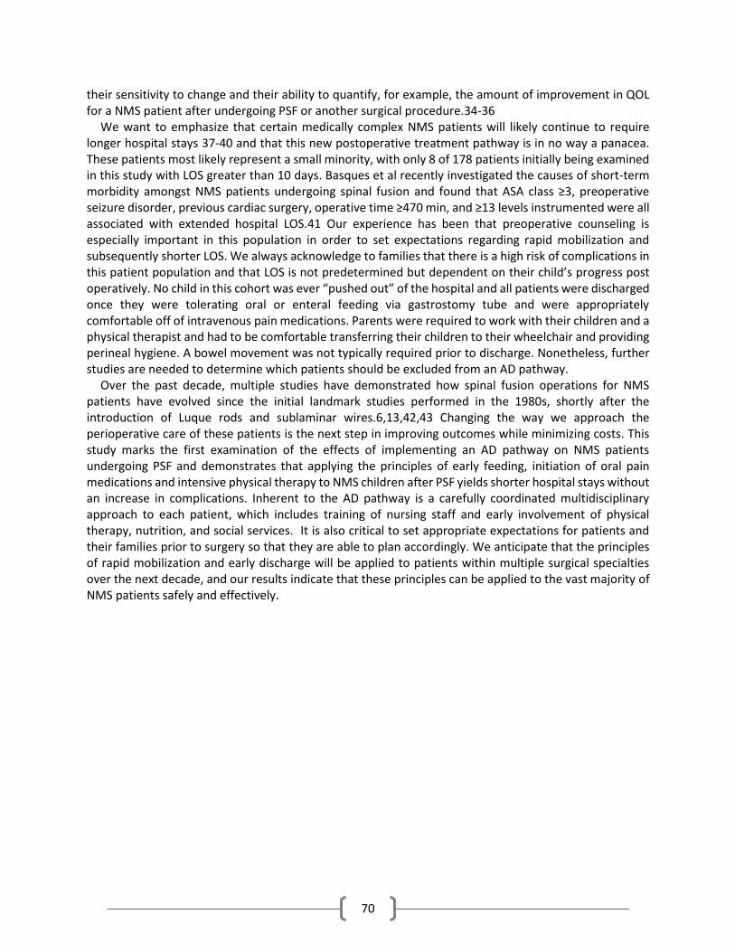
70
their sensitivity to change and their ability to quantify, for example, the amount of improvement in QOL for a NMS patient after undergoing PSF or another surgical procedure.34-36
We want to emphasize that certain medically complex NMS patients will likely continue to require longer hospital stays 37-40 and that this new postoperative treatment pathway is in no way a panacea. These patients most likely represent a small minority, with only 8 of 178 patients initially being examined in this study with LOS greater than 10 days. Basques et al recently investigated the causes of short-term morbidity amongst NMS patients undergoing spinal fusion and found that ASA class ≥3, preoperative seizure disorder, previous cardiac surgery, operative time ≥470 min, and ≥13 levels instrumented were all associated with extended hospital LOS.41 Our experience has been that preoperative counseling is especially important in this population in order to set expectations regarding rapid mobilization and subsequently shorter LOS. We always acknowledge to families that there is a high risk of complications in this patient population and that LOS is not predetermined but dependent on their child’s progress post operatively. No child in this cohort was ever “pushed out” of the hospital and all patients were discharged once they were tolerating oral or enteral feeding via gastrostomy tube and were appropriately comfortable off of intravenous pain medications. Parents were required to work with their children and a physical therapist and had to be comfortable transferring their children to their wheelchair and providing perineal hygiene. A bowel movement was not typically required prior to discharge. Nonetheless, further studies are needed to determine which patients should be excluded from an AD pathway.
Over the past decade, multiple studies have demonstrated how spinal fusion operations for NMS patients have evolved since the initial landmark studies performed in the 1980s, shortly after the introduction of Luque rods and sublaminar wires.6,13,42,43 Changing the way we approach the perioperative care of these patients is the next step in improving outcomes while minimizing costs. This study marks the first examination of the effects of implementing an AD pathway on NMS patients undergoing PSF and demonstrates that applying the principles of early feeding, initiation of oral pain medications and intensive physical therapy to NMS children after PSF yields shorter hospital stays without an increase in complications. Inherent to the AD pathway is a carefully coordinated multidisciplinary approach to each patient, which includes training of nursing staff and early involvement of physical therapy, nutrition, and social services. It is also critical to set appropriate expectations for patients and their families prior to surgery so that they are able to plan accordingly. We anticipate that the principles of rapid mobilization and early discharge will be applied to patients within multiple surgical specialties over the next decade, and our results indicate that these principles can be applied to the vast majority of NMS patients safely and effectively.

71
REFERENCES 1. Saito N ES, Ohotsuka K, Kumeta H, Takaoka K. Natural history of scoliosis in spastic cerebral
palsy. Lancet. 1998;351:1687–1692. 2. Hasler C. Operative treatment for spinal deformities in cerebral palsy. J Child Orthop.
2013(7):419-423. 3. Fletcher ND, Andras LM, Lazarus DE, et al. Use of a Novel Pathway for Early Discharge Was
Associated With a 48% Shorter Length of Stay After Posterior Spinal Fusion for Adolescent Idiopathic Scoliosis. J Pediatr Orthop. Jul 24 2015.
4. Jevsevar D KL. The relationship between preoperative nutritional status and complications after an operation for scoliosis in patients who have cerebral palsy. J Bone Joint Surg Am. 1993;75:880-884.
5. Barsdorf AI SD, Kaufmann P. Scoliosis surgery in children with neuromuscular disease: findings from the US National Inpatient Sample, 1997 to 2003. . Arch Neurol. 2010;67:231-235.
6. Eric R. Benson M, Jeffrey D. Thomson, MD, Brian G. Smith, MD, and John V. Banta, MD. Results and Morbidity in a Consecutive Series of Patients Undergoing Spinal Fusion for Neuromuscular Scoliosis. Spine. 1998;23(21):2308-2318.
7. Nicholas D. Fletcher NS, Philip M. Mitchell, Timothy S. Oswald, Dennis P. Devito, Robert W. Bruce. Clinical and economic implications of early discharge following posterior spinal fusion for adolescent idiopathic scoliosis. J Child Orthop. 2014(8):257-263.
8. Cobb J. Outline for the study of scoliosis. Am Acad Orthop Surg Instr Course Lect. 1948. 1948;5:261-275.
9. Watanabe K LL, Daubs MD, Bridwell KH, Stobbs G, Hensley M. Is spine deformity surgery in patients with spastic cerebral palsy truly beneficial?: a patient/parent evaluation. Spine. 2009;34:2222-2232.
10. Mercado E AB, Wright JG. Does spinal fusion influence quality of life in neuromuscular scoliosis? . Spine. 2007;32:S120–S125.
11. Jones KB SP, Shindle MK, McCarthy ML Longitudinal parental perceptions of spinal fusion for neuromuscular spine deformity in patients with totally involved cerebral palsy. J Pediatric Orthop 2003;23:143–149.
12. Larsson ELC AS, Normelli HCM, Oberg BE Longterm follow-up of functioning after spinal surgery in patients with neuromuscular scoliosis. Spine. 2005;30:2145–2152.
13. Nancy Alice Murphy M, Sean Firth, PhD, MPH, Trisha Jorgensen, BS, and Paul C. Young, MD. Spinal Surgery in Children with Idiopathic and Neuromuscular Scoliosis. What’s the Difference? J Pediatr Orthop. 2006;26(2):216-220.
14. Christopher Diefenbach MD, Marc N. Ialenti, M.D., Baron S. Lonner, M.D., Jonathan R, Kamerlink, M.D., Kushagra Verma, M.D., and Thomas J. Errico, M.D. Hospital Cost Analysis of Neuromuscular Scoliosis Surgery. Bulletin of the Hospital for Joint Diseases. 2013;71(4):272-277.
15. Master DL, Poe-Kochert C, Son-Hing J, Armstrong DG, Thompson GH. Wound infections after surgery for neuromuscular scoliosis: risk factors and treatment outcomes. Spine (Phila Pa 1976). Feb 1 2011;36(3):E179-185.
16. Master DL, Son-Hing JP, Poe-Kochert C, Armstrong DG, Thompson GH. Risk factors for major complications after surgery for neuromuscular scoliosis. Spine (Phila Pa 1976). Apr 1 2011;36(7):564-571.
17. Modi HN, Suh SW, Yang JH, et al. Surgical complications in neuromuscular scoliosis operated with posterior- only approach using pedicle screw fixation. Scoliosis. 2009;4:11.

72
18. Pugely AJ, Martin CT, Gao Y, Ilgenfritz R, Weinstein SL. The incidence and risk factors for short-term morbidity and mortality in pediatric deformity spinal surgery: an analysis of the NSQIP pediatric database. Spine (Phila Pa 1976). Jul 1 2014;39(15):1225-1234.
19. Mohamad F PS, Pawelek J, Marks M, Bastrom T, Faro F, Newton P. Perioperative complications after surgical correction in neuromuscular scoliosis. . J Pediatric Orthop. 2007;27:392–397.
20. Szoke G LG, Miller F, Dabney K. Wound infection after spinal fusion in children with cerebral palsy. . J Pediatric Orthop. 1998;18:727-733.
21. Tsirikos AI CW, Dabney KW, Miller F, Glutting J. Life expectancy in pediatric patients with cerebral palsy and neuromuscular scoliosis who underwent spinal fusion. . Dev Med Child Neurol. 2003;45:677–682.
22. Master DL CP, Jochen SH, Armstrong DG, Thompson GH. Wound infections after surgery for neuromuscular scoliosis: risk factors and treatment outcomes. . Spine (Phila Pa 1976). 2011;36:E179–E185.
23. Sponseller PD LD, Hungerford MW, Eck K, Bridwell KH, Lenke LG. Deep wound infections after neuromuscular scoliosis surgery: a multicenter study of risk factors and treatment outcomes. . Spine. 2000;25:2461–2466.
24. Geiger F PD, Carstens C. Complications of scoliosis surgery in children with myelomeningocelE. Eur Spine J. 1999;8:22-26.
25. Sponseller PD SS, Abel MF, Newton PO, Letko L, Marks M Infection rate after spine surgery in cerebral palsy is high and impairs results multicenter analysis of risk factors and treatment. Clin Orthop Relat Res. 2010;468:711-716.
26. Sharma S, Wu C, Andersen T, Wang Y, Hansen ES, Bunger CE. Prevalence of complications in neuromuscular scoliosis surgery: a literature meta-analysis from the past 15 years. Eur Spine J. Jun 2013;22(6):1230-1249.
27. Sarwahi V SJ, Schafer MF, et al. Standards in anterior spine surgery in pediatric patients with neuromuscular scoliosis. J Pediatr Orthop. 2001;21(6):756.
28. Yuan N SD, Dorey F, Keens TG. Preoperative predictors of prolonged postoperative mechanical ventilation in children following scoliosis repair. Pediatr Pulmonol. 2005;40:414-419.
29. Perrin C UJ, Ambrosio CD, Hill NS. Pulmonary complications of chronic neuromuscular diseases and their management. Muscle Nerve. 2004;29:5-27.
30. Kang GR SS, Lee IO. Preoperative predictors of postoperative pulmonary complications in neuromuscular scoliosis. J Orthop Sci Official J Jpn Orthop Assoc. 2011;16:139–147.
31. Edler A MD, Forbes RB Blood loss during posterior spinal fusion surgery in patients with neuromuscular disease: is there an increased risk? Paediatr Anaesth. 2003;13:818–822.
32. Seruya M OA, Boyajian MJ, Myseros JS, Yaun AL, Keating RF. Unreliability of intraoperative estimated blood loss in extended sagittal synostectomies. J Neurosurg Pediatr. 2011;8(5):443-449.
33. Razvi K CS, Arulkumaran S, Ratnam SS. A comparison between visual estimation and laboratory determination of blood loss during the third stage of labour. Aust N Z J Obstet Gynaecol. 1996;36(2):152-154.
34. Carlon S SN, Yong K, Gilmore R, Sakzewski L, Boyd R. A systematic review of the psychometric properties of Quality of Life measures for school aged children with cerebral palsy. BMC Pediatr. 2010;10(81):1-11.
35. Comstock CP LJ, Wenger DR. Scoliosis in totalbody-involvement cerebral palsy: analysis of surgical treatment and patient and caregiver satisfaction. Spine. 1998;23:1412–1425.
36. Ersberg A GP. Pre- and postoperative quality of life in patients treated for scoliosis. Acta Orthop. 2013;84(6):537-543.

73
37. David I. Rappaport M, Judy Adelizzi-Delany, MSN, CPNP, Kenneth J. Rogers, PhD, ATC, Chalanda E. Jones, MD, Maria E. Petrini, MD, Kate Chaplinski, MD, Peter Ostasewski, RN,, BSN M, Iman Sharif, MD, MPH, and David M. Pressel, MD, PhD. Outcomes and Costs Associated With Hospitalist Comanagement of Medically Complex Children Undergoing Spinal Fusion Surgery. Hospital Pediatrics. 2013;3(3):233-241.
38. Hod-Feins R AY, Mirovsky Y, Barr J, Abu-Kishk I, Lahat E, Eshel G. Pediatric scoliosis surgery—the association between preoperative risk factors and postoperative complications with emphasis on cerebral palsy children. Neuropediatrics. 2007;38:239–243.
39. Lipton GE MF, Dabney KW, Altiok H, Bachrach SJ. Factors predicting postoperative complications following spinal fusions in children with cerebral palsy. J Spinal Disord. 1999;12:197-205.
40. Klein JD GS. Nutritional status in the patient with spinal infection. . Orthopedic Clin North Am. 1996;27:33-36.
41. Basques B CS, Lukasiewicz A, Webb M, Samuel A, Bohl D, Smith B, Grauer J. Predicting Short-Term Morbidity in Patients Undergoing Spinal Fusion for Neuromuscular Scoliosis. Spine Publish Ahead of Print. 2015.
42. John Sarwark MVS, MDc. New Strategies and Decision Making in the Management of Neuromuscular Scoliosis. Orthop Clin N Am. 2007(38):485-496.
43. Stanitski CL ML, Hall JE, Rosenthal RK. Surgical correction of spinal deformity in cerebral palsy. Spine (Phila Pa 1976). 1982;7:563–569.

74
Humerus Fracture Fixation: Incidence Rates and
Complications as Reported by American Board of
Orthopaedic Surgery Part II Candidates
Michael B. Gottschalk, MD, William Carpenter, MD, Elise Hiza, MD, William Reisman, MD, James Roberson, MD BACKGROUND Despite extensive research regarding patient outcomes after operative fixation of humeral shaft fractures by means of open reduction and internal fixation (ORIF) or intramedullary nailing (IMN), no current consensus exists regarding the optimal surgical treatment. The objective of this study was to compare IMN and plate fixation (ORIF) of humeral shaft fractures by using the American Board of Orthopaedic Surgery (ABOS) Part II operative database to analyze incidence rate, changes in management trends over time, early complications, and factors affecting the management choice METHODS The ABOS database is a collection of surgical cases that are self- reported by orthopaedic candidates approved for admission to the ABOS oral examination. The database was searched for records from 2004 to 2013 for humeral shaft surgical cases as indicted by Current Procedural Terminology (CPT) codes 24515 (open reduction internal fixation) and 24516 (insertion of intramedullary nail) pertaining to humeral shaft fractures. The geographic region and fellowship training of the candidates; the year of surgery, diagnosis code, age, and sex of the patients; and the surgeon- reported complications were analyzed. RESULTS The search identified 3,430 surgically treated humeral shaft fractures that were reported to the ABOS database from 2004 to 2013. A significant decline in IMN use was seen from 2004 (42.9%) to 2013 (21.2%, p < 0.001). The IMN cohort had lower complication rates pertaining to both infections (1.5% compared with 3.0% for ORIF, p = 0.007) and nerve palsies (3.1% compared with 7.8%, p < 0.001). No significant difference was seen in the rate of nonunion (1.3% for IMN compared with 1.6% for ORIF, p = 0.63), although follow-up may be too short to demonstrate a difference. The IMN cohort did have significantly higher mortality (4.9% compared with 0.7% for ORIF, p < 0.001). Subset analysis demonstrated that the IMN cohort had significantly more pathologic fractures (26.8% compared with 1.5% of the fractures treated with ORIF, p < 0.001). CONCLUSIONS Although the overall incidence of fixation of humeral fractures was unchanged from 2004 to 2013, there was a significant shift from IMN to ORIF using plate fixation during this time period. Possible reasons for this shift in treatment to ORIF include the potential impact of recent publications highlighting complications of IMN and increased surgeon attention to cost containment. LEVEL OF EVIDENCE Level III

75
Cellular Characterization of Rotator Cuff Muscle Atrophy
Jimmy H Daruwalla, MD, Lianne Tellier, BSc, Marijke DeVos, BSc, Jennifer McFaline-Figueroa, BSc, Johnna Temenoff, PhD, Nick Willett, PhD, Claudius Jarrett, MD
BACKGROUND Rotator cuff muscle degeneration and fatty infiltration occur as a result of rotator cuff tendon tears. Even after surgical tendon repair, this irreversible process portends a poor prognosis.1,2 The cellular pathophysiology of rotator cuff tear-induced muscle atrophy is poorly understood, and it is currently unknown why regeneration of rotator cuff muscle is not observed clinically. Research from a myocardial infarction model suggests that promoting recruitment of bone-marrow derived cells, such as mesenchymal stem cells (MSCs) may encourage repair of muscle after injury.3,4 However, to date, the participation of these cells has not been evaluated in the setting of rotator cuff injury. Our study aims to characterize the response of endogenous regenerative muscle cells after injury in humans and in a rat model of rotator cuff muscle atrophy. We hypothesize that a small up-regulation of pro-regenerative cell populations, such as MSCs, would be observed within the injured rotator cuff muscle of humans and rats near the time of injury when compared to controls. METHODS We utilized a validated rat model of rotator cuff muscle atrophy.5 The injured and contralateral/control supraspinatus muscles of each rat were harvested at standardized timepoints up to 6 weeks. Samples were examined histologically using Oil red-O staining to detect the presence and degree of fibrofatty infiltration. Muscle samples from the rats were also processed and analyzed using flow cytometry in order to quantify changes in the cell populations of interest. Human muscle cell samples were also similarly analyzed to see if replication of the same cell populations was possible. RESULTS 12 rats underwent tendon and nerve transection. The contralateral limb was used as the control. Histological analysis demonstrated the presence of fibrous and fatty infiltration within the injured supraspinatus muscle starting at week 3, with the most pronounced effect observed at week 6, when compared to uninjured muscle (Figures 1, 2). At 1 week, flow cytometry demonstrated a significant increase in MSC populations in the injury group vs. uninjured muscle (Figure 3). We also validated the ability to identify these same cell populations using flow cytometry in human samples CONCLUSION Histological changes observed within the rat model musculature over 6 weeks replicate the processes of muscle atrophy and fatty infiltration seen clinically. MSC populations are present at increased numbers within the muscle at 1 week following injury, despite evidence of muscle degeneration over time. Thus, further increasing recruitment of these pro-regenerative cells may be a promising therapeutic target for rotator cuff muscle atrophy.

76
Figures
Figure 1. H&E staining reveals fibrous infiltration (black arrows) in the supraspinatus muscle of the injured shoulder in as little as 1 week after injury (Tiles D-F), as compared to the contralateral control (Tiles A-C). Over time, an increase in fibrous infiltration was observed.

77
Figure 2. Oil red-O staining for adipose tissue reveals fatty infiltration (black arrows) in the supraspinatus muscle of the injured shoulder (Tiles C-D) starting at week 1, with an increased response seen over time. Little to no fatty infiltration was observed in control muscles (Tiles A-B).

78
Figure 3. Graph demonstrating 2-fold increase (# - p ≤ 0.05) in MSC recruitment to the supraspinatus muscle of injured shoulders compared to controls at 1 week after injury.
0
0.5
1
1.5
2
2.5
3
3.5
InjuryOnly
FoldChangetoContralateral
Control
MSCRecruitment7DaysPost-Injury
#

79
REFERENCES
1. Laron D, Samagh SP, Liu X, Kim HT, Feeley BT. Muscle degeneration in rotator cuff tears. Journal
of shoulder and elbow surgery / American Shoulder and Elbow Surgeons [et al] 2012; 21(2): 164-74.
2. Gladstone JN, Bishop JY, Lo IK, Flatow EL. Fatty infiltration and atrophy of the rotator cuff do not improve after rotator cuff repair and correlate with poor functional outcome. The American journal of sports medicine 2007; 35(5): 719-28.
3. Ghadge SK, Muhlstedt S, Ozcelik C, Bader M. SDF-1alpha as a therapeutic stem cell homing factor in myocardial infarction. Pharmacology & therapeutics 2011; 129(1): 97-108.
4. Abbott JD, Huang Y, Liu D, Hickey R, Krause DS, Giordano FJ. Stromal cell-derived factor-1alpha plays a critical role in stem cell recruitment to the heart after myocardial infarction but is not sufficient to induce homing in the absence of injury. Circulation 2004; 110(21): 3300-5.
5. Liu X, Manzano G, Kim HT, Feeley BT. A rat model of massive rotator cuff tears. Journal of orthopaedic research : official publication of the Orthopaedic Research Society 2011; 29(4): 588-95.

80
Risk Factors of Lumbar Spinal Epidural Fat
Anuj Patel, MD, Cathy Vu, BS, Douglas Robertson, MD, Michael Gottschalk, MD, John Rhee, MD BACKGROUND Spinal epidural lipomatosis (SEL) is an excessive deposition of adipose tissue in the spinal epidural space that deforms the thecal sac. SEL causes symptoms by directly compressing the spinal cord or nerve roots. Even without causing direct compression, excessive epidural fat can cause symptoms by decreasing the accommodative ability of the epidural space. Otherwise clinically insignificant spinal pathology can then become acutely symptomatic. Patients can present with spinal stenosis, myelopathy, radiculopathy, or even cauda equina syndrome as a result of excessive epidural adipose tissue. Despite these serious consequences, there is a general lack of knowledge on the risk factors that can lead to increased volume of epidural fat. We will aim to determine which risk factors correlate to excessive deposition of adipose tissue in the epidural space. METHODS We performed a retrospective review of a consecutive cohort of 248 adult patients who were seen as new patients by physicians in the Emory University Spine Department and had 3T Magnetic Resonance T-weighted images of their lumbar spines taken at the Emory University Orthopaedic and Spine Center. Patient charts, patient reported data, and MRIs were analyzed for each patient. The quantity of epidural fat was measured on MRI T1 weighted images. Measurements were taken on axial and sagittal cuts. MRI measurements were also performed using a program created at the Emory University Orthopaedic and Spine Center to volumetrically quantify the amount of fat in the epidural space of the lumbar spine based on axial cuts. DICOM files with all patient identifiers removed of axial cuts starting at the level of the lamina of L1 caudally to the level of the lamina of S1 were measured. Statistical analysis was performed to determine the relationship of volume of lumbar epidural fat to age, BMI, pain scores, posterior subcutaneous fat, number of epidural steroid injections within a year, HBA1c, LDL, HDL, and Triglycerides. RESULTS Several risk factors were found to directly correlate with volume of epidural adipose tissue in the lumbar spine. There was a direct correlation between BMI and volume of epidural fat (p<0.001). Patients classified by BMI also had significantly different mean volumes. Based on WHO classification, morbidly obese and obese patients had significantly greater volumes compared to overweight and normal weight patients. The mean volume of epidural fat in patients classified as morbidly obese was 8952.77 mm3 compared with 6287.70 mm3 in patients classified as overweight and 4711.59 mm3 in those classified as normal weight (p <0.0001). The mean volume of epidural fat in patients classified as obese was 7656.87 mm3, also significantly higher than that of the overweight and normal weight patients (p <0.0001). Overweight patients also had a significantly higher mean volume of epidural fat compared to normal weight patients (p=0.0002). HbA1c also directly correlated with volume of epidural fat (p= 0.021) though only patients with risk factors for diabetes had HbA1c labs drawn. Posterior subcutaneous fat measured on the sagittal MRI tangentially to the L4 lamina was also found to be a significant predictor of epidural fat (p < 0.0001). Using a multivariate analysis correlating volume of epidural fat to BMI, HBA1c, and subcutaneous fat together, we found that only posterior subcutaneous fat was an independent variable (p=0.021) due to all variables being a part of metabolic syndrome. Unrelated to metabolic syndrome, sex was found to be an independent variable as males have a higher mean of volume of fat (8581.86 mm3 ± 330.91) compared

81
to females (6452.45 mm3 ± 339.64) (p<0.0001). Age, LDL, triglycerides, HDL, and number of epidural steroid injections within a year of the MRI were not found to correlate with total epidural fat volume. Increasing volumes of lumbar epidural fat directly correlated with increasing pain scores when patients initially presented for evaluation (p = 0.001). Using axial MRI cuts, we also determined which patients had SEL, defined as direct deformation of their thecal sac by this epidural fat. 67% of all patients (163/143) were found to have SEL at a minimum of 1 level. SEL was most commonly found at the L2-3 and L3-4 disc spaces. We found that the mean volume of epidural fat was significantly higher in patients that had SEL at 7467.25mm3 compared to those without SEL at 5016.30 mm3 (p<0.0001). The mean BMI of patients with SEL was significantly higher at 31.47 ± 5.07 than in patients without SLE at 25.55 ± 3.37 (p<0.0001). There is also a higher probability that patients with diabetes had SEL compared to non-diabetics (p = 0.0052). Patients with SEL had a higher average pain score of 6.03 than patients without SEL of 4.41 (p<0.0001). CONCLUSIONS We defined spinal epidural lipomatosis as excessive deposition of adipose tissue in the epidural space that deforms the thecal sac. We determined that SEL in the lumbar spine was more commonly found in patients with a greater total volume of epidural adipose tissue. BMI, HbA1c, and posterior subcutaneous fat were all found to directly correlate with the total volume of epidural fat in the lumbar spine. We therefore emphasize the importance of taking these risk factors into consideration when treating patients with persisting symptoms suggesting compression of spinal neural elements. As nearly one-third of the population of the United States is obese and 5% is morbidly obese, this issue is a major health concern. Using this data, practitioners will be able to educate patients on ways to decrease their risk of depositing excessive adipose tissue into their epidural space, and in turn decrease their risk of having symptomatic spinal pathology. It will also aid practitioners in determining treatment algorithms for obese patients with symptomatic spinal pathology.

82
Linear regression model of the correlation of Body Mass Index to the total volume of epidural fat in the lumbar spine as an independent variable.
Figure 1. Distribution of Volume of epidural fat in the lumbar spine in patients classified by BMI based on WHO classification. 2 = normal, 3 = overweight, 4 = obese, 5 = morbidly obese

83
BMI class N (%) Mean volume of epidural adipose tissue
95% CI
Class 1 -- --
Class 2 48 (19.7%) 4711.59 4114.91 5308.27
Class 3 98 (40.2%) 6287.70 5870.11 6705.28
Class 4 71 (29.1%) 7656.87 7166.26 8147.48
Class 5 27 (11.1%) 8952.77 8157.20 9748.34
P value table for difference in epidural adipose tissue volume between BMI classes
Class 2 Class 3 Class 4 Class 5
Class 2 0.0002 <0.0001 <0.0001
Class 3 0.0002 0.0002 <0.0001
Class 4 <0.0001 0.0002 0.0340
Class 5 <0.0001 <0.0001 0.0340
Comparison of means of volumes of epidural fat in patients with different BMI classifications based on
WHO classification. 1 = underweight, 2 = normal, 3 = overweight, 4 = obese, 5 = morbidly obese
Linear regression model of the correlation of HbA1c to the total volume of epidural fat in the lumbar spine
as an independent variable
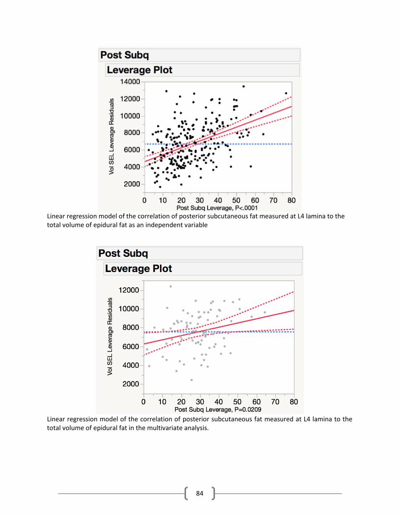
84
Linear regression model of the correlation of posterior subcutaneous fat measured at L4 lamina to the total volume of epidural fat as an independent variable
Linear regression model of the correlation of posterior subcutaneous fat measured at L4 lamina to the total volume of epidural fat in the multivariate analysis.

85
Comparison of total volume of epidural fat in the lumbar spine in males compared to females.
Linear regression model of the correlation of total volume of epidural fat to patient reported pain scores.
0 2 4 6 8 10
Pain Score
2500
5000
7500
10000
12500
Vol S
EL
95% Prediction Limits95% Confidence LimitsFit
0.0402Adj R-Square
0.0442R-Square
5.79E6MSE
242Error DF
2Parameters
244Observations
Fit Plot for Vol_SEL

86
Comparison of total volume of epidural fat in patients with SEL vs patients without SEL defined as
deformation of thecal sac by epidural fat
N With compression Without Compression
T-statistic p-value
Mean BMI 163 31.47 (5.08) 80 25.55 (3.38) -10.80 <0.0001
Z test p-value
Mean HbA1c
65 6.3 (1.26) 18 5.57 (0.54) -2.37 0.0176
Mean fat volume
163 7467.25 (2304.81) 80 5016.30 (1867.81)
-7.43 <0.0001
Mean Post SubQ fat
163 29.56 (13.80) 80 17.37 (9.63) -6.76 <0.0001
Mean LDL 69 100.52 (47.32) 26 98.15 (40.97) -0.0334 0.9734
Table 1. Comparison of means of risk factors in patients with SEL to patients without SEL

87
Comparison of pain scores in patients with SEL vs patients with SEL
Comparison of BMI of patients with SEL vs patients with SEL

88
Continued Delay in Diagnosis of Slipped Capital Femoral
Epiphysis
Mathew D. Schur BA, Lindsay M. Andras MD, Alexander M. Broom BA, Kody K. Barrett BA, Christine A. Bowman, BA2 Herman Luther, Rachel Y. Goldstein MD MPH, Nicholas D. Fletcher MD, Michael B. Millis MD, Robert Runner MD, David L. Skaggs MD, MMM STRUCTURED ABSTRACT BACKGROUND More than a decade ago, Skaggs and Kocher et al. reported significant delays in the diagnosis of slipped
capital femoral epiphysis (SCFE). The purpose of this study was to identify if the time to diagnosis has
improved.
METHODS A retrospective review was performed of 481 patients admitted with a diagnosis of SCFE at three large pediatric hospitals between 01/2003-12/2012. RESULTS
The average time from symptom onset to diagnosis was 17 weeks (range 0-169). There were no significant differences in time from symptom onset to diagnosis across two year intervals of the study period (p=0.94). Time from presentation to first provider to diagnosis was significantly shorter for patients presenting to an orthopaedic clinic (mean 0 weeks, range 0-0) versus PCP (mean 4 weeks, 0-52) (p=0.003, r=0.24) or ED (mean 6 weeks, range 0-104) (p=0.008, r=0.36). 52 patients (10.8%) developed a second SCFE after treatment of the first. Time from symptoms to diagnosis for the second SCFE was significantly shorter (p<0.001, r=0.19) with mean delay in diagnosis of 11 weeks (range 0-104). The number of second SCFE patients diagnosed while mild in severity as defined by Wilson classification was significantly larger (p=0.001, OR=4.44) than those with a first SCFE.
METHODS Despite reports documenting a lag in the diagnosis of SCFE over a decade ago, there has been no improvement in the delay in diagnosis. Decreased delay and severity for the second SCFE suggests that the education of at risk children and their families may be of benefit in improving this delay. INTRODUCTION
Concerns about delay in diagnosis of slipped capital femoral epiphysis (SCFE) in the pediatric population were brought to light over a decade ago.1, 3, 6 In 2002, Skaggs et al. reported on cases of SCFE where the mean time from initial presentation to diagnosis was 16 weeks.2 Similarly, Kocher et al. reviewed 196 cases of SCFE and found a median delay of 8 weeks from first symptom to diagnosis.3 In 2005, Green et al. also reported on a delay from first symptom to diagnosis with an average of 20 weeks in 102 patients.1 This delay can lead to development of increased slip severity and poor outcomes in both the short and long term including accelerated degenerative changes, decreased range of motion, and avascular necrosis (AVN). 1-10, 15 The purpose of this study was to evaluate if there has been any improvement in the time to diagnosis of SCFE over time and if there were any regional differences.

89
PATIENTS AND METHODS
An IRB approved retrospective radiographic and chart review was performed of children with a diagnosis of SCFE at three geographically distinct (west coast, northeast, and south) high volume pediatric centers between January 2003 and December 2012. Patients were excluded if the time between onset of symptoms and diagnosis or pain site were not documented. Patients were also excluded if they had a SCFE on the contralateral side that was treated either prior to the study period or at an outside institution. Time from symptom onset to diagnosis, as well as time of initial presentation to a healthcare provider were recorded. Symptoms (hip vs knee pain), demographic information and medical comorbidities were examined. Charts and radiographs were reviewed to determine slip stability, slip severity, type of procedure, and surgical management.
Stability of the SCFE was classified as “stable” or “unstable” as described by Loder et al. Southwick angles (the difference in epiphyseal shaft angle from the contralateral side for unilateral SCFE, or the difference from 15 degrees for bilateral SCFE) and Wilson classification (% displacement of femoral head epiphysis on metaphysis) were used to measure severity (Figure 1).15, 24, 25
Statistical analysis was performed using Stata12 (College Station, TX: StataCorp LP) and Microsoft Excel (2010). The Mann-Whitney U test was used to determine statistical significance between binary data, the Kruskal-Wallis test was used for categorical data, and Spearman’s Rho for continuous data. Multiple regression was used to control for covariates in determining significance of association. P-value was considered significant at <0.05. RESULTS
The total study population included 496 hips in 481 patients diagnosed with SCFE. Demographic and radiographic data are listed in Tables 1 and 2, respectively. The mean duration between symptom onset and time of diagnosis was 17 weeks (range 0 to 169). Mean durations on the West Coast, the South, and the Northeast United States were 17 weeks (range 0 to 157), 15 weeks (range 0 to 104), and 17 weeks (range 0 to 169), respectively, and were not significantly different (p=0.11). Separated into two year intervals there were the following delays in diagnosis: 2003-2004=13 weeks; 2005-2006=16 weeks; 2007-2008=19 weeks; 2009-2010=16 weeks; 2011-2012=20 weeks. There were no significant differences in time from symptom onset to diagnosis across two year intervals of the study period for the entire study sample (p=0.94) (Table 3) or for any of the individual geographic samples (West Coast p=0.52; Northeast p=0.40; South p=0.56). The majority of patients (51.9%, 240/462) presented to primary care physicians (PCP) initially (Figure 2). The mean weeks for all presenting locations are found in Figure 2. Of the 481 patients, 213 had complete data on the duration from presentation to first provider to diagnosis. The mean duration between presentation to a healthcare provider and diagnosis of SCFE was 5 weeks (range 0 to 104). Mean durations on the West Coast, in the South, and in the Northeast United States were 7 weeks (range 0 to 52), 5 weeks (range 0 to 46), and 3 weeks (range 0 to 104), respectively. A significant difference was identified between the Northeast and the South (p=0.02, r=0.21) and the Northeast and the West Coast (p=0.04, r=0.15). There was no significant difference across two year intervals of the study period (p=0.85). Of those with available data, 70.7% (145/205) presented to a PCP initially, compared to 19.5% (40/205) to the ED, 6.3% (13/205) to an orthopaedic clinic, and 0.5% (1/205) to an urgent care. The duration from presentation to first provider to diagnosis was significantly shorter for patients presenting to an orthopaedic clinic (mean 0 weeks, range 0 to 0) than either a PCP (mean 4 weeks, 0 to 52) (p=0.003, r=0.24) or the ED (mean 6 weeks, range 0 to 104) (p=0.008, r=0.36).
Longer time from first symptom to diagnosis was associated with both stable slips (p<0.001, r=0.24) and slips of moderate versus mild severity (p<0.001, r=0.24). In patients with a delay longer than 7 weeks 49.7% (85/171) of slips were moderate/severe. Similarly, longer delays were also found to be associated with a larger Southwick angle (p<0.001, Spearman’s rho=0.31). Association with longer delay

90
was also identified in patients with knee pain as their primary presenting symptom (p<0.001, r=0.16), patients initially presenting with bilateral SCFE (p<0.001, r=0.14), and with increased BMI (p<0.001, Spearman’s Rho=0.24). No significant association was found with age at first symptom (p=0.40), sex (p=0.13), or type of insurance (p=0.12). Multivariate regression analysis demonstrated continued significant association with BMI (p=0.02).
Overall, 8.5% (41/480) of patients underwent prophylactic pinning of the contralateral hip. 10.8% (52/480) of patients went on to subsequently receive a second SCFE diagnosis. Of those patients who sustained a contralateral slip with available data, 86% (31/36) had a mild slip, 3% (1/36) had a moderate slip, and 11% (4/36) had a severe slip at diagnosis. Second SCFE patients were significantly more likely to be diagnosed while mild in severity compared to patients with a first SCFE (p=0.001, OR=4.44) (Figure 3a, 3b). Patients presenting with a second diagnosis of SCFE had a significantly shorter time to diagnosis (mean = 11 weeks, range 0-104 weeks) compared to the initial SCFE group (p<0.001, r=0.19). DISCUSSION
This study of 481 patients from 3 high volume pediatric centers is, to our knowledge, the largest series on delay in diagnosis of slipped capital femoral epiphysis. Overall, there was a 17 week (CI 14.7 to 19.3) interval between development of symptoms and diagnosis. This issue gained attention over a decade ago. Green et al. reported an average time from first symptom to diagnosis of 20 weeks in a cohort of 102 patients, treated from 1989 to 1997 at the same West Coast pediatric hospital that participated in this study.1 Kocher et al. (from a center included in our study), Wilson et al., Seigal et al., Loder et al., Matava et al., and Pihl et al. also reported delays in diagnosis of SCFE as outlined in Table 4 3, 4, 6, 11, 16, 17 Given the data both from this and previous studies, it is clear that little progress has been made in shortening the time to diagnosis of SCFE.
Previous studies have identified numerous other risk factors for delay in diagnosis of SCFE. These include knee/distal thigh pain, public insurance, stable slips, and slip angle as significant predictors of delay from symptom onset to diagnosis.1, 3, 4, 16 This study identified a number of these associations with longer delay as well, including knee pain as the presenting symptom and stable slips. One association with longer delay found in this study that was not previously identified was with higher BMI. This is particularly concerning given that a higher BMI is widely cited in the literature as an independent risk factor for SCFE itself. 18-21 However, there is also an increased incidence of hip and knee pain in the overweight adolescent population that may contribute to it being overlooked.22, 23
In examining the reasons behind the continued delay, one apparent contributor is presentation to either a primary care provider or an ED, with a mean delay from initial provider presentation to diagnosis of 4 weeks and 6 weeks, respectively, versus a mean of 0 weeks for an outpatient orthopaedic clinic. Previous studies by Wilson et al., Matava et al., and Green et al. have identified a significant incidence of diagnostic errors by PCPs, with Matava et al. identifying a significantly higher number of diagnostic errors in patients with an initial symptom of knee rather than hip pain.1, 4, 11 Misdiagnoses were identified in the population of this study as well, evidenced by radiographs from one patient’s initial presentation and follow-up demonstrating significant slip but not resulting in a diagnosis of SCFE by the initial provider (Fig 4a-c). These findings, combined with the fact that the vast majority of patients first present to either a PCP or an ED, indicate that these two groups of providers may have the greatest opportunity to substantially impact the overall delay in diagnosis. Given these considerations, it may be important to prioritize further education on the prevalence of SCFE and its risk factors in both the primary and emergent care communities.
Another possible opportunity to decrease delay in diagnosis is through parental education on SCFE. This study found that patients who received a second diagnosis of SCFE had a significantly shorter delay in diagnosis than the overall mean for the initial SCFE. Within this group, nearly 90% were diagnosed while the slip was mild, compared to 58% at diagnosis of first SCFE. With the benefit of experience and

91
education, it is likely that these patients’ families were able to recognize the signs of SCFE earlier. This demonstrates the overall impact that counseling at risk patients may have on delay in diagnosis of the initial SCFE.
This study is limited by its retrospective nature. Authors were limited at all sites by the extent of documentation in the medical record. Nevertheless, this review of nearly 500 cases over the past 10 years at 3 major pediatric centers, showed no significant improvement was seen in the time to diagnosis of slipped capital femoral epiphysis. Each possible contributor to delay in diagnosis is important to consider, given the significant morbidity that may result without timely intervention. Delay provides time for development of greater slip severity and an increase in Southwick angle, as seen in this and other studies on the topic. These, in turn, lead to a decreased range of motion and early degenerative changes as well as an increased risk of AVN. 1-10, 15 Decreasing the delay in diagnosis would allow many of these significantly morbid risks to be avoided.
CONCLUSION In review of nearly 500 cases over the past 10 years at 3 major pediatric centers, no significant
improvement was seen in the time to diagnosis of slipped capital femoral epiphysis. This holds true for all 3 geographically distinct areas and, in examining previous studies, has been true for decades. However, opportunities exist for progress in the education of both physicians and families. By increasing the overall awareness of SCFE in the community, we may have an opportunity to decrease the delay in diagnosis that remains.

92
Figure Legend
Figure 1: Southwick Angle: Difference of the femoral head-shaft angle of each hip on a frog lateral radiograph. Reproduced with permission from the Children’s Orthopaedic Center, Los Angeles.

93
Figure 2: Percent of index presentations of SCFE by type of provider. Reproduced with permission from the Children’s Orthopaedic Center, Los Angeles.
Figure 3: Percent of mild, moderate, and severe slips in patients diagnosed with a SCFE. A: first SCFE diagnosis; B: second SCFE diagnosis. Reproduced with permission from the Children’s Orthopaedic Center, Los Angeles.

94
Figure 4: Progression of SCFE in a misdiagnosed patient in the study population. A: Initial radiograph; B: Progression of slip on follow-up radiograph; C: Intraoperative image. Reproduced with permission from the Children’s Orthopaedic Center, Los Angeles.
Patient Demographics and Association to Delay in Diagnosis
Covariant Category Median/Percent
Mean delay in
diagnosis
(weeks)
Significance of Association
to Delay in Diagnosis (effect
size)
Age 12 years (range 5 - 18) p=0.40
Gender Male 61% (295/481) 19.1
p=0.13 Female 39% (186/481) 13.4
BMI 28 kg/m^2,
(range 14-45)
P<0.001 (rs=0.24)
Insurance
status
Public 62% (294/467) 17.6
Private 36% (167/467) 15.7 p=0.12
Self-pay 2% (6/467) 14.3
Major pain
location
Hip 92% (441/481) 15.7 P<0.001 (r=0.16)
Knee 8% (40/481) 30.6
Slip
stability
Stable 81% (387/480) 17.9 p<0.001 (r=0.24)
Unstable 19% (93/480) 13.0
Table 1: Demographic information and significance of covariate association to delay in diagnosis. rs=Spearman’s rho

95
Patient Radiographic Measurements
Southwick angle 29° (range (-13°) - 87°)
Slip severity
Mild 61% (221/363)
Moderate 27% (97/363)
Severe 12% (45/363)
Table 2: Radiographic measurements of Southwick angle and severity of SCFE.
Delay in Diagnosis Over Two Year Intervals
Years Mean weeks from first symptom to diagnosis (range)
2003 - 2004 13.0 (0 - 130)
2005 - 2006 16.3 (0 - 169)
2007 - 2008 19.1 (0 - 157)
2009 - 2010 15.7 (0 - 155)
2011 - 2012 20.2 (0 - 104)
2003 - 2012 16.9 (0 - 169)
Table 3: Mean delay in diagnosis over two year intervals and for the entire study period from 2003 – 2012.
Previous Reports on Delay in diagnosis
Authors (years of study) Mean weeks from first symptom to diagnosis
Pihl et al (1996 - 2011) 26
Green et al (1989 - 1997) 20
Kocher et al (1988 - 2002) 8*
Matava et al (1985 - 1994) Group 1 = 33 Group 2 = 26
Seigal et al (1985 - 1987) 12
Loder et al (1975 - 1989) Detroit = 20 Ann Arbor = 32
Wilson et al (1936 - 1960) Slight slip = 13 Severe slip = 28
Table 4: Reports from previous literature on delay in diagnosis of SCFE. *Reported median. 1, 3, 4, 6, 11, 16, 17

96
REFERENCES
1. Green DW, Reynolds RA, Khan SN, Tolo V. The delay in diagnosis of slipped capital femoral epiphysis: a review of 102 patients. HSS J. Sep 2005;1(1):103-106.
2. Skaggs DL, Roy AK, Vitale MG, et al. Quality of evaluation and management of children requiring timely orthopaedic surgery before admission to a tertiary pediatric facility. J Pediatr Orthop. Mar-Apr 2002;22(2):265-267.
3. Kocher MS, Bishop JA, Weed B, et al. Delay in diagnosis of slipped capital femoral epiphysis. Pediatrics. Apr 2004;113(4):e322-325.
4. Matava MJ, Patton CM, Luhmann S, Gordon JE, Schoenecker PL. Knee pain as the initial symptom of slipped capital femoral epiphysis: an analysis of initial presentation and treatment. J Pediatr Orthop. Jul-Aug 1999;19(4):455-460.
5. Boyer DW, Mickelson MR, Ponseti IV. Slipped capital femoral epiphysis. Long-term follow-up study of one hundred and twenty-one patients. J Bone Joint Surg Am. Jan 1981;63(1):85-95.
6. Pihl M, Sonne-Holm S, Christoffersen JK, Wong C. Doctor's delay in diagnosis of slipped capital femoral epiphysis. Dan Med J.. Sep 2014;61(9):A4905.
7. Harris WH. Etiology of osteoarthritis of the hip. Clin Orthop Relat Res. Dec 1986(213):20-33. 8. Cowell HR. The significance of early diagnosis and treatment of slipping of the capital femoral
epiphysis. Clin Orthop Relat Res. Sep-Oct 1966;48:89-94. 9. Dreghorn CR, Knight D, Mainds CC, Blockey NJ. Slipped upper femoral epiphysis--a review of 12 years
of experience in Glasgow (1972-1983). J Pediatr Orthop. May-Jun 1987;7(3):283-287. 10. Rattey T, Piehl F, Wright JG. Acute slipped capital femoral epiphysis. Review of outcomes and rates of
avascular necrosis. J Bone Joint Surg Am. Mar 1996;78(3):398-402. 11. Wilson PD, Jacobs B, Schecter L. SLIPPED CAPITAL FEMORAL EPIPHYSIS: AN END-RESULT STUDY. J Bone
Joint Surg Am. Sep 1965;47:1128-1145. 12. Carney BT, Weinstein SL. Natural history of untreated chronic slipped capital femoral epiphysis. Clin
Orthop Relat Res. Jan 1996(322):43-47. 13. Ledwith CA, Fleisher GR. Slipped capital femoral epiphysis without hip pain leads to missed diagnosis.
Pediatrics. Apr 1992;89(4 Pt 1):660-662. 14. Rahme D, Comley A, Foster B, Cundy P. Consequences of diagnostic delays in slipped capital femoral
epiphysis. J Pediatr Orthop B. Mar 2006;15(2):93-97. 15. Loder RT, Richards BS, Shapiro PS, Reznick LR, Aronson DD. Acute slipped capital femoral epiphysis:
the importance of physeal stability. J Bone Joint Surg Am. Aug 1993;75(8):1134-1140. 16. Loder RT, Aronson DD, Greenfield ML. The epidemiology of bilateral slipped capital femoral epiphysis.
A study of children in Michigan. J Bone Joint Surg Am. Aug 1993;75(8):1141-1147. 17. Siegel DB, Kasser JR, Sponseller P, Gelberman RH. Slipped capital femoral epiphysis. A quantitative
analysis of motion, gait, and femoral remodeling after in situ fixation. J Bone Joint Surg Am. Jun 1991;73(5):659-666.
18. Nasreddine AY, Heyworth BE, Zurakowski D, Kocher MS. A reduction in body mass index lowers risk for bilateral slipped capital femoral epiphysis. Clin Orthop Relat Res. Jul 2013;471(7):2137-2144.
19. Lehmann CL, Arons RR, Loder RT, Vitale MG. The epidemiology of slipped capital femoral epiphysis: an update. J Pediatr Orthop. May-Jun 2006;26(3):286-290.
20. Murray AW, Wilson NI. Changing incidence of slipped capital femoral epiphysis: a relationship with obesity? J Bone Joint Surg Br. Jan 2008;90(1):92-94.
21. Riad J, Bajelidze G, Gabos PG. Bilateral slipped capital femoral epiphysis: predictive factors for contralateral slip. J Pediatr Orthop. Jun 2007;27(4):411-414.
22. Krul M, van der Wouden JC, Schellevis FG, van Suijlekom-Smit LWA, Koes BW. Musculoskeletal Problems in Overweight and Obese Children. Ann Fam Med. Jan 2009;7(4):352-356.

97
23. Smith SM, Sumar B, Dixon KA. Musculoskeletal pain in overweight and obese children. Int J Obes. Jan 2014;38(1):11-15.
24. Southwick WO. Osteotomy through the lesser trochanter for slipped capital femoral epiphysis. J Bone Joint Surg Am. Jul 1967;49(5):807-835.
25. Wilson pd. The treatment of slipping of the upper femoral epiphysis with minimal
displacement. J bone joint surg am. 1938;20(2):379-399.

98
2015 – 2016 Orthopaedic Surgery Residents
PGY4 – PGY1
P
G
Y
4
P
G
Y
3
P
G
Y
2
P
G
Y
1
Briggs Ahearn
Medical University of South Carolina
College of Medicine
Hometown: Georgetown, SC
Ian Gao Northwestern
University
The Feinberg School of Medicine
Hometown: Clemson, SC
Sandra Hobson University of
Virginia School of Medicine
Hometown: Lynchburg, VA
Rishin Kadakia Vanderbilt
University School of Medicine
Hometown: Nashville, TN
Jeffrey Konopka Indiana University School of Medicine
Hometown: Greenwood, IN
Timothy McCarthy Creighton University
School of Medicine Hometown:
West Des Moines, IA
Adam
Boissonneault Trinity College Dublin, Ireland
Hometown: Long Valley, NJ
Zach Grabel Brown University
The Warren Alpert School of Medicine
Hometown: Palm Beach Gardens,
FL
Greg Kurkis University of
Virginia School of Medicine
Hometown: Roswell, GA
Nick Patel Emory University
School of Medicine Hometown:
North East, MD
Dale Sega Florida International
University Hometown: Queens, NY
David Shau Vanderbilt
University School of Medicine
Hometown: Flower Mound, TX
Timothy Borden Baylor University
Hometown: Ft. Myers, FL
Charles Daly Medical University of
South Carolina Hometown:
Charleston, SC
Eli Garrard University of Texas –
Houston Hometown:
Katy, TX
Joel Huleatt Brown University
Hometown: Vail, CO
Bryan Sirmon University of South
Alabama Hometown: Fairhope, FL
Laura Bellaire Emory University
School of Medicine Hometown: Atlanta, GA
William Carpenter University of Texas -
San Antonio Hometown:
Waco, TX
Jimmy Daruwalla Emory University
School of Medicine Hometown:
Rockville, MD
Anuj Patel University of South
Alabama Hometown: Gadsden, AL
Robert Runner Emory University
School of Medicine Hometown: Atlanta, GA

99
Emory Orthopaedics Surgical Faculty James R. Roberson, MD Professor & Chairman, Adult Reconstruction
Thomas L. Bradbury, MD Assistant Professor, Adult Reconstruction Director, Orthopaedics Residency Program
Scott Boden, MD Professor & Vice Chairman, Spine
John G. Heller, MD Baur Professor, Spine
Gerald Rodts, MD Professor, Spine
John 'X' Xerogeanes, MD Professor, Sports Medicine
Spero Karas, MD Associate Professor, Sports Medicine
Sameh (Sam) Labib, MD Associate Professor, Sports Medicine/Foot & Ankle
Thomas Moore, MD Associate Professor, Trauma
John Rhee, MD Associate Professor, Spine
George Wright, MD Associate Professor, Trauma/Hand & Upper Extremity
S. Tim Yoon, MD Associate Professor, Spine
Dheera Ananthakrishnan, MD Assistant Professor, Spine
Jason Bariteau, MD Assistant Professor, Foot & Ankle
Robert W. Bruce, Jr., MD Assistant Professor, Pediatrics
Greg Erens, MD Assistant Professor, Adult Reconstruction
Nicholas Fletcher, MD Assistant Professor, Pediatrics
Matthew Gary, MD Assistant Professor, Spine
George Guild, MD Assistant Professor, Adult Reconstruction
Michael Gottschalk, MD Assistant Professor, Hand & Upper Extremity
Kyle Hammond, MD Assistant Professor, Sports Medicine
Claudius Jarrett, MD Assistant Professor, Hand & Upper Extremity
John Louis-Ugbo, MD Assistant Professor, Foot & Ankle
T. Scott Maughon, MD Assistant Professor, Sports Medicine
Gary McGillivary, MD Assistant Professor, Hand & Upper Extremity
Keith Michael, MD Assistant Professor, Spine
David Monson, MD Assistant Professor, Musculoskeletal Oncology
Thomas Moore, Jr., MD Assistant Professor, Trauma
Shervin Oskouei, MD Assistant Professor, Musculoskeletal Oncology
Diane Payne, MD Assistant Professor, Trauma/Hand & Upper Extremity
Mathew Pombo, MD Assistant Professor, Sports Medicine
Daniel Refai, MD Assistant Professor, Spine
Nickolas Reimer, MD Assistant Professor, Musculoskeletal Oncology
William Reisman, MD Assistant Professor, Trauma
Christopher Sadlack, MD Assistant Professor, Adult Reconstruction
Richard Thomas, MD Assistant Professor, Trauma

100
Kelly Day Visiting Professors
1978 William Murray MD Professor & Chairman of The Department of Orthopedic Surgery at UCSF.
1979 Robert E. Leach MD Boston University
1980 Carl L. Nelson MD Chairman of the Department of Orthopaedic Surgery at the University of Arkansas for Medical Science
1981 Sir John Charnley Wrightington Hospital Professor Emeritus of the University of Manchester, the Royal College of Surgeons of England and Ireland, and the Universities of Edinburgh and Glasgow.
1982 Howard H. Steel MD Shriner’s Hospital-Philadelphia
1983 Robert H. Fitzgerald, Jr. MD Chairman - Wayne State University
1984 Joseph Schatzker MD Professor Emeritus of Surgery at the University of Toronto
1985 Larry Matthews MD The University of Michigan
1986 John P. Kostuik MD Professor Johns Hopkins University School of Medicine
1987 Richard H. Gelberman MD Washington University, Department of Orthopaedic Surgery
1988 J. Leonard Goldner MD Duke University
1989 Henry J. Mankin MD Massachusetts General Hospital
1990 Bernard F. Morrey MD Professor & Chairman, of Orthopaedics at the Mayo Clinic
1991 Gary G. Poehling MD Professor of Orthopaedic Surgery at Bowman Gray School of Medicine
1992 Michael W. Chapman MD Professor & Chairman,
Department of Orthopaedic Surgery University of California at Davis
1993 Michael F. Schafer MD Ryerson Professor & Chairman Department of Orthopaedic Surgery Northwestern School of Medicine
1994 James R. Urbaniak MD Virginia Flowers Baker Professor & Chief of Orthopaedic Surgery, Duke University Medical Center
1995 Dan M. Spangler MD Professor & Chairman of Orthopaedic Surgery & Rehabilitation, Vanderbilt University
1996 James H. Herndon MD David Silver Professor & Chairman of Orthopaedic Surgery, University Pittsburgh’s Medical School & Chief of Orthopaedics & Rehabilitation at the UPMC.
1997 S. Terry Canale MD Professor, Department of Orthopaedic Surgery, University of Tennessee College of Medicine.
1998 Angus M. McBryde, Jr. MD Professor & Chairman, Orthopaedic Surgery,The Medical University of South Carolina.
1999 L. Andrew Koman MD Professor & Chairman, Department of Orthopaedic Surgery, Duke University Medical Center
2000 Louis U. Bigliani MD Frank E. Stinchfield Professor & Chairman Department of Orthopaedic Surgery College of Physicians & Surgeons
2001 Robert S. Adelaar MD Professor & Vice-Chairman, Department of Orthopaedic Surgery Medical College of Virginia
2002 John S. Gould MD Alabama Sports Medicine
101
2003 Freddie H. Fu MD Professor & Chairman, Department of Orthopaedic Surgery University of Pittsburgh
2004 Peter Stern MD Professor & Chairman, University of Cincinnati
2005 James N. Weinstein DO Chairman Department of Orthopaedics Dartmouth
2006 Marc F. Swiontkowski MD Chairman Department of Orthopaedics University of Minnesota
2007 Michael Coughlin MD Coughlin Foot and Ankle Clinic at St. Alphonsus Hospital, Boise, Idaho
2008 Michael Simon MD Chairman, Department of Orthopaedics University of Chicago
2009 Richard J. Hawkins MD, Clinical Professor, University of Colorado Clinical Professor, Team Physician: Denver Broncos, Colorado Rockies & UT Southwestern. Principal, Steadman Hawkins Clinic of the Carolinas
2010 Joseph A. Buckwater, MD Professor & Head of The Department of Orthopaedic Surgery
2011 Jesse B. Jupiter, MD Professor of Orthopaedic Surgery at Massachusetts General Hospital
2012 J.A. “Tony” Herring, MD Chief of Staff, Emeritus at Texas Scottish Rite Professor of Orthopaedic Surgery University of Texas Southwestern Medical School
2013 Steven Garfin, MD Professor and Chair Department of Orthopaedic Surgery at UCSD
2014 William Levine, MD Frank E. Stinchfield Professor and Chairman, Department of Orthopedic Surgery Columbia University Medical Center
2015 Kevin Bozic, MD, MBA Inaugural Chair of the Department of Surgery and Perioperative Care, and Professor of Orthopaedic Surgery at the Dell Medical School University of Texas at Austin
2016 Samir Mehta, MD, MBA Associate Professor, Department of Orthopaedic Surgery Chief, Orthopaedic Trauma and Fracture Service University of Pennsylvania
at the University of Iowa Hospitals & Clinics

102
PUBLICATIONS & PRESENTATIONS
Peer-Reviewed Journals: 2015 - 2016
Eslam Pour A, Bradbury TL, Horst PK, Harrast JJ, Erens GA, Roberson JR. Trends in Primary and
Revision Hip Arthroplasty Among Orthopedic Surgeons Who Take the American Board of Orthopedics
Part II Examination. J Arthroplasty. 2016 Feb 26. pii: S0883-5403(16)00012-7.
Gottschalk MB, Premkumar A, Sweeney K, Boden SD, Heller J, Yoon ST, Rhee JM,
Leckie SK, Braly B, Simpson AK, Lenehan E. Posterolateral Lumbar Arthrodesis with and Without
Interbody Arthrodesis for L4-L5 Degenerative Spondylolisthesis: A Comparative Value Analysis. Spine
(Phila Pa 1976). 2015 Jun 15;40(12):917-25.
Hiza EA, Gottschalk MB, Umpierrez E, Bush P, Reisman WM. Effect of a Dedicated Orthopaedic
Advanced Practice Provider in a Level I Trauma Center: Analysis of Length of Stay and Cost. J Orthop
Trauma. 2015 Jul;29(7):e225-30.
Hinds RM, Gottschalk MB, Capo JT. The Pronator Quadratus and Distal Anterior Interosseous Nerve: A
Cadaveric Study. J Wrist Surg. 2015 Aug;4(3):183-7.
Hinds RM, Gottschalk MB, Capo JT. National Trends in Carpal Tunnel Release and Hand Fracture
Procedures Performed During Orthopaedic Residency: An Analysis of ACGME Case Logs. J Grad Med
Educ. 2016 Feb;8(1):63-7.
Hinds RM, Gottschalk MB, Strauss EJ, Capo JT. Trends in Arthroscopic Procedures Performed During
Orthopaedic Residency: An Analysis of Accreditation Council for Graduate Medical Education Case Log
Data. Arthroscopy. 2016 Apr;32(4):645-50.
Guild GN 3rd, Runner RP, Rickels TD, Oldja R, Faizan A. Anthropometric Computed Tomography
Reconstruction Identifies Risk Factors for Cortical Perforation in Revision Total Hip Arthroplasty. J
Arthroplasty. 2016 Apr 13. pii:S0883-5403(16)30054-7. doi: 10.1016/j.arth.2016.04.006. [Epub ahead of
print] PubMed PMID: 27212394
Shani RH, Umpierrez E, Nasert M, Hiza EA, Xerogeanes J. Biomechanical Comparison of Quadriceps
and Patellar Tendon Grafts in Anterior Cruciate Ligament Reconstruction. Arthroscopy. 2016 Jan;32(1):71-
5.
Pecha FQ, Nicolello TS, Xerogeanes JW, Karas S, Labib SA. Patient Perceptions of Athletic Trainers
and Orthopaedic Medical Residents as Primary Clinical Support Staff in Sports Medicine Practice: A
Randomized, Double-Blinded Prospective Survey. J Allied Health. 2015 Winter;44(4):225-8.
Tayrose GA, Karas SG, Bosco J. Biceps Tenodesis for Type II SLAP Tears. Bull Hosp Jt Dis (2013).
2015 Jun;73(2):116-21.
Labib SA, Hoffler CE 2nd, Shah JN, Rolf RH, Tingan A. The Gift Box Open Achilles Tendon Repair
Method: A Retrospective Clinical Series. J Foot Ankle Surg. 2016 Jan-Feb;55(1):39-44.

103
Peer-Reviewed Journals continued
Yoon ST. Richard A. Deyo, MD, MPH: 2015 ISSLS Wiltse Lifetime Achievement Award, Sponsored by
Nuvasive. Spine (Phila Pa 1976). 2015 Jun 1;40(11):757-8.
Liu S, Lafage R, Smith JS, Protopsaltis TS, Lafage VC, Challier V, Shaffrey CI, Radcliff K, Arnold PM,
Chapman JR, Schwab FJ, Massicotte EM, Yoon ST, Fehlings MG, Ames CP. Impact of dynamic
alignment, motion, and center of rotation on myelopathy grade and regional disability in cervical
spondylotic myelopathy. J Neurosurg Spine. 2015 Dec;23(6):690-700.
Liu S, Tetreault L, Fehlings MG, Challier V, Smith JS, Shaffrey CI, Arnold PM, Scheer JK, Chapman JR,
Kopjar B, Protopsaltis TS, Lafage V, Schwab F, Massicotte EM, Yoon ST, Ames CP. Novel Method Using
Baseline Normalization and Area Under the Curve to Evaluate Differences in Outcome Between Treatment
Groups and Application to Patients with Cervical Spondylotic Myelopathy Undergoing Anterior Versus
Posterior Surgery. Spine (Phila Pa 1976). 2015 Dec;40(24):E1299-304.
Kepler CK, Vaccaro AR, Fleischman AN, Traynelis VC, Patel AA, Dekutoski MB, Harrop J, Wood KB,
Schroeder GD, Bransford R, Aarabi B, Okonkwo DO, Arnold PM, Fehlings MG, Nassr A, Shaffrey C,
Yoon ST, Kwon B. Treatment of Axis Body Fractures: A Systematic Review. J Spinal Disord Tech. 2015
Oct 14.
Melikian R, Yoon ST, Kim JY, Park KY, Yoon C, Hutton W. Sagittal Plane Correction Using the Lateral
Transpsoas Approach: A Biomechanical Study on the Effect of Cage Angle and Surgical Technique on
Segmental Lordosis. Spine (Phila Pa 1976). 2016 Mar 10.
Wang Z, Hutton WC, Yoon ST. The effect of capacitively coupled (CC) electrical stimulation on human
disc nucleus pulposus cells and the relationship between CC and BMP-7. Eur Spine J. 2016 Feb 18.
Kepler CK, Vaccaro AR, Chen E, Patel AA, Ahn H, Nassr A, Shaffrey CI, Harrop J, Schroeder GD,
Agarwala A, Dvorak MF, Fourney DR, Wood KB, Traynelis VC, Yoon ST, Fehlings MG, Aarabi B.
Treatment of isolated cervical facet fractures: a systematic review. J Neurosurg Spine. 2015 Oct 30:1-8.
Buck JS, Yoon ST. The Incidence of Durotomy and its Clinical and Economic Impact in Primary, Short-
segment Lumbar Fusion: An Analysis of 17,232 Cases. Spine (Phila Pa 1976). 2015 Sep 15;40(18):1444-
50.
Hsu RY, Lee Y, Hayda R, DiGiovanni CW, Mor V, Bariteau JT. Morbidity and Mortality Associated with
Geriatric Ankle Fractures: A Medicare Part A Claims Database Analysis. J Bone Joint Surg Am. 2015 Nov
4;97(21):1748-55.
Chao J, Choi JH, Grear BJ, Tenenbaum S, Bariteau JT, Brodsky JW. Early radiographic and clinical results
of Salto total ankle arthroplasty as a fixed-bearing device. Foot Ankle Surg. 2015 Jun;21(2):91-6.
Bariteau JT, Hsu RY, Mor V, Lee Y, DiGiovanni CW, Hayda R. Operative versus nonoperative treatment
of geriatric ankle fractures: A Medicare Part A claims database analysis. Foot Ankle Int. 2015
Jun;36(6):648-55.
Michael KW, Neustein TM, Rhee JM. Where should a laminoplasty start? The effect of the proximal
level on post-laminoplasty loss of lordosis. Spine J. 2016 Jan 28. pii: S1529-9430(16)00301-6.

104
Peer-Reviewed Journals continued
Kim JH, Rhee JM, Enyo Y, Hutton WC, Kim SS. A biomechanical comparison of 360° stabilizations for
corpectomy and total spondylectomy: a cadaveric study in the thoracolumbar spine. J Orthop Surg Res.
2015 Jul 1;10:99.10.1186/s13018-015-0240-6.
Baird EO, Rhee JM, Dodwad SN, Patel AA. Do You Need to Use a Collar After a 2-level Instrumented
ACDF? J Spinal Disord Tech. 2015 Jul;28(6):199-201.
Rhee JM, Chapman JR, Norvell DC, Smith J, Sherry NA, Riew KD. Radiological Determination of
Postoperative Cervical Fusion: A Systematic Review. Spine (Phila Pa 1976). 2015 Jul 1;40(13):974-91.
Schell A, Rhee JM, Allen A, Andras L, Zhou F. Surgical management of Gorham disease involving the
upper cervical spine with occipito-cervical-thoracic fusion: a case report. Spine J. 2016 Mar 11. pii: S1529-
9430(16)00373-9.
Whitmore MJ, Hawkins CM, Prologo JD, Marshall KW, Fabregas JA, Yim DB, Monson D, Oskouei SV,
Fletcher ND, Williams RS. Cryoablation of Osteoid Osteoma in the Pediatric and Adolescent Population.
J Vasc Interv Radiol. 2016 Feb;27(2):232-7.
Monson D, Oskouei SV, Reimer NB. PARITY Investigators. Prophylactic antibiotic regimens in tumour
surgery (PARITY): a pilot multicentre randomised controlled trial. Bone Joint Res. 2015 Sep;4(9):154-62.
Fletcher ND, Lazarus DE, Desai MJ, Patel NN, Bruce RW Jr. Medicaid insurance is associated with
larger curves in patients who require scoliosis surgery. Am J Orthop (Belle Mead NJ). 2015
Nov;44(11):E454-7.
Fletcher ND, Andras LM, Lazarus DE, Owen RJ, Geddes BJ, Cao J, Skaggs DL, Oswald TS, Bruce RW
Jr. Use of a Novel Pathway for Early Discharge Was Associated with a 48% Shorter Length of Stay After
Posterior Spinal Fusion for Adolescent Idiopathic Scoliosis. J Pediatr Orthop. 2015 Jul 24.
Daly CA, Hutton WC, Jarrett CD. Biomechanical effects of rotator interval closure in shoulder
arthroplasty. J Shoulder Elbow Surg. 2016 Feb 17. pii: S1058-2746(15)00722-3.
Slone HS, Walton ZJ, Daly CA, Chapin RW, Barfield WR, Leddy LR, Hartsock LA. “The Impact of Race
on the Development of Severe Heterotopic Ossification Following Acetabular Fracture Surgery.” Injury.
2015. Jun;46(6):1069-73. PMID: 25744171
Gillig JD, Smith MD, Hutton WC, Jarrett CD. The effect of flexor digitorum profundus tendon
shortening on jersey finger surgical repair: a cadaveric biomechanical study. J Hand Surg Eur Vol. 2015
Sep;40(7):729-34.
Becher S, Oskouei S. PET Imaging in Sarcoma. Orthop Clin North Am. 2015 Jul;46(3):409-15, xi.
King JJ, Nystrom LM, Reimer NB, Gibbs CP Jr, Scarborough MT, Wright TW. Allograft-prosthetic
composite reverse total shoulder arthroplasty for reconstruction of proximal humerus tumor resections. J
Shoulder Elbow Surg. 2016 Jan;25(1):45-54.
Desai MJ, Reisman WM, Hutton WC. Improving Spanning-Knee External Fixator Stiffness: A
Biomechanical Study. Am J Orthop (Belle Mead NJ). 2016 Feb;45(2):E42-5.

105
Peer-Reviewed Journals continued
Reisman WM, Shuler MS, Roskosky M, Kinsey TL, Freedman BA. Use of Near-Infrared Spectroscopy to
Detect Sustained Hyperaemia Following Lower Extremity Trauma. Mil Med. 2016 Feb;181(2):111-5.
Gelbard RB, Edwards J, Reisman WM, Shafii SM, Rajani RR. Surgeon-Performed Duplex Ultrasound
Facilitates Diagnosis and Management of Blunt Popliteal Artery Injury. Am Surg. 2015 Jul;81(7):E271-3.
Runner R, Moore T Jr, Reisman W. Value of a Dedicated Saturday Orthopaedic Trauma Operating
Room. J Orthop Trauma. 2016 Jan;30(1):e24-9.
Goda Y, Higashino K, Toki S, Suzuki D, Kobayashi T, Matsuura T, Fujimiya S, Hutton WC, Fukui Y,
Sairyo K. The Pullout Strength of Pedicle Screws following Re-direction after Lateral Wall Breach or End-
plate Breach. Spine (Phila Pa 1976). 2016 Apr 1.
Branch TP, Stinton SK, Siebold R, Freedberg HI, Jacobs CA, Hutton WC. Assessment of knee laxity using
a robotic testing device: a comparison to the manual clinical knee examination. Knee Surg Sports Traumatol
Arthrosc. 2015 Dec 24.