the journal of urology copyright © 1981 by the williams
TRANSCRIPT

0022-5347/81/1255-0614$02.00/0 T H E J O U R N A L OF U R O L O G Y Copyright © 1981 by The Williams & Wilkins Co.
Vol. 125, May Printed in U.S.A.
ADVANCES IN INTRAOPERATIVE RENAL RADIOGRAPHY: 3-DIMENSIONAL RADIOGRAPHY OF THE KIDNEY
J. M. GIL-VERNET AND A. CULLA From the Department of Urology, University of Barcelona School of Medicine, Barcelona, Spain
A B S T R A C T
A renal contact chassis that allows for the discovery of the smallest calculi and calcifications is described. The corrected images are far superior to commercially available plates and there is less radiation exposure to the patient and staff of the operating room. The bases have been established for intraoperative, 3-dimensional radiographic exploration of the kidney, which presently has not been achieved and is no doubt of great significance in operations for lithiasis. The technique represents a decisive aid for the intraoperative localization of residual calculi that allows for complete removal with a minimum of trauma to the kidney.
The methods and materials presently used for intraoperative radiographic exploration of the kidney are somehow deficient. This deficiency is reflected in the great number of relapses that, in most cases, are owing to residual calculi not detected at the time of operation because of the poor quality of the radiographic material or because of the lack of an adequate method to localize them precisely in the renal space. The operations and the exploration of the lithiasic kidney presently have been done in a simplistic manner.
The surgeon is satisfied with a simple radiograph that indi-cates the existence of the calculus, and some excretory urogra-phy (IVP) plates that show the repercussions on the intrarenal excretory ducts. The surgeon extracts the calculus as he would a tooth, with little or ineffective radiological control during the operation. Radiographic exploration of the patient is not done at the time of discharge from the clinic and, thus, if faced with a calculus in the future, he will not know whether it is an authentic or false relapse, or whether the kidney had been damaged in the course of the operation. This attitude is correct in the case of a simple pyelic calculus but it cannot be accepted in cases of multiple pyelocaliceal lithiasis and even less in cases of staghorn calculi, in which the problem must be tackled in depth. This means that we cannot be satisfied with a routine preoperative radiographic examination, nor can we accept as valid the present-day rudimentary and ineffective techniques of operative radiological control.
A present-day operation should be subject to modern postu-lates:1 1) An operation should be conservative because it is necessary to avoid nephrectomy and even the resection of a part of the parenchyma that has been abused under the pretext of the "lithogenic focus". 2) It should be complete (the extrac-tion must be total) because, if not, the operation lacks sense since a relapse is certain when there is infection. If the surgeon is unable to extract all of the calculi because, among other reasons, he does not have the necessary material means, it is preferable not to operate since the next operation will be more difficult. 3) The operation should be atraumatic because an effort must be made to avoid a nephrotomy, that is a lesion of the renal parenchyma. If this is unavoidable a minimal, radial, controlled and directed nephrotomy should be done.
An operation also should be subject to new guide lines.2 We believe that in cases of pyelocaliceal and staghorn calculi: 1) meticulous and exhaustive preoperative radiological explora-tion should be done and 2) there should be perfect radiological control during the operation, which will make it possible to detect and locate even the smallest caliceal calculi in a precise
Accepted for publication May 2, 1980.
manner. These guide lines will make the complete removal of the calculi easier and will provide the surgeon with more precise information, such as 1) the shape and size of the primary calculus and the number and localization of the secondary calculi, and 2) the shape and size of the cavities in which the calculus is lodged and, particularly, the morphological charac-teristics of the calices and their infundibula.
A series of simple radiographic plates is used for the preop-erative radiological study of the calculus, including anteropos-terior views, internal profile of the Abreu, oblique views from different angles and tomography. This set of plates will make possible a study of the number and orientation on different planes of all of the branches of the staghorn calculus, of its possible articular surfaces and, also, the number and location of the accessory calculi. This is important not only to avoid leaving behind a fragment or free calculus but also for studying the force lines of the calculus, its branches and the axis and direc-tion in which the traction is to be applied to follow the suitable rules for the removal of staghorn calculi.3 An IVP taken accord-ing to the direction of the beams and planes described will make it possible to see the characteristics of the cavities in which the calculi are lodged and the ducts through which they eventually will be removed.
RENAL CONTACT RADIOGRAPHY
For operative radiological control in the removal of calculi different techniques have been used, such as radioscopy with an image-intensifier, television, simple radiography and intra-abdominal contact radiography. We consider the last mentioned technique to be the one offering the best guarantees and the following advantages: 1) better detail since the plate is situated in close contact with the kidney, 2) less enlargement of the image for the same reason and 3) a decrease in all of the radiological characteristics (kilovolts, exposure and milliam-peres) because half of the thickness is eliminated by the intra-abdominal position of the plate and, thus, there is no need for antidiffusors.
Materials and techniques. There are commercially available photographic plates prepared for this technique. However, a drawback in the use of these plates is their fragility, which makes them bend easily during handling for placement in the lumbar cell, distorting the image and causing alterations in the emulsion with the subsequent appearance of photographic spots. In addition, the greatest and most serious defect of these plates is that they do not have intensifying screens, which means that an increase in the exposure time is necessary, or lead protection for preventing subsequent secondary radiation,

A D V A N C E S I N I N T R A O P E R A T I V E R E N A L R A D I O G R A P H Y 615
FIG. 1. A, cover of chassis. B, metal plate. C and C', intensifier screens. D, film.
with the resulting loss of contrast medium. In short, the com-mercially available renal contact radiographic plates provide images of low quality and lacking definition, since small calculi of low density go unnoticed.
To improve the radiographic quality with these plates the surgeon releases the kidney from attachments and brings it outwards from the lumbar cell, which prevents the interference of tissue between the kidney and the x-ray tube. With this traumatic maneuver a somewhat better contact radiograph will be made than the one performed with the kidney in situ. However, this traction or stretching of the pedicle is dangerous since it could cause irreversible anatomical lesions in the divi-sion branches of the renal artery, especially when they emerge prematurely, and in the polar arteries. Often, these lesions are responsible for a truncular vasospasm despite the anesthetic infiltration that causes an ischemic tubulopathy to occur. Clin-ically and experimentally, we have verified that forced exterior-ization of the kidney causes hemodynamic and biochemical changes with repercussions on its functioning.
The radiographic image obtained with the method described herein is not only much clearer but, also, it is performed with the kidney in situ, without bringing it outwards.
Renal contact chassis. In 1968 the aforementioned draw-backs were solved by the creation of a small chassis4 composed of a metal sheet and 2 intensifying screens with the radiographic film between them.* This entire apparatus is contained within an opaque cover that can be sterilized by ethylene oxide or by immersion in iodine or other antiseptic liquids (fig. 1). In this way the equivalent, but reduced in size, of a normal radio-graphic chassis is obtained with the advantages of intensifica-tion produced by the reinforcement screen, the absorption of the secondary radiation by the metal sheet and a certain rigidity of the unit, which gives greater security to the film and provides images of great radiographic quality. With a portable x-ray machine and this minichassis perfect radiographs may be ob-tained since, generally, an output of 50 to 55 kv. and 12 mA. at a focus-plate distance of 70 cm. is used.
Direct and indirect radiation. Direct and indirect radiation dosage is calculated as 0.048R (100 cm.2 field) at a distance of 1 m., 0.052 rad on the kidney surface and 0.021 rad at a depth of 1.5 cm. This calibration of the radiation dose demonstrates that the x-rays made with our contact minichassis have no dangerous effects, even when the accumulative factor is consid-ered, since 0.021 rad allows a far greater number of radiographs to be made than those normally necessary.
Before obtaining the radiograph it should be determined that the kidney is placed well against the minichassis and that the central beam is perpendicular to the kidney-chassis as a whole
* V. Mueller Surgical Instruments, Chicago, Illinois.
(fig. 2). Arrangements of the plates. In the course of the intraopera-
tive radiographic exploration normal practice has been to ar-range the contact plates of the posterior surface of the kidney, by which we obtain an anteroposterior radiograph, that is from front to back (fig. 3, A). However, we have found that by placing the plate in contact with the anterior surface of the kidney and obtaining a posteroanterior x-ray, that is from back to front (fig. 3, B), the superimposition of calcified costal cartilages and ribs, which could lead to error, is avoided.
We believe that it is important to make the first contact x-rays after the kidney has been released and immediately before removal of the calculi (fig. 3) so that the radiological character-istics can be adjusted to the density of the calculus. In this way good quality of the x-rays to be obtained later to localize the small calculi or residual concretions can be ensured. In practice, the important fact is to locate the calculi in the renal calices. For this purpose we believe that it is absolutely necessary to use metal clips that, when placed under the fibrous capsule, will serve in the course of operative radiological control as points of reference for the localization of the residual calculi. Four to 6 clips are placed on the posterior surface of the kidney, 1 being situated so as to facilitate identification of 1 of the poles (fig. 3, C). The position of these clips eventually can be modified during the operation so that they may be superimposed on or brought near the remaining calculus or calculi (fig. 4), which will allow their removal.
3 - D I M E N S I O N A L R A D I O G R A P H Y
To know exactly where the residual caliceal calculus is lo-cated is the great question that often faces the surgeon during removal of a staghorn calculus. The location, which at times is distressing, can be learned by means of 3-dimensional radio-graphic exploration of the kidney. Until now the conventional intraoperative contact x-rays have provided us with 2 dimen-sions of the kidney and calculus: the length and width, whereby the height of the renal zone in which it is projected is known. However, we still do not know the third dimension of the kidney, its thickness, that is its depth. To obtain the radiograph-ical third dimension of the calculus means to pinpoint its exact position.
Anatomicosurgical considerations. The excretory duct be-gins in the small or second-order calices that together form the major calices. The number of second-order calices averages from 6 to 8, except when there are morphological variants. These calices are arranged in 3 rows according to the height in a primary superior group and in a secondary inferior group, and according to thickness of the kidney (fig. 5): 1 row on the frontal median plane W to A'), 1 in front of the anterior plane (B to B') and 1 in the posterior plane (C to C'). Thus, a nephrotomy cannot open all of the calices at the same time, in contrast with what occurs with the large calices located on the frontal plane itself.4
There usually are 3 large or first-order calices. The large
i
FIG. 2. Kidney must be well against plate and central beam must be perpendicular to kidney-chassis unit.
616 GIL-VERNET AND CULLA
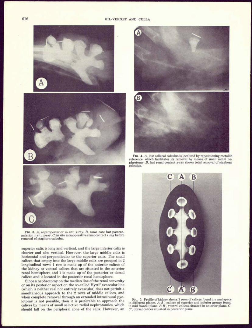
FIG. 4. A, last caliceal calculus is localized by repositioning metallic reference, which facilitates its removal by means of small radial ne-phrotomy. B, last renal contact x-ray shows total removal of staghorn calculus.
FIG. 3. A, anteroposterior in situ x-ray. B, same case but postero-anterior in situ x-ray. C, in situ intraoperative renal contact x-ray before removal of staghorn calculus.
superior calix is long and vertical, and the large inferior calix is shorter and also vertical. However, the large middle calix is horizontal and perpendicular to the superior calix. The small calices that empty into the large middle calix are grouped in 2 longitudinal rows: 1 row is made up of the anterior calices of the kidney or ventral calices that are situated in the anterior renal hemisphere and 1 is made up of the posterior or dorsal calices and is located in the posterior renal hemisphere.
Since a nephrotomy on the median line of the renal convexity or on its posterior aspect on the so-called Hyrtl5 avascular line (which is neither real nor entirely avascular) does not permit a simultaneous approach to the 2 rows of middle calices, and when complete removal through an extended intrasinusal pye-lotomy is not possible, then it is preferable to approach the calices by means of small selective radial nephrotomies, which should fall on the peripheral zone of the calix. However, an
FIG. 5. Profile of kidney shows 3 rows of calices found in renal space in different planes. A-A', calices of superior and inferior groups found in mid frontal plane. B-B', ventral calices situated in anterior plane. C-C', dorsal calices situated in posterior plane.

ADVANCES IN INTRAOPERATIVE RENAL RADIOGRAPHY 617
by another from a posteroanterior direction. Then, the 2 exter-nal profile x-rays are obtained. If the type I plate-holder is used disassembled 1 of its branches is placed in front of and the other behind the renal pedicle (fig. 8, A), and when type II is used afterwards 1 of its branches is placed above and the other below the pedicle (fig. 8, B). The plate-holder is closed the moment the x-ray is taken. This closing, which is not complete, does not involve even the slightest trauma to the vessels of the kidney. The relative ischemia time of 2 to 4 seconds needs no comment.
The technical characteristics of these radiographs are of minimum power, since the x-rays on their way to the plate only encounter the weak opacity of the kidney, because the surgical incision eliminates the interposing of tissue between the x-ray tube and the kidney. The characteristics of the radiographs are 45 to 50 kw. and 10 mA. for 0.5 seconds at a distance of 70 cm. It is important to check previously the central beam. The beam must be completely perpendicular to the plane of the radio-graphic plates (figs. 9 and 10).
Although the radiation doses are minimal, the person holding the plate-holder clamp is protected by using radiological beam-localizing diaphragms. A diaphragm with a circular opening of 1 cm. in diameter, at a focus-plate distance of 70 cm., provides a focal area of 18 cm., sufficient in size to cover the entire kidney and, thus, avoid direct radiation around it.
RESULTS
exact knowledge of the location of the calculus is an indispens-able prior condition.
Material. To obtain the third dimension of the calculus, that is to discover its depth and, subsequently, its situation in the renal space, it is necessary to obtain external profile x-rays, that is at a 90-degree angle. For this purpose, we use a set of minichassis (fig. 6) and 2 articulated clamps with 2 flat surfaces that can be taken apart like forceps; these plate-holders are of 2 different models. Each model has the shape of a half ellipse, in transverse (fig. 7, A) or in longitudinal section (fig. 7, B) and is made of metal. The radiographic plates are arranged on these surfaces in the same manner. The 2 halves of the radiographic film are placed on both metal plates of the clamp, thus, avoiding radiation from behind.
The half elliptical shape, either in transverse or longitudinal section, that we have selected for the radiographic film is the most appropriate. In this way each renal hemisphere, whether it be anterior or posterior, or the superior or inferior renal half, can be projected onto it and, when both halves are joined, we have the complete kidney in a profile view.
First, a conventional anteroposterior x-ray is made followed
FIG. 6. Set of minichassis for 3-dimensional radiographic exploration of kidney.
The x-ray in a 90-degree angle with respect to the chassis anteroposterior x-ray, that is the radiography of an external profile, provides the third dimension of the calculus in the renal space. The importance of this radiography technique rests in the fact that it situates the calculus in a front-to-back direction, that is in the thickness of the organ, which constitutes a valuable and decisive aid for its localization and removal.
An external profile contact x-ray shows whether the remain-ing calculus is found in a calix of the anterior renal hemisphere or in 1 of the posterior renal hemispheres. In addition, it reveals the exact distance between the calculus and the surface of the organ, that is the thickness of the parenchyma covering it which, when combined with the anteroposterior x-ray, gives us the 3 dimensions of the calculus whereby we learn its exact location.
This technique is of great help in the localization of the
FIG. 7. A, plate carrier type I to x-ray anterior and posterior faces. B, plate earner type II to x-ray superior and inferior renal halves

618 G I L - V E R N E T A N D CULLA
FIG. 8. A, to obtain third dimension of kidney x-ray is made in external profile with plate carrier I; 1 branch is placed in front and 1 behind renal pedicle, both in contact with internal edge of kidney. B, with plate carrier II 1 branch is placed above and 1 below renal pedicle. Metallic reference clips are placed on posterior surface and opaque thread is placed on external renal edge.
platform.
FIG. 10. A, posteroanterior x-ray with kidney in situ to obtain 2-dimensions of kidney and its contents. B, x-ray in external profile with plate carrier I, obtaining third dimension of kidney.
lithiasic calix and for the removal of its contents by means of an extended pyelotomy. However, when this is not possible because there is a marked lack of proportion between the size of the calculus and the infundibulum of the calix then, because we know exactly in which calix the calculus is found, it is possible to make a nephrotomy incision tha t is minimal, tha t is of a length equivalent to the thickness of the calculus and in a radial direction in the most peripheral zone of the calix, which is the shortest t ransparenchymatous approach and the 1 with the least vascular risk.
Since there is no superimposition of tissue between the x-ray tube and the kidney radiographs with greater clarity than even those done with the anteroposterior contact chassis are ob-tained, which is important when concretions or small mineral-ized calculi are to be detected. It must be emphasized tha t in 16 per cent of the cases we have been able to discover calculi or calcifications that had not been revealed either in the preoper-ative radiographic explorations or in the anteroposterior contact intraoperative x-ray.
REFERENCES 1. Truc, E. and Grasset, D.: Lithiase rénale. Encyclop. Méd. Chir., 9:
5, 1960. 2. Gil-Vernet, J.: New surgical concepts in removing renal calculi.
Urol. Int., 20: 255, 1965. 3. Gil-Vernet, J. M.: La cirugia intrasinusal de los cálculos corali-
formes. Rapport XV o Congrès de la Société Internationale d'Urologie, Tokyo, July 12-18, vol. 1, p. 11, 1970.
4. Paitre, F., Giraud, D. and Dupret, S.: Práctica Anatomoquirúrgica Ilustrada. Abdomen. Los Órganos Retroperitoneales. Barcelona: Salvat Editores, fase. 3, p. 255, 1941.

ADVANCES IN INTRAOPERATIVE RENAL RADIOGRAPHY 619
5. Hyrtl, J.: Das Nierenbecker der sáugetiere und des Menschen. Wiener Sitzungsberichte, B, LIV, 1870.
EDITORIAL COMMENT
This detailed review certainly exhibits considerable knowledge and appreciation of the fundamentals of direct renal radiography during surgical procedures. We concur in the appraisal of the importance of 3-dimensional radiography and have found this technically extremely difficult to achieve owing to the fact that heavy portable x-ray equip-ment must be transported first to 1 side and then the other side of the operating table. Even these techniques provide films that are at less than right angles to each other (oblique rather than true lateral views). The plate-holder for maintaining the films in position against the hilar surface of the kidney and, thus, properly positioning the kidney for a convex to hilar surface exposure certainly presents the most technically advanced method for this procedure and the only one I know in which 2 films truly at right angles to each other may provide a 3-dimensional concept of the surgically exposed kidney. Quite obviously, surgical mobilization of the kidney will need to be virtually complete and there may be some technical difficulties with the clamp but these are aggra-vations that the surgeon should be able to overcome. Surely this is a progressive approach to a problem that has been addressed in America by the "hole-in-the-film technique". Few have been able to find any constructive use for the commercial hole-in-the-film kidney films.
The use of image intensification screens unquestionably will reduce the amount of radiation required to make comparable films of the kidney. However, the amount of reduction is so exceedingly small in rad that I do not believe this is significant. The disadvantage in using the screens has been one of economics. The plastic-encased disposable kidney films available commercially have a good shelf-life and provide
excellent technical films with as great a resolution as the cassette intensification films, albeit requiring a slight increase in kilovolts peak. The image intensification screens are too expensive to be discarded and, therefore, must be loaded by technicians in the individual hospital. In America most institutions will find it exceedingly difficult to provide consistently reliable loading, evacuation and sealing of the cassettes so that they may be sterilized properly and available consistently at the time of the operation.
William H. Boyce Section of Urology Bowman Gray School of Medicine Winston-Salem, North Carolina
REPLY BY AUTHORS
Technically, it is extraordinarily simple to obtain the 3-dimensional exploration of the kidney. The amount of time necessary, including placement of the forceps carrying the plaques, is not >5 minutes. The small portable x-ray machine does not need to be moved from 1 side of the operating table to the other. The importance of obtaining the exact location of the calculi and, therefore, their total removal with a mini-mum of trauma, as well as the detection of those calculi that are slightly mineralized (and tha t are not seen with the regular commercial films available) relegate the prosaic economic aspects to a secondary consid-eration, although the intensifying plaques can be recuperated easily. These cassettes have been on the market for about 1 year and their sterilization can be obtained by using the usual system of ethylene oxide or immersing them in iodine for 3 minutes. The significance of this method is that it has solved the problem of the residual calculus.