the journal of experimental microbiology & immunology+vol4... · all the primers used in pcr...
TRANSCRIPT
Vol 4:1-15
August 2018 Volume 4: 1-15 Undergraduate Research Article - Refereed https://jemi.microbiology.ubc.ca/ 1
Deletion of the capsule phosphatase gene wzb renders Escherichia coli strain K30 sensitive to the antibiotic nitrofurantoin. Michelle Gu, Almas Khan, Desiree S. Pagulayan, Wai Lam Tam Department of Microbiology and Immunology, University of British Columbia, Vancouver, British Columbia, Canada SUMMARY The group I capsular polysaccharides (CPS) are assembled through the Wzy-dependent pathway that involves Wza, Wzb and Wzc proteins. CPS has been characterized as a virulence factor that confers antimicrobial resistance in Escherichia coli (E. coli). Increasing antimicrobial resistance among uropathogens has led to increased interest in using the antibiotic nitrofurantoin. While previous studies have investigated the effect of single gene deletions of wza, wzb, and wzc on erythromycin sensitivity in E. coli K30, susceptibility of these individual gene knockout strains to nitrofurantoin has not yet been explored. We aimed to investigate the relationship between nitrofurantoin sensitivity and E. coli group I CPS production by studying its biosynthesis genes wza and wzb. Strains were confirmed via polymerase chain reaction (PCR) and Sanger sequencing. Zones of inhibition for each strain were quantified via a disc diffusion assay, and the minimum inhibitory concentration (MIC) of nitrofurantoin for each strain was determined. Results of the disc diffusion assay revealed significant differences in zone of inhibition sizes for wza-wzb-wzc and wzb mutant strains relative to wild type (WT) E. coli K30. In contrast to the E. coli K30 wzb mutant, the BW25113 wzb mutant from the Keio collection showed less sensitivity than its WT parent strain. We conclude that other factors besides the presence of capsule may be contributing to nitrofurantoin susceptibility, and that Wzb may be involved in complex interactions with substrates that affect the activation of nitrofurantoin. INTRODUCTION
antigen or capsular polysaccharides (CPS) are large molecular weight surface polysaccharides found in a variety of Gram-positive and Gram-negative bacteria (1).
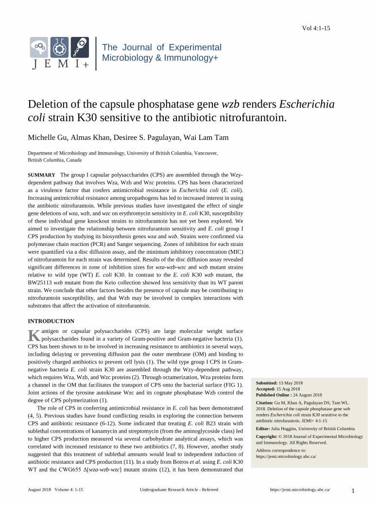
CPS has been shown to to be involved in increasing resistance to antibiotics in several ways, including delaying or preventing diffusion past the outer membrane (OM) and binding to positively charged antibiotics to prevent cell lysis (1). The wild type group I CPS in Gram-negative bacteria E. coli strain K30 are assembled through the Wzy-dependent pathway, which requires Wza, Wzb, and Wzc proteins (2). Through octamerization, Wza proteins form a channel in the OM that facilitates the transport of CPS onto the bacterial surface (FIG 1). Joint actions of the tyrosine autokinase Wzc and its cognate phosphatase Wzb control the degree of CPS polymerization (1).
The role of CPS in conferring antimicrobial resistance in E. coli has been demonstrated (4, 5). Previous studies have found conflicting results in exploring the connection between CPS and antibiotic resistance (6-12). Some indicated that treating E. coli B23 strain with sublethal concentrations of kanamycin and streptomycin (from the aminoglycoside class) led to higher CPS production measured via several carbohydrate analytical assays, which was correlated with increased resistance to these two antibiotics (7, 8). However, another study suggested that this treatment of sublethal amounts would lead to independent induction of antibiotic resistance and CPS production (11). In a study from Botros et al. using E. coli K30 WT and the CWG655 Δ[wza-wzb-wzc] mutant strains (12), it has been demonstrated that
K
Submitted: 13 May 2018 Accepted: 15 Aug 2018 Published Online : 24 August 2018
Citation: Gu M, Khan A, Pagulayan DS, Tam WL. 2018. Deletion of the capsule phosphatase gene wzb renders Escherichia coli strain K30 sensitive to the antibiotic nitrofurantoin. JEMI+ 4:1-15
Editor: Julia Huggins, University of British Columbia
Copyright: © 2018 Journal of Experimental Microbiology and Immunology. All Rights Reserved.
Address correspondence to: https://jemi.microbiology.ubc.ca/
The Journal of Experimental Microbiology & Immunology+
JEMI+ Gu et al.
August 2018 Volume 4: 1-15 Undergraduate Research Article - Refereed https://jemi.microbiology.ubc.ca/
2
mutants with a full deletion of wza, wzb, and wzc genes were more resistant to macrolide antibiotics, but the resistance mechanism may be independent from capsule production. Discrepancy between studies demonstrates the need for studying the capsular involvement in conferring antibiotic resistance in an antibiotic-, bacterial gene-, and strain-specific manner.
Nitrofurantoin is a synthetic nitrofuran antibiotic specifically used for treating uncomplicated lower urinary tract infections against a range of Gram-positive and Gram-negative bacteria (13, 14). Increasing antimicrobial resistance among uropathogens has led to increased interest in using nitrofurantoin (15). Nitrofurantoin is a derivative of imidazolidinedione and has a balance of hydrophobic and hydrophilic properties (16, 17). It has a relatively low molecular weight (238.159 g/mol) and is a zwitterion (16). Similar to other antibiotic drugs used for Gram-negative bacterial targets, these physiochemical properties allow nitrofurantoin to pass through the capsule and OM, and diffuse through the cytoplasmic membrane into the cell more rapidly than charged antibiotics (17). The mechanism for the action of nitrofurantoin is not well understood. One proposed mechanism is that once inside the bacterial cell, the aryl nitro group in nitrofurantoin is converted by bacterial nitroreductase into the reactive nitroso group that damages DNA, RNA, and proteins through redox reactions (18).
In previous study, Botros et al. used a range of antibiotics including nitrofurantoin to examine the effect of capsule on antibiotic sensitivity (12). In their screen for changes in antibiotic sensitivity between the WT E. coli K30 strain and the mutant E. coli K30 CWG655 strain with deletion of wza, wzb, and wzc genes, they found that the mutant strain showed a significant increase in sensitivity to nitrofurantoin on both Luria-Bertani (LB) plates and Mueller-Hinton (MH) plates (12). While other studies have investigated the effect of single gene deletions of wza, wzb, and wzc on erythromycin sensitivity, the sensitivity of these mutant strains to nitrofurantoin is yet to be explored (19-21).
As all three gene products are required for assembly of CPS, we hypothesized that single knockout mutants for wza and wzb will exhibit an increase in sensitivity to nitrofurantoin, similar to the pattern previously observed in the mutant strain CWG655 in which all three genes are deleted.
FIG. 1 Simplified diagram of the structure of group I capsule in K30 strain (1, 3). Wza is an octamer that forms a channel within the outer membrane. Wzb is a cytosolic phosphatase that associates with Wzc, which is a tyrosine autokinase. Wzc is a tetrameric, integral inner membrane protein that can form a higher-order complex with Wza to connect the inner membrane and outer membrane.

JEMI+ Gu et al.
August 2018 Volume 4: 1-15 Undergraduate Research Article - Refereed https://jemi.microbiology.ubc.ca/
3
METHODS AND MATERIALS
Bacterial strains, media preparation, and growth conditions. All bacterial strains used in this study are listed in TABLE 1. E. coli K30 CWG343 (E69 with wzbK30::aphA3 and wza22
min ::aadA insertion on wzb22min and etk), CWG655 [wza22 min::aadA Δ(wza-wzb-wzc)K30::aphA3], and CWG281 (wza22 min::aadA wzaK30::aacC1) were previously obtained from the laboratory of Dr. Chris Whitfield from the Department of Molecular and Cellular Biology at the University of Guelph. Frozen stocks of mutant strains were kept in the MICB 421 laboratory from the Department of Microbiology and Immunology at the University of British Columbia (UBC), which also provided the wild type (WT) E69 (serotype: O9a:K30:H12) strain. CWG343 contained a kanamycin resistance cassette inserted in wzb gene and CWG655 contained mutations where genes of interest were knocked out with a kanamycin resistance cassette while a gentamicin resistance cassette insertion was used to inactivate the wza gene in the creation of CWG281 (22). JW2046-1 [F-, Δ(araD-araB)567, ΔlacZ4787(::rrnB-3), λ-, Δwzb-759::kan, rph-1, Δ(rhaD-rhaB)568, hsdR514] and the parent K-12 Keio strain, BW25113, were obtained from the Coli Genetic Stock Centre at Yale. Overnight liquid cultures were grown on a shaker at 150-200 rpm and plates were grown in an incubator. All growth experiments were conducted at 37°C for 16-20 hours. Cells were grown on Lysogeny Broth (LB) agar plates (1.0% w/v tryptone, 0.5% w/v yeast extract, 0.5% w/v NaCl, pH 7, 1.5% agar) for antibiotic testing, and on BD Difco™ Dehydrated Mueller Hinton (MH) agar plates as well as in BBLTM MH broth (MHB) for disc diffusion and MIC assays.
Identification of K30 strains via PCR amplification and agarose gel electrophoretic analysis of wza, wzb, and wzc region. PCR amplification was performed using a modified protocol described by Su et al. (21). All the primers used in PCR are listed in TABLE 2. EB6 and EB7, which anneal within the region containing wza, wzb, and wzc genes, were used in Reid and Whitfield’s study (22). They were ordered from IDT and resuspended with sterile distilled water to reach a stock concentration of 100 μM. pUC19-193F and -355R, which anneal to a 162 bp sequence for beta-lactamase in the pUC19 plasmid that was used as a PCR positive control, were provided by the Microbiology and Immunology Department, UBC at a concentration of 100 μM. All primers were further diluted to 10 μM as the working concentration. Genomic DNA of each of the four K30 strains were extracted following the protocol from Invitrogen PureLink Genomic DNA kit for Gram-negative bacterial cell lysate. Concentrations of the isolated genomic DNA were then determined using NanoDrop200 spectrophotometer to ensure a similar amount of template was used for individual PCR reactions. A master mix for 8 PCR reactions was made from 40 μL of 10X PCR Buffer (no
TABLE 1 Strains used in the study.

JEMI+ Gu et al.
August 2018 Volume 4: 1-15 Undergraduate Research Article - Refereed https://jemi.microbiology.ubc.ca/
4
MgCl2), 12 μL of 50 mM MgCl2, 8 μL of 10mM dNTP mix, 1.6 μL of PlatinumTM Taq Polymerase (Thermofisher), and 314.4 μL of sterile distilled water. In each PCR tube, 47 μL of Master Mix, 1 μL each of the templates, corresponding forward and reverse primers were added to reach a total volume of 50 μL. In the negative control, 1 μL of each of the following was added: sterile distilled water, EB6 and EB7. The PCR reactions were run overnight in a Bio-Rad thermocycler, with an initial denaturation step at 94°C for 120 seconds, followed by 35 cycles of 94°C for 30 sec, 50°C for 30 sec, 72°C for 4 min 50 sec, and a final extension step at 72°C for 10 min. The PCR products were kept in the thermocycler at 4°C until the next day for visualization via gel electrophoresis. 0.8% agarose gel was used in gel electrophoretic analysis of PCR amplification products. The band sizes were compared to a 1kb Plus ladder (Invitrogen) to confirm correct product size.
PCR product purification and Sanger sequencing of purified products to further elucidate the structure of the wzy operon. Products were purified in accordance with the protocol in the Thermofisher GeneJet PCR Purification Kit. Sanger sequencing of purified PCR products for all four K30 strains was performed. The NanoDrop200 spectrophotometer was used to measure the concentrations of each purified PCR product and 200-500 ng of each was submitted for sequencing from EB6 and EB7 primers. Additional sequencing of the wzb region using a custom designed forward primer was also done on the WT and CWG343 strains. All Sanger sequencing results were aligned using NCBI Nucleotide BLAST with the search limited to E. coli (taxa id:562). A forward primer for the wzb region was designed using the tool PrimerQuest from IDT based on the K30 capsule sequence obtained from NCBI (Accession ID AF104912). Following suggested guidelines from Genewiz, the primer was 20 bp in length, had 50% G-C content and was 105 bp upstream from the wzb gene (TABLE 2). Visualization of the primer annealing to the wzb region was done using SNAPGene.
Disc diffusion assay. A modified version of the Kirby-Bauer method, as used by Botros et al., was followed (12, 23). A colony from each strain was inoculated with 5 mL of MH media and grown overnight. The optical density (OD) of each culture was measured the next day at 660nm using the Beckman spectrophotometer. The cultures were then diluted with sterile MHB to ~0.8-1.0 OD. 200 μL of each diluted culture was spread on separate MH agar plates and allowed to dry (24). Three to four pre-hole punched, autoclaved Whatman paper discs were evenly placed on each plate using sterile forceps. In the fume hood, 2.5 μL of a 30 mg/ml nitrofurantoin stock solution (0.03 g powder dissolved in 1 mL dimethylformamide (DMF) split into 100-200 μL aliquots) was pipetted onto each disc. 2.5 μL of DMF was also pipetted onto negative control discs placed on separate MH agar plates for each strain. All plates were incubated for 18-20 hours at 37℃. Diameters of the resulting inhibition zones were measured in millimetres. A clear area around the disc indicates sensitivity to nitrofurantoin and a wider diameter relative to WT indicates increased sensitivity. For trials 1-3, statistical significance between all four strains was determined using ANOVA, followed by a two sample t-test using MSerror for planned comparisons against WT, and the Tukey-Kramer test for unplanned comparisons between mutant strains. The Tukey-Kramer test results across all samples were
TABLE 2 Primer sequences and melting temperatures used in the study.
JEMI+ Gu et al.
August 2018 Volume 4: 1-15 Undergraduate Research Article - Refereed https://jemi.microbiology.ubc.ca/
5
represented in box plots with numerical and categorical variables on the y versus x axis. For trials 4-5, statistical significance between two strains was determined using a two-sample t-test. This test was chosen as it was determined via histogram plots and Levene’s test that the data was approximately normal and met the equal variance assumption of the two sample t-test. Bar graph instead of box plot representation of the data was chosen as non-overlapping box plot whiskers may be misinterpreted as significance which only applies if the Tukey-Kramer test was used for analysis.
The MIC assay. Overnight cultures of each strain were grown in 5 mL MHB at 37°C on a shaker at 150-200 rpm. Cultures were measured the next day at OD660 using the Beckman spectrophotometer and diluted to 1.0 OD660. In a 96-well plate, 100 μL of 160 μg/mL nitrofurantoin solution (diluted from a stock concentration of 30 mg/mL) was added to the first column of wells before a series of two-fold dilution was carried out (50 μL of nitrofurantoin was transferred to the next column of wells containing 50 μL of MHB, until the concentration reached 2.5 μg/mL in the last column). From the final column of wells, 50 μL of nitrofurantoin-MHB was discarded to maintain the same volume across all wells. In duplicates, 50 μL of each diluted strain was added to the serially diluted nitrofurantoin wells. As the nitrofurantoin solution was yellow, a column of nitrofurantoin concentrations (80 - 1.25 μg/mL) mixed with 50 μL of MHB, served as a base reading to normalize readings for each column of nitrofurantoin concentration. A volume of 50 μL of each of diluted strain and MHB were added in duplicate down a column to serve as the positive control to confirm strain growth without nitrofurantoin. To the remaining empty wells, 100 μL of MHB was added to serve as sterility control. The plate was incubated at 37°C for 16-20 hours and was read with BioTek Micro-volume Plate Reader at OD600.
RESULTS
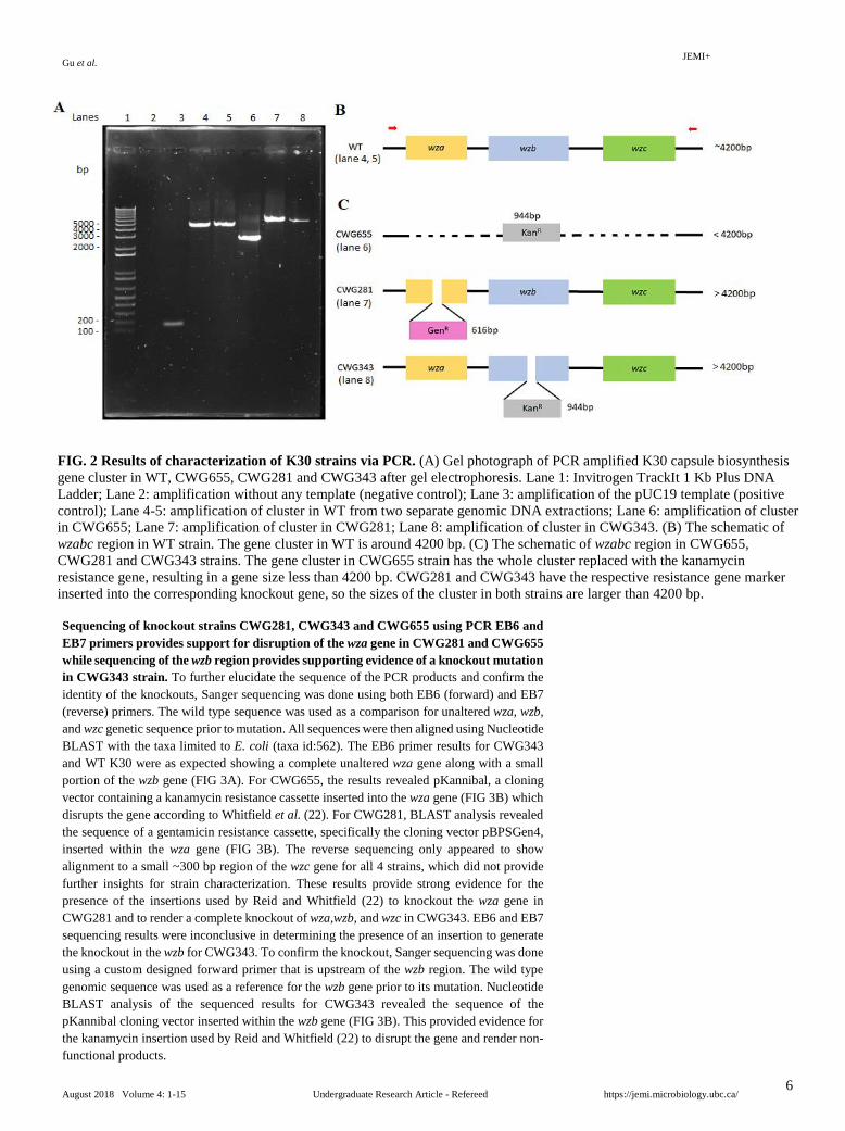
Characterization of knockout strains CWG281, CWG343 and CWG655 using PCR and agarose gel electrophoresis. To identify the K30 strains, we performed PCR using extracted genomic DNA from each of the strains. The capsule biosynthesis gene cluster containing wza, wzb and wzc genes was amplified. EB6 and EB7 primers that flank the region were used. Agarose gel electrophoretic analysis resolved the PCR amplicons from the strains (FIG 2A), and the structures of the wzabc cassette of the strains are shown for comparison (FIG 2B-C). Lane 2 shows the negative control without template for amplification. The absence of band in this lane suggests no contamination of the reagents. Lane 3 shows the positive control with the expected PCR product size of 162 bp. According to the sequence (accession number AF104912) from GenBank, the target amplification region in WT is 4200 bp, which is shown in both lanes 4 and 5. Lane 6 shows the PCR product from CWG655 corresponding to a band size that is smaller than 4200 bp. This is consistent with our prediction since this strain was generated by replacing the wzabc region with the kanamycin resistance cassette with a size of 944bp (22), thus this region is smaller than that in the WT. Lanes 7 and 8 show the PCR products from CWG281 and CWG343 respectively. Both bands sizes are larger than 4200 bp. Since these mutant strains have their respective resistance gene marker inserted into the corresponding knockout gene (21), the overall sizes of the target cluster for amplification are larger than WT. Based on gel electrophoretic analysis, the bands of the strains did show the approximate expected sizes, suggesting the correct genotype for the strains.
TABLE 3 Minimum inhibitory concentrations of nitrofurantoin for E. coli K30 WT, CWG281, CWG343, and CWG655 strains, incubated in MHB for 16-20 hours.
JEMI+ Gu et al.
August 2018 Volume 4: 1-15 Undergraduate Research Article - Refereed https://jemi.microbiology.ubc.ca/
6
Sequencing of knockout strains CWG281, CWG343 and CWG655 using PCR EB6 and EB7 primers provides support for disruption of the wza gene in CWG281 and CWG655 while sequencing of the wzb region provides supporting evidence of a knockout mutation in CWG343 strain. To further elucidate the sequence of the PCR products and confirm the identity of the knockouts, Sanger sequencing was done using both EB6 (forward) and EB7 (reverse) primers. The wild type sequence was used as a comparison for unaltered wza, wzb, and wzc genetic sequence prior to mutation. All sequences were then aligned using Nucleotide BLAST with the taxa limited to E. coli (taxa id:562). The EB6 primer results for CWG343 and WT K30 were as expected showing a complete unaltered wza gene along with a small portion of the wzb gene (FIG 3A). For CWG655, the results revealed pKannibal, a cloning vector containing a kanamycin resistance cassette inserted into the wza gene (FIG 3B) which disrupts the gene according to Whitfield et al. (22). For CWG281, BLAST analysis revealed the sequence of a gentamicin resistance cassette, specifically the cloning vector pBPSGen4, inserted within the wza gene (FIG 3B). The reverse sequencing only appeared to show alignment to a small ~300 bp region of the wzc gene for all 4 strains, which did not provide further insights for strain characterization. These results provide strong evidence for the presence of the insertions used by Reid and Whitfield (22) to knockout the wza gene in CWG281 and to render a complete knockout of wza,wzb, and wzc in CWG343. EB6 and EB7 sequencing results were inconclusive in determining the presence of an insertion to generate the knockout in the wzb for CWG343. To confirm the knockout, Sanger sequencing was done using a custom designed forward primer that is upstream of the wzb region. The wild type genomic sequence was used as a reference for the wzb gene prior to its mutation. Nucleotide BLAST analysis of the sequenced results for CWG343 revealed the sequence of the pKannibal cloning vector inserted within the wzb gene (FIG 3B). This provided evidence for the kanamycin insertion used by Reid and Whitfield (22) to disrupt the gene and render non-functional products.
FIG. 2 Results of characterization of K30 strains via PCR. (A) Gel photograph of PCR amplified K30 capsule biosynthesis gene cluster in WT, CWG655, CWG281 and CWG343 after gel electrophoresis. Lane 1: Invitrogen TrackIt 1 Kb Plus DNA Ladder; Lane 2: amplification without any template (negative control); Lane 3: amplification of the pUC19 template (positive control); Lane 4-5: amplification of cluster in WT from two separate genomic DNA extractions; Lane 6: amplification of cluster in CWG655; Lane 7: amplification of cluster in CWG281; Lane 8: amplification of cluster in CWG343. (B) The schematic of wzabc region in WT strain. The gene cluster in WT is around 4200 bp. (C) The schematic of wzabc region in CWG655, CWG281 and CWG343 strains. The gene cluster in CWG655 strain has the whole cluster replaced with the kanamycin resistance gene, resulting in a gene size less than 4200 bp. CWG281 and CWG343 have the respective resistance gene marker inserted into the corresponding knockout gene, so the sizes of the cluster in both strains are larger than 4200 bp.

JEMI+ Gu et al.
August 2018 Volume 4: 1-15 Undergraduate Research Article - Refereed https://jemi.microbiology.ubc.ca/
7
Deletion of wzb in E. coli strain K30 is sufficient to confer sensitivity to nitrofurantoin. In order to test if single capsule gene knockout on each of wza, wzb and wzc in E. coli strain K30 leads to increased nitrofurantoin sensitivity, we chose to measure zones of inhibition via a disc diffusion assay. Overnight cultures of each strain were prepared in MHB. OD660 measurements were taken the next day and cultures were diluted before spreading on MH agar plates. Three nitrofurantoin discs were placed on each plate which were incubated overnight at 37°C. Negative control discs were treated with DMF alongside the nitrofurantoin discs. No inhibition zones appeared around the negative control discs after incubation (data not shown). FIG 4A-D show zones of clearance from all strains in the initial trial of the disc diffusion assay, which indicated their sensitivity to nitrofurantoin. CWG343 and CWG655 were more sensitive relative to WT. Statistical analysis revealed significance in non-overlapping boxplot whiskers (FIG 4B). The zone of clearance for the wza mutant appeared similar in size to WT and their boxplot whiskers, which denote the smallest and largest non-extreme data points, overlapped indicating that there was no significant difference between the two strains. The second trial of the disc diffusion assay confirmed initial CWG343 and CWG655 findings relative to WT, but no significant difference was found between CWG343 and CWG655 (FIG 5). The average size of the inhibition zones for CWG343 and CWG655
FIG. 3 Sanger sequencing results after analysis with nucleotide database in BLAST. (A) Primer and wild type sequences aligned onto the wza,wzb and wzc regions. The EB6 PCR forward primer shown in orange aligned outside of the wza region. The wzb sequencing primer in purple aligned onto a portion of the wza region (upstream of the wzb), and EB7 PCR reverse primer in red aligned just downstream of wzc. Sequencing results for the WT showed a 1049 bp sequence aligning onto wza region and a portion of the wzb for the EB6, and 1049 bp of its sequence map onto the entire wzb gene and a portion of the wzc gene with the wzb sequencing primer and the reverse primer showed a ~300 bp alignment onto wzc. (B) Alignments onto cloning vectors with resistance markers inserted into the genes are in green. CWG343 sequencing results showed a 1055 bp sequence aligning primarily onto the wza gene and a portion of the wzb gene with the EB6 primer. CWG281 and CWG655 both had ~580 bp of their sequence map onto the wza gene. CWG281 had 471 bp of its sequence map onto a pBPSGen4 cloning vector containing a gentamicin resistance cassette. CWG655 had 405 bp of the sequence map onto a pKannibal cloning vector containing a kanamycin resistance marker. For the wzb sequencing primer, only WT and CWG343 were sequenced. CWG343 had only 672 bp of its sequence align partially onto the wzb gene and 672 bp of its sequence align onto a pKannibal cloning vector with the kanamycin resistance cassette. The reverse primer showed a ~300 bp alignment onto wzc for all 3 strains.

JEMI+ Gu et al.
August 2018 Volume 4: 1-15 Undergraduate Research Article - Refereed https://jemi.microbiology.ubc.ca/
8
FIG. 4 Susceptibility of WT K30 and mutant strains to nitrofurantoin via disc diffusion assay: Trial 1. Zones of inhibition for (A) WT, (B) CWG655 (Δwza-wzb-wzc), (C) CWG343 (Δwzb), and (D) CWG281 (Δwza). The sizes of clearance around the discs indicate that WT and mutant strains are sensitive to nitrofurantoin. CWG343 followed by CWG655 show greater sensitivity to nitrofurantoin compared to WT, as indicated by larger zones of clearance. CWG281 showed a similar size of zone as the WT. Scale bars = 6 mm. (E) Box plot representation of (A-D), where n = 3 for each strain, and whiskers denote the smallest and largest non-extreme data points. Statistical significance was confirmed using the Tukey-Kramer test for unplanned comparisons of inhibition zones between strains, and non-overlapping whiskers generally indicate significance (p-value < 0.05). Incubation conditions: 18-20 hours at 37°C on MH agar plates. Zone of clearance refers to zone of inhibition. The diameter of the zone was measured; a larger diameter relative to WT indicates greater susceptibility whereas a smaller diameter indicates greater resistance.
FIG. 5 Susceptibility of WT K30 and mutant strains to nitrofurantoin via disc diffusion assay: Trial 2. Zones of inhibition for (A) WT, (B) CWG655 (Δwza-wzb-wzc), (C) CWG343 (Δwzb), and (D) CWG281 (Δwza). The sizes of clearance around the discs indicate that WT and mutant strains are sensitive to nitrofurantoin. CWG343 followed by CWG655 and then CWG281 show greater sensitivity to nitrofurantoin compared to WT, as indicated by larger zones of clearance. Scale bars = 6 mm. (E) Box plot representation of (A-D), where n = 3 for each strain, and whiskers denote the smallest and largest non-extreme data points. Statistical significance was confirmed using the Tukey-Kramer test for unplanned comparisons of inhibition zones between strains, and non-overlapping whiskers generally indicate significance (p-value < 0.05). There is no significant difference between CWG343 and CWG655 which have only one whisker. Incubation conditions: 18-20 hours at 37°C on MH agar plates. Zone of clearance refers to zone of inhibition. The diameter of the zone was measured; a larger diameter relative to WT indicates greater susceptibility whereas a smaller diameter indicates greater resistance.

JEMI+ Gu et al.
August 2018 Volume 4: 1-15 Undergraduate Research Article - Refereed https://jemi.microbiology.ubc.ca/
9
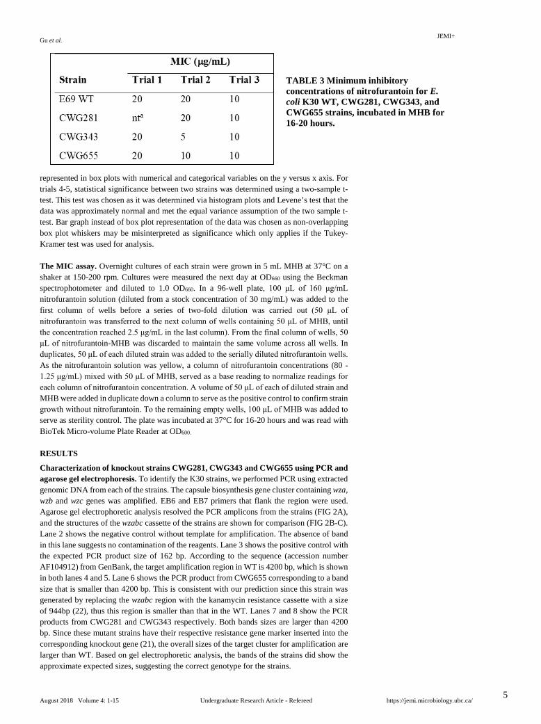
for all trials were 22.33 mm and 20.67 mm, respectively. However, the data also suggested significant difference between WT and CWG281 (wza mutant). We investigated this phenomenon in a separate trial comparing only WT and CWG281. More replicates were included with four nitrofurantoin discs on each of two MH agar plates for both strains. The results showed no significant mean difference (7). We conclude that deletion of wzb but not wza is sufficient for nitrofurantoin sensitivity in a disc diffusion assay conducted on solid MH agar media. MIC assay of nitrofurantoin performed on E. coli K30 (WT), CWG281, CWG343, and CWG655 strains were inconsistent between trials. The MIC assay was performed in order to determine the minimum inhibitory concentration of the experimental WT and E. coli K30 mutant strains for nitrofurantoin in liquid media. Strains were plated in 96-well plates containing concentrations of nitrofurantoin (80 to 1.25 μg/mL) prepared in two-fold serial dilutions in wells down each column. Cells in MHB without nitrofurantoin was included as the positive control column to verify that all strains were growing without nitrofurantoin selection. A negative control with just nitrofurantoin and MHB was included due to the yellow colour of the nitrofurantoin solution, which could potentially affect readings. A DMF control was also included as DMF has been implicated in a previous study to affect growth of E. coli and it was the solvent used for nitrofurantoin (25). The positive and DMF control showed high growth. This confirmed that DMF did not affect the growth of experimental strains. MHB was added to all unused wells which served as sterility controls to check for contamination. CWG281 was not tested in trial 1, as the overnight culture did not reach the required OD to perform the MIC assay. Three trials were performed on different days and thus were not compared by statistical analysis. The MIC for all strains were below 32 μg/mL (TABLE 3), which is the published cutoff for identifying strains sensitive to nitrofurantoin (26). In trial 1 and 3, the MIC of all strains were the same within each trial, at 20 μg/mL and 10 μg/mL respectively (TABLE 3). Trial 2 showed that the MIC of WT and CWG281 was 20 μg/mL, which is higher than that of CWG343 and CWG655, which were at 5 μg/mL and 10 μg/mL respectively (TABLE 3). The exact MIC of each strain could not be determined due to varying MIC results between trials.
Deletion of wzb in BW25113 is sufficient to confer resistance to nitrofurantoin. To investigate a potential interaction between Wzb and nitrofurantoin, another wzb mutant from the Keio strain collection, JW2046-1, was tested in a disc diffusion assay and compared to its WT parent strain, BW25113. Each strain was spread on two MH agar plates, and replicate
FIG. 6 Susceptibility of WT K30 and CWG281 (Δwza) to nitrofurantoin via disc diffusion assay. (A, B) Zones of clearance around the discs indicate that WT and CWG281 were sensitive to nitrofurantoin. The zones appear similar in size, suggesting that there is no difference in sensitivity to nitrofurantoin between the two strains. Scale bars = 6 mm. (C) Bar graph in lieu of box plot representation of (A, B) as non-overlapping box plot whiskers may be misinterpreted as significance, which can showcase Tukey-Kramer analysis results but not those of a two-sample t-test. Error bars denote the standard error of the mean. n = 7 for WT (a replicate was removed due to possible experimental error) and n = 8 for CWG281. Statistical non-significance (p-value > 0.05) was confirmed using a two-sample t-test. Incubation conditions: 18-20 hours at 37°C on MH agar plates. Zone of clearance refers to zone of inhibition. The diameter of the zone was measured; a larger diameter relative to WT indicates greater susceptibility whereas a smaller diameter indicates greater resistance.

JEMI+ Gu et al.
August 2018 Volume 4: 1-15 Undergraduate Research Article - Refereed https://jemi.microbiology.ubc.ca/
10
discs were placed in equal number of quadrants on each plate. In a previous trial, we included negative control discs treated with DMF alongside the nitrofurantoin discs for the JW2046-1 strain. As expected, no inhibition zones appeared around the negative control discs after incubation (data not shown), similar to the results for the four K30 strains. Statistical analysis using a test of unequal variance revealed significant difference between the two sample mean zone of inhibition sizes (FIG 7). The average size of the inhibition zones for JW2046-1 and BW25113 were 16.75 mm and 18.88 mm, respectively. JW2046-1 K-12 wzb mutant is more resistant than its wild type counterpart in contrast to CWG343. DISCUSSION
Nitrofurantoin susceptibility tests were performed via disc diffusion and MIC assays to investigate whether single knockout mutants for wza, wzb and wzc in E. coli strain K30 would exhibit an increase in sensitivity, similar to the observations of the triple knockout mutant strain CWG655 reported by Botros et al. (12) We predicted that mutant strains would be more sensitive compared to WT and we therefore expected larger inhibition zones as well as lower MIC for mutant strains. We replicated the findings from Botros et al. that CWG655 is significantly more sensitive than WT (12).
While active transport of nitrofurantoin has been observed in eukaryotic cells, the mode of entry of this antibiotic into bacterial cells, in addition to its mechanism of action, is not fully understood (27). Based on our understanding of nitrofurantoin, it may be able to enter into the cell via the Wza translocator that is involved in CPS production and assembly. The octameric structure of Wza allows for a wider pore to facilitate CPS export to the cell surface (1), and it has been proposed that Wza can function as a channel for large, polar antibiotics, such as erythromycin, to enter the cell (21). As well, Wzc may be involved as it has been found that Wzc interacts with Wza to regulate the opening of a component ring in Wza (22). However, Botros et al.’s findings revealed that the deletion of three genes essential for capsule formation conferred higher nitrofurantoin sensitivity (12), suggesting that CPS may promote antibiotic resistance toward nitrofurantoin. Therefore, we decided to investigate this discrepancy via disc diffusion assays.
We found that CWG281 (wza mutant) is similar to WT, and that CWG343 (wzb mutant) displayed the largest zones of clearance, suggesting that this strain is the most sensitive to nitrofurantoin among all the K30 strains. In FIG 4, the zone of inhibition for CWG343 seems to have an irregular shape, but we performed more trials of the disc diffusion assay and obtained similar results for CWG343, which consistently demonstrates higher sensitivity to nitrofurantoin compared to WT K30, as indicated by larger inhibition zones. It may be that
FIG. 7 Susceptibility of BW25113 (Keio parent strain) and JW2046-1 (Keio Δwzb) to nitrofurantoin via disc diffusion assay. (A, B) Zones of clearance around the discs indicate that BW25113 and JW2046-1 were sensitive to nitrofurantoin. BW25113 shows greater sensitivity to nitrofurantoin compared to JW2046-1, as indicated by larger zones of clearance. Scale bars = 6 mm. (C) Bar graph in lieu of box plot representation of (A, B). Refer to FIG 6 for explanation. n = 8 for both strains and error bars denote the standard error of the mean. Statistical significance (p-value < 0.05 as marked by *) was confirmed using a two-sample t-test. Incubation conditions: 18-20 hours at 37°C on MH agar plates. Zone of clearance refers to zone of inhibition. The diameter of the zone was measured; a larger diameter relative to Keio WT parent strain indicates greater susceptibility whereas a smaller diameter indicates greater resistance.

JEMI+ Gu et al.
August 2018 Volume 4: 1-15 Undergraduate Research Article - Refereed https://jemi.microbiology.ubc.ca/
11
with the amount of nitrofurantoin we used, the further the antibiotic diffuses away from the disc to produce larger inhibition zones, the more likely it loses some potency, or the strain may still be highly sensitive to low amounts of diffused nitrofurantoin. Slight differences in gel consistency across MH agar plates and in pipetting the antibiotic may also contribute to lags in bacterial growth and affect nitrofurantoin diffusion to thus lead to an uneven clearance zone (28). Although we observed a significant difference in the second trial for CWG281, increased replicates in a subsequent trial involving comparison of CWG281 against WT only confirmed no significant mean difference (FIG 6). We thus conclude that the significant difference initially observed in the second trial involving all mutant strain comparisons against WT was an anomaly.
Based on our disc diffusion results, we expected the same susceptibility pattern in the MIC assay, with CWG343 having the lowest MIC followed by CWG655, then CWG281 and WT with similar MIC. We obtained these results in one of the trials, however, the same MIC was observed across all strains in the remaining two trials which differed in precision (20 µg/mL vs. 10 µg/mL). Interestingly, in trials 2 and 3, we noticed a decrease in cell growth at a concentration below the MIC for CWG343, which might indicate gene expression changes in nitrofurantoin susceptibility and/or different growth stage effect. However, it is likely that the strains exhibit different levels of sensitivity when exposed to antibiotic in liquid versus solid media (19-21). Su et al. found that their MIC results were contradictory to their disc diffusion assay results for erythromycin after two trials of five replicates (21). The same observation was noted by Rana et. al (19) and Jazdarehee et al. (20), so we presume that there may be changes in cell physiology and behaviour towards external stressors like antibiotics when cells grow in different environments (29). Higher moisture content at low antibiotic concentration may have enabled faster growth and nutrient depletion. As well, the solubility of nitrofurantoin in the MIC assay may be reduced after mixing with MH media. Its solubility
FIG. 8 Proposed model for the role of Wzb in conferring nitrofurantoin resistance in E. coli K30 strain. Normally, Wzb may inhibit nitroreductase activation by removing an associated phosphate group for proper functioning. Without Wzb, nitroreductase activity is less inhibited, allowing the breakdown of nitrofurantoin into toxic intermediate compounds (e.g. nitroso group) that kill the cells.

JEMI+ Gu et al.
August 2018 Volume 4: 1-15 Undergraduate Research Article - Refereed https://jemi.microbiology.ubc.ca/
12
in water is 0.19 mg/mL while that in solvent DMF is 80 mg/mL (16), which may result in a discrepancy in resistance between the two assays. In general, we found that the disc diffusion and MIC data provide evidence to suggest that nitrofurantoin sensitivity may not be due to capsule formation. We then modified our research focus to investigate whether Wzb, a phosphotyrosine phosphatase, was a contributing factor to nitrofurantoin sensitivity instead of capsule.
Since nitrofurantoin is activated by bacterial nitroreductase, we proposed that Wzb normally inhibits nitroreductase activation (FIG 8). In the absence of Wzb, nitroreductase activity may be less inhibited, resulting in an increase in nitrofurantoin sensitivity in the wzb mutant. However, since CWG655 full knockout strain also displayed sensitivity, an alternative model would be that Wzc phosphorylates nitroreductase to confer resistance. Wzc is an autotyrosine kinase that phosphorylates Wzb, which in turn dephosphorylates Wzc.
To test our proposed Wzb model, we used JW2046-1, which is another wzb mutant created from a different E. coli strain, BW25113 (a derivative of K-12 strain). Unlike the disc diffusion results for CWG343 and K30 WT, the opposite effect was observed for JW2046-1 when compared to its WT parent strain. JW2046-1 had smaller zones of clearance, which indicated that this wzb mutant was more resistant than its parent strain. This may be due to the nature of Wzb in different E. coli strains. Wzb in K-12 is mainly involved in colanic acid production to protect against desiccation and acidic environments, while Wzb in K30 is mainly involved in group I capsule production (30). Moreover, the capsular biosynthetic gene cluster that includes wzb is regulated by different mechanisms in the two strains. In K-12, it has been observed to express colanic acid at negligible amounts at 37℃, which was the temperature used in our experiments; in K30, the expression of group I CPS is not temperature-dependent and the genes related to their production including wza,wzb, and wzc are constitutively expressed (22, 31). Although some studies have showed increased colanic acid production in K-12 in response to some antibiotics such as beta-lactams (9, 32), nitrofurantoin has not been tested and protein expression was not measured in our experiments. Thus, it is difficult to determine levels of expression that have occurred given both these factors. Therefore, even though functionally similar, the differences among regulations of these genes, the different pathways involved in producing distinct polysaccharides by each strain, and other potential strain specific changes that may occur in response to nitrofurantoin with these gene mutations may contribute to the observed variation in resistance that was seen between our testing strains BW25113 and K30.
A previous study identified that nitrofurantoin resistance-conferring mutations could occur due to a nonsense or frameshift mutation in genes that are involved in reductase-mediated activation of nitrofurantoin (33). Our K30 mutants were created by insertion (22), which could result in frameshift mutations in downstream expression. These polar effects, especially in reductase genes, might contribute to the difference in nitrofurantoin sensitivity between CWG655, CWG281, and CWG343. However, based on the genes located up to 12 kbp downstream from wzc and 1 kbp upstream of wza (data not shown), we did not find reductase genes in the immediate vicinity but instead we found genes related to capsule production. Sequencing results with our reverse primer (EB7) showed that a large portion of the Sanger sequenced product mapped onto the first 640 bp of the wbaP gene that occurs after wzc in the wzy locus (data not shown), which indicates an intact, non-disrupted gene. Therefore, we speculated that polar effect was not a conclusive factor in our study of nitrofurantoin sensitivity based on the distance we could investigate. However, it could be that our genomic scope was too narrow for the investigation of polar effects in K30 strains.
Overnight cultures diluted to ~1.0 OD660 had been used throughout our investigation. As a result, the cells might have entered stationary phase and so were not actively growing. Moreover, the cellular expression system might have been altered to influence the cells’ behavior towards nitrofurantoin and thus our observations. Further repeats of our experiments with a lower concentration of cells would ensure exponential cell growth and a more accurate interpretation of our results. Limitations The main pitfalls of this study include not obtaining the wzc mutant strain (CWG285) and observing inconsistent MIC results. The wzc mutant would allow further investigation of the relationship between the capsule knockout genes and nitrofurantoin to

JEMI+ Gu et al.
August 2018 Volume 4: 1-15 Undergraduate Research Article - Refereed https://jemi.microbiology.ubc.ca/
13
more conclusively determine whether capsule plays a role in nitrofurantoin susceptibility. The small number of replicates for each trial of the MIC assay made it difficult to determine potential outliers and perform statistical analysis on the obtained results. An alternative method using Etest strips on solid media may provide more comparable MIC data to the disc diffusion assay results.
For our initial research focus, we also encountered challenges with obtaining data regarding capsule formation of the K30 strains in order to establish a relationship between the presence of capsule and nitrofurantoin resistance. We first quantified capsule formation using a phenol-sulphuric assay but the results from the three trials were inconsistent and no definitive interpretation could be made (data not shown). Alternatively, we attempted to use differential interference contrast microscopy to investigate the capsule in a qualitative way, but we could only visualize WT and CWG281 strains due to technical issues in the first trial (FIG S2). Owing to time constraints, we could not repeat the experiment with an optimized protocol to collect more data that might support capsule formation of the strains. Therefore, we could not conclude the extent of capsule formation in relation to the difference in nitrofurantoin sensitivity among the strains. Conclusion In conclusion, we confirmed the identities of all four strains via PCR and sequencing. We found significant differences in inhibition zone for CWG343 and CWG655 relative to WT K30 strain, as well as for JW2046-1 relative to its parent strain, BW25113. The results from the MIC assay were inconclusive. Our data suggests that Wzb may play a more predominant role than capsule in conferring nitrofurantoin resistance for the K30 strain. Future Directions While our data suggests that Wzb is a key contributor to nitrofurantoin resistance, there may be other underlying mechanisms that involve a complex network of protein interactions and signalling which have yet to be identified. Hence, the potential role of Wzc or Wza cannot be completely excluded in our proposed model. It would be interesting to repeat our experiment with the wzc mutant to determine a potential interplay between Wzc and Wzb (1). To examine the potential role of Wzb/Wzc in our proposed model, several assays can be done in the future. To verify the potential role of these proteins in conferring nitrofurantoin resistance in the strains, a complementation assay can be done with the mutants. After the corresponding genes are restored, a similar level of nitrofurantoin resistance in the mutant strains compared to WT would suggest that wzb/wzc does contribute to observed resistance (34). Considering that the phosphorylation state of nitroreductase may also be a key determinant of nitrofurantoin sensitivity in the strains, a phosphorylation assay can be performed to investigate the potential interaction between Wzb/Wzc and nitroreductase. Additionally, as the phosphorylation state of Wzc appears to play a key role in the production of polysaccharides in both K-12 and K30, phosphorylation states of this protein for both strains can be also explored (30).
More trials of the disc diffusion assay with increased replicates (e.g. n = 10) for each strain should be performed to strengthen the association of zone inhibition sizes to nitrofurantoin susceptibility. We observed that adding antibiotic solution to each disc one by one may introduce experimental errors. Using thicker, premade nitrofurantoin discs with set concentrations might increase the reliability of disc diffusion assay results as the rate of antibiotic diffusion would be proportional among the discs and more evenly-shaped clearance zones may be produced. Further tests on JW2046-1 may elucidate why it is more resistant than its wild type counterpart in contrast with the K30 mutants strains, especially CWG343. As well, additional testing of a BW25113 Keio wzc mutant strain and WT BW compared to WT K30 may elucidate the role of the wzc gene in antibiotic resistance towards nitrofurantoin and determine if nitrofurantoin sensitivity varies between different strains.
Future studies may wish to conduct more replicates and trials of the MIC assay to increase the statistical power of MIC results for each strain. Alternatively, Etest strips on solid media can be used to determine precise MIC results that may be more consistent with the observed differences in nitrofurantoin sensitivity between strains from the disc diffusion assay. Etest strips also eliminate the potential effect of liquid media on nitrofurantoin sensitivity (26).
Finally, sequencing of other genes within the wzy operon for all strains including the K-12 WT and mutant strains can be carried out along with functional assays for gene products

JEMI+ Gu et al.
August 2018 Volume 4: 1-15 Undergraduate Research Article - Refereed https://jemi.microbiology.ubc.ca/
14
within this operon. These assays may help to determine if disruption of one gene may have polar effects on other downstream genes in the operon, which in turn might affect sensitivity to nitrofurantoin as K-12 colanic acid operon genes have been implicated in biofilm production and in antibiotic sensitivity (35, 36). Also, these assays may help determine if the wza, wzb, and wzc genes are being expressed in K-12 at the temperatures used for the experiments and also quantify the corresponding levels of expression, as previous studies have shown negligible expression at these temperatures. Moreover, based on the K-12 gene map, there may be other proteins, particularly multidrug resistant proteins such as MdtA-C, that confer resistance despite mutations in the colanic acid biosynthesis gene cluster (37). Therefore, sequencing results may clarify whether nitrofurantoin sensitivity depends on the nature of the strain. ACKNOWLEDGEMENTS
This project was funded by the Department of Microbiology and Immunology at the University of British Columbia. We would like to thank Dr. Chris Whitfield at the University of Guelph for providing us with the E. coli K30 E69 and CWG281 strains used in this study. We would also like to thank Dr. David Oliver and Gyles Ifill for their constant support and guidance throughout this project. Furthermore, we would like to thank the staff of the media room for providing us with necessary supplies and equipment. CONTRIBUTIONS MG: Wrote portion of methods, results, and limitations sections. Edited abstract, introduction, materials/methods, and future directions sections. Generated FIG 1 and TABLE 3. AK: Wrote portion of materials/methods, results, discussion, and future directions sections. Edited title, introduction, abstract, and conclusion sections. Generated FIG 3. DP: Wrote the abstract; portions of the introduction, materials/methods, results, discussion, limitations section, conclusion and future directions sections, the acknowledgement section, and generated FIG 4-7, TABLE S1, and part of FIG 8. KT: Wrote the title, portions of materials/methods, results, discussion, limitation, future directions, reference sections, and overall editing. Generated TABLE 1-2, FIG 2, S1 and S2, part of FIG 8, processed pictures in FIG 4-7
REFERENCES
1. Sachdeva, S, Palur, RV, Sudhakar, KU, Rathinavelan, T. 2017. E. coli Group 1 capsular polysaccharide exportation nanomachinary as a plausible antivirulence target in the perspective of emerging antimicrobial resistance. Front Microbiol. 8:. doi: 10.3389/fmicb.2017.00070.
2. Whitfield, C. 2006. Biosynthesis and assembly of capsular polysaccharides in Escherichia coli. Annu Rev Biochem. 75:39-68. doi: 10.1146/annurev.biochem.75.103004.142545.
3. Collins RF, Beis K, Dong C, Botting CH, Mcdonnell C, Ford RC, Clarke BR, Whitfield C, Naismith JH. 2007. The 3D structure of a periplasm-spanning platform required for assembly of group 1 capsular polysaccharides in Escherichia coli. Proceedings of the National Academy of Sciences. 104(7):2390–2395.
4. Campos, MA, Vargas, MA, Regueiro, V, Llompart, CM, Albertí, S. 2004. Capsule polysaccharide mediates bacterial resistance to antimicrobial peptides. Infect Immun. 72:7107-7114. doi: 10.1128/IAI.72.12.7107-7114.2004.
5. Llobet, E, Tomas, JM, Bengoechea, JA. 2008. Capsule polysaccharide is a bacterial decoy for antimicrobial peptides. Microbiology. 154:3877-3886. doi: 10.1099/mic.0.2008/022301-0.
6. Slack, MP, Nichols, WW. 1982. Antibiotic penetration through bacterial capsules and exopolysaccharides. J Antimicrob Chemother. 10:368-372. doi: 10.1093/jac/10.5.368.
7. Lu, E, Trinh, T, Tsang, T, Yeung, J. Effect of growth in sublethal levels of kanamycin and streptomycin on capsular polysaccharide production and antibiotic resistance in Escherichia coli B23. J Exp Microbiol Immunol. 12:21-26.
8. Ganal, S, Guadin, C, Roensch, K, Tran, M. Effects of streptomycin and kanamycin on the production of capsular polysaccharides in Escherichia coli B23 cells. J Exp Microbiol Immunol. 11:54-99.
9. Al Zahrani, F, Huang, M, Lam, B, Vafaei, R. Capsule formation is necessary for kanamycin tolerance in Escherichia coli K-12. J Exp Microbiol Immunol. 17:24-28.
10. Song, C, Sun, X, Xing, S, Xia, P, Shi, Y, Wang, S. 2014. Characterization of the interactions between tetracycline antibiotics and microbial extracellular polymeric substances with spectroscopic approaches. Environ Sci Pollut Res. 21:1786-1795. doi: 10.1007/s11356-013-2070-6.
11. Drayson R, Leggat T, Wood M. Increased antibiotic resistance post-exposure to sub-inhibitory concentrations is independent of capsular polysaccharide in Escherichia coli. J Exp Microbiol Immunol. 15:36-42.
JEMI+ Gu et al.
August 2018 Volume 4: 1-15 Undergraduate Research Article - Refereed https://jemi.microbiology.ubc.ca/
62
12. Botros, S, Mitchell, D, Ommen, CV. 2015. Deletion of Escherichia coli K30 Group I Capsule Biosynthesis Genes wza, wzb and wzc Confers Capsule-Independent Resistance to Macrolide Antibiotics. J Exp Microbiol Immunol. 19:.
13. McOsker, CC, Fitzpatrick, PM. 1994. Nitrofurantoin: mechanism of action and implications for resistance development in common uropathogens. J Antimicrob Chemother. 33 Suppl A:23-30. doi: 10.1093/jac/33.suppl_A.23.
14. Guay, DR. 2001. An Update on the Role of Nitrofurans in the Management of Urinary Tract Infections. Drugs. 61:353-364. doi: 10.2165/00003495-200161030-00004.
15. Gupta, K, Scholes, D, Stamm, WE. 1999. Increasing prevalence of antimicrobial resistance among uropathogens causing acute uncomplicated cystitis in women". JAMA: the Journal of the American Medical Association. 281(8): 736–738. doi:10.1001/jama.281.8.736
16. National Center for Biotechnology Information. PubChem Compound Database; CID=6604200. https://pubchem.ncbi.nlm.nih.gov/compound/nitrofurantoin#section=3D -Conformer
17. Dougherty, TJ, Pucci, MJ. 2012. Antibiotic discovery and development. Springer Verlag, Bos, MA 18. Blass BE. 2015. Chapter 13-Case Studies in Drug Discovery. Basic Principles of Drug Discovery
and Development. 499–529. doi: 10.1016/B978-0-12-411508-8.00013-X. 19. Rana, G, Ahn, P, Jang, Y, Nan, J. 2016. Single Deletion of Escherichia coli K30 Group I Capsule
Biosynthesis System Component, wzb, Is Not Sufficient to Confer Capsule-Independent Resistance to Erythromycin. J Exp Microbiol Immunol. 20:19-24.
20. Jazdarehee, A, Anderson, JJ, Morrison, D, Pardoe, W. 2017. Deletion of Escherichia Coli K30 Type I Capsule Assembly Gene wzc Confers Resistance to The Antibiotic Erythromycin in Solid Media. J Exp Microbiol Immunol. 21:108-112.
21. Su, AM, Wang, A, Yeo, L. 2017. Deletion of the group 1 capsular gene wza in Escherichia coli E69 confers resistance to the antibiotic erythromycin on solid media but not in liquid media. J Exp Microbiol Immunol +. 3:1-8.
22. Reid, AN, Whitfield, C. 2005. functional analysis of conserved gene products involved in assembly of Escherichia coli capsules and exopolysaccharides: evidence for molecular recognition between wza and wzc for colanic acid biosynthesis. J Bacteriol. 187: 5470-5481. doi:10.1128/JB.187.15.5470–5481.2005.
23. Hudzicki, J. 2009. Kirby-Bauer disk diffusion susceptibility test protocol. American Society of Microbiology. Accessed on 8th March 2018 at http://www.microbelibrary.org/component/resource/laboratorytest/3189-kirby-bauer-disk-diffusion-susceptibility-test-protocol
24. Clinical and Laboratory Standards Institute. 2015. M02-A12: performance standards for antimicrobial disk susceptibility tests--approved standard twelfth edition.
25. Barry, AL, Lasner, RA. 1976. Inhibition of bacterial growth by the nitrofurantoin solvent dimethylformamide. Antimicrob Agents Chemother. 9(3): 549–550. doi:10.1128/aac.9.3.549.
26. Shanmugam, D, Esak, S, Narayanaswamy, A. 2016. Molecular characterisation of nfsA gene in nitrofurantoin resistant uropathogens. J Clin Diagn Res. 10(6):5-9.
27. Karl, FW, Weaver, R, Neville, MC. 1997. Active transport of nitrofurantoin across the mammary epithelium in vivo. J Pharmacol Exp Ther. 280(2): 664-668.
28. Cooper, A, Dean, A, Hinshelwood, C. 1968. Factors Affecting the Growth of Bacterial Colonies on Agar Plates. Proceedings of the Royal Society of London. Series B, Biological Sciences. 171(1023)
29. Miller, M. B., and B. L. Bassler. 2001. Quorum sensing in bacteria. Annu. Rev. Microbiol. 55:165 30. Obadia, B, Lacour, S, Doublet, P, Baubichon-Cortay, H, Cozzone, AJ, Grangeasse, C. 2007.
Influence of tyrosine-kinase Wzc activity on colanic acid production in Escherichia coli K12 Cells. J Mol Biol. 367(1): 42–53. doi:10.1016/j.jmb.2006.12.048
31. Chris, W, Roberts, IS. 1999. Structure, assembly and regulation of expression of capsules in Escherichia coli. Molecular Microbiology. 31:1307-1319. doi: 10.1046/j.1365-2958.1999.01276.x.
32. Sailer, FC, Meberg, BM, Young, KD. 2003. β-Lactam induction of colanic acid gene expression in Escherichia coli. FEMS Microbiol. Lett. 226:245-249.
33. Sandegren, L, Lindqvist, A, Kahlmeter, G, Andersson, DI. 2008. Nitrofurantoin resistance mechanism and fitness cost in Escherichia coli. J Antimicrob Chemother. 64:495-503. doi:10.1093/jac/dkn222
34. Yuen, B, Ting, J, Kang, K, Wong T. 2017. Investigation of Wza in erythromycin sensitivity of Escherichia coli K30 E69 by genetic complementation. J Exp Microbiol Immunol. 21:52-57.
35. Limoli, DH, Wozniak, DJ, Jones, CJ. 2015. Bacterial extracellular polysaccharides in biofilm formation and function. Microbiol Spec. 3(3):. doi:10.1128/microbiolspec.mb-0011-2014
36. Prigent-Combaret, C, Vidal, O, Corinne Dorel, C, Lejeune, P. 1999. Abiotic surface sensing and biofilm-dependent regulation of gene expression in Escherichia coli. J. Bacteriol. 181(19
37. Universal Protein Resource. 2018. UniProtKB - P0AAB2 (WZB_ECOLI). Accessed 15th April 2018 at http://www.uniprot.org/uniprot/P0AAB2.
38. Hughes RB, Smith, A. 2011. Capsule stain protocols. ML Microbe Library, ASM. Accessed on 9th March 2018 at http://www.microbelibrary.org/component/resource/laboratorytes t/3041-capsule-stain-protocols.