the journal of biological chemistry printed in u.s.a ... · tumor necrosis factor signaling to...
TRANSCRIPT

Tumor Necrosis Factor Signaling to Stress-activated Protein Kinase(SAPK)/Jun NH2-terminal Kinase (JNK) and p38GERMINAL CENTER KINASE COUPLES TRAF2 TO MITOGEN-ACTIVATED PROTEIN KINASE/ERKKINASE KINASE 1 AND SAPK WHILE RECEPTOR INTERACTING PROTEIN ASSOCIATES WITHA MITOGEN-ACTIVATED PROTEIN KINASE KINASE KINASE UPSTREAM OF MKK6 AND p38*
(Received for publication, April 21, 1998, and in revised form, June 17, 1998)
Takashi Yuasa‡, Shigeo Ohno§, John H. Kehrl¶, and John M. Kyriakisi
From the Diabetes Research Laboratory, Medical Services, Massachusetts General Hospital, Charlestown, Massachusetts02129, the §Department of Molecular Biology, Yokohama City University School of Medicine, 3-9, Fukuura, Kanazawa-ku,Yokohama, 236, Japan, the ¶B Cell Molecular Immunology Section, Laboratory of Immunoregulation, NIAID, NationalInstitutes of Health, Bethesda, Maryland 20892
Tumor necrosis factor (TNF) elicits a diverse array ofinflammatory responses through engagement of itstype-1 receptor (TNFR1). Many of these responses re-quire de novo gene expression mediated by the activatorprotein-1 (AP-1) transcription factor. We investigatedthe mechanism by which TNFR1 recruits the stress-ac-tivated protein kinases (SAPKs) and the p38s, two mito-gen-activated protein kinase (MAPK) families that to-gether regulate AP-1. We show that the human SPS1homologue germinal center kinase (GCK) can interactin vivo with the TNFR1 signal transducer TNFR-associ-ated factor-2 (TRAF2) and with MAPK/ERK kinase ki-nase 1 (MEKK1), a MAPK kinase kinase (MAPKKK) up-stream of the SAPKs, thereby coupling TRAF2 to theSAPKs. Receptor interacting protein (RIP) is a secondTNFR signal transducer which can bind TRAF2. Weshow that RIP activates both p38 and SAPK; and thatTRAF2 activation of p38 requires RIP. We also demon-strate that the RIP noncatalytic intermediate domainassociates in vivo with an endogenous MAPKKK thatcan activate the p38 pathway in vitro. Thus, TRAF2 ini-tiates SAPK and p38 activation by binding two proximalprotein kinases: GCK and RIP. GCK and RIP, in turn,signal by binding MAPKKKs upstream of the SAPKs andp38s.
Tumor necrosis factor (TNF)1 is a multifunctional cytokine
that induces a broad spectrum of responses, both at the cellularand organismal level. These include fever, shock, cachexia,tumor necrosis, leukocyte adhesion, and extravasation, induc-tion of other cytokines, cell growth, and apoptosis (1). TNF isimportant in the regulation of normal immune developmentwhere it governs in part the ablation of autoreactive lympho-cytes during immune cell selection. In addition, TNF has beenimplicated in the pathogenesis of noninsulin-dependent diabe-tes, as well as acute and chronic inflammatory diseases such asendotoxic shock and arthritis. TNF exerts its effects by bindingto one of two receptors, the 55-kDa TNF receptor (TNFR)-1/CD120a or the 75-kDa TNFR2/CD120b (1, 2). TNF bindingresults in receptor trimerization and the consequent initiationof signal transduction (2).
The protein recruitment model for TNF signaling posits thatligand-induced TNFR trimerization results in the binding, tothe TNFR intracellular extensions, of signal transducing pro-teins which then relay signals to downstream effectors. Theintracellular extension of TNFR1 contains a death domain.Death domains mediate homotypic and heterotypic protein-protein interactions (2, 3) and are critical for nucleating recep-tor-effector complexes and implementing several signaling pro-grams including apoptosis (2, 3). Among the first deathdomain-containing proteins shown to bind TNFR1 was TNFR-associated death domain protein (TRADD). The binding ofTRADD and TNFR1 requires the death domains of bothpolypeptides (4). TRADD can also bind two additional signaltransducers: TNFR-associated factor-2 (TRAF2), a member ofthe TRAF family of signal transducers, and receptor interact-ing protein (RIP), a death domain-containing Ser/Thr kinase.RIP can also bind TRAF2 and, accordingly, TNF treatment isthought to result in the formation of a TRADDzRIPzTRAF2complex (5–13).
The activation of gene expression is an important conse-quence of TNFR1 engagement and is essential for many of thebiological responses to TNF. TNF can recruit the multimerictranscription factors activator protein-1 (AP-1) and nuclearfactor kB (NF-kB) (14, 15). TNF recruitment of AP-1 is pivotalto the inflammatory response. AP-1 is a heterodimer consistingof the c-Jun transcription factor and either a member of the Fosor activating transcription factor (ATF) family of transcription
* This work was supported in part by United States Public HealthService Grant GM46577 and Grant DAMD17–94-J-4397 from the U. S.Army MRMC Breast Cancer Research Program (to J. M. K.). The costsof publication of this article were defrayed in part by the payment ofpage charges. This article must therefore be hereby marked “advertise-ment” in accordance with 18 U.S.C. Section 1734 solely to indicate thisfact.
‡ Supported by a fellowship from the Japan Society for the Promotionof Science.
i To whom correspondence should be addressed: Diabetes ResearchLaboratory, Massachusetts General Hospital East, 149 13th Street,Charlestown, MA 02129. Tel.: 617-726-9451; Fax: 617-726-9452;E-mail: [email protected].
1 The abbreviations used are: TNF, tumor necrosis factor; AP-1, ac-tivator protein-1; ATF, activating transcription factor; CD, cluster ofdifferentiation; CT, GCK carboxyl-terminal extension; CTD, GCK car-boxyl-terminal regulatory domain; ERK, extracellular signal-regulatedkinase; GCK, germinal center kinase; GCKR, GCK-related; GSH glu-tathione; GST, glutathione S-transferase; HA, hemagglutinin; ID, in-termediate domain; MAPK, mitogen-activated protein kinase; MBP,myelin basic protein; MEK, MAPK/ERK kinase; MEKK, MEK-kinase;MKK, MAPK kinase; MAPKKK, MAPK kinase kinase; NF-kB, nuclearfactor-kB; PAGE, polyacrylamide gel electrophoresis; PAK, p21-acti-
vated kinase; PEST, Pro/Glu/Ser/Thr-rich; RING, really interestingnew gene; RIP, receptor interacting protein; SAPK, stress-activatedprotein kinase; SEK, SAPK/ERK kinase; SPS, sporulation specific;STE, sterile; TAK, TGF-b-activated kinase; TNFR, TNF receptor; Tpl-2/Cot, tumor progression locus-2; TRADD, TNFR-associated death do-main protein; TRAF, TNFR-associated factor.
THE JOURNAL OF BIOLOGICAL CHEMISTRY Vol. 273, No. 35, Issue of August 28, pp. 22681–22692, 1998Printed in U.S.A.
This paper is available on line at http://www.jbc.org 22681
by guest on August 14, 2019
http://ww
w.jbc.org/
Dow
nloaded from

factors (16). Upon activation, AP-1 binds to cis elements in thegenes for cytokines such as interleukin-2 and TNF itself. Inaddition, AP-1 is necessary for the expression of inflammatoryproteases such as collagenase, and cell surface adhesion mole-cules such as E-selectin, which promote leukocyte adhesion andextravasation (16, 17).
TNF can activate two subfamilies of the mitogen-activatedprotein kinase (MAPK) family of Ser/Thr kinases that togetherare largely responsible for the regulation of AP-1 in response toinflammatory stimuli (18–20): the stress-activated protein ki-nases (SAPKs, also referred to as Jun NH2-terminal kinases,JNKs (18, 21)) and the p38s. The SAPKs and p38s regulateAP-1 both by directly phosphorylating AP-1 components, andthrough the phosphorylation of transcription factors that par-ticipate in regulating the expression of c-jun and c-fos (18–25).
MAPK pathways have been strongly conserved in eukaryoticevolution and always employ a central, three tiered “core mod-ule” of protein kinases, wherein MAPKs are activated by Tyrand Thr phosphorylation catalyzed by members of the MAPK/extracellular signal-regulated kinase (ERK) kinase (MEK)family. MEKs, in turn are activated by Ser/Thr phosphoryla-tion catalyzed by several protein kinase families collectivelyreferred to as MAPK kinase kinases (MAPKKKs) (26). TheSAPKs are activated by at least two MEKs, SAPK/ERK ki-nase-1 (SEK1, also called MAPK-kinase (MKK)-4) and MKK7(27–30). Likewise, p38 is activated by at least two MEKs,MKK3 and MKK6 (28, 31). MAPKKKs upstream of the SAPKsand p38s are highly divergent (26). These include MAPK/ERKkinase kinases (MEKKs) 1–4, TGF-b-activated kinase-1, apop-tosis signal-regulating kinase-1, Tpl-2/Cot, and the mixed lin-eage kinases (26, 32–39). With the exception of MEKK1 and, toa lesser extent, the mixed lineage kinases, which are selectivefor the SAPKs, each of these MAPKKKs can activate multipleMAPK pathways (26, 32–39).
Mammalian protein kinases homologous to S. cerevisiaeSPS1 have been implicated in the selective activation of theSAPKs, possibly through the direct recruitment of MAPKKKs(40–45). Germinal center kinase (GCK) was the first mamma-lian SPS1 shown to activate the SAPK pathway (40). Endoge-nous GCK is activated by TNF (40). Subsequently, four addi-tional SPS1 homologues were shown to activate co-expressedSAPK. None of these can activate p38 or ERK1/2 in vivo(40–45).
Despite the clear cut activation of AP-1, the SAPKs, p38s,and GCK by TNF, and the recent finding that TRAF2 is re-quired for TNF activation of SAPK (46–49), the molecularmechanisms coupling the TNFR signaling complex to SAPKand p38 have not been well characterized. We report hereinthat the GCK COOH-terminal regulatory domain can bind bothTRAF2 and MEKK1 and that MEKK1 is a likely physiologictarget of GCK. The TRAF2-GCK and GCK-MEKK1 interac-tions effectively couple one SAPK regulatory pathway toTNFR1. We also show that RIP can activate both the SAPK andp38 pathways in vivo; and our results indicate that RIP isrequired for TRAF2 activation of p38. The RIP intermediatedomain is both necessary and sufficient for SAPK and p38activation; and we demonstrate that the RIP intermediate do-main, when overexpressed, can associate in vivo with an en-dogenous MAPKKK activity. This complex can be employed inassays to reconstitute the p38 pathway in vitro. We concludefrom these results that GCK and RIP are proximal componentsin redundant, bifurcating mechanisms for TRAF2-mediatedSAPK activation; by contrast, RIP is a dominant effector forTRAF2 activation of p38. In addition, we propose that bothGCK and RIP elicit SAPK and p38 activation by binding, andpossibly triggering activation of MAPKKKs.
EXPERIMENTAL PROCEDURES
Plasmids and Constructs—We used the following vectors: pEBG,which expresses a GST-tagged polypeptide (27), pCMV5, which ex-presses an M2-FLAG-tagged polypeptide, pMT3, which expresses anHA-tagged polypeptide, pCDM12, which expresses a Myc-taggedpolypeptide and pEBV, which expresses an untagged polypeptide.pMT3-SAPK-p46b1 and p38a have been described (18, 40). GCK andMEKK1 constructs were prepared by polymerase chain reaction ampli-fication and cloning according to standard methods (50). RIP constructswere prepared as described (51). Human TRAF2 was amplified bypolymerase chain reaction from human T cell cDNA.
Kinase Assays and in Vivo GCK or RIP Activation of SAPK andp38—293 cells were cultivated in 10-cm dishes and, as shown in thefigures, were co-transfected (by the CaPO4 method) with either 0.3 mgof pCMV5-GCK, 0.3 mg of pEBG-GCK (unless indicated), 7 mg of theindicated pCDM12-Myc-RIP or 7 mg of pEBV-TRAF2 and 1 mg of eitherpMT3-SAPK-p46b1 or p38a. As necessary, transfected DNA levels werebalanced with empty plasmid. After 18 h, SAPK and p38 were immu-noprecipitated with anti-HA and assayed, respectively, for c-Jun orATF2 kinase activity as described (18). GCK was assayed as described(38, 52). For SEK1 phosphorylation assays, cells were transfected with5 mg of pEBG-GCK-CTD or pEBG and either 5 mg of pCMV5-MEKK1 orpCMV5. GST polypeptides were isolated as described (27). GST-SEK1-KR was purified from transfected 293 cells as described below forMKK6 and p38. Phosphorylation of GST-SEK1-KR was performed as inYan et al. (32). For dominant inhibitory experiments, 3 mg of pCMV5-MEKK1 (817–1340) were co-transfected along with 0.3 mg of pEBG-GCK and 1 mg of pMT3 SAPK.
Coimmunoprecipitation Assays—293 cells were transfected by theCaPO4 method. Unless indicated, 1–5 mg of plasmid was used. Asnecessary, transfected DNA levels were balanced with empty plasmid.Immunoprecipitations and GST pulldowns were performed as described(18, 27) with the following modifications. Lysis buffer was 20 mM Tris,pH 7.4, 2 mM EGTA, 10 mM MgCl2, 0.1% (v/v) b-mercaptoethanol, 1%(w/v) Triton X-100, 100 mM phenylmethylsulfonyl fluoride, 10 kallekreininhibiting units/ml aprotinin, 2 mM leupeptin, 2 mM pepstatin. Immu-noprecipitates were washed twice with lysis buffer, twice with highstringency wash buffer (lysis buffer prepared with 0.1% (w/v) TritonX-100 and containing 1 M LiCl) and twice with wash buffer (no LiCl).Immunoblotting was performed using the enhanced chemiluminescencemethod (Amersham) according to the manufacturer’s instructions. An-ti-FLAG antibody was from Kodak, anti-GST and MEKK1 antibodieswere from Upstate Biotechnology, anti-TRAF2 antibody was fromSanta Cruz.
For in vitro binding of GCK to MEKK1, 293 cells were transfectedwith 5 mg of pCMV5-M2-FLAG-MEKK1 (817–1221). After 20 h, theMEKK1 was immunoprecipitated and washed under stringent condi-tions as described above. To the beads were added 10 ng of GST orGST-GCK which had been purified from transfected cell extracts asdescribed below. As controls, mock immunoprecipitations were pre-pared using extracts of cells transfected with empty vector. The GCKwas allowed to incubate with the MEKK1 at which time the beads werewashed under stringent conditions as described above. To the beadswere added kinase assay buffer (20 mM Tris, pH 7.4, 1 mM EGTA, 1 mM
dithiothreitol, 0.1% (w/v) Triton X-100) containing MgCl2 (10 mM) and[g-32P]ATP (100 mM). Autophosphorylation/phosphorylation was al-lowed to proceed for 20 min at 30 °C. For mock immunoprecipitations,the supernatants containing GST-GCK were removed and allowed toautophosphorylate as above.
Purification of GST-GCK, -SEK1-KR, -MKK6, and -p38 from Trans-fected Cells—GST, GST-p38, -SEK1-K129R, and -MKK6 were purifiedfrom 7–10 plates of 293 cells transfected with the relevant pEBGconstructs. GST-GCK was purified from 20 plates of transfected cells.The purification protocol was the same for all four proteins. Cells werelysed by Dounce homogenization in 5 ml of lysis buffer (20 mM Hepes,pH 7.4, 2 mM EGTA, 1 mM dithiothreitol, 250 mM sucrose, 200 mM
phenylmethylsulfonyl fluoride, 2 mM pepstatin, 10 kallekrein inhibitingunits of aprotinin). Lysates were cleared by centrifugation (100,000 3 g,30 min) and Triton X-100 (0.1% w/v) was added to the supernatants.Supernatants were then loaded onto 250-ml glutathione-agarose col-umns pre-equilibrated with column buffer (lysis buffer prepared with-out sucrose and with 0.1% (w/v) Triton X-100). Columns were washedtwice with column buffer, three times with high stringency columnwash buffer (column buffer containing 1 M LiCl), and twice again withcolumn buffer. Bound proteins were eluted with 100 mM glutathione incolumn buffer and the purified proteins dialyzed into storage buffer(column buffer containing 50% (v/v) glycerol). Proteins prepared in this
TNF Signaling to SAPK and p3822682
by guest on August 14, 2019
http://ww
w.jbc.org/
Dow
nloaded from

manner were stable for up to 6 months at 220 °C. GST-ATF2(4–94) orGST-c-Jun(1–135) were expressed in bacteria and purified as described(18).
In Vitro Activation of MKK6 by RIP Mutants—For activation assays,each of the Myc-RIP constructs was immunoprecipitated from at least
seven 10-cm plates of 293 cells expressing the relevant pCDM12 con-struct. Cells were transfected with 10 mg of RIP plasmid for theseexperiments. The lysis and immunoprecipitation procedure have beendescribed (Ref. 18 and see above). Immune complexes were washed athigh stringency as described above for the co-immunoprecipitation ex-
FIG. 1. Endogenous and recombinant MEKK1 interact with the carboxyl-terminal regulatory domain of GCK. A, interaction ofendogenous MEKK1 with GCK. 293 cells were transfected with GST-tagged constructs of either full-length (FL) GCK or the GCK-CTD. GCKconstructs were isolated on glutathione-agarose and probed with anti-GST antibody or an antibody to the MEKK1 catalytic domain as indicated.Numbers correspond to GCK constructs transfected. B, recombinant MEKK1 interacts with the GCK-CTD. MEKK1 also interacts with SEK1 butnot with PAK1. 293 cells were co-transfected with GST-tagged GCK-FL, GCK-CTD, PAK1, or SEK1 as indicated, and M2-FLAG-taggedMEKK1(817–1493). GST polypeptides were isolated on glutathione-agarose and probed with anti– FLAG antibody as indicated. In order to judgeexpression of the various constructs, crude extracts were also subjected to SDS-PAGE and immunoblotted with anti-GST and anti-FLAG asindicated. Numbers correspond to the GST-tagged constructs transfected. C, immobilized FLAG-MEKK1 (817–1221) can bind purified GST-GCK;the bound GCK can then phosphorylate the immobilized MEKK1. Left panel, purification of GST-GCK from 293 cells transfected with pEBG-GCK.Ten percent of the material was subjected to SDS-PAGE and Coomassie Blue staining. Right panel, 293 cells were transfected with M2-FLAG-MEKK1 (817–1221), the MEKK1 was immunoprecipitated with anti-FLAG and exposed to purified GST-GCK (full-length) as indicated. TheFLAG-MEKK1 beads were washed at high stringency and incubated with [32P]ATP. As a control, an identical amount of GST-GCK was incubatedwith blank beads and the supernatant incubated with [32P]ATP. Proteins were resolved by SDS-PAGE and the gels subjected to autoradiography.D, the MEKK1 associated with the GCK-CTD is catalytically active. 293 cells were co-transfected with either GST (lane numbers 1 and 2) orGCK-CTD (lane numbers 3 and 4) and either empty FLAG vector (lanes 1 and 3) or M2-FLAG-MEKK1 (lanes 2 and 4). FLAG immunoprecipitatesor GST isolates were prepared and assayed for phosphorylation of the MEKK1 substrate GST-SEK1-KR (top panels). Crude extracts were subjectedto SDS-PAGE and immunoblotting with anti-FLAG or anti-GST as indicated (bottom panels).
TNF Signaling to SAPK and p38 22683
by guest on August 14, 2019
http://ww
w.jbc.org/
Dow
nloaded from

FIG. 2. Characterization of the GCK-MEKK1 interaction. A, residues 817–1221 of MEKK1 are required for binding GCK; the kinase domainof MEKK1 is dispensable for GCK binding. 293 cells were co-transfected with M2-FLAG-MEKK1(817–1493) (lane numbers 1–3), MEKK1(817–1340) (lane numbers 4–6), or MEKK1(817–1221) (lane numbers 7–9), and either GST (lane numbers 1, 4, and 7), GST-GCK-CTD (lane numbers2, 5, and 8) or GST-full length GCK (FL, lane numbers 3, 6, and 9). GST pulldowns were performed and these as well as the crude extracts weresubjected to SDS-PAGE and immunoblotting with either anti-FLAG antibody or anti-GST antibody as indicated (WB denotes Western blot). B,K44M-GCK is devoid of detectable MBP kinase activity. 293 cells were transfected with the indicated amounts of M2-FLAG-tagged wild type orK44M-GCK as indicated. FLAG immunoprecipitations were performed and assayed for MBP phosphorylation as described (55). Crude extractswere subjected to SDS-PAGE and immunoblotting with anti-FLAG as indicated (WB). C, kinase-inactive GCK binds MEKK1 poorly. The CT andPEST3 motifs of the GCK-CTD are necessary for MEKK1 binding. 293 cells were co-transfected with the indicated M2-FLAG-tagged-GCK mutantand deletion constructs (described in the text and shown in the top panel) and GST-MEKK1 (817–1221). GST pulldowns were performed. These
TNF Signaling to SAPK and p3822684
by guest on August 14, 2019
http://ww
w.jbc.org/
Dow
nloaded from

periments. 20-ml RIP beads or blank beads (prepared from mock immu-noprecipitations performed with nonimmune serum) were suspended ina total of 40 ml of assay buffer (20 mM Tris, pH 7.4, 2 mM EGTA, 1 mM
dithiothreitol, 0.1% (w/v) Triton X-100). To this were added 20 ml ofassay buffer containing 6 ng of inactive GST-MKK6 or an equivalentamount of MKK6/p38 storage buffer. Reactions were started with theaddition of 15 ml of [32P]ATP/MgCl2 mixture to give final concentrationsof 100 mM [32P]ATP and 10 mM MgCl2. The reactions were allowed toproceed for 30 min at 30 °C. Reaction tubes were then centrifuged and30 ml of supernatant were removed to separate tubes. To each of thesetubes were added 30 ml of assay buffer containing 6 ng of GST-p38 or anequivalent amount of storage buffer. Additional ATP/Mg (100 mM/10 mM
plus that already present) was added and the reactions continued for 30min at 30 °C. At this time, 1 mg of GST-ATF2(4–94) plus additional ATP(100 mM/10 mM plus that already present) was added and the reactionscontinued for 20 min at 30 °C. Reactions were quenched with Laemmlisample buffer and the products subjected to SDS-PAGE andautoradiography.
RESULTS
The GCK COOH-terminal Regulatory Domain (CTD)Strongly Binds MEKK1—MEKK1, like GCK, selectively acti-vates the SAPK pathway (32, 40). The specificity of both GCKand MEKK1 for the SAPK pathway compelled us to ask ifMEKK1 might be a target of GCK. Accordingly, we expressedin 293 cells glutathione S-transferase (GST)-tagged constructsof either full-length GCK or the GCK-CTD (residues 271–819).The GCK polypeptides were purified on glutathione-agarose.We then looked for an association between the GCK constructsand endogenous MEKK1. To do this, we immunoblotted theGCK isolates with an antibody to the MEKK1 COOH-terminalcatalytic domain. Probing of the GCK isolates with this anti-body indicated that a portion of the endogenous MEKK1 wasselectively associated with the GCK-CTD (Fig. 1A).
The preferential interaction between MEKK1 and the GCK-CTD was somewhat counterintuitive. Both the GCK-CTD andfull-length GCK, when overexpressed, can activate the SAPKsin vivo; however, full-length GCK activates coexpressed SAPK5–10-fold more effectively than does the GCK-CTD (40). Thuswe wished to explore in detail the molecular basis of thisinteraction and clarify its physiologic significance. First, inorder to determine if recombinant MEKK1 behaved in a man-ner similar to that of the endogenous protein, we coexpressedan M2-FLAG-tagged form of MEKK1 (residues 817–1493) andeither full-length, GST-tagged GCK, or a GST-tagged GCK-CTD construct. As specificity controls, we co-transfected paral-lel 293 cell cultures with a GST-tagged form of the mammalianSTE20 homologue p21-activated kinase (PAK)-1 (26) and M2-FLAG-tagged MEKK1. Extracts were prepared and GST-tagged polypeptides were purified on glutathione-agarose,washed, and immunoblotted with anti-FLAG antibody. Immu-noblotting of the immobilized GCK constructs with anti-FLAGantibody demonstrated that the 110-kDa FLAG-taggedMEKK1 species bound preferentially to the GCK-CTD con-struct. The interaction between GCK and MEKK1 was ex-tremely stable and was resistant to washing with 1% TritonX-100 and 1 M LiCl. Moreover, MEKK1’s interaction with GCKwas not a promiscuous binding event insofar as we did notdetect an interaction between MEKK1 and GST-PAK1 (Fig.1B). This, despite the fact that constitutively active PAK mu-tants can activate coexpressed SAPK (53), and models devel-oped from early yeast studies had indicated that kinases of theSTE20 family such as PAK1 could regulate MAPKKK3MEK
3 ERK/MAPK core signaling modules (54). Inasmuch asMEKK1 interacts preferentially with the GCK-CTD as com-pared with full-length GCK (Figs. 1, A and B, and 2), weconclude that MEKK1 (817–1493) interacts with GCK in amanner similar to that of endogenous MEKK1.
While an interaction between full-length GCK and MEKK1is not apparent in Fig. 1, A and B, it can occur in vivo (see alsoFig. 2). Moreover, it is possible to recreate this interaction invitro to a extent sufficient to allow for the phosphorylation, bythe bound GCK, of an MEKK1 construct consisting of residues817–1221. Thus, cells were transfected with vector or M2-FLAG-MEKK1 (817–1221) (Fig. 1C). The MEKK1 was immu-noprecipitated from the transfected cells with anti-FLAG, theimmunoprecipitates washed with high salt buffer (1 M LiCl)and incubated with GST or GST-full-length GCK that had beenpurified (Fig. 1C, left) from transfected cells expressing thecognate constructs. The resulting GCK z MEKK1 complexeswere again washed in high salt (1 M LiCl) buffer and incubatedwith [g-32P]ATP. As a control for the kinase activity of thepurified GCK, an identical amount of purified GST-GCK wasallowed to autophosphorylate in the presence of empty pro-tein-G beads. This autophosphorylation is shown in Fig. 1C,right panel, right lanes. Incubations containing both GCK andMEKK1 (817–1221) show both autophosphorylated GCK andphosphorylated MEKK1 (817–1221) (Fig. 1C, right panel). Nei-ther 32P-labeled polypeptide is detected in the absence of addedGST-GCK, and the phosphorylated MEKK1 polypeptide is notseen in the absence of immunoprecipitated MEKK1. Moreover,no detectable protein kinase activity is present in the MEKK1immunoprecipitates (Fig. 1C, right panel). Thus, the immobi-lized MEKK1 can bind sufficient purified full-length GCK tosupport detectable phosphorylation in vitro of the MEKK1polypeptide. While the GCK preparation used in Fig. 1C ishighly purified, and the MEKK1 immunoprecipitate waswashed at high stringency prior to incubation with GCK, it isconceivable (albeit remotely) that phosphorylation of MEKK1by the purified GCK might be catalyzed by a contaminatingkinase, present in the GCK preparation or in the MEKK1immunoprecipitate, that is activated by the bound GCK. How-ever, insofar as the GCKzMEKK complex was washed at highstringency before adding ATP and proceeding with the kinaseassay, phosphorylation by a contaminating kinase would re-quire as a prerequisite the binding of GCK to MEKK1. Thus,the results in Fig. 1C lend further support to the notion thatfull-length GCK can bind MEKK1.
The MEKK1 associated with the GCK carboxyl terminus iscatalytically active; and the GCK-CTD-MEKK1 interaction issufficiently stable to support MEKK1’s phosphorylation of itssubstrate, SEK1. Glutathione-agarose isolates from cells coex-pressing either empty M2-FLAG vector or M2-FLAG-MEKK1,and either GST or GST-GCK-CTD were assayed for phospho-rylation of kinase-inactive SEK1 (SEK1-K129R) in vitro.SEK1-K129R phosphorylation was only observed in GST iso-lates containing both the GCK-COOH terminus and MEKK1(Fig. 1D).
In addition to interacting with the GCK CTD, MEKK1 caninteract in vivo with SEK1. Coexpressed GST-tagged SEK1and M2-FLAG-tagged MEKK1 form a complex that can bedetected in anti-M2-FLAG blots of GST-SEK1 isolates (Fig.1B). Inasmuch as SEK1-SAPK in vivo interactions have been
and the crude extracts were then subjected to SDS-PAGE and immunoblotting with anti-FLAG or -GST as indicated. A control (untransfected)crude extract is labeled C in the anti-GST immunoblot. D, expression of the GCK-binding site of MEKK1 blocks SAPK activation by coexpressedGCK. 293 cells were co-transfected with GST-GCK, HA-SAPK-p46b1, and FLAG-MEKK1(817–1340) as indicated. SAPK was immunoprecipitatedand assayed in immune complexes as described (18). The effect of FLAG-MEKK1 (817–1340) on GCK activation of SAPK is corrected for any SAPKactivating activity over basal contributed by FLAG-MEKK1 (817–1340) itself, which is shown in the figure. Expression of the transfected constructsis shown in the bottom panels.
TNF Signaling to SAPK and p38 22685
by guest on August 14, 2019
http://ww
w.jbc.org/
Dow
nloaded from

reported previously, GCK may form part of a multimeric SAPKsignaling module.
GCK Binds MEKK1 at Amino Acids 817–1221 of the MEKK1Amino-terminal Regulatory Domain—MEKK1 contains an ex-tensive (1221 amino acids) amino-terminal regulatory domain(33, 55). Previous studies have demonstrated that Nck-inter-acting kinase, a mammalian SPS1 homologue with similarproperties to GCK, can bind only full-length MEKK1 (42). TheNck-interacting kinase-MEKK1 interaction requires residues1–719 of the MEKK1 polypeptide (42). While endogenous, full-length MEKK1 can also interact with the GCK-CTD (Fig. 1A),our recombinant MEKK1 construct, which, like endogenous,full-length MEKK1, binds the GCK-CTD strongly and prefer-entially (Fig. 1B), consists of residues 817–1493; thus, GCKbinds MEKK1 at a region on the MEKK1 polypeptide distinctfrom that which interacts with Nck-interacting kinase. Fig. 1Cindicates that full-length GCK can bind to a MEKK1 constructconsisting of residues 817–1221, a domain just outside theMEKK1 catalytic domain (amino acids 1221–1493). To confirmthat the MEKK1 catalytic domain was not necessary for GCKbinding, we employed two constructs, MEKK1(817–1340) andMEKK1(817–1221) wherein either subdomains V-XI (residues1341–1493) of the catalytic domain, or all of the catalytic do-main (residues 1221–1493), respectively, were deleted. M2-FLAG-tagged MEKK1(817–1493), MEKK1(817–1340), orMEKK1(817–1221) were coexpressed with either GST full-length GCK or GST-GCK-CTD. GSH-agarose isolates wereprobed with anti-M2-FLAG to detect bound MEKK1. From Fig.2A, it is clear that all three MEKK1 constructs can interactwith GCK. While binding of MEKK1 to full-length GCK isclearly detectable, these MEKK constructs preferentially bindthe GCK CTD. Again, the binding of MEKK1 to either full-length GCK or the GCK-CTD is strikingly stable and is resist-ant to washing in 1 M LiCl and 1% Triton X-100. Similarexperiments using the MEKK1 catalytic domain (amino acids1222–1493) showed that this region of the MEKK1 polypeptidecould not bind GCK-CTD (data not shown). We conclude thatthe amino-terminal 817 amino acids and the catalytic domainof MEKK1 (residues 1222–1493) are not required for bindingGCK, and that the GCK-binding site on MEKK1 lies betweenresidues 817 and 1221.
Kinase-inactive GCK Binds MEKK1 Less Strongly Than DoesWild Type—The preferential association between MEKK1 andthe free GCK-CTD suggested that wild type catalytically activeGCK might not interact with MEKK1 with sufficient avidity tobe readily detected, whereas kinase-inactive GCK might inter-act more stably with GCK. Precedent for stable interactionsbetween inactive protein kinases and their downstream effec-tors has been demonstrated (25, 56, 57). To compare theMEKK1 binding properties of kinase-inactive and wild typeGCK, we expressed M2-FLAG-tagged GCK-K44M (Fig. 2C),which is completely devoid of MBP kinase activity (Fig. 2B) andGST-MEKK1 in 293 cells. To our surprise, while K44M-GCKdoes interact with MEKK1, it does so less stably than does wildtype, full-length GCK (Fig. 2C). This lesser degree of MEKK1binding correlates with the lesser degree of SAPK pathwayactivation elicited by K44M-GCK.2 Thus, MEKK1 binds moststably to the free GCK-CTD; binding of GCK to full-lengthGCK, while detectable, is less avid, and kinase-inactive GCKbinds MEKK1 less stably than does wild type GCK.
Binding of GCK to MEKK1 Is Mediated by the COOH-termi-nal 141 Amino Acids and the COOH-terminal PEST Motif(PEST3) of GCK—We wished next to identify the region(s) onthe GCK-CTD necessary for MEKK1 binding. The GCK-CTD
consists of three PEST motifs, and a leucine-rich region fol-lowed by a short, 141-amino acid extension, the CT (CT, Fig.2C, top) (52). The leucine-rich and CT tail regions are modestlyconserved in other mammalian SPS1 family kinases, includinghematopoietic progenitor kinase-1, Nck-interacting kinase,and GCK-related (GCKR), and appear necessary for the inter-action between Nck-interacting kinase and MEKK1 (39, 40, 41,52). A series of deletion constructs of the GCK-CTD, as well asfull-length GCK were prepared and cloned into the pCMV5-M2-FLAG vector (Fig. 2C, top). These constructs were coex-pressed in 293 cells with GST-tagged-MEKK1. As observedabove (Fig. 1C), the interaction between full-length GCK andMEKK1, while clearly detectable, is comparatively less stablethan that between MEKK1 and the GCK-CTD (Fig. 2C). Aconstruct of the GCK-CTD wherein the amino-terminal PESTdomain is deleted still readily interacts with MEKK1. By con-trast, deletion of the short CT tail abrogates completely MEKKbinding to the GCK-CTD. Deletion of the Leu-rich region re-stores binding while subsequent deletion of the COOH-termi-nal PEST domain again abolishes MEKK1 binding to the GCK-CTD (Fig. 2C).
Thus, the structural elements of GCK that contribute toMEKK1 binding are complex. While we do not know if PEST3and CT are sufficient for MEKK1 binding (constructs consist-ing of these domains alone are unstable in vivo); from theresults in Fig. 2C, we conclude that the CT and COOH-termi-nal PEST motif (PEST3) are necessary for MEKK1 binding toGCK. Insofar as deletion of the CT domain results in a loss ofMEKK1 binding to the GCK-CTD, whereas subsequent dele-tion of the Leu-rich domain restores MEKK1 binding, the CTdomain may act to relieve an inhibition to MEKK1 bindingconferred by the Leu-rich domain. While the CT and PEST3domains and, possibly, the Leu-rich domain appear to play adirect role in MEKK1 binding, PEST1 and -2 may contribute toa conformation that allows for MEKK1 binding; however,PEST1 and -2 are insufficient per se to support MEKK1binding.
The GCK-binding Domain of MEKK1 Can Block GCK Acti-vation of SAPK—What is the functional significance of theGCK interaction with MEKK1? To explore this question, weexpressed GST-GCK and HA-SAPK (the p46b1 isoform) withthe MEKK1 construct, M2-FLAG-MEKK1(817–1340) (Fig. 2A),wherein the COOH-terminal catalytic domain was deleted af-ter subdomain IV, removing the substrate binding loop. Thisconstruct is essentially devoid of SAPK activating activity invivo (Fig. 2D). However, expression of this construct results ina dramatic 80% inhibition of GCK’s ability to activate theSAPK pathway (Fig. 2D). MEKK1(817–1340) expression doesnot inhibit GCK’s kinase activity (Fig. 2D) and likely acts bypreventing the binding of endogenous MEKK1 to the expressedGCK. The results in Figs. 1 and 2 are consistent with thehypothesis that MEKK1 is a physiologic target of GCK.
The GCK-CTD Can Bind TRAF2 in Vivo—Upon immunopre-cipitation from cell extracts, endogenous GCK exhibits signifi-cant basal activity; however, the enzyme can be activated sub-stantially in vivo by TNF (40). Thus we wished to determine ifGCK could interact with any of the signaling molecules knownto couple the SAPKs to TNFR1. Previous studies have shownthat both TRAF2 and RIP overexpression can activate theSAPKs (44, 45). However, targeted disruption of TRAF2 abol-ishes TNF activation of the SAPKs. Thus, TRAF2 is essential toTNF recruitment of the SAPKs (48, 49), while the role of RIP inSAPK regulation is less clear. In light of these findings, wewished to ascertain if TRAF2 could associate in vivo with GCK.Accordingly, we coexpressed either GST-tagged full-lengthGCK or GST-GCK-CTD in 293 cells along with TRAF2 (un-2 T. Yuasa and J. M. Kyriakis, unpublished observations.
TNF Signaling to SAPK and p3822686
by guest on August 14, 2019
http://ww
w.jbc.org/
Dow
nloaded from

tagged). GST-GCK isolates were then immunoblotted with anantibody to TRAF2 to detect TRAF2 bound to GCK. From Fig.3A, it is clear that both full-length GCK and the GCK-CTD canform complexes in vivo with TRAF2. As is the case with theGCKzMEKK1 complexes, the GCKzTRAF2 complexes are stableto washing in 1% Triton X-100 and 1 M LiCl. We do not see anassociation between GCK and RIP (data not shown). In addi-tion, as with MEKK1, the binding of full-length GCK toTRAF2, while clearly detectable and resistant to high strin-gency washing, is less stable than that between TRAF2 and thefree GCK-CTD (Fig. 3A).
The GCK-TRAF2 Interaction Requires the PEST1 and CTMotifs of the GCK-CTD—We next wished to identify the do-main(s) on the GCK-CTD necessary for TRAF2 binding. Ac-cordingly, 293 cells were co-transfected with untagged TRAF2and an M2-FLAG-tagged deletion series (Fig. 3B) of the GCKCTD. From Fig. 3C, it is clear that deletion of either theamino-terminal PEST motif (PEST1) or the 141-amino acid CTregion of the GCK CTD eliminates the binding of TRAF2 to theGCK-CTD. It is noteworthy that the binding of MEKK1 to GCKis also inhibited upon deletion of the CT region of the GCK-CTD, and restored upon subsequent deletion of the GCK-Leu-rich region (Fig. 2C). By contrast, deletion of the GCK-CTDLeu-rich motif does not restore TRAF2 binding (Fig. 3C). Thus,the CT and PEST1 regions of GCK-CTD are necessary forbinding TRAF2. PEST2 and -3 may contribute to stabilize thebinding of TRAF2; however, these domains are insufficient, inand of themselves, to mediate TRAF2 binding.
The TRAF Domain of TRAF2 Mediates the GCK-TRAF2 In-teraction—TRAF2 consists of an amino-terminal RING fingerdomain, a conserved TRAF domain, comprised of two tandemTRAF motifs, and an intermediate region containing five zincfingers (Fig. 3D, top) (7–11). TRAF domains are thought tomediate heterotypic and homotypic protein-protein interac-tions (13). Deletion of the RING finger renders TRAF2 incapa-ble of activating the SAPK pathway, indicating that the acti-vation (but not necessarily the binding) of downstreamsignaling elements is mediated by this domain (46, 47). Wegenerated a series of GST-tagged TRAF2 deletion constructs(Fig. 3D, top) in order to determine the portion of TRAF2responsible for binding GCK. These were coexpressed in 293cells with M2-FLAG-GCK-CTD. Anti-FLAG immunoprecipi-tates were probed with anti-TRAF2 to detect bound TRAF2deletion constructs. From Fig. 3D, it is clear that deletion of theRING and/or zinc finger domains does not compromise GCKbinding to TRAF2. By contrast, deletion of the TRAF domainscompletely eliminates the interaction between TRAF2 andGCK. Interestingly, RIP also binds to the TRAF domains ofTRAF2 (13). Our findings are consistent with the idea that theTRAF domains can mediate the interaction between TRAFproteins and their effectors. Once bound to TRAF2, the RINGdomain may promote GCK activation.
Activation of the SAPK and p38 Pathways by RIP Requiresthe RIP Intermediate Domain—While the GCK-TRAF2 andGCK-MEKK1 interactions provide one mechanism whereby
FIG. 3. GCK binds TRAF2. A, 293 cells were co-transfected withGST, GST-GCK-CTD, or GST-full length GCK (FL) and untaggedTRAF2. Crude extracts were immunoblotted with anti-GST or anti-TRAF2 antibody as indicated. GST pulldowns were prepared and sub-jected to SDS-PAGE and immunoblotting with anti-TRAF2 as indi-cated. Numbers correspond to the GST-GCK constructs transfected. B,diagram of the GCK mutant constructs employed in part C. C, Theinteraction between GCK and TRAF2 requires PEST1 and CT of theGCK-CTD. 293 cells were co-transfected with TRAF2 (untagged) andthe indicated GCK constructs which are diagrammed in B; numberscorrespond to the GCK construct used (illustrated in B). Crude extracts
were subjected to SDS-PAGE and immunoblotting with anti-TRAF2and anti-FLAG as indicated. FLAG immunoprecipitates were preparedand subjected to SDS-PAGE and immunoblotting with anti-TRAF2antibody. D, the TRAF domain of TRAF2 mediates the binding of GCK.The diagram illustrates the TRAF2 constructs used in this experiment.The numbers indicate the TRAF2 constructs (shown in the diagram)transfected. 293 cells were transfected with M2-FLAG-GCK-CTD andthe indicated GST-TRAF2 constructs. GST pulldowns and M2-FLAGimmunoprecipitates were subjected to SDS-PAGE and immunoblottingwith anti-GST as indicated. Crude extracts were subjected to immu-noblotting with anti-FLAG as indicated to determine GCK-CTDexpression.
TNF Signaling to SAPK and p38 22687
by guest on August 14, 2019
http://ww
w.jbc.org/
Dow
nloaded from

the SAPKs are recruited to TNFR1, these interactions do notaccount for TNF activation of p38 inasmuch as GCK andMEKK1 are highly selective for the SAPK pathway, and nei-ther can effectively activate p38 in vivo (26, 32, 40). Moreover,whereas overexpressed TRAF2 and RIP can activate theSAPKs, gene disruption studies indicate that TRAF2 is re-quired for TNF activation of the SAPKs, while the role of RIPin SAPK activation is nebulous (44, 45, 48, 49). Accordingly, wewished to determine the relationship between TRAF2 and RIPwith regard to SAPK and p38 regulation.
RIP consists of an amino-terminal protein Ser/Thr kinasedomain, a carboxyl-terminal death domain, and an intermedi-ate domain (12, 51) (Fig. 4A). In order to identify the region ofRIP necessary for SAPK and p38 activation, we tested theability of a panel of RIP mutants (Fig. 4A) to activate coex-pressed SAPK and p38. Thus, 293 cells were transiently trans-fected with the mutant RIP constructs (Myc-tagged) and eitherHA-SAPK or HA-p38. SAPK and p38 were immunoprecipitatedand assayed. Transient coexpression of Myc-tagged wild type(wt) RIP in 293 cells with either HA-tagged SAPK (the p46b1isoform) or HA-tagged p38 (the a isoform) results in robustactivation of both kinase pathways (Fig. 4B). From Fig. 4B, it isalso clear that the RIP catalytic domain alone is incapable ofSAPK or p38 activation. Moreover, D138N-RIP, which has beenmutated within the phosphotransferase loop of subdomain VI,and is devoid of detectable kinase activity (51), activates boththe SAPK and p38 pathways strongly. We observe no activationof the SAPKs or p38 upon overexpression of the RIP deathdomain (Fig. 4B).
Interestingly, deletion of the RIP intermediate domain abol-ishes completely SAPK and p38 activation, even if the kinasedomain is intact (Fig. 4B). Thus the RIP intermediate domainappears necessary for SAPK and p38 activation while the cat-alytic domain is dispensable. The D391–422-RIP mutant har-bors a deletion of a charged motif within the intermediatedomain (12, 51). This mutant is unable to activate NF-kB (51).However, expression of D391–422-RIP results in vigorous ac-tivation of both SAPK and p38. Thus, the region of the RIPintermediate domain required for SAPK and p38 activation isdistinct from that required for NF-kB activation, suggestingthat the SAPK, p38, and NF-kB pathways emanating from RIPactually diverge at the RIP-ID (Fig. 4B).
The RIP Intermediate Domain Is Sufficient for SAPK andp38 Activation and Couples TNF to the SAPK and p38 Path-ways, and TRAF2 to the p38 Pathway—The results in Fig. 4indicate that the intermediate domain is necessary for RIPactivation of coexpressed SAPK. To determine if this domainwas sufficient for SAPK and p38 activation, we coexpressed theMyc-tagged RIP intermediate domain with either HA-SAPK orHA-p38 (Fig. 5A). It is clear that expression of the RIP inter-mediate domain results in dramatic activation of both theSAPK and p38 pathways. Thus, the RIP death domain, theregion of RIP that associates with TRADD and is required forapoptosis (6, 12), is not necessary for SAPK or p38 activation.Insofar as RIP and TRAF2 associate through the RIP-ID andkinase domains (6, 13), RIP may couple TNFR1 to the SAPKsand p38s solely through its association with TRAF2.
We next asked if the RIP intermediate domain was necessaryfor TNF activation of the SAPKs. Accordingly, we examined theability of a mutant RIP construct devoid of the intermediatedomain, RIP-DID, to act as a dominant inhibitor of SAPK andp38 activation by TNF. Thus, 293 cells were co-transfected with
FIG. 4. Activation of both the SAPK and p38 pathways by RIP.Requirement for the RIP intermediate domain (ID) outside the chargedmotif necessary for NF-kB activation (54). A, diagram of the RIP con-structs employed in Figs. 4–6. K45R and D138N are mutations thatabolish RIP Ser/Thr kinase activity. Cat is the RIP kinase domain, DDis the death domain. Residues 391–422 comprise a charged region of theRIP-ID necessary for NF-kB activation. B, activation of the SAPK andp38 pathways by various RIP constructs: requirement for the RIP-ID.293 cells were co-transfected with the indicated Myc-tagged RIP con-structs (part A) and either HA-SAPK-p46b1 or HA-p38a. Crude ex-tracts or Myc immunoprecipitates were subjected to SDS-PAGE andimmunoblotting with anti-HA or anti-Myc, respectively. IgG(H) andIgG(L) denote the heavy and light chains of murine immunoglobulin-G
detected in the immunoblots of Myc immunoprecipitates. Anti-HA im-munoprecipitates were assayed for c-Jun (SAPK transfections) or ATF2(p38 transfections) as indicated.
TNF Signaling to SAPK and p3822688
by guest on August 14, 2019
http://ww
w.jbc.org/
Dow
nloaded from

either HA-SAPK or HA-p38 and either empty vector or Myc-RIP-DID. From Fig. 5A, it is clear that expression of the RIP-DID construct completely blocks TNF activation of the SAPKand p38 pathways. These results combined with the results inFig. 4 suggest that the intermediate domain mediates RIPsignaling to the SAPKs and p38s, and that RIP is important forboth SAPK and p38 activation by TNF. However, inasmuch asdeletion of the TRAF2 gene abrogates TNF activation of SAPK(48, 49), and the RIP-DID, upon overexpression, might competewith endogenous RIP and/or TRAF2 for access to TRADD andTNFR1, the relationship between, and relative contribution ofTRAF2 and RIP to SAPK and p38 signaling cannot be assessedfrom these experiments.
Thus, we wished to determine if RIP was an effector forTRAF2. Overexpression of TRAF2 has been reported to acti-vate the SAPKs (46, 47). We too see activation of HA-SAPK andHA-p38 by coexpressed TRAF2 (Fig. 5B). To our surprise, how-ever, RIP-DID could completely block activation of p38 byTRAF2, but failed to inhibit TRAF2 activation of coexpressedSAPK (Fig. 5B). We conclude from these results that RIP actsdownstream of TRAF2 in the p38 activation pathway. By con-trast, although this same pathway may be responsible for RIPactivation of SAPK, the TRAF23GCK3MEKK1 mechanism,or similar mechanisms (43) may bypass RIP and form a parallelpathway for SAPK activation by TNF. Such a redundant path-way would protect TRAF2 activation of SAPK from inhibitionby RIP-DID.
Association of an Endogenous MAPKKK Activity with theRIP Intermediate Domain—RIP, like GCK, is a putative effec-tor for TRAF2 (6, 13). The RIP-ID-dependent activation of p38by TRAF2 impelled us to investigate the molecular basis of p38activation by RIP. We sought to determine if RIP, through itsID, could associate with MAPKKKs in vivo, just as GCK does.Accordingly, we immunoprecipitated wild type RIP and a seriesof RIP mutants from transfected cells and examined them forthe presence of an associated endogenous MAPKKK able toactivate in vitro purified MKK6, the major MEK upstream ofp38.
For these experiments, MKK6, and p38 were expressed asGST fusion proteins in 293 cells and purified to apparent ho-mogeneity. Fig. 6A is a Coomassie Blue-stained gel of thepurified MKK6 and p38. RIP immunoprecipitates were incu-bated with purified, inactive MKK6 and Mg-ATP. A portion ofthe activated MKK6 was then removed and used in assays toactivate purified p38. Consequent activation of p38 was meas-ured using GST-ATF2(8–94) as a substrate. As indicated in thefigures, p38, MKK6, or RIP immunoprecipitates were replacedwith buffer or nonimmune serum immunoprecipitates in con-trol incubations.
Fig. 6B shows that both wild type and kinase-inactive(K45R) mutant RIP immunoprecipitates can activate MKK6 invitro. Overall, the activation of MKK6 is between 6- and 10-fold, and the resulting activation of p38 is up to 30-fold. Fig. 6Cdemonstrates that all of the RIP mutants which possess p38activating activity in vivo can also reconstitute MKK6 and p38activation in vitro. Notably, the RIP intermediate domain alonecan associate with an endogenous MAPKKK activity capable ofMKK6 activation in vitro. The identity of this MAPKKK is notyet known. We performed similar experiments to detect acti-vation of SEK1 and MKK7 in vitro. We did not observe activa-tion of SEK1 or MKK7 in these experiments. Thus the MAP-KKK associated with RIP may target SAPK-specific MEKsother than SEK1 or MKK7. Alternatively, the conditions em-ployed may not favor in vitro SEK1/MKK7 activation, or RIPmay not form a stable association with a MAPKKK upstream ofSAPK.
FIG. 5. The RIP intermediate domain is necessary and suffi-cient for SAPK and p38 activation. RIP is required to couple TRAF2to the p38 pathway. A, activation of SAPK and p38 by the RIP-ID,inhibition of TNF activation of SAPK and p38 by a RIP dominantinhibitory construct devoid of the ID (RIP-DID). 293 cells were co-transfected with the indicated Myc-tagged RIP constructs and eitherHA-SAPK or HA-p38. Cells were treated with 50 ng/ml human TNF orvehicle (phosphate-buffered saline, 1 mg/ml bovine serum albumin,labeled none) as indicated. Crude extracts were subjected to SDS-PAGEand immunoblotting with anti-HA and Myc immunoprecipitates withanti-Myc antibodies to judge expression of the transfected constructs.SAPK and p38 were assayed in anti-HA immunoprecipitates usingc-Jun or ATF2, respectively, as substrates. B, RIP is required forTRAF2 activation of p38, but not for TRAF2 activation of SAPK. 293cells were co-transfected with HA-SAPK, HA-p38, untagged TRAF2,and Myc-RIP-DID as indicated. Crude extracts were subjected to SDS-PAGE and immunoblotting with anti-HA and anti-TRAF2. Myc immu-noprecipitates were subjected to SDS-PAGE and immunoblotting withanti-Myc. Anti-HA immunoprecipitates were assayed for c-Jun kinase(SAPK transfections) or ATF2 kinase (p38 transfections) as indicated.
TNF Signaling to SAPK and p38 22689
by guest on August 14, 2019
http://ww
w.jbc.org/
Dow
nloaded from
DISCUSSION
The findings presented herein expand upon the protein re-cruitment model of TNFR signaling and point to a molecularmechanism by which the TNFR1 signaling complex couples tothe SAPKs and p38s. Our results show that GCK can form amolecular bridge linking TRAF2 and, by extension, the TNFR1signaling complex to MEKK1, SEK1, and the SAPKs. GCK islikely not the only mammalian SPS1 that can serve down-stream of TRAF2. GCKR, a kinase highly similar to GCK, canbe activated in vivo by TNF and, like GCK, is an effector forTRAF2 (41). Likewise, at least two additional members of theTRAF family, TRAF6 and TRAF5, can activate the SAPKs (58),
and, insofar as TRAF2 is critical to TNF signaling to theSAPKs (48, 49), activation of the SAPKs by TRAFs-5 and -6might represent mechanisms by which other ligands in theTNF family such as interleukin 1, CD40, CD27, or lympho-toxin-b (8–11) recruit the SAPKs. We propose that GCK, and atleast a subset of other members of the SPS1 family upstream ofthe SAPKs, are effectors for TRAFs.
We also show that RIP can activate both the SAPK and p38pathways, and that TRAF2 activation of p38 proceeds throughRIP and is dependent on the RIP-ID. We conclude that TRAF2-RIP forms a second mechanism for recruiting both SAPK andp38. Our results shown in Fig. 6 indicate that at least p38activation by RIP may involve the association of an endogenousMAPKKK activity with the RIP intermediate domain. Whetheror not the TRAF2-GCK, GCK-MEKK1, or the RIP-MAPKKKinteractions are direct or mediated by additional polypeptidesremains to be determined. Fig. 7 illustrates these findings. Theobservations that TRAF2 associates with RIP (6) and GCK invivo, and uses RIP to relay signals to p38 (and possibly SAPK),point to the pivotal position of TRAF2 in TNF signaling toSAPK and p38, and are consistent with results from recentstudies of mice in which the TRAF2 gene has been disrupted.TNF activation of SAPK is completely absent in these animals,while NF-kB activation is intact (48, 49).
Our results demonstrate that both GCK and RIP can per-form a recruiting function, coupling TRAF2 to MAPKKKs. Inaddition, both GCK and RIP are protein kinases. What, then, isthe role of the GCK and RIP catalytic domains? Deletion orabrogation of GCK’s or RIP’s kinase activity compromises, butdoes not eliminate effector activation (Fig. 4 and Refs. 40 and56). Conversely, the isolated GCK noncatalytic CTD interactsmore stably with MEKK1 than does wild type GCK, and ki-nase-inactive (K44M) GCK interacts with MEKK1 less stablythan does wild type GCK. The simplest explanation for theseresults is that activation of GCK’s kinase activity both allowsfor enhanced MEKK1 binding and promotes efficient turnoverof activated MEKK1. The RIP kinase domain may function in asimilar manner.
We do yet not know how GCK and RIP are regulated. Whileboth of these kinases can associate with elements of the TNFR1complex (Ref. 6 and Fig. 3), simple overexpression of eitherresults in constitutive activation (6, 12, 46, 47, 56). Inhibitorspresent in limiting concentrations could be overcome by over-expression of GCK or RIP. Alternatively, overexpression of
FIG. 6. RIP associates in vivo with an endogenous MAPKKKactivity that can be employed to reconstitute the p38 pathwayin vitro. A, purification of recombinant GST-p38 and MKK6. GST-p38and MKK6 were purified by glutathione-agarose chromatography from293 cells expressing the cognate pEBG construct. Ten percent of thepurified material was subjected to SDS-PAGE and stained with Coo-massie Blue. B, activation in vitro of the p38 pathway by immunopre-cipitates of wild type or K45R-RIP. The indicated Myc-RIP constructswere immunoprecipitated from transfected 293 cells and used to acti-vate recombinant GST-MKK6. Activated MKK6 was then removed to aseparate tube and used to activate p38. Activation of p38 was deter-mined by assaying ATF2 kinase activity. Nonimmune serum immuno-precipitates and/or kinase storage buffer replaced RIP, MKK6, or p38 incontrol assays as indicated. C, same as B, except that a panel of RIPmutants was used in the MKK6 activation assays. The MAPKKK ac-tivity associated with RIP interacts with the RIP-ID.
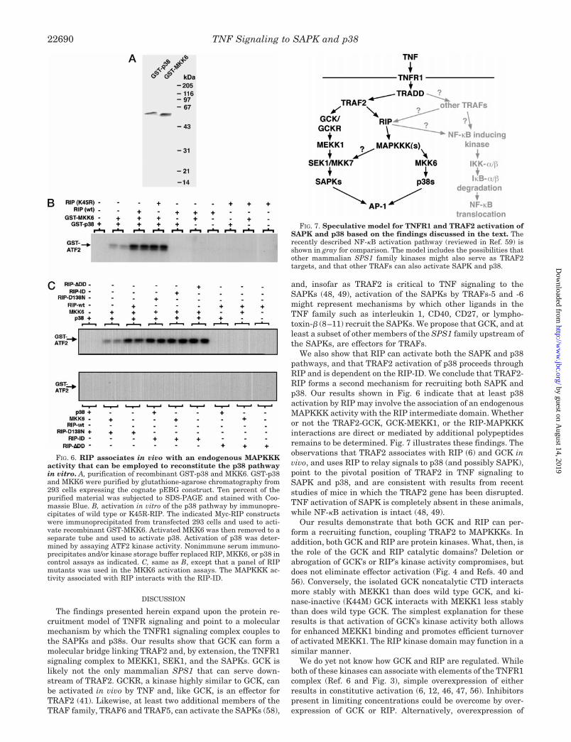
FIG. 7. Speculative model for TNFR1 and TRAF2 activation ofSAPK and p38 based on the findings discussed in the text. Therecently described NF-kB activation pathway (reviewed in Ref. 59) isshown in gray for comparison. The model includes the possibilities thatother mammalian SPS1 family kinases might also serve as TRAF2targets, and that other TRAFs can also activate SAPK and p38.
TNF Signaling to SAPK and p3822690
by guest on August 14, 2019
http://ww
w.jbc.org/
Dow
nloaded from

GCK or RIP could force homoligomerization and/or interactionswith TRAF2 that would not normally occur in resting cells. TheTRAF domains mediate the association between TRAF2 andboth GCK and RIP (Ref. 13 and Fig. 3D). However, the RINGdomains are necessary for SAPK pathway activation by TRAF2(46, 47). Thus, it is possible that upon the association of GCK(or RIP) with the TRAF2 TRAF domains, the TRAF2 RINGfinger domains could either promote activating conformationalchanges, or the dissociation of GCK or RIP inhibitors. Thesestructural changes would, in turn, permit interactions withdownstream effectors. As with MEKK1, TRAF2 binds the GCK-CTD more strongly than it binds full-length GCK. Again, acti-vation of GCK’s kinase activity may promote efficient turnoverof activated GCK, thereby reducing the amount of recoverablewild type GCKzTRAF2 complexes.
It is intriguing to compare the interaction domains on theGCK-CTD for TRAF2 and MEKK1. Fig. 2 indicates that the CTand PEST3 motifs of GCK-CTD are necessary for MEKK1binding while the CT and PEST1 motifs are needed for TRAF2binding. There is significant homology between the GCK CTextension and the extreme COOH terminus of GCKR (43).TRAF2 binding to PEST1 and CT on GCK, or to analogousdomains on GCKR, perhaps by promoting conformationalchanges, autophosphorylation, or other activating events,might render GCK (specifically PEST3) or GCKR accessible toMEKK1. Deletion of the Leu-rich domain of GCK-CTD restoresthe binding of MEKK1 lost upon deletion of CT. This maymerely reflect the fact that deletion of CT destabilizes theGCK-CTD, an effect reversed upon subsequent deletion of theLeu-rich region. Alternatively, the Leu-rich domain may in-hibit MEKK1 binding, perhaps by occluding PEST3; TRAF2binding to GCK might relieve this inhibition.
Inasmuch as GCK activation of the SAPK pathway is inhib-ited upon expression of the GCK-binding site of MEKK1 (Fig.2D), it is reasonable to propose that MEKK1 is a physiologictarget of GCK. However, we do not yet know how GCK mighttrigger MEKK1 activation, nor do we know how RIP mightregulate its associated MAPKKK activity. Efforts to elucidatethe mechanisms of stress-regulated MAPKKK regulation havebeen hampered by the fact that all mammalian MAPKKKsidentified thus far as activators of the SAPKs and p38s areconstitutively active upon overexpression (26, 33–39). In addi-tion, the endogenous forms of most of these MAPKKKs emergefrom cell extracts in a highly activated state (26, 33–39). GCKor RIP might simply phosphorylate and activate MAPKKKs.Indeed, we observe that GCK can phosphorylate MEKK1 (Fig.1C); however, such a mechanism fails to take into account theobservation that the kinase domains of GCK and RIP are notnecessary for SAPK or p38 activation. MEKK1 or the MAP-KKK associated with RIP might be regulated negatively, orthrough activating structural changes arising as a consequenceof binding GCK, as described above for the TRAF2-GCKinteraction.
The activation of AP-1 is critical to the inflammatory re-sponses elicited by TNF. Our results provide a framework for aplausible mechanism linking the TNFR1 signaling complex totwo ERK/MAPK pathways that regulate AP-1: the SAPKs andp38s. We propose that TNFR1 couples to the SAPKs and p38sthrough redundant and bifurcating pathways mediated inlarge part by TRAF2 (Fig. 7). TRAF2 associates with GCKwhich, in turn, associates with MEKK1. TRAF2 can also asso-ciate with RIP which activates the SAPKs and p38s, possiblythrough association with a distinct MAPKKK. Future studieswill focus on the identification and characterization of theRIP-associated MAPKKK, the mechanisms of GCK and RIP
recruitment and activation of MAPKKKs, and the mechanismsof GCK and RIP regulation.
Acknowledgments—We thank Felipe Pimentel-Muinos and BrianSeed for the RIP and TRAF2 constructs without which these studieswould not have been possible. We also thank Joseph Avruch andThomas Force for encouragement and comments on the manuscript.
REFERENCES
1. Bazzoni, F., and Beutler, B. (1996) The Tumor Necrosis Factor Ligand andReceptor Families (Flier, J.S., ed) pp. 1717–1725, Massachusetts MedicalSociety, Boston
2. Vandenabeele, P., Declercq, W., Beyaert, R., and Fiers, W. (1995) Trends CellBiol. 5, 392–399
3. Tartaglia, L. A., Ayres, T. M., Wong, G. H. W., and Goeddel, D. V. (1993) Cell74, 845–853
4. Hsu, H., Xiong, J., and Goeddel, D.V. (1995) Cell 81, 495–5045. Hsu, H., Shu, H.-B., Pan, M.-G., and Goeddel, D.V. (1996) Cell 84, 299–3086. Hsu, H., Huang, J., Shu, H.-B., Baichwal, V., and Goeddel, D. V. (1996)
Immunity 4, 387–3967. Rothe, M., Wong, S. C., Henzel, W. J., and Goeddel, D. V. (1994) Cell 78,
681–6928. Hu, H. M., O’Rourke, K., Boguski, M. S., and Dixit, V. M. (1994) J. Biol. Chem.
269, 30069–300729. Cheng, G., Cleary, A. M., Ye, Z.-S., Hong, D. I., Lederman, S., and Baltimore,
D. (1995) Science 267, 1494–149810. Cao, Z., Xiong, J., Takeuchi, M., Kurama, T., and Goeddel, D. V. (1996) Nature
1996, 383, 443–44611. Nakano, H., Oshima, H., Chung, W., Williams-Abbot, L., Ware, C. F., Yagita,
H., and Okumura, K. (1996) J. Biol. Chem. 271, 14661–1466412. Stanger, B. Z., Leder, P., Lee, T.-H., Kim, E., and Seed, B. (1995) Cell 81,
513–52313. Takeuchi, M., Rothe, M., and Goeddel, D. V. (1996) J. Biol. Chem. 271,
19935–1994214. Brenner, D. A., O’Hara, M., Angel, P., Chojkier, M., and Karin, M. (1989)
Nature 337, 661–66315. Verma, I. M., Stevenson, J. K., Schwartz, E. M., Van Antwerp, D., and
Miyamoto, S. (1995) Genes Dev. 9, 2723–273516. Karin, M., Liu, Z.-g., and Zandi, E. (1997) Curr. Opin. Cell. Biol. 9, 240–24617. Read, M. A., Whitley, M. Z., Gupta, S., Pierce, J. W., Best, J., Davis, R. J., and
Collins, T. (1997) J. Biol. Chem. 272, 2753–276118. Kyriakis, J. M., Banerjee, P., Nikolakaki, E., Dai, T., Rubie, E. A., Ahmad,
M. F., Avruch, J., and Woodgett, J. R. (1994) Nature 369, 156–16019. Han, J., Lee, J.-D., Bibbs, L., and Ulevitch, R. J. (1994) Science 265, 808–81120. Raingeaud, J., Gupta, S., Rogers, J. S., Dickens, M., Han, J., Ulevitch, R. J.,
and Davis, R. J. (1995) J. Biol. Chem. 270, 7420–742621. Derijard, B., Hibi, M., Wu, I.-H., Barrett, T., Su, B., Deng, T., Karin, M., and
Davis, R. J. (1994) Cell 76, 1025–103722. Gupta, S., Campbell, D., Derijard, B., and Davis, R.J. (1995) Science 267,
389–39323. Whitmarsh, A. J., Shore, P., Sharrocks, A. D., and Davis, R. J. (1995) Science
269, 403–40724. Treisman, R. (1994) Curr. Opin. Genet. Dev. 4, 96–10125. Han, J., Jiang, Y., Li, Z., Kravchenko, V. V., and Ulevitch, R. J. (1997) Nature
386, 563–56626. Kyriakis, J. M., and Avruch, J. (1996) J. Biol. Chem. 271, 24313–2431627. Sanchez, I., Hughes, R. T., Mayer, B. J., Yee, K., Woodgett, J. R., Avruch, J.,
Kyriakis, J. M., and Zon, L. I. (1994) Nature 372, 794–79828. Derijard, B., Raingeaud, J., Barrett, T., Wu, L.-H., Han, J., Ulevitch, R. J., and
Davis, R. J. (1995) Science 267, 682–68529. Tournier, C., Whitmarsh, A. J., Cavanagh, J., Barrett, T., and Davis, R. J.
(1997) Proc. Natl. Acad. Sci. U. S. A. 94, 7337–734230. Holland, P. M., Suzanne, M., Campbell, J. S., Noselli, S., and Cooper, J. A.
(1997) J. Biol. Chem. 272, 24994–2499831. Raingeaud, J., Whitmarsh, A. J., Barett, T., Derijard, B., and Davis, R. J.
(1996) Mol. Cell. Biol. 16, 1247–125532. Yan, M., Dai, T., Deak, J. C., Kyriakis, J. M., Zon, L. I., Woodgett, J. R., and
Templeton, D. J. (1994) Nature 372, 798–80033. Xu, S., Robbins, D. J., Christerson, L. B., English, J. M., Vanderbilt, C., and
Cobb, M. H. (1996) Proc. Natl. Acad. Sci. U. S. A. 93, 5291–529534. Gerwins, P., Blank, J. L., and Johnson, G. L. (1997) J. Biol. Chem. 272,
8288–829535. Takekawa, M., Posas, F., and Saito, H. (1997) EMBO J. 16, 4973–498236. Ichijo, H., Nishida, E., Irie, K., ten Dijke, P., Saitoh, M., Moriguchi, T., Takagi,
M., Matsumoto, K., Miyazono, K., and Gotoh, Y. (1997) Science 275, 90–9437. Yamaguchi, K., Shirakabi, K., Shibuya, H., Irie, K., Oishi, I., Ueno, N.,
Taniguchi, T., Nishida, E., and Matsumoto, K. (1995) Science 270,2008–2011
38. Salmeron, A., Ahmad, T. B., Carlile, G. W., Pappin, D., Narsimhan, R. P., andLey, S. C. (1996) EMBO J. 15, 817–826
39. Rana, A., Gallo, K., Godowski, P., Hirai, S., Ohno, S., Zon, L., Kyriakis, J. M.,and Avruch, J. (1996) J. Biol. Chem. 271, 19025–19028
40. Pombo, C. M., Kehrl, J. H., Sanchez, I., Katz, P., Avruch, J., Zon, L. I.,Woodgett, J. R., Force, T., and Kyriakis, J. M. (1995) Nature 377, 750–754
41. Kiefer, F., Tibbles, L. A., Anafi, M., Janssen, A., Zanke, B. W., Lassam, N.,Pawson, T., Woodgett, J. R., and Iscove, N. R. (1996) EMBO J. 15,7013–7025
42. Su, Y.-C., Han, J., Xu, S., Cobb, M., and Skolnik, E.Y. (1997) EMBO J. 16,1279–1290
43. Shi, C.-S., and Kehrl, J. H. (1997) J. Biol. Chem. 272, 32102–3210744. Tung, R. M., and Blenis, J. (1997) Oncogene 14, 653–65945. Diener, K., Wang, X. S., Chen, C., Meyer, C. F., Keesler, G., Zukowski, M., Tan,
TNF Signaling to SAPK and p38 22691
by guest on August 14, 2019
http://ww
w.jbc.org/
Dow
nloaded from

T.-H., and Yao, Z. (1997) Proc. Natl. Acad. Sci. U. S. A. 94, 9687–969246. Liu, Z.-g., Hsu, H., Goeddel, D. V., and Karin, M. (1996) Cell 87, 565–57647. Natoli, G., Costanzo, A., Ianni, A., Templeton, D. J., Woodgett, J. R., Balsano,
C., and Levrero, M. (1997) Science 275, 200–20348. Yeh, W.-C., Shahinian, A., Speiser, D., Kraunus, J., Billia, F., Wakeham, A.,
de la Pompa, J. L., Ferrick, D., Hum, B., Iscove, N., Ohashi, P., Rothe, M.,Goeddel, D. V., and Mak, T. W. (1997) Immunity 7, 715–725
49. Lee, S. Y., Reichlin, A., Santana, A., Sokol, K. A., Nussenzweig, M. C., andChoi, Y. (1997) Immunity 7, 703–713
50. Sambrook, J., Fritsch, E. F., Maniatis, T. (1989) Molecular Cloning: ALaboratory Manual, 2nd Ed., Cold Spring Harbor Laboratory Press, ColdSpring Harbor, NY
51. Ting, A. T., Pimentel-Muinos, F.-X., and Seed, B. (1996) EMBO J. 15,
6189–619552. Katz, P., Whalen, G., and Kehrl, J. H. (1994) J. Biol. Chem. 269, 16802–1680953. Bagrodia, S., Derijard, B., Davis, R. J., and Cerione, R. A. (1995) J. Biol. Chem.
270, 27995–2799854. Herskowitz, I. (1995) Cell 80, 187–19755. Lange-Carter, C. A., Pleiman, C. M., Gardner, A., Blumer, K. J., and Johnson,
G. L. (1993) Science 260, 315–31956. Fukunaga, R., and Hunter, T. (1997) EMBO J. 16, 1921–193357. Waskiewicz, A. J., Flynn, A., Proud, C. G., and Cooper, J. A. (1997) EMBO J.
16, 1909–192058. Song, H. Y., Regnier, C. H., Kirschning, C. J., Goeddel, D. V., and Rothe, M.
(1997) Proc. Natl. Acad. Sci. U. S. A. 94, 9792–979659. Stancovski, I., and Baltimore, D. (1997) Cell 91, 299–302
TNF Signaling to SAPK and p3822692
by guest on August 14, 2019
http://ww
w.jbc.org/
Dow
nloaded from

Takashi Yuasa, Shigeo Ohno, John H. Kehrl and John M. KyriakisUPSTREAM OF MKK6 AND p38
WITH A MITOGEN-ACTIVATED PROTEIN KINASE KINASE KINASE 1 AND SAPK WHILE RECEPTOR INTERACTING PROTEIN ASSOCIATES
TRAF2 TO MITOGEN-ACTIVATED PROTEIN KINASE/ERK KINASE KINASE -terminal Kinase (JNK) and p38: GERMINAL CENTER KINASE COUPLES2
Tumor Necrosis Factor Signaling to Stress-activated Protein Kinase (SAPK)/Jun NH
doi: 10.1074/jbc.273.35.226811998, 273:22681-22692.J. Biol. Chem.
http://www.jbc.org/content/273/35/22681Access the most updated version of this article at
Alerts:
When a correction for this article is posted•
When this article is cited•
to choose from all of JBC's e-mail alertsClick here
http://www.jbc.org/content/273/35/22681.full.html#ref-list-1
This article cites 57 references, 31 of which can be accessed free at
by guest on August 14, 2019
http://ww
w.jbc.org/
Dow
nloaded from