the isolated chicken eye test - wur
TRANSCRIPT

1
THE ISOLATED CHICKEN EYE TEST
TO REPLACE THE DRAIZE EYE TEST IN RABBITS
From development to implementation: “The long and winding road”.
Menk K. Prinsen

2
THESIS COMMITTEE
Promotors
Prof. Dr R.A. Woutersen
Professor Translational Toxicology
Wageningen University/TNO, Zeist
Prof. Dr C.F.M.Hendriksen
Professor Alternatives to Animal Testing
Utrecht University/Institute for Translational Vaccinology, Bilthoven
Co-promotor
Prof. Dr C.A.M. Krul
Professor Innovative Testing in Life Sciences & Chemistry
Utrecht University of Applied Sciences/TNO, Zeist
Other members
Dr A. van der Lelij, Utrechts Medisch Centrum Utrecht
Prof. Dr R.F. Witkamp, Wageningen University
Prof. Dr B.J. Blaauboer, Utrecht University
Prof. Dr V. Rogiers, Free University Brussel, Belgium
This research was conducted under the auspices of the Graduate School VLAG
(Advanced studies in Food Technology, Agrobiotechnology, Nutrition and Health Sciences).

3
Thesissubmitted in fulfilment of the requirements for the degree of doctor
at Wageningen Universityby the authority of the Rector Magnificus,
Prof. Dr M.J. Kropff,in the presence of the Thesis Committee appointed by the Academic Board
to be defended in publicon Friday 3 October 2014
at 11 a.m. in the Aula.
THE ISOLATED CHICKEN EYE TEST
TO REPLACE THE DRAIZE EYE TEST IN RABBITS
From development to implementation: “The long and winding road”.
Menk K. Prinsen

4
Menk K. PrinsenThe Isolated Chicken Eye test to replace the Draize test in rabbits.From development to implementation: “The long and winding road”, 184 pages.
PhD thesis, Wageningen University, Wageningen, NL (2014)With references, with summaries in Dutch and EnglishISBN 978-94-6257-003-0

5
Voor Cin

6
7
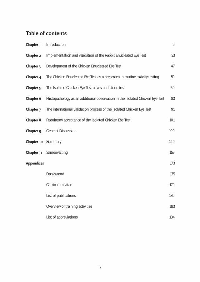
Table of contents
Chapter 1 Introduction 9
Chapter 2 Implementation and validation of the Rabbit Enucleated Eye Test 33
Chapter 3 Development of the Chicken Enucleated Eye Test 47
Chapter 4 The Chicken Enucleated Eye Test as a prescreen in routine toxicity testing 59
Chapter 5 The Isolated Chicken Eye Test as a stand-alone test 69
Chapter 6 Histopathology as an additional observation in the Isolated Chicken Eye Test 83
Chapter 7 The international validation process of the Isolated Chicken Eye Test 91
Chapter 8 Regulatory acceptance of the Isolated Chicken Eye Test 101
Chapter 9 General Discussion 109
Chapter 10 Summary 149
Chapter 11 Samenvatting 159
Appendices 173
Dankwoord 175
Curriculum vitae 179
List of publications 180
Overview of training activities 183
List of abbreviations 184

8

Introduction
The Draize Eye Irritation Test and alternatives.

10

11
Background
Before industrialization, eye defects were mainly caused by physical trauma or by diseases caused by malnutrition, bacterial infection or parasites. In the twentieth century, when (chemical) industrialization strongly developed, it became apparentthat conditions at the workplace could have distinct adverse effects on health andsafety of employees. Acute and long-term exposure to a variety of industrial chemicalswere responsible for a range of diseases, varying from relatively mild, non-life threate-ning phenomena, such as dermatitis, to incurable, lethal conditions such as cancer.After World War II, the chemical industry rapidly increased and workers became orga-nized and more concerned with the potential risks they could encounter in the work-place. Consequently, the need for identifying health hazards and worker’s protectionbecame an important issue in most industrial countries. Moreover, people could af-ford more luxury products and the household and personal care industry becamemore and more innovative using new technologies and (chemical) ingredients. There-fore, an even larger population of people needed to be safeguarded from potential hazardous substances. To establish the potential risk of exposure of the eyes to compounds, the Food andDrug Administration of the United States (US-FDA) adopted the Draize eye irritationtest using rabbits already in 1961. At first sight, this test is simple and straightfor-ward and provides a useful tool for regulators. However, the controversial character of this type of animal testing became known to the general public – on 15 April 1980, Henry Spira, a Belgian-American advocate, member and founder of theAnimal Rights International group bought a full-page advertisement (Figure 1) in the New YorkTimes, with the header: “How many rabbits doesRevlon blind for beauty's sake?” – and the need todevelop alternative non-animal tests became apparent. Within a year after Spira’s advertisment,Revlon had donated $750,000 to a fund to investi-gate alternatives to animal testing, followed bysubstantial donations from Avon, Bristol Meyers,Estée Lauder, Max Factor, Chanel, and Mary KayCosmetics. These donations led to the creation of the Center for Alternatives to Animal Testing (Wikepedia; Henry Spira).The attempts to validate alternative tests for eye irritation in the early nineteen-eightieswere considered to be relatively simple by comparing in vitro and in vivo irritationindex scores. What was expected to be a process of several years, however, turned outto be a decades spanning process still not fully completed.
Figure 1. Spira’s advertisement
(www.onegreenplanet.org).
1

12
For a large part, this can be attributed to the nature of the in vivo test in rabbits, whichis more complicated and compromised than originally believed. This thesis describes the development, performance, validation and acceptance of oneof the first alternatives, namely the in vitro isolated eye test.
Introduction
The eyeOur eyes are one of the most important sense organs we possess in order to keep incontact with our environment. We have only two eyes and as such we are vulnerable toaccidental damage, caused for instance by mechanical trauma or exposure to foreignmaterials such as chemicals. The exposure to chemicals can be intentional (ophthal-mologic formulations and contact lens fluids) or unintentional, at the workplace or athome, using household and/or personal care products. The natural defence mechanisms against possible damage to our eyes are obvious:protection by i) their embedded position in the eye-socket and protection by the eye-lids (blinking, closing of the eyes); ii) lachrymation (production of tears by innervationof the sensory nerves of the cornea) in order to dilute/remove foreign materials; iii) the tear film (protection against bacteria and drying out of the corneal surface),and iv) reflexes (turning away the head, protection of the eyes/head with our hands).
This thesis deals mainly with a critical part of the eye, namely the cornea, the eye's outermost layer and gateway to the perception of light (Figure 2), which is the maintarget tissue for eye irritation caused by chemical exposure.
Figure 2. The cornea (www.visionfortomorrow.org).
1

13
The composition or histology of the cornea and its functions are described in manyhandbooks and in general is as follows (NEI, 2011): “Although the cornea is clear and seems to lack substance, it is actually a highly orga-nized group of cells and proteins. Unlike most tissues in the body, the cornea containsno blood vessels to nourish or protect it against infection. Instead, the cornea receivesits nourishment from the tears and aqueous humor that fills the chamber behind it.The cornea must remain transparent to refract light properly, and the presence of eventhe tiniest blood vessels can interfere with this process. To see well, all layers of thecornea must be free of any cloudy or opaque areas.The corneal tissue is arranged in five basic layers (Figure 2), each having an importantfunction. These five layers are:
EpitheliumThe epithelium is the cornea's outermost region, comprising about 10 percent of the tissue's thickness. The epithelium functions primarily to: (1) block the passage of foreignmaterial, such as dust, water, and bacteria, into the eye and other layers of the corneaand (2) provide a smooth surface that absorbs oxygen and cell nutrients from tears anddistributes these nutrients to the rest of the cornea. The epithelium is filled with thou-sands of tiny nerve endings that make the cornea extremely sensitive to pain when rub-bed or scratched. Cold receptors are abundant in the cornea, but heat and touchreceptors are lacking. The part of the epithelium that serves as the foundation on whichthe epithelial cells anchor and organize themselves is called the basement membrane.
Bowman's LayerLying directly below the basement membrane of the epithelium is a transparent sheetof tissue known as Bowman's layer. It is composed of strong layered protein fibers called collagen. Once injured, Bowman's layer can form a scar as it heals. If thesescars are large and centrally located, some vision loss can occur.
StromaBeneath Bowman's layer is the stroma, which comprises about 90 percent of the cornea'sthickness. It consists primarily of water (78 percent) and collagen (16 percent), anddoes not contain any blood vessels. Collagen gives the cornea its strength, elasticity,and form. The collagen's unique shape, arrangement, and spacing are essential inproducing the cornea's light-conducting transparency.
Descemet's MembraneBeneath the stroma is Descemet's membrane, a thin but strong sheet of tissue thatserves as a protective barrier against infection and injuries. Descemet's membrane iscomposed of collagen fibers (different from those of the stroma) and is made by theendothelial cells that lie below it. Descemet's membrane can regenerate readily after injury.
1
14
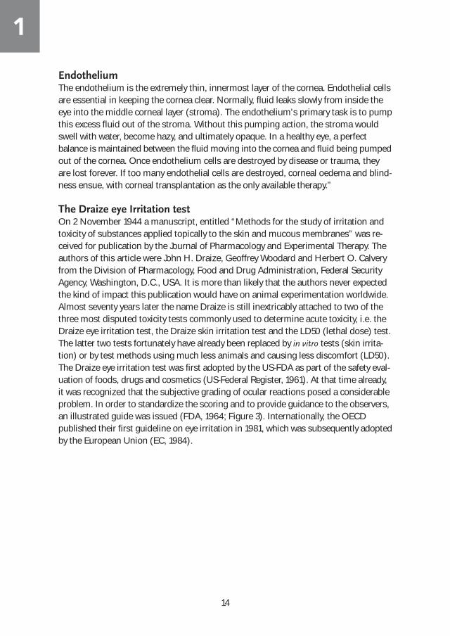
EndotheliumThe endothelium is the extremely thin, innermost layer of the cornea. Endothelial cellsare essential in keeping the cornea clear. Normally, fluid leaks slowly from inside theeye into the middle corneal layer (stroma). The endothelium's primary task is to pumpthis excess fluid out of the stroma. Without this pumping action, the stroma wouldswell with water, become hazy, and ultimately opaque. In a healthy eye, a perfect balance is maintained between the fluid moving into the cornea and fluid being pumpedout of the cornea. Once endothelium cells are destroyed by disease or trauma, theyare lost forever. If too many endothelial cells are destroyed, corneal oedema and blind-ness ensue, with corneal transplantation as the only available therapy.”
The Draize eye Irritation testOn 2 November 1944 a manuscript, entitled “Methods for the study of irritation andtoxicity of substances applied topically to the skin and mucous membranes” was re-ceived for publication by the Journal of Pharmacology and Experimental Therapy. Theauthors of this article were John H. Draize, Geoffrey Woodard and Herbert O. Calveryfrom the Division of Pharmacology, Food and Drug Administration, Federal SecurityAgency, Washington, D.C., USA. It is more than likely that the authors never expectedthe kind of impact this publication would have on animal experimentation worldwide.Almost seventy years later the name Draize is still inextricably attached to two of thethree most disputed toxicity tests commonly used to determine acute toxicity, i.e. theDraize eye irritation test, the Draize skin irritation test and the LD50 (lethal dose) test.The latter two tests fortunately have already been replaced by in vitro tests (skin irrita-tion) or by test methods using much less animals and causing less discomfort (LD50). The Draize eye irritation test was first adopted by the US-FDA as part of the safety eval-uation of foods, drugs and cosmetics (US-Federal Register, 1961). At that time already,it was recognized that the subjective grading of ocular reactions posed a considerableproblem. In order to standardize the scoring and to provide guidance to the observers,an illustrated guide was issued (FDA, 1964; Figure 3). Internationally, the OECD published their first guideline on eye irritation in 1981, which was subsequently adoptedby the European Union (EC, 1984).
1

15
Since then several revisions of the guideline have followed, mostly not affecting the actual exposure procedure, but providing guidance for refinement and reduction of animal use and discomfort (Table 1). Examples are the exemption of testing skin corrosives and substances with pH lower than 2.0 or higher than 11.5, the use of well-validated alternatives as a screen for severe irritancy, and a tiered approach of testing(i.e. starting with one animal and continue only if non-severe irritancy is observed).
Figure 3. FDA guidance on scoring of ocular lesions; Plate 2 (FDA,1964).
1

16
The design of the eye irritation test is actually quite simple and straightforward: a rabbitis placed on a worktable and restrained either manually or in a fixation-box (Figure 4).Next, the lower eye-lid is pulled out and the test substance is instilled in the conjunctivalcul de sac formed; the upper and lower eye lids are then closed and subsequently heldtogether for at least one second before releasing the animal. The other eye remainsuntreated and serves as a control.
The animal is returned to its cageand is free to remove the material.The control and test eyes are examined (without optical aid) atapproximately one hour, and atapproximately 24, 48, and 72hours after treatment. Ocular reactions of the test eye are judgedusing a scoring scale (Table 2).Residual eye effects are recordedat regular intervals, if necessaryup to about 3 weeks after treat-ment, in order to allow the evalu-ation of the reversibility orirreversibility of the effects elici-ted. Liquids are tested in a volume of 0.1 mL and solids (ground to a fine powder) in an amount of 0.1 g or a volume of 0.1 mL. In general, 0.1 mL is the amount the conjunctival cul de sac can hold when the lower eye-lid is pulled out.
Table 1
OECD TG 405 Procedure Guidance on interpretation of results Ethical considerations
1981 - 0.1 mL or 0.1 g substance - wash out only after 24 hr
- Extrapolation of the results of eye irritation studies in animals to man is valid only to a limited degree. The albino rabbit is more sensitive than man to ocular irritants or corrosives in most cases.
- Similar results in tests on other animal species can give more weight to extrapolation from animal studies to man.
- Care should be taken in the interpretation of data to exclude irritation resulting from secondary infection.
Local anaesthetics proposed - Three instead of six rabbits No testing of: - Strongly acidic or alkaline
substances - Corrosive or severe skin irritants
1987 - 0.1 mL or 0.1 g substance - wash out only after 24 hr
Identical to 1981 Guidance Addition of: - Animals showing severe and
enduring signs of distress and pain may need to be humanely killed.
Addition of: - severe eye irritants identified in
well-validated alternative studies
2002 - 0.1 mL or 0.1 g substance - wash out after 1 hr
Similar to 1981 and 1987 Guidance Addition of: - End points for humane sacrifice - Tiered testing
Addition of: - Weight-of-the-evidence analysis
on the existing relevant data - Conduct of validated and
accepted in vitro tests - One rabbit first
2012 - 0.1 mL or 0.1 g substance - wash out after 1 hr
Similar to 1981, 1987 and 2002 Guidance Addition of: - Extensive directions for the use
of topical anaesthetics and systemic analgesics
Addition of: - ICE test (OECD 438) - BCOP test (OECD 437)
Table 1. OECD test guideline no. 405 and its revisions (procedures, interpretation results, ethics and 3 R’s).
Figure 4. Instillation of the test substance in the Draize eye
test (TNO)
1
17
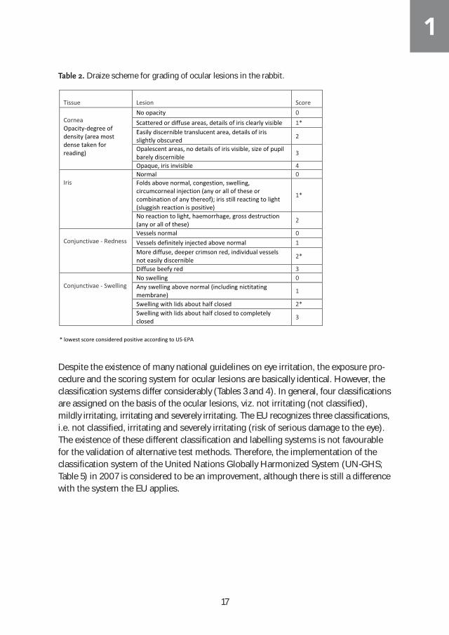
Despite the existence of many national guidelines on eye irritation, the exposure pro-cedure and the scoring system for ocular lesions are basically identical. However, theclassification systems differ considerably (Tables 3 and 4). In general, four classificationsare assigned on the basis of the ocular lesions, viz. not irritating (not classified),mildly irritating, irritating and severely irritating. The EU recognizes three classifications,i.e. not classified, irritating and severely irritating (risk of serious damage to the eye).The existence of these different classification and labelling systems is not favourablefor the validation of alternative test methods. Therefore, the implementation of theclassification system of the United Nations Globally Harmonized System (UN-GHS;Table 5) in 2007 is considered to be an improvement, although there is still a differencewith the system the EU applies.
Table 2. Draize scheme for grading of ocular lesions in the rabbit.
Tissue
Lesion
Score
Cornea Opacity-degree of density (area most dense taken for reading)
No opacity 0 Scattered or diffuse areas, details of iris clearly visible 1* Easily discernible translucent area, details of iris slightly obscured 2
Opalescent areas, no details of iris visible, size of pupil barely discernible 3
Opaque, iris invisible 4 Iris
Normal 0 Folds above normal, congestion, swelling, circumcorneal injection (any or all of these or combination of any thereof); iris still reacting to light (sluggish reaction is positive)
1*
No reaction to light, haemorrhage, gross destruction (any or all of these) 2
Conjunctivae - Redness
Vessels normal 0 Vessels definitely injected above normal 1 More diffuse, deeper crimson red, individual vessels not easily discernible 2*
Diffuse beefy red 3 Conjunctivae - Swelling
No swelling 0 Any swelling above normal (including nictitating membrane) 1
Swelling with lids about half closed 2* Swelling with lids about half closed to completely closed 3
* lowest score considered positive according to US-EPA
Table 2. Draize scheme for grading of ocular lesions in the rabbit.
1

18
The UN-GHS system subdivides the irritating category (Category 2) into mild irritant(Category 2B) and irritant (Category 2A), whereas the EU only uses the category irritant(Category 2; EC, 2008).
Table 3. European Union (19931) classification system for eye irritation/corrosion. Eye effects
R36 (Irritating to eyes)
R41 (Risk of serious damage
to eyes)4
3 animals2 6 animals3 3 animals1 6 animals2
Corneal opacity 2.0, but <3.0 2.0, but <3.0 3.0 3.0
Iris lesion 1.0, but <2.0 1.0, but 1.5 2.0 > 1.5
Conjuntiva redness 2.5 2.5
Conjunctiva chemosis
2.0 2.0
1 Official Journal of the European Communities, L 110 A, Volume 36, 4 May 1993
2 The classification is assigned if the mean tissue effect (averaged over the 24h, 48h and 72 h time points) exceeds the threshold value in
at least two of the three animals. 3 The classification is assigned if the mean tissue effect (averaged over the three time points and over the
six animals) exceeds the threshold value. 4 R41 is also assigned if, in at least one animal, one of the eye effects has not reversed at the end of the
observation period. Table 4. US-EPA (19981) classification system for eye irritation. Toxicity categories
Category I Category II Category III Category IV
Eye effects
Corrosive (irreversible destruction of ocular tissue) or corneal involvement or irritation persisting for more than 21 days
Corneal involvement or irritation clearing in 8-21 days
Corneal involvement or irritation clearing in 7 days or less
Minimal effects clearing in less than 24 hours
1 Health Effects Test Guideline OPPTS870.1000, EPA 712-C-98-189, August 1998
Table 5. GHS (20071) classification system for eye irritation/corrosion.
Eye effects Category 2A2 Category 13
Corneal opacity 1.0 3.0
Iris lesion 1.0 > 1.5
Conjunctiva redness 2.0
Conjunctiva chemosis 2.0
1 Globally Harmonised System of Classification and Labelling of Chemicals (UN-GHS). UN, New York and Geneva, 2007
2 All effects have to be reversible within 21 days of treatment. Subcategory of 2B: mildly irritating to the eyes, i.e. eye effects reversible within 7 days of treatment. 3 Category 1 is also applicable if, in at least one animal, an eye effect has not reversed, or is not expected to reverse, within 21 days of treatment.
Table 3. European Union (19931) classification system for eye irritation/corrosion.
Table 4. US-EPA (19981) classification system for eye irritation.
Table 5. UN-GHS (20071) classification system for eye irritation/corrosion.
1

19
Awareness of alternatives for animal testingThe publication of Russell and Burch in 1959 entitled: “The principles of humane experi-mental technique” stood at the basis of most initiatives relating to the use and develop-ment of alternatives for animal experiments. In their publication they postulated thefamous and often cited three R’s: Reduction, Refinement and Replacement of animalexperiments. Nowadays, the 3 R’s have become a mantra for scientists and regulatorsin research areas involving animal experimentation. The initiatives concerning theDraize eye test mainly involved reduction of the number of animals from six to threeper test and replacement by the implementation of non-animal alternatives. Certainaspects of the Draize eye test causing considerable pain and discomfort to the animalwere dealt with only at a much later stage, i.e. reduction of the time for a wash-out ofthe test substance from 24 hours to 1 hour after instillation in 2002, and the use ofsystemic pain relief and topical sedation in 2012 updates of the OECD guideline 405(Table 1).In the early nineteen-eighties, some toxicologists within the TNO-CIVO Toxicology andNutrition Institute in Zeist, the Netherlands, had growing concerns about the use ofexperimental animals in toxicity testing. One of them, Drs. H.B.W.M. Koëter, exploredthe possibilities of introducing alternative test methods for standard acute toxicitytests, such as the Draize eye and skin irritation tests. At that time the Netherlands Society of Toxicology (NVT) started a working group named “Kritische Evaluatie Toxiciteitstesten” (KET; Critical Evaluation of Toxicity Testing) of which Drs. Koëter wasa member. In Europe, the European Research Group for Alternatives in Toxicity Testing(ERGATT) was founded to stimulate innovative toxicological research and to act as acounterpart to the John Hopkins Center of Alternatives to Animal Testing in the USAwhich was founded in 1981.
Several alternatives had already been published varying from simple cell toxicity (cyto-toxicity tests), through sperm motility, to damage to the chorioallantois membrane ofhen’s eggs (HET-CAM; Figure 5).
Figure 5. Chorioallantois membrane of the
hen’s egg (www.eurochemricerche.it).
1

20
In 1982, TNO-CIVO was invited by the Commission of the European Community towrite a report on the reduction of numbers of animals in toxicity testing (Koëter andvan Vliet, 1983).Part of the assignment was to make an inventory of alternative testmethods used in toxicity testing, and it became apparent that numerous in vitro testshad already been developed. Several of these alternative test methods appeared verypromising, but standardization and validation had almost never received sufficient attention, because they were developed within a university or company setting, andapplied in most cases on a limited scale and not for regulatory purposes. On the basis of this report, TNO decided to include alternative approaches in the Institute’s toxicological research program, which was an important decision at thattime. For eye irritation, the policy was not to develop yet another test method but toselect one of the most promising existing alternative methods and to focus on furtherdevelopment, standardization and validation in order to develop a method that wouldbe acceptable for regulatory purposes. In addition, recommendations for a tiered approach to eye irritation testing were made, viz. testing skin irritation first, and startingthe eye irritation test with one rabbit. Because the cornea is such a highly relevant target tissue in eye irritation, it was takenas the basic principle for the development of a relevant and practical in vitro alternativeto the animal test that had been in use as the sole test for the screening of eye irritationworldwide since the early forties of the twentieth century.
Isolated Eye Test method (Rabbit)In 1981, A.B.G. Burton from Unilever published a method using isolated rabbit eyesfor the in vitro assessment of severe eye irritants (Burton, 1981). Previously, he haddiscovered that the measurement of corneal thickness (swelling) by slit-lamp exami-nation provided an objective assessment of eye irritation in the in vivo rabbit eye irrita-tion test (Burton, 1972). He had examined 100 different cosmetic formulations in 600rabbits and found not only a close relationship between the total corneal Draize scoreand the recorded corneal swelling, but also a relationship between corneal swellingand the conjunctival effects scored subjectively. Around that time another article onthe usefulness of slit-lamp examination in the rabbit eye irritation test, including cornealthickness, was published (McDonald, 1973). Between 1972 and 1981, Burton did not publish further on this subject, but it is assumedthat he played with the idea of replacing the live rabbit by isolated rabbit eyes only. Inhis 1981 publication no further considerations for using isolated eyes were given, but a possible clue may be found in the literature reference he used for the design of thesuperfusion apparatus (used for maintaining the isolated eyes in good condition),which he had modified from the one described by Mishima and Kudo in 1967. Remarkably, Burton had already referred to publications by Mishima in his 1972 article,and surely have thought about the possibility of using isolated rabbit eyes in a super-fusion apparatus at that time. It remains unclear why he did not pursue the use of isolated rabbit eyes sooner.
1

21
The idea of using isolated rabbit eyes was very appealing from a scientific point ofview. After all one uses an ex vivo eye for an eye in vivo and, moreover, the parametersmeasured (corneal swelling, corneal opacity and epithelial cytoxicity by fluoresceindye) are directly comparable to the parameters measured in vivo (both in rabbit and inman). Therefore, Koëter proposed to introduce an in vitro eye irritation test (with isolat-ed rabbit eyes) as a possible contribution to the reduction of experimental animal use
(Koëter and Prinsen, 1984). The project was approvedand partly funded by the “Dutch Society for the Protectionof Animals” and the foundation “Beauty without Cruelty”.Equipment for the Rabbit Enucleated Eye Test (REET;initial name for the Isolated Rabbit Eye Test) was purchased (slit-lamp microscope; Figure 6) or built bythe Technical Service Department of TNO-CIVO (super-fusion apparatus and eye-holders; Figure 7).
Figure 6. Haag-Streit slit lamp microscope (www.medwow.com).
Figure 7. Schematic presentation of the superfusion apparatus and eye holder (TNO).
1

22
The test method was evaluated by investigating the effects of several substances fromthe Burton publication (1981). The test method was further validated with 34 substancesthat had been investigated in the in vivo eye irritation test in rabbits as part of the standard toxicity testing at TNO-CIVO in 1983-1984 (Koëter and Prinsen, 1985).During the same period, several other investigators explored the use of isolated rabbiteyes as an alternative for the in vivo test. Unilever continued their work (York, 1982),while Shell Research Centre, Sittingbourne, UK, started their research program (Price,1985). The Institute for Hygiene and Epidemiology (IHE), Brussels, Belgium startedtheir investigations in 1988 (Jacobs, 1988 and 1990). The research and publications on isolated rabbit eyes resulted in the participation ofthis test method in the first EC Collaborative Study on the Evaluation of AlternativeMethods to the Eye Irritation Test (EC, 1991). In this study, five in vitro cell toxicitytests, the REET and the HET-CAM were selected to undergo validation by testing 21chemicals of different classes in at least 3 different laboratories. Some of the conclusionswere: i) The Isolated Rabbit Eye test did not misclassify many non-irritants and alsohad the capability to discriminate between moderate and severe effects, although irritating (R36) chemicals were underrepresented. ii) The REET produced resultswhich were consistent across all three laboratories and generally correctly predictedthe in vivo grade. The protocol and the method for calculating final irritancy grades (in validation studies later on called “Prediction Model”) needed harmonization before a wider interlaboratory study could be conducted. iii) The test is nearest to thehuman situation and has the advantage that all types of chemicals can be investigatedwithout the need for testing dilutions, therefore it is easier to interpret than the otherassays in this trial. The trial was, however, considered to be too limited to make firmconclusions and it was recommended to perform further interlaboratory trials with alarger number of laboratories and chemicals and according to the principles of GLP.An important remark in the EC report was the fact that toxicological profiles of thechemicals investigated were prepared by collecting and critically evaluating the literaturedata available, because it was not possible to repeat in vivo eye determinations for animal protection considerations. The availability, evaluation and appraisal of in vivoeye irritation data and the test method itself constitute the main cause of the exceptionallylong, and not yet completed, acceptance of alternative methods to the Draize eye test.Until recently, the in vivo data were taken as the “Golden Standard”, which in practicemeant that the in vitro data had to almost exactly match the in vivo result.
Isolated Eye Test method (Chicken)During the introduction and validation of the REET, it was recognized that the use oflaboratory rabbit eyes - although available from rabbits used for standard eye and skinirritation tests in vivo - was not ideal, especially for laboratories not using rabbits fortheir experiments. Moreover, the alternative test would still be associated with the useof the laboratory rabbit and eventually, if an alternative would replace the in vivo skinand/or eye irritation test, the rabbit as a source for eyes would dry up. The suggestion
1

23
had been made that an alternative approach could be to use eyes obtained from rabbit-abattoirs, but use of bovine or chicken eyes was also suggested (Koëter and Prinsen,1987). Therefore, in 1990, a proposal to examine the suitability of eyes of slaughter-house animals as an alternative for rabbit eyes in the enucleated eye test was submittedto the “Platform Alternatives for Animal Experimentation” (PAD; Platform Alternatievenvoor Dierproeven), which was granted after being reviewed by the NWO (Dutch organization for scientific research). During the period October - December 1990, pigand cow slaughterhouses (Hilversum) and chicken slaughterhouses (Breukelen andNijkerkerveen), all within a 1-hour drive from the test facilities of TNO, were visited tomake preliminary investigations concerning the practical aspects of obtaining eyes. InChapter 3 the selection of the most suitable eye donor and the development of themethod with the selected eye donor species is described. An important aspect was thevalidation of the test method, i.e. to put it simply: comparing the in vitro data withdata from the in vivo test. Most alternatives are validated with in vivo data obtainedfrom literature, a process with many drawbacks, which will be discussed in more detailin Chapters 7 and 8 of this thesis. The use and suitability of eyes of slaughterhouseanimals was first established by testing the same reference chemicals (Prinsen andKoëter, 1993) that had been tested in the EU Collaborative Study on the Evaluation ofAlternative Methods to the Eye Irritation Test (EC, 1991). Although the in vivo datawere obtained from literature this study was considered quite valuable because the invitro data obtained with the slaughter eyes could be directly compared with the in vitrodata obtained in the REET. Ideally, the in vitro test should be performed in parallel withthe in vivo test, hence enabling a more direct comparison of the data. Fortunately,TNO is also a Contract Research Organization (CRO) and many different substancesfrom various international and national industries were submitted for acute toxicitytesting including the eye irritation test. This offered the unique possibility to first testthe substances in the isolated eye test prior to the conduct of the in vivo eye irritation test.
Other alternativesIn the early nineteen-nineties another alternative method using corneas was developed,namely the Bovine Corneal Opacity test (BCOP; Gautheron, 1992). Gautheron, whoworked for Merck, Sharpe and Dhome located in the Auvergne, France used bovinecorneas, not in situ, but excised from the eye-ball and clamped inside a chamber (Figure 8). At first sight the method appears quite similar to the Isolated Chicken Eye(ICE) test, i.e. using corneas and measuring opacity and fluorescein penetration, butthe differences are remarkable. Corneal opacity is measured quantitatively as the amountof light transmission through the cornea. Permeability is measured quantitatively asthe amount of sodium fluorescein dye that passes across the full thickness of the cornea, as detected in the medium in the posterior chamber (OECD TG437, 2013). Anempirically-derived formula is used to calculate an In Vitro Irritancy Score (IVIS =mean opacity value + (15 x mean permeability OD490 value)).
1

24
Figure 8. BCOP test chambers with bovine cornea (www.iivs.com)
The BCOP, the ICE test and 7 other test systems were considered to be the most promising alternatives to be further validated and in 1992 the British Home Office(HO) and the Directorate General XI of the European Commission (EC) commissioneda validation study on alternatives to the Draize eye irritation test, to be known as theEC/HO validation study. The first priority was given to evaluate the possibility of iden-tification of substances severely irritating to the eye, while also evaluating the methodsfor predicting the irritants and non-irritants (Balls, 1995). The methods selected, theirprinciple, expression of results together with the pros and cons are presented in Table 6.
1

25
Other alternatives using reconstructed (human) corneal tissue, the so-called 3D models,such as the SkinEthic Human Corneal Epithelium test and the EpiOcular™ test weredeveloped in the late 20th early 21st century and were also validated in several studies.A drawback of these corneal models is that only the epithelial layer of the cornea is reconstituted which might pose a problem in discriminating irritating from moderately/severely irritating substances. At present, only the ICE and the BCOP tests are officially adopted by the OECD forIdentifying i) Chemicals Inducing Serious Eye Damage (OECD TG 437 and TG 438,2009) and ii) Chemicals Not Requiring Classification for Eye Irritation or Serious EyeDamage (OECD TG 437 and TG 438, 2013). The Fluorescein Leakage test has beenadopted by the OECD for Identifying Ocular Corrosives and Severe Irritants (OECD TG460, 2012), but with specific limitations, i.e. only applicable to water soluble chemicals
Table 6. Alternative In vitro tests for eye irritancy considered most promising (based on EC/HO study).
Alternative Principle Expression of results
Red blood cell haemolysis test
Leakage of haemoglobin (H) from red blood cells and denaturation (D)
H50 and D values equivalent to MMAS (Modified Maximum Average Score)
- relatively simple set-up - relatively simple performance
- single index score - no direct relation with ocular tissue - no reversibility - testing of non-soluble substances
Neutral red uptake test Inhibition of neutral red uptake (NRU) into mouse 3T3 cells
NRU50 values equivalent to MMAS
- relatively simple set-up - relatively simple performance
- single index score - no direct relation with ocular tissue - no reversibility - extreme PH, non-soluble substances
Fluorescein leakage test Fluorescein leakage (FL) by damage to the tight junctions of Madin-Darby canine kidney cells
FL20 score equivalent to MMAS
- relatively simple set-up - relatively simple performance
- single index score - no direct relation with ocular tissue - no reversibility - viscous materials, extreme PH, non-soluble
substances EYTEX method Turbidity of reagent EYTEX Draize equivalent
(EDE) score equivalent to MMAS
- relatively simple set-up - relatively simple performance
- single index score - no direct relation with ocular tissue - no reversibility - testing of solids, coloured samples, surfactants, water-solubles - interference/inhibition with matrix
HET-CAM method Haemorrhage, lysis and coagulation in the chorioallantoic membrane of embryonated chicken eggs
Reaction time for occurrence of haemorrhages, lysis and coagulation within 5 minutes combined into a Q score equivalent to MMAS
- relatively simple set-up - relatively simple performance
- single index score - no direct relation with ocular tissue - limited testing of solids and sticky materials - use of live embryo - subjective scoring - no reversibility
Silicon microphysiometer test
Reduction in the metabolic acidification rate of L929 fibroblasts
MRD5) values equivalent to MMAS
- assesses functional cell changes - single index score - no direct relation with ocular tissue - very limited testing of substances (37-48%) - laborious - complex and expensive system
Bovine corneal opacity/ permeability test
Changes in opacity and in permeability of isolated bovine corneas
In vitro irritancy score (IVIS) equivalent to MMAS
- highly standardized - - ocular tissue - eyes relatively easy attainable - objective scoring
- single index score - no direct observation (black box) - cornea excised - thick cornea compared to rabbit/human - laborious - no reversibility - no conjunctival damage - testing of solids, coloured substances
Isolated chicken eye test Isolated rabbit eye test
Corneal swelling, corneal opacity and fluorescein staining of damaged epithelial cells of the cornea
Degree of severity (categories) for each endpoint separately and combination of the three categories into regulatory classification
- eyes easy attainable - relatively simple set-up - relatively simple performance - ocular tissue - - slit-lamp microscopical assessment - objective scoring corneal swelling - direct translation to human ocular
damage - all substances can be assayed neat
- no reversibility - no conjunctival damage - subjective scoring opacity, fluorescein retention - experienced observer
Draize rabbit eye test Corneal opacity, iritis and conjunctival damage of one eye treated in the conjunctival sac
Degree of severity for each endpoint separately and classification on the basis of the most affected tissue (degree and/or persistency)
- simple set-up - simple performance - rabbits easily attainable - large eye - in vivo response including recovery
- unrealistic exposure area (inside eye-lid) - undefined exposure time (seconds to 24 hr) - no conjunctival damage - subjective scoring - experienced observer - animal behaviour influencing eye effects - unrealistic assessment of recovery (no aftercare)
Table 6. Alternative in vitro tests for eye irritancy considered most promising
(based on EC/HO study).
1

26
and excluding strong acids and bases, cell fixatives and highly volatile chemicals.
Outline of the thesisChapter 2 describes the results of the first validation of the rabbit enucleated eye test(REET) at TNO-CIVO. Substances, already tested in the in vivo eye irritation test at therequest of various industries and representing the average supply of substances incontract research, were tested in the REET. The test results were used to further developand optimize the test method and to establish the prediction model for classifying thesubstances according to their eye irritating properties. Chapter 3 describes the searchfor suitable animal species from slaughter-houses as a source for eyes to be completelyindependent from laboratory rabbits. The most promising candidate, the chicken eye,was further tested with 21 reference compounds to prove its reliability. Thereafter, theisolated chicken eye (ICE) test was immediately incorporated as a prescreen in theroutine in vivo assessment of eye irritation testing in the frame of contract research at TNO. In Chapter 4 the successful implementation of the ICE test is described bypresenting the parallel in vitro and in vivo data of a number of substances which represent the average supply of substances to be investigated by a CRO. The ICE testwas also used as a stand-alone test, especially by the household and personal care industry which increasingly adopted non-animal testing strategies. The Procter &Gamble Company was one of these companies that employed the ICE test to their eyeirritation safety program, and Chapter 5 describes the application of the ICE test totheir domain of household cleaning products. Chapter 6 deals with investigations inthe search for additional parameters that could be helpful to discriminate between thedifferent irritancy levels in the ICE. Histopathology of the cornea with different stainingtechniques and the determination of the corneal “Depth-of-Injury” could provide moredecisive data, especially in those borderline cases between irritant and severely irritant. The need for accepted alternative methods led to international validation studies in-volving several promising alternatives. In Chapter 7 the use of the Modified MaximumAverage Score (MMAS) as the sole parameter for evaluation of in vivo eye irritation isdiscussed. One of the most comprehensive international validation studies with ninealternative methods including the ICE was held in the mid nineteen-nineties. The results,however, were very disappointing and one of the reasons for that was believed to bethe use of the MMAS. Recommendations for handling of data in future validation studies are given. Obtaining regulatory acceptance of in vitro methods for eye irritation has been, andstill is, a time-consuming activity. The main obstacle is that regulatory bodies such as ICCVAM (Interagency Coordinating Committee on the Validation of Alternative Methods) demand that any alternative method must have an almost perfect matchwith the in vivo eye irritation test. Chapter 8 discusses the problems that developers ofalternative methods for eye irritation are facing when they are urged to strictly use thein vivo eye irritation data to validate their method and to gain regulatory acceptance. Chapter 9 contains the general discussion of the results and conclusions on the
1

27
development, validation and practical application of the ICE test with the emphasis onthe reasons for the long and still continuing process of regulatory acceptance.
1

28
References
Balls, M., Botham, P.A., Bruner, L.H., Spielmann, H. (1995). The EC/HO internationalvalidation study on alternatives to the Draize eye irritation test. Toxicology in Vitro 9,871-929.
Burton A.B.G. (1972). A method for the objective assessment of eye irritation. Foodand Cosmetics Toxicology 10, 209-217.
Burton A.B.G., York M., Lawrence, R.S. (1981).The in vitro assessment of severe eye irritants.; Food and Cosmetics Toxicology 19, 471-480.
Draize, J.H., Woodard, G., Calvery, H.O. (1944). Methods for study of irritation andtoxicity of substances applied topically to the skin and mucous membranes. Journal ofPharmacology and Experimental Therapeutics 82, 377-390.
EC (1984). Directive 67/548 (6th adaption); Annex V, Part B: Methods for the Determination of Toxicity B.5. Acute toxicity, eye irritation. Official Journal of the European Community 27, L251, 109.
EC (1991). Collaborative study on the evaluation of alternative methods to the eye irritation test. Document XI/632/91-V/E/1/131/91, part I (pp. 54) and part II (pp. 196).Brussels: European Commission.
Gautheron, P., Dukic, M., Alix, D., Sina, J.F. (1992). Bovine corneal opacity and permeability test: An in vitro assay of ocular irritancy. Fundamental and Applied Toxicology 18, 442-449.
Jacobs, G., Martens, M. (1988). The enucleated eye test: a comparison of the use of ultrasonicand optic pachometers. Toxicology In Vitro 2(4), 253-256.
Jacobs, G., Martens, M. (1990). Quantification of eye irritation based upon in vitrochanges of corneal thickness. ATLA 17, 255-262.
Koëter, H.B.W.M., van Vliet, J.C.J. (1983). Possibilities for reduction of the use of laboratory animals in toxicity testing. TNO report no. V83.249/000231, September1983, Zeist, the Netherlands.
Koëter, H.B.W.M., Prinsen, M.K. (1984). Introduction of an in vitro eye irritation test asa possible contribution to the reduction of the number of animals in toxicity testing.TNO report no. V84.150/140322, May 1984, Zeist, the Netherlands.
1

29
Koëter, H.B.W.M., Prinsen, M.K. (1985). Comparison of in vivo and in vitro eye irritationtest systems: A study with 34 substances. Alternative Methods in Toxicology 3, ChapterA9. Mary Ann Liebert, Inc., publishers, New York.
Koëter, H.B.W.M., Prinsen, M.K. (1987). Validation of an in vitro eye irritation study; A first step. Alternative Methods in Toxicology 5, Chapter E5. Mary Ann Liebert, Inc., publishers, New York.
McDonald, T.O., Baldwin H.A., Beasley, C.H. (1973). Slit-lamp examination of experimental animal eyes. Journal of the Society of Cosmetic Chemists 24, 163-180.
Mishima, S., Kudo, T. (1967). in vitro incubation of rabbit cornea. Investigative Ophthalmology 6 (4), 329-339.
National Eye Institute (NEI) (2011). Facts about the cornea and corneal disease, available at http://www.nei.nih.gov/health/cornealdisease (accessed April 2011).
OECD (1981). OECD Guideline for Testing of Chemicals No. 405: Acute eye irritation/corrosion, adopted 12 May 1981. Organisation for Economic Co-operation and Development, Paris.
OECD (2009). OECD Guideline for Testing of Chemicals No. 437: Bovine Corneal Opacity and Permeability Test Method for Identifying Ocular Corrosives and Severe Irritants, adopted 7 September 2009. Organisation for Economic Co-operation andDevelopment, Paris.
OECD (2009). OECD Guideline for Testing of Chemicals No. 438: Isolated Chicken EyeTest Method for Identifying Ocular Corrosives and Severe Irritants, adopted 7 September 2009. Organisation for Economic Co-operation and Development, Paris.
OECD (2012). OECD Guideline for Testing of Chemicals No. 405: Acute eyeirritation/corrosion, adopted 2 October 2012. Organisation for Economic Co-operationand Development, Paris.
OECD (2012). OECD Guideline for Testing of Chemicals No. 460: Fluorescein LeakageTest Method for Identifying Ocular Corrosives and Severe Irritants, adopted 2 October2012. Organisation for Economic Co-operation and Development, Paris.
OECD (2013). OECD Guideline for Testing of Chemicals No. 437: Bovine Corneal Opacityand Permeability Test Method for Identifying i) Chemicals Inducing Serious Eye Damageand ii) Chemicals Not Requiring Classification for Eye Irritation or Serious Eye Damage,adopted 26 July 2013. Organisation for Economic Co-operation and Development, Paris.
1
30
OECD (2013). OECD Guideline for Testing of Chemicals No. 438: Isolated Chicken EyeTest Method for Identifying i) Chemicals Inducing Serious Eye Damage and ii) Chemicals Not Requiring Classification for Eye Irritation or Serious Eye Damage,adopted 26 July 2013. Organisation for Economic Co-operation and Development, Paris.
Price, J.B., Andrews, I.J. (1985). The in vitro assessment of eye irritation using isolatedeyes. Food and Cosmetic Toxicology 23(2), 313-315.
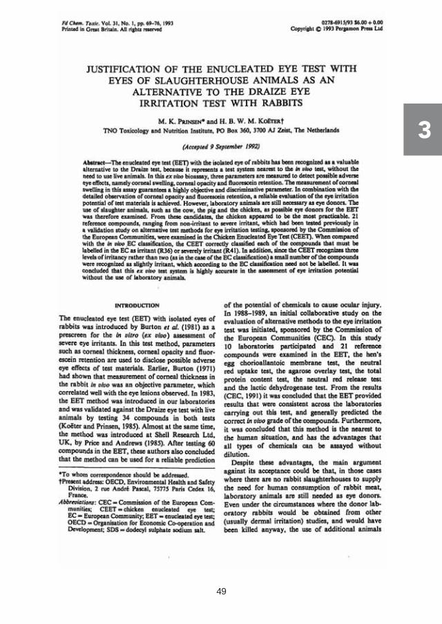
Prinsen, M.K., Koëter, H.B.W.M. (1993). Justification of the Enucleated Eye Test witheyes of slaughterhouse animals as an alternative to the Draize eye irritation test withrabbits. Food and Chemical Toxicology 31(1), 69-76.
Russell W.M.S., Burch R.L. (1959). The Principles of Humane Experimental Technique.South Mimms, Potters Bar, Herts:Universities Federation for Animal Welfare.
United Nations-Economic Commission for Europe (UN/ECE) (2007). Globally Harmonised System of Classification and Labelling of Chemicals (GHS). UN, New York and Geneva, 2007. Available at:http://www.unece.org/trans/danger/publi/ghs/ghs_rev02/02files_e.html.
US-Federal Register (1961). Title 21 Food and Drugs, Part 191 Hazardous substances, § 191.12 Test for eye irritants.
US-FDA (1964). Illustrated guide for grading eye irritation by hazardous substances.Washington, D.C. 20204, USA.
York, M., Lawrence, R.S., Gibson, G.B. (1982). An in vitro test for the assessment ofeye irritancy in consumer products – preliminary findings. International Journal of Cosmetic Science 4, 223-234.
1

31

32

Implementation and validation of the Rabbit Enucleated Eye Test
Herman B.W.M. Koëter and Menk K. Prinsen.Comparison of In Vivo and In Vitro Eye Irritancy Test Systems: A Study With 34 Substances.
Alternative Methods in Toxicology Volume 3, In Vitro Toxicology: A Progress ReportFrom The Johns Hopkins Center for Alternatives to Animal Testing. Editor Alan M.
Goldberg. Chapter A9. Mary Ann Liebert, Inc., New York, 1985

34

35
2
36
2

37
2

38
2

39
2
40
2

41
2

42
2
43
2

44
2

45

46
Development of the Chicken Enucleated Eye Test
M.K. Prinsen and H.B.W.M. Koëter.Justification of the Enucleated Eye Test with eyes of slaughterhouse animals
as an alternative to the Draize Eye Irritation Test with rabbits.
Food and Chemical Toxicology Vol. 31, No. 1, pp. 69-76, 1993

48
49
3

50
3

51
3

52
3

53
3

54
3

55
3

56
3

57

58

The Chicken Enucleated Eye Test as a prescreen in routine toxicity testing
M.K. Prinsen.The Chicken Enucleated Eye Test (CEET): A practical (pre)screen for the
assessment of eye irritation/corrosion potential of test materials.
Food and Chemical Toxicology Vol. 34, No. 3, pp. 291-296, 1996

60

61
4

62
4
63
4
64
4

65
4
66
4

67

68
The Isolated Chicken Eye Test as a stand-alone test
K. Schutte, M.K. Prinsen, P.M. McNamee, R. Roggeband.The isolated chicken eye test as a suitable in vitro method for determining the eye
irritation potential of household cleaning products.
Regulatory Toxicology and Pharmacology 54 (2009) 272-281

70

The isolated chicken eye test as a suitable in vitro method for determiningthe eye irritation potential of household cleaning products
K. Schutte a,*, M.K. Prinsen b, P.M. McNamee c, R. Roggeband d
a Procter & Gamble, Product Safety and Regulatory Affairs, Temselaan 100, B-1853 Strombeek-Bever, Belgiumb TNO Quality of Life, Toxicology and Applied Pharmacology Department, Utrechtseweg 48, 3704 HE Zeist, The Netherlandsc The Procter & Gamble Company, Whitehall Lane, Egham, Surrey TW20 9NW, UKd Procter & Gamble Service GmbH, Berliner Allee 65, 64274 Darmstadt, Germany
a r t i c l e i n f o
Article history:Received 6 November 2008Available online 19 May 2009
Keywords:Isolated chicken eye testICEChicken enucleated eye testCEETEye irritationHousehold cleaning productsIn vitro test methodsAlternative testing methodLow volume eye testLVET
a b s t r a c t
Eye irritation is an important endpoint in the safety evaluation of consumer products and their ingredi-ents. Several in vitro methods have been developed and are used by different industry sectors to assesseye irritation. One such in vitro method in use for some time already is the isolated chicken eye test(ICE). This investigation focuses on assessing the ICE as a method to determine the eye irritation potentialof household cleaning products, both for product safety assurance prior to marketing and for classifica-tion and labeling decisions. The ICE involves a single application of test substances onto the cornea of iso-lated chicken eyes. Endpoints are corneal swelling, corneal opacity and fluorescein retention. The ICEresults were compared to historic LVET data in this study due to availability of such in vivo data andthe ability to correlate LVET to human experience data on the outcome of accidental exposures to house-hold cleaning products in general. The results of this study indicate that the ICE test is a useful in vitromethod for evaluating the eye irritation/corrosion potential and establishing classification and labelingfor household cleaning products. For new product formulations, it is best used as part of a weight-of-evi-dence approach and benchmarked against data from comparable formulations with known eye irritation/corrosion profiles and market experience.
� 2009 Elsevier Inc. All rights reserved.
1. Introduction
Historically the rabbit Draize eye irritation test has been used toassess eye irritation potential of substances and mixtures thereof.The assay is accepted by regulatory agencies worldwide (e.g.,OECD, 2002; EC, 2004; USEPA, 1998) and is based on a methoddeveloped by Draize and colleagues in 1944 (Draize et al., 1944).The Draize test provides a quantitative scoring which is used asthe basis for hazard classification of eye irritants and corrosivesin international classification systems such as the European Union(EU) as well as under the United Nations (UN) Globally Harmo-nized System (GHS) classification and labeling scheme (EC, 2001;UNECE, 2003). Both classification systems are based on the severityof the ocular tissue lesions and/or persistence of effects. The EUhazard classification of eye irritants uses the risk phrases ‘R36’(Irritating to eyes) and ‘R41’ (Risk of serious damage to eyes), basedon whether the levels of damage, averaged across the 24, 48 and72 h observation times for each ocular tissue lesion, fall within orabove certain ranges of scores. The UN GHS considers two
harmonized categories, one for irreversible effects/serious damageto the eye (Category 1), and one for reversible effects (Category 2).Reversible effects are further sub-classified, based on the durationof persistence (Category 2A: Irritating to eyes, reverses within21 days and Category 2B: Mildly irritating to eyes, reverses within7 days).
Though the Draize test has served the community well for dec-ades there are, as with any assay, generally recognized limitationsof this assay. Scientific publications describe challenges of theDraize test related to variability, subjectivity of scoring and over-prediction of the human response (Weil and Scala, 1971; Yorkand Steiling, 1996; Buehler, 1974; Heywood and James, 1978;Jacobs et al., 1987; Daston and Freeberg, 1991). These challenges,added to concerns about animal welfare and a scientific desire tohave available eye irritation assays that are based on better under-standing of eye injury at the tissue and cellular level, have ledresearchers to investigate 3Rs alternative methods both in vivo(refinement) and in vitro (replacement) ones.
A number of in vitromethods, most notably organotypic models,have been evaluated for their ability to identify eye irritants/corro-sives. Organotypic models employ eye tissues (e.g., isolated eyesand corneas) from food-chain animals and include the bovine cor-neal opacity and permeability test (BCOP), the isolated chicken eye
0273-2300/$ - see front matter � 2009 Elsevier Inc. All rights reserved.doi:10.1016/j.yrtph.2009.05.008
* Corresponding author. Fax: +32 2 568 35 91.E-mail addresses: [email protected] (K. Schutte), [email protected] (M.K.
Prinsen), [email protected] (P.M. McNamee), [email protected] (R.Roggeband).
Contents lists available at ScienceDirect
Regulatory Toxicology and Pharmacology
journal homepage: www.elsevier .com/locate /yr tph
71
K. Schutte et al. / Regulatory Toxicology and Pharmacology 54 (2009) 272–281
5

test (ICE), also known as chicken enucleated eye test (CEET) and theisolated rabbit eye test (IRE) all ofwhichassess total corneal damage.
The ICE was introduced by Prinsen and Koëter in 1993 as a mod-ificationof the IRE (Burtonetal., 1981). Inbrief, the ICE involvesa sin-gle dose application of a test substance directly onto the cornea ofisolated chicken eyes. The endpointsmeasured are corneal swelling,corneal opacity and fluorescein retention. Corneal swelling, mea-sured as thickness, has been identified as a quantitative and reliableendpoint for the evaluation of corneal injury both in vivo and in vitro(Burton, 1972; Burton et al., 1981). Corneal opacity provides anassessment of corneal damage in the ICE that can be directly corre-lated to corneal damage observed in the in vivo rabbit eye test. Final-ly, fluorescein retention provides information on cornealpermeability, indicative of damage to the corneal surface. Theproce-dures for conduct of the ICE are described in INVITTOX protocol 80(http://evcam-sis.jrc.it/invittox/published/indexed_80.html).
Early use of the ICE assay for 21 chemicals with a known Draizeprofile identified that the ICE test correctly classified all chemicalsthat require R36 or R41 classification within the EU (Prinsen andKoëter, 1993). Further, the assay showed good correlationwith dataobtained in Draize on industrial materials and certain formulationstested in standard contract toxicity evaluations at the TNO laborato-ries (Prinsen, 1996). Together with other alternative assays, the ICEhas been reviewed in a range of validation or evaluation studies(Balls et al., 1999; Worth and Balls, 2002) with the outcome thatno single test was found capable of fully replacing the Draize test,but some of the assays, including the ICE, showed considerablepromiseas screening tools for eye irritancy/corrosion.Most recently,the ICE along with other organotypic assays has been reviewed bythe Interagency Coordination Committee on the Validation of Alter-native Methods (ICCVAM, 2006). ICCVAM accepted the ICE in 2006as a screening test to identify substances as ocular corrosives and se-vere irritants (i.e., EPA Category I, UN GHS Category 1, EU R41) in atiered testing strategy and as part of aweight-of-evidence approach.The ECVAM Scientific Advisory Committee then endorsed this con-clusion in 2007 (ECVAM, 2007). Furthermore, the ICE is now ac-cepted by both EU and US regulatory authorities for this purpose.
This study examined the suitability of the ICE test for its potentialto predict eye irritation/corrosion and its utility for classification andlabeling in the context of household cleaning products. For this pur-pose, a total of 20 household cleaning products and raw materialswere tested in the ICE test. The results were compared with in vivodata from a rabbit assay that is a refinement of the Draize test – thelow volume eye test (LVET). It is recognized that LVET is not aregulatoryapproved invivoeye irritationassay.However, for thepur-pose of this evaluation for household and cleaning products thiswasconsidered to be an appropriate in vitro to in vivo correlation forseveral reasons that include: (1) LVET is an established, mechanisti-cally-based (Maurer et al., 2002) in vivo eye irritation test that usesbiological and physiological endpoints that are relevant to humansfor which there is an identified prediction model that is the sameas that for the Draize test; (2) with the exception of the dosing regi-men in which a lower volume (10 lL versus 100 lL) of test materialis instilled directly onto the cornea instead of into the lower conjunc-tival sac, LVET is the sameassay as theDraize test in termsof the ocu-lar tissues evaluated (cornea, conjunctiva, iris), scoring system andinterpretation of individual ocular tissues data used for calculationof a classification/labeling within different regulatory schemes; (3)considering anatomical and physiological differences between spe-cies (rabbit andhuman) thedosevolumeof10 lL inLVET isanappro-priate dose volume (Swanston, 1985; Mishima et al., 1966; Ehlers,1976; Chrai et al., 1973); (4) LVET has been correlated as being pre-dictive of the human response from accidental exposure to house-hold and cleaning products through clinical studies (Freeberg et al.,1986a; Ghassemi et al., 1993; Roggeband et al., 2000) and humanexperience (accidental consumer and industrial exposures and
Poison Control Centres) (Freeberg et al., 1984, 1986b; Cormieret al., 1995) and (5) the LVET has been used successfully for manyyears by members of the household and cleaning products industryto support the consumer safety of such products. Since the ultimateobjectivehere is topredict thehumaneye response, the ICEdatahavebeen compared with historically available LVET data.
2. Materials and methods
2.1. Test substances
The study was conducted with 15 common household cleaningproducts and five raw materials, for most of which historical LVETdata was available. The test samples varied in terms of their formu-lation characteristics (i.e., low and high pH, bleaches, surfactant-based liquids or powders) and their potential to cause eye irritationas observed in the in vivo LVET assay. The principal test samplecharacteristics as well as the availability of LVET results or litera-ture data are presented in Table 1.
2.2. ICE procedure
Approximately 7 week old male or female ROSS spring chickensof 2.5–3.0 kg bodyweight from the slaughterhouse were used aseye donors. Chicken heads were taken immediately after sedationof the animals by electric shock and incision of the neck for bleed-ing and transported to the test facilities of TNO Quality of Life,Toxicology and Applied Pharmacology, Zeist, The Netherlands.Within 2 h after kill, the eyes were dissected and placed in a super-fusion apparatus as described below.
In a first step, the eyelidswere carefully removed from the chick-enheadwithoutdamaging thecorneaanda small dropoffluoresceinsodiumBP2%w/v (MinimsTM, disposable single-usedroppers, Smith& Nephews Ltd., Romford, England) was applied to the corneal sur-face for a few seconds, then rinsed offwith isotonic saline at ambienttemperature. The fluorescein-treated cornea was examined with aslit-lamp microscope (Slit-lamp 900 CN, Haag-Streit AG, Liebefeld-Bern, Switzerland) to ensure that no damage occurred. Undamagedeyes were then carefully removed from the head, placed in a stain-less steel clamp with the cornea positioned vertically and trans-ferred to one of the eleven chambers of the superfusion apparatus.The clamp was positioned in such a way that the entire cornea wassupplied with an isotonic saline drop at a rate of ca. 0.10–0.15 mL/min throughaperistalticpump(Watson-Marlow295CA,Rotterdam,The Netherlands). The chambers of the superfusion apparatus andthe saline were kept at 32 ± 1.5 �C (water pump, Thermomix 1441,B. Braun Melsungen AG, Melsungen, Germany).
The studies were conducted with three test eyes per sample andone control eye per assay (one assay covered three samples). Onecontrol eye was used to demonstrate the suitability of the generalconditions in the superfusion apparatus during the test period, i.e.,saline drip and temperature. Additional control eyes were notrequired because each test eye acted as its own control, by provid-ing baseline values for corneal swelling, corneal opacity and fluo-rescein retention prior to dosing. Before test start, all eyes wereexamined once again with the slit-lamp to ensure they were notdamaged. Corneal thickness was measured at the corneal apexusing the Depth Measuring Attachment No. I for the Haag-Streitslit-lampmicroscope. Eyes with a corneal thickness deviating morethan 10% from the mean value, unacceptably stained with fluores-cein (score higher than 0.5, indicating a permeable cornea) orshowing other signs of damage were discarded.
After an equilibration period of 45–60 min, corneal thicknesswas measured again to determine the zero reference value for cor-neal swelling calculations. Immediately afterward, at time t = 0, the
72
5
K. Schutte et al. / Regulatory Toxicology and Pharmacology 54 (2009) 272–281

test substance was applied. For this, the clamp holding the eye wasplaced on tissue paper outside the chamber with the cornea facingupwards. The standard testing protocol involves the application ofeither 30 lL of a liquid test substance or 30 mg of a solid test sub-stance to cover the entire surface of the cornea. This volume of30 lL was selected for the standard ICE protocol to mimic the Dra-ize test where 100 lL are used, taking into account that the chickencornea is approximately 1/3 the size of a rabbit cornea. In thisstudy, additional volumes (i.e., 3 and/or 10 lL) or masses (i.e., 3and/or 10 mg) of the test samples were also applied, in order toevaluate if they would provide better comparisons to the LVET as-say (where 10 lL are used).
Ten seconds after application, the corneal surface was thor-oughly rinsed by application of exactly 20 mL of isotonic salineand the eyes were then returned to the superfusion chamber. Usinga slit-lamp microscope, corneal swelling and corneal opacity weredetermined after 30, 75, 120, 180 and 240 min, and fluoresceinretention after 30 min.
At the end of the study, test and control eyes were preserved ina neutral aqueous phosphate-buffered 6% formaldehyde solution,
to be later embedded in paraffin wax, sectioned at 5 lM and exam-ined histologically for morphological effects after staining withhematoxylin and eosin.
2.3. Criteria and scoring system
The severity level for each study endpoint was evaluated for thethree test eyes of each sample according to the following set of cri-teria and scoring systems.
2.3.1. Corneal swellingThe mean percentage of corneal swelling was calculated for
each observation time point as follows:
corneal thickness at time t�corneal thickness at time¼0corneal thickness at time¼0
�100
Based on the highest mean value obtained at any of the obser-vation time points, a category score for corneal swelling was thendetermined, as shown in Table 2.
2.3.2. Corneal opacityCorneal opacitywas defined as ‘opacity degree of density’ and as-
sessed by scoring the area of the cornea that was most denselyopacified.
Score Observation
0 No opacity0.5 Very faint opacity1 Scattered or diffuse areas; details of the iris are clearly
visible2 Easily discernible translucent area; details of the iris are
slightly obscured3 Severe corneal opacity; no specific details of the iris are
visible; size of the pupil is barely discernible4 Complete corneal opacity; iris invisible
Table 1Test samples and availability of LVET results.
Test sample Product category LVET in vivo data available?
Household cleaning productsLiquidsAcidic cleaner (1) Low pH liquid (pH = 1.7 at 100%) (acid, stabilizers, perfume) YesAcidic cleaner (2) Low pH liquid (pH = 3.0 at 100%) (acid, stabilizers, perfume) YesFabric softener Low pH liquid (pH = 3.5 at 3%) (cationic surfactant-based, stabilizers, perfume) Yesa
Acidic peroxide bleach Low pH liquid (pH = 4.0 at 100%) (hydrogen peroxide-based, surfactant, perfume) YesAlkaline cleaner High pH liquid, aqueous (solvent, alkali, stabilizers, surfactant, perfume) (pH = 12 at 10%) YesAlkaline bleach cleaner High pH liquid, (hypochlorite-based, surfactants) (pH 11 at 1%) YesAutomatic dishwashing bleach gel High pH dishwasher gel (pH = 11.7 at 1%) (hypochlorite, silicates, alkali, stabilizers) YesDishwashing liquid Surfactant-based liquid (nonionic and anionic surfactants, stabilizers, perfume) (pH = 9 at 10%) YesPowdersPowder detergent (1) Laundry detergent powder (surfactants, builders, chelants, polymers, perfume) (pH = 10.5 at 1%) YesPowder detergent (2) Detergent powder like (1) plus 1% sulphamic acid Nob
Powder detergent (3) Detergent powder like (1) plus 5% sulphamic acid Nob
Powder detergent (4) Detergent powder like (1) plus 7% sulphamic acid Nob
Powder detergent (5) Detergent powder like (1) plus 5% citric acid Nob
Automatic dishwashing powder High pH powder based on silicates, alkali, stabilizer and surfactants (pH = 10.7 at 1%) YesBleach additive powder Percarbonate bleach-based laundry additive powder (pH = 10 at 1%) YesRaw materialsPowdersBleach catalyst Powder raw material, confidential (pH = 5.2 at 1%) YesCitric acid Powder raw material, pure substance (pH = 2.5 at 1%) Noc
Sulphamic acid Powder raw material, pure substance (pH = 1.2% at 1%) Noc
Silicate 2-ratio Powder raw material, pure substance (pH = 11.7 at 1%) YesMetasilicate 1-ratio Powder raw material, pure substance (pH = 12.8 at 1%) Yes
a LVET data available on a closely related formulation.b LVET data on powder detergent (1) are used as reference.c Literature data (Draize) are used as reference.
Table 2Determination of the category scores for corneal swelling, corneal opacity andfluorescein retention.
Corneal swelling(Max. mean %swelling)
Corneal opacity(Max. meanopacity score)
Fluoresceinretention (Meanretention score)
Corresponding category
0–5 0.0–0.5 0.0–0.5 Category I (no effect)>5–12 0.6–1.5 0.6–1.5 Category II (slight effect)>12–18a
>12–18ba 1.6–2.5 1.6–2.5 Category III (moderate effect)>18–26>26–32>26–32b 2.6–4.0c 2.6–3.0 Category IV (severe effect)>32
Prediction scheme is described in Prinsen and Koëter (1993) and Prinsen (1996).a >75 min after treatment.b <75 min after treatment.c In case of score 4, thickness assessment not possible.
73
K. Schutte et al. / Regulatory Toxicology and Pharmacology 54 (2009) 272–281
5

Mean corneal opacity was calculated for each observation time point.Basedonthehighestmeanscoreobtainedatanyof theobservationtimepoints, a category score for corneal opacity was determined (Table 2).
2.3.3. Fluorescein retentionFluorescein retention was scored as shown below:
Score Observation
0 No fluorescein retention0.5 Very minor single cell staining1 Single cell staining scattered throughout the treated area of
the cornea2 Focal or confluent dense single cell staining3 Confluent large areas of the cornea retaining fluorescein
When test substances adhered to the cornea, fluorescein retentionwas determined after adequate removal of the test substance. Basedon the mean fluorescein retention score obtained at 30 min, acategory score for corneal opacity was determined (Table 2).
2.3.4. Morphological effectsMorphological changes in the test eyes were recorded for each
of the test substances. The effects included pitting of corneal epi-thelial cells, loosening of the epithelium, roughening of the cornealsurface and sticking of the test substance to the cornea. These find-ings could vary in severity and could occur simultaneously.
2.3.5. Microscopic effectsCorneal lesions were determined by histological examination.
The effects included but were not limited to erosion, necrosisand vacuolation of the epithelium, disorder of stromal fibers, pyc-notic nuclei in the stroma (anterior/posterior region) and necrosisof the endothelium. The description of these findings was subjec-tive to the interpretation of the investigator.
2.3.6. ICE overall eye irritancy categorizationBased on the category scores obtained for corneal swelling,
corneal opacity andfluorescein retentionand, if present, onmorpho-logical effects, an overall eye irritancy class was established for eachof the products tested. As shown in Table 3, the substances wereclassed as not irritating, slightly, moderately or severely irritating,depending on the outcome of evaluation of the ICE endpoints.
2.3.7. Comparison of ICE and EU/UN GHS eye irritation classificationresults
In order to assess the suitability of the ICE test for determiningeye irritation classification, the results obtained in this study weretranslated into the corresponding EU classification based on a con-version scheme developed using scientific judgment and manyyears of experience (Prinsen and Koëter, 1993; Prinsen, 1996,2004). In addition, the general eye irritancy categorization schemeof the ICE also allowed for translation to UN GHS classification. Theconversion scheme is shown in Table 3.
2.4. LVET procedure
The LVET procedure was conducted in accordance with thestandard protocol published by the American Society for Testingand Materials originally in 1985 and reapproved in 2003 (ASTM,2003). Three animals were used in each LVET. A preliminary mac-roscopic examination of the eyes of each rabbit was conductedusing fluorescein dye. A minimum of 1 h after the preliminary ocu-lar examination, the test article was placed directly on the cornea
of the right eye of each animal. Liquids were administered at a vol-ume of 10 lL using a glass syringe. Solids were administered as aweight equivalent to 10 lL volume not to exceed 10 mg. Followinginstillation, the eyelids were released without forced blinking ormanipulation. The contra-lateral eye remained untreated to serveas a control.
Responses of the cornea, iris and conjunctiva in the test andcontrol eyes of each rabbit were evaluated macroscopically usingan auxiliary light source at 1, 24, 48 and 72 h after dosing accord-ing to the Draize scale for scoring ocular lesions (17). Followingmacroscopic observations at the 24-h scoring interval, the fluores-cein examination procedure was repeated on all test and controleyes and any residual test article gently rinsed from the eye at thistime (if possible) using physiological saline. If any fluorescein find-ings were noted, a fluorescein examination was further conductedat each subsequent interval until a negative response was obtainedand/or until all corneal opacity had cleared. If there was noevidence of treatment-related ocular irritation at the 72-h scoringinterval, the study was terminated. If ocular irritation persisted inany test eye, the observation period was extended for the affectedanimals (scored on days 7, 10, 14 and 21). Animals requiring anextended observation period remained on test until the irritationhad resolved or permanent injury was evident.
2.4.1. Scoring systemThe Draize scale for scoring ocular lesions was used to evaluate
the effect on the ocular tissues (cornea, conjunctiva and iris) ofexposure to test material (Draize, 1959). The scoring scale usedhere was the same as that which is used in the DangerousSubstances Directive Annex V Draize test (OECD, 2002).
2.4.2. Data interpretation2.4.2.1. Ocular evaluation. Using the Draize scoring scale, the groupmean irritation score was calculated for each scoring interval basedon the number of animals initially dosed in each group. The calcu-lated group mean ocular irritation scores for each interval was thenused to categorise the test article according to the ocular evalua-tion criteria as defined by Kay and Calandra (1962).
2.4.2.2. EU ocular evaluation. The total ocular irritation score for the24, 48 and 72 h intervals were individually added for corneal opac-ity, iris lesion, conjunctival redness and conjunctival edema. For atest containing three rabbits, the group mean scores for cornealopacity, iris lesion, conjunctival redness and conjunctival swellingwere then calculated. The resulting mean ocular irritation scoreswere then classified according to the existing European Union(EU) hazard classification and labeling scheme within chemicalslegislation (European Union, 2001).
3. Results
The results obtained in the ICE protocol for all 20 test materialsare summarized in the Tables 4–6. Table 4 presents the highestmean scores for corneal swelling and corneal opacity, and themean score for fluorescein retention, as well as the ICE eyeirritation categorization derived therefrom. The acidic to neutraltest materials generally caused mostly slight to moderate irritationeffects, while the clearly alkaline cleaner formulations as well aspowder raw material sulphamic acid and metasilicate producedsevere irritation effects. Related histopathological findings for alltest materials (only for eyes treated with 30 lL or mg) aregiven in Table 5. Finally, Table 6 then compares regulatory classifi-cations derived from the ICE test results and those derived fromLVET data previously. This comparison is visually illustrated inFig. 1.
74
5
K. Schutte et al. / Regulatory Toxicology and Pharmacology 54 (2009) 272–281

A direct comparison of the ICE and LVET-based EU classificationswas possible for 14 out of the 20 products tested. For 4 products(powder detergents (2)–(5)), no LVET testing was conducted, there-fore LVET data from a historic and similar formulation, powderdetergent (1), was used as the in vivo reference. For the two acids,literature data on in vivo studies were used as reference.
Overall, as summarized in Table 6, the ICE results were either inline with or more conservative than the eye irritation profile pre-dicted on the basis of the LVET test. When a dosing volume of30 lL or mg was applied, 14 out of 20 test samples revealed ICE-based regulatory classifications which were comparable to thoseof the LVET, 5 out of 20 were over-predicted and only one wasunder-predicted (silicate 2-ratio powder). At 10 lL or mg, theICE-based regulatory classification was in line with LVET in 13out of 20 cases, over-predicted in 4 out of 20 cases, and under-pre-dicted 3 out of 20 times. These data are illustrated in a visual wayin Fig. 1, where those test substances which showed a matchbetween ICE and LVET in terms of irritation category results arehighlighted in green. At 3 lL or mg there were only few datapoints; results were generally identical to what was seen at10 lL or mg. As a general trend, there was a good match betweenICE and LVET results for surfactant-based and low pH products.High pH products showed more often over-prediction in the ICEversus the LVET assay.
At the histological level, all but two liquids (‘acidic peroxidebleach’ and ‘dishwashing liquid’), showed some degree of effecton the epithelium, mainly necrosis and vacuolation. A few samplesalso induced pycnotic nuclei in the outer region of the stroma. For
the two remaining liquids mentioned above no effects were seen.All powder products had very slight to moderate effects on theepithelium. Several powders also led to pycnotic nuclei in the ante-rior region of the stroma. Metasilicate 1-ratio was the only testsubstance to induce effects in the posterior region of the stromaand the endothelium.
4. Discussion
The ICE testwas accepted in a range of EUMember States to iden-tify substances as ocular corrosives and severe irritants in a tieredtesting strategy and as part of a weight-of-evidence approach, evenbefore and also in line with the ICCVAM (2006) and ECVAM (2007)conclusions on the assay. It has been used by industry for screeningpurposes, predominantly early in the product development cycle toidentify product formulations that may have an unacceptable eyeirritation/corrosion profile. Whereas parallel in vitro ICE andin vivo Draize eye irritation testing (OECD 405) showed good corre-lation with about 100 test substances (Prinsen, 1996), it has provendifficult to compare ICE test resultswith thoseof in vivoeye irritationtests of various other sources (ICCVAM, 2006; Worth and Balls,2002). This could in part be due to reported limitations of thein vivo tests (e.g., theDraize test), particularly (1) the often unknownexposure conditions (up to 24 h for solids), and (2) the subjectivescoring of tissue lesions in the Draize test resulting in variable esti-mates of eye irritancy (Prinsen, 2006).
This study examined the suitability of the ICE test for itspotential to predict eye irritation/corrosion and its utility for
Table 3Overall ICE eye irritancy categorization based on the scores for corneal swelling, corneal opacity and fluorescein retention, and consequent conversion into corresponding EU andUN GHS classification.
Prediction scheme is described in Prinsen and Koëter (1993) and Prinsen (1996).aCombinations of these categories are less likely to occur.bCombination can be considered a borderline case between non-irritating and irritating (R36).cCombination can be considered as a borderline case between mildly irritating (2B) and irritating (2A).dCombination can be considered a borderline case between irritating (R36/2A) and severely irritating (R41/1).
75
K. Schutte et al. / Regulatory Toxicology and Pharmacology 54 (2009) 272–281
5

classification and labeling in the context of household cleaningproducts which were previously mostly evaluated in the low vol-ume eye test (LVET) and for which good quality data were avail-able. The LVET is a modification of the Draize protocol and uses alower dose volume of 10 lL (versus 100 lL in the Draize) that bet-ter fits the anatomical and physiological characteristics of the hu-man eye. In addition to giving responses closer to those observed inhumans, it has been used to provide data on mechanistic under-standing of the eye response to chemical injury (Maurer et al.,2002; Jester, 2006). The LVET was developed over 25 years agoby Griffith et al. (1980). Since then, it has been correlated as beingpredictive of the human response from accidental exposure tohousehold and cleaning products through clinical studies (Rogge-band et al., 2000) and through decades of market place experienceof the Procter & Gamble Company and other members of industry.
For a total of 20 household cleaning products and rawmaterials,a comparison was made between the EU and UN GHS classifica-tions derived using ICE and LVET test data (see Table 6). The out-come of this investigation shows that the eye irritancy/corrosionclassifications obtained for the sampled products based on ICEresults were, in a majority of cases, either in line with or more con-servative than the eye irritation profile predicted on the basis ofthe LVET test. Some conservatism or over-prediction was in factto be expected because the ICE model was developed to predictthe Draize test. The dose of 30 lL or mg proved to yield betterand more consistent results than 10 or 3 lL or mg. As a generaltrend, there was a good match between ICE and LVET results forsurfactant-based and low pH products, both in liquid as well asin powder form. High pH products showedmore often over-predic-tion in the ICE as compared to the LVET assay, especially the threeliquid ones alkaline cleaner, alkaline bleach cleaner and automaticdishwashing bleach gel. Only one sample, i.e., silicate 2-ratio, wasunder-predicted at the 30 mg dose; this can be explained by the
fact that this raw material is a powder that dissolves only slowlyin water and therefore did not lead to irritating effects during thestandard exposure time of 10 s. It was however correctly predictedupon longer exposure (data not shown).
This study confirms the suitability of the ICE test for the deter-mination of the eye irritation/corrosion potential of householdcleaning products and raw materials when compared with LVETwhich is the in vivo assay for which most data are available forhousehold and cleaning products and which has been successfullycorrelated with the human response through both clinical studiesand many years of human experience for these types of products.In the frame of product developments, ICE could be an appropriateassay to determine the profile of new cleaning product formula-tions by comparison to similar formulations for which historicin vivo eye irritation data and market experience exists. ICE dataare regularly used by Procter & Gamble in a weight-of-evidenceapproach to determine the eye safety and appropriate classificationof new candidate cleaner and detergent formulations. The ICE testis also useful to screen candidate formulations. Where for exampleseveral prototypes containing various concentrations of the sameingredients are being developed, ICE test results can help selectthe prototype(s) with the best irritation versus efficacy profile.Recent examples for which the ICE test was used within aweight-of-evidence approach for risk assessment and classificationdecisions included an acidic cleaner, a powder detergent and a con-centrated liquid detergent. These three products were related tothe test materials evaluated in the study but explored new innova-tion directions.
This study further suggests the utility of the ICE test for eyeirritancy/corrosion classification and labeling purposes in theframe of existing classification schemes (e.g., EU, UN GHS). ICEresults can be translated into the EU and UN GHS eye irritationschemes (as per Table 3) and benchmarked against data for
Table 4ICE test results – scores and eye irritation categorization.
aBased on immediate opacity (score 3) after application.
76
5
K. Schutte et al. / Regulatory Toxicology and Pharmacology 54 (2009) 272–281

comparable formulations for which EU classifications have beenderived.
Furthermore, microscopic evaluation of the cornea can provideimportant information on the severity of the injury at the tissueand cellular level. Maurer et al. have proposed that differences inocular irritation are related to differences in extent of initial injury,and that regardless of the process leading to tissue damage, extentof injury is a principal factor determining the outcome of ocularirritation (Maurer et al., 2002, which reviews all studies conductedin the mechanistic program). Using light microscopy they
characterized the general ocular irritancy of a broad sampling ofsurfactantsandnon-surfactants (acid, alkali, ketone,alcohol, aromaticamine, aldehyde) causing slight to severe irritation in the contextof the LVET. Additionally, using in vivo confocal microscopy, theyquantitatively characterized the corneal changes occurring withthese irritants in the LVET. Despite differences in the mechanismsby which these materials may cause injury, the results collectivelysupport their hypothesis that by defining the initial extent of injuryassociated with ocular irritation it is possible to predict the subse-quent response and final outcome. Importantly, they proposed that
Table 5Morphological findings for chicken eyes treated with 30 lL or mg of test substance (figures in brackets are number of eyes affected out of three; only indicated if different from 3/3).
– = not observed; ½, very slight; 1 = slight; 2 = moderate; 3 = severe.
77
K. Schutte et al. / Regulatory Toxicology and Pharmacology 54 (2009) 272–281
5

a mechanistically based approach to the development of alterna-tive ocular irritation tests would be to include histological evalua-tion to dimension the extent of initial injury using either ex vivo orin vitro corneal equivalent systems.
In the context of eye safety risk assessment or classificationdecisions, such histological assessments could add to the weight-of-evidence analysis in assays such as the ICE, especially whendecisions between non-irritant and irritant (i.e., not classified orR36/2A) and between moderate and severe irritants (i.e., R36/2Aor R41/1) have to be made. In borderline cases, a decision towardsa more severe eye irritant categorization or classification is justi-fied the more pronounced and deeper the injury. This is based onmechanistic work conducted by Maurer et al. suggesting that thedeeper the corneal injury, the smaller the likelihood of fast andcomplete recovery (Maurer et al., 2002; Jester, 2006). For example,pronounced damage to the epithelium and the posterior region ofthe stroma or at endothelial level indicate high irritation potentialjustifying a more severe eye irritant categorization or a R41/1 clas-sification. The mechanistic work also indicates that damage to theepithelium and anterior stromal region alone is typical of mild–moderate irritants and linked with potential for good recovery.
5. Conclusions
This study indicates that the ICE test is a useful in vitro method-ology for evaluating the eye irritation/corrosion potential andestablishing the EU classification and labeling of powder and liquidhousehold cleaning products when compared with LVET which isthe in vivo assay for which most data are available for householdand cleaning products and which has been successfully correlatedwith the human response through both clinical studies and manyyears of human experience for these types of products. The goodcorrelation of ICE results with LVET data for an identified rangeof products (in particular low pH and surfactant-based products)suggest that the ICE can be used to predict eye irritation responsesin man for these product types. For new product formulations, it isbest used as part of a weight-of-evidence tiered approach andbenchmarked against available data from comparable formulationswith known eye irritancy/corrosion profile and market experience.The number of samples comprised in the present research programwas limited; further investigations will be conducted to betterqualify the ICE test for this purpose and evaluate whether its usecan be extended to a wider range of product types.
Table 6Comparison of EU and GHS classification based on ICE versus LVET test results.
CCM – conventional classification method according to Directive 1999/45/EC: The CCM is a method described in the EU Dangerous Preparations Directive (1999/45/EC) toestimate the theoretical hazard of a preparation based on a simple summation of the individual hazards of the substances in that preparation. NC, Not classified; NA, Notapplicable as CCM method cannot be applied to substances.aNC but close to R36.bDose volume not tested.cR36/2A but close to R41/1.dPowder detergent (1) used as LVET reference.eEstimated based on literature data. Green boxes indicate cases where the ICE and LVET results are in line. Yellow and orange are when the ICE over or underpredicts,respectively, compared to the LVET.
78
5
K. Schutte et al. / Regulatory Toxicology and Pharmacology 54 (2009) 272–281

References
American Society for Testing and Materials, 2003. Standard Method for Evaluationof Eye Irritation in Albino Rabbits, E1055-93 (reapproved in 2003) in AnnualBook of ASTM Standards. Philadelphia, PA.
Balls, M., Berg, N., Bruner, L.H., Curren, R.D., de Silva, O., Earl, L.K., Esdaile, D.J.,Fentem, J.H., Liebsch, M., Ohno, Y., Prinsen, M.K., Spielmann, H., Worth, A.P.,1999. Eye irritation testing: the way forward. The report and recommendationsof ECVAM workshop 34. Altern. Lab. Anim. 27, 53–77.
Buehler, E.V., 1974. Testing to predict potential ocular hazards of householdchemicals. In: Winek, C.L. (Ed.), Toxicology Annual 1974. Marcel Dekker,p. 53.
Burton, A.B.G., 1972. A method for the objective assessment of eye irritation. FoodCosmet. Toxicol. 10, 209.
Burton, A.B.G., York, M., Lawrence, R.S., 1981. The in vitro assessment of severe eyeirritants. Food Cosmet. Toxicol. 19, 471–480.
Cormier, E.M., Hunter, J.E., Billhimer, W., May, J., Farage, M., 1995. Use of clinical andconsumer eye irritation data to evaluate the Low Volume Eye Test. J. Toxicol.Cut. Ocular Toxicol. 14, 197–205.
Chrai, S., Patton, T.F., Mehta, A., Robinson, J.R., 1973. Lacrimal and instilled fluiddynamics in rabbit eyes. J. Pharmaceut. Sci. 62, 1112–1121.
Daston, G.P., Freeberg, F.E., 1991. Chapter 16: ocular irritation testing. In: Hobson,D.W. (Ed.), Dermal and Ocular Toxicology. Fundamentals and Methods. CRCPress, Boca Raton, pp. 510–539.
Draize, J.H., Woodward, G., Calvery, H.O., 1944. Methods for the study of irritationand toxicity of substances applied directly on the skin and mucous membranes.J. Pharmacol. Exper. Ther. 82, 377–390.
Draize, J.H., 1959. Appraisal of the Safety of Chemicals in Food, Drugs andCosmetics. Association of Food and Drug Officials of the US EditorialCommittee, 1959. Second Printing 1965. Topeka, Kansas. pp. 46–51.
Ehlers, N., 1976. Pharmacology of the conjunctival sac. In: Dickstein, S. (Ed.), Drugsand Ocular Tissues (Second Meeting of the International Society for EyeResearch, Jerusalem, 1976). Karger, S.Basel. p. 23.
European Center for the Validation of Alternative Methods (ECVAM), 2007. ESACstatement on the conclusions of the ICCVAM retrospective study on organotypicin vitro assays as screening tools to identify potential ocular corrosives and severeirritants as determined byUSEPA, EU (R41) and UNGHS classifications in a tieredtesting strategy, as part of a weight of evidence approach. <http://ecvam. jrc.it/publication/ESAC26_statement_Organotypic_20070510c.pdf>(accessed July2008).
European Commission (EC), 2001. Commission Directive 2001/59/EC of 6 August2001 adapting to technical progress for the 28th time Council Directive 67/548/EEC on the approximation of the laws, regulations and administrativeprovisions relating to the classification, packaging and labelling of dangeroussubstances. Official Journal of the European Communities L255, 1–333.
European Commission (EC), 2004. Corrigendum to Commission Directive 2004/73/EC of 29 April 2004 adapting to technical progress for the 29th time CouncilDirective 67/548/EEC on the approximation of the laws, regulations andadministrative provisions relating to the classification, packaging andlabelling of dangerous substances. Official Journal of the European Union L152, 30 April 2004, 3–310.
Freeberg, F.E., Griffith, J.F., Bruce, R.D., Bay, P.H.S., 1984. Correlation of animal testmethods with human experience for household products. J. Toxic. Cut. Ocul.Toxicol. 1 (3), 53–64.
Freeberg, F.E., Hooker, D.T., Griffith, J.F., 1986a. Correlation of animal test methodswith human experience for household products: an update. J. Toxic. Cut. Ocul.Toxicol. 5 (2), 115–123.
Freeberg, F.E., Nixon, G.A., Reer, P.J., Weaver, J.E., Bruce, R.D., Griffith, J.F., Sanders III,L.W., 1986b. Human and rabbit eye responses to chemical insult. Fund. Appl.Toxic. 7, 626–634.
Ghassemi, A., Sauers, L.H., Bruner, L.H., Reer, P.J., Hall, R.H. (1993). Demonstratingthe human safety of a new household cleaning (HSC) product using alternativesto the Draize eye irritation test. Presentation made at the US Society ofToxicology meeting.
Griffith, J.F., Nixon, G.A., Bruce, R.D., Reer, P.J., Bannan, E.A., 1980. Dose–responsestudies with chemical irritants in the albino rabbit eye as a basis for selectingoptimum testing conditions for predicting hazard to the human eye. Toxicol.Appl. Pharmacol. 55, 501–513.
Heywood, R., James, L.W., 1978. Towards objectivity in the assessment of eyeirritation. J. Soc. Cosmet. Chem. 29, 25.
Interagency Coordination Committee on the Validation of Alternative Methods(ICCVAM), 2006. In vitro test methods for detecting ocular corrosives and severeirritants. Background review document for isolated chicken eye (ICE) test.<www.iccvam.niehs.nih.gov/docs/ocutox_docs/ocubrd/ice/iceappA.pdf>.
Jacobs, G., Martens, M., De Beer, J., 1987. Selecting optimal dosage volumes for eyeirritation tests in the rabbit. J. Toxicol. Cut. Ocul. Toxicol. 6, 109–116.
Jester, J.V., 2006. Extent of corneal injury as a biomarker for hazard assessment andthe development of alternative models to the Draize rabbit eye test. Cutan.Ocul. Toxicol. 25 (1), 41–54.
Kay, J.H., Calandra, J.C., 1962. Interpretation of eye irritation tests. J. Soc. Cosmet.Chem. 13, 281.
Maurer, J.K., Parker, R.D., Jester, J.V., 2002. Extent of initial corneal injury as themechanistic basis for ocular irritation: key findings and recommendations forthe development of alternative assays. Reg. Toxicol. Pharm. 36 (1), 106–117.
Mishima, S., Gasset, A., Klyce, S.D., Baum, J.L., 1966. Determination of tear volumeand tear flow. Invest. Ophthalmol. 5, 264–276.
Organization for Economic Co-Operation and Development (OECD), 2002. Guidelinefor the testing of chemicals: acute eye irritation/corrosion. Guideline 405,adopted 24 April 2002.
Prinsen, M.K., 1996. The chicken enucleated eye test (CEET): a practical (pre)screenfor the assessment of eye irritation/corrosion potential of test materials. FoodChem. Toxicol. 34 (3), 291–296.
Prinsen, M.K., Koëter, H.B.W.M., 1993. Justification of the enucleated eye test witheyes from slaughterhouse animals as an alternative to the Draize eye irritationtest with rabbits. Food Chem. Toxicol. 31, 69–76.
Prinsen, M.K., 2006. The Draize eye test and in vitro alternatives: a left-handedmarriage? Toxicol. In Vitro 20 (1), 78–81.
Prinsen M.K., 2004. Chicken enucleated eye test. INVITTOX protocol. <http://evcam-sis.jrc.it/invittox/published/indexed_80.html>.
Roggeband, R., York, M., Pericoi, M., Braun, W., 2000. Eye irritation responses inrabbit and man after single applications of equal volumes of undiluted modelliquid detergent products. Food Chem. Toxicol. 38, 727–734.
Fig. 1. Visualization of the data reported in Table 6 (green: matching prediction, blue: underprediction, red: overprediction).
79
K. Schutte et al. / Regulatory Toxicology and Pharmacology 54 (2009) 272–281
5

Swanston, D.W., 1985. Assessment of the validity of animal techniques in eyeirritation testing. Food Chem. Toxic. 23 (2), 169–173.
United Nations Economic Commission for Europe (UNECE), 2003. Globallyharmonized system of classification and labelling of chemicals (GHS). <http://www.unece.org/trans/danger/publi/ghs/ghs_rev00/00files_e.html>.
United States Environmental Protection Agency (USEPA), 1998. Health effects. Testguidelines OPPTS 870.2400. Acute eye irritation. EPA 712–C–98–195. <http://iccvam.niehs.nih.gov/SuppDocs/FedDocs/EPA/EPA_870_2400.pdf> (accessedMay 2008).
Weil, C.S., Scala, R.A., 1971. Study of intra- and interlaboratory variability in theresults of rabbit eye and skin irritation tests. Toxicol. Appl. Pharmacol. 19, 276–360.
Worth, A., Balls, M., 2002. Local toxicity: acute dermal and ocular effects. In: Worth,A., Balls, M. (Eds.), Alternative (non-animal) Methods for Chemicals Testing:Current Status and Future Prospects. A Report Prepared by ECVAM and theECVAM Working Group on Chemicals, vol. 30. ATLA, p. 125 (Suppl. 1).
York, M., Steiling, W., 1996. A critical review of the assessment of eye irritationpotential using the Draize rabbit eye test. J. Appl. Pharmac. 18, 233–240.
80
5
K. Schutte et al. / Regulatory Toxicology and Pharmacology 54 (2009) 272–281

81

82

Histopathology as an additional observation in the Isolated Chicken Eye Test
Menk K. Prinsen, M.K. , M.E.I. Schipper, M.V.W. Wijnands.Histopathology in the isolated chicken eye test and comparison of different
stainings of the cornea.
Toxicology in Vitro 25 (2011) 1475-1479

84

Histopathology in the isolated chicken eye test and comparisonof different stainings of the cornea
Menk K. Prinsen a,⇑, M.E.I. Schipper b, M.V.W. Wijnands a
a TNO Triskelion, Toxicology and Applied Pharmacology Department, P.O. Box 844, 3700 AV Zeist, The NetherlandsbUniversity Medical Centre Utrecht, Department of Pathology, Utrecht, The Netherlands
a r t i c l e i n f o
Article history:Received 14 December 2010Accepted 29 April 2011Available online 7 May 2011
Keywords:ICEStaining methodHistopathologyIsolated eyePAS stainingDepth-of-InjuryEye irritation in vitroAlternatives
a b s t r a c t
The isolated chicken eye (ICE) test, developed at our Institute, is accepted by the OECD for identificationof severe eye irritants. The OECD ICE Guideline (No. 438) encourages preservation of the treated eyes forpossible histopathology of the cornea, which is believed to strengthen evidence of absence or presence ofirritation and to help clarify borderline effects by assessment of the corneal Depth-of-Injury. Histopathol-ogy of the cornea in addition to the normal slit-lamp microscope assessment of corneal effects hasalready been performed routinely in ICE tests at our Institute, using two standard stainings (H&E andPAS). In this study, three other stainings (AZAN, EVG and Trichrome), more specific for collagen-richmembranes such as basement- and Bowman’s membranes were examined with corneas exposed to fourmodel compounds ranging from non- to severely irritating (corrosive). PAS appeared to be the superiorstaining method. Surprisingly, the well-known eye corrosive sodium hydroxide (NaOH, solid) did not vis-ibly compromise the integrity of Bowman’s or the basement membrane. Based on our experience, histo-pathology of the treated cornea is confirmative in relation to the standard assessment of eye irritation byslit-lamp observation in the ICE and in certain cases can help to evaluate borderline effects. Besides estab-lishing the depth of injury, additional investigation of corneal limbal stem cell damage after chemicalexposure might be appropriate to determine reversibility or irreversibility of eye effects.
� 2011 Elsevier Ltd. All rights reserved.
1. Introduction
The isolated chicken eye (ICE) test, developed at our Institute, isone of two validated alternative methods accepted by the OECD foridentification of severe irritants (OECD, 2009; Prinsen and Koëter,1993; Prinsen, 1996). The OECD ICE Test Guideline No. 438 encour-ages preservation of the treated eyes in order to perform histopa-thology. Histopathology of the cornea is believed to strengthenevidence (absence or presence) of eye irritation and to help clarifyborderline effects, especially those that are expected to be revers-ible or irreversible. The corneal Depth-of-Injury approach wasintroduced as an additional tool to more precisely determine theextent of initial corneal injury (Maurer et al., 2002; Jester, 2006;Jester et al., 2010; Scott et al., 2010). This approach is based on his-topathology performed by light microscopy and in vivo confocalmicroscopy of rabbit corneas and by using biomarkers of cell death
and viability. In general, it is believed that with non- or (mild) irri-tants the effects are limited to the epithelium of the cornea, whilemoderate to severe irritants also affect the deeper layers of the cor-nea such as stroma and endothelium. In their well thought-outpublication, Maurer et al. recommended that any ex vivo orin vitro replacement of the rabbit eye irritation test should meetthe following criteria: (1) assessment of injury should be three-dimensional, as injury is a three-dimensional process, (2) extentof injury may be assessed by extent of cytotoxicity within the cor-nea, (3) assessment of injury to epithelium, stroma and endothe-lium, (4) differentiate injury that is diffuse from injury that isfocally extensive, and (5) assessment of injury at different timepoints. The ICE test including histopathology meets by far thesepre-requisites and, moreover, histopathology of the cornea in addi-tion to the normal slit-lamp microscope assessment of corneal ef-fects is already performed in ICE tests at our Institute for more thanten years. Most of these tests are performed for sponsors and theresults are confidential. However, an article concerning the perfor-mance of the ICE test with household cleaning products andincluding histopathology has been published (Schutte et al., 2009).
From the perspective of reversibility/irreversibility, damage notonly to the epithelium, stroma and endothelium of the cornea, butalso to the other structures present in the cornea, such as basementmembrane or Bowman’s- and to a lesser extent Descemet’s
0887-2333/$ - see front matter � 2011 Elsevier Ltd. All rights reserved.doi:10.1016/j.tiv.2011.04.028
Abbreviations: AZAN, Azocarmine & Aniline; EU-CLP, European Union-Classifi-cation, Labelling and Packaging; EVG, Elastic Van Gieson; H&E, haematoxylin &eosin; ICE, isolated chicken eye; OECD, Organisation for Economic Co-operation andDevelopment; PAS, periodic acid-schiff; TNO, Toegepast NatuurwetenschappelijkOnderzoek (Organization for Applied Scientific Research); Trichrome, Masson’strichrome; UN-GHS, United Nations-Globally Harmonised System.⇑ Corresponding author.
E-mail address: [email protected] (M.K. Prinsen).
Contents lists available at ScienceDirect
Toxicology in Vitro
journal homepage: www.elsevier .com/locate / toxinvi t
85
Toxicology in Vitro 25 (2011) 1475–1479
6

membranes, are important to evaluate. After all, extracellularmatrices, such as the basement membrane, are the essential frame-work for wound healing (Wagoner, 1997).
Initially, the traditional haematoxylin & eosin (H&E) stainingwas used for the histopathological evaluation of the cornea. In anattempt to further improve the evaluation of the cornea, thePeriodic Acid-Schiff (PAS) staining was applied. This turned outto provide a colour spectrum with more contrast. The various spe-cific structures of the cornea became better discernable, which washelpful during microscopic evaluation of the corneal lesions.Therefore, it was decided to use PAS as the standard staining meth-od in the ICE test from then on. Three other stainings, known to bemore specific for collagen-rich membranes, were examined andcompared to the H&E and PAS stainings. Chicken corneas were ob-tained from standard ICE tests with substances classified accordingto the usual classification systems, i.e. UN-GHS (2007) and EU-CLP(2008); (formerly EC criteria for labelling of EC, 1993). Three sub-stances represented the categories non-classified (NC), irritating(Cat2 sub-divided into 2A and 2B in the UN-GHS classification sys-tem) and severely irritating (Cat1), and one represented a border-line case between Cat2 and Cat1. The focus of this investigationwas on the quality and applicability of the different staining tech-niques in relation to the morphology of the various cell structuresand the pathology. Publications on the ex vivo histopathology ofthe cornea in the open literature are scarce or the work is confiden-tial. Statements with respect to the corneal Depth-of-Injury theoryand histopathological evaluation of the cornea in the ICE test arebased on the many years of experience in this field at our Institute.
2. Materials and methods
2.1. Test substances
The four materials and their regulatory classifications selectedwere:
- physiological saline; non-classified/negative control (Eurovet,Bladel, The Netherlands)
- liquid surfactant containing cleaning product; Category 2/2A/R36 (source: confidential)
- liquid surfactant containing cleaning product; Category 2/2A/R36 borderline Category 1/R41 (source: confidential)
- NaOH, solid (purity > 97%); Category 1/R41 (Sigma–Aldrich,Germany).
Chicken corneas treated with these materials were obtainedfrom routinely performed ICE tests, which constituted assessmentof corneal swelling, opacity, fluorescein retention by damaged epi-
thelial cells using the Haag–Streit slit-lamp microscope over a 4 hperiod and after a 10 s treatment with the test substance (OECD TG438, 2009; Prinsen and Koëter, 1993; Prinsen, 1996). On the basisof the severity (maximum mean score of three eyes) of the ob-served findings for corneal swelling, corneal opacity and fluores-cein retention, the effects were divided into four classes, viz.I = no effect; II = slight effect; III = moderate effect; IV = severe ef-fect. The final irritation classification is determined by the combi-nation of the three classes obtained for the three endpoints(corneal swelling, corneal opacity and fluorescein retention) intopredefined classification schemes (Prinsen and Koëter, 1993; Prin-sen, 1996). In addition, to allow for numerical ranking and compar-ison an Irritation Index was calculated. This index is based on theaddition of the maximummean scores obtained for the parametersaccording to the following formula: Irritation Index = maximummean corneal swelling + maximum mean opacity (�20) + meanfluorescein score (�20). The factor of 20 is included to give equalweight to the scores obtained for opacity and fluorescein retentionin the index compared to the maximum swelling possible (ca. 60%).
2.2. Preservation of the cornea
Our experience with histopathology of the chicken corneashowed that specific fixatives, e.g. Davidsons, often suggested forfixation of eyes appeared not to be necessary. The treated corneas(eyes) were collected in a neutral aqueous phosphate-buffered 4%solution of formaldehyde at termination of the ICE test, i.e. 4 h aftertreatment. For that purpose, the eyes were first cut in half with ascalpel just behind the level of the lens and through the vitreousbody. The half with the cornea and lens was placed in a glass con-tainer with approximately 20 ml of formalin. After fixation for atleast 24 h, the tissue was trimmed with scissors in such a way thata thin piece containing the entire cornea and the adjacent sclerawere embedded in paraffin wax. Longitudinal serial slides (sec-
Table 2PAS staining of the cornea.
Step Treatment of the slides
1 Deparaffinization2 Periodic acid 0.5% for 10 min3 Rinsing in water for 5 min4 Rinsing in aqua dest. two times for 1 min5 Schiff reagent for 30 min6 Rinsing in water for 30 min7 Haematoxyline for 30 s8 Short rinse in water9 Dehydration, xylol, malinol
10 Pertex mounting medium
Table 1Slit-lamp examination: maximum mean scores for corneal swelling, opacity and fluorescein retention, irritation categories assigned, Irritation Index, and regulatoryclassifications.
Test material Maximum mean score for Irritation class1 Irritation Index2 Classifications (UN-GHS3/EU-CLP4/EC-standards5)
Swelling % Opacity Fluorescein retention
Saline 0 0.0 0.0 I; I; I 0 NC/NC/NCCleaning product 1 11 2.2 2.0 II; III; III 94 Category 2A/Category 2/R36Cleaning product 2 186 3.0 2.0 II; IV; III 118 Category 2A7/Category 27/R367
NaOH, solid 44 4.0 3.0 IV; IV; IV 184 Category 1/Category 1/R41
1 I = no effect; II = slight effect; III = moderate effect; IV = severe effect.2 Irritation Index = maximum mean corneal swelling + maximum mean opacity (�20) + mean fluorescein score (�20).3 NC = not classified; Category 2B = mild irritant; Category 2A = irritant; Category 1 = irreversible effects on the eye/serious damage to the eye.4 NC = not classified; Category 2 = Irritating to eyes; Category 1 = irreversible effects on the eye/serious damage to the eye.5 NC = not classified; R36 = Irritating to eyes; R41 = risk of serious damage to eyes. EC-standards as published in the Official Journal of the European Communities, L 110 A,Volume 36, 4 May 1993.
6 Wrinkling of the epithelium.7 Considered borderline with Category 1 and R41 because of the severe opacity and wrinkling of the epithelium.
86
6
M.K. Prinsen et al. / Toxicology in Vitro 25 (2011) 1475–1479

tioned at 5 lM) were prepared from the central area of the corneaand further processed with the five different stainings.
2.3. Staining methods
In general, the directions given in the manual AFIP LaboratoryMethods in Histotechnology (Prophet 1992) were followed forthe H&E, the PAS, the Masson’s trichrome (Trichrome), the Azocar-mine & Aniline (AZAN) and the Elastic Van Gieson (EVG) stainings.Because PAS is used as the standard staining in the ICE test, its pro-cedure is given in more detail (Table 2).
2.4. Histopathology
The stained slides were evaluated by the pathologist with re-spect to:
- general quality of the morphology of the epithelium, stromaand endothelium;
- visibility of the basement membrane;- visibility of Bowman’s membrane.
Evaluation of Descemet’s membrane was not included becausedamage to this deepest situated membrane is not considered to re-sult in borderline effects, but in severe, irreversible effects.
The routine semi-quantitative microscopic evaluation of PASstained corneas was done by the pathologist according to the crite-ria shown in Table 3. The criteria for semi-quantitative microscopicevaluation were set after evaluating a large number of corneasranging from unexposed to being exposed to severely irritatingor corrosive materials.
3. Results
3.1. ICE test
Table 1 presents the summary results of the four materials as-sessed by slit-lamp observation in the ICE test.
3.2. Quality of the morphology
The results (Table 4 and Figures in Supplement) showed thatgeneral morphology could be assessed adequately with H&E andPAS and less so with Trichrome, AZAN and EVG. Bowman’s mem-brane was visible with all stainings, whereas the basement mem-brane was visible with PAS only. H&E staining resulted in overallgood quality, except for the visibility of the basement membrane,which was poor. PAS staining resulted in overall good quality withgood discrimination of all relevant structures. The overall quality ofTrichrome, AZAN and EVG was limited. The epithelium was diffi-cult to evaluate after staining with Trichrome or AZAN, being eithervery dark or very red, respectively. The EVG staining resulted inhardly discernable stromal nuclei. Bowman’s membrane was visi-ble with all stainings, whereas the basement membrane was bestvisible with PAS.
3.3. Histopathological changes
Table 5 presents the routine semi-quantitative microscopicevaluation of PAS stained corneas treated with the non-classified/negative control, the Category 2/2A, the Category 2/2A borderlineCategory 1 and Category 1 materials. The evaluation comprised ef-fects on the epithelium (erosion, necrosis and/or vacuolation),integrity of Bowman’s- and basement membranes, the stroma (dis-order of fibres and presence of pyknotic nuclei), and endothelium
(necrosis). As expected the negative control cornea showed noabnormalities. With the irritating (Cat2/Cat2A/R36) test material,the corneal effects were limited to the epithelium, i.e. only veryslight erosion and very slight vacuolation of the epithelium. Noabnormalities were observed in stroma or endothelium. With the
Table 3Semi-quantitative evaluation isolated chicken eyes.
EpitheliumErosionVery slight Few single cells up to the entire
single superficial layer.Slight Up to 3 layers.Moderate Up to 50% of the epithelial layer is
gone.Severe Epithelial layer is gone up to the
basement membrane.Necrosis(Normal <10 necrotic cells)Very slight 10–20 necrotic cells.Slight 20–40 necrotic cellsModerate Many necrotic cells but<50% of the
epithelial layer.Severe 50–100% of the epithelial layer is
necrotic.VacuolationVery slight Single to few scattered cells.Slight Groups of vacuolated cells or
single string of cells with smallvacuoles.
Moderate Up to 50% of the epitheliumconsists of vacuolated cells.
Severe 50–100% of the epitheliumconsists of vacuolated cells.
StromaPyknotic nuclei(Normal <5 pyknotic nuclei)Few 5–10 pyknotic nucleiSeveral >10 pyknotic nucleiAdditional termsUndulating Epithelial layer including the
basement membrane is wrinkled.GeneralUnless otherwise indicated, lesions are
always diffuse. However, in ‘diffuse’lesions the central part of the corneais usually more affected than theperipheral part. This may be due tothe fact that the test substance, if anon-viscous liquid, which is appliedon the centre of the cornea, diluteswhen it flows to the peripheral partsof the cornea. In contrast, lesions canbe classified focal or multifocal ifthey are actually confined to certainspots. This may be observed whenthe test substance is a powder
Table 4General quality of the morphology and the visibility of the Bowman’s membrane andbasement membrane.
87
M.K. Prinsen et al. / Toxicology in Vitro 25 (2011) 1475–1479
6

borderline severely irritating (Cat2/Cat2A/R36-Cat1) test material,the corneal effects were also limited to the epithelium but morepronounced, i.e. severe vacuolation of the epithelium. In addition,the epithelial layer was (partly) detached from the epitheliumand had an undulating appearance, which was also noticeable bythe routine slit-lamp observation (observed as wrinkling of the epi-thelium). With the severely irritating (Cat1) test material, the cor-neal effects were limited to the epithelium and the upper part ofthe stroma, consisting of severe erosion of the epithelium and sev-eral pyknotic nuclei in the stroma. No abnormalities were observedin the lower stroma or endothelium. Other ICE studies includingthe routine testing of this positive control (NaOH, solid), regularlyshow microscopic changes such as pyknotic nuclei in the lowerstroma and necrosis of the endothelium (unpublished data).
4. Discussion and conclusions
This study compared five different stainings of the chicken cor-nea exposed in the ICE test to substances with different eye irritat-ing properties. PAS, followed by H&E, was clearly the more optimaland preferred staining for evaluation of histopathological changesof the cornea in ICE tests. Its staining was superior with respect tothe quality of the morphology of the corneal structures and the vis-ibility of the different membranes present in the cornea. Trichome,AZAN and EVG were less successful with respect to the staining ofthe critical parts of the (chicken) cornea. Solid NaOH, one of themost corrosive materials, did not visibly compromise the integrityof Bowman’s or basement membrane. In other routine ICE testswith severe irritants or corrosives, including NaOH solid, whichcould show damage to the deeper region of the cornea, damage toBowman’s or basement membrane was also not noticed (unpub-lished data). It is not clear if this lack of visible damage to Bowman’sor basement membrane also means that the functionality of thesemembranes is uncompromised. Nevertheless, the examination ofBowman’s membrane in the ICE test might be of importance be-cause chicken corneas are similar to primate and human corneasin that they possess a Bowman’s membrane, whereas the rabbitcornea lacks a Bowman’s membrane (Fowler et al., 2004).
In general, information on histopathology of the human corneain relation to chemical exposure is almost absent, but an interest-ing observation was the fact that vacuolation of the corneal epithe-lium in humans can occur after exposure to certain chemicals,whereas this lesion did not occur in rabbit corneas (Morton,1986). In the ICE test, vacuolation of the corneal epithelium is partof the assessment and regularly observed (Schutte et al., 2009;Tables 3 and 5 in this article).
Other information, i.e. communications with an ophthalmolo-gist specialized in the human cornea and publications on chemical
injuries of the human eye and stem cell therapy (Wagoner, 1997;Rama et al., 2010), indicate that apart from degree of penetration,corneal limbal stem cells play an important role in regeneration ofthe cornea after chemical insult. Recovery from injury or successfulgrafting of corneal tissue is only possible if the corneal limbal stemcell deficiency is not too extensive after chemical insult. ‘‘If limbalstem cell loss is complete, severe superficial pannus invariably oc-curs (often in association with stromal vascularization) resulting incomplete conjunctivalization of the new ocular surface’’ (Wagoner1997). Because of their rather superficial location (early contact)and the role they play in recovery of corneal injury, i.e. by renewedepithelialization, one would expect that establishing corneal limbalstem cell survival after exposure could be of equal or more impor-tance as the postulated Depth of Injury theory (Maurer et al., 2002;Jester, 2006; Jester et al., 2010; Scott et al., 2010). Preliminaryinvestigations with respect to immunostaining of the chicken cor-neal limbal stem cells with p63 as a stem cell marker are ongoingat TNO.
If repair (reversibility) of corneal damage is depending on theresultant of the degree of penetration and limbal stem cell survivalafter exposure, routine histopathology of the cornea may not besufficient to determine reversibility or irreversibility of eye effects.Remarkably, for labelling and classification of materials – this con-stitutes the purpose of the majority, if not all, of the acute eye irri-tation tests – there is no difference in labelling of severe irritantswith reversible eye effects and severe irritants with irreversibleeye effects. They are all labelled with Category 1 (irreversible ef-fects on the eye/serious damage to the eye).
Based on our experience, we are of the opinion that, in general,histopathology of the treated cornea is confirmative in relation tothe standard and robust assessment of eye irritation (damage) byslit-lampmicroscope in the ICE test. Furthermore, it can clarify cer-tain morphological phenomena observed by slit-lamp observation,such as wrinkling of the cornea, and assist in the evaluation of bor-derline effects.
Acknowledgements
The authors thank Dr. A. van der Lelij, ophthalmologist of theUniversity Medical Centre of Utrecht and Dr C.F. Kuper of TNOQuality of Life for their contribution to the discussion.
Appendix A. Supplementary data
Supplementary data associated with this article can be found, inthe online version, at doi:10.1016/j.tiv.2011.04.028.
Table 5Routine semi-quantitative microscopic evaluation of the cornea in the ICE (PAS staining; Fig. b1–b5).
Test Material Epithelium Bowman’s andbasement membrane
Stroma Endothelium
Erosion Necrosis Vacuolation Integrity Disorderof fibres
Pyknotic nuclei Necrosis
outer region(adjacent to epithelium)
inner region(adjacent to endothelium)
Control (saline) 0 0 0 Not compromised 0 0 0 0Category 2A Very
slight0 Very slight Not compromised 0 0 0 0
Category 2A/1 0� 0 Severe Not compromised� 0 0 0 0Category 1 (NaOH) Severe 0 0 Not compromised 0 Several 0 0*
0 = No abnormalities; ½ = very slight; 1 = slight; 2 = moderate; 3 = severe� Epithelial layer (partly) detached from stroma.� Undulating appearance of epithelial layer, including Bowman’s membrane (observed as wrinkling by slit-lamp observation).* Occasionally in ICE tests with NaOH as the positive control, pyknotic nuclei in the inner region of the stroma and necrosis of the endothelium are also observed.
88
6
M.K. Prinsen et al. / Toxicology in Vitro 25 (2011) 1475–1479

References
EC (1993). Commission Directive 93/21/EEC of 27 April 1993 adapting to thetechnical progress for the 18th time Council Directive 67/548/EEC on theapproximation of laws, regulations and administrative provisions relating tothe classification, labelling and packaging of dangerous substances. OfficialJournal of the European Communities, L 110A, 4 May.
EU-CLP (2008). Regulation (EC) No 1272/2008 of the European Parliament and ofthe Council of 16 December 2008 on classification, labelling and packaging ofsubstances and mixtures, amending and repealing Directives 67/548/EEC and1999/45/EC, and amending Regulation (EC) No 1907/2006.
Fowler, W.C., Daniel, H.C., Roberts, Bruce.C., Zarovnaya, Elena.L., Proia, Alan.D., 2004.A new paradigm for corneal wound healing research: the white leghorn chicken(Gallus gallus domesticus). Current Eye Research 28 (4), 241–250.
Jester, J.V., 2006. Extent of corneal injury as a biomarker for hazard assessment andthe development of alternative models to the draize rabbit eye test. Cutaneousand Ocular Toxicology 25, 41–54.
Jester, J.V., Ling, Joseph., Harbell, John., 2010. Measuring depth of injury (DOI) in anisolated rabbit eye irritation test (IRE) using biomarkers of cell death andviability. Toxicology In Vitro 24, 597–604.
Maurer, J.K., Parker, R.D., Jester, J.V., 2002. Extent of initial corneal injury as themechanistic basis of ocular irritation: key findings and recommendations forthe development of alternative assays. Regulatory Toxicology andPharmacology 36, 106–117.
Morton, W.M., 1986. Toxicology of the eye, vol. 1. Charles c. Thomas Publishers,Springfield, Illinois, USA, p. 9.
OECD guideline no. 438: ‘‘Isolated chicken eye test method for identifying ocularcorrosives and severe irritants’’, adopted on 7 September 2009, Paris, France.
Prinsen, M.K., Koëter, H.B.W.M., 1993. Justification of the enucleated eye test witheyes of slaughterhouse animals as an alternative to the draize eye irritation testwith rabbits. Food and Chemical Toxicology 31 (1), 69–76.
Prinsen, M.K., 1996. The chicken enucleated eye test (CEET): a practical (pre)screenfor the assessment of eye irritation/corrosion potential of test materials. Foodand Chemical Toxicology 34 (3), 291–296.
Prophet, E.B., Mills, B., Arrington, J.B., Sobin, L.H., 1992. Armed forces institute ofpathology, laboratory methods in histotechnology. American Registry ofPathology, Washington DC, United States.
Rama, P., Matuska, S., Paganoni, G., Spinelli, A., De Luca, M., Pellegrini, G., 2010.Limbal stem-cell therapy and long-term corneal regeneration. The New EnglandJournal of Medicine 363 (2), 147–155.
Schutte, K., Prinsen, M.K., McNamee, P.M., Roggeband, R., 2009. The isolated chickeneye test as a suitable in vitromethod for determining the eye irritation potentialof household cleaning products. Regulatory Toxicology and Pharmacology 54,272–281.
Laurie, Scott. et al., 2010. A proposed eye irritation testing strategy to reduce andreplace in vivo studies using Bottom-Up and Top-Down approaches. ToxicologyIn Vitro 24, 1–9.
UN-GHS, 2007. United Nations-Economic Commission for Europe (UN/ECE). 2003.Globally Harmonised System of Classification and Labelling of Chemicals (GHS).UN, New York and Geneva, 2007.
Wagoner, M.D., 1997. Chemical Injuries of the eye: current concepts inpathophysiology and therapy. Survey of Ophthalmology 41 (4), 275–313.
89
M.K. Prinsen et al. / Toxicology in Vitro 25 (2011) 1475–1479
6

90

The international validation process of the Isolated Chicken Eye Test
Menk K. Prinsen.An Evaluation of the OECD Proposal for the Harmonised Classification of Eye Irritants and Corrosives.
Report of ECVAM Workshop 34, Eye Irritation Testing: The Way Forward, Appendix 1.
ATLA 27, 72-77, 1999

92

Introduction
The failure of recent validation studies (1,2) to find a suitable replacement to theDraize test is partly a result of the statisti-cal method chosen for evaluating the per-formance of the in vitro tests. Therelevance of these methods was assessed bycorrelating the in vitro test scores with theModified Maximum Average Draize TestScore (MMAS), which is problematicbecause the MMAS shows considerablevariability, particularly in the middle of theirritancy range. The use of the MMAS asthe in vivo endpoint in an international val-idation study could also be regarded asinappropriate on the grounds that mostregulatory systems (for example, the Euro-pean Union [EU], US Environmental Pro-tection Agency, US Food and DrugAdministration, and Canadian workplacesystems) do not classify chemicals on thebasis of their MMAS values, but accordingto their effects in individual tissues of theeye (conjunctiva, cornea and iris), taking
into account the recovery from or irre-versibility of these effects. However, itwould be difficult to accommodate all of thedifferent classification systems when con-ducting a validation study. Fortunately,this should soon be unnecessary, since anOECD proposal for the global harmonisa-tion of criteria for the classification of eyeirritants (3, 4) is in its final phase of accep-tance. The aim of this study was to examinethe effect of applying the OECD criteria tochemicals which have already been classi-fied according to EU criteria (5).
Materials and Methods
The animal data for 59 of the European Com-mission/British Home Office (EC/HO) chemi-cals were taken from the ECETOC referencechemicals data bank (6). These data wereused to classify the 59 chemicals according toboth EU criteria (5) and the proposed OECDcriteria (3, 4), which are summarised inTables I and II, respectively. Chemicals which
Appendix 1
An Evaluation of the OECD Proposal for theHarmonised Classification of Eye Irritants andCorrosives
Menk K. Prinsen
TNO Nutrition and Food Research Institute, Division of Toxicology, 3700 AJ Zeist, TheNetherlands
Summary — Classifications of eye irritation/corrosion were assigned to 59 of the chemicalsused in the European Commission/British Home Office (EC/HO) validation study by applyingboth European Union (EU) criteria and the harmonised criteria proposed by the OECD. It wasfound that the application of the two classification systems to the 59 chemicals resulted in com-parable classifications: all of the chemicals classified as R36 or R41 according to EU criteriawere classified as Category B or Category A, respectively, according to the proposed OECD cri-teria. Only two of the 59 chemicals, ethanol and ethyl-2-methylacetoacetate, were classified dif-ferently by the two systems; they were unclassified by the EU system, and were classified asCategory B by the OECD system. It is concluded that: a) the proposed OECD classification sys-tem is broadly equivalent to the EU classification system; and b) future validation studies onalternatives to the Draize test would benefit from the application of the OECD classificationsystem to the test chemicals.
93
M. Balls et al.
7

Table I: European Union classification system for eye irritation/corrosion
R36 R41(irritating to eyes) (risk of serious damage to eyes)a
Effect Three animalsb Six animalsc Three animalsb Six animalsc
Corneal opacity ≥ 2.0, but < 3.0 ≥ 2.0, but < 3.0 ≥ 3.0 ≥ 3.0Iris lesion ≥ 1.0, but < 2.0 ≥ 1.0, but ≤ 1.5 ≥ 2.0 > 1.5Conjuctival ≥ 2.5 ≥ 2.5redness
Conjunctival ≥ 2.0 ≥ 2.0chemosis
aA classification of R41 is also assigned if one of the eye effects has not reversed at the end ofthe observation period in at least one animal.bThe classification is assigned if the mean tissue effect (averaged over the 24-hour, 48-hour and72-hour periods) is greater than or equal to the threshold value in at least two of the three ani-mals. In this study, the same criteria were applied if four rabbits were used.cThe classification is assigned if the mean tissue effect (averaged over the three periods andover the six animals) is greater than or equal to the threshold value.
Table II: Proposed OECD classification system for eye irritation/corrosion
Effect Category Ba Category Ab
Corneal opacity ≥ 1.0 ≥ 3.0Iris lesion ≥ 1.0 > 1.5Conjunctival redness ≥ 2.0Conjunctival chemosis ≥ 2.0
aAll effects have to be reversible within 21 days of treatment. The subcategory of B1 can be usedfor chemicals considered to be mildly irritating to the eyes, i.e. chemicals whose eye effects arereversible within 7 days of treatment.bCategory B is also applicable if an eye effect has not reversed, or is expected to reverse, within21 days of treatment in at least one animal.
94
7
ECVAM Workshop 34: eye irritation

Table III: Classifications of eye irritation/corrosion for 59 chemicals obtained byapplying European Union (EU) and OECD criteria
In vivo data Classificationa
No. Test chemical Referenceb MMAS EU OECD
1. Sodium hydroxide (10%) 82 108 R41 A2. Benzalkonium chloride (10%) 186 108 R41 A3. Trichloroacetic acid (30%) 36 106 R41 A4. Cetylpyridium bromide (10%) 193 90 R41 A5. Cetylpyridium bromide (6%) 191 86 R41 A
6. Benzalkonium chloride (5%) 184 84 R41 A7. Captan 90 concentrate 170 83 R41 A8. Chlorhexidine 231 82 R41 A9. Cyclohexanol 77 80 R41 A10. Quinacrine 230 82 R41 A
11. Promethazine hydrochloride 229 72 R41 A12. Parafluoroaniline 105 70 R41 A13. Triton X-100 (10%) 207 69 R36 B14. Acetone 157 66 R36 B15. Hexanol 74 65 R36 B
16. 1-Naphthalene acetic acid, sodium salt 168 64 R41 A17. Sodium oxalate 147 61 R41 A18. Isobutanol 70 60 R36 B19. Imidazole 124 59 R41 A20. Sodium lauryl sulphate (15%) 176 59 R36 B
21. 2-Ethyl-1-hexanol 68 51 R36 B22. 4-Carboxybenzaldehyde 79 50 R36 B23. Methyl ethyl ketone 155 50 R36 B24. Pyridine 123 48 R41 A25. 1-Naphthalene acetic acid 166 47 R41 A
26. Benzalkonium chloride (1%) 180/182 34/56 R41 A27. 2,2-Dimethylbutanoic acid 34 45 R41 A28. γ -Butyrolactone 228 43 R36 B29. Thiourea —c —c —c —c
30. Octanol 66 41 R36 B
31. Methyl acetate 28 40 R36 B32. L-Aspartic acid 33 37 R36 B33. Benzoyl-L-tartaric acid 32 37 R41 A34. Triton X-100 (5%) 203/205 32/34 NI/R36 NI/B35. Potassium cyanate 146 31 R36 B
95
M. Balls et al.
7

could not be classified as irritant or corrosiveto the eye were classified as non-irritant (NI).
In addition, the 59 chemicals were dividedinto three groups: MMAS ≤ 25; 25 < MMAS≤ 59; and MMAS > 59. These cut-off values
were arbitrarily chosen as means of classify-ing chemicals into three groups. Thisthree-fold categorisation of chemicals wascompared with the EU classification ofchemicals (NI/R36/R41).
Table III: continued
In vivo data Classificationa
No. Test chemical Referenceb MMAS EU OECD
36. Isopropanol 64 30 R36 B37. Sodium perborate 144 30 R41 A38. Dibenzyl phosphate 161 30 R36 B39. 2,5-Dimethylhexanediol 63 28 R41 A40. Methyl cyanoacetate 27 28 R36 B
41. Sodium hydroxide (1%) 80 26 R36 B42. Ethanol 62 24 NI B43. 2,6-Dichlorobenzoyl chloride 49 24 R36 B44. Ammonium nitrate 143 18 R36 B45. Ethyl-2-methylacetoacetate 26 18 NI B
46. Sodium lauryl sulphate (3%) 174 16 NI NI47. Ethyl acetate 24 15 NI NI48. Maneb 164 14 R36 B49. Fomesafen (acid form) 163 14 NI NI50. Tetraaminopyrimidine sulphate 122 10 NI NI
51. Toluene 101 9 NI NI52. Butyl acetate 20 8 NI NI53. Trichloroacetic acid (3%) 30 7 NI NI54. Methyl isobutyl ketone 149 5 NI NI55. Tween 20 201 4 NI NI
56. Ethyl trimethyl acetate 18 4 NI NI57. Methylcyclopentane 138 4 NI NI58. Cetylpyridinium bromide (0.1%) 187 3 NI NI59. Glycerol 56 2 NI NI60. Polyethylene glycol 400 195 0 NI NI
aNI = non-irritant; R36/category B = irritating to eyes; R41/category A = risk of serious dam-age to eyes.bData from reference 6.cAcutely toxic, and therefore discarded from study.
MMAS = Modified Maximum Average Draize Test Score.
Bold type denotes different classifications obtained by applying the EU and OECD systems.
96
7
ECVAM Workshop 34: eye irritation

Results
The EU and OECD classifications for the 59chemicals, ordered in terms of decreasingMMAS, are given in Table III.
R41 classifications
Twenty two of the 59 chemicals were clas-sified as R41, of which sodium hydroxide(10%; chemical 1) and benzalkonium chlo-ride (10%; chemical 2) had the highestMMAS values (108), whereas 2,5-di-methylhexanediol (chemical 39) had thelowest MMAS (28). The R41 classificationfor the latter chemical is due to the persis-tence of eye effects in at least one rabbit.
R36 classifications
Twenty chemicals were classified as R36, ofwhich the non-ionic surfactant Triton X-100(10%; chemical 13) had the highest MMAS(69) and the pesticide Maneb (chemical 48)had the lowest MMAS (14).
NI classifications
Seventeen chemicals were classified as NI;Triton X-100 (5%; chemical 34) had the high-est MMAS (32) and polyethylene glycol 400(chemical 60) had the lowest MMAS (0).
Harmonised OECD classifications
With the exception of two chemicals, all ofthe chemicals classified as R41 were alsoclassified as Category A, and all of the chem-icals classified as R36 were also classified asCategory B. The two exceptions, ethanol and
ethyl-2-methylacetoacetate, were classifiedas NI on the basis of EU criteria, but as Cat-egory B on the basis of the harmonisedOECD criteria. The subcategory B1 (mildlyirritating) was not taken into consideration.The difference in the classification of ethanolunder the two systems is interesting, giventhat ethanol is used as the positive control inthe bovine corneal opacity/permeability(BCOP) assay.
Application of MMAS cut-offs
The comparison between the EU classifica-tions and the classifications obtained byapplying the MMAS cut-offs of 25 and 59 issummarised in Table IV. The results showthat the cut-off values cannot be used to clas-sify chemicals reliably.
Conclusions
It is concluded that: a) the EU and proposedOECD systems for the classification of eye irri-tants/corrosives are broadly equivalent; b) theMMAS cut-offs of 25 and 59 are not appropri-ate for classifying chemicals according to thetwo systems; and c) future validation studieson alternatives to the Draize test would bene-fit from the use of classifications based on theproposed OECD harmonised system.
Acknowledgement
The author gratefully acknowledges IngridGerner (BgVV, Berlin, Germany) for her
Table IV: Classification of eye irritants on the basis of Modified MaximumAverage Draize Test Score (MMAS)
European Union classification
NI R36 R41 Total
MMAS ≤ 25 16 3 0 1925 < MMAS ≤ 59 1 13 8 22MMAS > 59 0 4 14 18
Total 17 20 22 59
NI = non-irritant; R36 = irritating to eyes; R41 = risk of serious damage of eyes.
97
M. Balls et al.
7

constructive comments regarding thiswork.
References
1. Balls, M., Botham, P.A., Bruner, L.H. & Spiel-mann, H. (1995). The EC/HO international valida-tion study on alternatives to the Draize eyeirritation test. Toxicology in Vitro 9, 871–929.
2. Brantom, P.G. et al. (1997). A summary report ofthe COLIPA international validation study onalternatives to the Draize rabbit eye irritation test.Toxicology in Vitro 11, 141–179.
3. OECD (1996). Final Report of the OECD Workshopon Harmonization of Validation and Acceptance
Criteria for Alternative Toxicological Test Meth-ods, 62pp. Paris, France: OECD.
4. OECD (1998). Revised Proposal for the Harmo-nization of Hazard Classification Based on EyeIrritation/Corrosion. ENV/MC/CHEM/HCL(98)5,10pp. Paris, France: OECD.
5. EC (1993). Commission Directive 93/21/EEC of 27April 1993 adapting to technical progress for the18th time Council Directive 67/548/EEC on theapproximation of laws, regulations and adminis-trative provisions relating to the classification,packaging and labelling of dangerous substances.Official Journal of the European CommunitiesL110A, 1–86.
6. ECETOC (1998). ECETOC Technical Report No.48, Eye Irritation: Reference Chemicals Data Bank,2nd edn, 236pp. Brussels, Belgium: ECETOC.
98
7
ECVAM Workshop 34: eye irritation

99

100
Regulatory acceptance of the Isolated Chicken Eye Test
M.K. Prinsen.The Draize Eye Test and in vitro alternatives; a left-handed marriage?
Toxicology In Vitro 20 (2006) 78-81

102

Discussion
The Draize Eye Test and in vitro alternatives; a left-handed marriage?
M.K. Prinsen *
TNO Quality of Life, Toxicology and Applied Pharmacology, P.O. Box 360, 3700 AJ, Zeist, Utrechtseweg 48, 3704 HE, The Netherlands
Received 20 June 2005; accepted 20 June 2005Available online 1 August 2005
Keywords: Draize Eye Test; Validation; In vitro assay
1. Variability of the Draize Test
No other animal test like the Draize Eye IrritationTest has been as controversial to replacement byin vitro methods, while initially it was believed to beone of the �simplest� animal tests to be replaced. Sincethe early 1980s numerous alternatives have beendeveloped, with some being submitted to validation,but without finding a single test or set of tests for replac-ing the animal test. Why is this? For many of the alter-natives, it soon became clear that the chosen test systemhad not enough relevance with respect to eye irritationas was hoped for. For instance, a test system measuringdecreased sperm mobility/motility provides someinformation on cytotoxicity in general, but not specifi-cally on toxicity to corneal or conjunctival tissue. Thefact that the toxicity measured by the test system hasto be translated to specific (rabbit) ocular toxicity isthe basis for most of the problems encountered. Fur-thermore, the variability of the Draize Eye Test, espe-cially in the middle range of irritancy adds to thisproblem. The factors contributing to this variabilityare meanwhile well-known and recognized by thescientific world. The variability is mainly caused by thesubjective scoring by different observers and interlabora-tory variability.
2. Exposure conditions in the Draize Test
What is not highlighted in the discussions so far,however, is surprisingly enough the conduct and courseof the test itself, although several investigators have dis-cussed the unrealistic exposure conditions of the DraizeEye Test, i.e., instillation in the conjunctival cul-de-sacof the rabbits eye, compared to potential human expo-sure (Freeberg et al., 1986; Roggeband et al., 2000).
For most routine acute and repeat toxicity tests, stan-dard exposure times and/or delivery of dosage (orally,intravenously, etc.) are well-defined. In the dermal irrita-tion test, for example, the entire dosage is held by apatch onto the skin for an exact period of time. In theeye irritation test, however, neither of these well-definedconditions exists. For liquids, pastes and solids, it isimpossible to estimate how much and for how longthe test substance stays in contact with the eye. Foraqueous, non-viscous formulations the standard instilla-tion of 0.1 ml in the conjunctival cul-de-sac of the rabbitand the holding of the eye-lids for 1 s, results in a rapidremoval of the material within seconds/minutes throughblinking with the nictitating membrane (third eye-lid)and grooming by the rabbit.
This contrasts with the situation for sticky pastes forexample, which cannot be removed that easily. The mostdramatic variation in contact time and dosage occurswith solids. Even if applied as a 0.1 ml equivalent (thecontent of the cul-de-sac), the actual amount of a pow-der/solid that stays in contact with the eye is unpredict-able. More importantly, the contact time may vary froma couple of minutes to 24 h, because rinsing the eye is
0887-2333/$ - see front matter � 2005 Elsevier Ltd. All rights reserved.doi:10.1016/j.tiv.2005.06.030
* Tel.: +31 30 69 44 558; fax: +31 30 69 60 264.E-mail address: [email protected]
www.elsevier.com/locate/toxinvit
Toxicology in Vitro 20 (2006) 78–81
103
8

not allowed before the 24-h reading (only recently chan-ged to 1 h for solids in the 2002 update of OECD guide-line no. 405).
3. Testing of solids and variability
From ethical and scientific points of view, it is unbe-lievable that this situation still exists. Having carriedout the test since 1981, it became increasingly difficultfor me to adhere to this non-rinsing practice. Uninten-tionally, I discovered that the problem could be solvedby manipulation of the eye-lids of the rabbit at the 1 hobservation time point in such a way that any remnantsof the test material present could be removed withoutrinsing. This process was always recorded in our reports,but never resulted in any comment on this deviation fromthe guideline. It is striking how few reports on eye irrita-tion even mention the presence of remnants of powders/solids in the eye at the 1-h and/or 24-h observation timepoints, whereas it should be a common finding. Theenclosure of solid materials up to 24 h in the conjunctivalcul-de-sac, sometimes in combination with mechanicaldamage, can have a devastating effect on the eye. In thecase of poorly water-soluble solids with distinct cytotoxicproperties, the entrapped solid can rapidly cause a con-siderable and increasing degree of swelling of the con-junctivae, making it even more difficult for the animalto remove the material. If, at the 1-h observation, thelower eye-lid is not pulled away far enough by the obser-ver, it can stay unnoticed that a bulk of test material laysdeeply hidden in the conjunctival cul-de-sac.
Often, this forced continuous exposure for the next24 h results in a complete closure of the eye-lids by theabundant production of colloidal discharge which oftenforms a sealing crust. Upon opening these sealed eye-lids, purulent discharge, and other inflammatory debrisare released. The degree of swelling of the conjunctivaecan be sufficiently severe such that removal of any re-mains of the test substance is hardly possible anymore.In the majority of these cases, the eye is permanentlydamaged or can only be saved by applying special care,such as regular daily cleaning and rinsing of the eye andeye-lids, often including cutting off the eye-lashes to pre-vent further sealing. In general, keeping the eye-lidsopen is essential for the recovery process, otherwisethe enclosed inflammatory exudate will further damagethe cornea. If no further extensive remedial treatmentis given to the animal, the described exposure conditionscan easily cause an initial opacity score of 1 or 2 to de-velop into a score of 3 or 4. Also, the eye can becomevulnerable to microbiological infection (the so-calledsecondary inflammatory process), causing initial mildto moderate effects during the first days after exposuredeveloping into more severe and prolonged effects dur-ing the 21 day observation period.
Without doubt such events will have occurred inother laboratories in the past, and probably will con-tinue to occur, even with application of the present 1-hrinsing protocol for solids now in place. The events de-scribed here are of course not typical for all solids.Many of the solids are inert and form an unharmfulbulk which can easily be removed by the animal or theobserver, or they are well water-soluble and have al-ready disappeared at the 1-h observation time point.However, the overall problem makes the Draize EyeTest highly variable, even before the actual scoring ofeffects takes place. Therefore, even if the scoring couldbe made more objective and less variable, the scores re-corded will still represent a large variation. To myknowledge, this important source of variability hasnever been discussed, while its implication for any vali-dation of alternative in vitro methods is very important.
4. Draize Test results and validation
Does this mean that we cannot use the data from theDraize Eye Test for validation purposes at all? It seemslogical to assume that non-irritating or severely irritat-ing hydrophilic liquids and non-irritating solids producereliable reactions in the Draize Eye Test. Extremelyvariable results, however, will be obtained with stickypastes and solids in the moderate to severe range of irri-tancy and with hydrophobic solutions. Such data will beunsuitable for the use as benchmarking data in the vali-dation of in vitro methods. Apart from that, the kind ofentrapment of solids in the rabbit eye has little rele-vance, because it is highly unlikely to occur in humans,accidentally or intentionally. For that reason, the low-volume eye test (LVET) has been developed by the Proc-ter and Gamble company (Griffith et al., 1980). In thistest, one-tenth of the original volume of 0.1 ml is admin-istered directly onto the cornea of the rabbit and this isbelieved to mimic human exposure more realistically.
From a safety standpoint, it is understandable thatthe Draize Eye Test is still required by regulatory agen-cies, mostly because of the perceived higher sensitivity ofthe rabbit eye compared to the human eye. However,this perception has more to do with these exaggeratedexposure conditions rather than with specific ocular tis-sue sensitivity. In that light, it is praiseworthy that, sev-eral years ago, European regulatory agencies took theinitiative to accept in vitro screening of severe eye irri-tants by using isolated eyes or corneas, or the Hen�s-egg chorioallantoic membrane (HET-CAM assay).
Recently, the Interagency Coordinating Committeeon the Validation of Alternative Methods (ICCVAM)and the National Toxicology Program Interagency Cen-ter for the Evaluation of Alternative ToxicologicalMethods (NICEATM) of the United States of Americainitiated a programme to officially adopt these alternatives
104
8
M.K. Prinsen / Toxicology in Vitro 20 (2006) 78–81

into the US-guidelines for the screening of severe eyeirritants. In this programme, the methods and the dataavailable are evaluated by a panel composed of nationaland international experts. Although there was a generalawareness amongst the panel members concerning thevariability of the Draize Eye Test, the general attitudewas still to attempt to fit in the in vitro methods withthe Draize Eye Test, rather than to address the validityof the latter test. All emphasis is again on the statisticalevaluation of the in vivo and in vitro data. It is true thatthe nature and quality of the in vivo and in vitro datawas examined in more detail, but mostly with theintention of modifying/optimizing the in vitro assays,rather than questioning the relevance of the in vivodata. To some extent the flaws of the rabbit test wereacknowledged, but this did not lead to any real changesto the conduct of the test itself, whereas in vitro methodsare still judged against this ‘‘Gold Standard’’ andrepeatedly forced to have their methodologies adaptedto the rather unrealistic conditions of the Draize EyeTest.
To give an example, ICCVAM evaluated the data ofthe EC/HO validation study (Balls et al., 1995) in whichfour candidates (Isolated Chicken Eye test, IsolatedRabbit Eye test, HET-CAM assay, and Bovine CornealOpacity and Permeability test) selected by ICCVAMparticipated. Twenty-two out of the 59 substances exam-ined in this study were severe irritants and, for the pur-pose of selecting in vitro methods for screening severeirritants, the data of these compounds (tested by fourlabs per method) were useful. For the Isolated ChickenEye test (ICE), the ICCVAM panel concluded that itcould identify severe irritants but with a high false-neg-ative rate, especially for solids. Of the 22 compounds,the ICE identified 13 as severely irritating, 4 as irritatingand 5 as non, or borderline, irritating. The latter fivecompounds were all solids. Remarkably, most of thein vitro methods participating in the EC/HO study alsodid not identify these compounds as severe irritants. In-stead of questioning the in vivo exposure conditions ofsolids, ICCVAM considered this to be a deficiency ofthe in vitro method. Therefore, ICCVAM recommendedthat the test method needed to be optimized with respectto the exposure conditions for solids. Considering thefact that we have to deal with in vivo exposures to solidsranging from a couple of minutes up to 24 h, then stan-dardization of the Draize Eye Test would be an appro-priate recommendation.
5. How to proceed?
All suggestions for optimization/modification—however well intended they may seem to be—are driven bythe thought that they are needed because there is notsufficient correlation with the in vivo ‘‘Gold Standard’’
test. The fact that these are totally uncontrolled andnon-standardized conditions in the in vivo test, whichcannot be modeled accurately by any of the in vitrotests, seems to be ignored or of no concern to regulatorybodies or to validation bodies like ICCVAM and EC-VAM (European Centre for the Validation of Alterna-tive Methods). Until the problems with the DraizeTest discussed in this paper are solved and taken ac-count of, all efforts to validate in vitro tests as completereplacements for the in vivo test will be doomed to fail.
Since the first international (pilot) validation (Com-mission of the European Communities, 1991) of alterna-tives for eye irritation was started in 1988, numerousvalidations using optimized/modified/standardizedin vitro protocols have been carried out without any sub-stantial success. We seem to be caught in a vicious circleand, by now, after almost 18 years of validation, I thinkit is time to conclude that further attempts will be futile,if we keep on using ‘‘old’’ in vivo data or new datagenerated by the current protocol for comparison. Infact, since the very first validation, most of the in vitrotests have been used in practice for decision making bymany companies and have been accepted in Europe forscreening of severe irritants. Having carried out theDraize Eye Test since 1981 and applied an in vitro/ex vivo screen prior to any in vivo testing since 1992,my recommendation would be a multi-way approach,as follows:
(a) Immediate implementation in the guidelines (legis-lation) of the most current in vitro methods in thetesting strategy for screening of severe irritants, fol-lowing current EC practice (CA, 2002). Many con-tract or company laboratories already haveextensive experience with the existing in vitro alter-natives. Moreover, severe irritancy is not based onthe in vitro screen alone but often confirmedby other tests, such as skin irritation/corrosion(in vivo and in vitro) and acute dermal toxicity.Also indications of the possible (severe) irritatingproperties of a compound are often known by themanufacturer. Furthermore, most new substanceswill be tested in a battery of acute toxicity tests,covering skin and eye irritation, oral, dermal andinhalatory toxicity and skin sensitization, whichrequire a tiered decision-making process by theinvestigator with respect to dose and test concen-tration selection. For that purpose, it is always use-ful to know as early as possible if one is dealingwith an irritating (reactive) compound or not.
(b) Internationally, the Draize Eye Test should be re-evaluated taking a more realistic procedure like theLVET into consideration. The exposure condi-tions should be standardized for liquids and solids,i.e., a fixed exposure time, amount and mode ofinstillation (directly onto the cornea instead of in
105
M.K. Prinsen / Toxicology in Vitro 20 (2006) 78–81
8

the conjunctival cul-de-sac). In the EC guidelinesthere is the provision that substances causing eyeirritation may also be examined for the effect ofrinsing after a fixed exposure time, but in practicethis possibility seems not to be followed. Forexceptional circumstances, like ocular therapeuticsor pesticides, the non-rinsing protocol could bemaintained because in daily practice rinsing theeyes after (accidental) exposure to pesticides bythe user may not always be possible.
(c) Together with the immediate implementation ofin vitro methods and standardization of the DraizeEye Test, the possibilities for a more mechanisti-cally-based development and optimization ofin vitro methods should be an ongoing process.The parallel testing mentioned under point (a)would also offer the unique possibility to furthervalidate the in vitro methods for the non-severeirritating category of compounds, and to test anynew (mechanistically-based) modification bothin vitro and in vivo.
References
Balls, M., Botham, P.A., Bruner, L.H., Spielmann, H., 1995. The EC/HO international validation study on alternatives to the draize eyeirritation test. Toxicology in Vitro 9 (6), 871–929.
Commission of the European Communities, 1991. Collaborative studyon the evaluation of alternative methods to the eye irritation test.EC Document XI/632/91, V/E/1/131/91.
CA, 2002. Competent Authority meeting of the European Communi-ties. Agreement on the use of positive results obtained in nonvalidated in vitro eye irritation tests.
Freeberg, F.E., Nixon, G.A., Reer, P.J., Weaver, J.E., Bruce, R.D.,Griffith, J.F., Sanders, L.W., 1986. Human and rabbit eyeresponses to chemical insult. Fundamental and Applied Toxicology7, 626–634.
Griffith, J.F., Nixon, G.A., Bruce, R.D., Reer, P.J., Bannan, E.A.,1980. Dose–response studies with chemical irritants in the albinorabbit eye as a basis for selecting optimum testing conditions forpredicting hazard to the human eye. Toxicology and AppliedPharmacology 55, 501–513.
Roggeband, R., York, M., Pericoi, M., Braun, W., 2000. Eye irritationresponses in rabbit and man after single applications of equalvolumes of undiluted model liquid detergent products. Food andChemical Toxicology 38 (8), 727–734.
106
8
M.K. Prinsen / Toxicology in Vitro 20 (2006) 78–81

107

108

General Discussion
To be Published in Regulatory Toxicology and Pharmacology

110
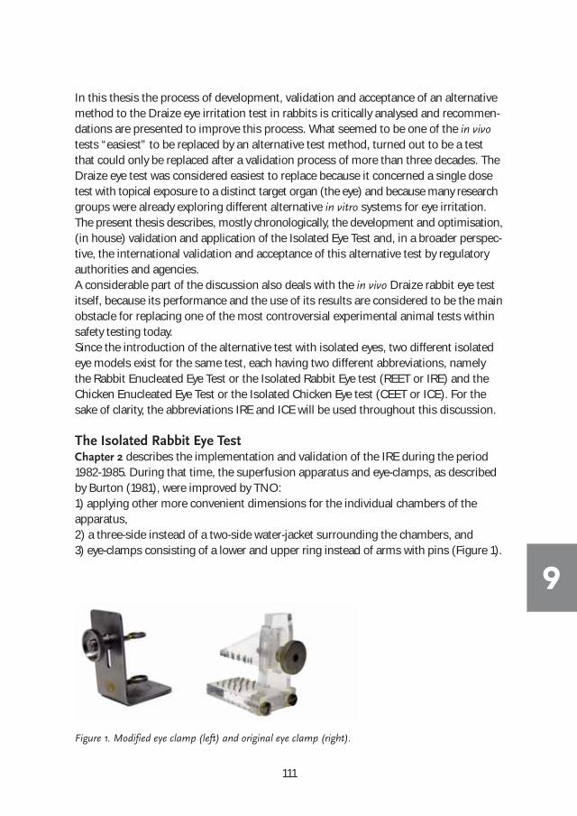
111
In this thesis the process of development, validation and acceptance of an alternativemethod to the Draize eye irritation test in rabbits is critically analysed and recommen-dations are presented to improve this process. What seemed to be one of the in vivotests “easiest” to be replaced by an alternative test method, turned out to be a testthat could only be replaced after a validation process of more than three decades. TheDraize eye test was considered easiest to replace because it concerned a single dosetest with topical exposure to a distinct target organ (the eye) and because many researchgroups were already exploring different alternative in vitro systems for eye irritation.The present thesis describes, mostly chronologically, the development and optimisation,(in house) validation and application of the Isolated Eye Test and, in a broader perspec-tive, the international validation and acceptance of this alternative test by regulatoryauthorities and agencies. A considerable part of the discussion also deals with the in vivo Draize rabbit eye testitself, because its performance and the use of its results are considered to be the mainobstacle for replacing one of the most controversial experimental animal tests withinsafety testing today.Since the introduction of the alternative test with isolated eyes, two different isolatedeye models exist for the same test, each having two different abbreviations, namelythe Rabbit Enucleated Eye Test or the Isolated Rabbit Eye test (REET or IRE) and theChicken Enucleated Eye Test or the Isolated Chicken Eye test (CEET or ICE). For thesake of clarity, the abbreviations IRE and ICE will be used throughout this discussion.
The Isolated Rabbit Eye TestChapter 2 describes the implementation and validation of the IRE during the period1982-1985. During that time, the superfusion apparatus and eye-clamps, as describedby Burton (1981), were improved by TNO: 1) applying other more convenient dimensions for the individual chambers of the apparatus, 2) a three-side instead of a two-side water-jacket surrounding the chambers, and3) eye-clamps consisting of a lower and upper ring instead of arms with pins (Figure 1).
Figure 1. Modified eye clamp (left) and original eye clamp (right).
9

112
9
With the improved equipment, thirty-four compounds already tested in vivo at TNOwere retested in the IRE test. The use of additional rabbits serving as eye donors wasavoided by reusing rabbits which had already been submitted to in vivo eye irritationtesting (unused control eye) or to in vivo skin irritation testing (both eyes), and whichhad to be sacrificed at termination of the in vivo study. The IRE results showed anoverall correlation of 82% with the in vivo results, using a general classification schemefor the grading of in vivo and in vitro eye irritation (i.e. not, slight, moderate or severeirritant). Four compounds, overpredicted (slight or moderate instead of non-irritant)by the IRE, were all moderate to severe irritants in the in vivo skin irritation test. A common physico-chemical characteristic of these compounds was their hydro-phobicity. A general observation in the in vivo rabbit eye test was that hydrophobiccompounds stayed in contact with the cornea (eye) for a relatively short period oftime, because they mixed poorly with the tear film on the cornea and because the nictitating membrane (third eye-lid) acted as a wiper, and rapidly removed the compoundfrom the eye. This was in distinct contrast to the skin irritation test where the compoundwas kept in contact with the skin with the aid of a patch and fixative tapes for at least 4hours. The presence of a third eye-lid is an example of a condition very specific to thein vivo rabbit eye test influencing the exposure. Hence this can cause a problem withrespect to the validation of alternative methods since an alternative method is notable to mimic the presence of a third eye-lid (which is also an irrelevant condition withrespect to humans). The issue of physico-chemical properties of the compounds andthe interpretation of the in vivo/in vitro results in relation to the practical aspects of thein vivo rabbit eye test is further described in the discussion of Chapters 7 and 8. On thebasis of the results with the 34 compounds, the IRE was considered to be a sensitiveand useful test system for the identification of eye irritants. Negative in vitro resultsshould be confirmed only in case of expected regular eye contact and in a maximumof three rabbits. At that time six rabbits was the usual number in the Draize eye test in order to comply with the US guidelines, although the first OECD guideline on eye irritation (OECD, 1981) recommended the use of 3 rabbits, which could mean a considerable reduction in the use of rabbits for eye irritation testing.
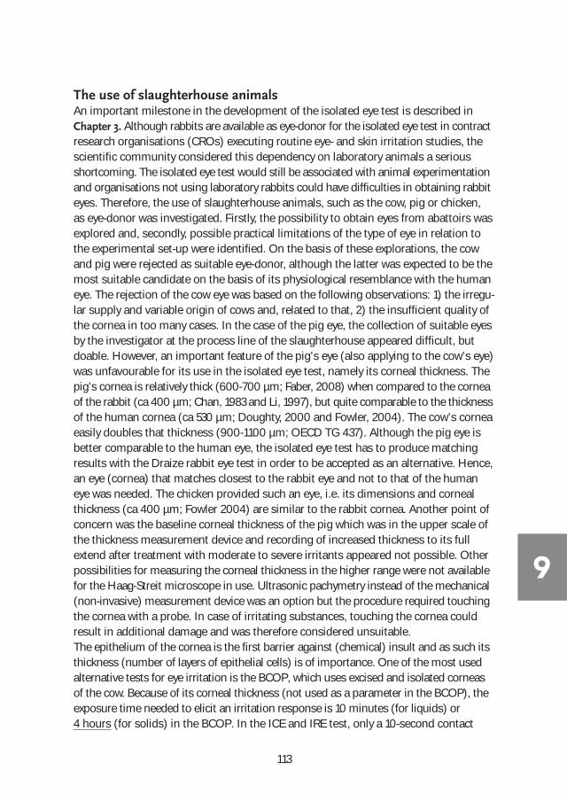
113
The use of slaughterhouse animals An important milestone in the development of the isolated eye test is described inChapter 3. Although rabbits are available as eye-donor for the isolated eye test in contractresearch organisations (CROs) executing routine eye- and skin irritation studies, thescientific community considered this dependency on laboratory animals a seriousshortcoming. The isolated eye test would still be associated with animal experimentationand organisations not using laboratory rabbits could have difficulties in obtaining rabbiteyes. Therefore, the use of slaughterhouse animals, such as the cow, pig or chicken,as eye-donor was investigated. Firstly, the possibility to obtain eyes from abattoirs wasexplored and, secondly, possible practical limitations of the type of eye in relation tothe experimental set-up were identified. On the basis of these explorations, the cowand pig were rejected as suitable eye-donor, although the latter was expected to be themost suitable candidate on the basis of its physiological resemblance with the humaneye. The rejection of the cow eye was based on the following observations: 1) the irregu-lar supply and variable origin of cows and, related to that, 2) the insufficient quality ofthe cornea in too many cases. In the case of the pig eye, the collection of suitable eyesby the investigator at the process line of the slaughterhouse appeared difficult, but doable. However, an important feature of the pig’s eye (also applying to the cow’s eye)was unfavourable for its use in the isolated eye test, namely its corneal thickness. Thepig’s cornea is relatively thick (600-700 µm; Faber, 2008) when compared to the corneaof the rabbit (ca 400 µm; Chan, 1983 and Li, 1997), but quite comparable to the thicknessof the human cornea (ca 530 µm; Doughty, 2000 and Fowler, 2004). The cow’s corneaeasily doubles that thickness (900-1100 µm; OECD TG 437). Although the pig eye isbetter comparable to the human eye, the isolated eye test has to produce matching results with the Draize rabbit eye test in order to be accepted as an alternative. Hence,an eye (cornea) that matches closest to the rabbit eye and not to that of the humaneye was needed. The chicken provided such an eye, i.e. its dimensions and cornealthickness (ca 400 µm; Fowler 2004) are similar to the rabbit cornea. Another point ofconcern was the baseline corneal thickness of the pig which was in the upper scale ofthe thickness measurement device and recording of increased thickness to its full extend after treatment with moderate to severe irritants appeared not possible. Otherpossibilities for measuring the corneal thickness in the higher range were not availablefor the Haag-Streit microscope in use. Ultrasonic pachymetry instead of the mechanical(non-invasive) measurement device was an option but the procedure required touchingthe cornea with a probe. In case of irritating substances, touching the cornea could result in additional damage and was therefore considered unsuitable. The epithelium of the cornea is the first barrier against (chemical) insult and as such itsthickness (number of layers of epithelial cells) is of importance. One of the most usedalternative tests for eye irritation is the BCOP, which uses excised and isolated corneasof the cow. Because of its corneal thickness (not used as a parameter in the BCOP), theexposure time needed to elicit an irritation response is 10 minutes (for liquids) or 4 hours (for solids) in the BCOP. In the ICE and IRE test, only a 10-second contact
9

114
9
period is needed to elicit a relevant irritation response. A relevant response means aresponse comparable to that observed in the in vivo rabbit test. Apart from a comparablethickness to the rabbit cornea and, to a certain extent to the human cornea, the chickencornea has another feature, which might be an advantage to predict (human) eye irritation, i.e. a well-developed Bowman’s membrane (Fowler 2004). This membrane,which is in between the epithelium and stroma, is also well developed in the humancornea, but poorly developed in the rabbit cornea. In the discussion of Chapter 6,more information on Bowman’s membrane in relation to histopathology performed inthe ICE is presented. With respect to the availability of eyes, attaining chicken eyes appeared relatively easyby simply collecting heads just after the sedation of the animals at the process line,transporting them to the laboratory and enucleating them from the heads within 2 hours after sacrifice. This time period is sufficient to guarantee corneas meeting theacceptance criteria for testing, i.e. no or very slight opacity, no or very slight fluoresceinstaining, and a corneal thickness in the normal range. The daily processing of thousandsof chickens at the slaughterhouse guaranteed a constant supply of suitable eyes (corneas).Since 1981 we have visited the same slaughterhouse (v.d. Bor, Nijkerkerveen) for oursupply and only during a period of about 6 months in 2003 could eyes not be obtainedbecause of an Avian flu break-out (H7N7 variant) in a large part of the Netherlands.Even then, eyes could be obtained from another slaughterhouse outside the affectedregion (van Miert, Breukelen).The use of chicken eyes was evaluated by testing the 21 reference chemicals previouslytested in the first (pilot) EC validation study of the IRE (EC, 1991) which were selectedto be representative of currently used industrial chemicals of different chemical classesand ranging from non-irritant to severe irritant. Describing the criteria and scoringsystem of the in vitro corneal effects together with a Prediction Model (PM) for matchingthe EC scheme for classification and labelling of compounds (see Table 3 of the intro-duction) was an important step forward. The development of the prediction schemewas primarily a theoretical exercise based on the range of physiological responses observed in the ICE (corneal swelling, opacity and fluorescein retention of damageepithelial cells). Because the corneal effects determined in the ICE have a direct relation-ship with the in vivo response (e.g. in vitro opacity for prediction of in vivo opacity), aPM could be established based on the magnitude/range of the effects and not byusing an empirically-derived mathematical algorithm to translate an in vitro effect toan in vivo effect. For instance, in the HET-CAM assay lysis of blood vessels of the chorioallantois membrane is measured as time (seconds) of first occurrence (Luepke,1985). The number of seconds recorded cannot be translated directly to an in vivo effect,which is considered a serious limitation of the method. A PM could be designed onlyafter computer calculations of data obtained for a relevant number of compounds byusing the in vivo MMAS (Modified Maximum Average Score) as the sole parameter forthe in vivo test. The computer calculation was based on a mathematical formula orconversion algorithm resulting in a single in vitro irritation index score comparable to

115
the in vivo MMAS. Computer calculations were also needed with the BCOP assay,using a cornea, because the opacity is measured as a reduced light transmission valueand the epithelial damage as the amount of fluorescein penetrated through the cornea,leading to an In Vitro Irritation Score (IVIS = mean opacity value + (15 x mean permeabilityOD490 value)). The ICE followed a theoretical approach not using in vitro data to be compared to thein vivo MMAS. By knowing the ranges of the in vitro ICE responses (swelling, opacityand fluorescein retention), cut-off values were chosen to identify different categoriesof effect, viz. a non (Category I), slight (Category II), moderate (Category III) or severe(Category IV) effect. Thus after testing a compound, three categories were established,i.e. one for swelling, one for opacity and one for fluorescein retention. The assignment of the final irritation classification to a non-irritant, (slight or moderate)irritant or severe irritant was obtained by the combination of these three categories.Again, a theoretical and weighted division of the different combinations possible wasmade for each final classification. For instance, at the low end of the ICE classificationsystem the combination of I/I/I is a non-irritant and at the high end the combinationof IV/IV/IV is a severe irritant. The combinations possible and respective irritationclassifications are shown in Table 1.
General classification UN-GHS classificationa Combinations of categories
Not irritating Not classified 3 x I 2 x I, 1 x II
Slightly irritating 2B : Mild irritant/causes eye irritation
2 x II, 1 x I 3 x II 2 x II, 1 x III 1 x I, 1 x II, 1 x III
Moderately irritating 2A : Irritant/causes eye irritation
2 x III, 1 x I 2 x III, 1 x II 3 x III 1 x IV, 2 x I 1 x IV, 2 x II 1 x IV, 2 x III 1 x IV, 1 x III, 1 x II
Severely irritating 1 : Irreversible effects on the eye/ serious damage to the eye
2 x IV, 1 x I 2 x IV, 1 x II 3 x IV
a Globally Harmonised System of Classification and Labelling of Chemicals (UN-GHS). UN, New York and Geneva, 2007
Table 1. ICE in vitro classification system.
9

116
9
Often the PM of other alternatives had to be revised after more compounds had beentested by the in vitro test or additional PM’s were especially designed for certain cate-gories of compounds. With the ICE, the criteria system for scoring the effects wasnever changed, while the classification system has been modified twice, i.e. once toaccommodate the introduction of the UN-GHS classification system and secondlyafter adoption as an OECD guideline for non-irritants (OECD 438, 2013). The use of the in vivo MMAS in establishing the PM proved to be less ideal thanthought. The EC and UN-GHS systems do not use the MMAS for determination of theirritation classification of a compound. Instead, the individual in vivo tissue scores areused. Evaluation of the use of the MMAS in the validation study of the European Commission and the British Home Office (EC/HO study) showed a poor correlationwith these classification systems (Prinsen, 1999). This is further presented in thesummary and discussion of Chapter 7.On the basis of the ICE study with 21 reference materials (Prinsen and Koëter, 1993)the conclusions were that: 1) although the ICE does not assess conjunctival damage,its sensitivity to predict ocular damage is not reduced, 2) the ICE correctly predictedthe EC classifications of the 21 chemicals and 3) the ICE fitted in the previously updatedEC B.5 and OECD 405 guidelines regarding acute eye irritation/corrosion now includingrecommendations to use alternatives for the prescreening or positive identification ofstrong eye irritants.
The Isolated Chicken Eye Test in practice The results described in the previous paragraph demonstrated that the ICE test wasready to act as a prescreen for the Draize eye test in rabbits. Apart from fundamentalresearch, TNO also performed routine contract toxicity tests with a regular flow ofacute toxicity testing (acute skin/eye irritation, acute oral/dermal toxicity and skin sensitization) which provided an excellent platform to incorporate the ICE as a prescreenfor the in vivo Draize eye irritation test. Acute toxicity tests are predominantly carriedout for regulatory purposes and conducting an in vitro test prior to the in vivo testwould mean additional costs for the sponsor. As a consequence, the sponsor could refuse inclusion of the ICE, whereas “parallel” testing of compounds in the ICE andthe in vivo Draize eye test would provide valuable information on the performance ofthe ICE. Moreover, it would introduce the ICE to sponsors and regulatory authorities,enhancing the chance for international acceptance, and for participation in validationprograms that were starting to be initiated. Therefore, it was decided to include theICE in addition to the routine Draize eye test without extra costs for the sponsor. In Chapter 4, the results are presented of this “parallel” testing of 44 compounds.These compounds were considered to be a relevant cross-section of compounds (rawchemicals, finished products and formulations) routinely produced by the (chemical)industry, and to provide evidence for the use of the ICE in the assessment of eye irritationfor regulatory purposes. Instead of only comparing single in vivo and in vitro irritationindex scores, as was the common practice in validation, the individual components

117
used for calculation of the index score were also analysed. This was a recommendationof the United States Interagency Regulatory Alternatives Group (IRAG) made during aworkshop on eye irritation testing in Washington DC, in 1993 (Scala, 1997). Fourteendifferent in vivo scores were derived from each of the 44 in vivo tests, covering timescores (1-hr, 24 hr, 1-72 hr, days to recovery), index scores (MAS and total eye score),and individual tissue cores (cornea, area of cornea involved, iris, cornea and iris combined, conjunctival redness, conjunctival swelling, conjunctival discharge, conjunctival scores combined). The in vivo critical scores were compared to the criticalscores of the ICE test, namely the scores for corneal swelling, corneal opacity, fluoresceinretention of damaged corneal epithelium and an index score (combination of thethree parameters). The overall correlations found for the in vivo scores with the ICE invitro scores were 0.90 (index score), 0.91 (corneal swelling), 0.86 (corneal opacity)and 0.82 (fluorescein retention). The correlation between the in vivo conjunctival scoresand the ICE scores were 0.92 (index score), 0.92 (corneal swelling), 0.93 (corneal opacity) and 0.86 (fluorescein retention). These correlations substantiated the conclusionmade by Burton (1972) that a relationship exists between the in vivo conjunctival damage and the corneal scores of the isolated eye test. Moreover, in ophthalmologythe term ocular surface was introduced to emphasize the potential interdependenceof the epithelium of the cornea and the epithelium of the conjunctivae (Thoft, 1977and Wagoner, 1997). “Subsequent clinical and research insights of Thoft and othersprovided compelling evidence of the functional relationships between these two adjacentcell populations”. Furthermore, the proven relationship and high correlation betweenthe critical scores of the ICE test and the Draize eye test demonstrated that the test isa relevant alternative to eye irritation, and that applying regulatory irritation classificationsystems is “just” a matter of choosing the appropriate threshold limits belonging tothe different irritation classes. This was supported by the in vivo and in vitro EC classifications obtained for the 44 compounds. Overall, it was concluded that the ICEprovided a practical prescreen for the Draize rabbit eye test and that only mild to moderate irritants in the ICE, generally showing the highest sensitivity to inter- andintra-laboratory variability, should be confirmed in the rabbit eye test. Eighteen yearslater, the OECD adopted this conclusion. Furthermore, the “parallel” testing showedthe ICE test to be robust in the sense that the practical aspects are not complicatedand relatively easy to control, i.e. a saline drip is sufficient to maintain the eyes ingood condition, and all compounds, regardless the physico-chemical properties, canbe assayed. The in-house repeatability of the ICE was assessed to be adequate during the ReferenceStandard Validation of in vitro tests sponsored by ECVAM (Brantom, 2000). Two refer-ence compounds for the group of siloxanes (decamethylcyclopentasiloxane and cyclo-hexylamino-functional PDMS) and two for the group of surfactants (Triton X-500 5%and cetylpyridium bromide 6%), representing non-irritants, Category 2B and 2A, andCategory 1 compounds, were tested five times each on different occasions.The publication of the results for the first 44 compounds did not result in termination
9

118
9
of the “parallel” testing program. The main reason for continuing the “parallel” testingwas that it was considered unethical to perform any toxicity test on live animals withoutprior information on the reactivity of the test compound using a “relevant” biologicalstructure such as the cornea. In those days, it was common practice to start differentacute toxicity tests with a new compound almost simultaneously and, if differentstudy directors were involved, often without consulting each other about the specificresults of their studies, whereas the result of an acute irritation test would have influ-enced or helped their decision concerning the study design to be followed. It becameapparent that the ICE fitted well in a tiered approach for acute toxicity testing. The resultsof the ICE provided not only information on eye irritation, but also gave informationthat could help to optimally design the other acute toxicity tests. If the ICE testshowed severe irritancy, the in vivo eye irritation test was waived and the skin irritationtest was initiated with one rabbit only. Important decisions for the conduct of theacute oral and dermal toxicity tests in rats could also be made on the basis of the outcome of the ICE. In most cases, these studies were started as a limit study withthe highest dose level of 2000 mg/kg body weight. In case of severely irritating or corrosive compounds, the local effects on the stomach or skin could lead to severesuffering or even mortality of the animals. When the ICE test showed severe effects,dosing of high levels or high concentrations of corrosive compounds could be avoided.When the ICE showed no or negligible signs of irritation (cytotoxicity), the decision toperform the acute oral and dermal tests with the highest dose level or a high test con-centration could be better justified. It was not until 2001 that the OECD adopted the use of results of any other in vitrotoxicity test on a compound in order to determine start levels to be used for the in vivoacute oral toxicity test in rats (OECD 423, 2001). For skin sensitization tests, this approach reduced the number of animals necessaryfor testing. At that time, the standard test for skin sensitization was the Guinea PigMaximization test (GPMT), requiring up to 40 animals for the main test and 6-9animals for the preliminary skin irritation test. The preliminary skin irritation test wasneeded to establish the appropriate (maximum tolerable) concentrations for the variousphases of the study (i.e. the intradermal and topical induction and the topical challenge).Normally ranges covering concentrations from 1% up to 100% had to be investigated.When the ICE showed no or negligible irritancy, the range to be examined could be limited to only 100% and one lower concentration, which in practice meant that only 3and not 6-9 animals were needed. At present, the GPMT is replaced by the LocalLymph Node Assay (LLNA, OECD TG 429, 2010), and this guideline also mentionsthe use of results of any other in vitro toxicity test on a substance as an aid in dose selection.In retrospect it can be concluded that the testing strategy was successful for the majority of compounds submitted for testing, i) identification of severe eye irritantswithout further in vivo testing, ii) tiered testing of skin irritants/corrosives, iii) determi-nation of acceptable (non-severe) dose levels of corrosive compounds in acute oral

119
and dermal toxicity testing, and iv) reduction of the number of animals used in thepreliminary irritation experiment of sensitization studies.“Parallel” in vivo and in vitro eye irritation testing was continued with another 50 com-pounds, meaning that each compound was first tested in the ICE and, in case of non-severe irritancy, directly followed by an in vivo rabbit test, both in full compliance withthe OECD principles of Good Laboratory Practice (GLP). These results were submittedto organizations dedicated to the validation of alternative non-animal tests, such asECVAM and ICCVAM. Because the performance of the ICE was considered sufficientlyestablished after the “parallel” testing of 94 compounds, another approach was intro-duced to make optimal use of the ICE at low extra costs. First a non-GLP ICE test withonly one eye was carried out and depending on the outcome either a full GLP ICE test(in case of severe irritancy) or an in vivo rabbit eye test (in case of non-severe irritancy)was carried out. This procedure was followed until the ICE OECD guideline 438 wasadopted to include the identification of non-irritants (OECD, 2013). From then on,only compounds identified by the ICE as irritating (Category 2) need to be tested inthe in vivo eye irritation test in rabbits.
The Isolated Chicken Eye Test as a stand-alone testThe ICE was increasingly being used as a stand-alone test by companies, which pursueda non-animal safety testing strategy of their products. One of the major internationalhousehold and personal care companies, the Procter & Gamble Company (P&G),adopted the ICE for the eye irritation screening of their products. A variety of productsand research formulations, ranging from hand or machine dish-wash products,household or industrial laundry products, householdcleaning products, cosmetics and hair dyes, were sub-mitted for testing in the ICE (Chapter 5). This was aboost for the application of the ICE and further develop-ment of the method. In the past, P&G had heavily investedin the introduction of a modification of the Draize eyetest, because the exposure conditions of the standardDraize eye test were considered unrealistic and exagger-ated compared to (recorded) accidental human exposure.Under the standard conditions of the Draize eye test aconsiderable number of their products, especially deter-gents in powder form, had to be labelled as severe eyeirritants. Their modification presented as an alternativemethod, the Low Volume Eye Test (LVET; Griffith,1980),uses only one tenth of the dose volume of the test material administered directly onto the cornea insteadof instilling it in the lower conjunctival sac (Figure 2).P&G employed the LVET since 1980 and the results correlated well with their database on human responses
Figure 2. Dose volume of 0.1 mL
versus 0.01 mL (Griffith, 1987).
9

120
9
to accidental exposure and to results of clinical studies (Griffith, 1980 and 1987). Although less burdening for the animals, the LVET was still an animal test and in viewof their non-animal testing policy they decided to explore the suitability of the ICE fortheir products.Twenty products (8 liquids and 12 powders) representing acidic or alkaline cleaners,dishwashing liquids or powder, powder detergents and raw materials of these productswere tested in the ICE. In addition to the ICE standard application of 30 µl or 30 mg,which is relatively equivalent to the amount used in the Draize eye test, volumes of 10and 3 µl and amounts of 10 and 3 mg were used to examine if this provided better correlation with the LVET results. Furthermore, histopathology of the treated corneassampled at the end of the observation period was now included. The inclusion of histopathology was one of the recommendations made by Maurer (2002), based ontheir microscopic examination of the cornea in LVET studies. They suggested a directcorrelation between the depth of injury in the cornea and possible recovery of eye lesions. Since measurement of recovery is not possible in the ICE (and in all other alternatives), it could be an important improvement of the method and helpful in theargumentation for absence or presence of microscopic lesions in the ICE that couldpredict (ir)reversibility. The outcome of this study (Schutte, 2009) was that, in general,the results of the ICE using the standard volume of 30 µl or mg were in line with ormore conservative than the LVET in terms of classification. The level of overpredictionfound in the ICE was expected and considered acceptable since the ICE was developedfor prediction of the Draize eye test and not the LVET. For P&G the ICE test was considered useful for several purposes, such as 1) EU/GHSclassification and labelling of powder and liquid household cleaning products, 2) screeningof candidate formulations, and 3) weight-of-evidence approach by determining the profile of new cleaning product formulations against benchmark products. A definiteconclusion on the usefulness of histopathology in the ICE could not be made, but thedata showed that assessment of the histopathological lesions in the various parts of thecornea was possible, enabling the application of the “Depth-of-Injury” theory of Maurer.The question remained if this theory established on the basis of an inflammatory processin the rabbit’s eye (consisting of initial ocular injury and subsequent repair over days/weeks) correlated with the irritation process or damage in the ICE. The outcome of thein vivo Draize rabbit eye test is heavily influenced by several unpredictable factors orevents which will not occur in the ICE test. These factors and their consequences for thefinal classification of compounds are part of the discussion of Chapter 8.
Optimizing histopathology in the Isolated Chicken Eye TestFor the histopathological observations in the ICE, as presented in Chapter 6, a quitebasic and routine procedure of processing the eye/cornea is used. The eye is preservedin formalin at the end of the study, i.e. 4 hours after the 10-second exposure, and furt-her processed into a paraffin block from which slides of the longitudinal section of thecorneal center are prepared. This area was considered appropriate since the application

121
of the test compound was made at the center of the cornea and in general the slit-lampobservation showed confluent, homogenous corneal effects. In case of non-homogenouseffects, such as focal spots with more severe opacity not present in the central area,these parts were also examined. The choice of the staining appeared an importantissue. In first instance, the most common staining by Haematoxylin & Eosin (H&E)was used, which was later replaced by the Periodic Acid-Schiff (PAS) staining, whichprovided a much better discrimination of the different layers of the cornea. The threemajor layers of the cornea, i.e. epithelium, stroma and endothelium were well visiblewith both stainings. The other structures, such as basement membrane and Bowman’smembrane (between epithelium and stroma) and Descemet’s membrane (betweenstroma and endothelium), were not that visible with H&E, whereas PAS providedmuch better results. The integrity of the membranes was considered to play a role inthe injury and recovery process of the cornea, and the visibility of these membranes bymicroscope was considered important for an adequate histopathological assessmentby the pathologist. Therefore, other staining methods specifically targeting collagen-rich membranes were tested on corneas treated with compounds representing a non-irritant, two irritants and a severe irritant (corrosive). The microscopic examinationfocused on the basement and Bowman’s membrane and not on Descemet’s membrane(see Introduction, Figure 2), because damage to this membrane adjacent to the endo-thelium was considered to result in severe, irreversible effects and not borderline effects.Of the five stainings selected, i.e. H&E, PAS, Trichrome, AZAN (Azocarmine & aniline)and EVG (Elastic Van Gieson), PAS was clearly superior with respect to visibility of themembranes and the quality of the morphology of the various corneal structures. Moreover, the histopathological examinations provided interesting facts and insights.After severe corrosive damage to the cornea by sodium hydroxide, observed macro-scopically through slit-lamp observation as maximum swelling and very severe opacityof the cornea, the basement and the Bowman’s membranes appeared undamagedwhile effects were seen in the underlying stroma. Does this mean that the functionalityof the membranes was not compromised or is light microscopy not able to detectsuch damage of the membranes? This observation led to the conclusion that depth ofinjury is not the only factor determining the seriousness of corneal injury. Personalcommunications with an ophthalmologist of the University Medical Centre of Utrecht,specialized in the cornea, and publications (Wagoner, 1997; Terry, 2002; Rama, 2010)indicated that in the clinic, emphasis is put on corneal opacity and corneal stem cellsurvival after (chemical) injury (see Table 2). With severe stem cell damage, recoveryof the corneal damage by re-epithelialization of migrating stem cells from the corneallimbus is not possible. In that case, the recovery process will result in complete, irrevers-ible conjunctivalization of the ocular surface. The stem cells are rather superficially located in the limbal region of the eye and as such involved in early contact with a topically applied compound. Therefore, the possibility of screening the viability ofstem cells after chemical injury could be of value. However, explorations to stain stemcells of the chicken cornea by p63 immunostaining appeared unsuccessful.
9
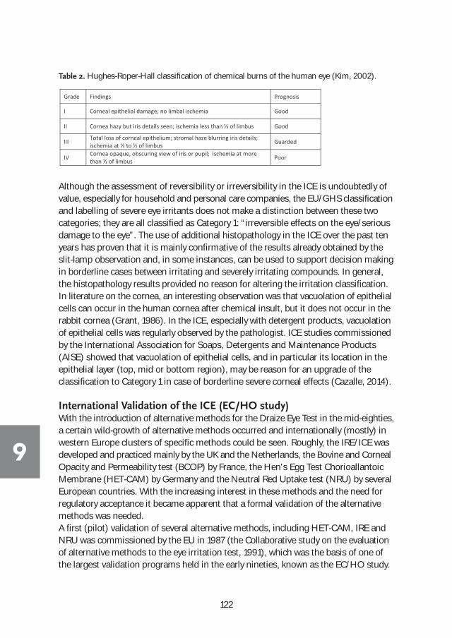
122
9
Although the assessment of reversibility or irreversibility in the ICE is undoubtedly ofvalue, especially for household and personal care companies, the EU/GHS classificationand labelling of severe eye irritants does not make a distinction between these two categories; they are all classified as Category 1: “irreversible effects on the eye/seriousdamage to the eye”. The use of additional histopathology in the ICE over the past tenyears has proven that it is mainly confirmative of the results already obtained by theslit-lamp observation and, in some instances, can be used to support decision makingin borderline cases between irritating and severely irritating compounds. In general,the histopathology results provided no reason for altering the irritation classification. In literature on the cornea, an interesting observation was that vacuolation of epithelialcells can occur in the human cornea after chemical insult, but it does not occur in therabbit cornea (Grant, 1986). In the ICE, especially with detergent products, vacuolationof epithelial cells was regularly observed by the pathologist. ICE studies commissionedby the International Association for Soaps, Detergents and Maintenance Products(AISE) showed that vacuolation of epithelial cells, and in particular its location in theepithelial layer (top, mid or bottom region), may be reason for an upgrade of the classification to Category 1 in case of borderline severe corneal effects (Cazalle, 2014).
International Validation of the ICE (EC/HO study) With the introduction of alternative methods for the Draize Eye Test in the mid-eighties,a certain wild-growth of alternative methods occurred and internationally (mostly) inwestern Europe clusters of specific methods could be seen. Roughly, the IRE/ICE wasdeveloped and practiced mainly by the UK and the Netherlands, the Bovine and CornealOpacity and Permeability test (BCOP) by France, the Hen's Egg Test ChorioallantoicMembrane (HET-CAM) by Germany and the Neutral Red Uptake test (NRU) by severalEuropean countries. With the increasing interest in these methods and the need forregulatory acceptance it became apparent that a formal validation of the alternativemethods was needed.A first (pilot) validation of several alternative methods, including HET-CAM, IRE andNRU was commissioned by the EU in 1987 (the Collaborative study on the evaluationof alternative methods to the eye irritation test, 1991), which was the basis of one ofthe largest validation programs held in the early nineties, known as the EC/HO study.
Grade Findings Prognosis
I Corneal epithelial damage; no limbal ischemia Good
II Good
III Total loss of corneal epithelium; stromal haze blurring iris details; Guarded
IV Cornea opaque, obscuring view of iris or pupil; ischemia at more than ½ of limbus Poor
Table 2. Hughes-Roper-Hall classification of chemical burns of the human eye (Kim, 2002).

123
In this comprehensive study, organized by the European Commission (EC) and theBritish Home Office (HO), nine methods including the ICE (see Introduction, Table 6)were each carried out by four laboratories testing 60 chemicals which represented dif-ferent chemical classes and irritation potential (Balls, 1995). The performance of the ICE in the EC/HO study, for which TNO was the lead laboratory, and the validationprocess in general is part of the discussion of Chapters 7 and 8.The chemicals were selected from the ECETOC database and were considered to havereliable in vivo eye irritation data. Basically that meant the tests having been performedunder GLP conditions and in compliance with OECD TG 405 (1981). No other assess-ment with respect to the quality of the individual data was made. The outcome of thestudy was quite disappointing; none of the methods were capable of identifying theeye irritation potential of the compounds (maximum overall correlation ranged from0.34 to 0.55). Breaking up the compounds into different categories such as liquids, solids, surfactants, non-irritants, severe irritants etc. did not improve the correlationsignificantly, although the group of surfactants showed the best results across all methods. One of the reasons for this disappointing result was considered to be the use of theMMAS as the sole parameter derived from the in vivo data. This score, ranging from0-110, is an average of the maximum individual tissue scores of the animals recordedfor a compound. By using certain cut-off values for the MMAS, a compound is classifiedas a non-irritant (score 0-25), irritant (score 25-59) or severe irritant (score >59). Assessment of the entire process of ocular inflammation by a single index instead ofusing the in vivo data to its full potential appeared rather inadequate. For instance, theclassification system used in the EC does not use a single irritation index score, but isbased on individual tissue scores (i.e. for cornea, iris and conjunctivae separately)and/or the (ir)reversibility of these effects within 21 days (Table 3 of the Introduction).In 1998, the OECD published a proposal for the harmonization of hazard classificationbased on eye irritation/corrosion, which is comparable to the EC classification system,because it also uses the individual tissue scores separately (Table 4 of the introduction).However, slightly lower thresholds were used for classification as an irritant (Category 2)or severe irritant (Category 1), and additionally recovery of eye effects within 7 dayswere used to discriminate between a mild irritant (2B) and an irritant (2A). Becausethe MMAS is not used for regulatory classification, the impact of the EC and proposedOECD classification criteria on the compounds tested in the EC/HO study was inves-tigated (Chapter 7). First of all, it was established that applying the two classificationsystems to the EC/HO compounds resulted in classifications that were comparablebetween the two systems, i.e. R36 compounds were also Category 2A/2B compounds,and R41 compounds were also Category 1 compounds. Subsequently, it was demon-strated that the MMAS cut-offs of 25 and 59 belonging to, respectively, irritant and severe irritant, were not appropriate for classification according to these two regulatorysystems. Eight compounds with an MMAS lower than 59 were in fact severe irritants(R41/Category 1) and 4 compounds with a MMAS higher than 59 and 3 compounds
9
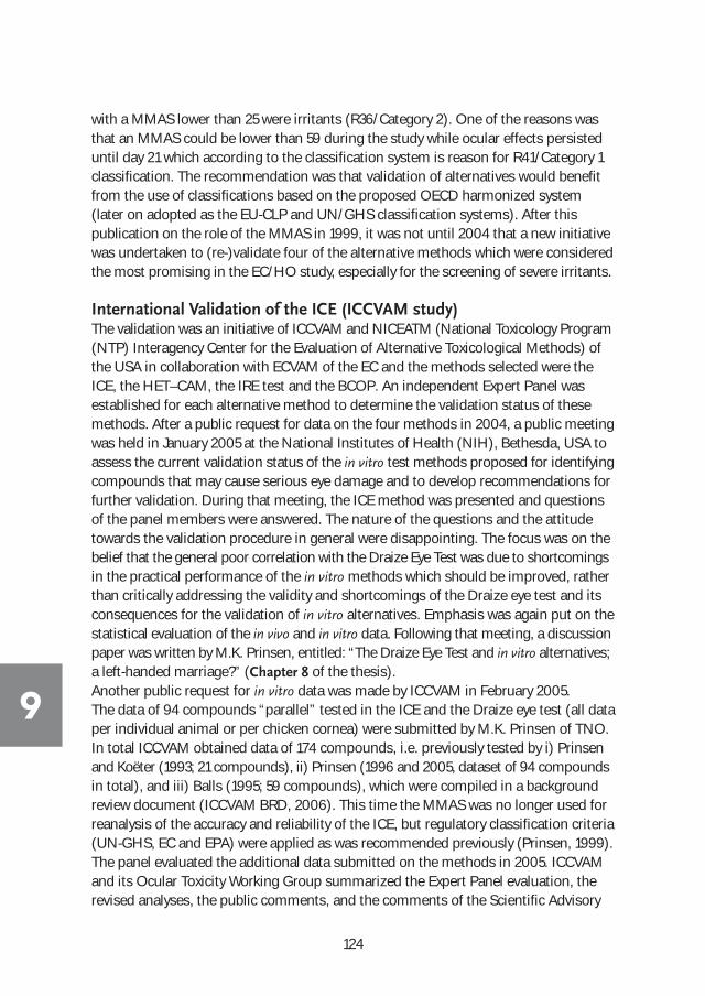
124
9
with a MMAS lower than 25 were irritants (R36/Category 2). One of the reasons wasthat an MMAS could be lower than 59 during the study while ocular effects persisteduntil day 21 which according to the classification system is reason for R41/Category 1classification. The recommendation was that validation of alternatives would benefitfrom the use of classifications based on the proposed OECD harmonized system(later on adopted as the EU-CLP and UN/GHS classification systems). After this publication on the role of the MMAS in 1999, it was not until 2004 that a new initiativewas undertaken to (re-)validate four of the alternative methods which were consideredthe most promising in the EC/HO study, especially for the screening of severe irritants.
International Validation of the ICE (ICCVAM study) The validation was an initiative of ICCVAM and NICEATM (National Toxicology Program(NTP) Interagency Center for the Evaluation of Alternative Toxicological Methods) ofthe USA in collaboration with ECVAM of the EC and the methods selected were theICE, the HET–CAM, the IRE test and the BCOP. An independent Expert Panel was established for each alternative method to determine the validation status of thesemethods. After a public request for data on the four methods in 2004, a public meetingwas held in January 2005 at the National Institutes of Health (NIH), Bethesda, USA toassess the current validation status of the in vitro test methods proposed for identifyingcompounds that may cause serious eye damage and to develop recommendations forfurther validation. During that meeting, the ICE method was presented and questionsof the panel members were answered. The nature of the questions and the attitude towards the validation procedure in general were disappointing. The focus was on thebelief that the general poor correlation with the Draize Eye Test was due to shortcomingsin the practical performance of the in vitro methods which should be improved, ratherthan critically addressing the validity and shortcomings of the Draize eye test and itsconsequences for the validation of in vitro alternatives. Emphasis was again put on thestatistical evaluation of the in vivo and in vitro data. Following that meeting, a discussionpaper was written by M.K. Prinsen, entitled: “The Draize Eye Test and in vitro alternatives;a left-handed marriage?” (Chapter 8 of the thesis). Another public request for in vitro data was made by ICCVAM in February 2005. The data of 94 compounds “parallel” tested in the ICE and the Draize eye test (all dataper individual animal or per chicken cornea) were submitted by M.K. Prinsen of TNO.In total ICCVAM obtained data of 174 compounds, i.e. previously tested by i) Prinsenand Koëter (1993; 21 compounds), ii) Prinsen (1996 and 2005, dataset of 94 compoundsin total), and iii) Balls (1995; 59 compounds), which were compiled in a backgroundreview document (ICCVAM BRD, 2006). This time the MMAS was no longer used forreanalysis of the accuracy and reliability of the ICE, but regulatory classification criteria(UN-GHS, EC and EPA) were applied as was recommended previously (Prinsen, 1999).The panel evaluated the additional data submitted on the methods in 2005. ICCVAMand its Ocular Toxicity Working Group summarized the Expert Panel evaluation, therevised analyses, the public comments, and the comments of the Scientific Advisory

125
Committee on Alternative Toxicological Methods in a final Test Method Evaluation Report (ICCVAM, 2006).The conclusion was that ”there are sufficient data to substantiate the use of the BCOP orthe ICE test methods, with certain limitations, as screening tests to identify compounds asocular corrosives and severe irritants in a tiered-testing strategy, using a weight-of-evidenceapproach, for regulatory hazard classification purposes”. The limitations of the ICE werethe testing of alcohols based upon the false positive rate and of solids and surfactantsbased upon the false negative rates. The limitations for the BCOP were the testing ofalcohols and ketones based upon the false positive rates and of solids based upon thefalse negative rate.Although the acceptance as a screen for ocular corrosives and severe irritants was asuccess for the ICE, the way the data were used by ICCVAM for reanalyses and for adjustments in the experimental procedures of the ICE was considered debatable. It was acknowledged that the in vivo rabbit eye test was subject to variability, but the in vivo data, however, were taken as absolutely accurate (the “Golden Standard”) in predicting the eye irritation potential of a compound. The in vivo irritation classifications were assigned if the study performance met withthe criteria set by ICCVAM. One of the criteria (assessment of full reversibility of anyeye effect) was the reason that the database (ICCVAM BRD of ICE: Appendix D, March2006) contained many gaps compared to the original data submitted, whereas sufficientin vivo data were available for classification. For example, two compounds (2,2-dimethylbutanoic acid and p-fluoroaniline) identified in the EC/HO validation study as severeirritants (R41) on the basis of the individual in vivo data (ECETOC, Technical documentno. 48: 2, June 1998) were rejected with the remark “study criteria not met”. The twocompounds were correctly identified as R41 by the ICE and by most other in vitromethods participating in the validation study. The in vivo classification was based onsound scientific judgment, but ICCVAM decided to exclude the compounds, becausea 21-day observation period was not completed. The OECD/EC guidelines (at the timeof testing) specified that the observation period should be long enough to evaluatethe reversibility or irreversibility of the lesions. The six rabbits treated with 2,2-dimethylbutanoic acid still showed slight to severe corneal opacity and neovascularization ofthe cornea at 14 days after treatment. It was considered evident that these lesionswould not have cleared within a 21-day observation period. Thus, the 14-day observationperiod applied was in agreement with the guidelines and should not have been a reasonfor discarding the results by ICCVAM. The same applied for p-fluoroaniline, which causedmoderate to severe corneal opacity and iritis score 2 (highest score possible; no reactionto light, haemorrhage, gross destruction). The test was terminated on day 3, which isalso in agreement with the guidelines which state that animals may be humanely sacrificed if the severity of the effects is considered too high. Similar cases occurred in the “parallel” data set of the 94 compounds provided byPrinsen (2005). These cases also mainly concerned in vivo eye irritation studies thatwere terminated earlier than 21 days after treatment because of the severity of eye effects
9

126
9
or compounds lacking an in vivo eye irritation study because of proven skin corrosivityin the in vivo rabbit skin irritation test (10 compounds), that was performed immediatelyafter the ICE had shown severe eye irritancy. In full agreement with the guidelines, thein vivo eye irritation test in rabbits was waived in these ten cases. The individual in vivoskin irritation data and the ICE data were provided to ICCVAM, but the 10 compoundsremained excluded from the analyses because of “classification assigned on the basisof skin corrosion assay” (SC). Remarkably, ICCVAM claimed to apply the criteria forclassification according to the EC (1993) and UN-GHS (2007), whereas both guidelinesunambiguously state “corrosive to skin” as one of the criteria for classifying a compoundas severely eye irritating. Another remarkable conclusion in the ICCVAM document was the underperformanceof surfactants by the ICE. This conclusion was primarily based on the 6 surfactantsexamined in the EC/HO validation study. The correlation percentages for surfactantsin the EC/HO study tested by the four participants that performed the ICE were 72, 82,83 and 76%, compared to an overall mean correlation percentage of 54%. In general,the chemical class of surfactants was best predicted by each of the nine in vitro methodsparticipating in the EC/HO study. The fact the ICE has been employed by P&G formore than ten years for their product development (Schutte, 2009), the majority ofwhich contains surfactants covering the whole spectrum of eye irritancy, is in contrastwith ICCVAM’s conclusion. More recently, member companies of the International Association for Soaps, Detergents and Maintenance Products (AISE) increasingly usethe ICE for their (surfactant-containing) products (Cazelle, 2014). With respect to the ICE study performance the ICCVAM expert panel identified twomajor issues: 1) the variability in swelling percentages obtained by the four laboratoriesperforming the ICE, and 2) the use of only one control eye per experiment.
Variability in swelling percentages (high CVs) The variation in swelling percentage was caused by the use of different pachymeterswith different slit width settings. This was already intensively discussed by the Management Team of the EC/HO study, but was considered of no concern becausethe in vivo MMAS was compared to the critical scores of the ICE (i.e. the max. meanswelling%, max. mean opacity score and mean fluorescein score) and not to the regulatory irritation classifications. ICCVAM overlooked this fact and decided to applythe TNO ICE system for categorizing effects to the other three participating labs aswell. This was a valid approach for the opacity and the fluorescein scores because thescoring is exactly the same for the four labs, but it could not be used integrally for theswelling %. The misconception of the variability in corneal swelling by the ICCVAM expert panel led to incorrect conclusions and recommendations. For example, centeringlights needed to be installed on the optical pachymeter to improve the determinationof corneal thickness by ensuring consistent central corneal thickness measurementsacross laboratories. The purpose of these lights in human ophthalmology is to guidethe patient’s eye to a fixed point and thus perform the reading at the centre of the cornea.

127
This is used because the subjects often (involuntarily) move their eyes making the(central) corneal thickness assessment difficult and variable. The chicken eye is isolatedand fixed, so there is no movement at all. Therefore, the corneal thickness can bemeasured in a very accurate and reproducible way at the centre of the cornea withoutany additional aid.
Use of control eyesICCVAM decided to increase the number of negative control eyes from the usual oneper experiment to three, because three was the accepted minimum of replicates in invitro testing in general. The use of only one negative control eye has been employedand approved by all users of the isolated eye test (both with rabbit eyes and with chickeneyes) since the introduction of the method by Burton in 1981 and during the EC/HOstudy. The use of only one negative control eye is justified since the effects of the corneatreated with the test material are not assessed or evaluated in any way against the effectsof the control eye. This is possible because, prior to testing, the quality and suitabilityof each cornea can be accurately assessed and, moreover, each cornea provides itsown pre-dose baseline thickness/opacity/fluorescein control values. Furthermore, allcompounds (liquids, pastes and solids) are tested neat and, therefore, effects of solventsneed not to be examined. The purpose of the negative control eye is only to demonstratethe appropriateness of the general conditions in the chambers of the superfusion apparatus, i.e. the saline drip onto the cornea and chamber temperature, necessary to maintain corneas in the proper condition during the 6-hour test period. All ICE experiments used for the ICCVAM BRD were performed with one negative control, representing 354 independent test runs or replicates. These negative controls nevershowed any unusual effects during the 6-hour test period and adequately demonstratedthe appropriateness of using only one negative control for the purpose of monitoringthe general conditions of the test system. With the practical limitations to the maximumnumber of chambers that can be constructed in the superfusion apparatus, the twoadditional control chambers could be better used to examine reference compounds orvehicle controls. Very rarely, an experiment was cancelled because the eyes (corneas)collected proved to be of insufficient quality at the first screening immediately afterdissection from the head. Not one experiment failed because the control eye showedcorneal effects during the experiment. Other alternatives like the BCOP need to usethree or more control corneas because pre-dose corneal observations, to assess theirsuitability for testing, are not possible. The BCOP control values at each observationtime point are needed for subtraction from the values of the test corneas.Following a public request for comments on the draft ICCVAM Test Method EvaluationReport: “Evaluation of the Current Validation Status of In Vitro Test Methods for Identifying Ocular Corrosives and Severe Irritants” (Federal Register notice 13513,March 2005), a document was submitted by M.K. Prinsen in which the above issueswere extensively highlighted and discussed. It was emphasized that TNO had been involved in the international validation process of alternative test systems for eye
9

128
9
irritation since the very first introduction of these methods in the early eighties.Throughout that validation process, one of the lessons learned was the extreme importance of exchanging information between experts, especially concerning thepractical aspects of the test methods. In that light, it was regrettable that ICCVAM declined the offer of TNO to organize and host a meeting for key experts of theICCVAM panel to demonstrate and discuss the ICE test method prior to the expertmeeting in Washington. The presence and interaction with the panel experts wouldhave been invaluable for understanding the practical aspects of the test method andprobably would have avoided considerable discussion about certain aspects of themethod.Upon a second public request for comments (Federal Register notice 43149, July2005), the “In Vitro Ocular Toxicity Draft Background Review Document (BRD)” onthe ICE test method was commented by M.K. Prinsen. This time the discussion concentrated on the exclusion of the ICE data of skin corrosive compounds and otherdata gaps in the BRD. ICCVAM was requested to revise the analyses with respect tothe screening of severe irritants by inclusion of the ten cases of skin corrosive compounds. Concern was also expressed on the decision to pool the data of the various ICE (validation) studies for analyses without analyzing the individual studiesseparately. In vivo and in vitro data obtained in “parallel” are of a higher quality thanICE data compared to in vivo data obtained from literature, because in the latter case it is obtained by different observers, with different batches of the compound, underdifferent laboratory conditions and often with only summarizing data reporting. Furthermore, reservations were made about the handling of the in vivo data, i.e. thatno lessons were learned from previous validation studies, which made clear that another approach for validation was needed. The disadvantages and shortcomings ofthe in vivo rabbit eye test and their implications on the test results should be addressedfirst before starting the validation process. To initiate a dialogue, the discussion paper(Chapter 8) was added to the comments sent to ICCVAM.
The Draize Eye Test and in vitro alternatives; a left-handed marriage?In Chapter 8, the difficulties and circumstances that are encountered when performingthe Draize eye test in rabbits and how they influence the final irritation classification ofa compound are summarized and discussed. Recognizing, understanding, and thecorrect appreciation of the in vivo test conditions and their effect on the results arecrucial for the evaluation of the in vitro results obtained by the alternative test method.The in vitro test conditions can usually be fully controlled in contrast to the conditionsof testing in the in vivo rabbit test. The in vivo results are used as the “Golden Standard”for all comparisons with the alternative tests. However, the shortcomings of this testshould not be ignored, especially if one is aware of the nature and extend of theseshortcomings. Up to now, in compliance of the rabbit test with the current guidelineswas the criterion used in validation studies for the validity and acceptance of the in vivo data. Once that was established, the in vivo results are treated as the absolute

129
truth without any room for interpretation or expert judgment. This means that a compound is classified as a non-irritant, an irritant or a severe irritant based on the in vivo test without any kind of nuance or deliberation. There is general agreement thatthe in vivo test in rabbits is far from perfect, but the implications of the inconsistenciesof the in vivo test for the validation of the alternatives were never taken into account. In fact, over the years an increasingly rigid attitude towards questioning the value ofthe in vivo eye irritation data can be noted. This can be considered the root of the problems encountered since the very first validation took place in 1989.There is a common believe and acknowledgement that the in vivo rabbit eye test produces variable results due to the fact that different labs and observers are involvedand data had been obtained over a very long period of time. However, this is only asmall part of the problem. There are more serious reasons to consider the in vivo datain a critical way.
There are several important issues that play a crucial role in the outcome of the in vivotest:
1. The kind of exposure By instillation of the compound in the conjunctival cul-de-sac (Figure 3), closing theeye-lids for one second and then releasing the rabbit, the exposure is undefined. It canbe anything from minutes (liquids) to 24 hours (solids). No washing out of remnantsfrom the conjunctival sac was allowed before 24 hours after treatment. Only after modification of the OECD guideline in 2002 was a wash-out allowed after one hour.Especially with poorly soluble/dissolving powders the results can be devastating if thepowder is present for one hour, let alone for up to 24 hours. It should be noted thatthe ICCVAM validation was mainly with in vivo data obtained before 2002. Remarkably,Draize only mentions the testing of liquids, solutions and ointments and not the
Figure 3. Conjunctival cul-de-sac (left) and a dose-volume equivalent of 0.01 mL
on the cornea (right) used in the LVET test, i.e. 10 times lower than used in the
Draize eye test (TNO).
9
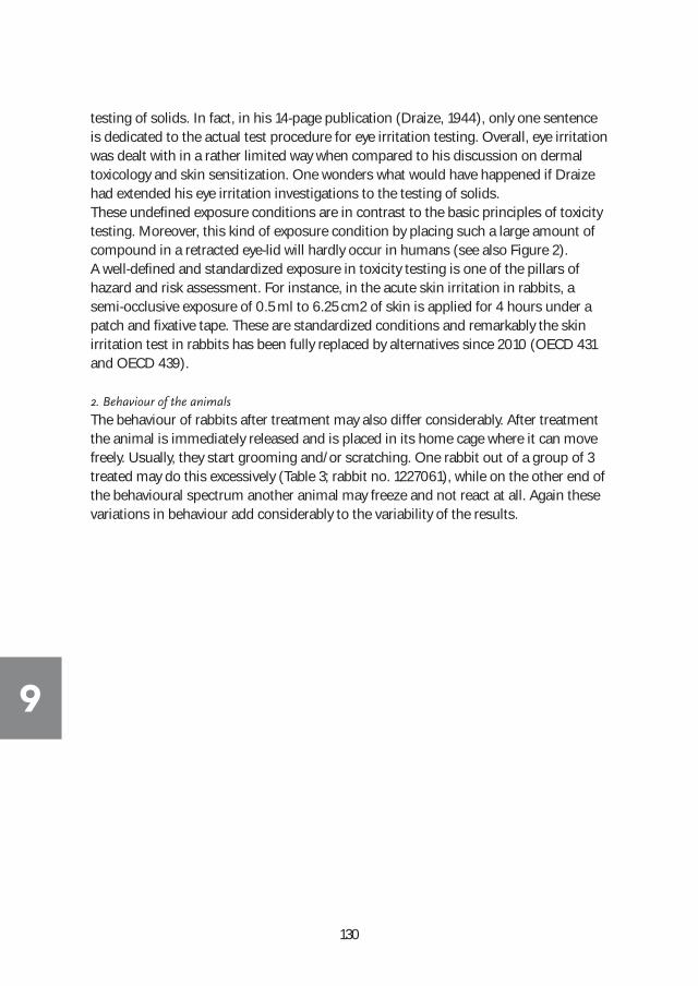
130
9
testing of solids. In fact, in his 14-page publication (Draize, 1944), only one sentenceis dedicated to the actual test procedure for eye irritation testing. Overall, eye irritationwas dealt with in a rather limited way when compared to his discussion on dermaltoxicology and skin sensitization. One wonders what would have happened if Draizehad extended his eye irritation investigations to the testing of solids.These undefined exposure conditions are in contrast to the basic principles of toxicitytesting. Moreover, this kind of exposure condition by placing such a large amount ofcompound in a retracted eye-lid will hardly occur in humans (see also Figure 2). A well-defined and standardized exposure in toxicity testing is one of the pillars of hazard and risk assessment. For instance, in the acute skin irritation in rabbits, asemi-occlusive exposure of 0.5 ml to 6.25 cm2 of skin is applied for 4 hours under apatch and fixative tape. These are standardized conditions and remarkably the skin irritation test in rabbits has been fully replaced by alternatives since 2010 (OECD 431and OECD 439).
2. Behaviour of the animals
The behaviour of rabbits after treatment may also differ considerably. After treatmentthe animal is immediately released and is placed in its home cage where it can movefreely. Usually, they start grooming and/or scratching. One rabbit out of a group of 3treated may do this excessively (Table 3; rabbit no. 1227061), while on the other end ofthe behavioural spectrum another animal may freeze and not react at all. Again thesevariations in behaviour add considerably to the variability of the results.

131
Table 3. Results of a three-animal in vivo test (TNO Triskelion report, 2012).
Animal No.
Corneal effects: Iris Conjunctivae
Discharge score area
redness swelling
1 HOUR
1227061 0 4 1 1 1 3
1228067 0 4 0 1 1 1
1228045 0 4 0 1 1 1
24 HOURS
1227061 1 4 1 1 1 1
1228067 0 4 0 0 0 0
1228045 0 4 0 0 0 0
48 HOURS
1227061 1 4 1 1 1 1
1228067 0 4 0 0 0 0
1228045 0 4 0 0 0 0
72 HOURS
1227061 0 4 0 1 1 0
1228067 0 4 0 0 0 0
1228045 0 4 0 0 0 0
6 DAYS
1227061 0 4 0 0 0 0
1228067 0 4 0 0 0 0
1228045 0 4 0 0 0 0
Table 3. Results of a three-animal in vivo test (TNO Triskelion report, 2012).
9
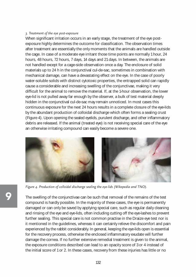
132
9
3. Treatment of the eye post-exposure
When significant irritation occurs in an early stage, the treatment of the eye post-exposure highly determines the outcome for classification. The observation timesafter treatment are essentially the only moments that the animals are handled outsidethe cage. In case of a moderate eye irritant those time points are normally 1 hour, 24hours, 48 hours, 72 hours, 7 days, 14 days and 21 days. In between, the animals arenot handled except for a cage-side observation once a day. The enclosure of solid materials up to 24 h in the conjunctival cul-de-sac, sometimes in combination withmechanical damage, can have a devastating effect on the eye. In the case of poorlywater-soluble solids with distinct cytotoxic properties, the entrapped solid can rapidlycause a considerable and increasing swelling of the conjunctivae, making it very difficult for the animal to remove the material. If, at the 1-hour observation, the lowereye-lid is not pulled away far enough by the observer, a bulk of test material deeplyhidden in the conjunctival cul-de-sac may remain unnoticed. In most cases this continuous exposure for the next 24 hours results in a complete closure of the eye-lidsby the abundant production of colloidal discharge which often forms a sealing crust(Figure 4). Upon opening the sealed eyelids, purulent discharge, and other inflammatorydebris are released. If the animal (treated eye) is not receiving special care of the eyean otherwise irritating compound can easily become a severe one.
Figure 4. Production of colloidal discharge sealing the eye-lids (Wikepedia and TNO).
The swelling of the conjunctivae can be such that removal of the remains of the testcompound is hardly possible. In the majority of these cases, the eye is permanentlydamaged or can only be saved by applying special care, such as regular daily cleaningand rinsing of the eye and eye-lids, often including cutting off the eye-lashes to preventfurther sealing. This special care is not common practise in the Draize eye test nor isit mentioned in the guidelines, whereas it can certainly relieve the discomfort and painexperienced by the rabbit considerably. In general, keeping the eye-lids open is essentialfor the recovery process, otherwise the enclosed inflammatory exudate will further damage the cornea. If no further extensive remedial treatment is given to the animal,the exposure conditions described can lead to an opacity score of 3 or 4 instead of the initial score of 1 or 2. In these cases, recovery from these injuries has little or no
133
relevance for man. As with the exposure conditions, these kind of circumstances arenot representative for the human situation. After accidental exposure, one will seek“immediate” care in case of ocular damage, and the victim will usually receive medicaltreatment, if required. The unrealistic exposure conditions in the Draize eye test impelled P&G to develop the in vivo rabbit Low Volume Eye Test (LVET) for their products. For instance, the testing of a dish wash detergent tab would result in dramaticocular effects in the standard in vivo Draize eye test, because the tab is ground to afine powder and instilled as a bulk in the conjunctival cul-de-sac of the rabbit and remains there for at least one hour (before 2002 up to 24 hours). Nobody would consider this as real exposure circumstances, nor will it occur in real life. In the ICE,the exaggerated test conditions can be mimicked by leaving the powder on the corneafor up to 60 seconds instead of the standard 10 seconds, but what relevance does ithave? The LVET was designed to mimic the possible human exposure and was extensivelyused for household care products. Now P&G uses the ICE for their purposes becausethe test also mimics the possible human exposure more closely. In general, it was astonishing that both ECVAM and ICCVAM urged that the ICE method needed to bemodified in order to mimic the extreme exposure conditions of the in vivo Draize eyetest, rather than modifying the exposure (to solids) in the in vivo Draize eye test.
Another phenomenon that occasionally occurred in the Draize eye test is the develop-ment of a secondary infection following the eye effects caused by the compound (initial infection). In the past, the hygiene standards in the laboratories were not ashigh as currently, and the treated eye could be infected by the scratching/grooming ofthe animal with its paws. In addition to the inflammation caused by a compound, theeye is more vulnerable to microbiological infection, causing initial mild to moderateeffects during the first days after exposure developing into more severe and prolongedeffects during the 21 day observation period. An interesting example of such an eventcan be found in one of the compounds tested in the EC/HO study and which was alsoused in the ICCVAM validation of the ICE. 1-Napthaleneacetic acid was tested in six rabbits of which one rabbit showed very unusual persisting and increasing effects after day 7, compared to the eye effects observed in the other five rabbits (ECETOC, 1998). The effects of the five rabbits followed a pattern which is normally expected for the initially slight to moderate eye effects, i.e. gradually decreasing in severity after day 3 and followed by a complete recovery on day 14 or day 21. In the sixth rabbit, a similar pattern was observed untilday 7, but thereafter the slight opacity observed increased to a moderate opacity onday 10 and finally a very severe opacity on days 14 and 21. This difference in the patternof the eye effects is remarkable and most probably caused by a secondary infection inthe eye of the animal. Based on the result in this rabbit, the compound was classifiedas Category 1, whereas the initial effects (24-72 hours) in the 6 rabbits would lead to aCategory 2(A) classification. The ICE test also classified the compound as Category 2. It is remarkable that the OECD guideline 405 of 1981 already stated that “Care should
9

134
9
be taken in the interpretation of data to exclude irritation resulting from secondary infection”. However, this issue was not addressed in the EC/HO validation or in theICCVAM validation.
4. Observation/grading of eye effects
In the early days of validation of alternatives for eye irritation it was recognized thatthe variability could be high in the Draize eye test, and this was considered to be caused by subjective scoring by different observers and by interlaboratory variability(Weil and Scala, 1971; Lordo, 1999; Ohno, 1999). Unfortunately, the publications byLordo and Ohno did not include the individual in vivo rabbit eye data which mighthave provided more insight in the underlying causes of the variation. Subjective scoring is indeed part of the problem but a large part of the variation presently ascribedto subjective scoring might in fact be caused by differences in animal behaviour, differences in exposure times, and absence (or presence) of post-treatment care. For instance in the study of Weil and Scala, ethanol 95% caused a combined score (all tissues combined) of 2, 9, 22, 15, 38 and 110 in the 6 rabbits at the 72-hour observation time point in one of the labatories. A score of 110 is the maximum scorepossible. Amongst the 24 laboratories the median score for ethanol 95% ranged from0 to 42. This cannot only be explained by subjective scoring.The subjective nature of the observation definitely plays a role with compounds causing effects near the thresholds for classification (not classified/irritant and irritant/severe irritant). First of all there is the grading/scoring of the effects itself. The Atlas of eye effects of the FDA (1964) already gave rise to debate. For example theredness of the conjunctivae of the eye no. 6 of Plate 1 (Figure 5) is stated to be score 2(moderate redness: more diffuse, deeper crimson red, individual vessels not easilydiscernible; see also Table 2 of the Introduction). Based on the grading of eye effectsof all compounds tested at TNO since 1981, it should be the maximum score of 3 (severe redness: diffuse beefy red), because a more intense redness cannot be observed.Eye no. 3 of Plate 1 is presented as a case of redness score 1 (slight redness: vesselsdefinitely injected above normal), whereas this would be a good example of score 2 forredness. Eye no. 2 of Plate 1 is more representative of a redness score 1 than of a normaleye (redness: vessels normal). The other plates of the Atlas contain more examples ofgrading that are considered questionable and subject to debate.

135
Figure 5. FDA guidance on scoring of ocular lesions; Plate 1 (FDA,1964).
9

Time point 24 Hours
Animal 1 2.0
Animal 2 2.0
Time point 48 Hours
Animal 1 2.0
Animal 2 2.0
Time point 72 Hours
Animal 1 2.0
Animal 2 2.0
Time point 24 Hours
Animal 1 2.0
Animal 2 2.0
Time point 48 Hours
Animal 1 2.0
Animal 2 2.0
Time point 72 Hours
Animal 1 2.0
Animal 2 1.0
136
9
Another issue concerning the subjective scoring is the decision the observer has tomake in certain cases where the score of one animal at one time point can make thedifference between, for instance, not classified and irritant (Category 2). For EU-CLPand UN-GHS classification, the individual tissue scores of each animal is first averagedover the 24-72 hour time points and next the average score of the two rabbits showingthe highest score for a specific tissue determines classification or not. The thresholdscore for redness or for swelling of the conjunctivae is an overall average of 2.0 forclassifying as a Category 2 compound. Table 4 shows a theoretical case where one ofthe 6 scores can make the difference between classifying or not. The score of 1.0 inTable 4 is assigned to animal 2 at the 72 hour time point, but can theoretically be atany of the 6 different places.
That this is not merely a theoretical example is demonstrated by the scores obtainedfor Fomesafen, acid form (ECETOC, 1998) which was also part of the EC/HO andICCVAM database (Table 5). The scores obtained for opacity are lacking just one score1 for classification as Category 2. Also the scores for redness of the conjunctivae arevery close to the threshold score of 2. The ICE test identified the compound as a Category 2B (UN-GHS criteria) or Category 2, but borderline to not classified (EU-CLPcriteria). In the ICCVAM validation this result was considered to be a false positive.
Table 4. Examples of conjunctival scores (redness or swelling); Category 2 (first table)
versus Not Classified (second table).

137
5. Appreciation of the in vivo data
With the knowledge of the factors influencing the in vivo results the “black or white”approach applied by ICCVAM can hardly be defended. Weil and Scala (1971) even concluded that the eye irritation test in rabbits as published by the Federal agencies ofthe US should not be recommended as standard procedure in any new regulations.However, the Draize eye test has been used practically unchanged until now.Michael Balls and Julia Fentum (1993), scientists in the field of validation, concluded:“It is very rare for any allowance to be made for the variability of the animal data, whichare thus given a status which they do not deserve. They wrongly become the “true” valueswhich the non-animal tests must struggle to reproduce. Also, insufficient allowance is madefor the doubt which must be placed on values which fall within the barrier zones on bothsides of category cut-off points. This is particular worrying when Cooper two-by-two way
Table 5. Ocular effects caused by Fomesafen, acid form (ECETOC, 1998).
9

138
9
plots are used as a basis for establishing the sensitivity, specificity, predictivity and concordanceof in vitro test data”. Bruner (1996), another scientist in the field of validation, concludedafter computer simulations that even if the alternative methods were perfectly repro-ducibly (if their coefficients of variation were 0), the variability in the Draize scoresalone would restrict the Pearson’s correlation coefficients to the range 0.65 - 0.80 whenthe Draize scores are between 0 and 40, which are typical for (mild) irritants.
ICE OECD Test guideline 438 One of the conclusions in the ICCVAM test method evaluation report (2006) was:“There are sufficient data to support the use of the ICE test method, in appropriate circumstances and with certain limitations, as a screening test to identify compounds asocular corrosives and severe irritants (i.e., EPA Category I, UN GHS Category 1, EU R41) ina tiered-testing strategy, as part of a weight-of-evidence approach. The identified limitationsfor this method are based on the false negative and false positive rates that are observed forcertain chemical and physical classes. Based on the available database, the false negativerates for alcohols, surfactants and solids range from 33% (1/3) to 50% (1/2), 44% (4/9) to57% (4/7), and 46% (6/13) to 70% (7/10), depending on the hazard classification system(EC, UN-GHS or EPA) used. Additionally, the false positive rates for alcohols range from27% (3/11) to 50% (5/10)”. Two of the alcohols in the data base were ethanol and butanol.Both caused severe irritancy in the ICE (and BCOP) but were Category 2 according tothe in vivo data of ECETOC (1998). As discussed previously the in vivo data of Weil andScala for ethanol (and also for butanol) showed very high inter- and intralaboratory variations, making the ICCVAM conclusion on performance of the ICE with respect toalcohols questionable. Also the surfactant examined in the study of Weil and Scalashowed high inter- and intralaboratory variations.In 2009, on the basis of the ICCVAM evaluation report the ICE and BCOP were adoptedas an OECD Test Guideline (TG 438 and TG 437, respectively) for the screening of severeeye irritants. The false negative rates for identifying severe irritants, i.e. compoundsidentified by the ICE as not severely irritating, were not considered critical since thesecompounds are still to be tested in the in vivo rabbit eye test. In the OECD test guideline,the limitations with respect to the screening of surfactants, alcohols and solids werealso mentioned. Specifically it was mentioned that: “The current validation database didnot allow for an adequate evaluation of some chemical or product classes (e.g. formulations).However, investigators could consider using this test method for testing all types of compounds(including formulations), whereby a positive result could be accepted as indicative of an ocularcorrosive or severe irritant response. However, positive results obtained with alcohols should beinterpreted cautiously due to risk of over-prediction”. The specific mentioning of formulationsis remarkable because the ICE is used more frequently for formulations than for purecompounds. Moreover, the Draize eye test also does not make any distinction betweenthe testing of pure compounds and formulations. In general, one should realize that atthat time the eye irritation potential of almost all, if not all, compounds had been deter-mined for regulatory purposes in one single type of eye (test), i.e. the rabbit eye (test).

139
A follow-up evaluation of the usefulness and limitations of alternatives for identifyingmild/moderate and non-irritant chemicals was made by NICEATM/ICCVAM, in collab-oration with ECVAM and JaCVAM. In early 2011, a proposal for updating the BCOP TG437 for the identification of chemicals not requiring a classification for eye irritationwas submitted to the OECD by means of a Standard Project Submission Form (SPSF).The BCOP database comprised 196 compounds of which 89 were non-irritants, andthese data were used to draft a Streamlined Summary Document (SSD). The BCOPwas considered appropriate because the percentage of false negatives for non-irritantswas 0%. However, the percentage of false positives was 69%. The ICE test was notproposed for such an update, because the review panel maintained the original recommendation to use the ICE only for classification of ocular corrosives and severeirritants. Specific objections against the use of the ICE for chemicals not requiring aclassification was the fact that two compounds of the “parallel” dataset (coded TNO-28and TNO-94) identified as non-irritants by the ICE turned out to be severe irritants inthe in vivo rabbit eye test. In collaboration with the National Coordinator for the OECD,it was argued by M.K. Prinsen that the review panel had not studied the nature of theeffects of these two false negative substances in detail. TNO-94 was an anti-foulingpaint for the shipping industry, a specific type of product, which produced reversibleirritating eye effects in two out of three rabbits. In the third rabbit an unusual effect occurred, i.e. adherence of the paint to the cornea which was reason to humanely sacrifice the animal on day 1. Anti-fouling paints are designed to be very durable whichmay explain the findings in this rabbit. Whether or not this peculiar effect is relevantfor humans, excluding (anti-fouling) paints from ICE testing would have no majorconsequences for the applicability of the method for screening of non-irritants in general. TNO-28 caused no corneal or iris effects; only conjunctival effects were observed.The conjunctival effects observed with this compound were below the threshold forclassification as an eye irritant. Eye effects had cleared completely in one rabbit after72 hours and in another rabbit after 7 days. The third rabbit showed increased con-junctival effects at 48 hours after treatment and on day 14 moderate redness andslight swelling of the conjunctivae were still observed. Importantly, a white oculardischarge was also observed which was a sign of secondary infection. The fact that the same effects were observed at 21 days after treatment supported this assumption.One week later, the eye effects in this rabbit had cleared completely. Overall, the ICE “parallel” data set provided by TNO showed a false negative rate fornon-irritants of 6% and a false positive rate of 1%. Therefore, the OECD was asked toreconsider the applicability of the ICE for the purpose of identifying non-irritants. During an OECD expert meeting (6-7 December 2012, Paris), M.K. Prinsen presentedthe issues of the in vivo Draize eye test as discussed previously.
9

140
9
As a result, the limitations of the in vivo Draize rabbit eye irritation test and their implications for validation purposes were recognized and summarized in the document (OECD SSD ICE, 2013) as follows:1. The in vivo rabbit eye irritation/corrosion test has no standardized exposure
regimen. Therefore, the duration of exposure of the test substance with the rabbit eyes remains unknown and can vary from a few minutes to several hours. In addition, for solids and sticky chemicals it is unclear how much of the compound (solid, paste or liquid) stays in contact with the eye;
2. The limited reproducibility of the Draize rabbit eye test method;3. The subjectivity in the allocation of the rabbit ocular tissue scores;4. The type of exposure which does not reflect a potential human accidental
exposure;5. The differences in physiology and sensitivity to tested chemicals between rabbit
and human eyes.
The re-evaluation of the ICE ICCVAM dataset showed that individual in vitro and in vivoclassifications of a number of compounds needed further considerations. Discrepancieswere found in the final in vivo and in vitro classifications for a number of compoundswhich had an impact on the final number of false negative compounds. After re-evaluation, the ICE test method had an overall accuracy of 82%, a false positiverate of 33%, and a false negative rate of 1% (instead of 6%) for non-irritants, whencompared to the in vivo data classified according to the UN-GHS. If anti-fouling organicsolvent containing paints were excluded from the database, the accuracy of the ICEtest method was 83%, the false positive rate 33% and the false negative rate 0%.In September 2013, the OECD TG 438 for the ICE was officially adopted also to includethe identification of non-irritants (in general about 80% of the chemicals tested arenon-irritants). This was a huge success for the ICE and for the 3 R’s in general, but itstill meant that compounds not identified by the ICE as non-irritant or severe irritanthave to be tested in the in vivo rabbit eye test.

141
Lessons learned, considerations and recommendations
• For eye irritation with a lack of human data, the combined in vitro/in vivo(parallel) testing instead of using in vivo rabbit eye irritation data from literature provided an ideal setting for i) developing and validating the alternative method, ii) introducing the method to industry and regulatory authorities, iii) getting insight in, and critically address the pros and cons of both the in vitro and in vivotest system.
• A meaningful validation of an in vitro alternative model in the middle range of irritancy (Category 2 classification) cannot be reached with the current in vivorabbit eye irritation data set due to the large variability.
• The selection procedure to select or to accept models suitable as an in vitroalternative to eye irritation should be more critical than in the past. Alternatives should have a direct relation to (human) ocular irritancy and be developed on the basis of the mechanistic principles of (human) ocular inflammation, instead of matching Draize eye test results only.
• It should be realized that the existing in vivo rabbit eye irritation data does not reflect the inflammatory and recovery processes in humans. Therefore, the data of the Draize eye test are not suitable for the development of in vitro models for eye irritation focusing on discrimination between severe, but reversible or severe, but irreversible eye effects.
• The in vivo eye irritation test in rabbits should no longer be allowed, and should be completely replaced by alternative methods, for instance the ICE.
• The household and personal care industry should share their strategy to fulfil regulatory demands without the use of the in vivo animal test with other (chemical)industries and regulatory authorities.
Recommendations for future validation
• The experimental animal should no longer be regarded as the “Golden Standard”,but reliable, preferably human, in vivo data of reference compounds should be available for validation.
• Equipment and protocols for new methods should be standardized.• An inventory of factors influencing the (regulatory) acceptance of alternatives
should be made with all parties involved prior to the practical start of the validation process.
9

142
9
In conclusion
Alternatives to the Draize eye irritation test should preferably make use of ex vivo eyes,eye tissue, or eye tissue equivalents in order to measure, both qualitatively and quanti-tatively parameters similar or identical to those in the clinic. The appropriateness ofthe presently available alternatives, based on the above mentioned criteria, is as follows:1. Models using intact isolated eyes with slit-lamp microscope observations and
histopathology, e.g. the ICE or IRE;2. Models using excised corneas with light transmission and fluorescein
measurements and histopathology, e.g. the BCOP (Bovine) or PCOP (Porcine);3. 2D or 3D human corneal epithelium reconstruction models, which have as major
disadvantage the lack of the different membranes of the cornea, corneal stroma and corneal endothelium.

143
References
Balls, M., Fentem, J.H. (1993). On Recognizing and Overcoming Barriers to the Acceptanceof Alternative Methods. Alternative Methods in Toxicology and the Life Sciences 11, Part 11 A. Mary Ann Liebert, Inc., publishers, New York.
Balls, M., Botham, P.A., Bruner, L.H., Spielmann, H. (1995). The EC/HO internationalvalidation study on alternatives to the Draize eye irritation test. Toxicology in Vitro 9,871-929.
Brantom, P.G., Cassidy, S., Eigler, D., Esdaile, D., Fentem, J.H., Liebsch, M. McPherson,J., Pfannenbecker, U., Prinsen, M.K. (2000). Preliminary evaluation of the applicationof reference standards in the prevalidation and validation of in vitro tests for eye irritation.TNO BIBRA Report No. 2/3387/00, Volume 2, December 2000.
Bruner, L.H., Carr, G.J., Chamberlain, M., Curren, R.D. (1996). Validation of alternativemethods for toxicity testing. Toxicology in Vitro 10(4), 479-501.
Burton, A.B.G. (1972). A method for the objective assessment of eye irritation.Food and cosmetics toxicology 10, 209-217.
Burton, A.B.G., York M., Lawrence, R.S. (1981).The in vitro assessment of severe eye irritants. Food and Cosmetics Toxicology 19, 471-480.
Cazelle, E., Eskes,C., Hermann, M., Jones, P., McNamee, P., Prinsen, M., Taylor, H.,Wijnands, M.V.W. (2014). Suitability of Histopathology as an Additional Endpoint tothe Isolated Chicken Eye Test for Classification of non-extreme pH Detergent andCleaning Products. Toxicology in Vitro (in press; Ref. No. TIV-D-13-00528).
Chan, T., Payor, S., Holden, B.A. (1983). Corneal thickness profiles in rabbits using anultrasonic pachometer. Investigative Ophthalmology and Visual Science 24(10), 1408-1410.
Doughty, M.J., Zaman, M.L. (2000). Human Corneal Thickness and Its Impact on Intraocular Pressure Measures: A Review and Meta-analysis Approach. Survey of Ophthalmology 44 (5), 367-408.
Draize, J.H., Woodard, G., Calvery, H.O. (1994). Methods for study of irritation andtoxicity of substances applied topically to the skin and mucous membranes. Journal ofPharmacology and Experimental Therapeutics 82, 377-390.
9

144
9
EC (1991). Collaborative study on the evaluation of alternative methods to the eye irritation test. Document XI/632/91-V/E/1/131/91, part I and part II. Brussels: EuropeanCommission.
EC (1993). Commission Directive 93/21/EEC of 27 April 1993 adapting to technical progress for the 18th time Council Directive 67/548/EEC on the Approximation ofLaws, Regulations and Administrative Provisions Relating to the Classification, Packaging and Labelling of Dangerous Substances. Official Journal of the EuropeanCommunities L110A, 1-86.
ECETOC (1998). Eye Irritation – Reference Chemicals Data Bank. Technical Report No. 48(2). European Centre for Ecotoxicology and Toxicology of Chemicals, Brussels.
Faber, C., Scherfig, E., Prause, J.U., Sørensen , K.E. (2008). Corneal thickness in pigsmeasured by ultrasound pachymetry in vivo. Scandinavian Journal of Laboratory AnimalScience 35, 39-43.
Federal Register notice 13513, Volume 70, No. 53, Monday, March 21, 2005: NationalToxicology Program; National Toxicology Program (NTP) Interagency Center for theEvaluation of Alternative Toxicological Methods (NICEATM); Availability of ExpertPanel Report on the Evaluation of the Current Validation Status of In Vitro Test Methods for Identifying Ocular Corrosives and Severe Irritants.
Federal Register notice 43149, Volume 70, No. 142, Tuesday, July 26, 2005: NationalToxicology Program (NTP); NTP Interagency Center for the Evaluation of AlternativeToxicological Methods (NICEATM); Availability of Revised Analyses and Proposed Reference Substances for In Vitro Test Methods for Identifying Ocular Corrosives andSevere Irritants.
Fowler, W. C., Chang, D.H., Roberts, B.C., Zarovnaya, E.L., Proia, A.D. (2004). A newparadigm for corneal wound healing research: The white leghorn chicken (Gallus gallus domesticus). Current Eye Research Vol. 28, No. 4, Pages 241-250.
Grant, W.M. (1986). Corneal epithelial Vacuoles. Toxicology of the Eye, 3rd edition, Volume 1, page 9. Charles C. Thomas Publishers, Springfield, Illinois, USA.
Griffith, J.F., Nixon, G.A., Bruce, R.D., Reer, P.J., Bannan, E.A. (1980). Dose-responsestudies with chemical irritants in the albino rabbit eye as a basis for selecting optimumtesting conditions for predicting hazard to the human eye; Toxicology and applied pharmacology 55, 501-513.

145
Griffith, J.F. (1987); The low volume eye irritation test.; Soap cosmetics chemical specialties 63(4), 32-63.
ICCVAM Background Review Document (2006); Current Status of In Vitro Test Methods for Identifying Ocular Corrosives and Severe Irritants: Isolated Chicken EyeTest Method. NIH Publication No: 06-4513.
Li, H.F., Petroll, W. M., Møller-Pedersen, T., Maurer, J.K., Cavanagh, H.D., Jester, J.V.(1997). Epithelial and corneal thickness measurements by in vivo confocal microscopythrough focusing (CMTF). Current Eye Research 16(3), 214-221.
Lordo, R.A., Feder, P.I., Gettings, S.D. (1999). Comparing and evaluating alternative(in vitro) tests on their ability to predict the Draize maximum average score. Toxicologyin vitro 13(1); 45-72.
Luepke, N. (1985). Hen's egg chorioallantoic membrane test for irritation potential.Food and Chemical Toxicology 23, 287-291.
Maurer, J.K., Parker, R.D. and Jester, J.V. (2002). Extent of corneal injury as the mechanistic basis for ocular irritation: key findings and recommendations for the development of alternative assays. Regulatory Toxicology and Pharmacology 36, 106-117.
OECD (1981). OECD Guideline for Testing of Chemicals No. 405: Acute eye irritation/corrosion, adopted 12 May 1981. Organisation for Economic Co-operation and Development, Paris.
OECD (2001). OECD Guidelines for the Testing of Chemicals No. 423: Acute oral toxicity;Acute Toxic Class method, Paris, France.
OECD (2002). OECD Guideline for Testing of Chemicals No. 405: Acute eye irritation/corrosion, adopted 24 April 2002. Organisation for Economic Co-operation and Development, Paris.
OECD (2004). OECD Guideline for Testing of Chemicals No. 431: In Vitro Skin Corrosion:Human Skin Model Test, adopted 13 April 2004. Organisation for Economic Co-operationand Development, Paris.
OECD (2009). OECD Guideline for Testing of Chemicals No. 437: Bovine Corneal Opacityand Permeability Test Method for Identifying Ocular Corrosives and Severe Irritants,adopted 7 September 2009. Organisation for Economic Co-operation and Develop-ment, Paris.
9

146
9
OECD (2009). OECD Guideline for Testing of Chemicals No. 438: Isolated Chicken EyeTest Method for Identifying Ocular Corrosives and Severe Irritants, adopted 7 September2009. Organisation for Economic Co-operation and Development, Paris.
OECD (2010). OECD Guidelines for the Testing of Chemicals No. 429: Skin Sensitisation;Local Lymph Node Assay, Paris, France.
OECD (2010). OECD Guideline for Testing of Chemicals No. 439: In Vitro Skin Irritation- Reconstructed Human Epidermis Test Method, adopted 22 July 2010. Organisationfor Economic Co-operation and Development, Paris.
OECD (2013). OECD Guideline for Testing of Chemicals No. 438: Isolated Chicken EyeTest Method for Identifying i) Chemicals Inducing Serious Eye Damage and ii) ChemicalsNot Requiring Classification for Eye Irritation or Serious Eye Damage, adopted 26 July2013. Organisation for Economic Co-operation and Development, Paris.
OECD (2013). Draft Streamlined Summary Document (SSD) for the Isolated ChickenEye (ICE) test method. ENV/JM/TG/RD(2013)19.
Ohno, Y., Kaneko, T., Inoue, T., Morikawa, Y., Yoshida, T., Fuji, A., Masuda, M., Ohno,T., Hayashi, M., Momma, J., Uchiyama, T., Chiba, K., Ikeda, N., Imanashi, Y., Itakagaki,H. (1999). Interlaboratory validation of the in vitro eye irritation tests for cosmetic ingredients. (1) Overview of the validation study and draize scores for the evaluationof the tests. Toxicology in Vitro 13(1), 73-98.
Prinsen, M.K. (1996). The chicken enucleated eye test (CEET): A practical (pre)screenfor the assessment of eye irritation/corrosion potential of test materials. Food andChemical Toxicology 34(4), 291-296.
Prinsen, M.K. (1999). An Evaluation of the OECD Proposal for the Harmonised Classification of Eye Irritants and Corrosives. Report of ECVAM Workshop 34, Eye Irritation Testing: The Way Forward, Appendix 1. ATLA 27, 72-77.
Prinsen, M.K. (2005). in vitro and in vivo data for 94 substances tested in the IsolatedChicken Eye test. Unpublished data provided directly to NICEATM by MK Prinsen,TNO Nutrition and Food Research Institute.
Rama, P., Matuska, S., Paganoni, G., Spinelli, A., De Luca, M., Pellegrini, G. (2010).Limbal Stem-Cell Therapy and Long-Term Corneal Regeneration. New England Journalof Medicine 363, 147-155.
147
Scala, R. A., Springer, J. (1997). IRAG working group 6; Guidelines for the Evaluationof Eye Irritation Alternative Tests: Criteria for Data Submission. Food and ChemicalToxicology 35, 13-22.
Schutte, K., Prinsen, M.K., McNamee, P.M., Roggeband, R. (2009).The isolated chickeneye test as a suitable in vitro method for determining the eye irritation potential ofhousehold cleaning products. Regulatory Toxicology and Pharmacology 54(3), 272-281.
Terry Kim, T., Khosla-Gupta, B. A. (2002). Chemical and Thermal Injuries to the OcularSurface. Ocular Surface Disease Medical and Surgical Management, Part III, pp 100-112.Springer-Verlag New York, Inc.
Thoft, R.A., Friend, J. (1977) Biochemical transformation of regenerating ocular surfaceepithelium. Investigative Ophthalmology and Visual Science 16, 14-20.
United Nations-Economic Commission for Europe (UN/ECE) (2007). Globally Harmonised System of Classification and Labelling of Chemicals (GHS). UN, New York and Geneva, 2007. Available at:http://www.unece.org/trans/danger/publi/ghs/ghs_rev02/02files_e.html.
US-FDA (1964). Illustrated guide for grading eye irritation by hazardous substances.Washington, D.C. 20204, USA.
Wagoner, M.D. (1997). Chemical Injuries of the Eye: Current Concepts Pathophysiologyand Therapy. Survey of Ophthalmology 41(4), 367-408.
Weil, C.S., Scala, R.A. (1971). Study of intra- and interlaboratory variability in the resultsof rabbit eye and skin irritation tests. Toxicology and applied pharmacology 19, 276-360.
9

148

149
Summary

150
151
Introduction
On 2 November 1944 a manuscript, entitled “Methods for the study of irritationand toxicity of substances applied topically to the skin and mucous membranes” wasreceived for publication by the Journal of Pharmacology and Experimental Therapy.The authors of this article were John H. Draize, Geoffrey Woodard and Herbert O. Calveryfrom the Division of Pharmacology, Food and Drug Administration, Federal SecurityAgency, Washington, D.C., USA. It is more than likely that the authors never expectedthe kind of impact this publication would have on animal experimentation worldwide.Almost seventy years later the name Draize is still inextricably attached to one of themost disputed toxicity tests commonly used to determine acute toxicity, i.e. the Draizeeye irritation test. To establish the potential risk of exposure of the eyes to compounds,the Food and Drug Administration of the United States (US-FDA) adopted the Draizeeye irritation test using rabbits already in 1961. Internationally, the Organisation forEconomic Co-operation and Development (OECD) published their first guideline oneye irritation in 1981, which was subsequently adopted by the European Union in 1984.In the early eighties, the controversial character of this type of animal testing becameknown to the general public and the development of alternative non-animal tests toreplace the Draize eye test started. The publication of Russell and Burch in 1959 entitled:“The principles of humane experimental technique” formed the basis of most initiativesrelating to the use and development of alternatives for animal experiments. In theirpublication they postulated the famous and often cited three R’s: Reduction, Refinementand Replacement of animal experiments. The 3 R initiatives concerning the Draize eyetest mainly involved reduction of the number of animals from six to three per test.Certain aspects of the Draize eye test causing considerable pain and discomfort to theanimal were dealt with only at a much later stage, i.e. in 2002 the reduction of thetime for a wash-out of the test substance from 24 hours to 1 hour after instillation,and in 2012 the use of systemic pain relief and topical sedation. The first attempts to validate alternative tests for eye irritation were considered to be relatively simple by comparing in vitro and in vivo irritation index scores. What was expected to be a process of several years, however, turned out to be a decades spanning process still not fully completed. For a large part, this can be attributed tothe nature of the in vivo test in rabbits, which is more complicated and compromisedthan originally believed.In the early nineteen-eighties, the TNO-CIVO Toxicology and Nutrition Institute inZeist, the Netherlands, introduced an alternative test for the Draize eye irritation test.This alternative, using isolated eyes, was based on a method developed by A.B.G. Burtonfrom Unilever. This thesis describes the development, performance, validation and acceptance of the in vitro isolated eye test which focuses on measuring the damage ofcompounds to the cornea.
10

152
10
Chapter 2 describes the results of the first in-house validation of the isolated rabbiteye test (IRE) at TNO-CIVO. Substances, already tested in the in vivo eye irritation testat the request of various industries and representing the average supply of substancesin contract research, were tested in the IRE with a modified superfusion apparatusand eye-clamps. The IRE results showed an overall correlation of 82% with the in vivoresults, using a general classification scheme for the grading of in vivo and in vitro eyeirritation (i.e. not, slight, moderate or severe irritant). Four compounds, all hydrophobic,were overpredicted by the IRE. A general observation was that hydrophobic compoundsstayed in contact with the cornea (eye) of the rabbit for a relatively short period oftime, mainly because its third eye-lid acted as a wiper. The presence of a third eye-lidis an example of a condition very specific to the in vivo rabbit eye test influencing theexposure to (hydrophobic) substances. This caused a problem with respect to the validation of alternative methods since alternative methods are not able to mimicsuch a condition. Moreover, it is a non-existing condition in man.
Although rabbits are available as eye-donor for the isolated eye test in contract research organisations executing routine eye- and skin irritation studies, the dependencyon laboratory animals was considered a serious shortcoming. Chapter 3 describes theuse of eyes of animals from slaughter-houses as a source for eyes. Slaughterhouseanimals, such as the cow, pig or chicken were considered as eye-donors. Although thepig eye was considered better comparable to the human eye, the chicken eye provedto be the most suitable candidate. The isolated eye test had to produce matching resultswith the Draize rabbit eye test in order to be accepted as an alternative. Hence, an eye(cornea) that matched closest to the rabbit eye and not to that of the human eye wasneeded. The chicken had such an eye because its corneal thickness is similar to therabbit cornea. The epithelium of the cornea is the first barrier against (chemical) insultand as such its thickness (number of layers of epithelial cells) is of importance. Thebaseline corneal thickness of the cow and pig was already that high that measurementof increased thickness to its full extend after treatment with moderate to severe irritantswas not possible. The suitability and sensitivity of chicken eyes was evaluated by testing21 reference chemicals, which were selected to be representative of currently used industrial chemicals of different chemical classes, ranging from non-irritant to severeirritant. Furthermore, a criteria and scoring system of the in vitro corneal effects togetherwith a Prediction Model (PM) for matching the EC scheme for classification and label-ling of compounds were introduced for the isolated chicken eye (ICE) test. On thebasis of the results it was concluded that: i) although the ICE does not assess conjunctival damage, its sensitivity to predict oculardamage is not reduced, ii) the ICE correctly predicted the EC classifications of the 21reference chemicals and iii) the ICE fitted in the previously updated EC and OECD guidelines regarding acute eye irritation/corrosion including recommendations to usealternatives for the prescreening or positive identification of strong eye irritants. Thereafter, the ICE was incorporated as a prescreen in the routine in vivo assessmentof eye irritation testing in the frame of contract research at TNO.

153
In Chapter 4 the results are presented of the “parallel” (first in vitro followed by invivo) testing of 44 compounds at TNO. These compounds were considered to be a relevant cross-section of compounds routinely produced by the (chemical) industry.The in vivo scores were compared to the critical scores of the ICE test, namely the scores for corneal swelling, corneal opacity, fluorescein retention of damaged cornealepithelium and an index score (combination of the three ICE parameters). The overallcorrelations found for the in vivo scores with the ICE in vitro scores were 0.90 (indexscore), 0.91 (corneal swelling), 0.86 (corneal opacity) and 0.82 (fluorescein retention).The correlation between the in vivo conjunctival scores and the ICE scores were 0.92(index score), 0.92 (corneal swelling), 0.93 (corneal opacity) and 0.86 (fluorescein retention). These correlations substantiated the conclusion made earlier by Burton in1972 that a relationship exists between the in vivo conjunctival damage and the cornealscores of the isolated eye test. Overall, it was concluded that the ICE provided a practicalprescreen for the Draize rabbit eye test and that only mild to moderate irritants in theICE, generally showing the highest sensitivity to inter- and intra-laboratory variability,should be confirmed in the rabbit eye test. Eighteen years later, the OECD adoptedthis conclusion. The “parallel” in vitro and in vivo eye irritation testing was continuedwith another 50 compounds and the results were submitted to organizations dedicatedto the validation of alternative non-animal tests, such as ECVAM (European Centre for the Validation of Alternative Methods) and ICCVAM (Interagency Center for theEvaluation of Alternative Toxicological Methods) of the USA.
At TNO the ICE test was also used as a stand-alone test, especially for the house-hold and personal care industry, which increasingly adopted non-animal testing strategies.The Procter & Gamble Company was one of these companies that used the ICE testfor their eye irritation safety program, and Chapter 5 describes the application of theICE test to their domain of household cleaning products. In the past, P&G had heavilyinvested in the Low Volume Eye Test (LVET), a modification of the Draize eye test. Because of the rather extreme exposure conditions of the standard Draize eye test,their products often needed labelling as severe eye irritants. The LVET used only onetenth of the dose volume of the test material (0.01 instead of 0.1 mL/gram) adminis-tered directly onto the cornea instead of instilling it in the lower conjunctival sac of the rabbit eye. The results of the LVET correlated well with their database on humanresponses to accidental exposure and to results of clinical studies. Although less burdening for the animals, the LVET was still an animal test and in view of their non-animal testing policy the suitability of the ICE for their products was explored. Twentyproducts representing cleaners, dishwashing liquids or powder, powder detergentsand raw materials of these products were tested at TNO in the ICE employing differentdosing volumes to examine the correlation with the LVET. Furthermore, histopathologyof the treated corneas sampled at the end of the observation period was included.With histopathology the correlation between the depth of injury in the cornea and possible recovery of eye lesions could be studied. Since measurement of recovery is not possible in the ICE (and in all other alternatives), it could be an important
10

154
10
improvement of the method. The outcome of this study was that, in general, the resultsof the ICE using its standard dosing volume of 30 µl or 30 mg were in line with or moreconservative than the LVET in terms of classification. The level of overprediction foundin the ICE was expected and considered acceptable since the ICE was developed forprediction of the Draize eye test and not the LVET. For P&G the ICE test was considereduseful for several purposes, such as 1) EU/GHS classification and labelling of powderand liquid household cleaning products, 2) screening of candidate formulations, and3) weight-of-evidence approach by determining the profile of new cleaning productformulations against benchmark products. A definite conclusion on the usefulness ofhistopathology in the ICE could not be made, but the data showed that assessment ofthe histopathological lesions in the various parts of the cornea was possible, enablingthe assessment of the depth of injury in the cornea.
Chapter 6 deals with investigations by TNO in the search for additional parametersthat could be helpful to discriminate between the different irritancy levels in the ICE.For that purpose, histopathology of the cornea using different staining techniqueswere examined. Originally, Haematoxylin & Eosin (H&E) was used for staining the microscopic slides of the cornea. Later on H&E staining was replaced by PeriodicAcid-Schiff (PAS) staining, which provided a better discrimination of the different layers of the cornea, i.e. the epithelium, stroma and endothelium and other structures,such as the basement membrane and the Bowman’s membrane (between epitheliumand stroma) and the Descemet’s membrane (between stroma and endothelium). The integrity of these membranes was considered to play a role in the injury and recovery process of the cornea, and visibility of these membranes by microscope wasconsidered important for an adequate histopathological assessment by the pathologist.Therefore, staining methods such as Trichrome, AZAN (Azocarmine & aniline) andEVG (Elastic Van Gieson), specifically targeting collagen-rich membranes, were testedon corneas treated with compounds representing a non-irritant, irritant and severe irritant (corrosive). PAS appeared clearly superior with respect to visibility of the membranes and the quality of the morphology of the various corneal structures. The histopathological examinations also showed that after severe corrosive damage tothe cornea by sodium hydroxide, the basement and the Bowman’s membranes appearedundamaged while lesions were seen in the underlying stroma. This observation led tothe conclusion that depth of injury is not the only factor determining the seriousnessof corneal injury. In the clinic, emphasis is put on corneal opacity and corneal stemcell survival after (chemical) injury in order to evaluate its severity. With severe stemcell damage, recovery of the corneal damage by re-epithelialization of migrating stemcells from the corneal limbus is not possible. In that case, the recovery process will result in complete, irreversible conjunctivalization of the ocular surface. The possibi-lity of screening the viability of stem cells after chemical injury could, therefore, be ofvalue. However, explorations to stain stem cells of the chicken cornea by p63 immuno-staining by the histopathology section of TNO appeared unsuccessful. Although theassessment of reversibility or irreversibility in the ICE is undoubtedly of value, especially

155
for household and personal care companies, the EU/GHS classification and labellingof severe eye irritants does not make a distinction between these two categories; theyare all classified as Category 1: “irreversible effects on the eye/serious damage to theeye”.
The need for accepted alternative methods led to international validation studiesinvolving several promising alternatives. One of the most comprehensive internationalvalidation was held by the European Commission (EC) and the British Home Office(HO) in the mid nineteen-nineties. Sixty chemicals were tested by nine alternative methods, including the ICE, and each performed by four independent labs. With amaximum overall correlation that ranged from 0.34 to 0.55, the outcome was very disappointing which was thought to be due to the use of the Modified Maximum Average Score (MMAS) as the sole parameter for evaluation of in vivo eye irritation(Chapter 7). The MMAS, which can range from 0-110, is an average of the maximumindividual tissue scores of the animals recorded for a compound. A compound wasclassified as a non-irritant with a MMAS of 0-25, irritant with a MMAS of 25-59, or severe irritant with a MMAS >59. Because the MMAS is not used for regulatory classification, the impact of the EC and proposed OECD harmonized classificationsystem (later on to be adopted as the EU-CLP and UN/GHS classification systems)was investigated by Menk Prinsen. MMAS cut-offs of 25 and 59 belonging to, respectively,irritant and severe irritant, appeared not appropriate for classification according tothese two regulatory systems. Eight compounds with an MMAS lower than 59 were infact severe irritants and 4 compounds with an MMAS higher than 59 and 3 compoundswith an MMAS lower than 25 were irritants. It was, therefore, recommended that futurevalidation of alternatives would benefit from the use of classifications based on theproposed OECD harmonized classification system.
In 2004, a new initiative was undertaken to (re-)validate alternative methodswhich were considered the most promising in the EC/HO study, especially for thescreening of severe irritants. It was an initiative of ICCVAM in collaboration withECVAM and the methods selected were the ICE, the Hen’s Test - ChorioallantoisMembrane (HET-CAM), the Isolated Rabbit Eye test (IRE) and the Bovine CornealOpacity and (fluorescein) Penetration test (BCOP). An independent Expert Panel wasestablished for each alternative method to determine the validation status of thesemethods. In general, the idea was that the poor correlation of the alternatives with theDraize Eye Test was due to shortcomings in the practical performance of the in vitromethods which should be improved, rather than also critically addressing the short-comings of the Draize eye test and its consequences for the validation of in vitroalternatives. Following a meeting of the panel experts and experts presenting the alternative methods, a discussion paper on this subject was written by Menk Prinsen,entitled: “The Draize Eye Test and in vitro alternatives; a left-handed marriage?” (Chapter 8) in which the issues of the in vivo test in relation to the development andvalidation of alternatives were addressed. In general, the in vivo results were used asthe “Golden Standard” for all comparisons with the alternative tests. Although there
10

156
10
was general agreement that the in vivo test in rabbits is far from perfect, the implicationsof the inconsistencies of the in vivo test for the validation of the alternatives werenever taken into account. It was generally believed that the in vivo rabbit eye test produced variable results due to the fact that different labs and observers were involvedand that data were used that had been obtained over a very long period of time. However, several other important issues played a crucial role in the outcome of the in vivo test. Instillation of the compound in the conjunctival cul-de-sac of the eye andthe almost immediately release of the rabbit thereafter resulted in an exposure, whichcan be anything from minutes (liquids) to 24 hours (solids). With solids, no washingout of remnants from the conjunctival sac was allowed before 24 hours after treatment.These undefined exposure conditions are in contrast to the basic principles of toxicitytesting that advocates a well-defined and controlled exposure to substances. Moreover,this kind of exposure condition by placing a large amount of compound in a retractedeye-lid will hardly occur in humans. Instead of questioning the in vivo exposure conditionswith solids, ICCVAM considered this to be a deficiency of the in vitro method and recommended that the test method needed to be optimized with respect to the exposureconditions for solids. Treatment of the eye post-exposure by the observer can also playan important role. Enclosure of solid materials up to 24 hours in the conjunctival cul-de-sac can result in a complete closure of the eye-lids by the abundant production ofcolloidal discharge which often forms a sealing crust. If the animal (treated eye) is notreceiving special care of the eye an otherwise irritating compound can easily become a severe one. Also these kind of circumstances are not representative for the humansituation in case of ocular damage. Another phenomenon that occasionally occurredin the Draize eye test and which was not taken into account when using in vivo data, isthe development of a secondary eye infection. Because of the inflammation caused bya compound, the eye is more vulnerable to microbiological infection, causing initialmild to moderate effects during the first days after exposure developing into more severe and prolonged effects during the 21 day observation period. It was concludedthat after almost 18 years of validation without any real success in replacing the Draizeeye test, that further attempts would be futile, if ‘‘old’’ in vivo data or new data generatedby the current OECD guideline were still used for validation. A multi-way approachwas recommended by i) implementation in the guidelines (legislation) of the mostcurrent in vitro methods in the testing strategy for screening of severe irritants, ii) re-evaluation of the Draize Eye Test by standardizing the exposure conditions for liquidsand solids and simultaneously iii) a more mechanistically-based development and optimization of in vitro methods as an ongoing process.
Chapter 9 of this thesis presents a general discussion of the results obtained. In addition, lessons learned and recommendations for future validation are presented.Overall, the results presented in this thesis showed that alternatives to the Draize eyeirritation test should preferably make use of ex vivo eyes, eye tissue (isolated cornea),or eye tissue equivalents (reconstructed corneal epithelium) in order to measure, bothqualitatively and quantitatively parameters similar or identical to those in the clinic.

157
Models using intact isolated eyes with slit-lamp microscope observations and histopathology, like the ICE or IRE are considered the most appropriate for that purpose.

158

Samenvatting

160

161
Met de sterke opkomst van de chemische industrie in de 20ste eeuw werd duidelijkdat de omstandigheden op de werkplek nadelige effecten op veiligheid en gezondheidvan de mens konden hebben. Kortdurende en langdurige blootstelling aan verschillendechemische stoffen veroorzaakten een scala aan ziektes, variërend van milde niet levens-bedreigende schade tot levensbedreigende ziektes als kanker. Naarmate werknemersen consumenten mondiger werden, nam de noodzaak toe om mogelijke blootstellingaan chemische stoffen te bepalen, de mensen vooraf te waarschuwen voor de gevarenen ze hiertegen te beschermen. Een van de vele risico’s bij het omgaan met (gevaar-lijke) stoffen is het in de ogen krijgen van het product. Al in 1961 schreef de Amerikaanse toelatingsautoriteit de “Food and Drug Administration”(FDA) een test voor met konijnen om stoffen te testen op hun oog irriterende eigen-schappen (de zogenaamde Draize test). Het controversiële karakter van de test werdpas bij het grote publiek bekend nadat Henry Spira, lid en oprichter van een dieren-rechtenorganisatie, een paginagrote advertentie in de New York Times plaatste met dekop ”Hoeveel konijnen maakt Revlon blind omwille van schoonheid”. Binnen een jaarwerd geld gedoneerd door diverse cosmetische bedrijven en werd een centrum voorhet ontwikkelen van alternatieven voor proefdiertesten opgezet; CAAT (Centre for Alternatives to Animal Testing). De ontwikkeling en validatie van alternatieven voor deoogirritatie test in konijnen, die als een relatief simpele test werd beschouwd, bleekeen taai en langdurig proces van vele jaren te worden. Sterker nog, de test is nu nameer dan 30 jaar nog steeds niet volledig vervangen. Dit proefschrift beschrijft de ontwikkeling, optimalisatie, validatie (binnen TNO) en toepassing van een alternatievetest met geïsoleerde ogen van kippen (de Isolated Chicken Eye, kortweg de ICE test)en in bredere zin de internationale validatie en acceptatie van de ICE test door overheids-instanties. Een groot gedeelte van de discussie betreft ook de uitvoering van de Draizetest zelf, omdat de manier waarop de resultaten van deze test gebruikt worden hetgrootste obstakel bleek te zijn bij de validatie van de alternatieven.
Het oog is één van onze belangrijkste zintuigen om contact te hebben met onzeomgeving. We hebben er maar twee en zijn als zodanig kwetsbaar voor beschadiging,veroorzaakt door opzettelijke of onopzettelijke blootstelling aan stoffen. Dit proefschriftbehandelt een belangrijk onderdeel van het oog, namelijk het hoornvlies (de cornea)die de buitenste laag vormt en “het venster naar de wereld” is. De cornea bestaat uitdiverse lagen waarin geen bloedvaten lopen. Dit laatste is belangrijk voor de geïso-leerde ogen test, omdat er geen directe bloedtoevoer nodig is om de cornea functioneelte houden gedurende langere tijd. De diverse lagen van buiten naar binnen zijn: 1) epithelium, 2) stroma en 3) endothelium, met tussen 1) en 2) het basaal membraanen Bowman’s membraan en tussen 2) en 3) het Descemet’s membraan (zie figuur 2,Introductie). De integriteit van de lagen en membranen zorgen voor een heldere corneawaarbij het epithelium een eerste barrière vormt met als doel om materialen, zoalsstof, water en bacteriën buiten te houden. Tevens vormt het epithelium een glad oppervlak dat zuurstof en voeding uit traanvocht kan absorberen dat vervolgens verderdoor de cornea wordt getransporteerd. Tevens zitten in het epithelium duizenden
11

162
11
zenuwuiteinden die de cornea zeer gevoelig maken voor pijn. Al in 1944 publiceerde de Amerikaan John Draize een artikel met daarin een beschrijvingvan onderzoeksmethoden voor het vaststellen van toxiciteit en irritatie van stoffen dieop huid en slijmvliezen worden toegediend, waaronder de oogirritatie test en de LD50(Lethale Dosis) test. Ruim zeventig jaar later is zijn naam nog steeds onlosmakelijkverbonden met deze zeer omstreden testen. Al bij het eerste gebruik als regulatoiretest werd erkend dat het subjectief scoren van de oogeffecten problematisch was. Om het scoren te standaardiseren werd in 1964 door de FDA een gids met foto’s uitgebracht die de onderzoeker kon gebruiken bij het scoren van effecten, zoals opaciteit (troebeling) van de cornea, roodheid en zwelling van de oogleden (zie figuur 3,Introductie). Internationaal adopteerde de OESO (Organisatie voor Economische Samenwerking en Ontwikkeling) de Draize oogirritatie test in 1981, gevolgd door deEuropese Unie in 1984. Sindsdien zijn de richtlijnen verschillende keren aangepast,maar de praktische uitvoering van de test bleef onveranderd. De aanpassingen betroffenvooral het aantal dieren per test en maatregelen om onnodig leed te voorkomen, zoalsuitsluiting van stoffen met extreme pH’s, huid corrosieve stoffen en een gefaseerdestart (eerst met één konijn en afhankelijk van de ernst van de effecten door tot drie konijnen). De uitvoering van de test is vrij simpel. De oogleden van het oog wordenopengehouden en 0.1 milliliter of 0.1 gram van de proefstof wordt in het onderste, uitgetrokken oogzakje gedeponeerd. Vervolgens worden de oogleden gedurende éénseconde gesloten waarna het dier losgelaten wordt. Het andere oog blijft onbehandelden dient ter controle. Het konijn wordt teruggezet in zijn kooi en is vrij om de proefstofuit zijn oog te verwijderen. De ogen worden 1, 24, 48 en 72 uur na blootstelling beoor-deeld volgens een score schaal (zie tabel 2, Introductie). De effecten worden wekelijks,tot 3 weken na blootstelling, vervolgd om vast te stellen of deze effecten reversibel(herstellen) of irreversibel zijn. Hoewel er verschillende internationale richtlijnenwaren op het gebied van oogirritatie was de uitvoering praktisch identiek. De systemenvoor de classificatie en het labelen van de stoffen op basis van de waargenomen oog-effecten waren echter zeer verschillend. Dit was zeer nadelig voor het valideren van dealternatieve methoden. Daarom was het implementeren van een geharmoniseerd classificatie systeem in 2007 door de UN (United Nations) een aanzienlijke verbetering.
Aan de basis van het vaak geciteerde 3 V principe, Vermindering, Verfijning enVervanging van proefdiergebruik, staat de publicatie van Russell en Burch uit 1959, getiteld: ”The Principles of Humane Experimental Technique”. De initiatieven met betrekking tot de Draize oogirritatie test betroffen vooral het verminderen van het aantal dieren per test van zes naar drie en mogelijke vervanging door proefdiervrije alternatieven. Pas in een veel later stadium werden maatregelen genomen om bepaaldeonderdelen van de test, die zeer veel leed kunnen veroorzaken, te verbeteren. Het werd bijvoorbeeld pas in 2002 toegestaan om proefstof (vooral vaste stoffen), dietot 24 uur na blootstelling nog in het oogzakje aanwezig kon zijn, na 1 uur i.p.v. 24 uurte verwijderen. In 2012 werd de OECD richtlijn aangepast met duidelijke aanwijzingenvoor lokale en systemische pijnverlichting voor de konijnen. Begin tachtiger jaren van

163
de vorige eeuw introduceerde Herman Koëter, lid van de werkgroep “Kritische EvaluatieToxiciteitstesten” van de Nederlandse Vereniging van Toxicologen, de geïsoleerdeogen test op TNO-CIVO in Zeist. Verschillende alternatieven voor oogirritatie, variërendvan bevruchte kippeneieren, cellijn toxiciteitstesten tot sperma motiliteit, waren al gepubliceerd, maar de geïsoleerde ogen test werd gekozen omdat bij oogirritatie decornea het belangrijkste doelorgaan is.
In 1981 publiceerde A.B.G. Burton van Unilever een methode met geïsoleerde konijnenogen om ernstig irriterende stoffen te identificeren. Hij had al in 1972 ontdektdat het meten van de dikte van de cornea na blootstelling bij konijnen een objectieveen nauwkeurige vaststelling van oogirritatie opleverde. Uit wetenschappelijk oogpuntis het gebruik van geïsoleerde ogen zeer aantrekkelijk, immers er wordt een oog (exvivo) voor een oog (in vivo) gebruikt en daarbij worden effecten (parameters) gemetendie direct vertaald kunnen worden naar die van het dier, maar ook naar die van demens. Met financiële steun van de Dierenbescherming en de Stichting SchoonheidZonder Wreedheid werd de benodigde apparatuur aangeschaft en gebouwd. Als eerstewerd de methode geëvalueerd door de stoffen die Burton getest had, ook te onderzoekenin de TNO opstelling. In Hoofdstuk 2 wordt de implementatie en validatie beschrevenvan de Isolated Rabbit Eye (IRE) gedurende de periode 1982-1985. Verbeteringen vanhet superfusie apparaat en ooghouders werden aangebracht en met de opstelling werden vervolgens 34 stoffen getest, die net daarvoor waren onderzocht in de Draizetest. Er waren geen extra dieren voor de IRE test nodig, omdat konijnen gebruikt werden die al waren ingezet voor huid of oogirritatie (controle oog). Gebruikmakendvan een algemeen classificatie schema (onderscheid in niet, licht, matig en ernstig irriterend) werd in 82% van de gevallen de juiste correlatie gevonden. Slechts in viergevallen was er een overschatting (overprediction), d.w.z. classificatie als licht ofmatig irriterend (in vitro) in plaats van niet irriterend (in vivo). Opvallend was dat dithydrofobe stoffen betroffen die bij konijnen snel uit het oog werden verwijderd doordatdeze stoffen slecht mengden met de aanwezige traanfilm en door het derde ooglid datals een soort wisser fungeerde. Dit laatste is een voorbeeld van een conditie in deDraize test die overduidelijk de blootstelling beïnvloedt, terwijl dit geen rol speelt bijhumane blootstelling. Op basis van deze resultaten werd geconcludeerd dat de IREeen gevoelig en bruikbaar testsysteem was voor het identificeren van oog irriterendestoffen. Niet irriterende stoffen in de IRE, waarbij frequent oogcontact werd verwacht,zouden dan alsnog in de Draize test moeten worden getest.
Hoofdstuk 3 beschrijft een belangrijke mijlpaal in de ontwikkeling van de geïso-leerde ogen test, namelijk het gebruik van slachtdieren als oog donor. Hoewel in dietijd de meeste onderzoekslaboratoria over voldoende, al eerder gebruikte, konijnenbeschikten om de test uit voeren, werd het hergebruik van proefdieren nog steeds alstekortkoming van de methode gezien. Daarom werd het gebruik van slachtdierenzoals het rund, het varken en de kip als donoren van ogen. Als eerste werd nagegaanof het mogelijk was om ogen te verzamelen in het slachthuis. Daarna werd de praktischekant van het gebruik van verschillende typen ogen in de testopstelling getoetst. Al snel
11

164
11
viel het rund als donor af - te onregelmatige aanvoer, te verschillende achtergrond vande dieren en teveel beschadigde ogen. Bovendien bleek de cornea (circa 1000 µm) extreem dikker t.o.v. de konijnen cornea (circa 400 µm). Het verzamelen van varkens-ogen op het abattoir was moeilijk, maar mogelijk. Toch viel ook het varkensoog af, ondanks dat dit oog vooraf als een goede kandidaat werd beschouwd vanwege deovereenkomsten in fysiologie met die van de mens, omdat ook deze cornea te dikbleek (circa 600 µm). Doordat de basisdikte van de cornea al in het top bereik van deafleesschaal van het diktemeetapparaat van de opstelling lag, zouden de waarden bijmatig tot ernstig irriterende stoffen mogelijk niet meer volledig bepaald kunnen worden.Andere mogelijkheden voor de meting in dat bereik waren ook niet voorhanden. De geïsoleerde ogen test moest vergelijkbare resultaten leveren t.o.v. de in vivo test inkonijnen om te worden geaccepteerd als alternatief. Daarom was een oog (cornea)nodig die het beste vergelijkbaar was met die van het konijn en niet met die van demens. De kip heeft een dergelijk oog, zowel de afmetingen als de dikte van de cornea(circa 400 µm) kwamen overeen met die van het konijn. Het verkrijgen van kippenogen bleek relatief eenvoudig door de koppen te verzamelennadat de kippen waren gedood aan de slachtlijn. De koppen werden vervoerd naar hetlaboratorium in plastic dozen om daar de ogen uit de koppen te ontleden en in het superfusie apparaat te plaatsen. Dit alles gebeurde binnen 2 uur na het verzamelenvan de koppen aan de slachtlijn. De ogen die ingezet werden moesten onbeschadigdecorneas hebben, d.w.z. geen of zeer geringe troebeling van de cornea, geen of zeer geringe opname van fluoresceïne door beschadigde of dode epitheel cellen en eendikte die in de normale range lag. De geschiktheid van het kippenoog werd vastgesteldmet 21 referentiestoffen (van niet irriteren tot ernstig irriterend) die al eerder in een EC(pilot) validatie studie van de IRE waren getest. Een belangrijke ontwikkeling daarbijwas het opstellen van de criteria voor het scoren van de effecten en een voorspellings-schema (Prediction Model = PM) voor classificatie op basis van het toen geldende ECsysteem voor labeling en classificatie van stoffen (zie tabel 3 van Hoofdstuk 2). Omdatde effecten die in de ICE gemeten werden een directe relatie hadden met de effectendie in de Draize test optraden (bijvoorbeeld ten aanzien van troebeling), kon de PMvooraf voor het grootste gedeelte theoretisch vastgesteld worden op basis van de reik-wijdte van de response (troebeling, zwelling en fluoresceïne opname). Dit in tegen-stelling tot de meeste andere alternatieven die effecten maten die geen directe relatiemet oogschade hadden. Deze alternatieven konden alleen na het testen van meerderestoffen achteraf door middel van een wiskundige vergelijking het in vitro effect(en) vertalen naar een in vivo effect. Bijvoorbeeld, in de HET-CAM test met bevruchte kippeneieren werd het chorioallantois membraan, rijk aan bloedvaten, blootgesteldaan de stof. Daarna werd de tijd bepaald van het oplossen van de bloedvaten en/ofontstaan van bloedingen na de start van de blootstelling. Vervolgens werd het aantalseconden via een wiskundige vergelijking omgezet naar een getal dat vergeleken konworden met een in vivo irritatie score. Pas na het testen van een groot aantal stoffenkon een dergelijke PM worden bepaald en vaak bleek dat de PM moest worden bijge-
165
steld als meer stoffen werden getest. De ICE gebruikte een andere benadering, nietdoor de ICE data te vertalen naar één in vitro score en die te vergelijken met de in vivoscore, maar door de vooraf bekende reikwijdte van elke parameter in te delen in logischecategorieën, namelijk in geen effect (categorie I), licht effect (categorie II), matig effect(categorie III) of ernstig effect (categorie IV). Dus bij het testen van een stof werd één categorie per parameter vastgesteld, namelijkvoor troebeling, voor zwelling en voor fluoresceïne opname. De vaststelling van deeind classificatie in niet irriterend, (licht/ matig) irriterend of ernstig irriterend werdbepaald door de combinatie van deze 3 categorieën. Opnieuw was dat een puur theo-retische aangelegenheid op basis van de mogelijke combinaties. Bijvoorbeeld in hetniet irriterende gebied hoort de combinatie van de categorieën I/I/I en in het ernstigirriterende gebied de combinatie van de categorieën IV/IV/IV. Alle mogelijke combinatiesen bijbehorende classificaties zijn terug te vinden in tabel 1 van Hoofdstuk 9. Pas in 2007 was het nodig het classificatie systeem van de ICE aan te passen omdater toen een internationaal schema, het United Nations - Globally Harmonized System(UN-GHS), in werking trad. Vervolgens moest het schema van de de ICE in 2013 nogeen keer aangepast worden om geaccepteerd te worden als OECD richtlijn voor hetidentificeren van niet-irriterende stoffen.
In toenemende mate werd de ICE ook gebruikt als een op zichzelf staande testzonder vervolg door een in vivo test. Vooral bedrijven die een veiligheidsprogrammahanteerden waarin geen dier testen meer voorkwamen, waren geïnteresseerd in deICE. Eén van de grootste producenten op het gebied van huishoudelijke en verzorgings-producten, de Procter & Gamble Company (P&G), adopteerde de ICE voor het screenenop oogirritatie. Dit was een enorme stimulans voor de verdere toepassing en ontwik-keling van de ICE. Voor P&G was het testen van hun zeepmiddelen in de Draize testeen groot probleem. Met de blootstellingscondities van de Draize test werden vooralzeeppoeders als ernstig irriterend geïdentificeerd. Daarom hadden ze in het verledensterk ingezet op een modificatie van de Draize test, de “Low Volume Eye Test (LVET)”waarbij het konijnenoog niet aan 0.1 mL of 0.1 gram, maar aan 0.01 mL of 0.01 gramwerd blootgesteld. Daarbij werden deze sterk gereduceerde hoeveelheden niet in hetoogzakje maar direct op de cornea toegediend. Deze condities, die veel realistischerwaren t.o.v. mogelijke humane blootstelling, gaven resultaten die meer in lijn lagenmet geregistreerde schadegevallen en klinische studies. Toch bleef de LVET een dierstudie, weliswaar met minder ongerief voor de dieren, maar niet langer passend in hun dierproefvrije strategie. Daarom besloot P&G de mogelijkheden van de ICE telaten onderzoeken bij TNO. In Hoofdstuk 5 worden de resultaten van de ICE met eenspecifieke groep van hun producten, namelijk de huishoudelijke schoonmaakmiddelenbeschreven. Naast de standaard hoeveelheid van 30 microliter of 30 milligram in deICE, die correleren met de hoeveelheden in de Draize test, werden ook 10 en 3 microliterof 10 en 3 milligram onderzocht om vast te stellen of de resultaten beter correleerdenmet de hoeveelheden gebruikt in de LVET. Ook werd voor het eerst histopathologischonderzoek van de cornea toegevoegd aan de ICE. Met dit extra onderzoek zou mogelijk
11

166
11
de diepte van de schade in de cornea (Depth-of-Injury) kunnen worden vastgesteld.Microscopisch onderzoek was ook al toegepast in de LVET en daaruit bleek een cor-relatie tussen de diepte van de schade en mogelijk herstel (reversibiliteit) van de oog-effecten. Aangezien de ICE geen herstel van oogschade kon vaststellen, zou het microscopisch vaststellen van de diepte van de effecten kunnen bijdragen aan het beargumenteren van mogelijk herstel van de vastgestelde oogschade. De uitkomstwas dat de resultaten met de standaard ICE hoeveelheid van 30 microliter of milli-gram in lijn of conservatiever waren dan die van de LVET. De mate van overschattingwerd acceptabel geacht omdat de ICE ontwikkeld was om de Draize test, en niet deLVET, te voorspellen. P&G achtte de ICE geschikt voor meerdere van hun doeleinden,zoals bijvoorbeeld de EU/GHS classificatie van hun schoonmaakproducten. Dit hieldin het vooraf testen van hun kandidaat producten en het toepassen van een benaderingmet meervoudige bewijsvoering (“weight-of-evidence” benadering) door middel vanvergelijking met bestaande producten. Een duidelijke uitspraak over de toegevoegdewaarde van histopathologie in de ICE kon niet worden gegeven, maar het bleek welmogelijk om effecten in de diverse delen van de cornea vast te stellen waardoor de“Depth-of-Injury” theorie getoetst zou kunnen worden. De vraag bleef of een theoriedie gebaseerd is op een herstelproces in een (levend) konijnenoog dat dagen of wekenkan duren, kan correleren met het schadeproces in de ICE. De uitkomst van de Draizetest wordt sterk beïnvloed door verschillende oncontroleerbare factoren die niet plaats-vinden in de ICE. Deze factoren en hun consequentie voor de irritatie classificatie vanstoffen worden in de discussie van de Hoofdstukken 8 en 9 besproken.
Om de histopathologische beoordeling van de corneas te optimaliseren werd bijTNO de mogelijkheid onderzocht om meer specifieke kleuringen toe te passen bij het maken van de microscopische preparaten van de cornea (Hoofdstuk 6). In eersteinstantie werden de microscopische preparaten van de cornea gekleurd met de standaardkleuring Haematoxylin & Eosin (H&E), maar deze werd later vervangen door de PeriodicAcid-Schiff (PAS) kleuring die een beter onderscheid van de diverse lagen van de corneagaf. In het bijzonder de diverse membranen van de cornea, het basaal membraan enBowman’s membraan (tussen epithelium and stroma) en het Descemet’s membraan(tussen stroma en endothelium) waren beter te onderscheiden. Deze membranenworden geacht een belangrijke rol te spelen bij het ontstekings- en herstelproces vande cornea. Daarom was de zichtbaarheid van deze membranen belangrijk voor devaststelling van hun integriteit bij de microscopische beoordeling door de patholoog.Andere meer specifieke kleuringen voor collageen rijke membranen zouden mogelijkde beoordeling kunnen verbeteren. Voor dit doel werden coupes van cornea’s, diewaren blootgesteld aan niet irriterende, irriterende of ernstig irriterende stoffen, bewerkt met de verschillende kleuringen en beoordeeld door de patholoog. Van de vijfgeselecteerde kleuringen, H&E, PAS, Trichrome, AZAN (Azocarmine & aniline) andEVG (Elastic Van Gieson), bleek PAS duidelijk superieur te zijn met betrekking totzichtbaarheid van de membranen en de kwaliteit van de morfologie van de diversestructuren van de cornea. Na ernstige corrosieve schade door natrium hydroxide

167
(natronloog) bleek de basaal membraan en Bowman’s membraan ogenschijnlijk onbeschadigd terwijl in het onderliggende stroma wel effecten werden waargenomen.Betekende dit dat de functionaliteit van de membranen niet was aangetast of waslichtmicroscopie niet in staat dit soort schade vast te stellen? Deze observatie leiddetot de conclusie dat “Depth-of-Injury” niet de enige factor is die de ernst van deschade bepaalt. Overleg met een oogarts van het Utrecht Medisch Universiteitscentrum,gespecialiseerd in de cornea, en diverse publicaties op dit gebied wezen uit dat in dekliniek de nadruk vooral ligt op de troebeling van de cornea en stamcel overleving na(chemische) schade. Bij zeer omvangrijke schade aan de stamcellen kan de corneaniet meer herstellen door re-epithelisatie van migrerende stamcellen. In dat geval resulteert de schade in onherstelbare conjunctivale ingroei van de cornea (conjuncti-valization). De stamcellen zijn oppervlakkig gelokaliseerd aan de rand van de corneaen als zodanig betrokken bij het eerste contact na blootstelling aan een stof. Daaromkan het vaststellen van de viabiliteit van de stamcellen van betekenis zijn. Pogingenom stamcellen van de kippen cornea met een p63 immuunkleuring te identificeren,bleken niet succesvol. Hoewel het vaststellen van reversibiliteit of irreversibiliteit vanoogschade belangrijk is, zeker voor de producenten van huishoudelijke en verzorgings-producten, maken de classificatie schema’s van de EU en GHS geen onderscheid tussendeze twee categorieën. Ze worden beide geclassificeerd als Categorie 1: “Veroorzaakternstig oogletsel”.
De behoefte aan geaccepteerde alternatieve testmethoden leidde tot een van degrootste validatiestudies ooit georganiseerd, namelijk de EC/HO studie in 1993-1995.Zestig stoffen werden in negen verschillende alternatieve test methoden, waaronderde ICE, onderzocht in vier verschillende laboratoria per methode. Met een maximalegemiddelde correlatie variërend van 0.34 tot 0.55 (1.0 is maximaal haalbaar) was deuitslag van deze studie erg teleurstellend. In Hoofdstuk 7 wordt beschreven dat hetgebruik van de MMAS (Modified Maximum Average Score) als enige parameter voorde mate van de in vivo oogirritatie, de oorzaak was van dit teleurstellende resultaat. De MMAS, die kan variëren van 0-110, is een gemiddelde van de hoogste oogweefselscores van de individuele konijnen, vastgesteld na het testen van een stof. Een stofwerd geclassificeerd als niet irriterend bij een MMAS van 0-25, irriterend bij eenMMAS van 25-59 of ernstig irriterend bij een MMAS hoger dan 59. Omdat de MMASniet gebruikt werd bij het vaststellen van de EU en GHS oogirritatie classificaties, werdonderzocht welke gevolgen deze classificaties hadden op het gebruik van de MMAS inde EC/HO studie. De MMAS grenzen van 25 en 59, behorend tot respectievelijk irriterenden ernstig irriterend, bleken niet goed overeen te komen met de EU en GHS classificatiesvoor irriterend en ernstig irriterend. Acht stoffen met een MMAS lager dan 59 warenernstig irriterend en 4 stoffen met een MMAS hoger dan 59 en 3 stoffen met een MMASlager dan 25 waren irriterend. De aanbeveling was dan ook om voortaan gebruik temaken van de EU en GHS classificaties bij nieuwe validatie studies.
In 2004 werd een nieuw initiatief gestart door het Interagency Coordinating Committee on the Validation of Alternative Methods (ICCVAM) in samenwerking met
11

168
11
het European Centre for the Validation of Alternative Methods (ECVAM) om alternatievemethoden, die het meest veelbelovend waren in de EC/HO studie, te (her)validerenvoor het identificeren van ernstig irriterende stoffen. Dit waren de ICE, de Hen’s Test -Chorioallantois Membrane (HET-CAM), de Isolated Rabbit Eye test (IRE) en de BovineCorneal Opacity and (fluorescein) Penetration test (BCOP). Om de validatiestatus vandeze methoden vast te stellen werd per alternatieve methode een onafhankelijk panelbenoemd. In het algemeen was de gedachte dat de slechte correlatie tussen de alter-natieven en de Draize test mede lag aan tekortkomingen in de praktische uitvoeringvan de alternatieve methoden. Aan eventuele tekortkomingen van de Draize test en demogelijke consequenties daarvan voor de validatie van de alternatieven werd weinigaandacht besteed. Na een openbare bijeenkomst waarin experts hun alternatieve methode konden presenteren en vragen daarover beantwoorden van de panelleden,schreef Menk Prinsen een discussiestuk over dit onderwerp, getiteld: “The Draize EyeTest and in vitro alternatives; a left-handed marriage?” (Hoofdstuk 8). Hierin wordende verschillende aspecten van de Draize ogen test besproken die van invloed zijn opde ontwikkeling, validatie en acceptatie van alternatieven. Over het algemeen wordende in vivo data gebruikt als de “Gouden” standaard bij het vergelijken met de resultatenvan de alternatieve methode, dus 100% accurate voorspelling. Hoewel men in het algemeen wel op de hoogte was van de tekortkomingen van de Draize test, werdendeze nooit meegewogen in het validatie en acceptatie proces van alternatieven. Menwist dat de Draize test variabele resultaten opleverde omdat verschillende laboratoriaen waarnemers bij het uitvoeren van deze test betrokken waren en de gebruikte dataover een zeer lange periode waren verzameld. Maar ook andere belangrijke zaken spelen een cruciale rol bij de uitkomst van deze in vivo test. De blootstellingsduur aaneen stof is hoegenaamd niet gedefinieerd of gestandaardiseerd. Doordat een stof inhet uitgetrokken oogzakje word gedeponeerd waarna het konijn wordt teruggezet inzijn kooi, kan de blootstelling variëren van minuten (bij vloeistoffen) tot uren (24 uurbij poeders), omdat resten van stoffen pas 24 uur na blootstelling verwijderd mogenworden. Deze ongedefinieerde blootstelling is tegenstrijdig met de basis principes vangoed toxiciteitsonderzoek; alles valt of staat bij een goed gedefinieerde en gecontro-leerde blootstelling. Daarbij, dit type blootstelling (grote hoeveelheid stof in een oog-zakje) is nauwelijks vergelijkbaar met een humane blootstelling. In plaats van dit terdiscussie te stellen, concludeerde ICCVAM dat de ICE in dit opzicht tekort schoot enkwam met de aanbeveling om de ICE te optimaliseren met betrekking tot de blootstel-lingprocedure voor vaste stoffen. Een ander onderbelichte factor vormde de verzorgingvan het oog na blootstelling. De opsluiting van bijvoorbeeld vaste stoffen in het oog-zakje kan leiden tot een volledige afsluiting van het oog door verkleving van de oogledenvanwege de productie van ontstekingseiwitten, die een afsluitende korst vormen. De ervaring was dat, als het oog en de oogleden niet regelmatig werden verzorgd(schoonmaken/spoelen met een fysiologische zoutoplossing), een irriterende stof gemakkelijk kon veranderen in een ernstig irriterende stof. Een dergelijke, in wezen,minimale verzorging van het blootgestelde oog en de oogleden vond in het algemeen

169
nooit plaats en werd ook niet genoemd in de richtlijnen. Deze omstandigheden zijnondenkbaar in het geval van humane blootstelling aan irriterende stoffen. Een anderfenomeen dat kon plaatsvinden in de Draize test was het optreden van een secundaireooginfectie. Door de irriterende effecten kan een oog verzwakt raken en gevoelig wordenvoor microbiële infecties. Daardoor kunnen in eerste instantie matige oogeffecten veranderen in ernstige aanhoudende effecten gedurende de observatieperiode van 21dagen. Deze gevallen van misinterpretatie van effecten, waarvoor al in de eerste OECDrichtlijn voor oogirritatie werd gewaarschuwd, zijn nooit in overweging genomen doorinstanties zoals ICCVAM en ECVAM, ondanks dat er voldoende aanwijzingen warenvoor zo’n infectie in de gebruikte in vivo data set.De conclusie van het discussiestuk was dat na 18 jaar van validatie zonder daadwerkelijksucces, het verder ontwikkelen en valideren van alternatieven zinloos zou zijn als mengebruik bleef maken van de bestaande in vivo data of nieuw te genereren in vivo datamet de bestaande richtlijn. Een meer-sporen beleid werd aanbevolen, waarbij de bestaande OECD richtlijn voor oogirritatie de blootstelling aan vloeibare en vaste stoffenzou standaardiseren en het gebruik van alternatieven voor ernstige oogirritatie zouopnemen (dit werd overigens al toegestaan binnen de EU). Parallel daaraan zoudende in vitro methoden meer op mechanistische grondslag ontwikkeld en geoptimaliseerdmoeten worden.
In Hoofdstuk 9 worden de resultaten samengevat, toegelicht en bediscussieerd.Daarbij wordt nog dieper ingegaan op de onmogelijkheid om van de alternatieve methoden een 100% correlatie te verlangen met de “onbetrouwbare” in vivo test. In 1971 werd door Amerikaanse onderzoekers vergelijkend in vivo oogirritatieonder-zoek uitgevoerd door dezelfde stoffen te laten testen door een twintigtal laboratoria.Op basis van die resultaten werd toen al geconcludeerd dat de Draize test niet geschiktwas om door regulerende instanties als “gouden standaard test” beschouwd te worden.Desondanks is de Draize test wereldwijd de test die gebruikt moet worden om de mogelijke oog irriterende werking van stoffen te onderzoeken. In 2009 kwam er eindelijkeen doorbraak door de acceptatie van de ICE en BCOP door de OECD als richtlijnvoor het screenen van stoffen op ernstige oogirritatie. Ondanks het feit dat er een aan-zienlijk percentage vals-negatieven was (niet ernstig irriterend in de in vitro test maarernstig irriterend in de in vivo test) werd dit niet als een belemmering gezien omdat de niet ernstig irriterende stoffen in vitro altijd nog onderzocht moesten worden in de in vivo test. Veel van de zogenaamde vals-negatieven in de ICE test laten in vivoresultaten zien die verklaard kunnen worden met de beschreven tekortkomingen vande Draize test. In 2011 volgde een nieuw initiatief om ook het screenen van niet-irriterendestoffen door de BCOP op te nemen in de OECD richtlijn van 2009. De ICE werd nietgeschikt geacht, voornamelijk omdat een tweetal stoffen die ernstig irriterend waren inde in vivo test niet irriterend waren in de ICE. Naar aanleiding daarvan werd doorMenk Prinsen de gebruikte dataset voorzien van inhoudelijk commentaar met betrekkingtot tekortkomingen of bijzonderheden in de in vivo data en ingestuurd met verzoek tot heroverweging van de ICE. Daaropvolgend werd bij de OECD een expert meeting
11
170
11
georganiseerd waar Menk Prinsen een presentatie gaf die toegespitst was op de problemen die spelen bij de in vivo test en de invloed daarvan op de resultaten. Naar aanleiding daarvan werden deze punten opgenomen in een OECD “Streamlined Summary Document” van de ICE en de resultaten na re-evaluatie geaccepteerd (82%accuratesse, 1% vals-negatieven en 33% vals-positieven). De BCOP werd geaccepteerdmet 69% accuratesse, 0% vals-negatieven en 69% vals-positieven. In september 2013werd het screenen van niet-irriterende stoffen door de ICE officieel toegevoegd aan deOECD richtlijn van de ICE. Dit was een groot succes voor de methode en voor de 3V’sin het algemeen, maar betekende nog wel dat stoffen die irriterend waren in de ICE (of BCOP) nog steeds in vivo getest moeten worden. In de praktijk blijkt dat de branchedie het meest te maken heeft met irriterende producten, namelijk die van de huishou-delijke- en industriële schoonmaakmiddelen, om te voldoen aan de regelgeving, al eendierproefvrije strategie toepast bij oogirritatieonderzoek. Het delen van informatieover deze dierproefvrije strategie met andere branches en industrieën is één van deaanbevelingen in dit proefschrift.
De eindconclusie van dit proefschrift is dat alternatieven voor de Draize oogirritatietest in konijnen bij voorkeur gebruik zouden moeten maken van geïsoleerde ogen,oogweefsel (geïsoleerde cornea) of oogweefsel equivalenten (gereconstrueerd corneaepithelium) om zowel kwalitatief als kwantitatief parameters te kunnen meten die gelijkwaardig of identiek zijn aan parameters voor humane oogschade. Modellen diegebruik maken van geïsoleerde “intacte” ogen met waarnemingen door middel vaneen oogmicroscoop en met de mogelijkheid om histopathologie toe te passen, zoalsde ICE en IRE, worden daarvoor het meest geschikt geacht.

171

172

Appendices
DankwoordCurriculum vitae
List of publicationsOverview of training activities

174

175
Dankwoord
Dertig jaar met een alternatieve test voor oogirritatie bezig zijn en meer dan 40 jaar bijTNO werken, dan wordt het lastig om iedereen te bedanken die een rol van betekenis heeftgespeeld in mijn loopbaan bij TNO. Toch ga ik een poging wagen en direct bij aanvangmijn verontschuldigingen aanbieden aan alle behulpzame mensen die ik vergeten ben tenoemen in dit dankwoord.
Ik begin met de twee personen die het belangrijkst zijn geweest voor mijn ontwikkeling bijTNO, namelijk Herman Koëter en Maarten Bosland. Met Maarten heb ik als onderzoeks-analist samen gewerkt aan zijn onderzoeksproject naar prostaatkanker voor het KWF van1980-1984. Zowel op werkgebied als privé konden we het goed met elkaar vinden en ikdenk met ontzettend veel plezier terug aan die tijd, waarin ik zoveel geleerd heb op het gebied van onderzoek doen. Maarten gaf mij alle vrijheid en vertrouwen om mee te denkenen voorstellen te doen voor het praktische gedeelte van het onderzoek. Het heeft mede debasis gelegd voor mijn ontwikkeling tot toxicoloog. Datzelfde geldt voor mijn samenwerkingmet Herman.
Herman, jij was mijn voorbeeld voor wat je kunt bereiken, wanneer je bereid bent omnaast je werk alle energie in je ontwikkeling te steken. Bij jou begon ik als biotechnicus bijhet teratologisch onderzoek dat je bij TNO aan het opzetten was. Na het behalen van mijnzoölogisch analisten diploma kreeg ik de kans om studie director te worden bij de sectieReproductie en Dermale Toxicologie waarvan jij toen hoofd was geworden. Begin jaren tachtig maakte Herman het mogelijk dat er een alternatief voor de oogirritatietest bij konijnen op TNO werd geïntroduceerd. Vanaf het moment dat er besloten was omgeïsoleerde ogen te gebruiken als alternatief, kreeg ik van hem alle vrijheid en vertrouwenom de methode op te zetten en te toetsen. Zijn vertrek naar de OECD begin jaren 90 betreurde ik vanwege het verlies van een mentor, maar het gaf mij ook de gelegenheid omde vleugels uit te slaan en de volledige verantwoordelijkheid voor de methode op mij tenemen.
Bij het eigen maken van de in vivo huid- en oogirritatie studies als studie director werd ik ingewerkt door Lammert van Beek, destijds verantwoordelijk voor de dermale toxiciteits-studies. Hij heeft mij alle “ins” en “outs” van de in vivo studies bijgebracht, met grote aandacht voor het beoordelen van de blootgestelde huiden en ogen van de konijnen, ietswaarvoor ik hem altijd nog zeer erkentelijk ben. Sinds die tijd heb ik samengewerkt met verschillende dierverzorgers en biotechnici, waarvan Theo Woertman en Herman van Vulpendie van het eerste uur waren. Zij en alle andere dierverzorgers en biotechnici, die mij sinds-dien hebben geassisteerd bij de uitvoering en beoordeling van huid- en oogirritatietesten,wil ik hierbij bedanken. De kennis die ik daarbij heb opgedaan was essentieel bij het ontwik-kelen van de ICE test en voor mijn ontwikkeling als deskundige op het gebied van oogirritatie.
&

176&
Reier van Barneveld van de (opgeheven) Technische Dienst van TNO heeft een grote bijdragegeleverd door het vervaardigen de eerste twee versies van de ooghouders en het superfusieapparaat voor de ogen. Nu 30 jaar later gebruik ik deze ooghouders nog steeds. De slachthuizen van de Bor in Nijkerkerveen en van de Miert in Breukelen ben ik zeer erkentelijk voor het beschikbaar stellen van de kippenkoppen al die jaren.
Lidy (helaas overleden in juli dit jaar), Darryl, Sebastiaan, Tim, Greetje, Lisanne en Hannievan de sectie Histologie van TNO, jullie bedank ik voor het verwerken van de ogen tot coupes.Tim, speciaal bedankt voor het transport van de kippenkoppen vanaf de slachthuizen. Darryl mijn dank voor de snelle potjes service en het excel(lente) verwerken van de data.Voor de microscopische beoordeling van de coupes van de cornea was de bijdrage van JoostBruijntjes en Marcel Wijnands onmisbaar. Dit onderdeel is een vaste waarde geworden in deeindbeoordeling van de stoffen. Marcel, jou wil ik speciaal bedanken voor het werk aan deatlas met de karakterisering van histopathologische afwijkingen van de cornea in de ICE test.
Naast de ICE test heb ik ook meegedaan aan de validatie van een ander alternatief vooroogirritatie, de BCOP test. Wilfred Maas wil ik bedanken voor al het praktische werk dat hijdaaraan heeft verricht. Als toenmalig (armlastig?) student was de leenauto, die je voor hetophalen van de runderogen in Den Bosch kreeg, natuurlijk een prettige bijeenkomst. Deervaring die we opdeden met deze test was ook waardevol voor het op de juiste waardeschatten van de ICE.
Ik ben nog lang niet klaar met bedanken. Ik wil graag Rob Roggeband noemen, die de ICE introduceerde bij de Procter and Gamble Company waar hij werkzaam is. Zonder zijn vertrouwen in de mogelijkheden van de ICE denk ik niet dat deze methode zover wasgekomen als nu het geval is. Verder zijn er de collega’s van P&G die zich bezighouden metoogirritatie, met name Pauline McNamee en Katrin Schutte, met wie ik zeer prettig heb samengewerkt.
De organisaties ECVAM en ICCVAM ben ik erkentelijk voor de manier waarop zij zich hebben gekweten van de moeilijke taak om alternatieven door het mijnenveld, dat validatieis gebleken, te loodsen, zeker op het gebied van de Draize oogirritatie test. Om heelhuidsdaar doorheen te komen is een cursus politiek en diplomatie voor onderzoekers zeer aante raden.
I warmly thank the organization AISE, her members and especially Elodie Cazalle for theircooperation in the further development and application of histopathology in the ICE test inthe domain of detergents.
Chantra Eskens, thank you so much for your work and effort as a consultant for the OECDin preparing and presenting the ICE dossier for the acceptance and adoption as an OECDguideline for the identification of non-irritants. Your diplomacy set an example for me.

177
Er zijn nog een aantal personen die ik graag wil noemen: Betty Hakkert, nationaal coördinator voor Nederland bij de OECD vanwege haar raad en ondersteuning en haar inspanningen om de ICE test geaccepteerd te krijgen voor deidentificering van niet-irriterende stoffen door de OECD.
Peter Davis voor het beoordelen van de hoofdstukken “Introduction” en “Discussion” metbetrekking tot de Engelse grammatica.
Mijn promotoren Ruud en Coenraad en mijn copromotor Cyrille voor hun inbreng en discussies bij de totstandkoming van dit proefschrift. Ruud, jou ben ik zeer erkentelijk voor de vrijheid die jij mij gaf om het onderzoek naar eigen inzicht in te vullen tijdens deperiode dat je hoofd van de afdeling TAP was. Ook motiveerde jij mij steeds om te gaan(en door te gaan met) promoveren op dit onderwerp.
Ik wil Alfred van Rossum bedanken voor zijn inspanningen en geduld en bij het grafischontwerpen van dit boekje. Alfred, ik hoop niet dat ik je lastigste klant ben geweest in al diejaren als grafisch ontwerper.
Micha Prinsen en Iwan, ontzettend bedankt voor jullie mooie ontwerp voor de omslag vanhet boekje. Veel succes verder met jullie kunstenaarschap en bierbrouwen (RotterdamseBocht) in Roffa.
Lieve Cindy, nu ben jij dan eindelijk aan de beurt. Bedankt dat je al die “TNO” jaren metmij hebt doorstaan. Bij het schrijven van dit dankwoord realiseerde ik mij opeens dat onzerelatie en dit onderzoek eigenlijk precies gelijk oplopen. Beide, oneerbiedig genoemd, projecten kenden hun “ups” en “downs”, maar zijn zeer succesvol gebleken, met natuurlijkals mooiste resultaat onze drie kinderen, Joeri, Sosja en Micha. Aan verdere vergelijkingenzal ik mij niet meer wagen. Het zal je, afkomstig uit een vegetariër nest, niet altijd gemakkelijkgevallen zijn om met iemand samen te leven die al vanaf zijn 18de met proefdieren werkten zeer rationeel denkt. Dat ik altijd zeer van de controle en vooruitdenken ben valt niet altijd mee, maar je hebt mij altijd alle ruimte gegeven. Wat ik heb bereikt op mijn werkgebiedis voor een groot deel aan jouw ondersteuning te danken. Jouw zorg voor het thuisfront tijdens de kinderjaren maakte het voor mij mogelijk om deze carrière bij TNO op te bouwen.Fantastisch dat je, nadat Micha naar de middelbare school ging, zelf nog een succesvollezaak (Stoel 67) hebt opgebouwd. Joeri, Sosja en Micha, jullie zijn als laatste aan de beurt.Vader van jullie te zijn, is het beste wat mij is overkomen en heeft mij in al die jaren (natuurlijk met de nodige kopzorgen) veel over mijzelf geleerd. Jullie zien opgroeien tot devolwassenen die jullie nu zijn, was en is een heel bijzondere ervaring. Enfin, jullie moetennu wel verder met een Doctor.
Menk K. PrinsenUtrecht, October 2014
&

178

179
Curriculum Vitae
Menk Prinsen werd geboren op 30 juli 1954 te Amersfoort. Na zijn middelbareschoolopleiding ging hij naar de Amersfoortse Laboratoriumschool en koos na heteerste algemene jaar voor een 3-jarige (avond)opleiding tot dierverzorger/biotechnischlaborant aan de Dr. Ir. W.L. Ghijsen Instituut te Utrecht. Tijdens die 3 jaren was hij eerstwerkzaam als leerling dierverzorger op het Centraal Proefbedrijf van TNO te Austerlitzen vervolgens als dierverzorger en biotechnisch laborant bij het Centraal Instituutvoor Voedingsonderzoek (CIVO) van TNO te Zeist. Na het behalen van het diplomabiotechnisch laborant ging hij terug naar de Amersfoortse Laboratoriumschool voorhet voorbereidende jaar HBO. Daarna keerde hij terug bij het CIVO en voltooide de(avond)opleiding Zoölogisch analist. Gedurende die periode was hij betrokken bij o.a.teratologisch onderzoek en later bij het KWF onderzoek naar de rol van voeding bij hetontstaan van prostaatkanker, waarvoor een specifiek model in ratten werd ontwikkeld.
In 1981 volgde een interne opleiding tot studie director van huid- en oogirritatieonderzoek dat later werd uitgebreid met acuut oraal en acuut en sub-chronisch dermaaltoxiciteitsonderzoek, dermale (foto)sensibilisatie, fototoxiciteit en testen op uterotropheactiviteit van stoffen. Met de ervaring die hij daarbij opdeed was hij betrokken als des-kundige bij het voorbereiden en opstellen van diverse OECD richtlijnen. Vanaf 1990was hij studie director van preklinische “safety pharmacology” studies en meer specifiekvan “vaccin safety” studies. Na het instellen van de erkenning toxicoloog via de toe-gepaste route door de Nederlandse Vereniging van Toxicologen volgde hij de postdocopleidingen Risico-evaluatie, Principles of Toxicological Pathology en Medische en Forensische toxicologie en verkreeg de erkenning tot toxicoloog.
Vanaf 1982 tot op heden houdt hij zich, naast zijn taak als studieleider van boven-genoemde toxiciteitstesten, bezig met het ontwikkelen en implementeren van de geïsoleerde ogen test als volwaardig dierproefvrij alternatief voor de Draize oogirritatie-test met konijnen. In 2010 ontving hij voor dit werk van de Stichting Bouwstenen voorDierenbescherming de “Hugo van Poelgeest” prijs.
&

180&
List of publications
Bosland, M.C., M.K. Prinsen and R. Kroes (1983). Adenocarcinomas of the prostate induced by N-nitrosomethyl urea in rats pretreated with Cyproterone acetate and testosterone. Cancer Letters, 18: 69-78.
Bosland, M.C., Prinsen, M.K., Dirksen, T.J.M. and Spit, B.J. (1990). Characterization ofAdenocarcinomas of the Dorsolteral Prostrate Induced in Wistar Rats by N-Methyl-N-nitrosourea, 7,12-Dimethylbenz(a)anthracene, and 3,2'-Dimethyl-4-amino-biphenyl, following Sequential Treatment with Cyproteropne Acetate and TestosteronePropionate. Cancer Research, 50: 700-709.
Bosland, M.C. and Prinsen, M.K. (1990). Induction of Dorsolateral Protstate Adeno-carcinomas and Other Accessory Sex Gland Lesions in Male Wistar Rats by a SingleAdministration of N-Methyl-N-nitrosourea, 7,12-Dimethylbenz(a)anthracene, and 3,2'-Dimethyl-4-animopiphenyl after Sequential Treatment with Cyproterone Acetateand Testosterone Propionate. Cancer Research, 50: 691-699.
Bosland, M.C., Menk K. Prinsen, Abraham Rivenson and John H. Weisburger (1992). Induction of skin and thyroid tumors in male rats by N-methyl-N-nitrosourea after sequentialtreatment with cyproterone acetate and testosterone propionate: effects of castration, ratstrain and time of carcinogen injection. Carcinogenesis, Volume 13, no. 4, 669-674.
Bunnik, G.S.J., M.C. Bosland, M.K. Prinsen and E. Hidajat (1982). Dietary influenceson plasma levels of testosterone, 5>-dihydrotestosterone and prolactin in male rats. J. Steroid Biochem., 17: LXXXIII, abstr. 249.
Fentem, J.H., M.K. Prinsen, H. Spielman, E. Walum and P.A. Botham (1990). Validation - lessons learned from practical experience. Toxicology In Vitro, Volume 9,no. 6, pp. 857-862.
Gautheron, P. and 17 co-authors (including M.K. Prinsen) (1994). Interlaboratory assessment of the Bovine Corneal Opacity and Permeability (BCOP) assay. ToxicologyIn Vitro, Volume 8, no. 3, pp. 381-392.
Koëter, H.B.W.M., Prinsen, M.K. (1985). Comparison of in vivo and in vitro eye irritationtest systems: A study with 34 substances. Alternative Methods in Toxicology 3, ChapterA9. Mary Ann Liebert, Inc., publishers, New York.
Koëter, H.B.W.M., Prinsen, M.K. (1987). Validation of an in vitro eye irritation study; A first step. Alternative Methods in Toxicology 5, Chapter E5. Mary Ann Liebert, Inc., publishers, New York.

181
Prinsen, M.K., Koëter, H.B.W.M. (1993). Justification of the Enucleated Eye Test witheyes of slaughterhouse animals as an alternative to the Draize eye irritation test withrabbits. Food and Chemical Toxicology 31(1), 69-76.
Prinsen, M.K. (1996). The chicken enucleated eye test (CEET): A practical (pre)screenfor the assessment of eye irritation/corrosion potential of test materials. Food andChemical Toxicology 34(4), 291-296.
Til, H.P., H.E. Falke, M.K. Prinsen and M.I. Willems (1997). Acute and subacute toxicity of Tyramine, Spermidine, Spermine, Putrescine and Cadaverine in rats. Food and Chemical Toxicology 35: 337-348.
Hempenius, R.A., J.H.M. van Delft, M. Prinsen and B.A.R. Lina (1997). PreliminarySafety Assessment of an Arochidonic Acid-enriched Oil derived from Mortierella alpina: Summary of Toxicological Data. Food and Chemical Toxicology 35: 573-581.
Prinsen, M.K., T. Romijn and N.J. Snoeij (1997). Skin sensitization testing the relevance of rechallenge and pretreatment with sodium lauryl sulfate in the guinea pig maximization test. Food and Chemical Toxicology, Volume 35, pp. 923-926.
Prinsen, M.K. and N.J. Snoeij (1999). Skin sensitization testing the relevance of rechallenge and pretreatment with sodium lauryl sulfate in the guinea pig maximizationtest. Answer to a Letter to the Editor. Food and Chemical Toxicology, Volume 37, pp. 265-269.
Prinsen, M.K. (1999). An Evaluation of the OECD Proposal for the Harmonised Classification of Eye Irritants and Corrosives. Report of ECVAM Workshop 34, Eye Irritation Testing: The Way Forward, Appendix 1. ATLA 27, 72-77.
Prinsen, M.K. and N. Gouko (2001). Determination of the Oestrogenic (Uterotrophic)Activity of Extracts of “General Purpose Polystyrene (GPPS)” using Immature FemaleRats. Journal of Applied Toxiciolgy 21, 235-239.
Emmen, H.H., H. Muijser, J.H.E. Arts, M.K. Prinsen (2003). Human volunteer studywith PGME; eye irritation during vapour exposure. Toxicology Letters 140-141: 249-259.
Prinsen, M.K. (2006). The Draize Eye Test and in vitro alternatives; a left-handed marriage? Toxicology In Vitro 20, 78-81.
Schutte, K., M.K. Prinsen, P.M. McNamee, R. Roggeband (2009). The isolated chickeneye test as a suitable in vitro method for determining the eye irritation potential ofhousehold cleaning products. Regulatory Toxicology and Pharmacology 54, 272–281.
&
182&
Prinsen, M.K., Dr. M.E.I. Schipper and Dr. M. Wijnands (2011).The role of histopathologyin the isolated chicken eye test and comparison of different stainings of the cornea.Toxicology In Vitro 25, 1475-1479.
Cazelle, E., Eskes, C., Hermann, M., Jones, P., McNamee, P., Prinsen, M., Taylor, H.,Wijnands, M.V.W. (2014). Suitability of Histopathology as an Additional Endpoint tothe Isolated Chicken Eye Test for Classification of non-extreme pH Detergent andCleaning Products. Toxicology In Vitro 28, 657-666.

183
Overview of training activities
a. Discipline specific activities Year
ICCVAM Ocular Toxicity Working Group meeting (invited speaker), NIEH, Bethesda, USA 2004
Expert meeting on alternatives for eye irritation (presentation ICE), NIEH, Bethesda, USA 2005
CAAT World Congress, Berlin, Germany 2005
DIA 1-day Workshop on non-clinical testing of Vaccines, Amsterdam 2007
DIA 1-day Workshop on Photosafety Evaluation of Drugs, Amsterdam 2007
Minipig Research Forum, Copenhagen, Denmark 2007
Training course (2 weeks) ICE of staff member University of Durban, Zeist 2008
1st International Conference on Dermatotoxicology, Vaalsbroek 2008
Minipig Research Forum, Copenhagen, Denmark 2008
CBER/NIAID Adjuvant Workshop, Bethesda MD, USA 2008
OECD Expert Consultation Meeting on Ocular Irritation/Corrosion (presentation ICE),
Washington, USA 2008
15th International Congress on Photobiology, Düsseldorf, Germany 2009
CAAT Poster presentation ICE staining of corneas, World Congress, Rome, Italy 2009
Presentation ICE award ceremony: “Hugo van Poelgeestprijs”, Poelgeest 2010
ICE 5-day Training course of staff University of Durban, South-Africa 2011
Key lecture Alternatives, SAALAS Congress, Johannesburg, South-Africa 2011
VWA workshop. ICE presentation, Den Hague 2011
NCV; ICE presentation, Houten 2011
NVT sectie Risicobeoordeling, ICE demonstration and presentation, Zeist 2011
Annual lecture ICE, Hogeschool Utrecht 2007-2011
Presentation/discussion OECD guideline 438, Paris, France 2012
BELTOX 1-day workshop vaccines, Wavre, Belgium 2014
b. General courses/activities
Post-doc education Toxicology, Risk Evaluation and Risk Assessment, Wageningen 1999
Post-doc education Toxicology, Medical and Forensic Toxicology, Utrecht 2000
Modular training programme, Principles of Toxicological Pathology, University of Surrey, UK 2001
Modular training programme, Haematology and clinical biochemistry, University of Surrey, UK 2004
GLP Training Seminar Refresher Study Director Training, Zeist 2006-2012
c. Optionals
Preparation PhD research proposal
Discussion meetings pathologist ICE, Zeist 2008-2014
Discussion meetings ophthalmologist, Zeist 2009-2014
Approved by the graduate school VLAG
&
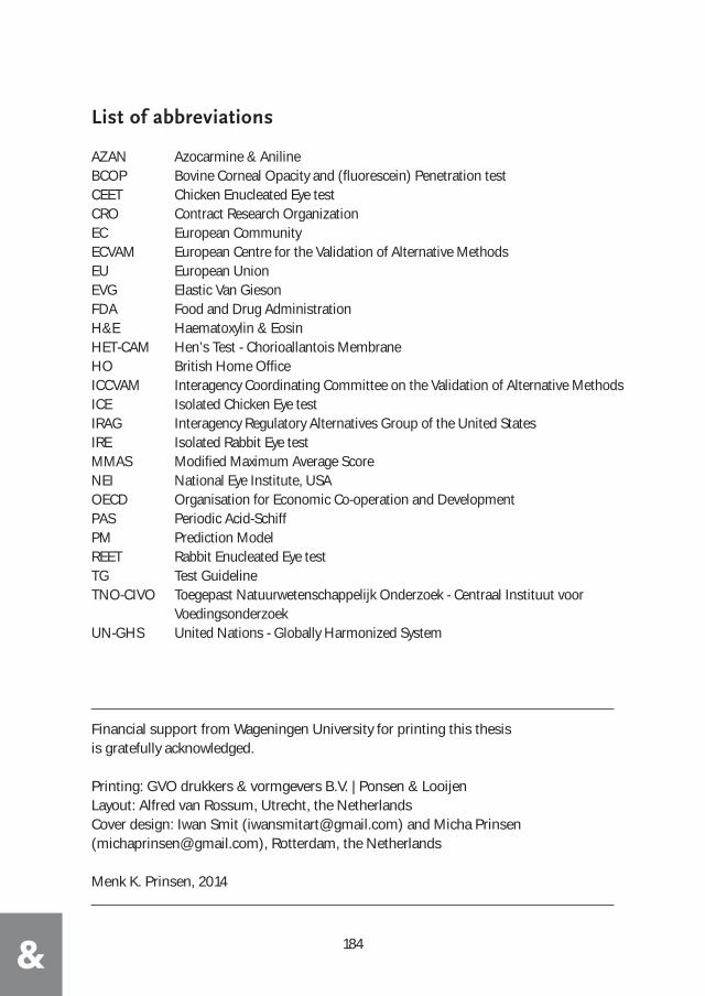
184&
Financial support from Wageningen University for printing this thesisis gratefully acknowledged.
Printing: GVO drukkers & vormgevers B.V. | Ponsen & LooijenLayout: Alfred van Rossum, Utrecht, the NetherlandsCover design: Iwan Smit ([email protected]) and Micha Prinsen([email protected]), Rotterdam, the Netherlands
Menk K. Prinsen, 2014
List of abbreviations
AZAN Azocarmine & A nilineBCOP Bovine Corneal Opacity and (fluorescein) Penetration test CEET Chicken Enucleated Eye testCRO Contract Research OrganizationEC European CommunityECVAM European Centre for the Validation of Alternative MethodsEU European UnionEVG Elastic Van GiesonFDA Food and Drug AdministrationH&E Haematoxylin & Eosin HET-CAM Hen’s Test - Chorioallantois MembraneHO British Home OfficeICCVAM Interagency Coordinating Committee on the Validation of Alternative MethodsICE Isolated Chicken Eye testIRAG Interagency Regulatory Alternatives Group of the United StatesIRE Isolated Rabbit Eye testMMAS Modified Maximum Average ScoreNEI National Eye Institute, USAOECD Organisation for Economic Co-operation and DevelopmentPAS Periodic Acid-SchiffPM Prediction ModelREET Rabbit Enucleated Eye testTG Test GuidelineTNO-CIVO Toegepast Natuurwetenschappelijk Onderzoek - Centraal Instituut voor
Voedingsonderzoek UN-GHS United Nations - Globally Harmonized System
