the intercostal nerve as a target for diagnostic biopsy
TRANSCRIPT

CLINICAL ARTICLEJ Neurosurg 128:1222–1225, 2018
PeriPheral nerve biopsy is a useful diagnostic tool in peripheral neuropathies when a diagnosis is not possible from clinical, laboratory, and neurophysi-
ological investigations. Broadly speaking, peripheral neu-ropathies are categorized as due to either axonal damage or demyelination. Determination of the cause of neuropa-thy is extremely important so that proper treatment can be instituted. The sural nerve is typically the nerve of choice for biopsy sampling, but biopsies of the superficial peroneal nerve and radial sensory nerve have also been described. However, the value of biopsy sampling of these purely sensory nerves is limited in patients who have pri-marily motor deficits, i.e., motor neuropathies and lower motor neuron diseases.1,2 Also, the resultant sensory loss that accompanies biopsy sampling of a small section of the sural nerve can be bothersome to some patients. Biopsy of the sural nerve is not recommended in patients who have normal sural nerve conduction.1 The reported techniques
for obtaining motor nerve tissue are limited in the litera-ture, and some involve risking significant motor impair-ment. The motor branch to the gracilis muscle is a target that is described in the literature. However, the anatomy is unfamiliar to many neurosurgeons and can be challenging in obese patients. Our institution proposes an alternative target for motor nerve biopsy: the intercostal nerve. It po-tentially has the advantages of minimal loss of function and contains an adequate amount of motor fibers.2,4
AnatomyThe intercostal nerve is a mixed peripheral nerve that
is easily accessible and exists in familiar anatomical ter-ritory. An intercostal nerve contains approximately 1200–1300 myelinated fibers, with about 40% of them being mo-tor fibers.3,5 The intercostal nerves are the somatic nerves that arise from the anterior divisions of the thoracic spinal
ABBREVIATIONS CIDP = chronic inflammatory demyelinating polyneuropathy. SUBMITTED November 6, 2015. ACCEPTED December 5, 2016.INCLUDE WHEN CITING Published online May 12, 2017; DOI: 10.3171/2016.12.JNS152565.
The intercostal nerve as a target for diagnostic biopsyKhoi D. Nguyen, MD, Haroon F. Choudhri, MD, and Samuel D. Macomson, MD
Department of Neurosurgery, Augusta University Medical Center, Augusta, Georgia
OBJECTIVE Peripheral nerve biopsy is a useful tool in diagnosing peripheral neuropathies. Sural and gracilis nerves have become the most common targets for nerve biopsy. However, the yield of sural nerve biopsy is limited in patients who have motor neuropathies, and gracilis nerve biopsy presents technical challenges and increased complications. The authors propose the intercostal nerve as an alternative motor nerve target for biopsy.METHODS A total of 4 patients with suspected peripheral neuropathies underwent intercostal nerve biopsy at the authors’ institution. A rib interspace that is inferior to the pectoralis muscle and anterior to the anterior axillary line is selected for the procedure. Generally the lower intercostal nerves (i.e., T7–11) are targeted. An incision is made over the inferior aspect of the superior rib at the chosen interspace. Blunt dissection is carried down to the neurovascular bundle and the nerve is isolated, ligated, and cut to send for pathological examination.RESULTS The average operative time for all cases was 73 minutes, with average blood loss of 8 ml. Biopsy results from 1 patient exhibited axonopathy, and the other 3 patients demonstrated axonopathy with demyelination. There were no short- or long-term postoperative complications. None of the patients reported sensory or motor deficits related to the biopsy at 6 weeks postoperatively.CONCLUSIONS The intercostal nerve can be an alternative target for biopsy, especially in patients with predominantly motor neuropathies, due to its mixed sensory and motor fibers, straightforward anatomy, minimal risk of serious sensory deficits, and no risk of motor impairment.https://thejns.org/doi/abs/10.3171/2016.12.JNS152565KEY WORDS intercostal nerve; nerve biopsy; peripheral neuropathy; peripheral nerve
J Neurosurg Volume 128 • April 20181222 ©AANS 2018, except where prohibited by US copyright law
Unauthenticated | Downloaded 03/23/22 05:50 PM UTC

Intercostal nerve biopsy
J Neurosurg Volume 128 • April 2018 1223
nerves from T-1 to T-11. The ventral primary ramus of the T-12 spinal nerve is the subcostal nerve and does not occupy an intercostal space. These nerves, in addition to supplying the thoracic wall, may also supply the pleura and the peritoneum. Intercostal nerves can be divided into atypical and typical groups based on their pattern of inner-vation. The typical nerves (T3–6) supply only the thoracic wall. The atypical nerves are T-1, T-2, and T7–11. They are considered atypical because in addition to innervating the thoracic wall, they also supply the brachial plexus (as is the case for T-1 and T-2) or the abdomen and peritoneum (as is the case for T7–11). In the intercostal space there are 3 muscle layers: the external intercostal muscle, the internal intercostal muscle, and the innermost intercostal muscle (Fig. 1). The upper intercostal nerves (T3–6) run parallel to their ribs in between the middle and innermost intercostal muscles, whereas the lower intercostal nerves (T7–11) lie superficial either to transversus thoracis or transversus abdominis muscles.
MethodsBetween 2010 and 2014 at our institution, we per-
formed intercostal nerve biopsy in 4 patients. The patient age range was 26–51 years. There were 2 men and 2 wom-en. All patients had a clinical course of progressive weak-ness in proximal extremities more than in distal, as well as gait disturbance. Three of the patients had preliminary diagnoses of chronic inflammatory demyelinating poly-neuropathy (CIDP), whereas 1 patient was being evaluat-ed for hereditary neuropathy. The CSF protein levels were elevated in 2 of the patients. In all of the patients, nerve conduction studies and electromyography showed normal sural nerve activity and were suggestive of mixed senso-rimotor polyneuropathy. One patient was morbidly obese, with a body mass index of 45.
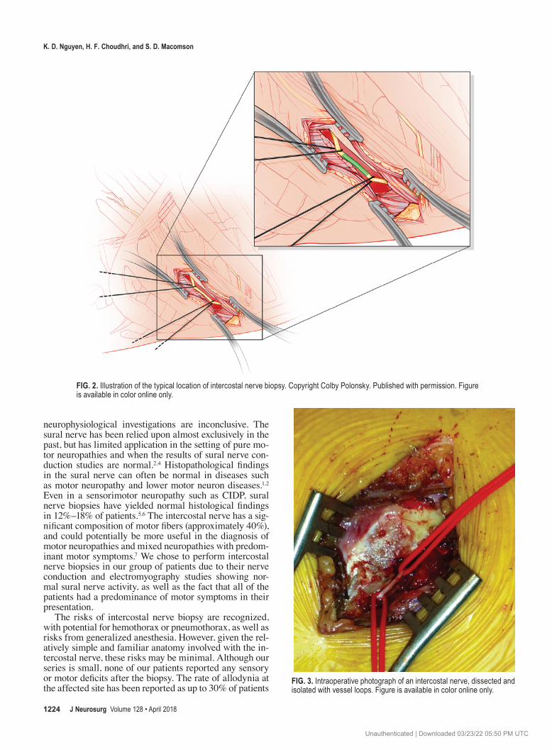
The biopsy procedure was performed under general anesthesia with the patient in the supine position with the arm at 90°, but can also be performed in the lateral posi-tion. We select a rib interspace that is inferior to the pecto-ralis muscle and anterior to the anterior axillary line (Fig. 2). We generally target the lower intercostal nerves (i.e., T7–11). The fourth intercostal nerve’s sensory component supplies the skin of the nipple-areolar area and must be avoided. An incision is made over the inferior aspect of the superior rib at the chosen interspace. Dissection is per-formed through the external and internal intercostal mus-cles to obtain access to the inferior pleural surface of the rib. A blunt dissector is then passed along the inferior sur-face of the rib, detaching the neurovascular bundle from the rib. A segment of the nerve is then isolated between 2 vessel loops and is sutured, ligated, and cut (Fig. 3).
ResultsThe average operative time for all cases was 73 min-
utes, with an average blood loss of 8 ml. Biopsy results from the patient with suspected hereditary neuropathy ex-hibited axonopathy, and the other 3 patients demonstrated axonopathy with demyelination. The patient with suspect-ed hereditary neuropathy did not elect to undergo genetic testing due to costs. The 3 patients with suspected CIDP
had their diagnoses confirmed as CIDP by their neurolo-gist at our institution. There were no short- or long-term postoperative complications. None of the patients reported sensory or motor deficits related to the biopsy at 6 weeks postoperatively.
DiscussionPeripheral nerve biopsy is essential in the diagnosis
of peripheral neuropathies when laboratory, clinical, and
FIG. 1. Illustration of a cross-section anatomy of intercostal space. Note the 3 muscular layers encountered from superficial to deep: external intercostal muscle, internal intercostal muscle, and innermost intercostal muscle. The green arrow points to the intercostal nerve within the neuro-vascular bundle. Copyright Colby Polonsky. Published with permission. Figure is available in color online only.
Unauthenticated | Downloaded 03/23/22 05:50 PM UTC
K. D. Nguyen, H. F. Choudhri, and S. D. Macomson
J Neurosurg Volume 128 • April 20181224
neurophysiological investigations are inconclusive. The sural nerve has been relied upon almost exclusively in the past, but has limited application in the setting of pure mo-tor neuropathies and when the results of sural nerve con-duction studies are normal.2,4 Histopathological findings in the sural nerve can often be normal in diseases such as motor neuropathy and lower motor neuron diseases.1,2 Even in a sensorimotor neuropathy such as CIDP, sural nerve biopsies have yielded normal histological findings in 12%–18% of patients.5,6 The intercostal nerve has a sig-nificant composition of motor fibers (approximately 40%), and could potentially be more useful in the diagnosis of motor neuropathies and mixed neuropathies with predom-inant motor symptoms.7 We chose to perform intercostal nerve biopsies in our group of patients due to their nerve conduction and electromyography studies showing nor-mal sural nerve activity, as well as the fact that all of the patients had a predominance of motor symptoms in their presentation.
The risks of intercostal nerve biopsy are recognized, with potential for hemothorax or pneumothorax, as well as risks from generalized anesthesia. However, given the rel-atively simple and familiar anatomy involved with the in-tercostal nerve, these risks may be minimal. Although our series is small, none of our patients reported any sensory or motor deficits after the biopsy. The rate of allodynia at the affected site has been reported as up to 30% of patients
FIG. 2. Illustration of the typical location of intercostal nerve biopsy. Copyright Colby Polonsky. Published with permission. Figure is available in color online only.
FIG. 3. Intraoperative photograph of an intercostal nerve, dissected and isolated with vessel loops. Figure is available in color online only.
Unauthenticated | Downloaded 03/23/22 05:50 PM UTC

Intercostal nerve biopsy
J Neurosurg Volume 128 • April 2018 1225
after sural nerve biopsy.4 Biopsy of the motor nerve to the gracilis muscle has been shown to carry risk of decrease in thigh adduction strength and hypesthesia.1 Another po-tential advantage over biopsy of the nerve to the gracilis muscle is that biopsy of the intercostal nerve may be less challenging in obese patients due to the straightforward anatomy of the intercostal space.
The emphasis on using the intercostal nerve as a target for biopsy is not to propose that it is superior to the yield and clinical relevance of sural nerve biopsy. Sural nerve biopsy is the gold standard and workhorse in the diagno-sis of peripheral neuropathies. However, the intercostal nerve can be useful as an aid in diagnosis of motor neu-ropathies or of sensorimotor neuropathies in which mo-tor symptoms predominate, especially in cases in which neurophysiological studies show normal sural nerve activ-ity. Additionally, intercostal nerve biopsy could be a good option in cases in which a motor nerve sample is needed. Biopsy of the nerve to the gracilis muscle can be challeng-ing due to anatomy unfamiliar to neurosurgeons.
ConclusionsWe have found the intercostal nerve to be an alternative
target for biopsy in motor neuropathies due to its mixed sensory and motor fibers, facile anatomy, minimal risk of serious sensory deficits, and no risk of motor impairment.
AcknowledgmentsColby Polonsky, MS, CMI, created the illustrations presented
in this work.
References 1. Abouzahr MK, Lange DJ, Latov N, Olarte M, Rowland LP,
Hays AP, et al: Diagnostic biopsy of the motor nerve to the gracilis muscle. Technical note. J Neurosurg 87:122–124, 1997
2. Barohn RJ, Kissel JT, Warmolts JR, Mendell JR: Chronic in-flammatory demyelinating polyradiculoneuropathy. Clinical characteristics, course, and recommendations for diagnostic criteria. Arch Neurol 46:878–884, 1989
3. Corbo M, Abouzahr MK, Latov N, Iannaccone S, Quattrini A, Nemni R, et al: Motor nerve biopsy studies in motor neu-ropathy and motor neuron disease. Muscle Nerve 20:15–21, 1997
4. Gabriel CM, Howard R, Kinsella N, Lucas S, McColl I, Sal-danha G, et al: Prospective study of the usefulness of sural nerve biopsy. J Neurol Neurosurg Psychiatry 69:442–446, 2000
5. Lykissas MG, Kostas-Agnantis IP, Korompilias AV, Vekris MD, Beris AE: Use of intercostal nerves for different target neurotization in brachial plexus reconstruction. World J Or-thop 4:107–111, 2013
6. Molenaar DS, Vermeulen M, de Haan R: Diagnostic value of sural nerve biopsy in chronic inflammatory demyelinating polyneuropathy. J Neurol Neurosurg Psychiatry 64:84–89, 1998
7. Riva N, Iannaccone S, Corbo M, Casellato C, Sferrazza B, Lazzerini A, et al: Motor nerve biopsy: clinical usefulness and histopathological criteria. Ann Neurol 69:197–201, 2011
DisclosuresThe authors report no conflict of interest concerning the materi-als or methods used in this study or the findings specified in this paper.
Author ContributionsConception and design: Macomson. Acquisition of data: Choudhri, Macomson. Analysis and interpretation of data: Nguy-en. Drafting the article: Nguyen. Critically revising the article: Nguyen, Macomson. Reviewed submitted version of manuscript: Macomson.
Supplemental InformationPrevious PresentationsPortions of this work have been given as an oral presentation at the Georgia Neurosurgical Society’s annual spring meeting, held on Sea Island, Georgia, on May 23, 2014.
CorrespondenceKhoi Nguyen, Department of Neurosurgery, Augusta University Medical Center, 1120 15th St., BI 3088, Augusta, GA 30912. email: [email protected].
Unauthenticated | Downloaded 03/23/22 05:50 PM UTC