the inner workings of the dna copying nanomachine
TRANSCRIPT

University of RichmondUR Scholarship Repository
Honors Theses Student Research
Spring 2013
The inner workings of the DNA copyingnanomachine : kinetic studies of DNA polymeraseI from the thermophilic bacterium RhodothermusmarinusEmily P. Kornberg
Follow this and additional works at: http://scholarship.richmond.edu/honors-theses
Part of the Biochemistry Commons, and the Molecular Biology Commons
This Thesis is brought to you for free and open access by the Student Research at UR Scholarship Repository. It has been accepted for inclusion inHonors Theses by an authorized administrator of UR Scholarship Repository. For more information, please [email protected].
Recommended CitationKornberg, Emily P., "The inner workings of the DNA copying nanomachine : kinetic studies of DNA polymerase I from thethermophilic bacterium Rhodothermus marinus" (2013). Honors Theses. Paper 55.

ABSTRACT

Kornberg 2
Kornberg 3
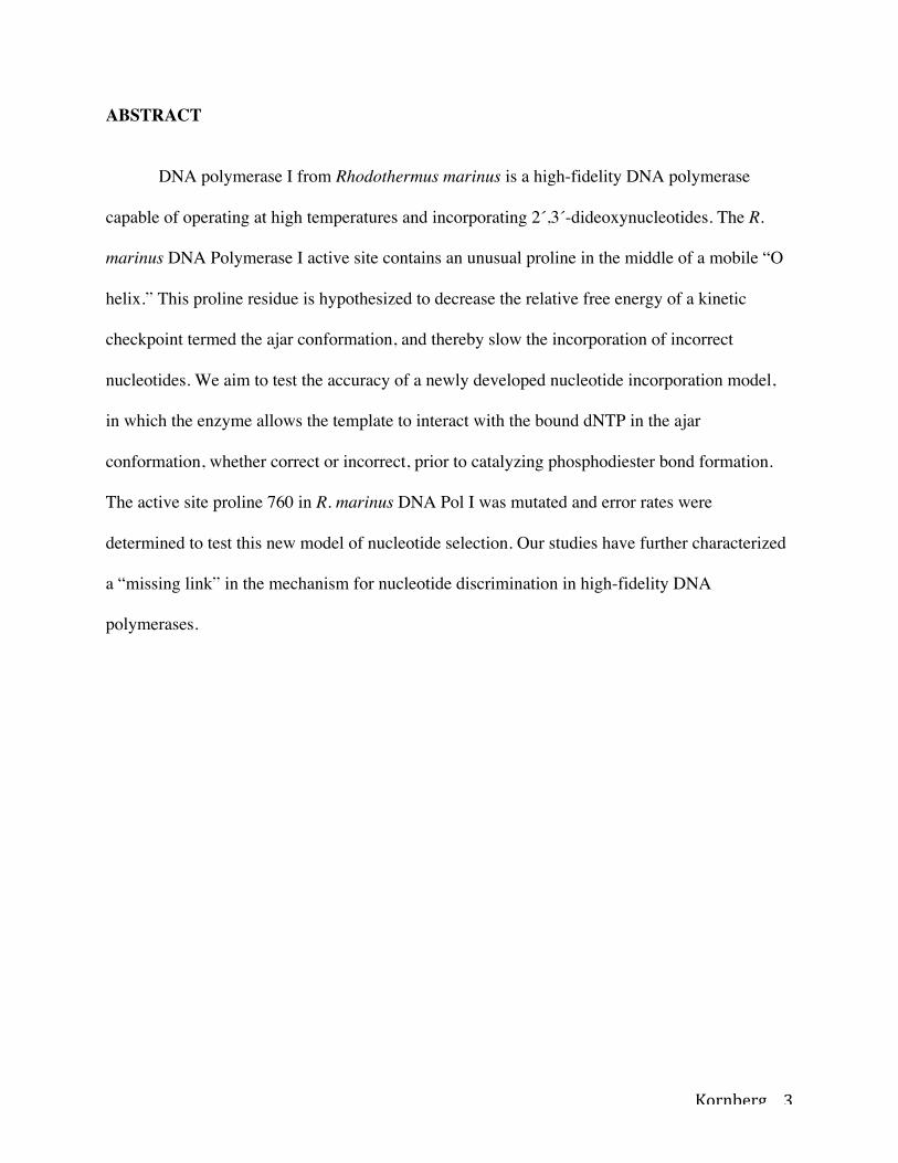
ABSTRACT
DNA polymerase I from Rhodothermus marinus is a high-fidelity DNA polymerase
capable of operating at high temperatures and incorporating 2´,3´-dideoxynucleotides. The R.
marinus DNA Polymerase I active site contains an unusual proline in the middle of a mobile “O
helix.” This proline residue is hypothesized to decrease the relative free energy of a kinetic
checkpoint termed the ajar conformation, and thereby slow the incorporation of incorrect
nucleotides. We aim to test the accuracy of a newly developed nucleotide incorporation model,
in which the enzyme allows the template to interact with the bound dNTP in the ajar
conformation, whether correct or incorrect, prior to catalyzing phosphodiester bond formation.
The active site proline 760 in R. marinus DNA Pol I was mutated and error rates were
determined to test this new model of nucleotide selection. Our studies have further characterized
a “missing link” in the mechanism for nucleotide discrimination in high-fidelity DNA
polymerases.

Kornberg 4
INTRODUCTION
DNA synthesis is a vital chemical reaction in biology because it is the mechanism by
which genetic material is copied and passed from one cell to another. This process is remarkably
fast and accurate, with nucleotide addition rates of tens or hundreds of nucleotides per second
and error rates between one error per ten thousand to one million nucleotides copied for
replicative polymerases 1. The high fidelity of DNA polymerases is achieved by employing a
complex induced-fit mechanism wherein the enzyme encloses around a complementary
incoming deoxynucleoside triphosphate (dNTP) and aligns the 3´-hydroxyl of the growing DNA
strand to the α-phosphate of the dNTP for in-line attack 2. Mismatches are misaligned in the
polymerase active site, leading to slower incorporation and dissociation 3.
Once the dNTP is bound, the enzyme transitions from an “open” to a “closed”
conformation, which encloses the free nucleotide opposite the base on the template DNA. During
this conformational change, a dislocation of a conserved tyrosine in the “O helix” enables the
template nucleotide to pair with the free dNTP in the newly formed “insertion site” (Figure 1).
Based on existing data, it is evident that DNA polymerase adopts the same conformation for all
four of the possible dNTPs and does not distinguish one base from another. Because of this, the
enzyme must allow the template base to preview all four of the dNTPs individually in order to
distinguish between the correct and incorrect dNTP match 8.
Many DNA polymerases also contain the ability to proofread their own work through an
attached 3´-to-5´ exonuclease domain. As the polymerase processively synthesizes DNA,
occasional errors occur and slow the rate of synthesis due to misalignment of substrates and
active site residues. Mismatched nucleotides in the growing DNA strand can then be transferred
to the 3´-to-5´ exonuclease active site, which cleaves 3´-terminal nucleotides using a two-metal-

Kornberg 5
ion catalytic mechanism 4,5. The enzyme can then resynthesize the DNA, resulting in higher
fidelity for DNA synthesis. Proofreading improves fidelity by up to two orders of magnitude 1.
In addition to their essential roles in all life forms, DNA polymerases also have important
functions in the laboratory setting. Technologies such as polymerase chain reaction and DNA
sequencing are essential and transformative to biology and biotechnology and have been
developed based on knowledge of DNA polymerase function. DNA polymerases from
thermophilic organisms have been particularly useful for biotechnology, allowing reactions to be
performed at high temperatures 8. The R. marinus DNA polymerase I contains a proline in place
of a highly conserved valine or isoleucine in the polymerase O helix (Figure 2), which prevents
the O helix from its normal functional flexibility. This proline “wedge” may play a key role in
the accuracy of the polymerase through the stabilization of the previously described ajar
conformation 8. This conformation falls between the open and closed conformations adopted by
the polymerase in which the O helix subdomain reaches an intermediate point in its movement,
allowing the incoming dNTP to be matched against the template DNA strand and identified as a
correct or incorrect match.
Previous studies conducted on DNA Polymerase I from another bacterium Bacillus
stearothermophilus have indicated that the polymerase adopts a previously characterized ajar
conformation prior to the closed conformation that allows the polymerase to incorporate
nucleotides into the growing DNA strand with more accurately, yet more slowly 8. In studies
conducted by Wu et. al. 8 a valine residue at position 713, the hinge of the O helix that allows for
its flexibility when incorporating nucleotides, was mutated to a proline (Figure 3). Kinetics
studies showed a slowed rate of incorporation of both correct and incorrect nucleotide bases,

Kornberg 6
which suggests that the transition from the ajar to the closed conformation is indeed part of the
enzyme mechanism.
We hypothesize that the R. marinus DNA polymerase I enzyme will more readily form
the ajar conformation in the presence of nucleotides due to having a proline “wedge” in the
active site, allowing the enzyme to make fewer mismatched nucleotide base pairs and have a
higher relative efficiency, which is an indicator of how often the enzyme incorporates incorrectly
matched base pairs into the growing DNA strand. To test this hypothesis, we will employ site-
directed mutagenesis to remove to the proline wedge from R. marinus DNA polymerase I. If the
proline wedge serves as a barrier to the closed conformation, as hypothesized in the model, then
its removal should speed up the enzyme. We aim to further characterize this conformation by
means of kinetics assays using quench flow analysis and capillary electrophoresis imaging to
quantify the incorporation of correctly and incorrectly-matched nucleotides by the R. marinus 3’-
5’ exonuclease-domain-deficient polymerase, its mutant with the proline “wedge” in the active
site mutated to a valine, and Taq DNA Polymerase I, which has a valine residue at the glycine
hinge in the O helix (Figure 3), into a growing DNA strand. These assays provide the
information necessary to calculate and compare the in vitro error rate of each enzyme used.
MATERIALS AND METHODS
Cloning. DNA fragment encoding the 5’-‘3 exonuclease deficient DNA polymerase I from
Rhodothermus marinus (RF; residues 329-924 plus an N-terminal methionine) was amplified by
PCR from genomic DNA (American Type Culture Collection, Manassas, Virginia), and inserted

Kornberg 7
into pCR2.1-TOPO vector (Invitrogen, Carlsbad, California) as XhoI restriction-enzyme
fragment. Oligonucleotides used for amplification were 5´-
CATATGGAAAAGGCGGACTACCGGATCGTC-3 ´and 5´-
TCAGTGGGCATCCAGCCAGTTGTC-3´. Gene was sequenced using Sanger sequencing and
inserted into a bacterial expression vector, pET-21a (Novagen), as a XhoI restriction-enzyme
fragment.
Site-Directed Mutagenesis: The D497A mutation was generated using the QuikChange site-
directed mutagenesis kit (Stratagene, La Jolla, California) with Phusion High-Fidelity DNA
Polymerase (New England Biolabs, Ipswich, Massachusetts). The oligonucletoides used for the
D497A and P760V mutagenesis reactions were 5´-
CCCTATGCCTGTGAAGCCACGGACATTGCACTG-3´/5´-
CAGTGCAATGTCCGTGGCTTCACAGGCATAGGG-3´ and 5´-
CCAAGATGGTCAACTACGGCATTGTCTACGGGATTTCGG-3´/5´-
CCGAAATCCCGTAGACAATGCCGTAGTTGACCATCTTGG-3´, respectively. The
mutations were confirmed by Sanger DNA sequencing using fluorescent terminators (Eurofins
MWG Operon, Huntsville, Alabama).
Protein expression and purification. The pET-21a plasmid containing the RF gene with the
D497A mutation was transformed into Escherichia coli BL21(DE3) cells (Lucigen, Middleton,
WI). Native D497A RF protein was produced using standard overexpression techniques.
Inoculated 500 mL LB/Amp Media with 20 mL overnight culture and grew at 37 °C to
OD600=0.600. Expression of protein was induced with IPTG (4 mM) and the cells were incubated
overnight at 37°C. Cells were harvested by centrifugation at ~3000Xg for 15 minutes, and stored
at 4 °C.

Kornberg 8
A bacterial pellet from cell culture was resuspended and lysed in B-PER (Bacterial
Protein Extraction Reagent 20 mM Tris Buffer, pH 7.5; Thermo Fisher Scientific, Rockford,
Illinois). Lysate was centrifuged at ~7500Xg for 30 minutes and supernatant was heated in 65 °C
water bath for 20 minutes. Lysates were centrifuged again at 7500 RPM for 30 minutes, and
supernatant containing heat-stable proteins was dialyzed in 1x Buffer A (50 mM Tris-HCl pH
7.5, 1 mM ethylenediamine tetraacetic acid, 6.5 mM 2-mercaptoethanol). Supernatant was
passed through heparin sepharose column equilibrated in 1x Buffer A using a linear gradient
containing 1x Buffer A and 50% 1x Buffer A & 1.5 M sodium chloride (Buffer B). Fractions
containing the protein were collected and exchanged back into 1x Buffer A through dialysis. The
fractions were then passed through S1 ion exchange column (BioRad) pre-equlibrated with 1x
Buffer A using a linear gradient containing 1x Buffer A and 1x Buffer B. Fractions containing
protein were combined and concentrated using 10 kDa Amicon centrifugal concentrators (EMD
Millipore). Protein sample was run through S1 ion-exchange column again to remove remaining
impurities, again using linear gradient of 1x Buffer A and 1x Buffer B. Fractions containing RF
were exchanged into 1x Buffer A through three cycles of concentration and dilution, and
concentrated to 1 mL in 50 kDa Amicon centrifugal concentrator.
Kinetics Assays and data collection. Complementary oligonucleotides used in the solution
studies were synthesized by Operon Biotechnologies, Inc. (Huntsville, AL). The template (5’-
TTACTTGACCAGATACACTGTCTTTGACACGTTGATGGATTAGAGCAATCACATCCA
AGACTGGCTATGCACGAA-3’) and fluorescently labeled primer (5’-6-carboxyfluorescein-
TCGTGCATAGCCAGTCTTGGATGTGATTGCTCTAATCCATCAACGTGTCAAAGACAG
TGTATCTGGT-3’) strands were annealed as described 18 (Figure 4). The DNA substrate and

Kornberg 9
wild type, P760V, or Taq DNA Polymerase I proteins were diluted with reaction buffer (50 mM
Tris-HCl, pH 8.0, 50 mM NaCl, 10 mM MgCl2, 1 mM DTT) to 0.1 and 2 μM, respectively. RF-
DNA or Taq-DNA complexes were mixed with equal volumes at various concentrations of
dCTP or dTTP. The reaction (50°C) was quenched at different time points using four reaction
volumes of quench solution (95% formamide (v/v), 25 mM EDTA). Reactions slower than 1000
ms (RF-dTTP) were executed manually. Reactions faster than 1000 ms (RF-dCTP) were
executed using a KinTek RQF-3 Rapid Quench Flow instrument (KinTek Corp., Austin, TX).
Primer extension was quantified by capillary electrophoresis with fluorescence detection, using
an ABI3100 Genetic Analyzer (Applied Biosystems, Foster City, CA). Pre-steady-state kinetic
constants were determined and fit as described using KaleidaGraph (Synergy Software) 8.
RESULTS
Bacterial DNA polymerase I enzymes contain an N-terminal 5´-to-3´ exonuclease
domain, a central 3´-to-5´ exonuclease domain for proofreading, and a C-terminal polymerase
domain. The N-terminal 5´-to-3´ exonuclease domain has been shown to be dispensable for the
DNA synthesizing activity of several DNA polymerases, including DNA polymerase I from
Escherichia coli (Klenow fragment), Thermus aquaticus (Klentaq1), and R. marinus9-12. Due to
the improved stability of the N-terminal truncated protein 9, we chose to express the large
fragment (amino acids 329-924) of the R. marinus DNA polymerase I, which we term
“Rhodothermus fragment”, or RF.

Kornberg 10
Site-directed mutagenesis was utilized to inactivate the 3´-to-5´ exonuclease domain of
RF for kinetic studies and future co-crystallization trials with DNA. An active nuclease activity
could potentially digest the DNA in complex with the enzyme, therefore not allowing for an
accurate measurement of nucleotide incorporation. The 3´-to-5´ exonuclease domain of DNA
polymerase I from R. marinus shares strong homology to the E. coli exonuclease domain and is
active 9, as opposed to the inactive domains of DNA polymerases from other thermophiles,
Bacillus stearothermophilus 13 and Thermus aquaticus which do not have a fully developed
active site to support this exonuclease activity. 14,15 All four acidic residues that bind two divalent
cations in the 3´-to-5´ exonuclease domain 4 are conserved. The single mutation that decreases
exonuclease activity the most in E. coli DNA polymerase I is aspartic acid 501 to alanine
(D424A) 5; thus we made the equivalent mutation, D497A, in RF to inactivate its exonuclease
activity.
Site-directed mutagenesis was again utilized to mutate a proline at position 760 in RF to a
valine. By removing this proline “wedge” at the hinge of the O helix, the enzyme without this
stabilized ajar conformation can be analyzed by monitoring its rate of incorporation of
nucleotides and the subsequent relative efficiency of the enzyme.
Kinetics Assays
RF, the P760V mutant, and Taq DNA Polymerase I were allowed to incorporate a single
nucleotide into a template strand 5’-deoxyguanosine overhang on a 6-FAM tagged template and
primer DNA complex (Figure 6). Quench flow assays were conducted via mixing our enzyme (2
μM)-DNA (.1 μM) complex with either deoxycytosine triphosphate (dCTP) or deoxythymine
triphosphate (dTTP) nucleotide at various concentrations (0.005-0.1 mM and 0.025-1.0 mM,

Kornberg 11
respectively) and quenching the reaction with Formamide and EDTA after a specified amount of
time (10-1000 ms and 20-3000 sec, respectively). A broad range of concentrations for each
nucleotide was used in order to acquire the most accurate constants for each enzyme. Each
sample was shipped to Dr. Scott Langdon at the Duke DNA Analysis Facility for capillary
electrophoresis analysis (Figure 4). Using this data the amount of extension of the primer DNA
strand could be quantified, and the fraction of primer extension could be plotted against time for
each concentration of nucleotide used in experiments (Figure 5). Initial rates of incorporation
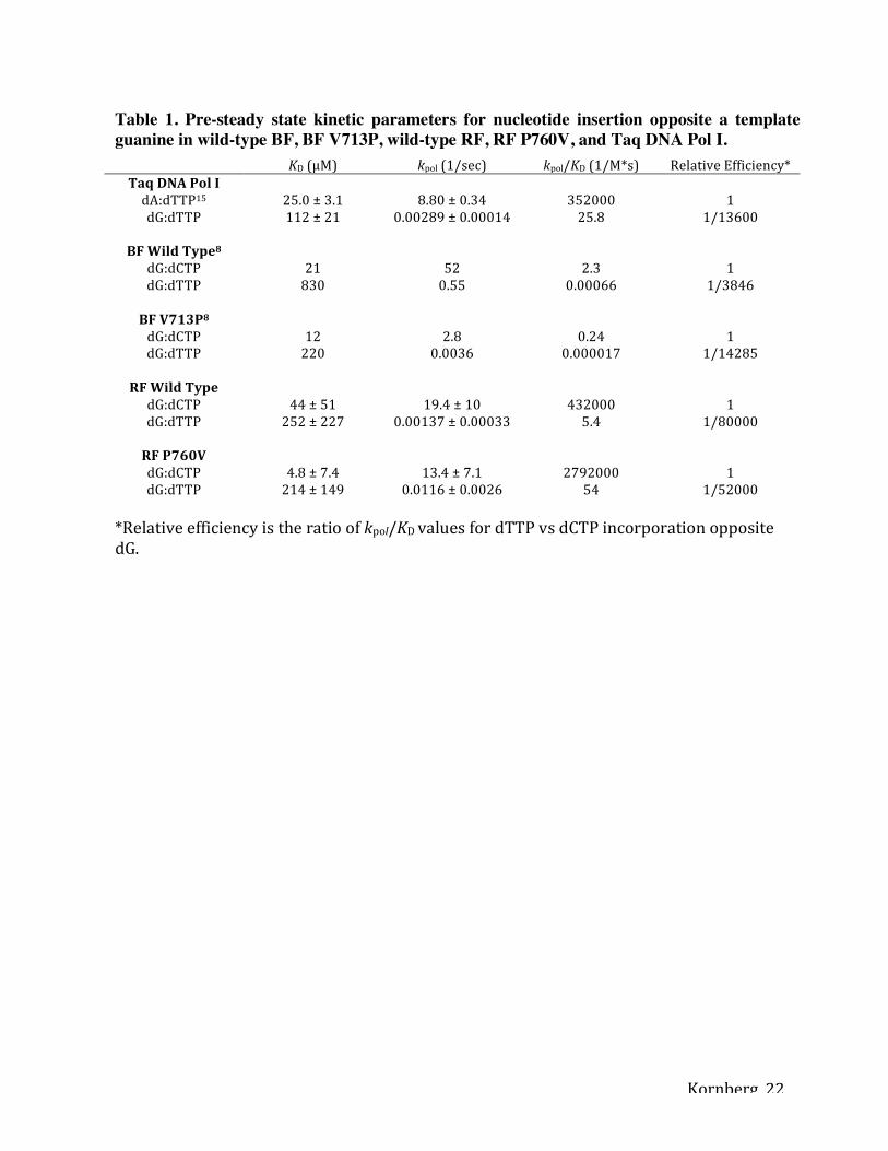
could be derived from these plots and, using Michaelis-Menten kinetics, the kpol, KD, and relative
efficiency could be determined for each enzyme. Each of these constants were calculated and the
results are reported in Table 1. Also included with the acquired data are the results from previous
studies using Bacillus fragment (BF).
DISCUSSION
From existing structural data 16, it is clear that DNA polymerase adopts the same open
conformation for all four template bases and does not distinguish one template base from
another. Thus, the enzyme, because it is capable of incorporating all four dNTPs, must allow the
template to preview all four nucleotides individually in order to distinguish between the correct
and incorrect dNTP. Therefore, there must exist a conformation in which the enzyme allows the
template to interact with the bound dNTP, whether correct or incorrect, before phosphodiester
bond formation is catalyzed 17. Previous structural studies have revealed a new conformation in
which the template base forms a base pair with the incoming nucleotide, but the enzyme does not

Kornberg 12
proceed to the closed conformation. Rather, the O helix adopts a conformation intermediate of
the open and closed conformations, which has been termed the “ajar” conformation. This ajar
conformation is made possible by a glycine hinge in that last turn of the O helix (Figure 3),
where the O helix bends sharply. The enzyme used in these previous experiments was mutated
such that a valine at position 713, in the hinge of the O helix, was replaced by a bulky proline
residue 8. This V713P mutant enzyme showed slower nucleotide incorporation rates for both
complementary pairs and mismatches, suggesting that the transition from ajar to closed
conformations is an important step in the polymerase mechanism. The V713P mutation actually
slowed G:T mismatch incorporation more than complementary G:C nucleotide addition, in turn
making the enzyme more accurate. Using these data, a new model for nucleotide selection in
DNA polymerase I enzymes was formulated. In this model (Figure 2), DNA polymerase I uses
the ajar conformation as a preview conformation to check the nucleotide for Watson-Crick base
pairing with the template. Good matches advance to the closed conformation 8. The ability to
adopt the ajar conformation is a critical part of the mechanism to distinguish mismatches.
Through our experiments with RF, we have further supported this model by altering the
ajar conformation in another DNA polymerase and measuring the enzyme fidelity. The presence
of proline 760 in the active site of RF DNA Polymerase I made possible the inverse experiment
of that with BF. We hypothesized that the mutation of proline 760 to valine in RF will remove an
energy barrier in the transition from the ajar to closed conformation, increase the rate of catalysis
for both correct and incorrect nucleotides, and also decrease the fidelity of the enzyme. Based on
the data reported in Table 1, it is clear that rates of catalysis in this mutant RF did in fact increase
for mismatched base pair incorporation. The rates of catalysis, however, for complementary
matches in the wild type and mutant RF enzymes were not statistically different, and our

Kornberg 13
hypothesis was not supported in this respect. The mutant RF had a substantially increased G:T
mismatch incorporation rate than that of the wild-type RF, suggesting that this stabilized ajar
conformation in the wild type RF enzyme plays a critical role in the high-fidelity of the enzyme.
Pre-steady state kinetics assays were also performed with Taq DNA Polymerase I in order to
provide a frame of reference for our results because this polymerase does not have a proline in an
equivalent position in the active site (Figure 3). RF wild-type turned out to be much more
accurate than Taq DNA Polymerase I, owing to the location of the proline 760 “wedge” in the
active site of the polymerase. Further supporting this observation was the observation that Taq
DNA Polymerase I had a higher rate of catalysis for the incorporation of mismatched base pairs
compared to the wild type RF enzyme. Data from previous experiments with BF is also included
in Table 1 8. Comparing the data between the wild type and mutant BF, there is an order of
magnitude difference in the incorporation of both complementary and mismatched nucleotides
between the two BF enzymes. As previously stated, we found no statistical difference in the kpol
for incorporating complementary nucleotides between our wild type and mutant RF enzymes.
However, as also seen with BF, we did see an order of magnitude change in the kpol for the
incorporation of mismatched nucleotides when comparing our wild type to our mutant RF
enzymes. In BF, the KD for the mutant enzyme decreased dramatically from that of the wild type
enzyme when incorporating mismatched nucleotides, indicating a much stronger affinity, and
thus a lower KD, for the incoming substrate due to the stabilized ajar conformation in the mutant
enzyme, which allows for greater interactions between the incoming dNTP and the growing
DNA strand. With our RF enzymes, we did not see a similar change in affinity for the
incorporation of complementary or mismatched nucleotides given the error rates for our KD
values measured for each enzyme. With the stabilization of the ajar conformation in the BF

Kornberg 14
mutant, the relative efficiency of the enzyme dramatically increased. When we made our
backwards mutation in the RF enzyme, in essence de-stabilizing the ajar conformation, we see a
dramatic decrease in the enzyme’s relative efficiency, thus exhibiting the importance of the role
this ajar conformation plays in maintaining the high fidelity of the enzyme.
With these experiments, we have expanded upon the current knowledge in regards to the
function of high-fidelity DNA polymerases and further characterized a critical intermediate step
in nucleotide incorporation which leads to the high accuracy of these enzymes. These studies and
future studies with other polymerases may answer some unresolved questions of how the initial
binding of a complementary dNTP encourages the enzyme to move through the initial weak
contacts that then develop and increase in strength as the enzyme proceeds to the closed
complex. Future studies may also help us understand how the initial weak binding of a
mismatched dNTP is recognized by the enzyme and helps it to proceed to a newly characterized
ajar conformation, which favors release of the incorrectly matched dNTP over catalysis.
ACKNOWLEDGMENTS
We thank Dr. Scott Langdon and the staff of the Duke University DNA Analysis Facility
for their assistance. We thank fellow lab members, Lori Spicer, Brooke Duffany, and Natalie
Omattage for assistance with cloning and protein purification.

Kornberg 15
FIGURE LEGENDS
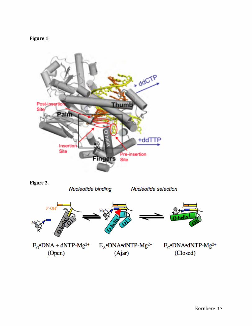
Figure 1. DNA polymerase I structure preceding phosphodiester bond formation.
Complementary nucleotide addition is accompanied by the transition from an open to the closed
conformation. This conformational change is characterized by rotation of the O helix in the
fingers subdomain, thereby enclosing the correct dNTP within the active site cleft. Reproduced
from 8, with permission.
Figure 2. Model for nucleotide sampling and selection. Nucleotides are sampled in the ajar
conformation (EA, cyan) and are released if it incorrect, whereupon the enzyme returns to the
open conformation (EO, gray), or entrapped in the closed conformation (EC, green) if it is
complementary to the template base. Cartoon representations of each state are shown in the
center. The red wedge in the hinge of the O helix represents the proline residue at position 760 in
RF. Adapted from 8, with permission.
Figure 3. Structure-based manual sequence alignment of nucleotide binding helices of A family
DNA polymerases. Phosphate interaction, blue; aromatic residues in the nucleotide binding sites,
green; Proposed glycine hinges in the helices; red. BstPolI, EcoPolI, and TaqPolI, DNA
polymerase I from B. stearothermophilus, E. coli, and T. aquaticus; hPolG, hPolQ and hPolNu,
human DNA polymerases γ, θ, and ν. Reproduced from 8, with permission.
Figure 4. Detection of nucleotide incorporation into a growing primer strand by RF. Capillary
electropherograms of primer DNA strand before and after nucleotide addition. The DNA

Kornberg 16
substrate in Figure 6 was pre-bound to RF and run in an ABI 3100 DNA Analyzer before (A)
and after (B) mixing with dCTP. Note the disappearance of the main peak (54.13) and the growth
of a new peak (54.87) upon addition of dCTP.
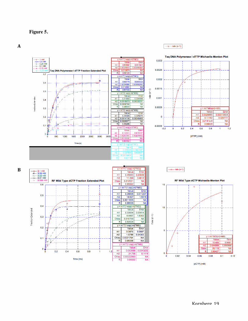
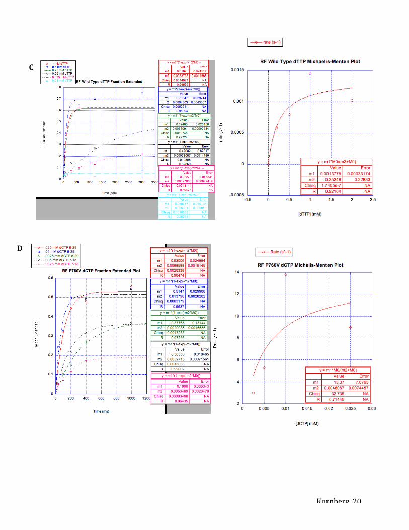
Figure 5. Measuring incorporation of nucleotides as determined by capillary electrophoresis.
Each concentration of nucleotide was plotted on a graph with fraction of primer extension on the
y-axis and time (s or ms) on the x-axis. Slope of fraction extension plots (m1), or the initial rate
of the enzyme in incorporating nucleotides, was used to plot rate (s-1) on the y-axis and
concentration (mM) of nucleotide on the x-axis. m1 and m2 values in Michaelis-Menten plots
represent kpol and KD, respectively. Fraction extended and Michaelis-Menten Plot for dTTP
incorporation using Taq DNA Polymerase I (A), dCTP incorporation using RF Wild Type (B),
dTTP incorporation using RF Wild Type (C), dCTP incorporation using RF P760V (D), dTTP
incorporation using RF P760V (E).
Figure 6. 6-FAM-labeled 59-mer primer strand (top) is annealed to a complementary 60-mer
template strand, leaving a single G 5’-overhang.
Kornberg 17
Figure 1. Figure 2.

Kornberg 18
Figure 3. Figure 4.
A
B
Kornberg 19
Figure 5.
A
B
Kornberg 20
D
C

Kornberg 21
Figure 6. 6-FAM-5’-…58nt-C-3’ 3’-…58nt-G-G-5’
E
Kornberg 22
KD (μM) kpol (1/sec) kpol/KD (1/M*s) Relative Efficiency* Taq DNA Pol I dA:dTTP15 25.0 ± 3.1 8.80 ± 0.34 352000 1 dG:dTTP 112 ± 21 0.00289 ± 0.00014 25.8 1/13600
BF Wild Type8
dG:dCTP 21 52 2.3 1 dG:dTTP 830 0.55 0.00066 1/3846
BF V713P8 dG:dCTP 12 2.8 0.24 1 dG:dTTP 220 0.0036 0.000017 1/14285
RF Wild Type dG:dCTP 44 ± 51 19.4 ± 10 432000 1 dG:dTTP 252 ± 227 0.00137 ± 0.00033 5.4 1/80000
RF P760V dG:dCTP 4.8 ± 7.4 13.4 ± 7.1 2792000 1 dG:dTTP 214 ± 149 0.0116 ± 0.0026 54 1/52000
*Relative efficiency is the ratio of kpol/KD values for dTTP vs dCTP incorporation opposite dG.
Table 1. Pre-steady state kinetic parameters for nucleotide insertion opposite a template guanine in wild-type BF, BF V713P, wild-type RF, RF P760V, and Taq DNA Pol I.

Kornberg 23
References 1. Kunkel TA (2004) DNA replication fidelity. J Biol Chem 279:16895–16898.
2. Joyce CM, Benkovic SJ (2004) DNA polymerase fidelity: kinetics, structure, and checkpoints. Biochemistry 43:14317–14324.
3. Kunkel TA, Bebenek K (2000) DNA replication fidelity. Annu. Rev. Biochem. 69:497–529.
4. Derbyshire V, Freemont PS, Sanderson MR, Beese L, Friedman JM, Joyce CM, Steitz TA (1988) Genetic and crystallographic studies of the 3“,5-”exonucleolytic site of DNA polymerase I. Science 240:199–201.
5. Derbyshire V, Grindley ND, Joyce CM (1991) The 3“-5” exonuclease of DNA polymerase I of Escherichia coli: contribution of each amino acid at the active site to the reaction. EMBO J 10:17–24.
6. Astatke M, Grindley ND, Joyce CM (1998) How E. coli DNA polymerase I (Klenow fragment) distinguishes between deoxy- and dideoxynucleotides. J Mol Biol 278:147–165.
7. Wang W, Wu EY, Hellinga HW, Beese LS (2012) Structural factors that determine selectivity of a high-fidelity DNA polymerase for deoxy-, dideoxy-, and ribo-nucleotides. J Biol Chem.
8. Wu, Eugene Y. and Beese, Lorena S. 2011 The structure of a high fidelity DNA polymerase bound to a mismatched nucleotide reveals an "ajar" intermediate conformation in the nucleotide selection mechanism. Journal of Biological Chemistry Vol. 286, published online March 19, 2011
9. Blondal T, Thorbjarnardottir SH, Kieleczawa J, Hjorleifsdottir S, Kristjansson JK, Einarsson JM, Eggertsson G (2001) Cloning, sequence analysis and functional characterization of DNA polymerase I from the thermophilic eubacterium Rhodothermus marinus. Biotechnol Appl Biochem 34:37–45.
10. Klenow H, Henningsen I (1970) Selective elimination of the exonuclease activity of the deoxyribonucleic acid polymerase from Escherichia coli B by limited proteolysis. Proc. Natl. Acad. Sci. U.S.A. 65:168–175.
11. Setlow P, Brutlag D, Kornberg A (1972) Deoxyribonucleic acid polymerase: two distinct enzymes in one polypeptide. I. A proteolytic fragment containing the polymerase and 3“ leads to 5” exonuclease functions. J Biol Chem 247:224–231.
12. Barnes WM (1992) The fidelity of Taq polymerase catalyzing PCR is improved by an N-terminal deletion. Gene 112:29–35.
13. Kiefer JR, Mao C, Hansen CJ, Basehore SL, Hogrefe HH, Braman JC, Beese LS (1997) Crystal structure of a thermostable Bacillus DNA polymerase I large fragment at 2.1 A resolution. Structure 5:95–108.

Kornberg 24
14. Chien A, Edgar DB, Trela JM (1976) Deoxyribonucleic acid polymerase from the extreme thermophile Thermus aquaticus. J. Bacteriol. 127:1550–1557.
15. Tindall KR, Kunkel TA (1988) Fidelity of DNA synthesis by the Thermus aquaticus DNA polymerase. Biochemistry 27:6008–6013.
16. Johnson, S.J., Taylor, J.S., and Beese, L.S. (2003) Proc Natl Acad Sci USA 100, 3895-3900.
17. Joyce, C. M., and Benkovic, S. J. (2004) Biochemistry 43, 14317-14324.
18. Johnson S. J., Taylor J. S., Beese L. S. (2003) Processive DNA synthesis observed in a polymerase crystal suggests a mechanism for the prevention of frameshift mutations. Proc. Natl. Acad. Sci. U.S.A. 100, 3895–3900