the importance of a second opinion in the diagnosis of barrett’s...
TRANSCRIPT

1130-0108/2017/109/3/185-189Revista española de enfeRmedades digestivas© Copyright 2017. sepd y © ARÁN EDICIONES, S.L.
Rev esp enfeRm dig2017, Vol. 109, N.º 3, pp. 185-189
Villanacci V, Salemme M, Stroppa I, Balassone V, Bassotti G. Appendicopathy. The importance of a second opinion in the diagnosis of Barrett’s esophagus: a “real life” study. Rev Esp Enferm Dig 2017;109(3):185-189.
DOI: 10.17235/reed.2016.4505/2016
Received: 22-06-2016Accepted: 19-10-2016
Correspondence: Vincenzo Villanacci. Institute of Pathology. Spedali Civili di Brescia. Piazza Spedali Civili, 1. 25100 Brescia, Italye-mail: [email protected]
ORIGINAL PAPERS
The importance of a second opinion in the diagnosis of Barrett’s esophagus: a “real life” studyVincenzo Villanacci1, Marianna Salemme1, Italo Stroppa2, Valerio Balassone2 and Gabrio Bassotti3
1Institute of Pathology. Spedali Civili. Brescia, Italy. 2Operative and Emergency Endoscopy Unit. Department of Medicine. Tor Vergata University Hospital. Roma, Italy. 3Gastroenterology and Hepatology Section. Department of Medicine. University of Perugia Medical School. Perugia, Italy
ABSTRACT
Background: Barrett’s esophagus is a precancerous lesion, and its identification with the early detection of dysplasia is of para-mount importance to prevent adenocarcinoma onset. However, there is still debate on the correct pathological identification of Barrett’s esophagus (and of associated dysplasia), and most studies have been conducted in an experimental setting.
Aims: To assess previous uncertain diagnoses of Barrett’s (with and without dysplasia) via a second opinion of an expert pathologist in a real life setting.
Patients and methods: Histological sections of 32 suspected Barrett’s patients from ten general Pathology units were centralized into one single unit in which an expert pathologist reviewed the slides blindly.
Results: Overall, in 78% of cases there was diagnostic dis-cordance; in particular, in 64% of cases the presence of low grade dysplasia was not confirmed. Of interest, 28% of cases with the original diagnosis were reclassified as non-Barrett’s.
Conclusions: The pathological diagnosis of Barrett’s esopha-gus, especially with regard to the presence of dysplasia, is still mis-interpreted, particularly in the setting of general pathology units. Thus, a second opinion from an experienced pathologist may help in the interpretation of the results and in starting appropriate follow-up programs.
Key words: Barrett’s esophagus. Dysplasia. Endoscopy. Histo-logical concordance.
INTRODUCTION
Barrett’s esophagus (BE) is defined as the presence of columnar metaplasia of the distal esophagus (1) and is considered as a complication of chronic gastroesophageal reflux, representing a major risk factor for the development of esophageal adenocarcinoma, the incidence of which has rapidly increased over the past decades (2,3).
The prevalence of BE in patients undergoing endos-copy is 1%, and reaches 3% taking only into account
the patients with reflux symptoms (4). According to the Montreal definitions, the term endoscopically suspected esophageal metaplasia (ESEM) describes endoscopic findings consistent with BE that are pending histological evaluation (5). Thus, the aim of the endoscopist is that of documenting the location of the gastro-esophageal junction and the squamo-columnar junction (measured in centimeters from the incisors) and obtaining biopsy samples from any proximal displacement of the latter, that is, suspected BE (6). For this purpose, the more stan-dardized validated method to measure BE is represented by the Prague CM criteria (C = circumferential extent in centimeters; M = maximum extent in centimeters of columnar appearing mucosa in the tubular esophagus) (7). The importance of this classification is highlight-ed by the fact that the length of BE is associated with a higher prevalence of dysplasia (8). However, adher-ence to the Prague classification continues to be variable (9,10).
From a pathological point of view, the histological cri-teria to diagnose BE are variable on a worldwide scale (11). In fact, in Europe and the United States the diagnosis of BE requires the presence (or absence) in esophageal biopsies from endoscopically identified areas of columnar mucosa of intestinal metaplasia, characterized by goblet cells (12-15), whereas the British Society of Gastroenter-ology requires histological documentation of metaplastic columnar mucosa, but not the presence of goblet cells (16), and in Japan no histological confirmation of endoscopi-cally documented esophageal columnar lined mucosa is required (17).
BE is considered to be a premalignant condition, prin-cipally in the presence of intestinal metaplasia, predispos-ing to adenocarcinoma via a multistep sequence; in this sequence the crucial point is the appearance of dysplasia,

186 V. VILLANACCI ET AL. Rev esp enfeRm Dig
Rev esp enfeRm Dig 2017;109(3):185-189
defined as the histological expression of DNA damage that precedes malignancy. One of the main issues in the diagno-sis and grading of dysplasia in BE is the wide inter-observer variability between pathologists, especially when biopsies are assessed by general pathologists (18-20), although this is not much better among expert gastrointestinal pathol-ogists, since there is evidence of only moderate inter-ob-server agreement in diagnosing early and late dysplastic lesions in BE (21). Therefore, at present, recommendations are based on the agreement by at least two gastrointestinal pathologists after an initial diagnosis of dysplasia in BE is made (22). However, it must be kept in mind that all the above considerations originate from clinical trials, often investigating selected patients from referral centers, and almost no studies on “real life” situations are available.
The purpose of the present study was to assess the routinely diagnostic pathologic agreement with a second expert opinion after an ESEM is identified and an initial difficult/doubtful diagnosis of BE, with or without dys-plasia, is made.
MATERIALS AND METHODS
Study protocol
This was a retrospective study organized by the Italian Society of Digestive Endoscopy (Società Italiana di Endoscopia Digestiva, SIED), between December 2012 and September 2015. Ten Gastroen-terology and Pathology units centralized routinely stained hematox-ylin-eosin slides from patients diagnosed with ESEM with an initial uncertain diagnosis of BE, and these were assessed by an expert gastrointestinal pathologist with longstanding interest in BE (VV).
Data analysis
The pathological slides were blinded and coded by the sending pathologist, and then read in blind by another pathologist (VV), who was unaware of the diagnosis formulated in the original Pathology Unit. The diagnosis of BE and grading of dysplasia (if present) was made according to previously described criteria (14,15,23), which included the presence of intestinal metaplasia, characterized by gob-let cells.
The concordance between the original diagnosis and the second expert opinion was expressed as a percentage and also analyzed by means of the Cohen’s kappa statistic (K) (24). According to Fleiss et al. (25), K is 1 when there is perfect agreement between the clas-sification systems, K is 0 when there is no agreement other than that expected by chance, and K is negative when the agreement is lower than that expected by chance.
Ethical considerations
This was a retrospective study, no individual patient identification was used and no study-driven clinical intervention was performed. Therefore, no ethical approval was necessary.
RESULTS
In the study period, samples from 32 ESEM patients (10 women, 22 men, median age 59 years [95% CI 51-62]) were received. The median number of biopsy samples (obtained according to the Seattle protocol) for each patient was 7.4, and no patient had esophagitis at the time of sampling. Table I shows the description of ESEM, the original (first) pathological diagnosis, and the second expert pathological revision.
Overall, in 25/32 (78%) of cases there was no diagnostic concordance, with a K = -0.021 (agreement lower than that expected by chance). In particular, in 16 cases (64%) the presence of low-grade dysplasia (LGD) was not confirmed by the experienced pathologist, that in these cases reported only the presence of BE without dysplasia, and LGD was confirmed in only 3/19 (16%) cases. Of interest, 7/25 cases (28%) with an original diagnosis of BE were defined as “carditis without morphological features suggesting BE”. In one case (#12) the dysplasia was not correctly graded, while in another case (#19) the “second opinion” prevented the use of the term “indefinite for dysplasia”. Figures 1 and 2 show representative pathologic cases.
DISCUSSION
This study, carried out in real life conditions (i.e., out-side formal research trials), once again highlights the problem of biopsy interpretation in BE, not limited to the identification of dysplasia. In fact, the level of discrepancy we found in our series (78%) suggests that in the daily rou-tine, this issue may be even more worrisome than thought based on the data inferred by research trials (18-21), even though two recent studies on consistent cohorts of patients showed that only 27% of the initial diagnoses of LGD and 51% of those of high-grade dysplasia were confirmed after a second opinion by experienced pathologists (26,27). With regard to the classification of dysplasia, there are several objective reasons that make it sometimes difficult for the pathologist to provide an accurate grading: when erosion, ulcerations, or active inflammation is present, regenerating Barrett’s epithelium may feature cytologic aspects resembling dysplasia (28); in addition, biopsies may be poorly oriented or have artifacts, thus limiting an accurate evaluation of dysplasia that may be present. It is common knowledge that for the above considerations a wide variability exists among pathologist in formulating this diagnosis, that is mostly based on the personal experi-ence with this specific area (20,29). This is the reason why a “second look” by an experienced pathologist is usually recommended when dysplasia in BE is found (22).
Of interest, in our opinion, is the fact that almost 30% of ESEM classified as BE by the first pathological assess-ment were actually not confirmed by the second review. This aspect is not usually mentioned in the literature, and

2017, Vol. 109, N.º 3 THE IMPORTANCE OF A SECOND OPINION IN THE DIAGNOSIS OF BARRETT’S ESOPHAGUS: A “REAL LIFE” STUDY 187
Rev esp enfeRm Dig 2017;109(3):185-189
studies have mainly focused on recognition of intestinal metaplasia in BE (30) or dysplasia in BE (20,31,32). A recent study evaluating inter-pathologist variability in the interpretation of columnar-lined esophagus showed a poor/fair concordance rate (K = 0.17, CI 0.16-0.19) in identify-ing possible BE without intestinal metaplasia (33). Once again, the agreement was greater between gastrointestinal pathologists than in other general pathologists.
Without doubt, there is a strong need for a better approach to BE patients, especially from a pathologic
point of view. In fact, since it is well known that Barrett’s epithelium may progress to adenocarcinoma over a period of time (34,35), it is of paramount importance to detect the early phases of neoplastic transformation, that is, LGD. Since patients with BE usually enter a surveillance pro-gram (36), and the finding of dysplasia prompts repeated endoscopic and pathologic controls (37), thus raising the health costs (38), efforts should be made not to overem-phasize the diagnosis of dysplasia in this setting. On the other hand, further efforts should be made to obtain diag-
Table I. CM classification of ESEM, first diagnosis, and second diagnosis by an expert gastrointestinal pathologist in a series of 32 patients
Case ESEM (CM classification) First diagnosis “Second opinion” diagnosis
1 C2M5 LGD in BE LGD in BE
2 C3M4 LGD in BE LGD in BE
3 C0M1 LGD in BE BE without dysplasia
4 C10M10 LGD in BE BE without dysplasia
5 C2M3 LGD in BE BE without dysplasia
6 C1M1 LGD in BE BE without dysplasia
7 C1M1 LGD in BE BE without dysplasia
8 C3M4 LGD in BE LGD in BE
9 C4M11 BE without dysplasia BE without dysplasia
10 C1M3 BE without dysplasia Carditis
11 C0M1 LGD in BE BE without dysplasia
12 C9M10 HGD in BE LGD in BE
13 C3M3 BE without dysplasia Carditis
14 C2M2 BE without dysplasia Carditis
15 C1M2 BE without dysplasia Carditis
16 C0M2 LGD in BE BE without dysplasia
17 C4M5 LGD in BE BE without dysplasia
18 C7M9 LGD in BE BE without dysplasia
19 C1M4 Indefinite for dysplasia in BE BE without dysplasia
20 C3M2 BE without dysplasia Carditis
21 C0M0 LGD in BE BE without dysplasia
22 C0M4 LGD in BE BE without dysplasia
23 C3M5 LGD in BE BE without dysplasia
24 C0M1 LGD in BE BE without dysplasia
25 C0M1 LGD in BE BE without dysplasia
26 C2M1 LGD in BE BE without dysplasia
27 C3M2 BE without dysplasia BE without dysplasia
28 C2M3 BE without dysplasia BE without dysplasia
29 C2M3 BE without dysplasia Carditis
30 C2M3 BE without dysplasia BE without dysplasia
31 C2M3 BE without dysplasia Carditis
32 C0M2 LGD in BE BE without dysplasia
188 V. VILLANACCI ET AL. Rev esp enfeRm Dig
Rev esp enfeRm Dig 2017;109(3):185-189
noses as precise as possible, and the “second look” by an experienced pathologist might thus help in downstaging (or confi rming) dysplasia grade, in order to improve the effectiveness of surveillance programs and to reduce costs. However, the road leading to this goal still appears quite long, since even the newer endoscopic image processing have so far not proven to be superior to white light imaging in screening programs for BE (39,40), although other more sophisticated techniques might offer better perspectives (41,42). At the same time, the histological descriptions are strictly dependent on the experience of the pathologists. In particular, it is important to keep in mind the charac-teristics of the architecture of the crypts (back to back in LGD, fused in high-grade dysplasia), the cytological characteristics (hyperchromatic nuclei in LGD, vesicular in high-grade dysplasia), and nucleoli (inconspicuous in LGD prominent in high-grade dysplasia).
Of course, this study has limitations, being the main one due to the relatively small sample size. However, this study
was meant as a way to test the possible daily problems in diagnosing cases of BE in real life conditions, outside of the strict limits imposed by formal research trials in which there is a more specifi c focus on the topic.
In conclusion, the pathological assessment of BE and dysplasia in BE is still quite diffi cult, and especially in the daily routine subjected to sensitivity bias. Once again, the importance of a second opinion by an experienced patholo-gist (whenever possible or available) is stressed, especially when dysplasia is found. Educational programs should be implemented to improve the diagnostic accuracy in this fi eld.
ACKNOWLEDGEMENTS
We are grateful to Covidien Italia S.P.A. for its technical support during the study.
REFERENCES
1. Naini BV, Souza RF, Odze RD. Barrett’s esophagus: A comprehen-sive and contemporary review for pathologists. Am J Surg Pathol 2016;40:e45-66. DOI: 10.1097/PAS.0000000000000598
2. Shaheen NJ, Richter JE. Barrett’s oesophagus. Lancet 2009;373:850-61. DOI: 10.1016/S0140-6736(09)60487-6
3. Fiocca R, Mastracci L, Milione M, et al. Microscopic esophagitis and Barrett’s esophagus: The histology report. Dig Liver Dis 2011;43(Sup-pl 4):S319-30. DOI: 10.1016/S1590-8658(11)60588-4
4. Pera M. Trends in incidence and prevalence of specialized intestinal metaplasia, Barrett’s esophagus, and adenocarcinoma of the gastroe-sophageal junction. World J Surg 2003;27:999-1108. DOI: 10.1007/s00268-003-7052-2
5. Vakil N, Van Zanten SV, Kahrilas P, et al. The Montreal defi nition and classifi cation of gastroesophageal refl ux disease: A global evi-dence-based consensus. Am J Gastroenterol 2006;101:1900-20. DOI: 10.1111/j.1572-0241.2006.00630.x
6. Cohen J, Safdi MA, Deal SE, et al. Quality indicators for esophago-gastroduodenoscopy. Am J Gastroenterol 2006;101:886-91. DOI: 10.1111/j.1572-0241.2006.00676.x
7. Sharma P, Dent J, Armstrong D, et al. The development and validation of an endoscopic grading system for Barrett’s esophagus: The Prague C & M cri-teria. Gastroenterol 2006;131:1392-9. DOI: 10.1053/j.gastro.2006.08.032
8. Gopal DV, Lieberman DA, Magaret N, et al. Risk factors for dysplasia in patients with Barrett’s esophagus (BE): Results from a multicenter consortium. Dig Dis Sci 2003;48:1537-41.
9. Menezes A, Tierney A, Yang YX, et al. Adherence to the 2011 Ameri-can Gastroenterological Association medical position statement for the diagnosis and management of Barrett’s esophagus. Dis Esophagus 2015;28:538-46. DOI: 10.1111/dote.12228
10. Dunn SJ, Neilson LJ, Hassan C, et al. ESGE Survey: Worldwide prac-tice patterns amongst gastroenterologists regarding the endoscopic management of Barrett’s esophagus. Endosc Int Open 2016;4:E36-41. DOI: 10.1055/s-0034-1393247
11. Naini BV, Chak A, Ali MA, et al. Barrett’s oesophagus diagnostic criteria: Endoscopy and histology. Best Pract Res Clin Gastroenterol 2015;29:77-96. DOI: 10.1016/j.bpg.2014.11.004
12. Spechler SJ, Sharma P, Souza RF, et al. American gastroenterological association medical position statement on the management of Barrett’s esophagus. Gastroenterol 2011;140:1084-91.
13. Riddell RH, Odze RD. Definition of Barrett’s esophagus: Time for a rethink. Is intestinal metaplasia dead? Am J Gastroenterol 2009;104:2588-94. DOI: 10.1038/ajg.2009.390
14. Villanacci V, Bellone G, Battaglia E, et al. Ski/SnoN expression in the sequence metaplasia-dysplasia-adenocarcinoma of Barrett’s esopha-gus. Hum Pathol 2008;39:403-9. DOI: 10.1016/j.humpath.2007.07.009
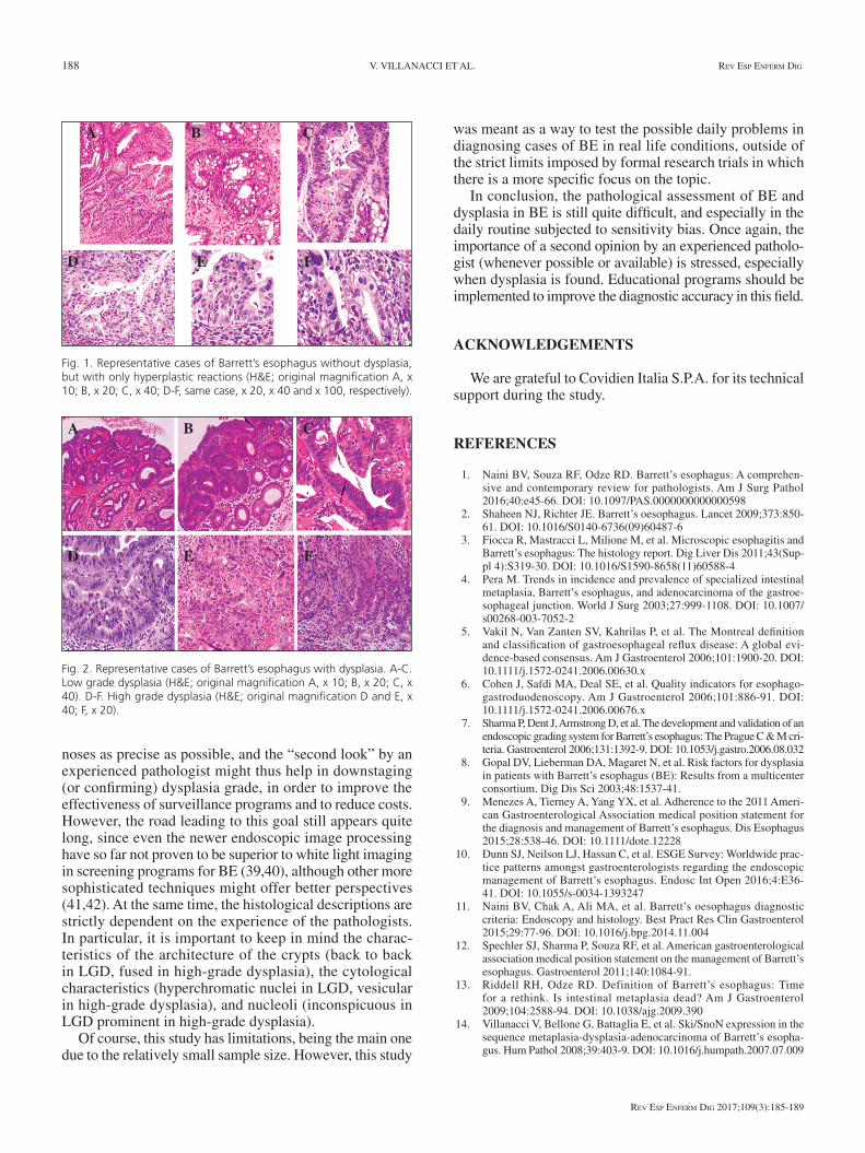
Fig. 1. Representative cases of Barrett’s esophagus without dysplasia, but with only hyperplastic reactions (H&E; original magnifi cation A, x 10; B, x 20; C, x 40; D-F, same case, x 20, x 40 and x 100, respectively).
Fig. 2. Representative cases of Barrett’s esophagus with dysplasia. A-C. Low grade dysplasia (H&E; original magnifi cation A, x 10; B, x 20; C, x 40). D-F. High grade dysplasia (H&E; original magnifi cation D and E, x 40; F, x 20).
A
D
B
E F
C
A
D
B
E
C
F

2017, Vol. 109, N.º 3 THE IMPORTANCE OF A SECOND OPINION IN THE DIAGNOSIS OF BARRETT’S ESOPHAGUS: A “REAL LIFE” STUDY 189
Rev esp enfeRm Dig 2017;109(3):185-189
15. Rossi E, Grisanti S, Villanacci V, et al. HER-2 overexpression/ampli-fication in Barrett’s oesophagus predicts early transition from dyspla-sia to adenocarcinoma: A clinicopathologic study. J Cell Mol Med 2009;13:3826-33. DOI: 10.1111/j.1582-4934.2008.00517.x
16. Fitzgerald RC, Di Pietro M, Ragunath K, et al. British Society of Gastroenterology guidelines on the diagnosis and management of Barrett’s oesophagus. Gut 2014;63:7-42 DOI: 10.1136/gutjnl-2013- 305372
17. Ogiya K, Kawano T, Ito E, et al. Lower esophageal palisade vessels and the definition of Barrett’s esophagus. Dis Esophagus 2008;21:645-9. DOI: 10.1111/j.1442-2050.2008.00825.x
18. Skacel M, Petras RE, Gramlich TL, et al. The diagnosis of low grade dysplasia in Barrett’s esophagus and its implication for disease pro-gression. Am J Gastroenterol 2000;95:3383-7. DOI: 10.1111/j.1572-0241.2000.03348.x
19. Montgomery E, Bronner MP, Goldblum JR, et al. Reproducibility of the diagnosis of dysplasia in Barrett esophagus: A reaffirmation. Hum Pathol 2001;32:368-78. DOI: 10.1053/hupa.2001.23510
20. Kaye PV, Haider SA, Ilyas M, et al. Barrett’s dysplasia and the Vienna classification: Reproducibility, prediction of progression and impact of consensus reporting and p53 immunohistochemistry. Histopathol 2009;54:699-712. DOI: 10.1111/j.1365-2559.2009.03288.x
21. Coco DP, Goldblum JR, Hornick JL, et al. Interobserver variability in the diagnosis of crypt dysplasia in Barrett esophagus. Am J Surg Pathol 2011;35:45-54. DOI: 10.1097/PAS.0b013e3181ffdd14
22. Bennett C, Vakil N, Bergman J, et al. Consensus statements for man-agement of Barrett’s dysplasia and early-stage esophageal adenocar-cinoma, based on a Delphi process. Gastroenterol 2012;143:336-46. DOI: 10.1053/j.gastro.2012.04.032
23. Villanacci V, Rossi E, Zambelli C, et al. COX-2, CDX2, and CDC2 immunohistochemical assessment for dysplasia-carcinoma progression in Barrett’s esophagus. Dig Liver Dis 2007;39:305-11. DOI: 10.1016/j.dld.2007.01.011
24. Cohen J. A coefficient of agreement for nominal scales. Educa-tion Psychol Measur 1960;20:37-46. DOI: 10.1177/00131644600 2000104
25. Fleiss JL, Levin B, Paik MC. Statistical methods for rates and propor-tions, 3rd ed. Hoboken: John Wiley & Sons; 2003.
26. Duits LC, Phoa KN, Curvers WL, et al. Barrett’s oesophagus patients with low-grade dysplasia can be accurately risk-stratified after histo-logical review by an expert pathology panel. Gut 2015;64:700-6. DOI: 10.1136/gutjnl-2014-307278
27. Sangle NA, Taylor SL, Emond MJ, et al. Overdiagnosis of high-grade dysplasia in Barrett’s esophagus: A multicenter, international study. Mod Pathol 2015;28:758-65. DOI: 10.1038/modpathol.2015.2
28. Yantiss RK. Diagnostic challenges in the pathologic evaluation of Bar-rett esophagus. Arch Pathol Lab Med 2010;134:1589-600.
29. Odze RD. Barrett esophagus: Histology and pathology for the clini-cian. Nat Rev Gastroenterol Hepatol 2009;6:478-90. DOI: 10.1038/nrgastro.2009.103
30. Corley DA, Kubo A, DeBoer J, et al. Diagnosing Barrett’s esophagus: Reliability of clinical and pathologic diagnoses. Gastrointest Endosc 2009;69:1004-10. DOI: 10.1016/j.gie.2008.07.035
31. Pech O, Vieth M, Schmitz D, et al. Conclusions from the histological diagnosis of low-grade intraepithelial neoplasia in Barrett’s oesophagus. Scand J Gastroenterol 2007;42:682-8. DOI: 10.1080/00365520601075803
32. Curvers WL, Ten Kate FJ, Krishnadath KK, et al. Low-grade dysplasia in Barrett’s esophagus: Overdiagnosed and underestimated. Am J Gas-troenterol 2010;105:1523-30. DOI: 10.1038/ajg.2010.171
33. Mastracci L, Piol N, Molinaro L, et al. Interobserver reproducibility in pathologist interpretation of columnar-lined esophagus. Virchows Arch 2016;468:159-67.
34. Villanacci V, Bassotti G, Salemme M, et al. Influence of genetics on tumoral pathologies: The example of the adenocarcinoma arising in Barrett’s esophagus. Rev Esp Enferm Dig 2012;104:592-602. DOI: 10.4321/S1130-01082012001100007
35. Salemme M, Villanacci V, Cengia G, et al. Intestinal metaplasia in Barrett’s oesophagus: An essential factor to predict the risk of dys-plasia and cancer development. Dig Liver Dis 2016;48:144-7. DOI: 10.1016/j.dld.2015.10.021
36. Rubenstein JH. Surveillance in Barrett’s esophagus: Utility and current recommendations. Gastroenterol Clin North Am 2015;44:285-97. DOI: 10.1016/j.gtc.2015.02.004
37. Singh R, Yeap SP, Cheong KL. Detection and characterization of early malignancy in the esophagus: What is the best management algorithm? Best Pract Res Clin Gastroenterol 2015;29:533-44. DOI: 10.1016/j.bpg.2015.06.004
38. Butt J, Kandel G. Barrett esophagus: When to endoscope. Clin Endosc 2014;47:40-6. DOI: 10.5946/ce.2014.47.1.40
39. Sharma P, Hawes RH, Bansal A, et al. Standard endoscopy with ran-dom biopsies versus narrow band imaging targeted biopsies in Barrett’s oesophagus: A prospective, international, randomised controlled trial. Gut 2013;62:15-21. DOI: 10.1136/gutjnl-2011-300962
40. Verna C, Feyles E, Lorenzi L, et al. I-SCAN targeted versus random biopsies in Barrett’s oesophagus. Dig Liver Dis 2014;46:131-4. DOI: 10.1016/j.dld.2013.10.005
41. Bertani H, Frazzoni M, Dabizzi E, et al. Improved detection of incident dysplasia by probe-based confocal laser endomicroscopy in a Barrett’s esophagus surveillance program. Dig Dis Sci 2013;58:188-93. DOI: 10.1007/s10620-012-2332-z
42. Xiong YQ, Ma SJ, Zhou JH, et al. A meta-analysis of confocal laser endomicroscopy for the detection of neoplasia in patients with Barrett’s esophagus. J Gastroenterol Hepatol 2016;31:1102-10. DOI: 10.1111/jgh.13267