the hippo signaling pathway: a potential therapeutic
TRANSCRIPT
RESEARCH ARTICLE Open Access
The Hippo signaling pathway: a potentialtherapeutic target is reversed by a Chinesepatent drug in rats with diabeticretinopathyGai-mei Hao1,3†, Tian-tian Lv1†, Yan Wu2, Hong-liang Wang1, Wei Xing1, Yong Wang1, Chun Li4, Zi-jian Zhang2,Zheng-lin Wang1, Wei Wang1*† and Jing Han2*†
Abstract
Background: The Hippo signaling pathway is reported to be involved in angiogenesis, but the roles of the Hippopathway in diabetic retinopathy have not been addressed. Fufang Xueshuantong Capsule has been used to treatdiabetic retinopathy in China; however, the effect of Fufang Xueshuantong Capsule on the Hippo pathway has notbeen investigated.
Methods: In this study, diabetes was induced in Sprague-Dawley rats with intraperitoneal injection of streptozotocin.Twenty weeks later, Fufang Xueshuantong Capsule was administered for 12 weeks. When the administration ended,the eyes were isolated for western blot and immunohistochemistry analyses. The levels of P- mammalian sterile 20-like(MST), large tumor suppressor homolog (Lats), P- yes-associated protein (YAP), transcriptional co-activator with PDZbinding motif (TAZ) and TEA domain family members (TEAD) were measured.
Results: Diabetic rats had a decreased P-MST level in the inner plexiform layer and reduced expression of P-YAP in thephotoreceptor layers of their eyes. In addition, diabetic rats displayed remarkable increases in Lats, TAZ and TEAD intheir retinas. Furthermore, Fufang Xueshuantong Capsule restored the changes in the Hippo pathway.
Conclusions: The Hippo signaling pathway is important for the progression of diabetic retinopathy and will hopefullybe a targeted therapeutic approach for the prevention of diabetic retinopathy.
Keywords: Diabetic retinopathy, Chinese medicine, Hippo
BackgroundDiabetic retinopathy (DR) is one of the most prominentcomplications of diabetes. DR remains a leading cause ofblindness and is characterized by vascular abnormalities,including increased permeability and the growth of newblood vessels [1]. At present, controlled systemic deregula-tion, laser photocoagulation, vitreoretinal surgery and in-travitreal anti-vascular endothelial growth factor (VEGF)
drugs remain the most common approaches to slow thedevelopment and progression of DR [2]; however, someexisting limitations and the fact that some patients re-spond poorly to these treatments has led to the need fornovel therapies for DR [3]. In addition, these findings sug-gest suggests that some other mediators or pathways mayparticipate and play an important role in DR. Therefore, itis important to elucidate the exact mechanisms of DR andthen investigate targeted ways to arrest the progression ofthis disease.The Hippo pathway is newly discovered and plays import-
ant roles in organ size control [4]. The pathway consists ofa large number of kinases and proteins, and the core mem-bers are mammalian sterile 20-like(MST), large tumor sup-pressor homolog(Lats), MOB kinase activator1(Mob1), and
* Correspondence: [email protected]; [email protected]†Equal contributors1College of Basic Medicine, Key Laboratory of Ministry of Education(Syndromes and formulas), Key Laboratory of Beijing (Syndromes andformulas), Beijing University of Chinese medicine, Beijing, China2Institute of Chinese Medicine, Beijing University of Chinese medicine,Beijing, ChinaFull list of author information is available at the end of the article
© The Author(s). 2017 Open Access This article is distributed under the terms of the Creative Commons Attribution 4.0International License (http://creativecommons.org/licenses/by/4.0/), which permits unrestricted use, distribution, andreproduction in any medium, provided you give appropriate credit to the original author(s) and the source, provide a link tothe Creative Commons license, and indicate if changes were made. The Creative Commons Public Domain Dedication waiver(http://creativecommons.org/publicdomain/zero/1.0/) applies to the data made available in this article, unless otherwise stated.
Hao et al. BMC Complementary and Alternative Medicine (2017) 17:187 DOI 10.1186/s12906-017-1678-3

salvador1(hsav1) [5]. The kinase cascade phosphorylatesyes-associated protein(YAP), transcriptional co-activatorwith PDZ binding motif(TAZ) and TEA domain familymembers(TEAD), which control cell proliferation [6, 7].It has been reported that Lats [8] and YAP [9] partici-
pate in the proliferation of endothelioma cells and vas-cular smooth muscle cell, respectively [10, 11]. Inaddition, Lats [8] and YAP [10] regulate angiogenesis inzebrafish. It is striking that YAP, which is the effector ofthe Hippo pathway, is expressed in retinal vessels andinvolved in endothelial sprouting [12] and angiogenesis[13]. It is well known that angiogenesis, which accom-panies the proliferation of endothelial cells, is a symp-tom of DR. Thus, these previous studies suggest that theHippo pathway plays roles in DR.To identify the specific roles of the Hippo signaling
pathway in the progression of DR, the influence ofFufang Xuesuhangtong (XST) Capsule, a patentedChinese drug that is reported to impede DR, on theHippo pathway was investigated. XST, which is com-posed of Panax notoginseng, Salvia miltiorrhiza, As-tragalus membranaceus and Scrophularia ningpoensis,has been noted for its medicinal effect against DR foralmost 20 years. Many clinical studies have indicatedthat XST could prevent fundus hemorrhage and exud-ation and stable vision in DR patients [14]. After-wards, XST has been found to ameliorate wholeblood viscosity, plasma viscosity, and erythrocyte ag-gregation indexes in STZ-induced rats [15]. Moreover,XST suppresses the acellular capillaries and increasethe pericyte numbers, which are the characteristics ofDR in rats [15]. In addition XST decreases the base-ment membrane thickness of the capillary and im-proved the pathological changes of the ganglion cellsin the retina of diabetic rats [16]. Meanwhile XST at-tenuates the aldose reductase activity and the contentof malondialdehyde (MDA), diminishes the expressionof VEGF, intercellular cell adhesion molecule-1(ICAM-1) and inducible nitric oxide synthase (iNOS),and augments the level of superoxide dismutase(SOD), pigment epithelium-derived factor (PEDF) andoccludin [15, 16]. Furthermore, recent studies haveshown that the characteristic constituents of XSThave the same pharmacological effect on DR [17].We hypothesize that the Hippo signaling pathway par-
ticipates in the process of DR and that XST has an impacton this pathway. Thus, the expression or distribution ofproteins in the Hippo pathway was investigated in the ret-inas of diabetic rats. In addition, the changes in the coremembers in the Hippo pathway were examined in ratretinas after XST administration. This study will enhancethe understanding of the mechanisms promoting the de-velopment of DR and provide valuable indications for anovel therapeutic target for DR.
MethodsEthics statementAll procedures involving animals and their care werecarried out according to the governmental guidelineson animal experimentation and the National Institutesof Health’s “Principles of Laboratory Animal Care”. Allexperimental protocols were approved by the Institu-tional Animal Ethics Committee of Beijing Universityof Traditional Chinese Medicine, Beijing, China (PermitNumber: 26–1514).
AntibodiesAnti-VEGF antibody, Abcam (Cambridge, UK), ab1316,mouse monoclonal, western blot (WB) dilution: 1:250;anti-extracellular signal-regulated kinas (Erk1) (pT202/pY204) + Erk2 (pT185/pY187) antibody, Abcam, ab4819,rabbit polyclonal, western blot (WB) dilution: 1:1000; anti-Erk1/2 antibody, Abcam, ab17942, rabbit polyclonal, west-ern blot (WB) dilution: 1:1000; anti-Lats antibody, SantaCruz (Dallas, Texas, U.S.A.), sc-9388, goat polyclonal,western blot (WB) dilution: 1:200; anti-TAZ antibody,Santa Cruz, sc-48,805, rabbit polyclonal, western blot(WB) dilution: 1:500; anti-TEAD antibody, Santa Cruz, sc-134,070, rabbit polyclonal, western blot (WB) dilution:1:1000; anti-β-actin antibody, Abcam, ab8226, mousemonoclonal, western blot (WB) dilution: 1:5000; P-MST,CST (Boston, Massachusetts, USA), #3681, rabbit poly-clonal, immunohistochemistry dilution: 1:250; P-YAP,CST, # 4911, rabbit polyclonal, immunohistochemistry di-lution: 1:250.
DrugXST (national medicine permission number Z20030017,lot number 130630) was purchased from ZhongshengPharmaceutical Co., Ltd. (Guangdong, China). Therewas 0.5 g drug per grain.
AnimalsThirty six Male healthy, Sprague-Dawley rats (8 weeksof age, 250–300 g) were supplied by Vital River Labora-tory Animal Technology Co. Ltd. (Beijing, China, Cer-tificate no SCXK (Beijing) 2007–0001). The animalswere kept at a room temperature of 22–24 °C, 40% hu-midity, and a 12-h daylight cycle. The rats were housedin 465 × 300 × 200 mm cages (Longdonghai Ltd., China,Type II) and provided with water and commercial ratfeed ad libitum. Three rats were raised in one cage. Thebeddings were changed every day, and the cages werechanged every week. When the experiments ended, theanimals were sacrificed using intraperitoneal injectionsof pentobarbital (50 mg/kg). The status of rats was ex-amined every day and the blood glucose was measuredevery 4 weeks.
Hao et al. BMC Complementary and Alternative Medicine (2017) 17:187 Page 2 of 10
Induction of diabetesAnimals were fasted for 14 h before streptozotocin(STZ, Sigma Chemical Co, USA, cat# S0130) injection.STZ was dissolved in10 mM citrate buffer (pH 4.4) andintraperitoneally injected within 5 min at 65 mg/kg bodyweight. Age-matched control rats received equal vol-umes of vehicle (citrate buffer). Seven days later, bloodwas obtained from the tail veins for glucose analysisusing a standard glucometer (One Touch Profile, Life-scan, Inc., USA). Rats with blood glucose levels higherthan 16.7 mmol/L were considered to be diabetic andused for the subsequent experiments.
Treatment scheduleAfter 20 weeks of diabetes induction, the diabetic ratswere divided into two groups according to the glucoseconcentration and the body weight: diabetic (n = 17),XST (n = 8). Then the treatment with XST by intragas-tric gavage was started. The daily dose of XST given was1.05 g/kg body weight, which was equivalent to approxi-mately 7 times the amount of the dose that patients re-ceive per day. The rats in normal group (n = 11) anddiabetic group were fed with water at the same time.Color Doppler imaging and trypsin digest preparationwere conducted after 12 weeks of XST treatment. Thenthe rats were sacrificed.
Observation by Color Doppler imagingColor Doppler imaging was used to monitor the flowvelocities before the rats were killed. The blood veloci-ties of the central retinal artery (CRA) were detected byColor Doppler (Vevo 2100, VisualSonics, Canada). Theprobe was placed on the opened eye following the appli-cation of sterile contact gel to minimize the force of theprobe on the globe. Peak systolic velocity(PSV), end-diastolic velocity(EDV), mean velocity(MV), resistanceindex(RI) and pulsatility index(PI) of the CRA weremeasured.
Immunohistochemical stainingThe paraffined slices were deparaffinized and dehy-drated. For immunohistochemistry, after the endogenousperoxidases were removed using 0.3% hydrogen peroxid-ase, the primary antibody against P-MST or P-YAP anti-body was added to the slices and incubated at 4 °Covernight. After three washes, the sample slices were in-cubated with the horseradish peroxidase-conjugated sec-ondary antibodies; 3, 3′-diaminobenzidine (DAB) wasused as the chromogen. In the end, the hematoxylinstaining was performed.
Trypsin digest methodThe retina was isolated and incubated at 37 °C in diges-tion buffer (0.1 mol/L Tris buffer, pH 7.8), containing 3%
trypsin (Amresco, USA). After 2–3 h of incubation,when the internal limiting membrane began to separatefrom the retina, the retina was transferred to phosphate-buffered saline (pH 7.4) at room temperature. The vas-cular tree was washed in distilled water to be freed ofany remaining neural tissue. The preparations were seton glass slides, air dried, and stained with hematoxylinand Periodic Acid-Schiff stain (PAS) to evaluate micro-vascular lesions. The acellular capillaries of the retinawere analyzed. The number of endothelial cells and peri-cytes were counted, and the ratio was calculated. Theendothelial cells were identified as elliptical and orientedalong the circumference of the capillary, and the peri-cytes were defined as round in shape and abutting theouter portion of the capillary wall.
Quantitative real-time PCRTotal RNA was extracted from frozen retina tissues.RNA concentration was determined by spectrophotom-eter (Nanodrop 2000, Thermo, USA). ComplementaryDNA was synthesized using a reverse transcription re-agent kit (Roche, USA). Primers were ordered fromShanghai Shenggong Co Ltd. Amplification and quanti-tation were performed by real-time PCR (ABI7500,USA), and β-actin served as the control. The primers forVEGF and β-actin were as followed: VEGF primer (for-ward 5′-3′): CAGAAGGGGAGCAGAAAGCC, reverse(5′-3′): AATGTTCAGCCCCAACCAAGA; β-actin pri-mer (forward 5′-3′): GCAGGAGTACGATGAGTCCG,reverse (5′-3′): ACGCAGCTCAGTAACAGTCC.
Western blot analysesRetinas were homogenized in RIPA buffer (Pulilai, China)containing protease inhibitors. The lysate was centrifuged,and the supernatant was collected. Protein content wasassayed using the BCA protein assay (Thermo, USA). Thetissue lysate, which contained 60 μg of protein, was sepa-rated on 12% SDS-polyacrylamide gels and was trans-ferred onto polyvinylidene fluoride membranes. Themembranes were blocked for 1.5 h at room temperaturein 5% nonfat dried milk with TBST (Pulilai, China) andthen incubated with a primary antibody overnight at 4 °C.The membranes were washed and incubated with a sec-ondary antibody at a dilution of 1:5000 for 1 h. Finally, themembranes were washed in triplicate with TBST and de-veloped using enhanced chemiluminescence (GE, USA).The bands on the film were measured, and density mea-surements were normalized to β-actin readings.
Statistical analysisThe Shapiro-Wilk test was applied to verify the normal-ity of the distributions. A two-way analysis of variance(ANOVA) was used to verify the differences between thenormal distributions, and the Kruskal-Wallis test was
Hao et al. BMC Complementary and Alternative Medicine (2017) 17:187 Page 3 of 10

used to assess differences between nonparametric distri-butions. For normal distributions, the results wereexpressed as the means ± S.D., and the differences wereconsidered significant when the probability of a Type Ierror was lower than 5% (p < 0.05).
ResultsBlood glucose and body weightBlood glucose and body weight were measured every 4weeks throughout the 32-week period. Diabetic ratsmaintained a higher blood glucose (19.1 mmol/L ~ 26.2 mmol/L), a 4–8-fold increase compared withthe age-matched control rats (vehicle treated; P < 0.001),
and lower body weight (P < 0.001) throughout the ex-periment period. In contrast to the diabetic group, XSThad no effect on blood glucose and body weight in thecontrols (Fig. 1c, d; P > 0.05). No side- effects of XSTwere observed.
Blood flow in the CRAColor Doppler imaging is a noninvasive, reproducible,and easily applied technique, and it has been shown tobe useful in evaluating hemodynamic changes in severalorbital and retinal vascular diseases [18]. It has been re-ported that blood flow velocity in the CRA was de-creased in rats with DR [14]. At the end of the present
A
B C
D E
Fig. 1 The blood glucose and body weight of rats. Diabetic rats were induced by streptozocin. The normal control rats received citrate buffer.The blood glucose and body weight were measured every 4 weeks. 20 weeks later, the rats whose blood glucose was higher than 16.7 mmol/Lwere divided into diabetic group and XST group. Then XST administration started and continued for 12 weeks. a Time schedule. b–c The bloodglucose and body weight before XST administration. ##: P < 0.01, compared to Normal. d–e The blood glucose and body weight after XSTadministration. There was no significant difference between diabetic group and XST group. **: P < 0.01, compared to Diabetic
Hao et al. BMC Complementary and Alternative Medicine (2017) 17:187 Page 4 of 10
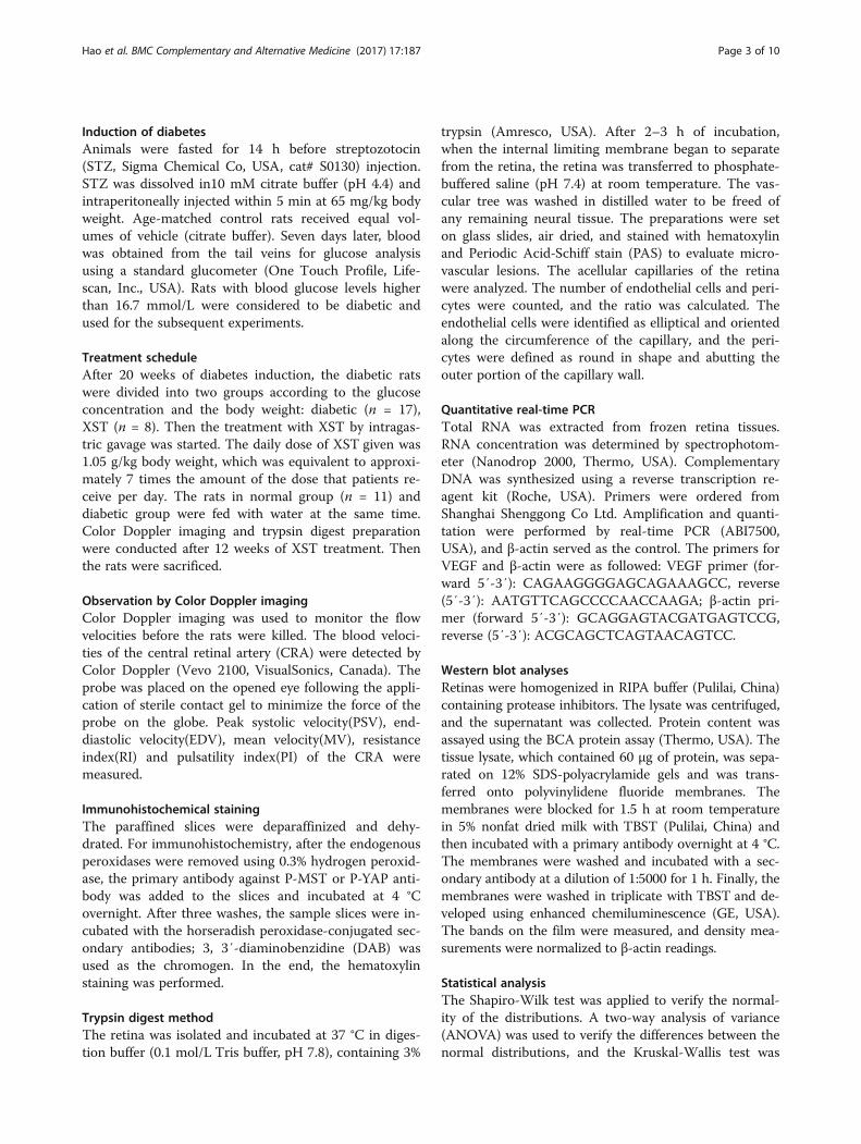
experiment, blood flow in the CRA was evaluated, andsimilar results were observed (Fig. 2). Diabetic rats ex-hibited a significant reduction in blood flow and an in-crease in PI and RI in the CRA compared with thecontrol rats (P < 0.05). PSV, EDV and MV were mark-edly increased in the XST group compared with the dia-betic group (P < 0.05, Fig. 2a). XST reduced the RI andPI remarkably (P < 0.05, Fig. 2b).
Retinal vascular histopathologyPrevious studies in animal models have indicated thatthe ratio of endothelial cells to pericytes increased sig-nificantly [19]. When the XST administration ended, theretinas were removed for trypsin digestion. The resultsshowed that acellular capillaries and the ratio of endo-thelial cells to pericytes in diabetic rat retinas increasedcompared with normal control rats (P < 0.001, Fig. 3).Additionally, XST reduced acellular capillaries and theratio of endothelial cells to pericytes (P < 0.001, Fig. 3).These findings suggest that a microvasculature lesiondeveloped in the diabetic rat retinas and that XST inhib-ited the pathological changes in diabetic retinopathy.
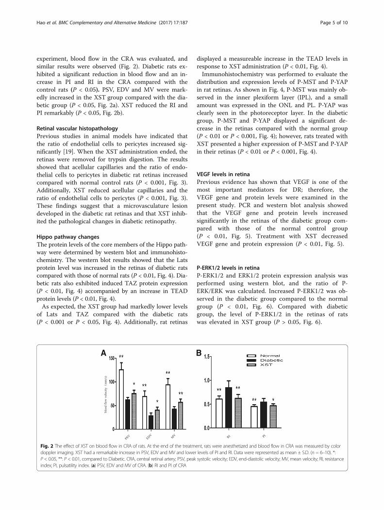
Hippo pathway changesThe protein levels of the core members of the Hippo path-way were determined by western blot and immunohisto-chemistry. The western blot results showed that the Latsprotein level was increased in the retinas of diabetic ratscompared with those of normal rats (P < 0.01, Fig. 4). Dia-betic rats also exhibited induced TAZ protein expression(P < 0.01, Fig. 4) accompanied by an increase in TEADprotein levels (P < 0.01, Fig. 4).As expected, the XST group had markedly lower levels
of Lats and TAZ compared with the diabetic rats(P < 0.001 or P < 0.05, Fig. 4). Additionally, rat retinas
displayed a measureable increase in the TEAD levels inresponse to XST administration (P < 0.01, Fig. 4).Immunohistochemistry was performed to evaluate the
distribution and expression levels of P-MST and P-YAPin rat retinas. As shown in Fig. 4, P-MST was mainly ob-served in the inner plexiform layer (IPL), and a smallamount was expressed in the ONL and PL. P-YAP wasclearly seen in the photoreceptor layer. In the diabeticgroup, P-MST and P-YAP displayed a significant de-crease in the retinas compared with the normal group(P < 0.01 or P < 0.001, Fig. 4); however, rats treated withXST presented a higher expression of P-MST and P-YAPin their retinas (P < 0.01 or P < 0.001, Fig. 4).
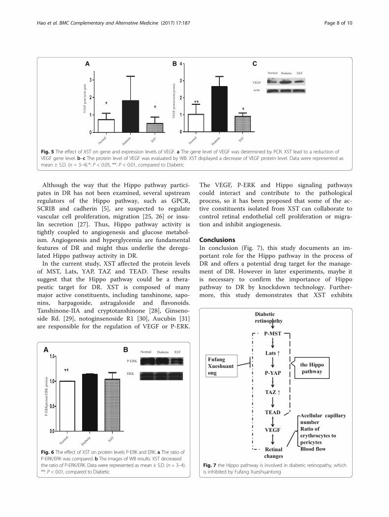
VEGF levels in retinaPrevious evidence has shown that VEGF is one of themost important mediators for DR; therefore, theVEGF gene and protein levels were examined in thepresent study. PCR and western blot analysis showedthat the VEGF gene and protein levels increasedsignificantly in the retinas of the diabetic group com-pared with those of the normal control group(P < 0.01, Fig. 5). Treatment with XST decreasedVEGF gene and protein expression (P < 0.01, Fig. 5).
P-ERK1/2 levels in retinaP-ERK1/2 and ERK1/2 protein expression analysis wasperformed using western blot, and the ratio of P-ERK/ERK was calculated. Increased P-ERK1/2 was ob-served in the diabetic group compared to the normalgroup (P < 0.01, Fig. 6). Compared with diabeticgroup, the level of P-ERK1/2 in the retinas of ratswas elevated in XST group (P > 0.05, Fig. 6).
A B
Fig. 2 The effect of XST on blood flow in CRA of rats. At the end of the treatment, rats were anesthetized and blood flow in CRA was measured by colordoppler imaging. XST had a remarkable increase in PSV, EDV and MV and lower levels of PI and RI. Data were represented as mean ± S.D. (n = 6–10). *:P < 0.05, **: P < 0.01, compared to Diabetic. CRA, central retinal artery; PSV, peak systolic velocity; EDV, end-diastolic velocity; MV, mean velocity; RI, resistanceindex; PI, pulsatility index. (a) PSV, EDV and MV of CRA. (b) RI and PI of CRA
Hao et al. BMC Complementary and Alternative Medicine (2017) 17:187 Page 5 of 10

DiscussionIn this study, the results showed that the Hippo pathwaybecame dysregulated in the retinas of diabetic rats, andXST restored the protein levels of this signaling path-way. Therefore, we describe here a novel molecularmechanism by which the Hippo pathway may be in-volved and play an important role in DR. Additionallythe Hippo pathway has been implicated in underlyingthe curative effect of XST.Previous results indicated that the Hippo pathway me-
diates angiogenesis, which is related to the damageswithin the retina in DR. In addition, VEGF has been im-plicated as one of the most important cytokines with
angiogenic and mitogenic actions, which has great ef-fects on DR. It has been confirmed that the overexpres-sion of TEAD increased VEGF promoter activity andVEGF expression in endothelial cells [20]. Additionally,silencing YAP inhibited the expression of VEGF [21].Taken together, these reports suggest that the Hippopathway is involved in cell proliferation and angiogenesisby regulating VEGF. Although not all of the participantsin the crosstalk between the Hippo pathway and VEGFare fully understood, ERK may function as an intermedi-ary. YAP, the reporter of the Hippo pathway, is able toinduce ERK phosphorylation [22], and the ERK pathwayhas been shown to increase VEGF mRNA stability [23]
A
D E
B C
Fig. 3 The effect of XST on morphology of rat retinas. The retinas were fixed in formalin for trypsin digestion. a–c The vasculature in midretina. Scalebar: 50 μm. The images showed the acellular capillaries (black arrow), endothelial cells (green arrows) and pericytes(red arrow). d The acellular capillarieswere counted and expressed as the total number/mm of retina area. There was a remarkable decrease in cellular capillaries in XST group. e Theendothelial cells and pericytes were identified and counted. Then the ratio of endothelial cells number /pericytes number was calculated. XSTadministration improved the ratio of endothelial cells number /pericytes number. Data were represented as mean ± S.D. (n = 3–5).*: P < 0.05, **:P < 0.01, ***: P < 0.001, compared to Diabetic
Hao et al. BMC Complementary and Alternative Medicine (2017) 17:187 Page 6 of 10
and promote VEGF expression [24]. Previous reportshave suggested that the Hippo pathway activates ERK,which modulates VEGF and facilitates angiogenesis.Our findings that MST, Lats, YAP, TAZ and TEAD
were all altered in the retinas of rats with DR led us to
conclude that the Hippo pathway may be the underlyingfactor in the process of DR. Additionally, VEGF andP-ERK were elevated in diabetic rats, which implies thatP-ERK-VEGF is the downstream target of the Hippopathway in DR.
A B C
D
G
E F
Fig. 4 The effect of XST on protein levels of Hippo pathway. a–d the protein levels of Lats, TAZ and TEAD were evaluated by WB. XST decreasedlevels of Lats, TAZ and TEAD protein. e–g Distribution and expression of P-MST and P-YAP were measured using IHC. P-MST was mainly observedin inner plexiform layer and small amount was expressed in outer nuclear layer and photoreceptor layer. P-YAP was clearly seen photoreceptorlayer. An elevation of P-MST and P-YAP were observed in XST group. Data were represented as mean ± S.D. (n = 3–4).*: P < 0.05, **: P < 0.01, ***:P < 0.001, compared to Diabetic
Hao et al. BMC Complementary and Alternative Medicine (2017) 17:187 Page 7 of 10
Although the way that the Hippo pathway partici-pates in DR has not been examined, several upstreamregulators of the Hippo pathway, such as GPCR,SCRIB and cadherin [5], are suspected to regulatevascular cell proliferation, migration [25, 26] or insu-lin secretion [27]. Thus, Hippo pathway activity istightly coupled to angiogenesis and glucose metabol-ism. Angiogenesis and hyperglycemia are fundamentalfeatures of DR and might thus underlie the deregu-lated Hippo pathway activity in DR.In the current study, XST affected the protein levels
of MST, Lats, YAP, TAZ and TEAD. These resultssuggest that the Hippo pathway could be a thera-peutic target for DR. XST is composed of manymajor active constituents, including tanshinone, sapo-nins, harpagoside, astragaloside and flavonoids.Tanshinone-IIA and cryptotanshinone [28], Ginseno-side Rd. [29], notoginsenoside R1 [30], Aucubin [31]are responsible for the regulation of VEGF or P-ERK.
The VEGF, P-ERK and Hippo signaling pathwayscould interact and contribute to the pathologicalprocess, so it has been proposed that some of the ac-tive constituents isolated from XST can collaborate tocontrol retinal endothelial cell proliferation or migra-tion and inhibit angiogenesis.
ConclusionsIn conclusion (Fig. 7), this study documents an im-portant role for the Hippo pathway in the process ofDR and offers a potential drug target for the manage-ment of DR. However in later experiments, maybe itis necessary to confirm the importance of Hippopathway to DR by knockdown technology. Further-more, this study demonstrates that XST exhibits
Fig. 7 the Hippo pathway is involved in diabetic retinopathy, whichis inhibited by Fufang Xueshuantong
A B
Fig. 6 The effect of XST on protein levels P-ERK and ERK. a The ratio ofP-ERK/ERK was compared. b The images of WB results. XST decreasedthe ratio of P-ERK/ERK. Data were represented as mean ± S.D. (n = 3–4).**: P < 0.01, compared to Diabetic
Fig. 5 The effect of XST on gene and expression levels of VEGF. a The gene level of VEGF was determined by PCR. XST lead to a reduction ofVEGF gene level. b–c The protein level of VEGF was evaluated by WB. XST displayed a decrease of VEGF protein level. Data were represented asmean ± S.D. (n = 3–4).*: P < 0.05, **: P < 0.01, compared to Diabetic
Hao et al. BMC Complementary and Alternative Medicine (2017) 17:187 Page 8 of 10

effects on the Hippo pathway in rats with DR andprovides molecular mechanisms underlying the use ofXST for the treatment of DR.
Additional file
Additional file 1: Data of the expreiment. (XLS 39 kb)
AbbreviationsCRA: Central retinal artery; DAB: 3, 3′-diaminobenzidine; DR: Diabetic retinopathy;EDV: End-diastolic velocity, EDV; ERK: Extracellular signal-regulated kinas;hsav1: Salvador1; ICAM-1: Intercellular cell adhesion molecule-1; iNOS: Induciblenitric oxide synthase; Lats: Large tumor suppressor homolog;MDA: Malondialdehyde; Mob1: MOB kinase activator1; MST: mammalian sterile20-like; MV: Mean velocity, MV; PAS: Periodic acid-schiff stain; PEDF: Pigmentepithelium-derived factor; PI: Pulsatility index, PI; PSV: Peak systolic velocity;RI: Resistance index, RI; SOD: Superoxide dismutase; TAZ: Transcriptional co-activator with PDZ binding motif; TEAD: TEA domain family members;VEGF: Vascular endothelial growth factor; WB: Western blot; XST: FufangXuesuhangtong; YAP: Yes-associated protein
AcknowledgementWe thank Yue-ying YUAN for conducting Color Doppler imaging.
FundingThis work was supported by the National Nature Science Foundation ofChina (No. 81673705; No. 81303083).
Availability of data and materialAll data generated or analysed during this study are included in thispublished article [and its Additional file 1].
Authors’ contributionsWW HJ conceived and designed the experiments. HGM LTT WHL XW ZZJperformed the experiments.WZL WY WY LC analyzed the data. HJ HGM LTTcontributed to the writing of the manuscript. All authors read and approvedthe final manuscript.
Authors’ informationGai-mei HAO, [email protected], College of Basic Medicine, KeyLaboratory of Ministry of Education (Syndromes and formulas), KeyLaboratory of Beijing (Syndromes and formulas), Beijing University of Chinesemedicine, Beijing, China; Institute of Basic Theory for Chinese Medicine,China Academy of Chinese Medical Sciences, Beijing, China.Tian-tian LV, [email protected], College of Basic Medicine, KeyLaboratory of Ministry of Education (Syndromes and formulas), KeyLaboratory of Beijing (Syndromes and formulas), Beijing University of Chinesemedicine, Beijing, China;Yan WU, [email protected], Institute of Chinese Medicine, BeijingUniversity of Chinese medicine, Beijing, China ;Hong-liang WANG, [email protected], College of Basic Medicine, KeyLaboratory of Ministry of Education (Syndromes and formulas), KeyLaboratory of Beijing (Syndromes and formulas), Beijing University of Chinesemedicine, Beijing, China;Wei XING, [email protected], College of Basic Medicine, Key Laboratory ofMinistry of Education (Syndromes and formulas), Key Laboratory of Beijing(Syndromes and formulas), Beijing University of Chinese medicine, Beijing,China;Yong WANG, [email protected], College of Basic Medicine, KeyLaboratory of Ministry of Education (Syndromes and formulas), KeyLaboratory of Beijing (Syndromes and formulas), Beijing University of Chinesemedicine, Beijing, China;Chun LI, [email protected], Modern Research Center for TraditionalChinese Medicine, Beijing University of Chinese Medicine, Beijing, China;Zi-jian ZHANG, [email protected], Institute of Chinese Medicine, BeijingUniversity of Chinese medicine, Beijing, China;Zheng-lin WANG, [email protected], College of Basic Medicine, KeyLaboratory of Ministry of Education (Syndromes and formulas), Key
Laboratory of Beijing (Syndromes and formulas), Beijing University of Chinesemedicine, Beijing, China;Wei WANG, [email protected], College of Basic Medicine, KeyLaboratory of Ministry of Education (Syndromes and formulas), KeyLaboratory of Beijing (Syndromes and formulas), Beijing University of Chinesemedicine, Beijing, China;Jing HAN, [email protected], Institute of Chinese Medicine, BeijingUniversity of Chinese medicine, Beijing, China.
Competing interestsThe authors declare that they have no competing interests.
Consent for publicationNot applicable.
Ethics approval and consent to participateAll procedures involving animals and their care were carried out accordingto the governmental guidelines on animal experimentation and the NationalInstitutes of Health’s “Principles of Laboratory Animal Care”. All experimentalprotocols were approved by the Institutional Animal Ethics Committee ofBeijing University of Traditional Chinese Medicine, Beijing, China (PermitNumber: 26–1514).
Publisher’s NoteSpringer Nature remains neutral with regard to jurisdictional claims inpublished maps and institutional affiliations.
Author details1College of Basic Medicine, Key Laboratory of Ministry of Education(Syndromes and formulas), Key Laboratory of Beijing (Syndromes andformulas), Beijing University of Chinese medicine, Beijing, China. 2Institute ofChinese Medicine, Beijing University of Chinese medicine, Beijing, China.3Institute of Basic Theory for Chinese Medicine, China Academy of ChineseMedical Sciences, Beijing, China. 4Modern Research Center for TraditionalChinese Medicine, Beijing University of Chinese Medicine, Beijing, China.
Received: 14 September 2016 Accepted: 11 March 2017
References1. Perrin RM, Konopatskaya O, Qiu Y, Harper S, Bates DO, Churchill AJ. Diabetic
retinopathy is associated with a switch in splicing from anti- to pro-angiogenic isoforms of vascular endothelial growth factor. Diabetologia.2005;48(11):2422–7.
2. Stitt AW, Lois N, Medina RJ, Adamson P, Curtis TM. Advances in ourunderstanding of diabetic retinopathy. Clin Sci (Lond). 2013;125(1):1–17.
3. Titchenell PM, Antonetti DA. Using the past to inform the future: anti-VEGFtherapy as a road map to develop novel therapies for diabetic retinopathy.Diabetes. 2013;62(6):1808–15.
4. Irvine KD, Harvey KF. Control of organ growth by patterning and hipposignaling in Drosophila. Cold Spring Harb Perspect Biol. 2015;7(6):a019224.
5. Harvey KF, Zhang X, Thomas DM. The Hippo pathway and human cancer.Nat Rev Cancer. 2013;13(4):246–57.
6. Mauviel A, Nallet-Staub F, Varelas X. Integrating developmental signals: aHippo in the (path)way. Oncogene. 2012;31(14):1743–56.
7. Zhou X, Wang Z, Huang W, Lei QY. G protein-coupled receptors: bridgingthe gap from the extracellular signals to the Hippo pathway. Acta BiochimBiophys Sin Shanghai. 2015;47(1):10–5.
8. Dai X, She P, Chi F, Feng Y, Liu H, Jin D, Zhao Y, Guo X, Jiang D, Guan KL, et al.Phosphorylation of angiomotin by Lats1/2 kinases inhibits F-actin binding, cellmigration, and angiogenesis. J Biol Chem. 2013;288(47):34041–51.
9. Tsuneki M, Madri JA. Adhesion molecule-mediated hippo pathway modulateshemangioendothelioma cell behavior. Mol Cell Biol. 2014;34(24):4485–99.
10. Xie C, Guo Y, Zhu T, Zhang J, Ma PX, Chen YE. Yap1 protein regulatesvascular smooth muscle cell phenotypic switch by interaction withmyocardin. J Biol Chem. 2012;287(18):14598–605.
11. Wang X, Hu G, Gao X, Wang Y, Zhang W, Harmon EY, Zhi X, Xu Z, LennartzMR, Barroso M, et al. The induction of yes-associated protein expressionafter arterial injury is crucial for smooth muscle phenotypic modulation andneointima formation. Arterioscler Thromb Vasc Biol. 2012;32(11):2662–9.
Hao et al. BMC Complementary and Alternative Medicine (2017) 17:187 Page 9 of 10
12. Choi HJ, Zhang H, Park H, Choi KS, Lee HW, Agrawal V, Kim YM, Kwon YG.Yes-associated protein regulates endothelial cell contact-mediatedexpression of angiopoietin-2. Nat Commun. 2015;6:6943.
13. Choi HJ, Kwon YG. Roles of YAP in mediating endothelial cell junctionalstability and vascular remodeling. BMB Rep. 2015;48(8):429–30.
14. Zheng YZ, Xie MS, Liu GH, Liu A, Jin WE. Effect of Jiawei Buyang HuanwuDecoction on Retinal Hemodynamics of Diabetic Rats. Journal of ShanxiCollege of Traditional Chinese Medicine. 2013;14:24–7.
15. Duan H, Huang J, Li W, Tang M. Protective effects of fufang xueshuantongon diabetic retinopathy in rats. Evid Based Complement Alternat Med. 2013;2013:408268.
16. Yuwei X, Junjie Z, Yongquan S, Zhimin L. Protection of FufangXueshuantong Capsule Against Retinal Oxidative Damage of Diabetic Rats. JMed Res. 2016;45(1):40–3.
17. Jian W, Yu S, Tang M, Duan H, Huang J. A combination of the mainconstituents of Fufang Xueshuantong Capsules shows protective effectsagainst streptozotocin-induced retinal lesions in rats. J Ethnopharmacol.2016;182:50–6.
18. Baydar S, Adapinar B, Kebapci N, Bal C, Topbas S. Colour Doppler ultrasoundevaluation of orbital vessels in diabetic retinopathy. Australas Radiol. 2007;51(3):230–5.
19. Sharma NK, Gardiner TA, Archer DB. A morphologic and autoradiographicstudy of cell death and regeneration in the retinal microvasculature ofnormal and diabetic rats. Am J Ophthalmol. 1985;100(1):51–60.
20. Shie JL, Wu G, Wu J, Liu FF, Laham RJ, Oettgen P, Li J. RTEF-1, a noveltranscriptional stimulator of vascular endothelial growth factor in hypoxicendothelial cells. J Biol Chem. 2004;279(24):25010–6.
21. Zhou Z, Zhu JS, Xu ZP. RNA interference mediated YAP gene silencinginhibits invasion and metastasis of human gastric cancer cell line SGC-7901.Hepato-Gastroenterology. 2011;58(112):2156–61.
22. Zhang J, Ji JY, Yu M, Overholtzer M, Smolen GA, Wang R, Brugge JS, DysonNJ, Haber DA. YAP-dependent induction of amphiregulin identifies a non-cell-autonomous component of the Hippo pathway. Nat Cell Biol. 2009;11(12):1444–50.
23. Essafi-Benkhadir K, Pouyssegur J, Pages G. Implication of the ERK pathwayon the post-transcriptional regulation of VEGF mRNA stability. Methods MolBiol. 2010;661:451–69.
24. Curry JM, Eubank TD, Roberts RD, Wang Y, Pore N, Maity A, Marsh CB. M-CSF signals through the MAPK/ERK pathway via Sp1 to induce VEGFproduction and induces angiogenesis in vivo. PLoS One. 2008;3(10):e3405.
25. Michaelis UR, Chavakis E, Kruse C, Jungblut B, Kaluza D, Wandzioch K, ManavskiY, Heide H, Santoni MJ, Potente M, et al. The polarity protein Scrib is essentialfor directed endothelial cell migration. Circ Res. 2013;112(6):924–34.
26. O'Hayre M, Degese MS, Gutkind JS. Novel insights into G protein and G protein-coupled receptor signaling in cancer. Curr Opin Cell Biol. 2014;27:126–35.
27. Yamagata K, Nammo T, Moriwaki M, Ihara A, Iizuka K, Yang Q, Satoh T, Li M,Uenaka R, Okita K, et al. Overexpression of dominant-negative mutanthepatocyte nuclear fctor-1 alpha in pancreatic beta-cells causes abnormalislet architecture with decreased expression of E-cadherin, reduced beta-cellproliferation, and diabetes. Diabetes. 2002;51(1):114–23.
28. Choi HS, Cho DI, Choi HK, Im SY, Ryu SY, Kim KM. Molecular mechanisms ofinhibitory activities of tanshinones on lipopolysaccharide-induced nitricoxide generation in RAW 264.7 cells. Arch Pharm Res. 2004;27(12):1233–7.
29. Liu XY, Zhou XY, Hou JC, Zhu H, Wang Z, Liu JX, Zheng YQ. Ginsenoside Rdpromotes neurogenesis in rat brain after transient focal cerebral ischemiavia activation of PI3K/Akt pathway. Acta Pharmacol Sin. 2015;36(4):421–8.
30. Yang BR, Hong SJ, Lee SM, Cong WH, Wan JB, Zhang ZR, Zhang QW, ZhangY, Wang YT, Lin ZX. Pro-angiogenic activity of notoginsenoside R1 inhuman umbilical vein endothelial cells in vitro and in a chemical-inducedblood vessel loss model of zebrafish in vivo. Chin J Integr Med.2016;22(6):420–9.
31. Park KS. Aucubin, a naturally occurring iridoid glycoside inhibits TNF-alpha-induced inflammatory responses through suppression of NF-kappaBactivation in 3T3-L1 adipocytes. Cytokine. 2013;62(3):407–12.
• We accept pre-submission inquiries
• Our selector tool helps you to find the most relevant journal
• We provide round the clock customer support
• Convenient online submission
• Thorough peer review
• Inclusion in PubMed and all major indexing services
• Maximum visibility for your research
Submit your manuscript atwww.biomedcentral.com/submit
Submit your next manuscript to BioMed Central and we will help you at every step:
Hao et al. BMC Complementary and Alternative Medicine (2017) 17:187 Page 10 of 10