the hammerhead ribozyme
TRANSCRIPT

The Hammerhead RibozymeFritz Eckstein
Birgit BramlageMax–Planck Institut fur
experimentelle Medizin,Hermann-Rein-Str. 3,
D-37075 Gottingen, Germany
Abstract: The hammerhead ribozyme is an intriguing RNA molecule with the ability to serve as acatalyst to cleavesequence-specifically RNA molecules in an intermolecular reaction. PreferentiallyMg21 is required for optimal activity by inducing the catalytically competent conformation and bypossibly acting as an acid–base catalyst. Even though the three-dimensional structure has beenelucidated details of the structure–function relationship and of the mechanism remain unanswered.The hammerhead ribozyme has stimulated the concept of the sequence-specific cleavage of mRNAsintracellularly and thusto inhibit geneexpression by preventing translation. Thisrepresentsan areaof considerable interest as it has the potential for the development of drugs. © 2001 John Wiley& Sons, Inc. Biopoly (Nucleic Acid Sci) 52: 147–154, 1999
Keywords: hammerhead ribozyme; structure; function; application
INTRODUCTION
Ribozymes are catalytically competent RNAs thatoccur either in nature or have been obtained by invitro selection.1,2 The most prominent and most re-cent example is the ribosomal 23S RNA, which con-stitutes the peptidyl transferase reaction center.3 Al-though ribozymes have been selected in vitro for thecatalysis of a large variety of reactions, all knownnaturally occurring ribozymes solely catalyzed RNAcleavage and ligation reactions. Thus the 23S RNAribozyme expands the scope of natural catalytic reac-tions and strengthens the hypothesis for an RNAworld where RNA was the predominant catalystrather than proteins. It has also been demonstratedthat ribozymes can evolve with few mutations from acommon ancestor, harboring modest cleavage andligase activities in this case, to two ribozymes withhigh efficiency for one of these activities.4 Thisclearly demonstrates the potential for evolution andadaptation of catalytic RNA.
This review wil l concentrate on the hammerheadribozyme which belongs to the class of small ri-bozymes.1 It has attracted considerable attention be-causeof its facilechemical synthesisand thusan entryinto mechanistic studies with nucleotide analogues. Italso rendered itself to theapplication for the inhibitionof gene expression on the RNA level. This ribozymewas originally discovered as a structural motif incertain plant pathogen viroids for the self-cleavage ofcatenated RNA, theproductsof a rolling circle typeofreplication, to monomeric units.5 This cleavage is anintramolecular reaction that wassubsequently devisedfor intermolecular reactions by placing substrate andribozyme on separate RNA molecules.6,7 This openedthe way to analyze the ribozyme kinetically like aconventional proteozyme and facilitated studies tounderstand the structure–function relationship of thismolecule. In addition, the separation of the substratefrom the ribozyme made the ribozyme suitable forcleaving any RNA and thus applicable for the se-quence-specific inhibition of gene expression.8,9
Correspondence to: Fritz Eckstein; email: [email protected] (Nucleic Acid Sciences), Vol. 52, 147–154 (1999)© 2001 John Wiley & Sons, Inc.
147

This article will concentrate on recent advances inunderstanding the reaction mechanism of the ham-merhead ribozyme and its application for the se-quence-specific inhibition of gene expression.
STRUCTURE AND REACTIONMECHANISM
The structure of the hammerhead ribozyme has beendetermined by x-ray crystallography in the groundstate as well as for a construct approaching the tran-sition state of the reaction.10–13 A two-dimensionalrepresentation of the hammerhead ribozyme in a com-plex with a substrate is given in Figure 1. The struc-ture–function relationship of this ribozyme has beenthe subject of several reviews.14–16For most of suchstudies the ribozyme is prepared by chemical synthe-sis as it permits the introduction of modified nucleo-
tides at specific positions. The ribozyme consists of acentral core with invariant nucleotides, a helix II thatstabilizes this core, and two binding arms to formhelices I and III with the substrate. Mg21 is requiredfor optimal activity and one of the roles of the metalion is to induce conformational changes to reach thecatalytically competent structure. These changes havebeen monitored most successfully by changes in flu-orescence. Fluorescence resonance energy transferanalysis quite clearly demonstrates a Mg21-dependenttwo-stage folding process for helices II and III tobecome coaxial, and helix I to form an angle to thisaxis consistent with the structure seen in the x-rayanalysis.17 Certain mutations in the core region pre-vent these transitions and thus nicely explain thecatalytic inactivity of the mutants.18 The Mg21 de-pendence of conformational changes was also fol-lowed by using the fluorescence of 2-ami-nopurine nucleoside as a signal.19 Temperature jumprelaxation studies indicate at least three relaxationtime constants in the range from a fewms to 200 ms,indicative of several steps to reach the final confor-mation (M. Menger, F. Eckstein, and D. Po¨rschke,unpublished data).
The ribozyme catalyses a phosphoryl transesterifi-cation reaction of an internucleotidic 39-59-phosphateof the substrate to a 29,39-nucleoside cyclic phosphatethat results in cleavage of the internucleotidic bond(Figure 2).14,20,21The reaction is sequence specific inthat cleavage occurs 39 to the triplet of the generalformula NUH where N is any nucleotide, U is uridine,and H is any nucleotide except guanosine. The target-ing of a particular NUH triplet for cleavage in anRNA is determined by the sequence of the ribozymebinding arms that have to be complementary to theupstream and downstream sequences of the triplet toform the ribozyme–substrate complex.
Although Mg21 is required for optimal activity, theprecise role of the metal ion is still a matter of debate.It is generally accepted that one role is the formationof the kinetically competent three-dimensional struc-ture, but opinions differ whether the metal ion, as thehydrate, also functions as the base, which abstracts theproton from the 29-OH group for nucleophilic attackon the phosphorus. Substrates with phosphorothioatemodification at the cleavage site suggest that oneMg21 is coordinated to the phosphate to be cleaved.Thus, whereas the Rp diastereomer of the phospho-rothioate is not cleaved in the presence of Mg21, theSp diastereomer is a good substrate. It is cleaved withinversion of configuration of phosphorus and thusindicates an in-line mechanism for the transesterifica-tion reaction. Phosphorothioate rescue experiments ofthe Rp diastereomer using soft metal ions such as
FIGURE 1 General sequence and structure of the ham-merhead ribozyme. (A) Two-dimensional representation;(B) structure based on the x-ray structural analysis.10–12N,any nucleotide; N9, nucleotide complementary to N; Y,pyrimidine nucleotide; R9, purine nucleotide complemen-tary to R; gray underlined, invariant nucleotides (core re-gion) except for position 7, which can be any nucleotide.
148 Eckstein and Bramlage

Mn21 or Cd21 are consistent with coordination of themetal ion to the pro-Rp oxygen. It has been proposedthat the Cd21 at this site represents a bridge to phos-phate at position 9 even though this is 20 Å away fromthe cleavage site.22 This model has been disputed onthe basis of model structures and cross-linking data.23
Yet in another interpretation of the rescue experi-ments, Cd21 is solely coordinated to phosphate 9.24
X-ray structures of the ribozyme–substrate complexin the ground state show the presence of several metalions but none at the cleavage site.10–12 In a structureapproaching the transition state, however, a Mg21 isindeed visible near the pro-Rp oxygen.13 Spectro-scopic studies with the phosphorothioate substrate andHg21 indicate coordination to the pro-Rp oxygen.25
Molecular dynamics simulations provide evidencethat am-bridged hydroxo bridged magnesium clusterprovides an OH2 for activation of the 29-hydroxylgroup.26 Furthermore, it is still unresolved whetherthe metal ion also coordinates to the leaving 59-oxy-gen to facilitate bond breakage.20,21Thus there is dataon metal ion presence consistent with an active role ofthe metal ion in catalysis. However, the final proof forsuch a role is still lacking. Although Mg21 is requiredfor optimal activity, high concentrations of monova-lent cations are also functional.27
There is still the possibility that a functional groupof one of the nucleobases might be involved in catal-ysis, although there is at present no evidence for thatfor the hammerhead ribozyme. However, a criticalcytidine at the active site has for example been im-
plicated as the general base for proton abstraction inthe HDV ribozyme or as the general acid to facilitatethe departure of the leaving group.28–30An adenosineof the 23S RNA has also been suggested as theacid-base for the ribosomal peptidyl transferase.31,32
Thus nucleobase functional groups can undergo pKachanges to near neutrality necessary to fulfill the taskof abstracting and donating a proton to promote ca-talysis. How general such involvement of nucleobasesfor RNA-catalyzed reactions is, will require more dataand certainly is not established as yet for the ham-merhead ribozyme.2,16,33 Thus, although the mecha-nism for the hammerhead ribozyme is simple inchemical terms and three-dimensional structures areavailable, a satisfactory analysis of the structure–function relationship remains still lacking.
TRIPLET SPECIFICITY
The conventional ribozyme cleaves 39 to NUH trip-lets. An exception is cleavage 39 to NCH when ino-sine is incorporated opposite in the ribozyme.34 How-ever, the basis for the triplet specificity is still unclear.This is surprising as the x-ray structure has beensolved not only in the ground but also close to thetransition state. This emphasizes the dynamics of thesystem that must adopt the active structure by as yetunknown conformational transformations, suggestingthat it might possibly be very short lived or presentonly with low occupancy. We thought that a sequence
FIGURE 2 Suggested mechanism of cleavage with the metal ion hydrate functioning as the base.
Hammerhead Ribozyme 149
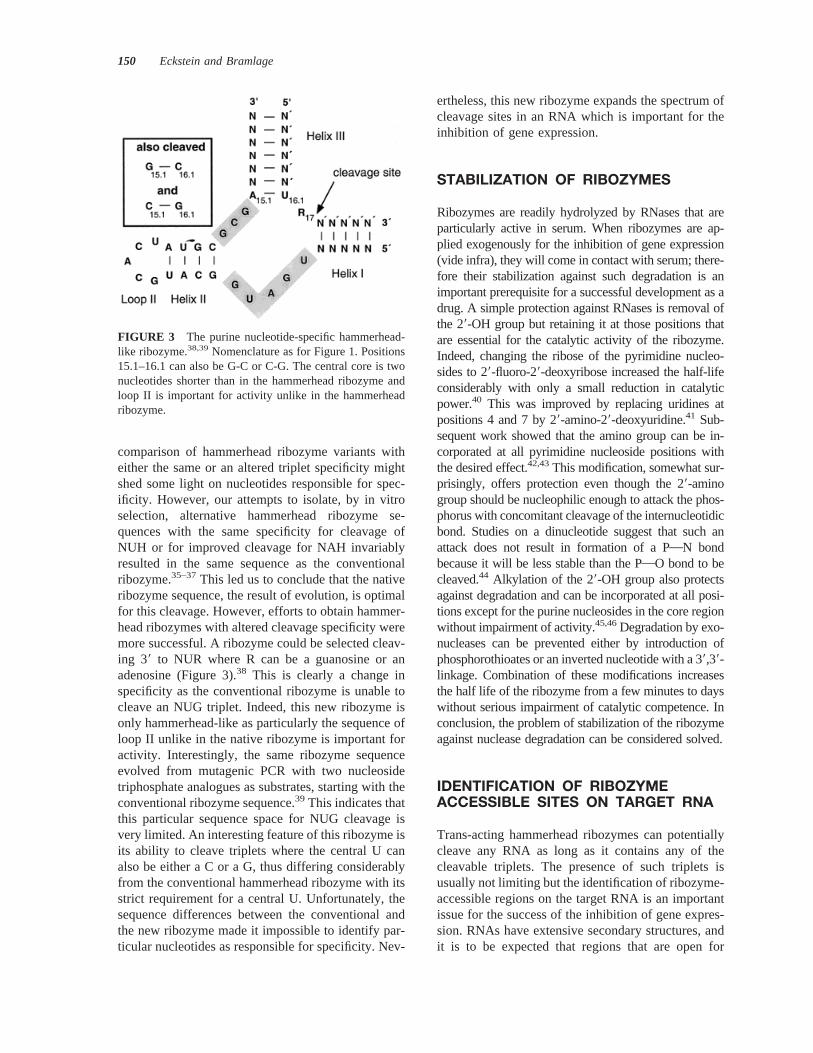
comparison of hammerhead ribozyme variants witheither the same or an altered triplet specificity mightshed some light on nucleotides responsible for spec-ificity. However, our attempts to isolate, by in vitroselection, alternative hammerhead ribozyme se-quences with the same specificity for cleavage ofNUH or for improved cleavage for NAH invariablyresulted in the same sequence as the conventionalribozyme.35–37This led us to conclude that the nativeribozyme sequence, the result of evolution, is optimalfor this cleavage. However, efforts to obtain hammer-head ribozymes with altered cleavage specificity weremore successful. A ribozyme could be selected cleav-ing 39 to NUR where R can be a guanosine or anadenosine (Figure 3).38 This is clearly a change inspecificity as the conventional ribozyme is unable tocleave an NUG triplet. Indeed, this new ribozyme isonly hammerhead-like as particularly the sequence ofloop II unlike in the native ribozyme is important foractivity. Interestingly, the same ribozyme sequenceevolved from mutagenic PCR with two nucleosidetriphosphate analogues as substrates, starting with theconventional ribozyme sequence.39 This indicates thatthis particular sequence space for NUG cleavage isvery limited. An interesting feature of this ribozyme isits ability to cleave triplets where the central U canalso be either a C or a G,thus differing considerablyfrom the conventional hammerhead ribozyme with itsstrict requirement for a central U. Unfortunately, thesequence differences between the conventional andthe new ribozyme made it impossible to identify par-ticular nucleotides as responsible for specificity. Nev-
ertheless, this new ribozyme expands the spectrum ofcleavage sites in an RNA which is important for theinhibition of gene expression.
STABILIZATION OF RIBOZYMES
Ribozymes are readily hydrolyzed by RNases that areparticularly active in serum. When ribozymes are ap-plied exogenously for the inhibition of gene expression(vide infra), they will come in contact with serum; there-fore their stabilization against such degradation is animportant prerequisite for a successful development as adrug. A simple protection against RNases is removal ofthe 29-OH group but retaining it at those positions thatare essential for the catalytic activity of the ribozyme.Indeed, changing the ribose of the pyrimidine nucleo-sides to 29-fluoro-29-deoxyribose increased the half-lifeconsiderably with only a small reduction in catalyticpower.40 This was improved by replacing uridines atpositions 4 and 7 by 29-amino-29-deoxyuridine.41 Sub-sequent work showed that the amino group can be in-corporated at all pyrimidine nucleoside positions withthe desired effect.42,43This modification, somewhat sur-prisingly, offers protection even though the 29-aminogroup should be nucleophilic enough to attack the phos-phorus with concomitant cleavage of the internucleotidicbond. Studies on a dinucleotide suggest that such anattack does not result in formation of a PON bondbecause it will be less stable than the POO bond to becleaved.44 Alkylation of the 29-OH group also protectsagainst degradation and can be incorporated at all posi-tions except for the purine nucleosides in the core regionwithout impairment of activity.45,46Degradation by exo-nucleases can be prevented either by introduction ofphosphorothioates or an inverted nucleotide with a 39,39-linkage. Combination of these modifications increasesthe half life of the ribozyme from a few minutes to dayswithout serious impairment of catalytic competence. Inconclusion, the problem of stabilization of the ribozymeagainst nuclease degradation can be considered solved.
IDENTIFICATION OF RIBOZYMEACCESSIBLE SITES ON TARGET RNA
Trans-acting hammerhead ribozymes can potentiallycleave any RNA as long as it contains any of thecleavable triplets. The presence of such triplets isusually not limiting but the identification of ribozyme-accessible regions on the target RNA is an importantissue for the success of the inhibition of gene expres-sion. RNAs have extensive secondary structures, andit is to be expected that regions that are open for
FIGURE 3 The purine nucleotide-specific hammerhead-like ribozyme.38,39Nomenclature as for Figure 1. Positions15.1–16.1 can also be G-C or C-G. The central core is twonucleotides shorter than in the hammerhead ribozyme andloop II is important for activity unlike in the hammerheadribozyme.
150 Eckstein and Bramlage

docking of an oligonucleotide such as antisense-oli-godeoxynucleotides or ribozymes will be limited. Un-til recently such sites have been identified mostly bytrial and error. However, experimental and theoreticalapproaches have been developed to facilitate the de-tection of accessible RNA sites. One such approach isscanning the labeled target RNA by hybridization to alibrary of oligonucleotides of known sequence on anarray. This method has so far only been used forantisense-oligodeoxynucleotides.47 Another is the an-nealing of a randomized oligodeoxynucleotide to alabeled transcript of the RNA and subsequent cleav-age of this complex by RNase H. The sites of cleav-age can then be scanned for triplets susceptible forcleavage by ribozymes.48 The analysis of transcriptsis not entirely satisfactory as the secondary structureof the mRNA in the cell might be quite different fromthat of the transcript. Additionally, part of the mRNAmight be covered by proteins preventing oligonucle-otide annealing. Thus extension of such analyses tonative mRNA is most desirable even though experi-mentally this is very challenging because of the lowstability of the cleavage products. Indeed, the analysisof murine DNA methyltransferase mRNA in a cellextracts has been successfully conducted with oligo-nucleotides and RNase H.49 Sites thus selected wereequally effective for inhibition by antisense oligode-oxynucleotides and ribozymes. Computational meth-ods have also been developed to identify suchsites.50,51 One direct comparison demonstrated thatsites identified by the computational and by the RNaseH methods were consistent.52 More recently, ri-bozymes with randomized substrate binding armshave been employed for cleavage of transcripts by thehairpin ribozyme.53,54Taking a randomized hammer-head ribozyme, we could find two cleavage sites inthe HIV-1 LTR in a construct were it served aspromoter for the expression for luciferase (E. Luzi, B.Bramlage, and F. Eckstein, unpublished data). Therandomized ribozyme strategy has the advantage thatpotential cleavage sites are directly detected and donot have to be deduced from the RNase H cleavagepatterns. This is important as shifts in the hybridiza-tion site by as little as one nucleotide can have seriousconsequences for the annealing and thus for the in-hibitory efficiency as has been detected for antisense-oligodeoxynucleotides.50 This phenomenon is not un-derstood and underlines the need for a better under-standing of the annealing process.
INHIBITION OF GENE EXPRESSION
Ribozymes are not only of interest as possible rem-nants of an RNA world and thus for an understanding
of the mechanism of RNA-catalyzed reactions, butalso for the application to the inhibition of gene ex-pression. As outlined in several reviews, particularlythe hammerhead ribozyme has been explored for thispurpose and many examples of successful applica-tions have been described.8,55–57 These approachescan be divided into two classes depending on themode of delivery of the ribozyme to cells. One, de-scribed as endogenous application, consists of cloningthe sequence for the ribozyme behind a suitable pro-moter into a vector, either a plasmid or a retroviralvector. After transfection or transduction, the ri-bozyme is transcribed in the cell to find its mRNAtarget. This approach has been adopted by severallaboratories and shown to be successful—for exam-ple, in interfering with HIV replication as discussed inseveral reviews.8,55–57One advantage of this deliverymode is that the gene can be stably integrated in thehost DNA, very much akin to a gene therapy protocol.Also, once integrated the supply of ribozyme is per-manent. However, the choice of vector is crucial,particularly for application to humans where vectorsafety is of considerable concern. In the other mode ofapplication, exogenous delivery, the ribozymes areprepared either by chemical synthesis or by transcrip-tion, and are added to the cells from the outside. Thismode of application will primarily be discussed here.
EXOGENOUS DELIVERY
The exogenous delivery of ribozymes circumvents theproblem of vector choice but is faced with the problemsof nuclease degradation of the supplied ribozymes in theserum and of efficient cellular uptake. The problem ofdegradation of exogenously delivered ribozymes by se-rum nucleases has essentially been solved by chemicalmodification as discussed above. Cellular uptake in cellculture is in general achieved by the use of cationic lipidsas carriers where the positive charge is localized on theoutside of the lipid for electrostatic interaction with thenegative charge of the oligonucleotide. Various compo-sitions of such lipids are available, some more suited forcertain cell lines than others. For the future it is hopedthat the derivatization of ribozymes with peptides ofknown transport properties might alleviate this situation.Such a strategy might also pave the way for cell type-directed delivery. At present, trial and error is the basisfor the carrier of choice. However, there are cell types inculture that take up ribozymes quite readily without suchaids as long as they are chemically modified.58 It isassumed that these cells, CHO cells in this example,have a very active membrane that facilitates oligonucle-otide transport. Most interestingly, chemically stabilized
Hammerhead Ribozyme 151

ribozymes are taken up by tissue upon local applicationin animal models without carrier.59–62This agrees withthe in vivo application of antisense-oligodeoxynucleo-tides, which is also achieved without carriers.63,64Thusuptake of oligonucleotides differs in cell culture and inanimals and our understanding of these processes is stillvery unsatisfactory.
Targets for chemically modified ribozymes in cellculture have been c-myb,65 N-ras,66 luciferase,67
VEGF receptor,62 multiple drug resistance gene,58,68
Hepatitis C virus 59 LTR,69 and HIV-1 LTR (B.Bramlage, E. Luzi, and F. Eckstein, unpublisheddata). In vivo model studies with synthetic ribozymeshave been reported for inhibition of expression ofamelogenin,59 of protein kinase C,42 of stromelysinmRNA,60 and of the dopamine D2 receptor.61
Only the cell culture experiments will be consid-ered here in more detail. The degrees of inhibitionsurprisingly do not vary very much in these studies,with between 51 and 77% inhibition (Table I). How-ever, the results are difficult to compare as they havebeen obtained with differences in cell lines, in mRNAtargets, in concentrations of ribozymes, in the types ofchemical modifications and carriers, and often with noscreen for the best ribozyme annealing or cleavagesite. Factors that affect the efficiency of ribozymeinhibition also include the lifetime of the protein andof the mRNA. Obviously the longer lived the proteinis, the more difficult it will be to achieve inhibition.Conversely, RNAs with a long half life are consideredbetter substrates for ribozymes.70
Interestingly, most authors observe a certain de-gree of inhibition of protein expression even by theinactive ribozyme (Table I). The simplest explanationput forward by most investigators is a simple anti-
sense effect by which the ribozyme prevents themovement of the ribosome during translation. Anadditional mechanism to explain the inhibition couldbe cleavage of the target RNA by a double-strandedRNase that has been identified in human cells.71 In-deed, degradation of the target RNA can be observedeven with the inactive ribozyme (B. Bramlage, E.Luzi, and F. Eckstein, unpublished data). However,the activity of such a double-stranded RNase has notbeen tested yet with chemically modified RNA aswould be the situation in the ribozyme-initiated inhi-bition of expression.
Colocalization
An important aspect for the efficient interference withgene expression is the colocalization of the ribozymewith its target RNA. Endogenous delivery has theadvantage in this respect in that the ribozyme can bedirected to the nucleus or to the cytoplasm either bythe choice of promoter or by combining it with sub-cellular localization signals.72,73 Colocalization of ri-bozyme and target in the nucleolus demonstrates thispoint very convincingly.74,75 At present synthetic ri-bozymes designed for exogenous delivery miss suchsignals and localization cannot be directed. However,it is hoped that the attachment of peptide signals, forexample important for nucleus import, might improvethis situation. Chemical modification can apparentlyinfluence localization somewhat. Thus, phosphoro-thioate groups, introduced to achieve nuclease stabil-ity, direct the ribozyme preferentially to the nucleusalthough interference with gene expression seems totake place in the cytoplasm.67
SUMMARY
The hammerhead ribozyme as a member of the ri-bozyme family is a most interesting molecule. Eventhough quite small in size, it poses many unansweredbut nevertheless interesting and challenging ques-tions. Thus the precise role of the essential metal ionsstill remains unresolved as does the basis for thecleavage specificity. One would have hoped that forsuch a small molecule details of structure–functionrelationship should be more easily tractable. As wecontinue to gain more insight into these problems, weenter uncharted territory in the field of RNA withrespect to its folding, its capacity for metal ion bind-ing, and its potential as a catalyst. Besides thesemechanistic aspects, the molecule has an as yet notfully explored potential for the sequence-specificcleavage of RNA. Inhibition of gene expression on
Table I Inhibition of Gene Expression by Active andInactive Ribozymes in Examples for ExogenousDeliverya
Target
Inhibition (%)
ReferenceActive Rz Inactive Rz
N-ras 54 20 66PKCa 70 50 42Luciferase 51 46 67HIV-1 LTR 67 53 c
c-myb 75 35 65VEGF receptor 56; 77b No inhibition 62HCV-UTR 55; 70b No data 69
a Ribozymes used in these studies have in general 7 nucleotidesin each substrate binding arm.
b Values for two different ribozymes.c B. Bramlage, E. Luzi, and F. Eckstein, unpublished data.
152 Eckstein and Bramlage

the RNA level by ribozymes can be very specific andwould represent a novel type of drug in the combat ofdiseases. However, cellular uptake, traffic, and local-ization of the ribozymes within the cell remain chal-lenges for the future.
REFERENCES
1. Sigurdsson, S. Th.; Thomson, J. B.; Eckstein, F. InRNA Structure and Function; Simons, R. W., Grun-berg-Manago, M., Eds.; Cold Spring Harbor Labora-tory Press, New York, 1998; pp 339–376.
2. Carola, C.; Eckstein, F. Current Opin Chem Biol 1999,3, 274–283.
3. Ban, N.; Nissen, P.; Moore, P. B.; Steitz, T. A. Science2000, 289, 905–920.
4. Schultes, E. A.; Bartel, D. P. Science 2000, 289, 448–452.
5. Symons, R. H. Ann Rev Biochem 1992, 61, 641–671.6. Uhlenbeck, O. C. Nature 1987, 328, 596–600.7. Haseloff, J.; Gerlach, W. L. Nature 1988, 334, 585–591.8. Bramlage, B.; Luzi, E.; Eckstein, F. Trends Biotechnol
1998, 16, 434–4383.9. Rossi, J. J. Chem Biol 1999, 6, R33–R37.
10. Pley, H. W.; Flaherty, K. M.; McKay, D. B. Nature1994, 372, 68–74.
11. Scott, W. G.; Finch, J. T.; Klug, A. Cell 1995, 81,991–1002.
12. Scott, W. G.; Murray, J. B.; Arnold, J. R. P.; Stoddard,B. L.; Klug, A. Science 1996, 274, 2065–2069.
13. Murray, J. B.; Terwey, D. P.; Maloney, L.; Karpeisky,A.; Usman, N.; Beigelman, L.; Scott, W. G. Cell 1998,92, 665–673.
14. Birikh, K. R.; Heaton, P. A.; Eckstein, F. Eur J Bio-chem 1997, 245, 1–16.
15. Wedekind, J. E.; McKay, D. B. Ann Rev BiophysBiomol Structure 1998, 27, 475–502.
16. Lilley, D. M. J. Current Opin Struct Biol 1999, 9,330–338.
17. Bassi, G. S.; Murchie, A. I. H.; Walter, F.; Clegg,R. M.; Lilley, D. M. J. EMBO J 1997, 16, 7481–7489.
18. Bassi, G. S.; Mollegaard, N. E.; Murchie, A. I. H.;Lilley, D. M. J. Biochemistry 1999, 38, 3345–3354.
19. Menger, M.; Tuschl, T.; Eckstein, F.; Porschke, D.Biochemistry 1996, 35, 14710–14716.
20. Kuimelis, R. G.; McLaughlin, L. W. Chem Rev 1998,28, 1027–1044.
21. Zhou, D.-M.; Taira, K. Chem Rev 1998, 28, 991–1026.22. Wang, S.; Karbstein, K.; Peracchi, A.; Beigelman, L.;
Herschlag, D. Biochemistry 1999, 38, 14363–14378.23. Murray, J. B.; Scott, W. G. J Mol Biol 2000, 296, 33–41.24. Yoshinari, K.; Taira, K. Nucleic Acids Res 2000, 28,
1730–1742.25. Cunningham, L. A.; Li, J.; Lu, Y. J Am Chem Soc
1998, 120, 4518–4519.26. Hermann, T.; Auffinger, P.; Scott, W. G.; Westhof, E.
Nucleic Acids Res 1997, 25, 3421–3427.
27. Murray, J. B.; Seyhan, A. A.; Walter, N. G.; Burke,J. M.; Scott, W. G. Chem & Biol 1998, 587–595.
28. Ferre´-D’Amare, A.; Doudna, J. A. J Mol Biol 2000,295, 541–556.
29. Perrotta, A. T.; Shih, I.-H.; Been, M. D. Science 1999,286, 123–126.
30. Nakano, S.-I.; Chadalavada, D. M.; Bevilacqua, P. C.Science 2000, 287, 1493–1497.
31. Nissen, P.; Hansen, J.; Ban, N.; Moore, P. B.; Steitz,T. A. Science 2000, 289, 920–930.
32. Muth, G. W.; Ortoleva-Donnelly, L.; Strobel, S. A.Science 2000, 289, 947–950.
33. Hanna, R.; Doudna, J. A. Current Opin Chem Biol2000, 4, 166–170.
34. Ludwig, J.; Blaschke, M.; Sproat, B. S. Nucleic AcidsRes 1998, 26, 2279–2285.
35. Nakamaye, K. L.; Eckstein, F. Biochemistry 1994, 33,1271–1277.
36. Vaish, N. K.; Heaton, P. A.; Eckstein, F. Biochemistry1997, 36, 6495–6501.
37. Kore, A. R.; Carola, C.; Eckstein, F. Bioorg Med Chem2000, 8, 1767–1771.
38. Vaish, N. K.; Heaton, P. A.; Fedorova, O.; Eckstein, F.Proc Natl Acad Sci USA 1998, 95, 2158–2162.
39. Kore, A. R.; Vaish, N. K.; Morris, J. A.; Eckstein, F. JMol Biol 2000, 301, 1113–1121.
40. Pieken, W. A.; Olsen, D. B.; Aurup, H.; Eckstein, F.Science 1991, 253, 314–317.
41. Heidenreich, O.; Benseler, F.; Fahrenholz, A.; Eckstein,F. J Biol Chem 1994, 269, 2131–2138.
42. Sioud, M.; Sorensen, D. R. Nature Biotechnol 1998, 16,556–561.
43. Beaudry, A.; DeFoe, J.; Zinnen, S.; Burgin, A.; Beig-elman, L. Chem Biol 1999, 7, 323–334.
44. Thomson, J. B.; Bhisma, K. P.; Jime´nez, V.; Eckart, K.;Eckstein, F. J Org Chem 1996, 61, 6273–6281.
45. Paolella, G.; Sproat, B. S.; Lamond, A. I. EMBO J1992, 11, 1913–1919.
46. Jarvis, T. C.; Wincott, F. E.; Alby, L. J.; McSwiggen,J. A.; Beigelman, L.; Gustofson, J.; Direnzo, A.; Levy,K.; Arthur, M.; Maculic-Adamic, J.; Karpeisky, A.;Gonzalez, C.; Woolf, T. M.; Usman, N.; Stinchcomb,D. T. J Biol Chem 1996, 271, 29107–29112.
47. Sohail, M.; Southern, E. M. Current Opin Mol Thera-peutics 2000, 2, 264–271.
48. Birikh, K. R.; Berlin, Y. A.; Soreq, H.; Eckstein, F.RNA 1997, 3, 429–437.
49. Scherr, M.; Reed, M.; Huang, C.-F.; Riggs, A. D.;Rossi, J. J. Mol Therapy 2000, 2, 26–38.
50. Patzel, V.; Steidl, U.; Kronenwett, R.; Haas, R.; Scza-kiel, G. Nucleic Acids Res 1999, 27, 4328–4334.
51. Mathews, D. H.; Burkard, M. E.; Freier, S. M.; Wyatt,J. R.; Turner, D. H. RNA 1999, 5, 1458–1469.
52. Scherr, M.; Rossi, J. J.; Sczakiel, G.; Patzel, V. NucleicAcids Res 2000, 28, 2455–2461.
53. Yu, Q.; Pecchia, D. B.; Kingsley, S. L.; Heckman, J. E.;Burke, J. M. J Biol Chem 1998, 273, 23524–23533.
Hammerhead Ribozyme 153

54. zu Putlitz, J.; Yu, Q.; Burke, J. M.; Wands, J. R.J Virology 1999, 73, 5381–5387.
55. James, H. A.; Gibson, I. Blood 1998, 91, 371–382.56. Muotri, A. R.; Veiga Pereira, L.; Reis Vasques, L. R.;
Menck, C. F. M. Gene 1999, 237, 303–310.57. Castanotto, D.; Scherr, M.; Rossi, J. J. Meth Enzymol
2000, 313, 401–506.58. Mariani, L.; Citti, L.; Nevischi, S.; Eckstein, F.; Rain-
aldi, G. Cancer Gene Therapy 2000, 7, 905–909.59. Lyngstadaas, S. P.; Risnes, S.; Sproat, B. S.; Thrane,
P. S.; Prydz, H. P. EMBO J 1995, 14, 5224–5229.60. Flory, C. M.; Pavco, P. A.; Jarvis, T. C.; Lesch, M. E.;
Wincott, F. E.; Beigelman, L.; Hunt III, S. W.; Schrier,D. J. Proc Natl Acad Sci USA 93, 745–758.
61. Salmi, P.; Sproat, B. S.; Ludwig, J.; Hale, R.; Avery,N.; Kela, J.; Wahlestedt, C. Eur J Pharmacol 2000, 388,R1–R2.
62. Parry, T. J.; Cushman, C.; Gallegos, A. M.; Agrawal,A. B.; Richardson, M.; Andrews, L. E.; Maloney, L.;Mokler, V. R.; Wincott, F. E.; Pavco, P. A. NucleicAcids Res 1999, 27, 2569–2577.
63. Crooke, S. T. Methods Enzymol 2000, 313, 3–45.64. Agrawal, S.; Kandimalla, E. R. Mol Medicine Today
2000, 6, 72–81.65. Jarvis, T. C.; Alby, L. J.; Beaudry, A. A.; Wincott,
F. E.; Beigelman, L.; McSwiggen, J. A.; Usman, N.;Stinchcomb, D. T. RNA 1996, 2, 419–428.
66. Scherr, M.; Grez, M.; Ganser, A.; Engels, J. W. J BiolChem 1997, 272, 14304–14313.
67. Bramlage, B.; Alefelder, S.; Marschall, P.; Eckstein, F.Nucleic Acids Res 1999, 27, 3159–3167.
68. Citti, L.; Eckstein, F.; Capecchi, B.; Mariani, L.; Ne-vischi, S.; Poggi, A.; Rainaldi, G. Antisense NucleicAcid Drug Devel 1999, 9, 125–133.
69. Macejak, D. G.; Jensen, K. L.; Jamison, S. F.; Do-menico, K.; Roberts, E. C.; Chaudhary, N.; von Car-lowitz, I.; Bellon, L.; Tong, M. J.; Conrad, A.; Pavco,P. A.; Blatt, L. M. Hepatology 2000, 31, 769–776.
70. Donahue, C. P.; Yadava, R. S.; Nesbitt, S. M.; Fedor,M. J. J Mol Biol 2000, 295, 693–707.
71. Wu, H.; MacLeod, R.; Lima, W. F.; Crooke, S. T. J BiolChem 1998, 273, 2532–2542.
72. Bertrand, E.; Castanotto, D.; Zhou, C.; Carbonnelle, C.;Lee, N. S.; Good, P.; Chatterjee, S.; Grange, T.; Pictet,R.; Kohn, D.; Engelke, D.; Rossi, J. J. RNA 1997, 3,74–88.
73. Lee, N. S.; Bertrand, E.; Rossi, J. J. RNA 1999, 5,1200–1209.
74. Samarsky, D. A.; Ferbeyre, G.; Bertrand, E.; Singer,R. H.; Cedergren, R.; Fournier, M. J. Proc Natl AcadSci USA 1999, 96, 6609–6614.
75. Michienzi, A.; Cagnon, L.; Bahner, I.; Rossi, J. J. ProcNatl Acad Sci USA 2000, 97, 8955–8960.
154 Eckstein and Bramlage