the great mismatch - national skill development
TRANSCRIPT
Published: February 03, 2011
r 2011 American Chemical Society 416 dx.doi.org/10.1021/mp100260c |Mol. Pharmaceutics 2011, 8, 416–429
ARTICLE
pubs.acs.org/molecularpharmaceutics
Hydrophobically Modified Dendrons: Developing Structure-ActivityRelationships for DNA Binding and Gene TransfectionSimon P. Jones,† Nathan P. Gabrielson,‡ Chun-Ho Wong,§ Hak-Fun Chow,§ Daniel W. Pack,‡
Paola Posocco,|| Maurizio Fermeglia,|| Sabrina Pricl,|| and David K. Smith*,†
†Department of Chemistry, University of York, Heslington, York, YO10 5DD, U.K.‡Department of Chemical and Biomolecular Engineering, University of Illinois at Urbana—Champaign, Urbana, Illinois 61801,United States§Department of Chemistry and The Center of Novel Functional Molecules, University Science Centre, The Chinese University of HongKong, Shatin, New Territories, Hong Kong
)Molecular Simulation Engineering Laboratory, Department of Materials and Natural Resources, University of Trieste, 34127 Trieste,Italy
bS Supporting Information
’ INTRODUCTION
In the emergent field of nanomedicine, gene therapy is one ofthe most widely investigated topics, however, as yet, a generalapproach to gene therapy In Vivo remains elusive.1 The devel-opment of vectors that are capable of delivering genetic material,such as DNA or siRNA, safely and efficiently into cells istherefore an important area of research, and could have adramatic impact on diseases such as cystic fibrosis.2 Viral vectorswere employed in some of the earliest clinical trials of gene therapy,but it was observed that they can cause problems with immuno-genicity and adverse patient response.3 For this reason, increasingattention has focused on the development of nonviral, syntheticvectors.4 Nonviral vectors such as Superfect5 or Lipofectamine6
are commercially available for in vitro transfections. Further-more, some nonviral vectors have been investigated in the clinic,with some promising results, for example, in phase 1 trials on
cystic fibrosis patients.7 Nonviral vectors can be divided into twomain classes: cationic polymers8 and cationic lipids.9 In bothcases, cationic charge is required to effectively bind and condensethe nucleic acid, which is a polyanion. Cationic polymers havehigh charge due to their polymeric nature; however this can leadto adverse toxicity.10 One particularly important class of cationicpolymer employed in transfection studies is well-defined branchedpolymers: dendrimers.11 Poly(amidoamine) (PAMAM) den-drimers5,12 and dendrimers based on L-lysine13 have been widelyinvestigated as DNA delivery agents. The second class of nonviralvector, cationic lipids, achieves high charge via self-assembly,with vector aggregates being responsible for gene delivery; work
Received: August 9, 2010Accepted: February 3, 2011Revised: October 21, 2010
ABSTRACT: This paper develops a structure-activity rela-tionship understanding of the way in which surfactant-likedendrons with hydrophilic spermine surface groups and avariety of lipophilic units at their focal points can self-assembleand subsequently bind to DNA with high affinity. The choice offunctional group at the focal point of the dendron and the hightunability of the molecular structure have a very significantimpact on DNA binding. Mesoscale modeling of the mode ofdendron self-assembly provides a direct insight into how themode of self-assembly exerts its effect on the DNA binding process. In particular, the hydrophobic unit controls the number ofdendrons in the self-assembled micellar structures, and hence their diameters and surface charge density. The DNA binding affinitycorrelates with the surface charge density of the dendron aggregates. Furthermore, these structure-activity effects can also beextended to cellular gene delivery, as surface charge density plays a role in controlling the extent of endosomal escape. It is reportedthat higher generation dendrons, although binding DNA less strongly than the self-assembling lower generation dendrons, are moreeffective for transfection. The impact of the lipophilic group at the focal point is less significant for the DNA binding ability of theselarger dendrons, which is predominantly controlled by the spermine surface groups, but it does modify the levels of genetransfection. Significant synergistic effects on gene delivery were observed when employing combinations of the dendrons andpolyethyleneimine (PEI, 25 kDa), with transfection becoming possible at low loading levels where the two components would nottransfect individually, giving practically useful levels of gene delivery.
KEYWORDS: dendron, DNA, gene delivery, mesoscale modeling, self-assembly

417 dx.doi.org/10.1021/mp100260c |Mol. Pharmaceutics 2011, 8, 416–429
Molecular Pharmaceutics ARTICLE
is ongoing to increase the load of genetic material that can bedelivered and decrease toxicity.9,10
Developing a detailed structure-activity relationship of trans-fection is of key importance. For example, Kirby and co-workersdemonstrated that gene expression in their cationic lipid derivedsystemwas dependent on chain length and amide bond linkage,14
and since this study, there has been intense interest in developingmeaningful structure-activity relationships which correlate mo-lecular-scale modifications with biological gene transfectionbehavior.15 In elegant research, the groups of Diederich, Florenceand Hammond have explored different series of well-defineddendritic amphiphiles.16 They revealed that the most activevectors in transfection were able to self-assemble under neutralto endosomal pH. Further studies from Diederich and co-work-ers showed that their most versatile transfection vectors self-assembled into vesicles or bilayers.17
We have developed a class of DNA binder based on a dendriticscaffold with multiple spermine surface groups (Figure 1).18
Spermine is a bioavailable DNA binder which operates at highmicromolar concentrations.19 We demonstrated that the pre-sence of multiple spermine groups on the dendron led to amultivalency effect, enhancing DNA binding. These dendronshad relatively poor transfection abilities, being several orders ofmagnitude less effective than polyethyleneimine (PEI), whichwas used as a positive control.20 However, we recently reportedthat modification of the focal point with a lipophilic cholesterolgroup could lead to significantly enhanced DNA binding andtransfection.21 This was attributed to the lipid-like self-assemblyof the dendron into a supramolecular aggregate improving theperformance of the system, a hypothesis which was confirmed bymesoscale molecular modeling.22 As such, we argued that ourdendrons combined aspects of both classes of nonviral vector:cationic polymers and lipids. It has also been noted thatcombining the advantages of both cationic polymers and lipidsin a single vector system can give rise to synergistic effects ongene delivery.23
We therefore decided to monitor the way in which thestructure of the hydrophobic unit modified the DNA binding and
gene delivery performance of this class of dendron. Indeed, thehigh synthetic tunability of dendron structures provides themwith a significant advantage over larger spherical dendrimerstructures. This can be done in a relatively straightforwardmanner and allows us to develop structure-activity relation-ships, in which the structural changes made to the dendron canbe correlated with DNA binding and transfection abilities. Byusing mesoscale molecular modeling, we are able to rationalizeour observations. In this way, we employ this combined experi-mental and theoretical approach to develop effective designprinciples for smart gene delivery vectors; these results willtherefore inform future research in this field.
’RESULTS AND DISCUSSION
Synthesis of First Generation Vectors. The first generationvectors investigated in this paper were synthesized from pre-viously reported dendron Z-G1-Boc which has a Z-protectinggroup at the focal point, and has three spermine surface groupsprotected by multiple N-tert-butyloxycarbonyl (Boc) protectinggroups (Scheme 1).18 The Z-protecting group was removed fromZ-G1-Boc by hydrogenolysis, to yield G1-Boc, which has areactive amine group at the focal point. Five different lipophilicunits were then selected for attachment to the free amine. Thesimplest of these were a commercially available dodecanoyl chainand a cholesterol unit. We also selected three synthetic branchedsystems, one based on lysine functionalized with dodecanoylchains, the other two being all-aliphatic dendrons previouslyreported (and provided) by Chow and co-workers.24
Dendron Chol-G1 was synthesized in a two step process usingthe previously reported methodology.21 First, cholesteryl chlor-oformate was reacted with G1-Boc in CH2Cl2 in the presence ofEt3N, and subsequently the Boc groups were removed bydissolving the product in methanol and bubbling HCl gasthrough the solution, providing the product in 56% yield.Novel dendron C12-G1 was synthesized in a similar way by
first reacting G1-Boc with dodecanoyl chloride, and then the Bocgroups being removed using HCl gas, giving product in a 66%
Figure 1. General structure of first and second generation dendrons with spermine surface groups (R-G1 and R-G2).

418 dx.doi.org/10.1021/mp100260c |Mol. Pharmaceutics 2011, 8, 416–429
Molecular Pharmaceutics ARTICLE
yield. In order to synthesize the novel dendron C12Lys-G1, weinitially reacted G1-Boc withN,N0-bis(benzyloxycarbonyl) lysineusing DCC and HOBt, in anhydrous THF with Et3N as base, toyield ZLys-G1-Boc. The benzyloxycarbonyl (Z) protectinggroups were then removed from the lysine unit by hydrogeno-lysis, and the unmasked free amines were reacted with dodeca-noyl chloride in anhydrous CH2Cl2 to yield C12Lys-G1-Boc in56% yield over the two steps . Finally, the Boc protecting groupswere removed with gaseous HCl in methanol to provide thedesired C12Lys-G1 in a 38% yield.We then attempted to conjugate the aliphatic dendrons (D1
and D2) to G1-Boc. However, we were unable to directly couplethese dendrons via standard peptide coupling methodologies,presumably due to the steric hindrance associated with reactingtogether the focal points of two dendritic building blocks. Wetherefore appended a glycine spacer unit onto Chow’s dendrons(see Supporting Information for the methodology).25 Theseextended hydrophobic dendrons (D1Gly and D2Gly) could besmoothly coupled to G1-Boc using DCC/HOBt methodology.The Boc groups were finally removed with HCl gas, providingthe desired novel products, D1Gly-G1 and D2Gly-G1, in goodyields of 61% and 57% respectively.DNA Binding Studies with First Generation Vectors. In-
itially we assayed the ability of these dendrons to bindDNA usingthe standard ethidium bromide (EthBr) displacement fluores-cence spectroscopy assay which we have employed in our previous
research.18,26 This method uses competition between the DNAbinder and EthBr to assess the concentration at which the DNAbinder becomes effective. This can be expressed as the concen-tration of DNA binder required for half of the EthBr to bedisplaced from binding to DNA: a C50 value. This concentrationcan more usefully be expressed as a charge excess (CE50) value,better suited for comparing structurally different DNA binderswith different molar masses and numbers of amines. To calculatethis CE50 value, it is assumed that each amine in the DNA binderis protonated, and each phosphate in the DNA is deprotonated.As such, the CE50 value is equivalent to a N:P ratio. Lower CE50values represent more effective binding, as a smaller amount ofpositive charge is required to bind the negative charge associatedwith the DNA. This assay therefore provides an excellentcomparative method for considering the DNA affinities of afamily of compounds such as this, in which each member has thesame spermine-based DNA binding motif and, as such, is ideallysuited for the development of structure-activity relationships.For this study, we compared all of our DNA binders underphysiologically relevant salt concentrations (150 mM NaCl).In our previous studies, we had demonstrated that Z-G1
bound to DNA under the conditions of this assay with a CE50value of 2.7 (Table 1). This was a significant improvement oversimple, unfunctionalized spermine, which had a CE50 value of1560, clearly demonstrating the benefits of using a multivalentDNAbinding scaffold. Furthermore, we have previously reported
Scheme 1. Synthesis of First Generation Vectors, Chol-G1, C12-G1, C12Lys-G1, D1Gly-G1 and D2Gly-G1, with DifferentHydrophobic Groups at the Focal Point

419 dx.doi.org/10.1021/mp100260c |Mol. Pharmaceutics 2011, 8, 416–429
Molecular Pharmaceutics ARTICLE
that modification of the dendron with a cholesterol unit sig-nificantly enhanced DNA binding as a consequence of dendronself-assembly, reducing the CE50 to just 0.55.21 This new studytherefore allows us to develop an understanding the precise roleof the lipophilic unit in mediating the self-assembly, and sub-sequent DNA binding affinity, of the dendrons. Clearly, mod-ification of the lipophilic unit at the focal point exerts a dramaticeffect on the affinity of the dendrons for DNA, even though thisgroup is not itself directly responsible for forming interactionswith DNA. Intriguingly, some of the focal point modificationsappeared to improve DNA binding, while others had an adverseeffect. Dendrons Chol-G1 and C12Lys-G1 had the lowest CE50values of 0.55 and 0.85 respectively, indicating very effectivebinding indeed, some of the best reported.27 Compound D2Gly-G1 was somewhat better than Z-G1 having a CE50 value of 1.8.On the other hand, compounds C12-G1 and D1Gly-G1 hadhigher CE50 values of 3.3 and 4.3 respectively, indicating theyhave lower affinities for DNA than the Z-G1 analogue.In order to support the results of these EthBr assays, we also
carried out gel electrophoresis using plasmid DNA. This methodhas the advantage of directly measuring the binding between thedendrons and DNA, i.e., it is not a competition assay. Further-more, it is performed under conditions equivalent to thetransfection assays reported later in this paper. In the gelelectrophoresis experiment, 1 μg of DNA was applied to eachwell, and the loading of DNA binder required to completelyretard the movement of the DNA could then be determined(Figure 2). Pleasingly, the data were in general agreement withthe EthBr exclusion assay. The most effective binder was Chol-G1, which proved to be a highly effective DNA binder, retardingDNA mobility above loadings of 0.153 nmoles (N:P ratio, 0.45).Once again, the least effective binders were C12-G1 and D1Gly-G1, which only retarded mobility at loadings above ca. 0.40 nmol(N:P ratio, 1.0). The other dendrons were intermediate inbehavior, although C12Lys-G1 was noted to be slightly lesseffective in this assay than in the EthBr exclusion experiment.It is therefore clear that although the DNA binding event is
located at the spermine surface groups, the unit at the focal pointalso has a profound influence on binding. In an attempt torationalize these observations and better understand the struc-ture-activity effects, we employed mesoscale modelingmethods.Modeling Dendron Self-Assembly. Many interesting pro-
blems in soft matter science occur at length and time scalessandwiched between the atomistic scale and the macroscopiccontinuum. Systems such as nanovector/DNA complexes for
gene transfer can involve spatial inhomogeneities over lengthscales ranging between 1 and 1000 nm, and exhibit phenomenaover time scales of 1ms or greater. Problems in such length-timespace cannot be addressed by traditional atomic-level moleculardynamics, or by conventional finite element approaches thatusually deal with phenomena at longer time scale. Rather, oneneeds to take recourse to computational techniques at theintermediate scale, called the mesoscale. Over the past few years,different approaches have been developed to address problems atthe mesoscale level, which could be broadly classified as particle-based or density-based. With the purpose of studying theeventual self-assembly morphology of the modified dendronseries developed in this work, and their interaction with DNA,we resorted to using a particle-based method called dissipativeparticle dynamics (DPD).28
In DPD, a group of atoms is coarse-grained into a bead,thereby substantially reducing the number of particles to besimulated. Further, rather than interact through Lennard-Jonesforces, the beads feel a simple soft pairwise conservative potentialwhich embodies the essential chemistry of the system. This forceis of short range, and has a simple analytical form, which results infast computation per time step and, hence, provides the oppor-tunity to expand the simulation from nanoseconds to real timeperiods.Accordingly, we began our DPD-based simulation study by
monitoring the dendron self-assembly processes and to gain aninsight into the types of aggregates which may be formed.Figure 3 shows that all hydrophobically modified dendrons formsupramolecular structures with nanometer dimensions (seeTable 2). All of these first generation dendrons form sphericalmonodisperse micelles, with diameters Dm of 3-5 nm. This is adirect consequence of their conical shape with a relatively largecationic headgroup connected to a comparatively small lipophilicpart (vide infra).A simple but effective molecular theory can be invoked to
qualitatively describe the evidence inferred from the mesoscale
Table 1. Binding Data for First Generation Compounds withDNA Obtained via Ethidium Bromide (EthBr) DisplacementAssaya
compound CE50 value (N:P ratio)
spermine 1560
D1Gly-G1 4.3
C12-G1 3.3
Z-G1 2.7
D2Gly-G1 1.8
C12Lys-G1 0.85
Chol-G1 0.55aCE50 represents the charge excess (N:P ratio) required to displace 50%of the EthBr. [NaCl] = 150 mM.
Figure 2. Agarose gel electrophoresis of polyamine/DNA complexesusing first generation dendrons. Polyamine:DNA loadings (w:w) are asfollows: lane 1, 0:1; lane 2, 0.1:1; lane 3, 0.2:1; lane 4, 0.3:1; lane 5, 0.4:1;lane 6, 0.5:1; lane 7, 0.6:1; lane 8, 0.7:1; lane 9, 0.8:1; lane 10, 0.9:1; lane11, 1:1, lane 12, 1.5:1; lane 13, 2:1; lane 14, 2.5:1; lane 15, 3:1.

420 dx.doi.org/10.1021/mp100260c |Mol. Pharmaceutics 2011, 8, 416–429
Molecular Pharmaceutics ARTICLE
simulations. Indeed, Israelachvili, Mitchell and Ninham pro-posed the concept of molecular packing parameter and demon-strated how the size and the shape of self-assembled molecules atequilibrium can be predicted from a combination of molecularpacking considerations and general thermodynamic principles.29
To a first approximation, the molecular packing parameter P of agiven amphiphile is defined as v0/al0 where v0 and l0 are thevolume and the length of the hydrophobic portion, and a is thesurface area of the hydrophobic core of the aggregate expressedper molecule in the aggregate (hereafter referred to as the areaper molecule). If we consider a generic micelle with a core radiusRc, made up of Nagg molecules, then the volume of the core V =Naggv0 = 4πRc
3/3, the surface area of the core A =Nagga = 4πRc2,
and, hence, Rc = 3v0/a, from simple geometrical relations. If themicellar core is densely packed with the hydrophobic moietieswithout any empty space, then the core radius cannot exceed theextended length l0 of the hydrophobic part. Introducing this
constraint in the expression for Rc, we obtain the well-knowncondition 0 e P e 1/3 if an amphiphile is to form sphericalmicelles. Simple molecular modeling considerations coupledwith the fundamental micellar parameters listed in Table 2allowed us to calculate the packing parameter for all of themodified dendrons in hydrated conditions. The calculated Pnumbers listed in Table 2 are between 0.24 and 0.32, inagreement with the observation from modeling studies that allof the dendrons self-assemble into spherical micelles.A further, key parameter in governing the DNA binding
properties of the dendrons and, eventually, the transfectionefficiency of the resulting DNA/dendron assembly complexesis the micelle surface charge density σm. In practical terms, σmcan be thought of as a measure of how positively charged amicelle is. Two micelles containing the same number of den-drons, and therefore carrying the same overall charge, mayexhibit different values of σm, if the micelle sizes, and hencetheir surface areas, are different. To calculate σm one thereforeneeds to know the aggregation number of the micelle Nagg, thecharge of each dendron head, and the micellar surface area Sm =4πRm
2, as σm = eNagg/Sm. Using the data reported in Table 2 andthe constant value of þ9 for the overall charge of each dendronheadgroup, the σm values shown in the last column of Table 2 canbe easily estimated. Summarizing the overall evidence stemmingfrom the analysis of data in Table 2 leads to the following,important considerations. First, the overall series of amphiphilicdendrons synthesized in this work assemble into small, sphericalmicelles in water and in the presence of physiological ionicstrength conditions (150 mM), as experimentally verified forsimilar systems.17,30 However, the different architectures of the
Figure 3. Mesoscale modeling of the amphiphilic dendrons synthesized in this work showing aggregation into spherical micellar objects: (a) Chol-G1;(b) C12Lys-G1; (c) D2Gly-G1; (d) C12-G1; (e) D1Gly-G1. In all pictures, the yellow sticks represent dendron units. Different colored spheres areadopted to represent the various hydrophobic regions. A light gray field is used to represent water.
Table 2. Values of Micellar Diameter Dm (nm), Core RadiusRc (nm), Aggregation Number Nagg, Packing Parameter P,and Micelle Surface Charge Density σm (e/nm2) for theDifferent Modified Dendrons as Obtained from DPDSimulations
compd Dm Rc Nagg P σm
Chol-G1 3.4( 0.1 0.8 21 0.24 5.2
C12Lys-G1 4.0( 0.2 1.3 24 0.24 4.3
D2Gly-G1 4.9( 0.2 1.5 32 0.32 3.8
C12-G1 4.0 ( 0.1 1.3 16 0.28 2.8
D1Gly-G1 4.0( 0.2 0.9 12 0.25 2.1

421 dx.doi.org/10.1021/mp100260c |Mol. Pharmaceutics 2011, 8, 416–429
Molecular Pharmaceutics ARTICLE
hydrophobic portion result in differently sized micelles and/or adifferent number of dendrons per micelle (the aggregationnumber Nagg), and, hence, a different micellar surface chargedensity σm. Pleasingly, the experimentally verified CE50 valuesdirectly correlate with the surface charge density, σm, valuesestimated from simulation, indicating that the micelles charac-terized by higher values of σm (i.e., Chol-G1, C12Lys-G1, andD2Gly-G1) are tighter DNA binders than their counterparts withlower σm values (i.e., C12-G1 and D1Gly-G1). Interestingly,comparing the best DNA binders, Chol-G1, C12Lys-G1 andD2Gly-G1, the former compound assembles into micelles ofmuch smaller diameter than the latter two. This is presumablydue to the less sterically demanding nature of cholesterol leadingto more effective packing within the micellar interior, comparedwith the branched hydrophobic units in the latter two dendrons,which will not be able to pack so efficiently. As such, even thoughthemicelles formed by Chol-G1 contain fewer dendron units andhave less total positive charge than the micelles formed byC12Lys-G1 and D2Gly-G1, their smaller size means that theyhave significantly higher surface charge density, and as such, theyare therefore much more effective DNA binders.From an energetic standpoint, the change in Gibbs free energy
of transfer of a single amphiphilic molecule from the monomericstate to a micelle of aggregation number Nagg, commonly calledthe free energy of micellization ΔGmic, can be modeled asconsisting of a hydrophobic part, ΔGmic,h, and an electrostaticpart, ΔGmic,e, so that ΔGmic = ΔGmic,h þ ΔGmic,e. The hydro-phobic part stems primarily from the favorable energy of transferof the hydrocarbon moieties from the aqueous phase to themicellar phase, and, secondarily, from the unfavorable residualinterfacial contact of water with the apolar components withinthe micelles. The electrostatic part of ΔGmic arises from therepulsion between the ionic head groups within themicellar shell.Following the theory proposed by Tanford31 and subsequentlymodified by other authors,32 we calculated the values of ΔGmic
for the five modified dendrons (Table 3). As can be seen, ΔGmic
at room temperature has large, negative values, indicating thatmicellization is a spontaneous and highly favorable process for allamphiphilic dendrons, although ΔGmic decreases on going fromChol-G1 to D1Gly-G1. The headgroup architecture is the samein all amphiphiles, and as such, the main differential contributionto ΔGmic stems from the ΔGmic,h term, which will reflectdifferences in the size and structure of the hydrophobiccomponent.The critical micelle concentration (CMC) is one of the most
commonly studied properties of a self-assembled system becauseit is a direct measure of the thermodynamic stability of themicelles in solution. Basic thermodynamic relationships allowCMC to be directly obtained from ΔGmic; accordingly, the lastcolumn in Table 3 lists the predicted CMC data for each systemsimulated in this work. Typically, micellar aggregates have CMCs
of the order of 10-3-10-5 M, while lower CMCs, even down tothe nanomolar range, are found for amphiphiles that form eithermembranes or cylindrical aggregates. Recently, however, elec-tron microscopy experiments performed on cholesterol-por-phyrin micelles revealed that these amphiphiles could formvirtually monodisperse spherical aggregates with a diameter ofapproximately 7 nm and a CMC value of 11 nM.33 Amphiphilesshowing low CMCs tend to have relatively large hydrophobicsegments, and this normally results in an assembly shape with alower curvature. However, our series of modified dendrons,combine a large hydrophobic portion with a very large head-group, resulting in a roughly conical amphiphile. The size of thehydrophobic segment is responsible for the low CMCs, while thelarge size of the headgroup results in the spherical geometry ofthe assembly. It is interesting to note that the predicted CMCvalues for C12-G1 and D1Gly-G1 lie above the concentrations ofthe DNA binding assays (i.e., low μm concentrations); as such, itis possible that the relatively poor DNA binding ability of thesecompounds reflects the fact that they are not aggregated underthe experimental conditions as a consequence of their relativelysmall hydrophobic segments. Although a word of caution is dueabout the fact that the calculated values of ΔGmic and CMC areobtained using validated but simplified theoretical approaches,the trends exhibited by these parameters are in line with theexperimental data. Indeed, we are currently carrying out fullexperimental aggregation studies on a closely related set ofhydrophobically modified dendrons, and for these systems, thein silico predictions of micelle diameters, charge densities andCMC values are closely mirrored by the experimental results,both in terms of trends and absolute values.We then performed mesoscale simulations of the dendron
micelles in the presence of DNA. These investigations reveal that,in all cases, the overall systems consist of parts of free, unfolded,single-chain DNA that connect micelles on which a partialamount of DNA has been adsorbed (see Figure 4). In otherwords, all dendron/DNA complexes present a typical beads-on-a-string structure, made of dendron micelles connected by a DNAthread. Importantly, this predicted morphology is supported bydetailed AFM studies between G4 PAMAM dendrimers andDNA,34 indicative that these self-assemblies of dendrons can beconsidered to be somewhat like covalently bound higher gen-eration spherical dendrimers. These structures are also somewhatreminiscent of the structure of open chromatin, which consistsof an array of nucleosome core particles, separated from eachother by up to 80 base pairs of linker DNA.35 However, in clearcontrast to the periodic structure of open chromatin, thedendron micelles appear to be distributed in a nonperiodic,more irregular way.As a consequence of this model, DNA molecules are partially
embedded within the micellar organization and partially exposedto the solution environment, where the Naþ ions originatingfrom both the DNA and the accompanying ionic strengthprovide the minimum 90% charge neutralization required forcondensation, according to Manning’s theory.36 According tothis picture, the cationic micelles act as a matrix in which theDNA chains are partially embedded within the micellar organiza-tion and partially free to condense. Figure 4 reveals also that,upon DNA addition, for all systems most of the micelles retaintheir spherical geometry. However, the appearance of somemicelles changes to an elliptical cross-section, resulting from astretching of the micelles induced by the linear geometry of DNAto maximize mutual electrostatic contacts.
Table 3. Predicted Free Energy of Micellization ΔGmic (kJ/mol) and Critical Micelle Concentration CMC (μM) for theDifferent Modified Dendrons Estimated According to SP5
compd ΔGmic CMC
Chol-G1 -87.56 0.021
C12Lys-G1 -80.42 0.089
D2Gly-G1 -77.97 0.15
C12-G1 -55.92 12.5
D1Gly-G1 -49.29 47.6

422 dx.doi.org/10.1021/mp100260c |Mol. Pharmaceutics 2011, 8, 416–429
Molecular Pharmaceutics ARTICLE
In all systems, but particularly for C12-G1 and D1Gly-G1,DNA localizes in the interstitial space and gives rise to smallbundles. At first sight the bundling of DNA helices may appear tobe unexpected, however, these DNA bundles are formed by theinterplay of the salt-induced screening of the electrostatic inter-actions and the depletion-attraction caused by the dendronmicelles. Depletion-attraction is a somewhat underappreciatedforce associated with the aggregation of two large colloidalobjects as a consequence of the osmotic pressure generated bythe exclusion of smaller objects from their interacting interface.37
While depletion-attraction has previously been reported for likecharges or neutral objects, the screening of the electrostaticinteractions enables this effect to be observed also between DNAand the oppositely charged dendron micelles, for which theelectrostatic interactions are attractive. The presence of salttherefore facilitates bundling of DNA by reducing the electro-static repulsion between DNA molecules, and also reduces theelectrostatic attraction between positively charged micelles andnegatively charged DNA. The existence of DNA bundles andcondensed regions can also be understood by taking into accountthe small dimensions of the micelles formed by all thesedendrons and the relevant values of σm. Indeed, extensivewrapping of DNA around small micelles will result in a quitehigh cost of DNA bending (or elastic) free energy; accordingly,this is not observed in any of the systems considered. Further-more, a decrease of the DNA adsorbed amount per particle isobserved as the surface charge density, σm, decreases. This is onlypartly compensated by the fact that some of the free part of DNAadsorbs on new particles, and therefore the bundling of unboundDNA becomes more favored.
In aqueous solutions both DNA and dendron micelles areassociated with their respective counterions. The high chargedensity of DNA actually results in counterion condensation: in itssolution structure, the base length between negative phosphategroups on the DNA backbone is equal to l0 = 1.7 Å. This issignificantly less than the Bjerrum length in water lB = e
2/ewkBT =7.1 Å, where εw is the dielectric constant of water (=80), kB is theBoltzmann constant, and T is the temperature. The Bjerrumlength corresponds to the distance where the Coulomb energybetween two unit charges is equal to the thermal energy kBT.Under these conditions, it has been shown that positive coun-terions will condense on the DNA backbone until the Manningparameter ξ = l0/l* approaches unity, l* being the renormalizeddistance between the negative charges after counterioncondensation.38 A similar analysis shows that, near the surfaceof a positively charged micelle, almost half of the negativecounterions are contained within the Gouy-Chapman lengthlG-C =e/2πlBσm. Combining DNA and our amphiphilic den-dron micelles allows the charges of the spermine head groups toneutralize the phosphate moieties on DNA. This replaces andreleases the tightly bound-counterions of both micelle andnucleic acid in solution. The resulting gain of translationalentropy by the counterions is a driving force for higher orderself-assembly into micelle/DNA complexes.39 It should bepointed out that the term bound counterions is used in a looseform: indeed, the counterions near the DNA or a micelle surfaceare bound and yet remain in their fully hydrated state. Thisimplies no change in the entropy of water molecules upon releaseof bound counterions into solution. The driving force of thecounterion release mechanism is reduced by added salt, as in the
Figure 4. Mesoscale modeling of the interaction of DNA with the amphiphilic dendrons synthesized in this work: (a) Chol-G1; (b)C12Lys-G1;(c) D2Gly-G1; (d) C12-G1; (e) D1Gly-G1. In all pictures, yellow sticks represent dendron units. Different color spheres are adopted torepresent the various hydrophobic regions along the series. A light gray field is used to represent water. DNA molecules are depicted asorange sticks.

423 dx.doi.org/10.1021/mp100260c |Mol. Pharmaceutics 2011, 8, 416–429
Molecular Pharmaceutics ARTICLE
experiments and simulations described in this work. This isparticularly true for counterion release from the dendritic micellarassembly, which relies on a concentration gradient between thelayer of ions confined close to the micelle and the bulk solution.Since lG-C scales with l/σm, the concentration of counterionsnext to each micellar entity scales with σm. Therefore, theaddition of salt to a solution in which the complex betweenDNA and micelles are formed has a stronger effect on thosecomplexes for which the micelles are characterized by lowervalues of σm. This is in agreement with the present experimentalevidence, for which the DNA binding ability of the amphiphilicdendrons, as quantified by the ethidium bromide assay, decreaseswith the decreasing charge surface density of the micelles.In Vitro Transfection with First Generation Vectors. A
number of studies have previously demonstrated that vectoraggregation could be correlated with gene transfection ability,14-17
and we therefore decided tomonitor the transfection potential ofour dendrons, in order to determine the effect of hydrophobicmodification on gene delivery. In order to determine the transfec-tion efficiency of these dendrons, we transfected HEK293 cellswith polyplexes comprising 1 μg of a luciferase-encoding plasmid(pGL3) and varying amounts of each dendron. After 4 h, themedium was changed and the cells were left for 20 h beforemeasuring luciferase activity in cell lysates. We employed PEI(polyethyleneimine, 25k, branched) as a positive control in thesestudies.40 Unfortunately, none of our first generation (G1)vectors were able to transfect cells directly; it was necessary toadd chloroquine (100 μM) in order to achieve some transfection.Chloroquine can act as a weak proton sponge in endosomes,raising endosomal pH, and ultimately leading to endosomerupture.41 We monitored the data from these chloroquine-enhanced experiments in order to determine the effect ofstructural modifications on transfection (Figure 5).Pleasingly, Chol-G1 and C12Lys-G1 gave the most effective
chloroquine-assisted tranfection: these were the compoundswhich demonstrated the most effective DNA binding. ForChol-G1 the optimum transfection, which reached 24% of PEI,was observed at a dendron:DNA ratio of 4:1 (w/w). For C12Lys-G1, the optimum transfection of 17% of PEI was at a 6:1 (w/w)
ratio. Compound C12-G1 exhibited lower levels of transfection:up to ca. 5% of PEI, as would have been predicted based on itslower affinity for DNA and lesser extent of hydrophobic functio-nalization. Surprisingly, however, compounds D1Gly-G1 andD2Gly-G1 exhibited very poor transfection over all mass ratios.We had thought that this type of compound, in particular D2Gly-G1 which bound DNA reasonably well, may be fairly effective intransfection assays, in analogy with the research of Diederich andco-workers.16 It is possible that the branched aliphatic dendronplays some other role in the cell which effectively inhibits DNAtransfection; the biological behavior of these hydrophobic den-drons in cellular systems is clearly worthy of further investigation.In summary therefore, Chol-G1 exhibited the most promisingDNA binding and the best transfection data; however, it must beremembered that transfection could only be obtained in thepresence of chloroquine.In order to determine whether endosomal escape really was
the limiting factor in the transfection, we decided to investigatetransfection mediated by mixtures of Chol-G1 and PEI. It isargued that PEI can act as a proton sponge and therefore assists inendosomal rupture.42 We therefore employed Chol-G1 and PEIas amixture in the absence of chloroquine, at concentrations suchthat no transfection would be observed with either individualcomponent. Specifically, we employed a mixture of 2:0.25:1Chol-G1:PEI:DNA (wt/wt/wt). When used individually, 2:1Chol-G1:DNA and 0.25:1 PEI:DNA showed no transfection atall, but we observed that the mixed delivery system gavesignificant levels of transfection; >10% of positive control(Figure 6). We argue that the small amount of PEI present inthe mixture is sufficient to assist endosomal rupture, while thedendron is effectively able to bind DNA and act as the carriermolecule. We reason that PEI cannot act as the major source ofDNA binding in this mixed system because it shows significantlyweaker DNA binding ability than Chol-G1 and is only present inlow amounts.This demonstrates the potential of mixed systems to enhance
gene delivery. In particular, PEI enhances the delivery potentialof our dendritic vector, allowing it to operate at low concentra-tions in the absence of chloroquine. Conversely, our dendriticvector may allow toxic cationic polymers, such as PEI, to beemployed while decreasing the loading required for transfection,hence reducing potential toxicity. It is our belief that this kind ofsynergistic approach to gene transfection holds considerablefuture potential, in agreement with other studies in which
Figure 5. Transfection efficiency of first generation dendritic vectors inHEK 293 cells. Luciferase expression was normalized by total cellularprotein; i.e., data were calculated in RLU/mg of protein and are thenquoted as percentages of the transfection efficiency of PEI (N = 6, errorbars represent standard deviation). Transfection is performed in thepresence of chloroquine (100 μM).
Figure 6. Transfection efficiency of Chol-G1 when mixed with PEI (nochloroquine). For comparison, transfection efficiencies with PEI (0.25:1)and PEI (1:1) are also presented.

424 dx.doi.org/10.1021/mp100260c |Mol. Pharmaceutics 2011, 8, 416–429
Molecular Pharmaceutics ARTICLE
mixtures of cationic lipids and cationic polymers have shownbeneficial synergistic effects.23
Understanding the pathways and mechanisms governing theinteractions of modified-dendrons/DNA complexes and cells iscrucial to making dendron-mediated gene delivery therapeuti-cally viable. The complexity of the transfection process—frominitial attachment of a nanovector/DNA complex to the plasmamembrane to internalization of the complex via endocytosis, itsrelease form the endosome followed by the dissociation of thevector from the DNA, and finally the transport of DNA into thenucleus followed by successful gene expression—suggests thatan interplay of many critically important parameters needs to beconsidered in order to achieve transfection. The nanoparticlesurface charge density σm discussed in the previous modelingsection is one such parameter, controlling at least some of theaspects outlined above. If cellular attachment and uptake were
the only limiting transfection efficiency factors via a σm-depen-dent mechanism, a linear increase of the transfection efficiencywith σm would be predicted. Furthermore, for complexes withlow σm, the transfection ability is also limited by endosomalescape. This is substantiated by transfection experiments per-formed in the presence of chloroquine, as illustrated in Figure 5.These experiments indicate that complexes with higher σmshould be more able to escape from endosomes. The escapefrom endosomes likely occurs via an activated fusion process ofthe oppositely charged endosome membrane and the micelle/DNA complex. The activation energy of this process can bewritten as δE = ak - bσm, where a and b are positive constants.The parameter k is the bending rigidity of the micelle, which ismainly determined by the hydrophobic portion of the moleculeand the area per hydrophobic moiety. Bending or deformation ofthe micelle, as required by fusion, results in an energy cost
Scheme 2. Synthesis of Second Generation Vectors, Chol-G2 and C12-G2
425 dx.doi.org/10.1021/mp100260c |Mol. Pharmaceutics 2011, 8, 416–429
Molecular Pharmaceutics ARTICLE
proportional to k. Since the interacting entities during fusion areoppositely charged, the activation energy decreases with increas-ing σm, making fusion more likely, in keeping with the currentexperimental results. In these ways the modeling studies are inagreement with the experimental results of the transfectionassays.Synthesis of Second Generation Vectors. Having explored
structure activity relationships in our first generation vectors, wethen went on to synthesize and experimentally investigate somesecond generation (G2) dendritic systems. We opted to synthe-size Chol-G2 and C12-G2, as the syntheses were straightforward(Scheme 2), and for the first generation vectors, these twomodifications had been at either extreme of DNA binding andtransfection behavior. Compound Z-G2-Boc was deprotected atthe focal point by hydrogenolysis, and then coupled with dode-canoyl chloride or cholesteryl chloroformate to yield C12-G2-BocandChol-G2-Boc respectively. The overall yields were somewhatlower than those for the first generation system, reflecting thegreater steric hindrance of the amine group at the focal point ofthe more highly branched dendron. The Boc groups were thenremoved by treatment with HCl gas in methanol to yield thetarget compounds C12-G2 and Chol-G2 in excellent yield.DNA Binding Studies with Second Generation Vectors.
Once again, we employed the EthBr exclusion assay in order tomonitor the binding of these vectors to DNA. In this case, unlikethe first generation system, there were minimal differencesbetween the CE50 values observed with Chol-G2 (CE50 = 1.4)and C12-G2 (CE50 = 1.7). For these second generation com-pounds, the hydrophilic dendron block is much larger than in theG1 systems, and clearly dominates the molecular structure andshape. As such, these second generation dendrons should haveless potential to self-assemble in a surfactant-like manner. Wepropose that the nine spermine surface groups, which contain 27amino groups, dominate the DNA binding process, and thefunctional group at the focal point therefore exerts much less ofan effect. In this way, at least for DNA binding, the data indicatethat focal point modification is less important in larger G2dendrons than in their smaller analogues.In order to probe DNA binding further, we performed gel
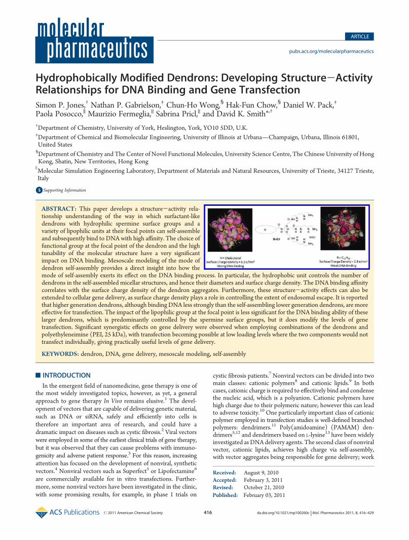
electrophoresis (Figure 7). This confirmed the observation thatthere was little difference between Chol-G2 and C12-G2. In thecase of Chol-G2, DNAmobility was retarded at a loading of 0.119nmoles (N:P ratio of 1.1). For C12-G2, DNA retardation wasobserved at a very similar loading level (0.123 nmoles, N:P ratioof 1.1).Interestingly, the CE50 values (from EthBr exclusion) and N:P
ratios (from gel electrophoresis) for Chol-G2 binding DNA aresignificantly higher than those observed with Chol-G1. This isperhaps surprising given that Chol-G2 has many more spermine
groups, and may have been be expected to be a more effectivebinder. Indeed, in our previous studies, Z-G2 was, as expected,more effective than Z-G1 in terms of its CE50 value.18 Theseobservations clearly reinforce the concept discussed above thatthe cholesterol unit can, in the case of Chol-G1, play a proactiverole in encouraging and promoting self-assembly of the dendron,generating a high density of spermine groups, with this processhaving amajor positive effect on the ability of Chol-G1 to interactwith DNA. Clearly this does not occur for the G2 dendron, andthe spermine surface groups alone control binding.In Vitro Transfectionwith SecondGeneration Vectors.We
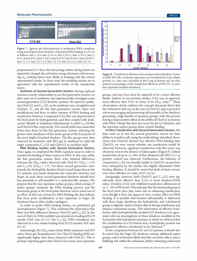
then went on to test the second generation vectors for theirability to transfect cells using the methodology described above.Given that Chol-G2 showed less effective DNA binding thanChol-G1, we were unsure whether any transfection would beobserved. However, significant transfection with this vector wasobserved, even in the absence of chloroquine (Figure 8). Indeedtransfection of up to ca. 10% of that obtained with PEI (1:1) aspositive control was observed. Furthermore, the behavior ofcompound C12-G2 was broadly similar to Chol-G1, as may havebeen anticipated by the similar, but slightly diminished DNAbinding affinities. It should be noted that both of these vectorswere most effective at a ratio of 6:1 (w/w).Intriguingly, however, both Chol-G2 and C12-G2 were sig-
nificantly more effective than Z-G2 at lower dendron/DNAratios. Dendron Z-G2 only exhibited transfection efficiencies ofca. 1-2% of PEI levels. This indicates that the functional group atthe focal point does play some role in enhancing transfection,even though it does not appear to have a major effect on DNAbinding. It is possible that because self-assembly is disfavoredwith these larger dendrons, the hydrophobic unit (cholesterolgroup or aliphatic chain) is better able to disrupt membranes andenhance endosomal escape. This observation of effective trans-fection with hydrophobically modified G2 dendrons is in agree-ment with our investigations of these dendrons modified at thefocal point with hydrophobin proteins, in which we observed thatthe combination of a G2 branch and a hydrophobic protein wasrequired for effective transfection to be observed.43
In the comparison between G1 and G2 systems, it should alsobe noted that the larger G2 dendrons contain additional aminegroups, and it is therefore likely that they are better able to play abuffering role within the endosome, further enhancing endosomal
Figure 7. Agarose gel electrophoresis of polyamine/DNA complexesusing second generation dendrons. Polyamine:DNA loadings (w:w) areas follows: lane 1, 0:1; lane 2, 0.1:1; lane 3, 0.2:1; lane 4, 0.3:1; lane 5,0.4:1; lane 6, 0.5:1; lane 7, 0.6:1; lane 8, 0.7:1; lane 9, 0.8:1; lane 10, 0.9:1;lane 11, 1:1, lane 12, 1.5:1; lane 13, 2:1; lane 14, 2.5:1; lane 15, 3:1.
Figure 8. Transfection efficiency of second generation dendritic vectorsin HEK 293 cells. Luciferase expression was normalized by total cellularprotein; i.e., data were calculated in RLU/mg of protein and are thenquoted as percentages of the transfection efficiency of PEI (N = 6, errorbars represent standard deviation).

426 dx.doi.org/10.1021/mp100260c |Mol. Pharmaceutics 2011, 8, 416–429
Molecular Pharmaceutics ARTICLE
escape. This may help explain why some transfection is observed,even in the absence of chloroquine.In order to probe transfection further, we once again applied
our most effective vector (Chol-G2) as a mixture with PEI inorder to determine whether the mixed vector exhibited synergis-tic transfection effects (Figure 9). Remarkably, even though aratio of 0.25:1 PEI:DNA (w/w) exhibits no transfection, theaddition of Chol-G2 to this mixture (4:0.25:1, Chol-G2:PEI:DNA) gave rise to very good levels of transfection (ca. 50% ofthat observed with positive control). Once again, this demon-strates the potential of PEI to enhance the transfection potentialof these dendrons (the level of transfection was enhanced 5-fold)and also allow them to be employed at lower loadings (theoptimal loading of Chol-G2 decreased from 6:1 to 4:1). Further-more, the PEI, which presumably acts by assisting buffering in theendosome, can be employed at lower concentrations, where it isunable to transfect at its own right, allowing it to be employed in atransfection vector at much lower loadings than previouslyrealized.
’CONCLUSIONS
In this paper, we have reported a structure-activity relation-ship for a set of dendritic vectors with DNA binding sperminesurface groups. In particular, we report that the hydrophobic/hydrophilic balance plays a subtle role in controlling DNAbinding and transfection ability. The easy synthetic tunabilityof these dendron structures can therefore be readily exploited tomaximize their biological activity. For the first generation system,the choice of the group at the focal point of the dendron had asignificant impact on the DNA binding. The binding affinity forDNA was enhanced by the presence of significant hydrophobicgroup. Mesoscale modeling demonstrated that these systemswere better able to pack into tighter micellar aggregates, whichhave higher surface charge density and as such, show enhancedelectrostatic interactions with DNA. In this way, self-assembly ofthe dendrons enhances DNA binding. Furthermore, the differ-ences in DNA binding affinity appeared to correlate with thetransfection ability of the dendrons, although chloroquine had tobe added to assist endosomal escape. In contrast, the secondgeneration dendrons bound DNA in a manner which wasrelatively independent of the hydrophobic group, presumablybecause the large hydrophilic spermine surface area dominatesthe binding. These dendrons were able to transfect in the absenceof chloroquine, presumably due to the greater number of amines
providing some buffering effect. The cholesterol functionalizeddendron was a significantlymore effective transfection agent thanthe dendron with a Z-protecting group, possibly due to the abilityof the cholesterol group to disrupt endosomal membranes.Finally, we observed significant synergistic effects when employ-ing combinations of our dendrons (Chol-G1 or Chol-G2) andPEI. Indeed, transfection was possible using mixtures at loadinglevels which were so low that the individual components did notexhibit significant transfection ability on their own. Using thisapproach, we were able to achieve gene transfection at practicallyuseful levels.
’ASSOCIATED CONTENT
bS Supporting Information. Details of synthetic methodsand characterization data for all dendrons, details of the biolo-gical assays performed in the work, and methods used formodeling. This material is available free of charge via the Internetat http://pubs.acs.org.
’AUTHOR INFORMATION
Corresponding Author*E-mail: [email protected].
’ACKNOWLEDGMENT
We gratefully acknowledge EPSRC (EP/C534395/1) forfunding this project. H.-F.C., a recipient of the Croucher SeniorResearch Fellowship, Hong Kong, wishes to thank CUHK for theFocused Investment into Areas of Strength: Specialized Areas(Scheme B). We acknowledge support for networking providedby COST network TD0802 (Dendrimers in Biomedicine).
’REFERENCES
(1) Edelstein, M. L.; Abedi, M. R.; Wixon, J. Gene therapy clinicaltrials worldwide to 2007—an update. J. Gene Med. 2007, 9, 833–842.
(2) (a) Griesenbach, U.; Geddes, D. M.; Alton, E. W. F. W. Genetherapy progress and prospects: cystic fibrosis. Gene Ther. 2006,13, 1061–1067. (b) Geiger, J.; Aneja, M. K.; Rudolph, C. Vectors forpulmonary gene therapy. Int. J. Pharm. 2010, 390, 84–88.
(3) Tomanin, R.; Scarpa, M.Why do we need new gene therapy viralvectors? Characteristics, limitations and future perspectives of viralvector transduction. Curr. Gene Ther. 2004, 4, 357–372.
(4) (a) Boulaiz, H.; Marchal, J. A.; Prados, J.; Melguizo, C.; Aranega,A. Non-viral and viral vectors for gene therapy. Cell Mol. Biol. 2005,51, 3–22. (b) Kodama, K.; Katayama, Y.; Shoji, Y.; Nakashima, H. Thefeatures and shortcomings for gene delivery of current non-viral carriers.Curr. Med. Chem. 2006, 13, 2155–2161. (c) Mintzer, M. A.; Simanek,E. E. Nonviral Vectors for Gene Delivery. Chem. Rev. 2009,109, 259–302.
(5) Tang, M. X.; Redemann, C. T.; Szoka, F. C., Jr. In vitro genedelivery by degraded polyamidoamine dendrimers. Bioconjugate Chem.1996, 7, 703–714.
(6) Dalby, B.; Cates, S.; Harris, A.; Ohki, E. C.; Tilkins, M. L.; Price,P. J.; Ciccarone, V. C. Advanced transfection with Lipofectamine 2000reagent: primary neurons, siRNA, and high-throughput applications.Methods 2004, 33, 95–103.
(7) (a) Rosenecker, J.; Huth, S.; Rudolph, C. Gene therapy for cysticfibrosis lung disease: Current status and future perspectives. Curr. Opin.Mol. Ther. 2006, 8, 439–445. (b) Conese, M.; Giola, S. D.; Castellani, S.Gene therapy for cystic fibrosis. Exp. Opin. Ther. Pat. 2008, 18, 929–943.
(8) (a) Wong, S. Y.; Pelet, J. M.; Putnam, D. Polymer systems forgene delivery-past, present, and future. Prog. Polym. Sci. 2007,
Figure 9. Transfection efficiency of Chol-G2 when mixed with a smallamount of PEI (no chloroquine present). For comparison, transfectionefficiencies with PEI (0.25:1), PEI (1:1) and Chol-G2 in the absence ofPEI, are also presented.

427 dx.doi.org/10.1021/mp100260c |Mol. Pharmaceutics 2011, 8, 416–429
Molecular Pharmaceutics ARTICLE
32, 799–837. (b) Tiera, M. J.; Winnik, F. M.; Fernandes, J. C. Syntheticand natural polycations for gene therapy: State of the art and newperspectives. Curr. Gene Ther. 2006, 6, 59–71. (c) Midoux, P.; Breuzard,G.; Gomez, J. P.; Pichon, C. Polymer-based gene delivery: A currentreview on the uptake and intracellular trafficking of polyplexes. Curr.Gene Ther. 2008, 8, 335–352.(9) (a) Karmali, P. P.; Chaudhuri, A. Cationic Liposomes as non-
viral carriers of gene medicines: Resolved issues, open questions, andfuture promises.Med. Res. Rev. 2007, 27, 696–722. (b) Bhattacharya, S.;Bajaj, A. Advances in gene delivery through molecular design of cationiclipids. Chem. Commun. 2009, 4632–4656.(10) Lv, H.; Zhang, S.; Wang, B.; Cui, S.; Yan, J. Toxicity of cationic
lipids and cationic polymers in gene delivery. J. Controlled Release 2006,114, 100–109.(11) (a) Dufes, C.; Uchegbu, I. F.; Schatzlein, A. G. Dendrimers in
gene delivery. Adv. Drug Delivery Rev. 2005, 57, 2177–2202. (b) Guillot-Nieckowski, M.; Eisler, S.; Diederich, F. Dendritic vectors for genetransfection. New J. Chem. 2007, 31, 1111–1127. (c) Smith, D. K.Dendrimers and the double helix—From DNA binding towards genetherapy. Curr. Top. Med. Chem. 2008, 8, 1187–1203.(12) (a) Haensler, J.; Szoka, F. C. Polyamidoamine cascade poly-
mers mediate efficient transfection of cells in culture. Bioconjugate Chem.1993, 4, 372–379. (b) Kukowska-Latallo, J.-F.; Bielinska, A. U.; Johnson,J.; Spindler, R.; Tomalia, D. A.; Baker, J. R. Efficient transfer of geneticmaterial into mammalian cells using Starburst polyamidoamine dendri-mers. Proc. Natl. Acad. Sci. U.S.A. 1996, 93, 4897–4902. (c) Bielinska, A.;Kukowska-Latallo, J. F.; Johnson, J.; Tomalia, D. A.; Baker, J. R.Regulation of in vitro gene expression using antisense oligonucleotidesor antisense expression plasmids transfected using starburst PAMAMdendrimers.Nucleic Acids Res. 1996, 24, 2176–2182. (d) Turunen,M. P.;Hiltunen,M.O.; Ruponen,M.; Virkamaki, L.; Szoka, F. C.; Urtti, A.; Yla-Herttuala, S. Efficient adventitial gene delivery to rabbit carotid arterywith cationic polymer-plasmid complexes. Gene Ther. 1999, 6, 6–11.(13) (a) Choi, J. S.; Lee, E. J.; Choi, Y. H.; Jeong, Y. J.; Park, J. S.
Poly(ethylene glycol)-block-poly(L-lysine) dendrimer: Novel linearpolymer/dendrimer block copolymer forming a spherical water-solublepolyionic complex with DNA. Bioconjugate Chem. 1999, 10, 62–65. (b)Ohsaki, M.; Okuda, T.; Wada, A.; Hirayama, T.; Niidome, T.; Aoyagi, H.In vitro gene Transfection using dendritic poly(L-lysine). BioconjugateChem. 2002, 13, 510–517.(14) (a) McGregor, C.; Perrin, C.; Monck, M.; Camilleri, P.; Kirby,
A. J. Rational approaches to the design of cationic gemini surfactants forgene delivery. J. Am. Chem. Soc. 2000, 123, 6215–6220. (b) Jennings,K. H.; Marshall, I. C. B.; Wilkinson, M. J.; Kremer, A.; Kirby, A. J.;Camilleri, P. Aggregation properties of a novel class of cationic geminisurfactants correlate with their efficiency as gene transfection agents.Langmuir 2002, 18, 2426–2429.(15) For typical examples see: (a) Smisterova, J.; Wagenaar, A.;
Stuart, M. C. A.; Polushkin, E.; ten Brinke, G.; Hulst, R.; Engberts, J. B. F.N.; Hoekstra, D. Molecular shape of the cationic lipid controls thestructure of cationic lipid/dioleylphosphatidylethanolamine-DNA com-plexes and the efficiency of gene delivery. J. Biol. Chem. 2001,276, 47615–47622. (b) Niculescu-Duvaz, D.; Heyes, J.; Springer, C. J.Structure-activity relationship in cationic lipid mediated gene transfec-tion. Curr. Med. Chem. 2003, 10, 1233–1261. (c) Castro, M.; Griffiths,D.; Patel, A.; Pattrick, N.; Kitson, C.; Ladlow, M. Effect of chain lengthon transfection properties of spermine-based gemini surfactants. Org.Biomol. Chem. 2004, 2, 2814–2820. (d) Wang, J.; Gao, S.; Zhang, P.;Wang, S.; Mao, M.; Leong, K. Polyphosphoramidate gene carriers: effectof charge group on gene transfer efficiency. Gene Ther. 2004,11, 1001–1010. (e) Horobin, R. W.; Weissig, V. A QSAR-modetingperspective on cationic transfection lipids. 1. Predicting efficiency andunderstanding mechanisms. J. Gene Med. 2005, 7, 1023–1034. (f) Liu,Y. M.; Reineke, T. M. Hydroxyl stereochemistry and amine numberwithin poly(glycoamidoamine)s affect intracellular DNA delivery. J. Am.Chem. Soc. 2005, 127, 3004–3015. (g) Karmali, P. P.; Majeti, B. K.;Sreedhar, B.; Chaudhuri, A. In vitro gene transfer efficacies and serumcompatibility profiles of novel mono-, di-, and tri-histidinylated cationic
transfection lipids: A structure-activity investigation. Bioconjugate Chem.2006, 17, 159–171. (h) Isobe, H.; Nakanishi, W.; Tomita, N.; Jinno, S.;Okayama, H.; Nakamura, E. Gene delivery by aminofullerenes: Struc-tural requirements for efficient transfection. Chem. Asian J. 2006,1, 167–175. (i) Rajesh, M.; Sen, J.; Srujan, M.; Mukherjee, K.; Sreedhar,B.; Chaudhuri, A. Dramatic influence of the orientation of linkerbetween hydrophilic and hydrophobic lipid moiety in liposomal genedelivery. J. Am. Chem. Soc. 2007, 129, 11408–11420. (j) Ghonaim,H. M.; Ahmed, O. A. A.; Pourzand, C.; Blagbrough, I. S. Varying thechain length in N-4,N-9-diacyl spermines: Non-Viral lipopolyaminevectors for efficient plasmid DNA formulation. Mol. Pharmaceutics2008, 5, 1111–1121. (k) Van Vliet, L. D.; Chapman, M. R.; Avenier,F.; Kitson, C. Z.; Hollfelder, F. Relating chemical and biological diversityspace: A tunable system for efficient gene transfection. ChemBioChem2008, 9, 1960–1967. (l) Dewa, T.; Asai, T.; Tsunoda, Y.; Kato, K.; Baba,D.; Uchida, M.; Sumino, A.; Niwata, K.; Umemoto, T.; Iida, K.; Oku, N.;Nango, M. Liposomal Polyamine-Dialkyl Phosphate Conjugates asEffective Gene Carriers: Chemical Structure, Morphology, and GeneTransfer Activity. Bioconjugate Chem. 2010, 21, 844–852. (m) Zhi, D. F.;Zhang, S.; Wang, B.; Zhao, Y.; Yang, B.; Yu, S. Transfection Efficiency ofCationic Lipids with Different Hydrophobic Domains in Gene Delivery.Bioconjugate Chem. 2010, 21, 563–577.
(16) (a) Joester, D.; Losson, M.; Pugin, R.; Heinzelmann, H.;Walter, E.; Merkle, H. P.; Diederich, F. Amphiphilic dendrimers: Novelself-assembling vectors for efficient gene delivery. Angew. Chem., Int. Ed.2003, 42, 1486–1490. (b) Guillot, M.; Eisler, S.; Weller, K.; Merkle,H. P.; Gallani, J. L.; Diederich, F. Effects of structural modification ongene transfection and self-assembling properties of amphiphilic dendri-mers. Org. Biomol. Chem. 2006, 4, 766–769. (c) Toth, I.; Sakthivel, T.;Wilderspin, A. F.; Bayele, H.; O’Donnell, M.; Perry, D. J.; Pasi, K. J.; Lee,C. A.; Florence, A. T. Novel cationic lipidic peptide dendrimer vectors -In vitro gene delivery. STP Pharm. Sci. 1999, 9, 93–99. (d) Shah, D. S.;Sakthivel, T.; Toth, I.; Florence, A. T.; Wilderspin, A. F. DNA transfec-tion and transfected cell viability using amphipathic asymmetric den-drimers. Int. J. Pharm. 2000, 208, 41–48. (e) Al-Jamal, K. T.;Ramaswamy, C.; Singh, B.; Florence, A. T. Structures from lysine-baseddendrons and dendrimers: monolayers, dendriplexes, dendrisomes,nanoparticles and micellar aggregates. J. Drug Delivery Sci. Technol.2005, 15, 11–18. (f) Bayele, H. K.; Sakthivel, T.; O’Donell, M.; Pasi,K. J.; Wilderspin, A. F.; Lee, C. A.; Toth, I.; Florence, A. T. Versatilepeptide dendrimers for nucleic acid delivery. J. Pharm. Sci. 2005,94, 446–457. (g) Bayele, H. K.; Ramaswamy, C.; Wilderspin, A. F.; Srai,K. S.; Toth, I.; Florence, A. T. Protein transduction by lipidic peptidedendrimers. J. Pharm. Sci. 2006, 95, 1227–1237. (h) Wood, K. C.; Little,S. R.; Langer, R.; Hammond, P. T. A family of hierarchically self-assembling linear-dendritic hybrid polymers for highly efficient targetedgene delivery. Angew. Chem., Int. Ed. 2005, 44, 6704–6708. (i) Wood,K. C.; Azarin, S. M.; Arap, W.; Pasqualini, R.; Langer, R.; Hammond,P. T. Tumor-targeted gene delivery using molecularly engineered hybridpolymers functionalized with a tumor-homing peptide. BioconjugateChem. 2008, 19, 403–405.(j) Poon, Z.; Lee, J. A.; Huang, S.; Prevost,R. J.; Hammond, P. T. Highly stable ligand clustered ‘patchy’ micellenanocarriers for systemic tumor targeting Nanomedicine 2010,DOI:10.1016/j.nano.2010.07.008.
(17) Guillot-Nieckowski, M.; Joester, D.; Stohr, M.; Losson, M.;Adrian, M.; Wagner, B.; Kansy, M.; Heinzelmann, H.; Pugin, R.;Diederich, F.; Gallani, J. L. Self-assembly, DNA complexation, and pHresponse of amphiphilic dendrimers for gene transfection. Langmuir2007, 23, 737–746.
(18) (a) Kostiainen, M. A.; Hardy, J. G.; Smith, D. K. High-affinitymultivalent DNA binding by using low-molecular-weight dendrons.Angew. Chem., Int. Ed. 2005, 44, 2556–2559. (b) Kostiainen, M. A.;Smith, D. K.; Ikkala, O.Optically triggered release ofDNA frommultivalentdendrons by degrading and charge-switching multivalency. Angew. Chem.,Int. Ed. 2007, 46, 7600–7604. (c) Welsh, D. J.; Jones, S. P.; Smith, D. K.“On-Off” multivalent recognition: Degradable dendrons for temporaryhigh-affinity DNA binding. Angew. Chem., Int. Ed. 2009, 48, 4047–4051.(d) Kostiainen, M. A.; Rosilo, H. Low-Molecular-Weight dendrons for

428 dx.doi.org/10.1021/mp100260c |Mol. Pharmaceutics 2011, 8, 416–429
Molecular Pharmaceutics ARTICLE
DNAbinding and release by reduction-triggered degradation of multivalentinteractions. Chem.—Eur. J. 2009, 15, 5656–5660. (e) Pavan, G. M.;Danani, A.; Pricl, S.; Smith, D. K. Modeling the multivalent recognitionbetween dendritic molecules and DNA: Understanding how ligand “sacri-fice” and screening can enhance binding. J. Am. Chem. Soc. 2009,131, 9686–9694. (f) Pavan, G.M.; Kostiainen, M. A.; Danani, A. Computa-tional approach for understanding the interactions of UV-degradabledendrons with DNA and siRNA. J. Phys. Chem. B 2010, 114, 5686–5693.(g) Jones, S. P.; Pavan, G.M.; Danani, A.; Pricl, S.; Smith, D. K. Quantifyingthe effect of surface ligands on dendron-DNA interactions: Insights intomultivalency through a combined experimental and theoretical approach.Chem.—Eur. J. 2010, 16, 4519–4532.(19) (a) Tabor, C. W.; Tabor, H. Polyamines. Annu. Rev. Biochem.
1984, 740–790. (b) Vijayanathan, V.; Thomas, T.; Shirahata, A.;Thomas, T. J. DNA condensation by polyamines: A laser light scatteringstudy of structural effects. Biochemistry 2001, 40, 13644–13651. (c)Vijayanathan, V.; Lyall, J.; Thomas, T.; Shirahata, A.; Thomas, T. J.Ionic, structural, and temperature effects on DNA nanoparticlesformed by natural and synthetic polyamines. Biomacromolecules2005, 6, 1097–1103.(20) Hardy, J. G.; Kostiainen, M. A.; Smith, D. K.; Gabrielson, N. P.;
Pack, D. W. Dendrons with spermine surface groups as potentialbuilding blocks for nonviral vectors in gene therapy. Bioconjugate Chem.2006, 17, 172–178.(21) Jones, S. P.; Gabrielson, N. P.; Pack, D. W.; Smith, D. K.
Synergistic effects in gene delivery—a structure-activity approach to theoptimization of hybrid dendritic-lipidic transfection agents. Chem.Commun. 2008, 4700–4702.(22) Posocco, P.; Pricl, S.; Jones, S. P.; Barnard, A.; Smith, D. K. Less
is more—multiscale modelling of self-assembling multivalency and itsimpact on DNA binding and gene delivery. Chem. Sci. 2010, DOI:10.1039/c0sc00291g.(23) (a) Gao, X.; Huang, L. Potentiation of cationic liposome-
mediated gene delivery by polycations. Biochemistry 1996,35, 1027–1036. (b) Sorgi, F. L.; Bhattacharya, S.; Huang, L. Protaminesulfate enhances lipid-mediated gene transfer. Gene Ther. 1997,4, 961–968. (c) Kukowska-Latallo, J. F.; Chen, C.; Eichman, J.; Bielinska,A. U.; Baker, J. R. Enhancement of dendrimer-mediated transfection usingsynthetic lung surfactant exosurf neonatal in vitro. Biochem. Biophys. Res.Commun. 1999, 264, 253–261. (d) Lampela, P.; Soininen, P.; Urtti, A.;M€annist€o, P.; Raasmaja, A. Synergism in gene delivery by small PEIs andthree different nonviral vectors. Int. J. Pharm. 2004, 270, 175–184. (e)Kim, T. H.; Kim, S. I.; Akaike, T.; Cho, C. S. Synergistic effect ofpoly(ethylenimine) on the transfection efficiency of galactosylated chit-osan/DNA complexes. J. Controlled Release 2005, 105, 354–366. (f)Pelisek, J.; Gaedtkel, L.; De Rouchey, J.; Walkerl, G. F.; Niko, S.; Wagner,E. Optimized lipopolyplex formulations for gene transfer to human coloncarcinoma cells under in vitro conditions. J. Gene Med. 2006, 8, 186–197.(g) Golda, A.; Pelisek, J.; Klocke, R.; Engelmann, M. G.; Rolland, P. H.;Mekkaoui, C.; Nikol, S. Small poly-L-lysines improve cationic lipid-mediated gene transfer in vascular cells in vitro and In Vivo. J. Vasc. Res.2007, 44, 273–283. (h) Chen, J.-L.; Wang, H.; Gao, J.-Q.; Chen, H.-L.;Liang, W.-Q. Liposomes modified with polycation used for gene delivery:Preparation, characterization and transfection in vitro. Int. J. Pharm. 2007,343, 255–261. (i) Garcia, L.; Bunuales, M.; Duzgunes, N.; de Ilarduya,C. T. Serum-resistant lipopolyplexes for gene delivery to liver tumourcells. Eur. J. Pharm. Biopharm. 2007, 67, 58–66. (j) Brito, L.; Little, S.;Langer, R.; Amiji, M. Poly(beta-amino ester) and cationic phospholipid-based lipopolyplexes for gene delivery and transfection in human aorticendothelial and smooth muscle cells. Biomacromolecules 2008,9, 1179–1187. (k) Hardy, J. G.; Love, C. S.; Gabrielson, N. P.; Pack,D. W.; Smith, D. K. Synergistic effects on gene delivery- co-formulationof small disulfide-linked dendritic polycations with Lipofectamine2000TM. Org. Biomol. Chem. 2009, 7, 789–793. (l) Santos, J. L.; Oliveira,H.; Pandita, D.; Rodrigues, J.; Pego, A. P.; Granja, P. L.; Tomas, H.Functionalization of poly(amidoamine) dendrimers with hydrophobicchains for improved gene delivery inmesenchymal stem cells. J. ControlledRelease 2010, 144, 55–64.
(24) Chow, H.-F.; Ng, K.-F.; Wang, Z.-Y.; Wong, C.-H.; Luk, T.; Lo,C.-M.; Yang, Y.-Y. Synthesis of new amphiphilic dendrons bearingaliphatic hydrocarbon surface sectors and a monocarboxylic or dicar-boxylic acid focal point. Org. Lett. 2006, 8, 471–474.
(25) Habicher, T.; Diederich, F.; Gramlich, V. Catalytic dendro-phanes as enzyme mimics: Synthesis, binding properties, micropolarityeffect, and catalytic activity of dendritic thiazolio-cyclophanes. Helv.Chim. Acta 1999, 82, 1066–1095.
(26) (a) Cain, B. F.; Baguley, B. C.; Denny, W. A. Potential anti-tumor agents 28. Deoxyribonucleic-acid polyintercalating agents. J. Med.Chem. 1978, 21, 658–668. (b) Gershon, H.; Ghirlando, R.; Guttman,S. B.; Minsky, A. Mode of formation and structural features of DNAcationic liposome complexes used for transfection. Biochemistry 1993,32, 7143–7151.
(27) Zadmard, R.; Schrader, T. DNA recognition with large calixar-ene dimers. Angew. Chem., Int. Ed. 2006, 45, 2703–2706.
(28) (a) Hoogerbrugge, P. J.; Koelman, J. Simulation microscopichydrodynamic phenomena with dissipative particle dynamics. Europhys.Lett. 1992, 19, 155–160. (b) Espa~nol, P.; Warren, P. Statistical-Mechanics of dissipative particle dynamics. Europhys. Lett. 1995,30, 191–196. (c) Groot, R. D.; Warren, P. B. Dissipative particledynamics: Bridging the gap between atomistic and mesoscopic simula-tion. J. Chem. Phys. 1997, 107, 4423–4435. (d) Groot, R. D.; Rabone,K. L. Mesoscopic simulation of cell membrane damage, morphologychange and rupture by nonionic surfactants. Biophys. J. 2001,81, 725–736.
(29) (a) Israelachvili, J. N.; Mitchell, D. J.; Ninham, B. W. Theory ofself-assembly of hydrocarbon amphiphiles into micelles and bilayers. J.Chem. Soc., Faraday Trans. 2 1976, 72, 1525–1568. (b) Israelachvili,J. N.; Mitchell, D. J.; Ninham, B. W. Theory of self-assembly of lipidbilayers and vesicles. Biochim. Biophys. Acta 1977, 470, 185–201.
(30) (a) Al-Jamal, K. T.; Ramaswamy, C.; Florence, A. T. Supramo-lecular structures from dendrons and dendrimers. Adv. Drug DeliveryRev. 2005, 57, 2238–2270. (b) Nguyen, P. M.; Hammond, P. T.Amphiphilic linear-dendritic triblock copolymers composed of poly-(amidoamine) and poly(propylene oxide) and their micellar-phase andencapsulation properties. Langmuir 2006, 22, 7825–7832. (c) J€ager,C. M.; Hirsch, A.; Schade, B.; B€ottcher, C.; Clark, T. Counterionscontrol the self-assembly of structurally persistent micelles: Theoreticalprediction and experimental observation of stabilization by sodium ions.Chem.—Eur. J. 2009, 15, 8586–8592. (d) Zidovska, A.; Evans, H. M.;Ewert, K. K.; Quispe, J.; Carragher, B.; Potter, C. S.; Safinya, C. R. Liquidcrystalline phases of dendritic lipid-DNA self-assemblies: Lamellar,hexagonal, and DNA bundles. J. Phys. Chem. B 2009, 113, 3694–3703.(e) Chooi, K. W.; Gray, A. I.; Tetley, L.; Fan, Y.; Uchegbu, J. F. Themolecular shape of poly(propylenimine) dendrimer amphiphiles has aprofound effect on their self assembly. Langmuir 2010, 26, 2301–2316.
(31) Tanford, C. The hydrophobic effect: formation of micelles andbiological membranes, 2nd ed.; Krieger Publishing Co.: Malabar, FL,1991; pp 60-78.
(32) (a) Patrickios, C. S. Micellization model for multivalent ionicsurfactants. J. Phys. Chem. 1995, 99, 17437–17441. (b) Nagarajan, R.Molecular packing parameter and surfactant self-assembly: The ne-glected role of the surfactant tail. Langmuir 2002, 18, 31–38.
(33) Tomas, S.; Milanesi, L. Hydrophobically self-assembled nano-particles as molecular receptors in water. J. Am. Chem. Soc. 2009,131, 6618–6623.
(34) Abdelhady, H. G.; Allen, S.; Davies, M. C.; Roberts, C. J.;Tendler, S. J. B.; Williams, P. M. Direct real-time molecular scalevisualisation of the degradation of condensed DNA complexes exposedto DNase I. Nucleic Acid Res. 2003, 31, 4001–4005.
(35) (a) Dorigo, B.; Schalch, T.; Kulangara, A.; Duda, S.; Schroeder,R.; Richmond, T. J. Nucleosome arrays reveal the two-start organizationof the chromatin fiber. Science 2004, 306, 1571–1573. (b) Olins, A. L.;Olins, D. E. Spheroid chromatin units. Science 1974, 183, 330–332.
(36) Manning, G. S. Thermodynamic stability theory for DNAdoughnut shapes induced by charge neutralization. Biopolymers 1980,19, 37–59.

429 dx.doi.org/10.1021/mp100260c |Mol. Pharmaceutics 2011, 8, 416–429
Molecular Pharmaceutics ARTICLE
(37) Marenduzzo, D.; Finan, K.; Cook, P. R. The depletion attrac-tion: an underappreciated force driving cellular organization. J. Cell Biol.2006, 175, 681–686.(38) Manning, G. S. Limiting laws and counterion condensation in
polyelectrolyte solutions 1. Colligative properties. J. Chem. Phys. 1969,51, 924–933.(39) (a) Harries, D.; May, S.; Gelbart, W. M.; Ben-Shaul, A.
Structure, stability, and thermodynamics of lamellar DNA-lipid com-plexes. Biophys. J. 1998, 75, 159–173. (b) Bruinsma, R. Electrostatics ofDNA cationic lipid complexes: isoelectric instability. Eur. Phys. J. B 1998,4, 75–88. (c) May, S.; Ben-Shaul, A. Modeling of cationic lipid-DNAcomplexes. Curr. Med. Chem. 2004, 11, 151–167.(40) (a) Thomas, M.; Klibanov, A. M. Enhancing polyethyleni-
mine’s delivery of plasmid DNA into mammalian cells. Proc. Natl. Acad.Sci. U.S.A. 2002, 99, 14640–14645. (b) Forrest, M. L.; Koerber, J. T.;Pack, D. W. A degradable polyethylenimine derivative with low toxicityfor highly efficient gene delivery. Bioconjugate Chem. 2003, 14, 934–90.(c) Gabrielson, N. P.; Pack, D. W. Acetylation of polyethylenimineenhances gene delivery via weakened polymer/DNA interactions.Biomacromolecules 2006, 7, 2427–2435. (d) Gabrielson, N. P.; Pack,D. W. Efficient polyethylenimine-mediated gene delivery proceeds via acaveolar pathway in HeLa cells. J. Controlled Release 2009, 136, 54–61.(41) (a) Guy, J.; Drabek, D.; Antoniou, M. Delivery of DNA into
mammalian cells by receptor-mediated endocytosis and gene therapy.Mol. Biotechnol. 1995, 3, 237–248. (b) Pless, D. D.; Wellner, R. B. Invitro fusion of endocytic vesicles: Effects of reagents that alter endoso-mal pH. J. Cellular Biochem. 1996, 62, 27–39.(42) Sonawane, N. D.; Szoka, F. C.; Verkman, A. S. Chloride
accumulation and swelling in endosomes enhances DNA transfer bypolyamine-DNA polyplexes. J. Biol. Chem. 2003, 278, 44826–44831.(43) (a) Kostiainen,M. A.; Szilvay, G. R.; Smith, D. K.; Linder,M. B.;
Ikkala, O. Multivalent dendrons for high-affinity adhesion of proteins toDNA. Angew. Chem., Int. Ed. 2006, 45, 3538–3542. (b) Kostiainen,M. A.; Szilvay, G. R.; Lehtinen, J.; Smith, D. K.; Linder, M. B.; Urtti, A.;Ikkala, O. Precisely defined protein-polymer conjugates: construction ofsynthetic DNA binding domains on proteins by using multivalentdendrons. ACS Nano 2007, 1, 103–113.