the glymphatic system: a beginner’s...
TRANSCRIPT

ORIGINAL PAPER
The Glymphatic System: A Beginner’s Guide
Nadia Aalling Jessen1• Anne Sofie Finmann Munk1
• Iben Lundgaard1•
Maiken Nedergaard1
Received: 26 January 2015 / Revised: 6 April 2015 / Accepted: 10 April 2015
� Springer Science+Business Media New York 2015
Abstract The glymphatic system is a recently discovered
macroscopic waste clearance system that utilizes a unique
system of perivascular tunnels, formed by astroglial cells,
to promote efficient elimination of soluble proteins and
metabolites from the central nervous system. Besides waste
elimination, the glymphatic system also facilitates brain-
wide distribution of several compounds, including glucose,
lipids, amino acids, growth factors, and neuromodulators.
Intriguingly, the glymphatic system function mainly during
sleep and is largely disengaged during wakefulness. The
biological need for sleep across all species may therefore
reflect that the brain must enter a state of activity that
enables elimination of potentially neurotoxic waste prod-
ucts, including b-amyloid. Since the concept of the glym-
phatic system is relatively new, we will here review its
basic structural elements, organization, regulation, and
functions. We will also discuss recent studies indicating
that glymphatic function is suppressed in various diseases
and that failure of glymphatic function in turn might con-
tribute to pathology in neurodegenerative disorders, trau-
matic brain injury and stroke.
Keywords The glymphatic system � Astrocytes �Perivascular spaces � Virchow–Robin spaces �Cerebrospinal fluid secretion � Sleep � Aging �Neurodegenerative diseases � Traumatic brain injury
Introduction
Clearance of excess fluid and interstitial solutes is critical for
tissue homeostasis. In the peripheral tissues soluble material,
proteins and fluid from the interstitial space are returned to the
general circulation by the lymphatic system [1]. The lymphatic
network extends throughout all parts of the peripheral tissues
and the density of lymph vessels correlates with the rate of
tissue metabolism. Although the brain and spinal cord are
characterized by a disproportionally high metabolic rate [2],
and synaptic transmission is exquisitely sensitive to changes in
their environment, the central nervous system (CNS) com-
pletely lacks conventional lymphatic vessels. A few older re-
ports have noted lymphatic vessel-like structures in dura, but
these channels are not lined by endothelial cells [3]. This re-
view addresses recent findings that shed light on the para-
dox that CNS lacks a lymphatic system and discusses these
findings within the broader context of what is known about
waste elimination from theCNS. Finally, we discuss our recent
findings indicating that the recently described glymphatic
system also might also serve to distribute non-waste com-
pounds such as lipids and glucose within the brain.
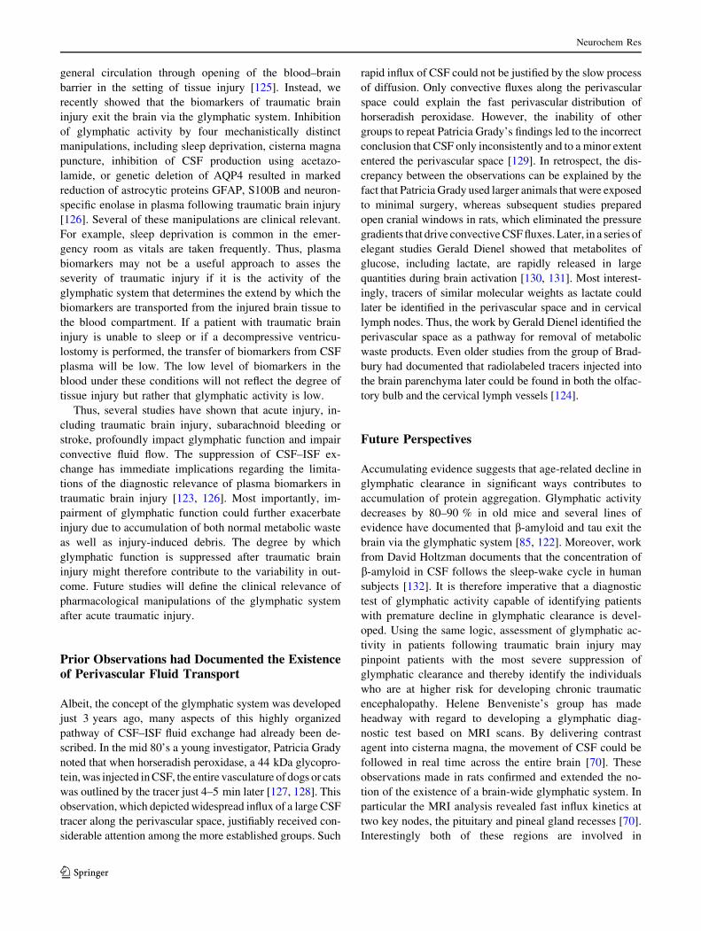
CSF Production
The brain consists of four fluid compartments: cerebrospinal
fluid (CSF), interstitial fluid, intracellular fluid, and the blood
vasculature (Fig. 1). The blood is kept separate from the
Nadia Aalling Jessen, Anne Sofie Finmann Munk and Iben Lundgaard
have contributed equally to the work.
Special Issue: In honor of Dr. Gerald Dienel.
& Nadia Aalling Jessen
1 School of Medicine and Dentistry, University of Rochester
Medical Center, 601 Elmwood Ave, Box 645, Rochester,
NY 14642, USA
123
Neurochem Res
DOI 10.1007/s11064-015-1581-6

brain parenchyma and CSF by the blood–brain and blood–
CSF barriers, respectively. These barriers are essential in
maintaining the extracellular environment of the brain be-
cause they regulate the ionic and biochemical composition of
the different fluid compartments [4]. The blood–brain barrier
is comprised by blood vessel endothelial cells coupled by
tight junctions, whereas the blood–CSF barrier is formed
primarily between the choroid plexus epithelial cells [5].
Because the choroid plexus capillaries, unlike other capil-
laries in the brain, are devoid of tight junctions, they are
permeable to macromolecules [4]. Choroid plexus epithelial
cells, in turn, are connected by tight junctions. At the choroid
plexus, the trans-epithelial transport of macromolecules fa-
cilitated byepithelial transporters therefore determines
which macromolecules that enter CSF from the blood
(Figs. 1, 2).
In mammals, CSF comprises 10 % of the total fluid vol-
ume within the cranial cavity [6]. The CSF flows through the
four ventricles linked by channels or foramina into the sub-
arachnoid space of the cortex and spinal cord (Fig. 1). From
the cortical subarachnoid space it penetrates the brain
parenchyma perivascularly and bathes the brain before it
exits the CNS and drains into the lymphatic system.
CSF is thought to be produced primarily by the choroid
plexuses, which are expansions of the ependymal epithe-
lium, lining the lateral, third, and fourth ventricles [7]. The
choroid plexuses are highly folded and vascularized
structures consisting of a single layered cuboidal or low
cylindrical epithelium residing on a basement membrane.
The luminal surface area of the choroid plexus epithelial
cells is densely covered by microvilli and possess either
one primary cilia or small tufts of motile cilia [5, 8].
CSF production at the choroid plexus is mediated by
exchange and transport of ions (especially Cl-, Na? and
HCO3-) across the epithelial cells, which generates an
osmotic gradient that drives the movement of water from
the blood to the ventricle lumen [4, 5, 9]. The importance
of ionic transporters in CSF production was originally
established using pharmacological tools, which in more
recent years have been supported by genetic studies. The
Choriod plexus
CSF
CapillaryInterstitial fluid
CSF (10%)Blood (10%)
Capillary
Astrocyteendfeet
Blood-brain barrierEndothelial cell w. tight junctions
Brain parenchyma:Interstitial uid (12-20%)Intracellular uid (60-68%)
Lateral ventricle
Third ventricle
Fourth ventricle
Blood-CSF barrierChoroid plexus epithelial cellw. tight junctions
Fenestrated endothelial cell
Basal lamina
Pericyte
AQP4
Fig. 1 Schematic representation of the brain’s fluid compartments
and barriers. The fluid compartments in the brain consist of
intracellular fluid (ICF) (60–68 %), interstitial fluid (ISF) (also
known as extracellular fluid) (12–20 %), blood (10 %) and the
cerebrospinal fluid (CSF) (10 %) [6, 11]. Blood is separated from the
CSF and interstitial fluid by the blood brain barrier (BBB) and blood-
CSF barrier, respectively. Tight junctions between the blood
endothelial cells constitute the BBB, restricting macromolecules to
move freely from the blood into the brain parenchyma. Fluid and
solutes diffusses into the brain parenchyma from the perivascular
space located between endothelial cells and astrocytic endfeet that
expresses the water channel aquaporin-4 (AQP4). The blood-CSF
barrier is formed by tight junctions between the choroid plexus
epithelial cells. Macromolecules from the blood can move freely
between the fenestrated endothelial cells to the interstitial fluid but is
restricted by tight junctions in the choroid plexus epithelial cells,
which therefore are believed to be the main players in determining
CSF composition
Neurochem Res
123
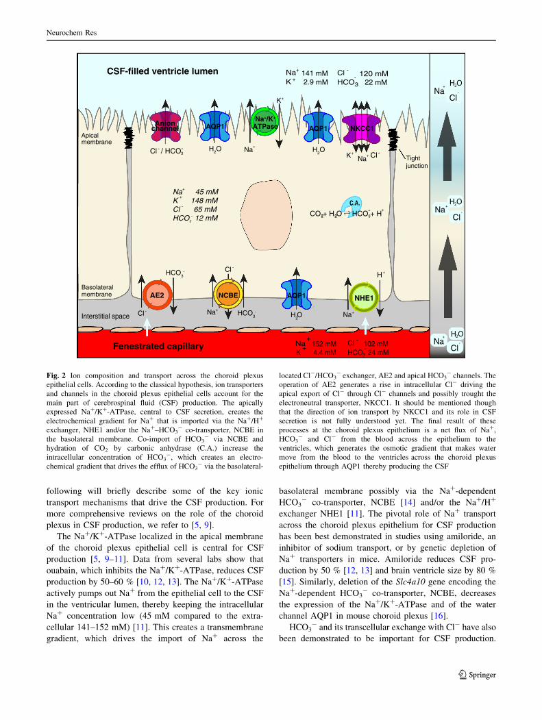
following will briefly describe some of the key ionic
transport mechanisms that drive the CSF production. For
more comprehensive reviews on the role of the choroid
plexus in CSF production, we refer to [5, 9].
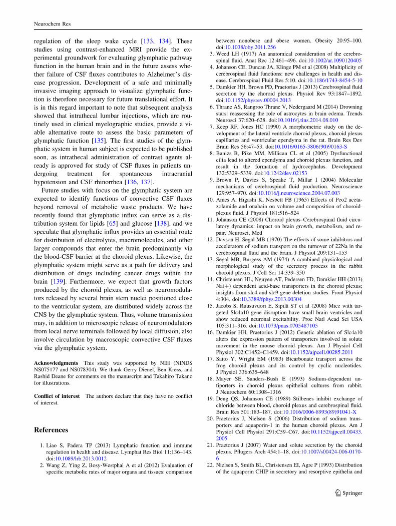
The Na?/K?-ATPase localized in the apical membrane
of the choroid plexus epithelial cell is central for CSF
production [5, 9–11]. Data from several labs show that
ouabain, which inhibits the Na?/K?-ATPase, reduces CSF
production by 50–60 % [10, 12, 13]. The Na?/K?-ATPase
actively pumps out Na? from the epithelial cell to the CSF
in the ventricular lumen, thereby keeping the intracellular
Na? concentration low (45 mM compared to the extra-
cellular 141–152 mM) [11]. This creates a transmembrane
gradient, which drives the import of Na? across the
basolateral membrane possibly via the Na?-dependent
HCO3- co-transporter, NCBE [14] and/or the Na?/H?
exchanger NHE1 [11]. The pivotal role of Na? transport
across the choroid plexus epithelium for CSF production
has been best demonstrated in studies using amiloride, an
inhibitor of sodium transport, or by genetic depletion of
Na? transporters in mice. Amiloride reduces CSF pro-
duction by 50 % [12, 13] and brain ventricle size by 80 %
[15]. Similarly, deletion of the Slc4a10 gene encoding the
Na?-dependent HCO3- co-transporter, NCBE, decreases
the expression of the Na?/K?-ATPase and of the water
channel AQP1 in mouse choroid plexus [16].
HCO3- and its transcellular exchange with Cl- have also
been demonstrated to be important for CSF production.
Fenestrated capillary
H2O
H2O
AQP1
AQP1
Na+/K+
ATPase
Na+
H2O
AQP1
AE2 NCBE
Na+HCO3
-
Cl -
CSF-filled ventricle lumen
Na+ 45 mMK
+ 148 mM
Cl - 65 mMHCO3
- 12 mM
Na+ 141 mMK +
+ 152 mM
K+
4.4 mMCl - 102 mMHCO3
- 24 mM
Cl - 120 mM
HCO3-
Basolateral membrane
Apicalmembrane
K+
Tight junction
Cl / HCO -
channelAnion
Cl -
HCO3-
NHE1
Na+
H+
Interstitial space
C.A.
CO + H O HCO + H2 3-
2+
3-
K+
Na+ Cl -
NKCC1
Na+
Cl-
Na+
Cl-
Na+
Cl-
H O2
H O2
H O2
2.9 mM 22 mM
Na
Fig. 2 Ion composition and transport across the choroid plexus
epithelial cells. According to the classical hypothesis, ion transporters
and channels in the choroid plexus epithelial cells account for the
main part of cerebrospinal fluid (CSF) production. The apically
expressed Na?/K?-ATPase, central to CSF secretion, creates the
electrochemical gradient for Na? that is imported via the Na?/H?
exchanger, NHE1 and/or the Na?–HCO3- co-transporter, NCBE in
the basolateral membrane. Co-import of HCO3- via NCBE and
hydration of CO2 by carbonic anhydrase (C.A.) increase the
intracellular concentration of HCO3-, which creates an electro-
chemical gradient that drives the efflux of HCO3- via the basolateral-
located Cl-/HCO3- exchanger, AE2 and apical HCO3
- channels. The
operation of AE2 generates a rise in intracellular Cl- driving the
apical export of Cl- through Cl- channels and possibly trought the
electroneutral transporter, NKCC1. It should be mentioned though
that the direction of ion transport by NKCC1 and its role in CSF
secretion is not fully understood yet. The final result of these
processes at the choroid plexus epithelium is a net flux of Na?,
HCO3- and Cl- from the blood across the epithelium to the
ventricles, which generates the osmotic gradient that makes water
move from the blood to the ventricles across the choroid plexus
epithelium through AQP1 thereby producing the CSF
Neurochem Res
123
Application of acetazolamide or DIDS, inhibitors of carbonic
anhydrases and anion exchange, respectively, reduces CSF
formation by 30–50 % [10, 17–19]. The mechanisms by
which HCO3- and Cl- and their exchange contribute to
regulation of CSF production is still unclear [9]. It is
speculated that intracellular accumulation of HCO3- (as a
consequence of HCO3- co-import with Na? via NCBE, and
intracellular HCO3- formation by carbonic anhydrase-cat-
alyzed hydration of CO2) drives the outward transport of
HCO3- down its electrochemical gradient via HCO3
- chan-
nels and the HCO3-/Cl- exchanger, AE2 in the basolateral
membrane. The exchange of HCO3- with Cl- then causes
accumulation of intracellular Cl- [9, 11], and generates an
electrochemical gradient for Cl-. As a result, Cl- leaves the
cell via apically-located Cl- channels and possibly via
transporters such as the electroneutral NKCC1 (that co-
transports Na? and K? to the ventricles) [9, 20, 21] (Fig. 2).
Overall, the abovementioned processes generate a net
movement of Na?, Cl- and HCO3- from the blood across the
choroid plexus epithelium to the ventricles. This outward
movement ofNa?, Cl- andHCO3- is believed to generate the
osmotic gradient that driveswater in the same direction across
the apical membrane [5, 9, 11]. Water fluxes across the
choroid plexus epithelium take place mainly through the
highly water permeable channel, AQP1, located primarily in
the apicalmembrane and to a smaller degree in the basolateral
membrane of the choroid plexus epithelial cells [20–24]. It is
debated whether AQP1 is the sole route for water transport
across the choroid plexus, however, AQP1 is critical for CSF
production since knockout of AQP1 in mice reduces the CSF
production rate by 35 % and choroid plexus water perme-
ability by 80 % compared to wildtype littermates [25, 26].
Overall, the net result of ion and water movement across the
choroid plexus epithelium is production of CSF that, com-
pared to the blood, is lower in protein and K? [27], and higher
in Na?, Cl- and Mg2? and has a 99 % water content com-
pared to a water content of 92 % in plasma [4, 6].
Despite decades of research, surprisingly little is known
about the physiological processes regulating CSF produc-
tion. It is expected that CSF production is regulated by
intracranial pressure, but existing reports are contradictory
and suggest that intracranial pressure must be increased
significantly or chronically to suppress CSF production [5,
28]. Additionally, CSF production might also be regulated
by the autonomic nervous system, but again the literature is
complex possibly reflecting the technical limitations asso-
ciated with quantifying CSF production [29].
The Choroid Plexus as the Sole Source of CSF is
Debated
CSF is continuously produced. In humans and mice CSF is
renewed approximately four and 12 times each 24 h,
respectively, and the total CSF volume of 150–160 mL in
human and 0.04 mL in mice is kept constant by removal of
CSF [4, 26, 30]. CSF is drained into the peripheral lym-
phatic system by efflux via the olfactory bulb and along
cranial and spinal nerves [21, 31, 32]. Recently, the im-
portance of the arachnoid granulations in CSF removal has
been questioned [33]. Hence, efflux along cranial and
spinal nerves and the olfactory route might represent the
most important efflux pathways for CSF [31, 34].
According to the classical model, the choroid plexuses
alone are responsible for the vast majority (80–90 %) of
CSF formation [35–37]. Evidence for the significant in-
volvement of the rodent choroid plexus in transport of
solutes was underscored in a proteomic study reporting that
6.7 % of the total number of proteins in the choroid plexus
is involved in transmembrane ion transport. This is a larger
proportion than in the kidney, where the proportion of
proteins estimated to be involved in ion transmembrane
transport activity was 4.8 % [38]. However, discrepancies
between experimental results from fundamental studies of
CSF formation and the classical hypothesis, have provided
the basis for a new model of CSF hydrodynamics [37, 39].
Basically, it has been proposed that CSF formation occurs
by filtration and flux of fluid through the capillary walls,
and that the respective volumes of CSF and interstitial fluid
mainly depend on hydrostatic and osmotic forces between
the CSF and brain parenchyma created by gradients of
proteins and inorganic ions across the capillary membrane
[37, 40]. Accordingly, under physiological conditions,
water is filtered from capillaries with high capillary pres-
sure, to the interstitial fluid and CSF. Since the perme-
ability of plasma electrolytes (Na? and Cl- ) is low, the
electrolytes are retained. This generates an osmotic counter
pressure that opposes the water filtration so when plasma
reaches the low hydrostatic pressure capillaries and
venules, water is reabsorbed into vessels from the inter-
stitial fluid and CSF [37]. Hence, according to this newer
hypothesis, CSF and interstitial fluid are continuously in-
terchanging and the volume occupied by each compartment
depends on hydrostatic and osmotic forces.
Recently, Buishas, Gould and Linninger introduced a
third hypothesis namely a computational model that is
based on existing empirical data. Their model attempts to
combine the two above-mentioned hypotheses and takes
into account the evidence of the glymphatic pathway [41]
explained below. The mathematical model is proposed to
‘‘predict the effects the osmolarity of ECS (extracellular
space), blood, and CSF on water flux in the brain, estab-
lishing a link between osmotic imbalances and pathological
conditions such as hydrocephalus and edema’’ [41]. This
novel model needs validation, but could turn out to provide
unique insight into CSF dynamics during normal physiol-
ogy and pathology.
Neurochem Res
123
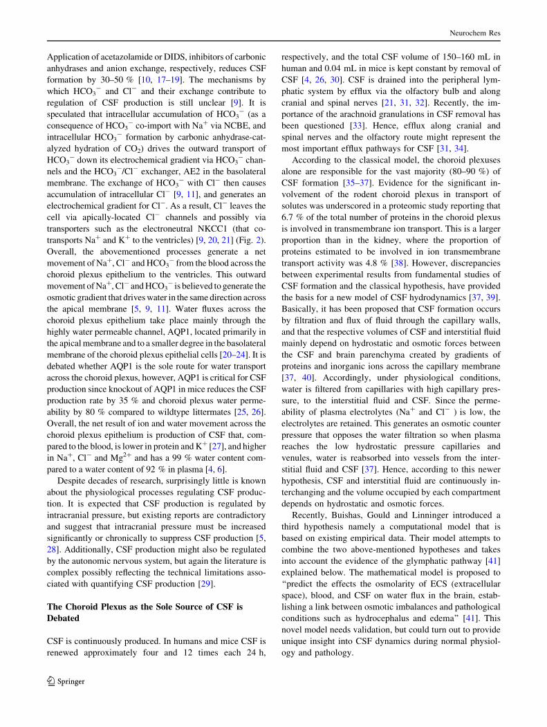
Brain Vasculature and the Perivascular Space
The brain’s vasculature has several unique features that
distinguishes it from the vasculature in the rest of the body
[42]. The arterial cerebral circulation consists of an anterior
cerebral circulation and posterior cerebral circulation sup-
plied by the internal carotid arteries and the vertebral ar-
teries, respectively. The anterior circulation, which includes
themiddle and anterior cerebral arteries, communicates with
the posterior circulation, the basilar artery and posterior
cerebral arteries, via anterior and posterior communicating
arteries at the Circle ofWillis [43]. From the Circle ofWillis,
the anterior circulation perfuse the evolutionary younger
parts of the brain including the neocortex of the cerebral
hemispheres, while the posterior circulation supplies the
brainstem and cerebellum [43]. At the cortical surface,
cerebral arteries extend into pial arteries running through the
CSF-containing subarachnoid space and the subpial space
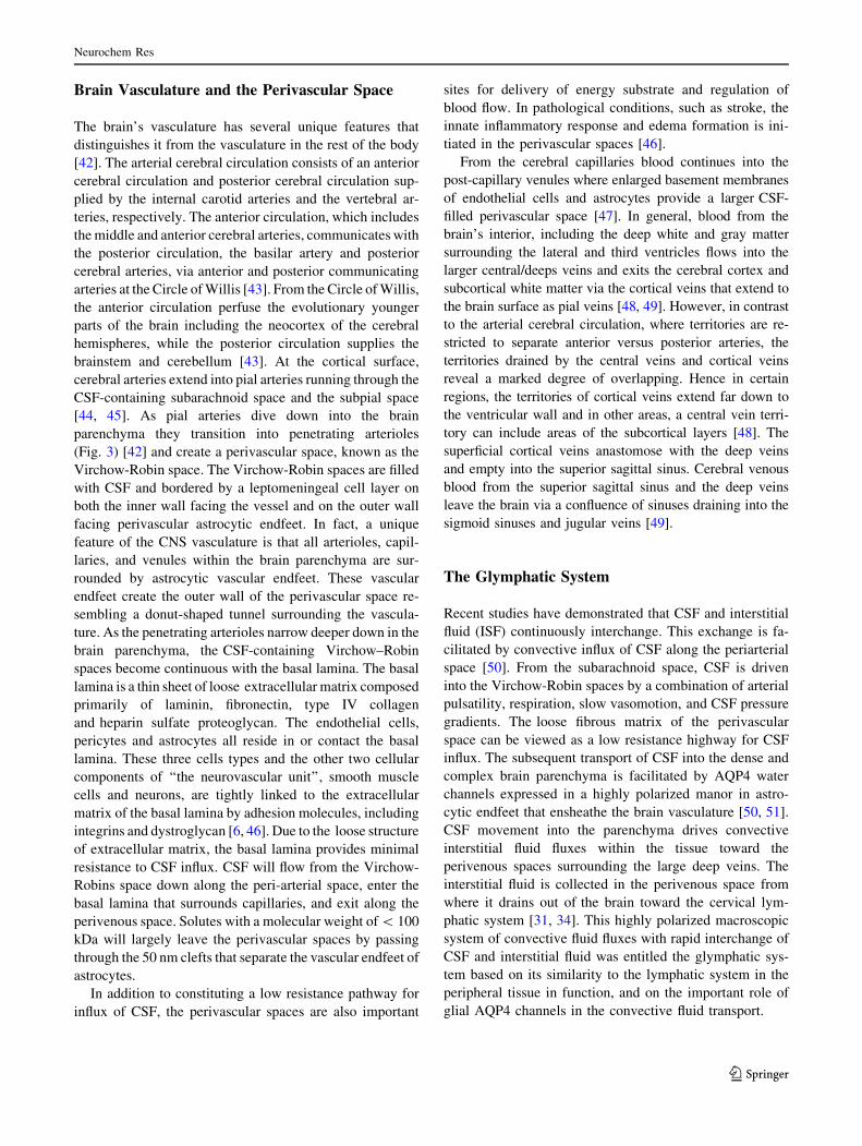
[44, 45]. As pial arteries dive down into the brain
parenchyma they transition into penetrating arterioles
(Fig. 3) [42] and create a perivascular space, known as the
Virchow-Robin space. The Virchow-Robin spaces are filled
with CSF and bordered by a leptomeningeal cell layer on
both the inner wall facing the vessel and on the outer wall
facing perivascular astrocytic endfeet. In fact, a unique
feature of the CNS vasculature is that all arterioles, capil-
laries, and venules within the brain parenchyma are sur-
rounded by astrocytic vascular endfeet. These vascular
endfeet create the outer wall of the perivascular space re-
sembling a donut-shaped tunnel surrounding the vascula-
ture. As the penetrating arterioles narrow deeper down in the
brain parenchyma, the CSF-containing Virchow–Robin
spaces become continuous with the basal lamina. The basal
lamina is a thin sheet of loose extracellular matrix composed
primarily of laminin, fibronectin, type IV collagen
and heparin sulfate proteoglycan. The endothelial cells,
pericytes and astrocytes all reside in or contact the basal
lamina. These three cells types and the other two cellular
components of ‘‘the neurovascular unit’’, smooth muscle
cells and neurons, are tightly linked to the extracellular
matrix of the basal lamina by adhesion molecules, including
integrins and dystroglycan [6, 46]. Due to the loose structure
of extracellular matrix, the basal lamina provides minimal
resistance to CSF influx. CSF will flow from the Virchow-
Robins space down along the peri-arterial space, enter the
basal lamina that surrounds capillaries, and exit along the
perivenous space. Solutes with a molecular weight of\ 100
kDa will largely leave the perivascular spaces by passing
through the 50 nm clefts that separate the vascular endfeet of
astrocytes.
In addition to constituting a low resistance pathway for
influx of CSF, the perivascular spaces are also important
sites for delivery of energy substrate and regulation of
blood flow. In pathological conditions, such as stroke, the
innate inflammatory response and edema formation is ini-
tiated in the perivascular spaces [46].
From the cerebral capillaries blood continues into the
post-capillary venules where enlarged basement membranes
of endothelial cells and astrocytes provide a larger CSF-
filled perivascular space [47]. In general, blood from the
brain’s interior, including the deep white and gray matter
surrounding the lateral and third ventricles flows into the
larger central/deeps veins and exits the cerebral cortex and
subcortical white matter via the cortical veins that extend to
the brain surface as pial veins [48, 49]. However, in contrast
to the arterial cerebral circulation, where territories are re-
stricted to separate anterior versus posterior arteries, the
territories drained by the central veins and cortical veins
reveal a marked degree of overlapping. Hence in certain
regions, the territories of cortical veins extend far down to
the ventricular wall and in other areas, a central vein terri-
tory can include areas of the subcortical layers [48]. The
superficial cortical veins anastomose with the deep veins
and empty into the superior sagittal sinus. Cerebral venous
blood from the superior sagittal sinus and the deep veins
leave the brain via a confluence of sinuses draining into the
sigmoid sinuses and jugular veins [49].
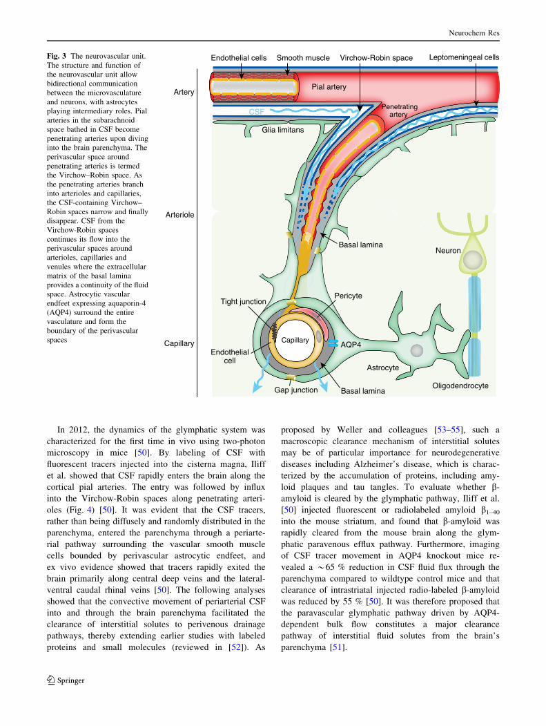
The Glymphatic System
Recent studies have demonstrated that CSF and interstitial
fluid (ISF) continuously interchange. This exchange is fa-
cilitated by convective influx of CSF along the periarterial
space [50]. From the subarachnoid space, CSF is driven
into the Virchow-Robin spaces by a combination of arterial
pulsatility, respiration, slow vasomotion, and CSF pressure
gradients. The loose fibrous matrix of the perivascular
space can be viewed as a low resistance highway for CSF
influx. The subsequent transport of CSF into the dense and
complex brain parenchyma is facilitated by AQP4 water
channels expressed in a highly polarized manor in astro-
cytic endfeet that ensheathe the brain vasculature [50, 51].
CSF movement into the parenchyma drives convective
interstitial fluid fluxes within the tissue toward the
perivenous spaces surrounding the large deep veins. The
interstitial fluid is collected in the perivenous space from
where it drains out of the brain toward the cervical lym-
phatic system [31, 34]. This highly polarized macroscopic
system of convective fluid fluxes with rapid interchange of
CSF and interstitial fluid was entitled the glymphatic sys-
tem based on its similarity to the lymphatic system in the
peripheral tissue in function, and on the important role of
glial AQP4 channels in the convective fluid transport.
Neurochem Res
123
In 2012, the dynamics of the glymphatic system was
characterized for the first time in vivo using two-photon
microscopy in mice [50]. By labeling of CSF with
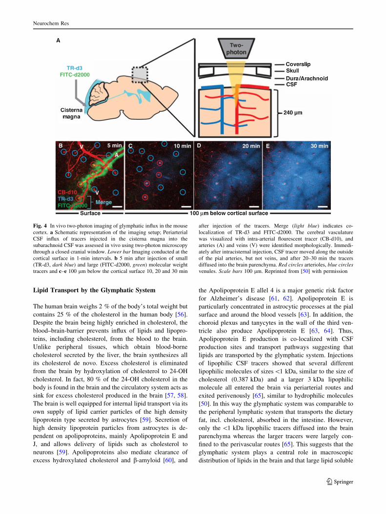
fluorescent tracers injected into the cisterna magna, Iliff
et al. showed that CSF rapidly enters the brain along the
cortical pial arteries. The entry was followed by influx
into the Virchow-Robin spaces along penetrating arteri-
oles (Fig. 4) [50]. It was evident that the CSF tracers,
rather than being diffusely and randomly distributed in the
parenchyma, entered the parenchyma through a periarte-
rial pathway surrounding the vascular smooth muscle
cells bounded by perivascular astrocytic endfeet, and
ex vivo evidence showed that tracers rapidly exited the
brain primarily along central deep veins and the lateral-
ventral caudal rhinal veins [50]. The following analyses
showed that the convective movement of periarterial CSF
into and through the brain parenchyma facilitated the
clearance of interstitial solutes to perivenous drainage
pathways, thereby extending earlier studies with labeled
proteins and small molecules (reviewed in [52]). As
proposed by Weller and colleagues [53–55], such a
macroscopic clearance mechanism of interstitial solutes
may be of particular importance for neurodegenerative
diseases including Alzheimer’s disease, which is charac-
terized by the accumulation of proteins, including amy-
loid plaques and tau tangles. To evaluate whether b-amyloid is cleared by the glymphatic pathway, Iliff et al.
[50] injected fluorescent or radiolabeled amyloid b1–40into the mouse striatum, and found that b-amyloid was
rapidly cleared from the mouse brain along the glym-
phatic paravenous efflux pathway. Furthermore, imaging
of CSF tracer movement in AQP4 knockout mice re-
vealed a *65 % reduction in CSF fluid flux through the
parenchyma compared to wildtype control mice and that
clearance of intrastriatal injected radio-labeled b-amyloid
was reduced by 55 % [50]. It was therefore proposed that
the paravascular glymphatic pathway driven by AQP4-
dependent bulk flow constitutes a major clearance
pathway of interstitial fluid solutes from the brain’s
parenchyma [51].
Glia limitans
Pial artery
Penetratingartery
Virchow-Robin spaceSmooth muscleEndothelial cells
Artery
CSF
Basal lamina
Capillary
Arteriole
Basal lamina
AQP4
Astrocyte
Neuron
Oligodendrocyte
PericyteTight junction
Endothelialcell
Gap junction
Capillary
Leptomeningeal cellsFig. 3 The neurovascular unit.
The structure and function of
the neurovascular unit allow
bidirectional communication
between the microvasculature
and neurons, with astrocytes
playing intermediary roles. Pial
arteries in the subarachnoid
space bathed in CSF become
penetrating arteries upon diving
into the brain parenchyma. The
perivascular space around
penetrating arteries is termed
the Virchow–Robin space. As
the penetrating arteries branch
into arterioles and capillaries,
the CSF-containing Virchow–
Robin spaces narrow and finally
disappear. CSF from the
Virchow-Robin spaces
continues its flow into the
perivascular spaces around
arterioles, capillaries and
venules where the extracellular
matrix of the basal lamina
provides a continuity of the fluid
space. Astrocytic vascular
endfeet expressing aquaporin-4
(AQP4) surround the entire
vasculature and form the
boundary of the perivascular
spaces
Neurochem Res
123
Lipid Transport by the Glymphatic System
The human brain weighs 2 % of the body’s total weight but
contains 25 % of the cholesterol in the human body [56].
Despite the brain being highly enriched in cholesterol, the
blood–brain-barrier prevents influx of lipids and lipopro-
teins, including cholesterol, from the blood to the brain.
Unlike peripheral tissues, which obtain blood-borne
cholesterol secreted by the liver, the brain synthesizes all
its cholesterol de novo. Excess cholesterol is eliminated
from the brain by hydroxylation of cholesterol to 24-OH
cholesterol. In fact, 80 % of the 24-OH cholesterol in the
body is found in the brain and the circulatory system acts as
sink for excess cholesterol produced in the brain [57, 58].
The brain is well equipped for internal lipid transport via its
own supply of lipid carrier particles of the high density
lipoprotein type secreted by astrocytes [59]. Secretion of
high density lipoprotein particles from astrocytes is de-
pendent on apolipoproteins, mainly Apolipoprotein E and
J, and allows delivery of lipids such as cholesterol to
neurons [59]. Apolipoproteins also mediate clearance of
excess hydroxylated cholesterol and b-amyloid [60], and
the Apolipoprotein E allel 4 is a major genetic risk factor
for Alzheimer’s disease [61, 62]. Apolipoprotein E is
particularly concentrated in astrocytic processes at the pial
surface and around the blood vessels [63]. In addition, the
choroid plexus and tanycytes in the wall of the third ven-
tricle also produce Apolipoprotein E [63, 64]. Thus,
Apolipoprotein E production is co-localized with CSF
production sites and transport pathways suggesting that
lipids are transported by the glymphatic system. Injections
of lipophilic CSF tracers showed that several different
lipophilic molecules of sizes\1 kDa, similar to the size of
cholesterol (0.387 kDa) and a larger 3 kDa lipophilic
molecule all entered the brain via periarterial routes and
exited perivenously [65], similar to hydrophilic molecules
[50]. In this way the glymphatic system was comparable to
the peripheral lymphatic system that transports the dietary
fat, incl. cholesterol, absorbed in the intestine. However,
only the\1 kDa lipophilic tracers diffused into the brain
parenchyma whereas the larger tracers were largely con-
fined to the perivascular routes [65]. This suggests that the
glymphatic system plays a central role in macroscopic
distribution of lipids in the brain and that large lipid soluble
Fig. 4 In vivo two-photon imaging of glymphatic influx in the mouse
cortex. a Schematic representation of the imaging setup; Periarterial
CSF influx of tracers injected in the cisterna magna into the
subarachnoid CSF was assessed in vivo using two-photon microscopy
through a closed cranial window. Lower bar Imaging conducted at the
cortical surface in 1-min intervals. b 5 min after injection of small
(TR-d3, dark blue) and large (FITC-d2000, green) molecular weight
tracers and c–e 100 lm below the cortical surface 10, 20 and 30 min
after injection of the tracers. Merge (light blue) indicates co-
localization of TR-d3 and FITC-d2000. The cerebral vasculature
was visualized with intra-arterial flourescent tracer (CB-d10), and
arteries (A) and veins (V) were identified morphologically. Immedi-
ately after intracisternal injection, CSF tracer moved along the outside
of the pial arteries, but not veins, and after 20–30 min the tracers
diffused into the brain parenchyma. Red circles arterioles, blue circles
venules. Scale bars 100 lm. Reprinted from [50] with permission
Neurochem Res
123

molecules might require specific carrier particles to be
delivered efficiently via the CSF. Astrocytes thus play
dual roles in lipid synthesis and lipid distribution by re-
leasing lipid carrier proteins, such as Apolipoprotein E, and
in maintaining the highway for lipid distribution, the
glymphatic system.
What Drives Glymphatic Influx?
Glymphatic transport of CSF along the periarterial spaces,
followed by convective flow through the brain parenchy-
ma, and exit of interstitial fluid (ISF) along the perivenous
space to the cervical lymph system, is an energy requiring
process that is driven by multiple mechanisms. The con-
stant production of CSF by the choroid plexus creates a
pressure that dictates the direction of the fluid flow through
the ventricular system to the subarachnoid space. In addi-
tion, several lines of work show that respiration is instru-
mental in movement of CSF through the aqueduct [66, 67].
Entry of CSF along the perivascular space is crucial for
facilitating glymphatic ISF–CSF exchange and clearance
function. Using reporter mice to distinguish arteries from
veins (Ds-red fluorescent protein expressed under the NG2
promoter in pericytes and smooth muscle cells) [68, 69], it
was demonstrated that CSF tracers follow arteries at the
pial surface running across the cortical surface and descend
along penetrating arteries, which dive perpendicularly into
the brain to reach capillary beds [50, 70]. What drives the
entry of CSF along perivascular space of penetrating lep-
tomeningeal arteries specifically? Particular to arteries,
pulsation generated by smooth muscle cells creates pulse
waves along the whole length of the pial artery and
penetrating arteries diving into the brain from the cortical
surface [71–74]. Dobutamine, an adrenergic agonist, in-
creased pulsatile effect significantly when administrated to
mice and resulted in a larger amount of CSF penetration
into the parenchyma. The opposite effect was obtained
when arterial pulsatility was dampened by internal carotid
artery ligation. Additionally, the reduction of pulse waves
decreased CSF–ISF exchange [71]. This suggests that
glymphatic activity, at least in part, is driven by arterial
pulsatility and explains why perivascular influx occurs
preferentially around pulsating arteries and not cerebral
veins.
The Glymphatic System is Turned on During Sleep
While sleep is essential for all mammals, sleep is also a
vulnerable state since the decreased alertness during sleep
increase the chance of being targeted by predators. This
compromise in alertness versus rest suggests that sleep
serves a fundamental biological function. Multiple studies
indicating that sleep enhances memory consolidation,
which could be important for competition amongst species
[75–78], however, the basic biological need for sleep is
unclear [79]. Brain energy metabolism only declines by
15–25 % during sleep suggesting that sleep does not sim-
ply serve to conserve energy [80]. Recent analysis shows
that the sleep state is unique in the sense that glymphatic
activity is dramatically enhanced, while its function is
suppressed during wakefulness. In vivo 2-photon imaging
of glymphatic function showed that the CSF influx in the
awake state was reduced by 90 % compared to anes-
thetized mice [81]. In order to test if this was specific to the
unconscious state or a side effect of the anesthetics used,
the same experiment was performed in naturally sleeping
mice. This analysis of CSF influx showed a striking simi-
larity between true sleep and anesthetized mice. The sleep-
wake difference in glymphatic influx correlated with the
volume fraction of interstitial space that was 13–15 % in
the awake state an expanded to 22–24 % in both sleep and
anesthetized mice [81]. This observation indicates that the
sleep state is particularly conducive to convective fluid
fluxes and thereby to clearance of metabolites. Thus, a
major function of sleep appears to be that the glymphatic
system is turned on and that the brain clears itself of
neurotoxic waste products produced during wakefulness.
The observation that glymphatic function is highly ac-
tive in both anesthetized mice and naturally sleeping mice
but not awake mice indicates that it is differences in the
sleep versus wakeful state and not daily circadian rhythms
that regulate glymphatic activity. A major driver of arousal
is the neuromodulator norepinephrine [82]. Our analysis
showed that norepinephrine also is a key regulator of
glymphatic activity and that norepinephrine might be re-
sponsible for suppression of glymphatic during wakeful-
ness. Local application of a cocktail of norepinephrine
receptor antagonists in awake mice resulted in an increase
in CSF tracer influx almost comparable to that observed
during sleep or anesthesia [81]. In contrast, norepinephrine
application, mimicking the wakeful state, significantly
decreased the interstitial volume fraction. An increase in
interstitial space volume in the sleep state reduces tissue
resistance towards convective flow thus permitting CSF–
ISF exchange. Thus the burst release of norepinephrine
during arousal increases the cellular volume fraction re-
sulting in a decrease in the interstitial space [83]. In turn,
the resistance toward convective exchange of CSF and ISF
increases and this results in a suppression of glymphatic
fluxes during wakefulness. Norepinephrine also acts di-
rectly on choroid plexus epithelial cells and inhibits CSF
production. Conversely, removal of norepinephrine sig-
naling, mimicking the sleep state, enhances CSF produc-
tion [84]. The concerted effect of norepinephrine thus acts
Neurochem Res
123
via different mechanisms on both fluid availability and
convective fluxes to suppress glymphatic function and
norepinephrine can therefore be considered both a key
regulator of the switch between the sleep and wakeful state
and solute clearance from the brain.
Convective CSF Fluxes in Aging and Pathology
Glymphatic Activity Decreases Sharply During
Aging
A recent assessment of glymphatic function in old versus
young mice showed a dramatic reduction by *80–90 % in
aged compared to young mice [85]. The suppression of
glymphatic activity included both influx of CSF tracers and
clearance of radiolabeled b-amyloid and inulin. Reactive
gliosis, defined by hypertrophy of GFAP? astrocyte pro-
cesses, increases with aging [86] and might contribute to
the age-related decline in glymphatic function, although
the mechanism of how changes in GFAP expression might
contribute to altered glymphatic function remains unclear.
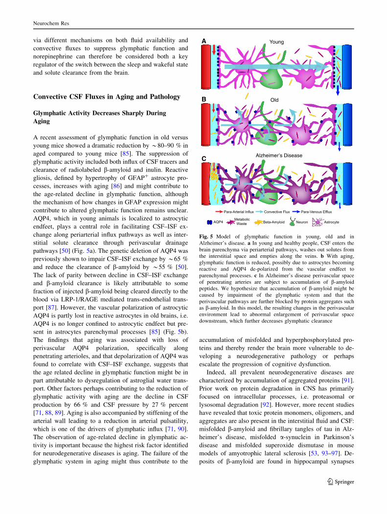
AQP4, which in young animals is localized to astrocytic
endfeet, plays a central role in facilitating CSF–ISF ex-
change along periarterial influx pathways as well as inter-
stitial solute clearance through perivascular drainage
pathways [50] (Fig. 5a). The genetic deletion of AQP4 was
previously shown to impair CSF–ISF exchange by *65 %
and reduce the clearance of b-amyloid by *55 % [50].
The lack of parity between decline in CSF–ISF exchange
and b-amyloid clearance is likely attributable to some
fraction of injected b-amyloid being cleared directly to the
blood via LRP-1/RAGE mediated trans-endothelial trans-
port [87]. However, the vascular polarization of astrocytic
AQP4 is partly lost in reactive astrocytes in old brains, i.e.
AQP4 is no longer confined to astrocytic endfeet but pre-
sent in astrocytes parenchymal processes [85] (Fig. 5b).
The findings that aging was associated with loss of
perivascular AQP4 polarization, specifically along
penetrating arterioles, and that depolarization of AQP4 was
found to correlate with CSF–ISF exchange, suggests that
the age related decline in glymphatic function might be in
part attributable to dysregulation of astroglial water trans-
port. Other factors perhaps contributing to the reduction of
glymphatic activity with aging are the decline in CSF
production by 66 % and CSF pressure by 27 % percent
[71, 88, 89]. Aging is also accompanied by stiffening of the
arterial wall leading to a reduction in arterial pulsatility,
which is one of the drivers of glymphatic influx [71, 90].
The observation of age-related decline in glymphatic ac-
tivity is important because the highest risk factor identified
for neurodegenerative diseases is aging. The failure of the
glymphatic system in aging might thus contribute to the
accumulation of misfolded and hyperphosphorylated pro-
teins and thereby render the brain more vulnerable to de-
veloping a neurodegenerative pathology or perhaps
escalate the progression of cognitive dysfunction.
Indeed, all prevalent neurodegenerative diseases are
characterized by accumulation of aggregated proteins [91].
Prior work on protein degradation in CNS has primarily
focused on intracellular processes, i.e. proteasomal or
lysosomal degradation [92]. However, more recent studies
have revealed that toxic protein monomers, oligomers, and
aggregates are also present in the interstitial fluid and CSF:
misfolded b-amyloid and fibrillary tangles of tau in Alz-
heimer’s disease, misfolded a-synuclein in Parkinson’s
disease and misfolded superoxide dismutase in mouse
models of amyotrophic lateral sclerosis [53, 93–97]. De-
posits of b-amyloid are found in hippocampal synapses
C
WasteAQP4 Beta-Amyloid Neuron Astrocyte
Para-Arterial Influx Para-Venous EffluxConvective Flux
Young
Old
Alzheimer’s Disease
A
B
Metabolic
Fig. 5 Model of glymphatic function in young, old and in
Alzheimer’s disease. a In young and healthy people, CSF enters the
brain parenchyma via periarterial pathways, washes out solutes from
the interstitial space and empties along the veins. b With aging,
glymphatic function is reduced, possibly due to astrocytes becoming
reactive and AQP4 de-polarized from the vascular endfeet to
parenchymal processes. c In Alzheimer’s disease perivascular space
of penetrating arteries are subject to accumulation of b-amyloid
peptides. We hypothesize that accumulation of b-amyloid might be
caused by impairment of the glymphatic system and that the
perivascular pathways are further blocked by protein aggregates such
as b-amyloid. In this model, the resulting changes in the perivascular
environment lead to abnormal enlargement of perivascular space
downstream, which further decreases glymphatic clearance
Neurochem Res
123
where activity of the neurons might lead to increased
shedding making synapses a source of extracellular b-amyloid [98]. In concert with this, b-amyloid production is
highest during the awake state when neuronal activity is
highest [99]. However, b-amyloid is not only produced by
neurons. In fact, all cells produce b-amyloid and in par-
ticular oligodendrocytes and their precursor cells [100],
which, at least in part, might account for the myelin dis-
orders observed in Alzheimer’s disease. The emerging
concept of a prion-like spread of neurotoxic proteins ag-
gregates highlights the importance of the interstitial space
regarding deposits of aggregated protein, such as b-amy-
loid in Alzheimer’s disease [101, 102]. The production and
turnover of b-amyloid is strikingly rapid in humans. In
healthy, young subjects 8.3 % of total b-amyloid is cleared
each hour via the CSF [103]. Perivascular drainage path-
ways function as a sink for interstitial b-amyloid in Alz-
heimer’s disease and perivascular spaces are a sites for
both amyloid deposits and Alzheimer’s disease pathology
[104]. Bulk clearance by the glymphatic system might in
combination with transport across the blood–brain barrier
[60] provide necessary and sufficient removal of extracel-
lular b-amyloid until the end of the reproductive life span
around which time failure in adequate CSF bulk flow leads
to accumulation of b-amyloid [85] (Fig. 5). This suggests
that low activity of the glymphatic system could be a major
risk factor for development of neurodegenerative diseases.
The anatomical routes of interstitial bulk flow in the
perivascular space are consistent with sites of b-amyloid
accumulation, although b-amyloid accumulates pre-
dominantly at cerebral arteries. Periarterial accumulation
of b-amyloid around arteries could reflect recirculation of
b-amyloid-rich CSF that deposits aggregates after being
taken up by smooth muscles cells [105], although the
concept of recirculation of CSF is controversial [45]. In
turn, vascular amyloidosis might reduce glymphatic CSF
influx and the stagnation of CSF influx might accelerate b-amyloid accumulation. In addition, monocytic engulfment
of b-amyloid appears to occur selectively at veins [106].
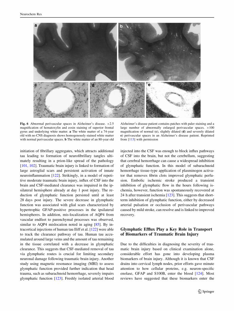
Abnormal enlargement of the perivascular space is more
frequently observed in Alzheimer’s disease compared to
aged-matched control subjects suggesting a spiral of pro-
tein accumulation, deformation of glymphatic routes and
further reduction in protein clearance and pathology
(Fig. 6).
However, abnormalities at the perivascular space are
also prominent in non-Alzheimer’s dementia. Only sur-
passed by Alzheimer’s disease, vascular dementias are the
second most common cause of dementia and these diseases
are also characterized by deformation of the perivascular
space [107, 108]. Changes in or surrounding cerebral blood
vessels due to hypertension, atherosclerosis or hereditary
diseases can cause vascular dementia [108]. Vascular
dementia is often caused by pathology in small cerebral
blood vessels and capillaries, collectively termed small
vessel disease. Enlargement of the perivascular space is
frequently observed in small vessel disease [109]. An ex-
ample of this is the disease ‘‘cerebral autosomal dominant
arteriopathy with subcortical infarcts and leukoen-
cephalopathy’’ [110, 111]. We speculate that these anato-
mical abnormalities in the perivascular space could have a
profound impact on glymphatic fluxes due to altered phy-
sical, and perhaps also cellular signaling pathways in ways
that are non-permissive for convective fluxes in the
perivascular spaces. In fact, numerous case reports found
abnormal widening of perivascular space in dementia pa-
tients otherwise in good health [112]. One explanation for
widening of the perivascular space could be periarterial
blockage leading to local obstruction of glymphatic fluxes,
perhaps affecting the width of the downstream periarterial
space (Fig. 5). Roher et al. [113] hypothesized that the
enlargement of perivascular space in the white matter oc-
curs as a secondary effect of clogging of perivascular space
in the upstream artery, usually located in the grey matter.
Myelin-rich tissue may be more sensitive to obstruction of
convective interstitial fluid fluxes, perhaps explaining why
vascular diseases, including ‘‘cerebral autosomal dominant
arteriopathy with subcortical infarcts and leukoen-
cephalopathy’’, primarily affect white matter [114]. It is
important to note, that the potential role of glymphatic
fluxes in diseases with prominent enlargement of perivas-
cular spaces has yet to be addressed experimentally, but
that gross changes of the perivascular space are likely to
negatively affect glymphatic functions. A suppression of
glymphatic function could in turn exacerbate pathology.
Traumatic Brain Injury and Its Implications
for Neurodegenerative Diseases
Traumatic brain injury, which most frequently affects
military personnel or athletes, increases the risk of prema-
ture dementia and Alzheimer’s disease [115, 116]. Multiple
studies have shown that repeated traumatic events, and even
single events of moderate to severe head trauma, can lead to
progressive neurodegeneration. However, it is currently not
understood why a subpopulation of individuals develops
chronic traumatic encephalopathy, whereas other exposed
to the same degree of initial brain injury are not affected
[117]. Traumatic brain injury induces accumulation of b-amyloid peptide and of C-tau, a proteolytically cleaved
product of MAP-tau, which is a highly abundant intracel-
lular microtubule protein in axons [118–120]. C-tau is a
biomarker of brain injury since it is released in vast quan-
tities and correlates with severity of traumatic brain injury
[121]. An emerging hypothesis is that the large amplitude
increases of interstitial tau lead to cellular uptake and
Neurochem Res
123
initiation of fibrillary aggregates, which attracts additional
tau leading to formation of neurofibrillary tangles ulti-
mately resulting in a prion-like spread of the pathology
[101, 102]. Traumatic brain injury is linked to formation of
large astroglial scars and persistent activation of innate
neuroinflammation [122]. Strikingly, in a model of repeti-
tive moderate traumatic brain injury, influx of CSF into the
brain and CSF-mediated clearance was impaired in the ip-
silateral hemisphere already at day 1 post injury. The re-
duction of glymphatic function persisted until at least
28 days post injury. The severe decrease in glymphatic
function was associated with glial scars characterized by
hypertrophic GFAP-positive processes in the ipsilateral
hemispheres. In addition, mis-localization of AQP4 from
vascular endfeet to parenchymal processes was observed,
similar to AQP4 mislocation noted in aging [85]. By in-
tracortical injections of human tau Iliff et al. [122] were able
to track the clearance pathway of tau. Human tau accu-
mulated around large veins and the amount of tau remaining
in the tissue correlated with a decrease in glymphatic
clearance. This suggests that CSF-mediated removal of tau
via glymphatic routes is crucial for limiting secondary
neuronal damage following traumatic brain injury. Another
study using magnetic resonance imaging (MRI) to assess
glymphatic function provided further indication that head
trauma, such as subarachnoid hemorrhage, severely impairs
glymphatic function [123]. Freshly isolated arterial blood
injected into the CSF was enough to block influx pathways
of CSF into the brain, but not the cerebellum, suggesting
that cerebral hemorrhage can cause a widespread inhibition
of glymphatic function. In this model of subarachnoid
hemorrhage tissue-type application of plasminogen activa-
tor that removes fibrin clots improved glymphatic perfu-
sion. Embolic ischemic stroke produced a transient
inhibition of glymphatic flow in the hours following is-
chemia, however, function was spontaneously recovered at
24 h after transient ischemia [123]. This suggests that short-
term inhibition of glymphatic function, either by decreased
arterial pulsation or occlusion of perivascular pathways
caused by mild stroke, can resolve and is linked to improved
recovery.
Glymphatic Efflux Play a Key Role in Transportof Biomarkers of Traumatic Brain Injury
Due to the difficulties in diagnosing the severity of trau-
matic brain injury based on clinical examination alone,
considerable effort has gone into developing plasma
biomarkers of brain injury. Although it is known that CSF
drains into cervical lymph nodes, prior efforts gave minute
attention to how cellular proteins, e.g. neuron-specific
enolase, GFAP and S100B, enter the blood [124]. Most
reviews have suggested that these biomarkers enter the
Fig. 6 Abnormal perivascular spaces in Alzheimer’s disease. 92.5
magnification of hematoxylin and eosin staining of superior frontal
gyrus and underlying white matter. a The white matter of a 74-year
old with no CNS diagnosis shows homogenously stained white matter
with normal perivascular spaces. b The white matter of an 80-year old
Alzheimer’s disease patient contains patches with paler staining and a
large number of abnormally enlarged perivascular spaces. 9100
magnification of normal (c), slightly dilated (d) and severely dilated
e) perivascular spaces in an Alzheimer’s disease patient. Reprinted
from [113] with permission
Neurochem Res
123
general circulation through opening of the blood–brain
barrier in the setting of tissue injury [125]. Instead, we
recently showed that the biomarkers of traumatic brain
injury exit the brain via the glymphatic system. Inhibition
of glymphatic activity by four mechanistically distinct
manipulations, including sleep deprivation, cisterna magna
puncture, inhibition of CSF production using acetazo-
lamide, or genetic deletion of AQP4 resulted in marked
reduction of astrocytic proteins GFAP, S100B and neuron-
specific enolase in plasma following traumatic brain injury
[126]. Several of these manipulations are clinical relevant.
For example, sleep deprivation is common in the emer-
gency room as vitals are taken frequently. Thus, plasma
biomarkers may not be a useful approach to asses the
severity of traumatic injury if it is the activity of the
glymphatic system that determines the extend by which the
biomarkers are transported from the injured brain tissue to
the blood compartment. If a patient with traumatic brain
injury is unable to sleep or if a decompressive ventricu-
lostomy is performed, the transfer of biomarkers from CSF
plasma will be low. The low level of biomarkers in the
blood under these conditions will not reflect the degree of
tissue injury but rather that glymphatic activity is low.
Thus, several studies have shown that acute injury, in-
cluding traumatic brain injury, subarachnoid bleeding or
stroke, profoundly impact glymphatic function and impair
convective fluid flow. The suppression of CSF–ISF ex-
change has immediate implications regarding the limita-
tions of the diagnostic relevance of plasma biomarkers in
traumatic brain injury [123, 126]. Most importantly, im-
pairment of glymphatic function could further exacerbate
injury due to accumulation of both normal metabolic waste
as well as injury-induced debris. The degree by which
glymphatic function is suppressed after traumatic brain
injury might therefore contribute to the variability in out-
come. Future studies will define the clinical relevance of
pharmacological manipulations of the glymphatic system
after acute traumatic injury.
Prior Observations had Documented the Existenceof Perivascular Fluid Transport
Albeit, the concept of the glymphatic system was developed
just 3 years ago, many aspects of this highly organized
pathway of CSF–ISF fluid exchange had already been de-
scribed. In the mid 80’s a young investigator, Patricia Grady
noted that when horseradish peroxidase, a 44 kDa glycopro-
tein,was injected inCSF, the entire vasculature of dogs or cats
was outlined by the tracer just 4–5 min later [127, 128]. This
observation, which depictedwidespread influx of a large CSF
tracer along the perivascular space, justifiably received con-
siderable attention among the more established groups. Such
rapid influx of CSF could not be justified by the slow process
of diffusion. Only convective fluxes along the perivascular
space could explain the fast perivascular distribution of
horseradish peroxidase. However, the inability of other
groups to repeat Patricia Grady’s findings led to the incorrect
conclusion that CSF only inconsistently and to a minor extent
entered the perivascular space [129]. In retrospect, the dis-
crepancy between the observations can be explained by the
fact that Patricia Grady used larger animals that were exposed
to minimal surgery, whereas subsequent studies prepared
open cranial windows in rats, which eliminated the pressure
gradients that drive convectiveCSFfluxes. Later, in a series of
elegant studies Gerald Dienel showed that metabolites of
glucose, including lactate, are rapidly released in large
quantities during brain activation [130, 131]. Most interest-
ingly, tracers of similar molecular weights as lactate could
later be identified in the perivascular space and in cervical
lymph nodes. Thus, the work by Gerald Dienel identified the
perivascular space as a pathway for removal of metabolic
waste products. Even older studies from the group of Brad-
bury had documented that radiolabeled tracers injected into
the brain parenchyma later could be found in both the olfac-
tory bulb and the cervical lymph vessels [124].
Future Perspectives
Accumulating evidence suggests that age-related decline in
glymphatic clearance in significant ways contributes to
accumulation of protein aggregation. Glymphatic activity
decreases by 80–90 % in old mice and several lines of
evidence have documented that b-amyloid and tau exit the
brain via the glymphatic system [85, 122]. Moreover, work
from David Holtzman documents that the concentration of
b-amyloid in CSF follows the sleep-wake cycle in human
subjects [132]. It is therefore imperative that a diagnostic
test of glymphatic activity capable of identifying patients
with premature decline in glymphatic clearance is devel-
oped. Using the same logic, assessment of glymphatic ac-
tivity in patients following traumatic brain injury may
pinpoint patients with the most severe suppression of
glymphatic clearance and thereby identify the individuals
who are at higher risk for developing chronic traumatic
encephalopathy. Helene Benveniste’s group has made
headway with regard to developing a glymphatic diag-
nostic test based on MRI scans. By delivering contrast
agent into cisterna magna, the movement of CSF could be
followed in real time across the entire brain [70]. These
observations made in rats confirmed and extended the no-
tion of the existence of a brain-wide glymphatic system. In
particular the MRI analysis revealed fast influx kinetics at
two key nodes, the pituitary and pineal gland recesses [70].
Interestingly both of these regions are involved in
Neurochem Res
123
regulation of the sleep wake cycle [133, 134]. These
studies using contrast-enhanced MRI provide the ex-
perimental groundwork for evaluating glymphatic pathway
function in the human brain and in the future assess whe-
ther failure of CSF fluxes contributes to Alzheimer’s dis-
ease progression. Development of a safe and minimally
invasive imaging approach to visualize glymphatic func-
tion is therefore necessary for future translational effort. It
is in this regard important to note that subsequent analysis
showed that intrathecal lumbar injections, which are rou-
tinely used in clinical myelographic studies, provide a vi-
able alternative route to assess the basic parameters of
glymphatic function [135]. The first studies of the glym-
phatic system in human subject is expected to be published
soon, as intrathecal administration of contrast agents al-
ready is approved for study of CSF fluxes in patients un-
dergoing treatment for spontaneous intracranial
hypotension and CSF rhinorrhea [136, 137].
Future studies with focus on the glymphatic system are
expected to identify functions of convective CSF fluxes
beyond removal of metabolic waste products. We have
recently found that glymphatic influx can serve as a dis-
tribution system for lipids [65] and glucose [138], and we
speculate that glymphatic influx provides an essential route
for distribution of electrolytes, macromolecules, and other
larger compounds that enter the brain predominantly via
the blood-CSF barrier at the choroid plexus. Likewise, the
glymphatic system might serve as a path for delivery and
distribution of drugs including cancer drugs within the
brain [139]. Furthermore, we expect that growth factors
produced by the choroid plexus, as well as neuromodula-
tors released by several brain stem nuclei positioned close
to the ventricular system, are distributed widely across the
CNS by the glymphatic system. Thus, volume transmission
may, in addition to microscopic release of neuromodulators
from local nerve terminals followed by local diffusion, also
involve circulation by macroscopic convective CSF fluxes
via the glymphatic system.
Acknowledgments This study was supported by NIH (NINDS
NS075177 and NS078304). We thank Gerry Dienel, Ben Kress, and
Rashid Deane for comments on the manuscript and Takahiro Takano
for illustrations.
Conflict of interest The authors declare that they have no conflict
of interest.
References
1. Liao S, Padera TP (2013) Lymphatic function and immune
regulation in health and disease. Lymphat Res Biol 11:136–143.
doi:10.1089/lrb.2013.0012
2. Wang Z, Ying Z, Bosy-Westphal A et al (2012) Evaluation of
specific metabolic rates of major organs and tissues: comparison
between nonobese and obese women. Obesity 20:95–100.
doi:10.1038/oby.2011.256
3. Weed LH (1917) An anatomical consideration of the cerebro-
spinal fluid. Anat Rec 12:461–496. doi:10.1002/ar.1090120405
4. Johanson CE, Duncan JA, Klinge PM et al (2008) Multiplicity of
cerebrospinal fluid functions: new challenges in health and dis-
ease. Cerebrospinal Fluid Res 5:10. doi:10.1186/1743-8454-5-10
5. Damkier HH, Brown PD, Praetorius J (2013) Cerebrospinal fluid
secretion by the choroid plexus. Physiol Rev 93:1847–1892.
doi:10.1152/physrev.00004.2013
6. Thrane AS, Rangroo Thrane V, Nedergaard M (2014) Drowning
stars: reassessing the role of astrocytes in brain edema. Trends
Neurosci 37:620–628. doi:10.1016/j.tins.2014.08.010
7. Keep RF, Jones HC (1990) A morphometric study on the de-
velopment of the lateral ventricle choroid plexus, choroid plexus
capillaries and ventricular ependyma in the rat. Brain Res Dev
Brain Res 56:47–53. doi:10.1016/0165-3806(90)90163-S
8. Banizs B, Pike MM, Millican CL et al (2005) Dysfunctional
cilia lead to altered ependyma and choroid plexus function, and
result in the formation of hydrocephalus. Development
132:5329–5339. doi:10.1242/dev.02153
9. Brown P, Davies S, Speake T, Millar I (2004) Molecular
mechanisms of cerebrospinal fluid production. Neuroscience
129:957–970. doi:10.1016/j.neuroscience.2004.07.003
10. Ames A, Higashi K, Nesbett FB (1965) Effects of Pco2 aceta-
zolamide and ouabain on volume and composition of choroid-
plexus fluid. J Physiol 181:516–524
11. Johanson CE (2008) Choroid plexus–Cerebrospinal fluid circu-
latory dynamics: impact on brain growth, metabolism, and re-
pair. Neurosci, Med
12. Davson H, Segal MB (1970) The effects of some inhibitors and
accelerators of sodium transport on the turnover of 22Na in the
cerebrospinal fluid and the brain. J Physiol 209:131–153
13. Segal MB, Burgess AM (1974) A combined physiological and
morphological study of the secretory process in the rabbit
choroid plexus. J Cell Sci 14:339–350
14. Christensen HL, Nguyen AT, Pedersen FD, Damkier HH (2013)
Na(?) dependent acid-base transporters in the choroid plexus;
insights from slc4 and slc9 gene deletion studies. Front Physiol
4:304. doi:10.3389/fphys.2013.00304
15. Jacobs S, Ruusuvuori E, Sipila ST et al (2008) Mice with tar-
geted Slc4a10 gene disruption have small brain ventricles and
show reduced neuronal excitability. Proc Natl Acad Sci USA
105:311–316. doi:10.1073/pnas.0705487105
16. Damkier HH, Praetorius J (2012) Genetic ablation of Slc4a10
alters the expression pattern of transporters involved in solute
movement in the mouse choroid plexus. Am J Physiol Cell
Physiol 302:C1452–C1459. doi:10.1152/ajpcell.00285.2011
17. Saito Y, Wright EM (1983) Bicarbonate transport across the
frog choroid plexus and its control by cyclic nucleotides.
J Physiol 336:635–648
18. Mayer SE, Sanders-Bush E (1993) Sodium-dependent an-
tiporters in choroid plexus epithelial cultures from rabbit.
J Neurochem 60:1308–1316
19. Deng QS, Johanson CE (1989) Stilbenes inhibit exchange of
chloride between blood, choroid plexus and cerebrospinal fluid.
Brain Res 501:183–187. doi:10.1016/0006-8993(89)91041-X
20. Praetorius J, Nielsen S (2006) Distribution of sodium trans-
porters and aquaporin-1 in the human choroid plexus. Am J
Physiol Cell Physiol 291:C59–C67. doi:10.1152/ajpcell.00433.
2005
21. Praetorius J (2007) Water and solute secretion by the choroid
plexus. Pflugers Arch 454:1–18. doi:10.1007/s00424-006-0170-
6
22. Nielsen S, Smith BL, Christensen EI, Agre P (1993) Distribution
of the aquaporin CHIP in secretory and resorptive epithelia and
Neurochem Res
123
capillary endothelia. Proc Natl Acad Sci USA 90:7275–7279.
doi:10.1073/pnas.90.15.7275
23. Damkier HH, Brown PD, Praetorius J (2010) Epithelial path-
ways in choroid plexus electrolyte transport. Physiology
(Bethesda) 25:239–249. doi:10.1152/physiol.00011.2010
24. Papadopoulos MC, Verkman AS (2013) Aquaporin water
channels in the nervous system. Nat Rev Neurosci 14:265–277.
doi:10.1038/nrn3468
25. Oshio K, Song Y, Verkman AS, Manley GT (2003) Aquaporin-1
deletion reduces osmotic water permeability and cerebrospinal
fluid production. Acta Neurochir 86(Suppl):525–528
26. Oshio K, Watanabe H, Song Y et al (2005) Reduced cere-
brospinal fluid production and intracranial pressure in mice
lacking choroid plexus water channel Aquaporin-1. FASEB J
19:76–78. doi:10.1096/fj.04-1711fje
27. Husted RF, Reed DJ (1976) Regulation of cerebrospinal fluid
potassium by the cat choroid plexus. J Physiol 259:213–221
28. Silverberg GD, Huhn S, Jaffe RA et al (2002) Downregulation
of cerebrospinal fluid production in patients with chronic hy-
drocephalus. J Neurosurg 97:1271–1275. doi:10.3171/jns.2002.
97.6.1271
29. Lindvall M, Owman C (1981) Autonomic nerves in the mam-
malian choroid plexus and their influence on the formation of
cerebrospinal fluid. J Cereb Blood Flow Metab 1:245–266.
doi:10.1038/jcbfm.1981.30
30. Szentistvanyi I, Patlak CS, Ellis RA, Cserr HF (1984) Drainage
of interstitial fluid from different regions of rat brain. Am J
Physiol 246:F835–F844
31. Johnston M, Zakharov A, Papaiconomou C et al (2004) Evi-
dence of connections between cerebrospinal fluid and nasal
lymphatic vessels in humans, non-human primates and other
mammalian species. Cerebrospinal Fluid Res 1:2. doi:10.1186/
1743-8454-1-2
32. Koh L, Zakharov A, Johnston M (2005) Integration of the
subarachnoid space and lymphatics: Is it time to embrace a new
concept of cerebrospinal fluid absorption? Cerebrospinal Fluid
Res 2:6. doi:10.1186/1743-8454-2-6
33. Biceroglu H, Albayram S, Ogullar S et al (2012) Direct venous
spinal reabsorption of cerebrospinal fluid: a new concept with
serial magnetic resonance cisternography in rabbits. J Neurosurg
Spine 16:394–401. doi:10.3171/2011.12.SPINE11108
34. Murtha LA, Yang Q, Parsons MW et al (2014) Cerebrospinal
fluid is drained primarily via the spinal canal and olfactory route
in young and aged spontaneously hypertensive rats. Fluids
Barriers CNS 11:12. doi:10.1186/2045-8118-11-12
35. Kimelberg HK (2004) Water homeostasis in the brain: basic
concepts. Neuroscience 129:851–860. doi:10.1016/j.neu
roscience.2004.07.033
36. Redzic Z (2011) Molecular biology of the blood-brain and the
blood-cerebrospinal fluid barriers: similarities and differences.
Fluids Barriers CNS 8:3. doi:10.1186/2045-8118-8-3
37. Oreskovic D, Klarica M (2010) The formation of cerebrospinal
fluid: nearly a hundred years of interpretations and misinter-
pretations. Brain Res Rev 64:241–262. doi:10.1016/j.brainres
rev.2010.04.006
38. Sathyanesan M, Girgenti MJ, Banasr M et al (2012) A molecular
characterization of the choroid plexus and stress-induced gene
regulation. Transl Psychiatry 2:e139. doi:10.1038/tp.2012.64
39. Bulat M, Lupret V, Orehkovic D, Klarica M (2008) Transven-
tricular and transpial absorption of cerebrospinal fluid into
cerebral microvessels. Coll Antropol 32(Suppl 1):43–50
40. Oreskovic D, Klarica M (2014) A new look at cerebrospinal
fluid movement. Fluids Barriers CNS 11:16. doi:10.1186/2045-
8118-11-16
41. Buishas J, Gould IG, Linninger AA (2014) A computational
model of cerebrospinal fluid production and reabsorption driven
by starling forces. Croat Med J 55:481–497. doi:10.3325/cmj.
2014.55.481
42. Kulik T, Kusano Y, Aronhime S (2008) Regulation of cerebral
vasculature in normal and ischemic brain. Neuropharmacology
55:281–288. doi:10.1016/j.neuropharm.2008.04.017
43. Prince E, Ahn S (2013) Basic vascular neuroanatomy of the
brain and spine: what the general interventional radiologist
needs to know. Semin Intervent Radiol 30:234–239. doi:10.
1055/s-0033-1353475
44. Zlokovic BV (2011) Neurovascular pathways to neurodegen-
eration in Alzheimer’s disease and other disorders. Nat Rev
Neurosci 12:723–738. doi:10.1038/nrn3114
45. Zhang ET, Inman CB, Weller RO (1990) Interrelationships of
the pia mater and the perivascular (Virchow–Robin) spaces in
the human cerebrum. J Anat 170:111–123
46. del Zoppo GJ, Moskowitz M, Nedergaard M (2015) The neu-
rovascular unit and responses to ischemia. In: Grotta J, Albers
G, Broderick J, Kasner S, Lo E, Medelow AD, Sacco R, Wong L
(eds) Stroke: pathophysiology, diagnosis, and management, 6th
Edn. Elsevier, Philadelphia.
47. Engelhardt B, Ransohoff RM (2012) Capture, crawl, cross: the T
cell code to breach the blood–brain barriers. Trends Immunol
33:579–589. doi:10.1016/j.it.2012.07.004
48. Schlesinger B (1939) The venous drainage of the brain, with
special reference to the galenic system. Brain 62:274–291.
doi:10.1093/brain/62.3.274
49. Cipolla M (2010) Anatomy and ultrastructure. Cereb, Circ
50. Iliff JJ, Wang M, Liao Y et al (2012) A paravascular pathway
facilitates CSF flow through the brain parenchyma and the
clearance of interstitial solutes, including amyloid b. Sci TranslMed 4:147ra111. doi: 10.1126/scitranslmed.3003748
51. Iliff JJ, Nedergaard M (2013) Is there a cerebral lymphatic
system? Stroke 44:S93–S95. doi:10.1161/STROKEAHA.112.
678698
52. Bradbury M, Cserr H (1985) Drainage of cerebral interstitial
fluid and of cerebrospinal fluid into lymphatics. In: Johnston M
(ed) Experimental Biology of the lymphatic circulation. Else-
vier, New York, pp 355–394
53. Weller RO, Subash M, Preston SD et al (2008) Perivascular
drainage of amyloid-beta peptides from the brain and its failure
in cerebral amyloid angiopathy and Alzheimer’s disease. Brain
Pathol 18:253–266. doi:10.1111/j.1750-3639.2008.00133.x
54. Carare RO, Bernardes-Silva M, Newman TA et al (2008) So-
lutes, but not cells, drain from the brain parenchyma along
basement membranes of capillaries and arteries: significance for
cerebral amyloid angiopathy and neuroimmunology. Neu-
ropathol Appl Neurobiol 34:131–144. doi:10.1111/j.1365-2990.
2007.00926.x
55. Hawkes CA, Hartig W, Kacza J et al (2011) Perivascular drai-
nage of solutes is impaired in the ageing mouse brain and in the
presence of cerebral amyloid angiopathy. Acta Neuropathol
121:431–443. doi:10.1007/s00401-011-0801-7
56. Bjorkhem I, Meaney S (2004) Brain cholesterol: long secret life
behind a barrier. Arter Thromb Vasc Biol 24:806–815. doi:10.
1161/01.ATV.0000120374.59826.1b
57. Bjorkhem I, Lutjohann D, Diczfalusy U et al (1998) Cholesterol
homeostasis in human brain: turnover of 24S-hydroxycholes-
terol and evidence for a cerebral origin of most of this oxysterol
in the circulation. J Lipid Res 39:1594–1600
58. Lutjohann D, Breuer O, Ahlborg G et al (1996) Cholesterol
homeostasis in human brain: evidence for an age-dependent flux
of 24S-hydroxycholesterol from the brain into the circulation.
Proc Natl Acad Sci USA 93:9799–9804. doi:10.1073/pnas.93.
18.9799
59. Fagan AM, Holtzman DM, Munson G et al (1999) Unique
lipoproteins secreted by primary astrocytes from wild type, apoE
Neurochem Res
123
(-/-), and human apoE transgenic mice. J Biol Chem
274:30001–30007. doi:10.1074/jbc.274.42.30001
60. Deane R, Sagare A, Hamm K et al (2008) apoE isoform—
specific disruption of amyloid b peptide clearance from mouse
brain. J Clin Invest 118:4002–4013. doi:10.1172/JCI36663DS1
61. Strittmatter WJ, Saunders AM, Schmechel D et al (1993)
Apolipoprotein E: high-avidity binding to beta-amyloid and
increased frequency of type 4 allele in late-onset familial Alz-
heimer disease. Proc Natl Acad Sci USA 90:1977–1981. doi:10.
1073/pnas.90.5.1977
62. Corder EH, Saunders AM, Strittmatter WJ et al (1993) Gene
dose of apolipoprotein E type 4 allele and the risk of Alzhei-
mer’s disease in late onset families. Science 261:921–923.
doi:10.1126/science.8346443
63. Boyles JK, Pitas RE, Wilson E et al (1985) Apolipoprotein E
associated with astrocytic glia of the central nervous system and
with nonmyelinating glia of the peripheral nervous system.
J Clin Invest 76:1501–1513. doi:10.1172/JCI112130
64. Xu Q, Bernardo A,Walker D et al (2006) Profile and regulation of
apolipoprotein E (ApoE) expression in the CNS in mice with tar-
geting of green fluorescent protein gene to the ApoE locus. J Neu-
rosci 26:4985–4994. doi:10.1523/JNEUROSCI.5476-05.2006
65. Rangroo Thrane V, Thrane AS, Plog BA et al (2013) Paravas-
cular microcirculation facilitates rapid lipid transport and as-
trocyte signaling in the brain. Sci Rep 3:2582. doi:10.1038/
srep02582
66. Klose U, Strik C, Kiefer C, Grodd W (2000) Detection of a
relation between respiration and CSF pulsation with an echo-
planar technique. J Magn Reson Imaging 11:438–444. doi:10.
1002/(SICI)1522-2586(200004)11:4\438:AID-JMRI12[3.0.
CO;2-O
67. Yamada S, Miyazaki M, Yamashita Y et al (2013) Influence of
respiration on cerebrospinal fluid movement using magnetic
resonance spin labeling. Fluids Barriers CNS 10:36. doi:10.
1186/2045-8118-10-36
68. Murfee WL, Skalak TC, Peirce SM (2005) Differential arterial/
venous expression of NG2 proteoglycan in perivascular cells
along microvessels: identifying a venule-specific phenotype.
Microcirculation 12:151–160. doi:10.1080/10739680590904955
69. Zhu X, Bergles DE, Nishiyama A (2008) NG2 cells generate
both oligodendrocytes and gray matter astrocytes. Development
135:145–157. doi:10.1242/dev.004895
70. Iliff JJ, Lee H, Yu M et al (2013) Brain-wide pathway for waste
clearance captured by contrast-enhanced MRI. J Clin Invest
123:1299–1309. doi:10.1172/JCI67677
71. Iliff JJ, Wang M, Zeppenfeld DM et al (2013) Cerebral arterial
pulsation drives paravascular CSF-interstitial fluid exchange in
the murine brain. J Neurosci 33:18190–18199. doi:10.1523/
JNEUROSCI.1592-13.2013
72. Schroth G, Klose U (1992) Cerebrospinal fluid flow—I. Phy-
siology of cardiac-related pulsation. Neuroradiology 35:1–9.
doi:10.1007/BF00588270
73. Stoodley MA, Brown SA, Brown CJ, Jones NR (1997) Arterial
pulsation-dependent perivascular cerebrospinal fluid flow into
the central canal in the sheep spinal cord. J Neurosurg
86:686–693. doi:10.3171/jns.1997.86.4.0686
74. Bilston LE, Stoodley MA, Fletcher DF (2010) The influence of
the relative timing of arterial and subarachnoid space pulse
waves on spinal perivascular cerebrospinal fluid flow as a pos-
sible factor in syrinx development. J Neurosurg 112:808–813.
doi:10.3171/2009.5.JNS08945
75. Buzsaki G (1998) Memory consolidation during sleep: a neu-
rophysiological perspective. J Sleep Res 7(Suppl 1):17–23.
doi:10.1046/j.1365-2869.7.s1.3.x
76. Fishbein W, Gutwein BM (1977) Paradoxical sleep and memory
storage processes. Behav Biol 19:425–464. doi:10.1016/S0091-
6773(77)91903-4
77. Huber R, Ghilardi MF, Massimini M, Tononi G (2004) Local
sleep and learning. Nature 430:78–81. doi:10.1038/nature02663
78. Tucker MA, Hirota Y, Wamsley EJ et al (2006) A daytime nap
containing solely non-REM sleep enhances declarative but not
procedural memory. Neurobiol Learn Mem 86:241–247. doi:10.
1016/j.nlm.2006.03.005
79. Siegel JM (2005) Clues to the functions of mammalian sleep.
Nature 437:1264–1271. doi:10.1038/nature04285
80. Madsen PL, Schmidt JF, Wildschiødtz G et al (1991) Cerebral
O2 metabolism and cerebral blood flow in humans during deep
and rapid-eye-movement sleep. J Appl Physiol 70:2597–2601
81. Xie L, Kang H, Xu Q et al (2013) Sleep drives metabolite
clearance from the adult brain. Science 342:373–377. doi:10.
1126/science.1241224
82. Berridge CW, Waterhouse BD (2003) The locus coeruleus-no-
radrenergic system: modulation of behavioral state and state-
dependent cognitive processes. Brain Res Rev 42:33–84. doi:10.
1016/S0165-0173(03)00143-7
83. O’Donnell J, Zeppenfeld D, McConnell E et al (2012) Nore-
pinephrine: a neuromodulator that boosts the function of mul-
tiple cell types to optimize CNS performance. Neurochem Res
37:2496–2512. doi:10.1007/s11064-012-0818-x
84. Nilsson C, Lindvall-Axelsson M, Owman C (1992) Neuroen-
docrine regulatory mechanisms in the choroid plexus-cere-
brospinal fluid system. Brain Res Rev 17:109–138. doi:10.1016/
0165-0173(92)90011-A
85. Kress BT, Iliff JJ, Xia M et al (2014) Impairment of paravas-
cular clearance pathways in the aging brain. Ann Neurol. doi:10.
1002/ana.24271
86. Sabbatini M, Barili P, Bronzetti E et al (1999) Age-related
changes of glial fibrillary acidic protein immunoreactive
astrocytes in the rat cerebellar cortex. Mech Ageing Dev
108:165–172
87. Deane R, Zlokovic BV (2007) Role of the blood-brain barrier in
the pathogenesis of Alzheimer’s disease. Curr Alzheimer Res
4:191–197. doi:10.2174/156720507780362245
88. Chen RL, Kassem NA, Redzic ZB et al (2009) Age-related
changes in choroid plexus and blood-cerebrospinal fluid barrier
function in the sheep. Exp Gerontol 44:289–296. doi:10.1016/j.
exger.2008.12.004
89. Fleischman D, Berdahl JP, Zaydlarova J et al (2012) Cere-
brospinal fluid pressure decreases with older age. PLoS ONE
7:e52664. doi:10.1371/journal.pone.0052664
90. Zieman SJ, Melenovsky V, Kass DA (2005) Mechanisms,
pathophysiology, and therapy of arterial stiffness. Arter Thromb
Vasc Biol 25:932–943. doi:10.1161/01.ATV.0000160548.
78317.29
91. Ross CA, Poirier MA (2004) Protein aggregation and neurode-
generative disease. Nat Med 10(Suppl):S10–S17. doi:10.1038/
nm1066
92. Takalo M, Salminen A, Soininen H et al (2013) Protein aggre-
gation and degradation mechanisms in neurodegenerative dis-
eases. Am J Neurodegener Dis 2:1–14
93. Frost B, Jacks RL, Diamond MI (2009) Propagation of tau
misfolding from the outside to the inside of a cell. J Biol Chem
284:12845–12852. doi:10.1074/jbc.M808759200
94. Grad LI, Yerbury JJ, Turner BJ et al (2014) Intercellular
propagated misfolding of wild-type Cu/Zn superoxide dismutase
occurs via exosome-dependent and -independent mechanisms.
Proc Natl Acad Sci USA 111:3620–3625. doi:10.1073/pnas.
1312245111
Neurochem Res
123

95. Kordower JH, Chu Y, Hauser RA et al (2008) Lewy body-like
pathology in long-term embryonic nigral transplants in Parkin-
son’s disease. Nat Med 14:504–506. doi:10.1038/nm1747
96. Li JY, Englund E, Holton JL et al (2008) Lewy bodies in grafted
neurons in subjects with Parkinson’s disease suggest host-to-
graft disease propagation. Nat Med 14:501–503. doi:10.1038/
nm1746
97. Yamada K, Cirrito JR, Stewart FR et al (2011) In vivo micro-
dialysis reveals age-dependent decrease of brain interstitial fluid
tau levels in P301S human tau transgenic mice. J Neurosci
31:13110–13117. doi:10.1523/JNEUROSCI.2569-11.2011
98. Palop JJ, Mucke L (2010) Amyloid-beta-induced neuronal
dysfunction in Alzheimer’s disease: from synapses toward
neural networks. Nat Neurosci 13:812–818. doi:10.1038/nn.
2583
99. Bero AW, Yan P, Roh JH et al (2011) Neuronal activity reg-
ulates the regional vulnerability to amyloid-beta deposition. Nat
Neurosci 14:750–756. doi:10.1038/nn.2801
100. Skaper SD, Evans NA, Rosin C et al (2009) Oligodendrocytes
are a novel source of amyloid peptide generation. Neurochem
Res 34:2243–2250. doi:10.1007/s11064-009-0022-9
101. Guo JL, Lee VM (2011) Seeding of normal Tau by pathological
Tau conformers drives pathogenesis of Alzheimer-like tangles.
J Biol Chem 286:15317–15331. doi:10.1074/jbc.M110.209296
102. Jucker M, Walker LC (2011) Pathogenic protein seeding in
Alzheimer disease and other neurodegenerative disorders. Ann
Neurol 70:532–540. doi:10.1002/ana.22615
103. Bateman RJ, Munsell LY, Morris JC et al (2006) Human
amyloid-beta synthesis and clearance rates as measured in
cerebrospinal fluid in vivo. Nat Med 12:856–861. doi:10.1038/
nm1438
104. Sagare AP, Bell RD, Zlokovic BV (2012) Neurovascular dys-
function and faulty amyloid beta-peptide clearance in Alzheimer
disease. Cold Spring Harb Perspect Med. doi:10.1101/cshper
spect.a011452
105. Maurizi CP (1991) Recirculation of cerebrospinal fluid through
the tela choroidae is why high levels of melatonin can be found
in the lateral ventricles. Med Hypotheses 35:154–158. doi:10.
1016/0306-9877(91)90041-V
106. Michaud JP, Bellavance MA, Prefontaine P, Rivest S (2013)
Real-time in vivo imaging reveals the ability of monocytes to
clear vascular amyloid beta. Cell Rep 5:646–653. doi:10.1016/j.
celrep.2013.10.010
107. Ferrer I (2010) Cognitive impairment of vascular origin: neu-
ropathology of cognitive impairment of vascular origin. J Neurol
Sci 299:139–149. doi:10.1016/j.jns.2010.08.039
108. Thal DR, Grinberg LT, Attems J (2012) Vascular dementia:
different forms of vessel disorders contribute to the development
of dementia in the elderly brain. Exp Gerontol 47:816–824.
doi:10.1016/j.exger.2012.05.023
109. Gouw AA, Seewann A, van der Flier WM et al (2011)
Heterogeneity of small vessel disease: a systematic review of
MRI and histopathology correlations. J Neurol Neurosurg Psy-
chiatry 82:126–135. doi:10.1136/jnnp.2009.204685
110. Groeschel S, Chong WK, Surtees R, Hanefeld F (2006) Vir-
chow–Robin spaces on magnetic resonance images: normative
data, their dilatation, and a review of the literature. Neuroradi-
ology 48:745–754. doi:10.1007/s00234-006-0112-1
111. Tournier-Lasserve E, Joutel A, Melki J et al (1993) Cerebral
autosomal dominant arteriopathy with subcortical infarcts and
leukoencephalopathy maps to chromosome 19q12. Nat Genet
3:256–259. doi:10.1038/ng0393-256
112. Buerge C, Steiger G, Kneifel S et al (2011) Lobar dementia due
to extreme widening of Virchow–Robin spaces in one hemi-
sphere. Case Rep Neurol 3:136–140. doi:10.1159/000329267
113. Roher AE, Kuo Y-M, Esh C et al (2003) Cortical and lep-
tomeningeal cerebrovascular amyloid and white matter pathol-
ogy in Alzheimer’s disease. Mol Med 9:112–122
114. Joutel A, Corpechot C, Ducros A et al (1996) Notch3 mutations
in CADASIL, a hereditary adult-onset condition causing stroke
and dementia. Nature 383:707–710. doi:10.1038/383707a0
115. Moretti L, Cristofori I, Weaver SM et al (2012) Cognitive de-
cline in older adults with a history of traumatic brain injury.
Lancet Neurol 11:1103–1112. doi:10.1016/S1474-
4422(12)70226-0
116. Plassman BL, Havlik RJ, Steffens DC et al (2000) Documented
head injury in early adulthood and risk of Alzheimer’s disease
and other dementias. Neurology 55:1158–1166. doi:10.1212/
WNL.55.8.1158
117. Irimia A, Wang B, Aylward SR et al (2012) Neuroimaging of
structural pathology and connectomics in traumatic brain injury:
toward personalized outcome prediction. Neuroimage Clin
1:1–17. doi:10.1016/j.nicl.2012.08.002
118. Pop V, Sorensen DW, Kamper JE et al (2013) Early brain injury
alters the blood-brain barrier phenotype in parallel with beta-
amyloid and cognitive changes in adulthood. J Cereb Blood
Flow Metab 33:205–214. doi:10.1038/jcbfm.2012.154
119. Shaw GJ, Jauch EC, Zemlan FP (2002) Serum cleaved Tau
protein levels and clinical outcome in adult patients with closed
head injury. Ann Emerg Med 39:254–257. doi:10.1067/mem.
2002.121214
120. Zemlan FP, Jauch EC, Mulchahey JJ et al (2002) C-tau
biomarker of neuronal damage in severe brain injured patients:
association with elevated intracranial pressure and clinical out-
come. Brain Res 947:131–139. doi:10.1016/S0006-
8993(02)02920-7
121. Gabbita SP, Scheff SW, Menard RM et al (2005) Cleaved-tau: a
biomarker of neuronal damage after traumatic brain injury.
J Neurotrauma 22:83–94. doi:10.1089/neu.2005.22.83
122. Iliff JJ, Chen MJ, Plog BA et al (2014) Impairment of glym-
phatic pathway function promotes tau pathology after traumatic
brain injury. J Neurosci 34:16180–16193. doi:10.1523/JNEUR
OSCI.3020-14.2014
123. Gaberel T, Gakuba C, Goulay R et al (2014) Impaired glym-
phatic perfusion after strokes revealed by contrast-enhanced
MRI: a new target for fibrinolysis? Stroke 45:3092–3096.
doi:10.1161/STROKEAHA.114.006617
124. Bradbury MW, Cserr HF, Westrop RJ (1981) Drainage of
cerebral interstitial fluid into deep cervical lymph of the rabbit.
Am J Physiol 240:F329–F336
125. Mondello S, Muller U, Jeromin A et al (2011) Blood-based
diagnostics of traumatic brain injuries. Expert Rev Mol Diagn
11:65–78. doi:10.1586/erm.10.104
126. Plog BA, Dashnaw ML, Hitomi E et al (2015) Biomarkers of
traumatic injury are transported from brain to blood via the
glymphatic system. J Neurosci 35:518–526. doi:10.1523/
JNEUROSCI.3742-14.2015
127. Rennels ML, Gregory TF, Blaumanis OR et al (1985) Evidence
for a ‘‘paravascular’’ fluid circulation in the mammalian central
nervous system, provided by the rapid distribution of tracer
protein throughout the brain from the subarachnoid space. Brain
Res 326:47–63. doi:10.1016/0006-8993(85)91383-6
128. Rennels ML, Blaumanis OR, Grady PA (1990) Rapid solute
transport throughout the brain via paravascular fluid pathways.
Adv Neurol 52:431–439
129. Ichimura T, Fraser PA, Cserr HF (1991) Distribution of extra-
cellular tracers in perivascular spaces of the rat brain. Brain Res
545:103–113. doi:10.1016/0006-8993(91)91275-6
130. Ball KK, Cruz NF, Mrak RE, Dienel GA (2010) Trafficking of
glucose, lactate, and amyloid-beta from the inferior colliculus
Neurochem Res
123

through perivascular routes. J Cereb Blood Flow Metab
30:162–176. doi:10.1038/jcbfm.2009.206
131. Gandhi GK, Cruz NF, Ball KK, Dienel GA (2009) Astrocytes
are poised for lactate trafficking and release from activated brain
and for supply of glucose to neurons. J Neurochem
111:522–536. doi:10.1111/j.1471-4159.2009.06333.x
132. Ju Y-ES, McLeland JS, Toedebusch CD et al (2013) Sleep
quality and preclinical Alzheimer disease. JAMA Neurol
70:587–593. doi:10.1001/jamaneurol.2013.2334
133. Tonsfeldt KJ, Chappell PE (2012) Clocks on top: the role of the
circadian clock in the hypothalamic and pituitary regulation of
endocrine physiology. Mol Cell Endocrinol 349:3–12. doi:10.
1016/j.mce.2011.07.003
134. Axelrod J (1974) The pineal gland: a neurochemical transducer.
Science 184:1341–1348. doi:10.1126/science.184.4144.1341
135. Yang L, Kress BT, Weber HJ et al (2013) Evaluating glym-
phatic pathway function utilizing clinically relevant intrathecal
infusion of CSF tracer. J Transl Med 11:107. doi:10.1186/1479-
5876-11-107
136. Aydin K, Terzibasioglu E, Sencer S et al (2008) Localization of
cerebrospinal fluid leaks by gadolinium-enhanced magnetic
resonance cisternography: a 5-year single-center experience.
Neurosurgery 62:584–589. doi:10.1227/01.neu.0000317306.
39203.24
137. Schick U, Musahl C, Papke K (2010) Diagnostics and treatment
of spontaneous intracranial hypotension. Minim Invasive Neu-
rosurg 53:15–20. doi:10.1055/s-0030-1247552
138. Lundgaard I, Li B, Xie L et al (2015) Direct neuronal glucose
uptake heralds activity-dependent increases in cerebral meta-
bolism. Nat Commun. doi:10.1038/ncomms7807
139. Hadaczek P, Yamashita Y, Mirek H et al (2006) The
‘‘perivascular pump’’ driven by arterial pulsation is a powerful
mechanism for the distribution of therapeutic molecules within
the brain. Mol Ther 14:69–78. doi:10.1016/j.ymthe.2006.02.018
Neurochem Res
123