the gastrointestinal nematode trichostrongylus colubriformis down
TRANSCRIPT

RESEARCH ARTICLE Open Access
The gastrointestinal nematode Trichostrongyluscolubriformis down-regulates immune geneexpression in migratory cells in afferent lymphJacqueline S Knight1*, David B Baird2, Wayne R Hein1, Anton Pernthaner1
Abstract
Background: Gastrointestinal nematode (GIN) infections are the predominant cause of economic losses in sheep.Infections are controlled almost exclusively by the use of anthelmintics which has lead to the selection of drugresistant nematode strains. An alternative control approach would be the induction of protective immunity tothese parasites. This study exploits an ovine microarray biased towards immune genes, an artificially inducedimmunity model and the use of pseudo-afferent lymphatic cannulation to sample immune cells draining from theintestine, to investigate possible mechanisms involved in the development of immunity.
Results: During the development of immunity to, and a subsequent challenge infection with Trichostrongyluscolubriformis, the transcript levels of 2603 genes of cells trafficking in afferent intestinal lymph were significantlymodulated (P < 0.05). Of these, 188 genes were modulated more than 1.3-fold and involved in immune function.Overall, there was a clear trend for down-regulation of many genes involved in immune functions includingantigen presentation, caveolar-mediated endocytosis and protein ubiquitination. The transcript levels of TNFreceptor associated factor 5 (TRAF5), hemopexin (HPX), cysteine dioxygenase (CDO1), the major histocompatabilitycomplex Class II protein (HLA-DMA), interleukin-18 binding protein (IL-18BP), ephrin A1 (EFNA1) and selenoproteinS (SELS) were modulated to the greatest degree.
Conclusions: This report describes gene expression profiles of afferent lymph cells in sheep developing immunityto nematode infection. Results presented show a global down-regulation of the expression of immune geneswhich may be reflective of the natural temporal response to nematode infections in livestock.
BackgroundIn sheep, infections with gastrointestinal nematodes arethe most important individual cause of economic losses.At present, control of nematode infections is dependenton the repeated use of anthelmintics, but this constantuse of drugs has enabled strong selection for drug-resis-tant nematode strains. Resistance to one or more of themajor anthelmintic drug families is common in allmajor sheep producing countries, putting the economicsurvival of sheep production at risk [1,2].Generating high levels of protective immunity would
provide an alternative control option. A degree ofimmunity does develop after repeated natural infections
acquired during grazing and this may be adequate toenable the host to reject incoming larvae and to elimi-nate existing infections. Effective immunity can also beinduced artificially by repeated infection with unnatu-rally large numbers of gastrointestinal nematodes. Forexample, repeated experimental infection with L3 larvae,followed by drug treatment at a later stage of parasitedevelopment, is recognized as an effective inducer ofprotective immunity [3,4]. In general, the developmentof protective immunity to nematode infection is markedby a Th2-type cytokine response. In sheep and cattle IL-13 and to some extent IL-5, are regarded as havingmajor roles in the induction of immunity to intestinalnematode infections [5-7].Analysis of global changes in gene expression using
microarray technology may aid the investigation ofimmune responses of sheep to gastrointestinal parasites.
* Correspondence: [email protected] Ltd., Hopkirk Research Institute, Grasslands Research Centre,Palmerston North 4442, New ZealandFull list of author information is available at the end of the article
Knight et al. BMC Immunology 2010, 11:51http://www.biomedcentral.com/1471-2172/11/51
© 2010 Knight et al; licensee BioMed Central Ltd. This is an Open Access article distributed under the terms of the Creative CommonsAttribution License (http://creativecommons.org/licenses/by/2.0), which permits unrestricted use, distribution, and reproduction inany medium, provided the original work is properly cited.

This technology has already become an important toolto examine complex biological processes in sheep com-plementing the extensive body of knowledge that existsfor rodent and human disease models. Studies in sheepinclude gene expression profiling of tissues relevantto nematode infection in selection lines genetically resis-tant or susceptible to GIN [8-12], in breeds withnatural resistance to GIN [13,14] and in non-selectionlines [11,15].The investigation of changes of gene expression pro-
files in the immune cells that migrate out of the intest-inal environment would help to dissect the specific andessential role that these cells play in the developmentand dissemination of immunity to GIN. A pseudo-affer-ent lymphatic cannulation procedure was used in thisstudy to enable access to large numbers of antigen pre-senting cells (APC), in particular dendritic cells (DC), asthey migrate out of the intestinal environment. Recently,we described and validated procedures to cannulatepseudo-afferent lymphatic vessels in sheep and therebygain access to large numbers of afferent lymph cells[16]. We also demonstrated differential expression inthese cells of specific cytokine genes relevant to nema-tode immunity in selection line sheep [5]. This presentstudy has expanded this earlier work by performing agenome wide screen for gene expression changes inafferent lymph cells. This global examination of thetranscriptional activity of this key migrating cell popula-tion extends the knowledge derived from immune tissuestudies [10,11,13]. Our results show that nematodesdown-regulate the expression of an unexpectedly highproportion of immune genes in host cells migratingfrom local tissue environments and that this is likely tocontribute to the slow development of natural immunityto nematodes. Better insights into these modulatedgenes and pathways should aid in identifying mechan-isms linked to immune suppression and lead to the dis-covery of novel immunomodulants.
ResultsValidation of sample pooling strategy and microarraydataFor this study, we used pseudo-afferent lymphatic can-nulation procedures to continuously access immunecells migrating in lymph, draining directly from themucosa of the sheep small intestine. The long-term col-lection of afferent lymph after cannulation has pre-viously been extensively validated for sheep with GINinfections [16]. The cell population in afferent intestinallymph contains approximately 15% DC and 85% lym-phocytes. RNA from samples collected over one week,from all experimental animals in each group, waspooled. As expected, a classical Th2 cytokine profilewith up-regulation of IL-5, IL-13 and to a lesser extent
of IL-4, but with no changes in IFNg transcript levels,was observed in these pooled samples with expressionlevels peaking after the third immunising infection andimmediately after challenge (Figure 1). This is evidencethat pooling RNA within treatment groups over a 7 dayperiod did not obscure gene expression profiles shownpreviously to be relevant to nematode immunity.To confirm the microarray data, the levels of 7 ran-
domly selected genes in the sample pools wereassessed by qRT-PCR. The genes assessed wereCXCR3 and GNLY, identified during the repeatedimmunising infections, and TRAF3, TLR6, SLA,LOC509457 and a novel ovine gene identified duringparasite challenge. These data showed similar changesover time between transcript levels assessed by qRT-PCR or by the microarray (Figure 2) demonstrating thevalidity of expression profile analysis using microarraytechnology. In some instances a number of differentESTs mapping to the same human or bovine genewere spotted onto the microarray. In the majority ofcases the changes observed were similar for all ESTs(for example HSP90AA1, 6 ESTs; HSPA8, 6 ESTs;HLA-DQB1, 2 ESTs; YARS, 2 ESTs; ATP2B4, 2 ESTs;CD151, 2 ESTs; CD47, 2 ESTs; ACTG2, 2 ESTs; seeAdditional files 1, 2 and 3) further validating the arrayexperiment.
0
5
10
15
20
25
30
35
40
45
PI I1 I2 I3 C1 C2 C3
Fold
Exp
ress
ion
Sample Pool
Figure 1 Analysis of transcript levels of Th2 effector cytokines.The level of IL-4 (open triangle), IL-5 (black square), IL-13 (blacktriangle) and IFNg (black circle) transcripts in sample pools weremeasured by qRT-PCR. Expression levels were normalised to PI level.PI = week 0, I1 = week 1, I2 = week 4, I3 = week 7, C1 = week 10,C2 = week 11, C3 = week 12.
Knight et al. BMC Immunology 2010, 11:51http://www.biomedcentral.com/1471-2172/11/51
Page 2 of 12
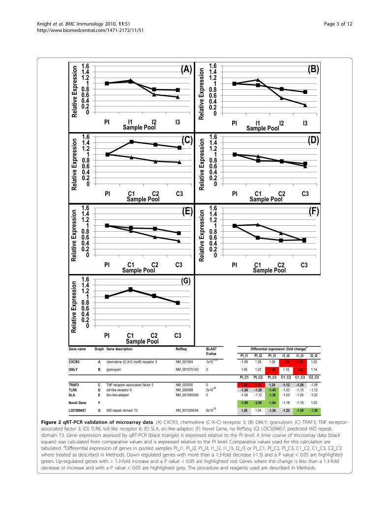
Figure 2 qRT-PCR validation of microarray data. (A) CXCR3, chemokine (C-X-C) receptor 3; (B) GNLY, granulysin; (C) TRAF3, TNF receptor-associated factor 3; (D) TLR6, toll-like receptor 6; (E) SLA, src-like-adaptor; (F) Novel Gene, no RefSeq; (G) LOC509457, predicted WD repeatdomain 73. Gene expression assessed by qRT-PCR (black triangle) is expressed relative to the PI level. A time course of microarray data (blacksquare) was calculated from comparative values and is expressed relative to the PI level. Comparative values used for this calculation aretabulated. aDifferential expression of genes in pooled samples PI_I1, PI_I2, PI_I3, I1_I2, I1_I3, I2_I3 or PI_C1, PI_C2, PI_C3, C1_C2, C1_C3, C2_C3where treated as described in Methods. Down regulated genes with more than a 1.3-fold decrease (-1.3) and a P value < 0.05 are highlightedgreen. Up-regulated genes with > 1.3-fold increase and a P value < 0.05 are highlighted red. Genes where the change is less than a 1.3-folddecrease or increase and with a P value < 0.05 are highlighted grey. The procedure and reagents used are described in Methods.
Knight et al. BMC Immunology 2010, 11:51http://www.biomedcentral.com/1471-2172/11/51
Page 3 of 12
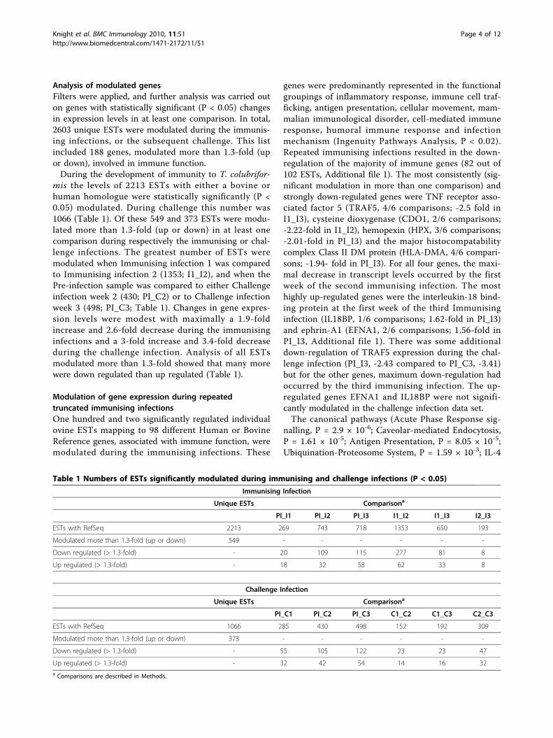
Analysis of modulated genesFilters were applied, and further analysis was carried outon genes with statistically significant (P < 0.05) changesin expression levels in at least one comparison. In total,2603 unique ESTs were modulated during the immunis-ing infections, or the subsequent challenge. This listincluded 188 genes, modulated more than 1.3-fold (upor down), involved in immune function.During the development of immunity to T. colubrifor-
mis the levels of 2213 ESTs with either a bovine orhuman homologue were statistically significantly (P <0.05) modulated. During challenge this number was1066 (Table 1). Of these 549 and 373 ESTs were modu-lated more than 1.3-fold (up or down) in at least onecomparison during respectively the immunising or chal-lenge infections. The greatest number of ESTs weremodulated when Immunising infection 1 was comparedto Immunising infection 2 (1353; I1_I2), and when thePre-infection sample was compared to either Challengeinfection week 2 (430; PI_C2) or to Challenge infectionweek 3 (498; PI_C3; Table 1). Changes in gene expres-sion levels were modest with maximally a 1.9-foldincrease and 2.6-fold decrease during the immunisinginfections and a 3-fold increase and 3.4-fold decreaseduring the challenge infection. Analysis of all ESTsmodulated more than 1.3-fold showed that many morewere down regulated than up regulated (Table 1).
Modulation of gene expression during repeatedtruncated immunising infectionsOne hundred and two significantly regulated individualovine ESTs mapping to 98 different Human or BovineReference genes, associated with immune function, weremodulated during the immunising infections. These
genes were predominantly represented in the functionalgroupings of inflammatory response, immune cell traf-ficking, antigen presentation, cellular movement, mam-malian immunological disorder, cell-mediated immuneresponse, humoral immune response and infectionmechanism (Ingenuity Pathways Analysis, P < 0.02).Repeated immunising infections resulted in the down-regulation of the majority of immune genes (82 out of102 ESTs, Additional file 1). The most consistently (sig-nificant modulation in more than one comparison) andstrongly down-regulated genes were TNF receptor asso-ciated factor 5 (TRAF5, 4/6 comparisons; -2.5 fold inI1_I3), cysteine dioxygenase (CDO1, 2/6 comparisons;-2.22-fold in I1_I2), hemopexin (HPX, 3/6 comparisons;-2.01-fold in PI_I3) and the major histocompatabilitycomplex Class II DM protein (HLA-DMA, 4/6 compari-sons; -1.94- fold in PI_I3). For all four genes, the maxi-mal decrease in transcript levels occurred by the firstweek of the second immunising infection. The mosthighly up-regulated genes were the interleukin-18 bind-ing protein at the first week of the third Immunisinginfection (IL18BP, 1/6 comparisons; 1.62-fold in PI_I3)and ephrin-A1 (EFNA1, 2/6 comparisons; 1.56-fold inPI_I3, Additional file 1). There was some additionaldown-regulation of TRAF5 expression during the chal-lenge infection (PI_I3, -2.43 compared to PI_C3, -3.41)but for the other genes, maximum down-regulation hadoccurred by the third immunising infection. The up-regulated genes EFNA1 and IL18BP were not signifi-cantly modulated in the challenge infection data set.The canonical pathways (Acute Phase Response sig-
nalling, P = 2.9 × 10⁻⁶; Caveolar-mediated Endocytosis,P = 1.61 × 10⁻⁵; Antigen Presentation, P = 8.05 × 10⁻⁵;Ubiquination-Proteosome System, P = 1.59 × 10⁻³; IL-4
Table 1 Numbers of ESTs significantly modulated during immunising and challenge infections (P < 0.05)
Immunising Infection
Unique ESTs Comparisona
PI_I1 PI_I2 PI_I3 I1_I2 I1_I3 I2_I3
ESTs with RefSeq 2213 269 743 718 1353 650 193
Modulated more than 1.3-fold (up or down) 549 - - - - - -
Down regulated (> 1.3-fold) - 20 109 115 277 81 8
Up regulated (> 1.3-fold) - 18 32 58 62 33 8
Challenge Infection
Unique ESTs Comparisona
PI_C1 PI_C2 PI_C3 C1_C2 C1_C3 C2_C3
ESTs with RefSeq 1066 285 430 498 152 192 309
Modulated more than 1.3-fold (up or down) 373 - - - - - -
Down regulated (> 1.3-fold) - 55 105 122 23 23 47
Up regulated (> 1.3-fold) - 32 42 54 14 16 32a Comparisons are described in Methods.
Knight et al. BMC Immunology 2010, 11:51http://www.biomedcentral.com/1471-2172/11/51
Page 4 of 12
signalling, P = 1.02 × 10⁻³) presented in Additional file 2had the lowest significance levels, an indication of theassociation between the pathway and the data set. Theexpression of genes associated with these pathwaystended to be modulated to the greatest degree betweenthe first week of the first Immunising infection (I1) andthe first week of the second Immunising infection (I2).The decrease in gene expression in early steps of the
acute phase response is consistent with the down-regu-lation of genes encoding TRAF6, TCF4 and STAT3, andup-regulation of HNRNPK. Modulation of transcriptlevels of genes encoding the acute phase response pro-teins FGG, HPX, C4, CRP and A2M (decrease) and RBP(increase) also support down-regulation of this response.Contrary to this, was the decrease in ALB, AMBP,APOH, ITIH2 and increase in ITIH4, FTL and LBPtranscript levels.Genes encoding proteins involved in Caveolar-mediated
Endocytosis, such as the integrins (ITGA3, ITGB1,ITGA9, ITGAV), the protein kinase, FYN, the regulatoryGTPase RAB5C, beta-2-microglobin (B2M), CD48 andalbumin (ALB) were all down-regulated, suggesting adown-regulation of this pathway. In contrast, the levels ofDMN2 encoding the GTPase dynamin 2 and ACTG2encoding actin increased. The genes encoding major histo-compatability Class II proteins (HLA-DMB, HLA-DRA,HLA-DRB1, HLA-DMA) responsible for MHC Class IIantigen presentation were down-regulated as were thegenes expressing TAP2, a transporter protein and PSEM2a component of the immunoproteosome involved in pre-sentation of antigen by major histocompatability complexClass I suggesting Antigen Presentation via both the ClassI and Class II pathways was affected during repeated trun-cated immunising infections. Further, a number of genesencoding ubiquitin-proteosome system componentsresponsible for targeting, via conjugation of multiple ubi-quitin units, of proteins for degradation by the proteosomewere modulated during the development of immunity.The expression of genes encoding the ubiquitinatingenzymes, UBE2D2 and UBE4A decreased while that ofUBE2J1 increased. Of the genes encoding proteinsinvolved in degradation; PSMD5 and USP15 weredecreased while USP10 and USP4 increased. Transcriptionof genes encoding the heat shock proteins, HSPA5,HSPA8, and HSP90AA1 was consistently reduced. Themajor histocompatability proteins, beta-2-microglobin(B2M) and the low affinity IgE receptor (FCER2 or CD23),all associated with IL-4 signalling, were down-regulatedwhile the ribosomal S6 kinase (RPS6KB2) was up-regu-lated in this data set.
Modulation of gene expression during challenge infectionOf the ESTs significantly regulated during thechallenge infection, 121 ovine ESTs mapping to 113
different Human or Bovine Reference genes werelinked to immune functions (Additional file 3), inclu-sive of genes associated with the inflammatoryresponse, immune cell trafficking, inflammatory dis-ease, antigen presentation, humoral immune response,cell-mediated immune response, lymphoid tissue struc-ture, infection mechanism, immunological disease andapoptosis of eukaryotic cells (P values < 0.02, IngenuityPathways Analysis Functional groupings). The majortrend during challenge was for a down-regulation inthe expression of these immune-associated genes, butthe degree of down-regulation was not as extensivewhen compared to the immunising infections (Addi-tional files 1 and 3). The most consistently up-regu-lated immune gene was selenoprotein S (SELS, 3/6comparisons). Expression increased 1.54-fold in thefirst week of the Challenge infection, compared to thePre-infection, and 1.75-fold in the third week afterChallenge infection, compared to the Pre-infectionsample. This gene was not significantly modulated inthe immunising infection data set. TNF receptor asso-ciated factor 5 (TRAF5) had the most consistently (3/6comparisons) down regulated transcript levels. Levelsdecreased 2.4-fold in the first week of the Challengeinfection, compared to the Pre-infection, and 3.4-foldafter the third week, compared to the Pre-infectionsample (Additional file 3). Over all, the most substan-tial fold-change for all genes modulated during thechallenge infection tended to occur when the firstweek of the Challenge infection (C1) was compared tothe Pre-infection (PI) sample pool.The most significant canonical pathways associated
with this data set were Acute Phase Response Signalling(P = 4.46 × 10⁻⁸), Complement Signalling (P = 4.5 × 10⁻³) and IL-4 Signalling (P = 1.22 × 10⁻²). During the chal-lenge infection 10% of genes encoding proteins involvedin the acute phase response were modulated (Additionalfile 4). Consistent with a decreased acute phase responsewere the down-regulation of STAT3 and RIPK1, up-regulation of HNRNPK and the modulation of acutephase response proteins C9, ITIH3, HPX, HP and ALBESTs. Inconsistent with the decreased acute phaseresponse was the observed increase in the expression ofTCF4, encoding transcription factor 4, and the down-regulation of acute phase response proteins AMBP, TF,ITIH2 and AHSG. A number of the key components ofthe complement system (C1QC, C3, C9, C7) weredown-regulated during the challenge infection. Theinterleukin 4 receptor (IL4R) was consistently up-regulated when either Challenge infection 1 or 2 poolswere compared to the Pre-infection pool. Major histo-compatability proteins (HLA-DMA, HLA-DQB1, B2M)and the low affinity IgE receptor (FCER2 or CD23), endproducts of IL4 signalling, were all down-regulated.
Knight et al. BMC Immunology 2010, 11:51http://www.biomedcentral.com/1471-2172/11/51
Page 5 of 12
DiscussionWe deliberately interrogated a large set of ESTs derivedprimarily from DCs on the arrays in order to maximiseour understanding of immune gene expression in a nat-ural cell pool which is highly enriched for DC. Thesecells migrate out of the intestinal mucosa which formsthe inter-face between host and nematode, into mesen-teric lymph nodes, where immunological responses areinitiated. This study describes global gene expressionprofiles of afferent lymph cells during the developmentof immunity to a gastrointestinal parasite. This under-standing is crucial for the development of immune mod-ulatory treatments, as well as for new treatment such asvaccines to control parasite infections.For this data we employed a pooling strategy of indivi-
dual samples. Analysis of cytokine gene expression inthese pools by qRT-PCR revealed the expected over-expression of Th2-type cytokines. This is evidence thatthe more persistent modulations of gene expression arenot obscured by this pooling strategy.Due to the difficult nature of experiments involving
collection of lymph for 13 weeks, not all the animalswere patent for the entire term of the experiment.Therefore we cannot exclude some degree of bias at thelate stages of the experiment.It is well established that many helminths are able to
produce a whole range of bioactive molecules that mod-ulate the immune response of a host [17]. Of thesemolecules, glycans have been shown to be key players ininducing Th2-type and anti-inflammatory responses[18-20]. Our data not only showed a down-regulation ofmany individual genes involved in immunity but alsoare indicative of a down-regulation of entire immunolo-gical pathways, although it remains to be determinedwhat aspects of the changes in gene expression are dueto the natural response of the host to GIN infection.These data are consistent with in vitro studies of hel-minth-stimulated monocyte-derived dendritic cells(MoDC) in which gene expression profiles are also indi-cative of non-responsiveness [21,22]. This may be dueto the production of immunomodulatory molecules bythe parasite effectively inducing an anti-inflammatoryenvironment sympathetic to parasite persistence [23].Soluble molecules from the eggs of the helminth Schis-tosoma mansoni (SEA) by themselves have a minimaleffect on MoDC but are capable of suppressing theexpression of pro-inflammatory genes usually presentafter LPS stimulation. It is hypothesised that suppressionof LPS activation may in part be due to increased pro-duction of IL-10, an anti-inflammatory cytokine [22,24].In general, stimulation of MoDC with helminth derivedproducts inhibit or partly inhibit DC maturationassessed by measurement of DC maturation markerssuch as CD86, CD40, OX40L, CD80 and MHC Class II
molecules [24-28]. Our data clearly show that underin vivo conditions the antigen presentation pathway isalso significantly down-regulated during repeated trun-cated immunising infections, but not during challengeof immune lambs. It remains to be determined if inhibi-tion of DC maturation also occurs during natural infec-tion with gastrointestinal nematode parasites, as seen inin vitro experiments. The predominant Th1-typeresponse of MoDC stimulated by bacterial lipopolysac-caride (LPS), marked by increased chemokine produc-tion [29] as well as expression of genes encodingproteins related to cell structure, antigen presentationand IFN-inducible proteins [30], was not detected at anystage, in lambs repeatedly infected and then challengedwith intestinal nematodes.Previous studies in sheep analysing changes in global
transcript levels associated with parasite resistance orinfection have involved analysis of abomasal, intestinaland associated immune tissues. In this study transcriptlevels have been assessed in immune cells that migratedirectly out of the tissue which harbours an intestinalnematode parasite. Microarray analysis of gene expres-sion profiles in both abomasal tissue and lymph nodeshas previously been used to investigate Haemonchuscontortus resistance in two different sheep breeds andsuggest that the more resistant breed has greater expres-sion of genes associated with the inflammatory response,gut motility, and cell differentiation and proliferation[13]. The comparisons of gene expression in intestinaltissue and associated immune tissues from sheep linesgenetically resistant or susceptible to GIN (H. contortus,T. colubriformis) [8,10,11], or by the sequential biopsy ofabomasal mucosa during H. contortus infection [15] sug-gest that genes involved in acquired immunity, oxidativestress, apoptosis and mucosal function are modulated.In these studies modulation of expression of ITGB1,THBS1 and GPX1 was found to be in common withthis study. The production of oxidants by the host, arethought to be anti-parasitic [31] and as such there isalso a requirement for antioxidants. The levels of theantioxidant glutathione are thought to fluctuate duringGIN infections [32]. A previous study [10] and thisstudy have found that transcription of glutathione per-oxidise (GPX1) increases during GIN infection, thussupporting an increase in antioxidant activities. Keaneet al [9] found the most highly expressed genes in intest-inal tissue of susceptible animals to be those involved inprotein degradation including a ubiquitin-like protein.Ingenuity Pathway Analysis of data presented in this
study is indicative of a down-regulation of many genesinvolved in protein ubiquitination, caveolar-mediatedendocytosis, both MHC Class I and Class II antigen pre-sentation during immunising infections, which togethercould contribute to the natural slow development of
Knight et al. BMC Immunology 2010, 11:51http://www.biomedcentral.com/1471-2172/11/51
Page 6 of 12

protective host immunity to gastrointestinal nematodeparasites. Protein ubiquitination is not only associatedwith the degradation of damaged proteins but in theregulation of cellular processes including immuneresponses such as antigen presentation and activation ofpro-inflammatory responses via the transcription factorNF-�B (reviewed in [33]). Intriguingly, the heat shockproteins encoded by HSP90AA1 and HSPA5 enhanceDC maturation. The former has also been shown toenhance the TLR-mediated production of pro-inflamma-tory cytokines [34,35]. In this study, the transcription ofboth the HSP90AA1 and HSPA5 genes was down-regulated during repeated immunising infections sugges-tive of an inhibition of DC maturation. Suppression ofDC maturation is well known from in vitro studies inwhich helminth products have been shown to regulateDC cytokine and cell surface molecule maturation mar-kers [24,25,28]. It remains to be established whether ornot heat shock proteins HSP90AA1 and HSPA5 areinvolved.A number of genes encoding proteins involved in
acute phase responses were also modulated in this dataset. Acute phase proteins (APPs) are predominantlysynthesised in the liver. However, their levels can alsobe modulated in non-hepatic tissues such as the lymphnode [36,37] and possibly in migrating lymph cells. Theexpression of genes encoding a group of APPs weredown-regulated, consistent with routine function duringthe acute phase response. One of the most highly down-regulated genes during the development of immunitywas hemopexin (HPX). The signalling adaptor moleculeTRAF6 and the transcription factor STAT3, bothinvolved in elicitation of an acute phase response, weredown-regulated while HNHKPK, a negative regulator ofthe response was up-regulated. The regulation of thesegenes was observed during both the development ofimmunity and the challenge infection. Modulation ofthese genes is consistent with a down-regulation of theacute phase response.Signalling is an essential process required for the inte-
gration of immune responses. A number of proteinsinvolved in such pathways were significantly modulatedduring the development of immunity to parasite infec-tion. TRAF proteins function as adaptor proteins inTNF signalling pathways and may play a role in immu-nity to gastrointestinal parasites [38]. TRAF5 transcriptlevels where the most significantly and consistentlymodulated during repeated immunising infections andremained low during the challenge infection. TRAF5 sig-nalling is involved in the development of Th2-typeresponses, as exemplified by knockout mice studies inwhich TRAF5-/- mice showed an enhanced Th2 pheno-type [39]. Our data clearly show the development of
Th2-type effector cytokine response during repeatedimmunising infections which is supported by the strongdown-regulation of TRAF5 signalling and the up-regula-tion of the IL-18 binding protein. Interleukin-18 bindingprotein (IL-18BP) binds and inhibits the biological activ-ity of the pro-inflammatory Th1-response promotingcytokine IL-18 [40]. IL-18 has also been implicated inthe susceptibility of mice to Trichuris muris infectionwhich is achieved via down-regulation of the productionof IL-13 [41]. The genes IL22RA1, IL20RA, IL10RB andIL10RA, encode receptors for members of the IL-10cytokine family (IL-10, IL-20, IL-22, IL-26). IL10RBassociates with IL22RA1 or with IL20RA to formrespectively the IL-22 or the IL-26 receptor whileIL20RA and IL20RB form the IL-20 receptor. The tran-scription of IL10RB, IL22RA1 and IL20RA were down-regulated during the development of immunity to T.colubriformis. Both IL-22 and IL-26 have been shown topromote pro-inflammatory gene expression in intestinalepithelial cells [42,43]. Recently the removal of IL-22was shown to have a protective effect in gut infectedwith the parasitic protozoan Toxoplasma gondii [44]. Itis feasible that these cytokines could also be involved inthe development of immunity to T. colubriformis. IL-22,IL-20, IL-26 and IL-18 have all been implicated ininflammatory conditions such as Crohn’s disease andPsoriasis in humans [42,43,45,46]. IL-4, a Th2 cytokine,plays a critical role in the host’s defence against gastro-intestinal parasites [47]. Our data show an up-regulationof the IL-4 receptor (IL4R) during the challenge infec-tion indicative of a Th2-type response. Other genessuch as those encoding major histocompatability pro-teins, the low affinity receptor for IgE (FCER2 or CD23)and beta-2-microgobulin (B2M) were down-regulated.These genes are associated with, but are not exclusiveto, IL-4 signalling.Under stress conditions, endoplasmic reticular function
is impaired resulting in activation of the transcription fac-tor NF-�B and the expression of pro-inflammatory cyto-kines. Maintenance of endoplasmic reticular integrityunder such conditions can involve selenoprotein S (SELS).A genetic variant of the SELS gene with impaired expres-sion and suppression of transcript levels with a SELS smallinterfering RNA suggests that a decrease in SELS expres-sion is linked to an increase in pro-inflammatory cytokinelevels [48]. Any elevation in SELS transcript levels duringimmunising infections and during parasite challenge istherefore consistent with the ability of parasites to inducethe suppression of an inflammatory response. Cysteinedioxygenase (CDO1) may be involved in the inflammatoryresponse to oxidative stress via the regulation of taurinemetabolism. Regulation of this involvement is howeverthought to be via a post-translational mechanism [49] andso further investigation would be required to determine
Knight et al. BMC Immunology 2010, 11:51http://www.biomedcentral.com/1471-2172/11/51
Page 7 of 12

the significance of the modulation of this gene during thedevelopment of immunity to gastrointestinal parasites.
ConclusionsOur findings present a global picture of the changes ingene expression in cells trafficking in afferent lymphover extended periods of time during the developmentof immunity and challenge with the gastrointestinalparasite T. colubriformis. This report describes geneexpression profiles in immune cells draining directlyfrom the intestinal mucosa; the interface between thehost and the nematode. Data are suggestive of a down-regulation of the expression of immune genes, a down-regulation that may be relevant to the development ofimmunity. These results lay the groundwork for furtherstudies on nematode mediated immune-modulationwhich results in this slow development of immunity togastrointestinal parasites. In particular, it remains to bedetermined if the observed changes in gene expressionare also seen on a protein level resulting in functionalchanges of pathways.
MethodsAnimals and experimental designAll animal experiments and surgical procedures wereapproved by the Animal Ethics Committee of the Walla-ceville Animal Research Centre. Animals were raisednematode free and were fed on a standardised diet con-sisting of sheep nuts and Lucerne chaff. Five outbrednematode naïve female Romney lambs were surgicallyfitted with lymphatic cannulae to enable continuous col-lection of afferent lymph for up to 13 weeks [16]. Lambswere immunized by orally infecting them with 50,000 T.colubriformis L3 larvae and the infection was terminatedwith oxfendazole (5 mg/kg; Systamex® COOPERS) 2weeks later. After a resting period of one week, the oralinfection followed by drenching procedure (= ‘truncated-immunising infection’) was repeated twice. A final drenchwas given at week 9 and sheep were then challenged with50,000 T. colubriformis a week later. The challenge infec-tion was allowed to develop for 3 weeks. Faecal eggcounts (FEC) of zero, determined at week 13, demon-strated the development of protective immunity. Thisregime of truncated-immunising infections and challengeinfection is shown diagrammatically in Figure 3A.
Sampling of lymphLymph was collected continuously into sterile flaskscontaining heparin (Sigma-Aldrich, St Louis, MO) andantibiotics (penicillin and streptomycin; Sigma). In addi-tion, up to four samples of 5 to 10 ml of fresh lymphwere collected under aseptic conditions over a period of15 to 30 min daily, into sample tubes containing heparinto allow sampling of cells for RNA extraction. Cells
(approximately 108) were promptly separated fromlymph plasma by centrifugation at 400 g for 3 min,washed once in cold PBS and re-suspended in RNAlater(Ambion, Austin, TX) to preserve integrity. Sampleswere stored at -20°C until subjected to RNA extraction.
ParasitologyInfective T. colubriformis larvae were cultured understandard procedures from eggs obtained from faeces ofa mono-specifically infected sheep. Faecal egg countswere performed on each animal at weekly intervalsthroughout the duration of the experiment using themodified McMaster method [50].
Microarray preparationOvine cDNA libraries were generated and single passsequenced to generate expressed sequence tags (ESTs).The amplification of cDNAs and array preparation wereas described previously [8]. An ovine microarray con-taining 10,458 amplified cDNAs and 119 control spotswas used. The majority (5215) of cDNAs printed on thearray were derived from immune tissue libraries, includ-ing isolated dendritic cells (5053) with the remaining162 from lymph node tissue, Peyers Patches and muco-sal lymphoid tissue. The other cDNAs printed onto thearray were from gall bladder (2638), liver (1748), foetaland reproductive tissue (734), wool follicles (96) andskin cells (30).
RNA isolation, fluorescent labelling and slidehybridisationTotal RNA was isolated from individual lymph cell sam-ples using Trizol (Invitrogen, Carlsbad, CA) and theappropriate RNA samples pooled and further purifiedusing an RNeasy kit (Qiagen, Hilden, Germany). RNAconcentrations were determined by the spectrophoto-metric measurement of absorption at 260 nm and everyRNA preparation was assessed for integrity and theabsence of genomic DNA contamination by agarose gelelectrophoresis. First strand cDNA labelled with eitherCy3 or Cy5 (Amersham Biosciences, Uppsala, Sweden)was produced from 20 μg of total pooled RNA using theSuperScript Indirect cDNA Labeling System (Invitro-gen). All procedures were as described by the manufac-turer. Cy3 and Cy5-labelled cDNA were pooled,concentrated to 10 μl by ethanol precipitation prior todenaturation at 95°C, combining with SlideHyb glassarray hybridisation buffer #1 (60 ul, Ambion) andapplied to the array. Array pre-hybridisation and hybri-disation conditions, scanning and image processing wereas described [8], except that hybridisation was at 52°C.Pools (PI, Pre-infection, week 0; I1, Immunising infec-
tion 1, week 1; I2, Immunising infection 2, week 4; I3,Immunising infection 3, week 7; C1, first week after
Knight et al. BMC Immunology 2010, 11:51http://www.biomedcentral.com/1471-2172/11/51
Page 8 of 12

Challenge infection, week 10; C2, second week afterChallenge infection, week 11; C3, third week after Chal-lenge infection, week 12; Figure 3A) consisted of equalamounts of total RNA from each animal and for eachday over a period of 7 days being pooled. Each pool wascompared with every other pool (Figure 3B). In total 12slides were hybridised to investigate the development ofimmunity (Immunising infections) and another 12 toinvestigate the challenge of immunised sheep (Challengeinfection), and included slides where first strand cDNAwas labelled with the opposite dye (dye swap). Thenumber of animals contributing to each pool changed as
not all animals had a patent cannula for the entireexperimental period of 13 weeks (Figure 3A).
Microarray normalisation and analysisData for individual slides were normalised as describedpreviously [8,51]. ESTs with either a normalised meanlog intensity < 9 and with > 6 bad (unreadable) spotsout of 12 were excluded from further analysis. The stan-dardized residual of the normalized log ratio of themean (SR_mean) was calculated for each EST and con-verted to a fold change. An EST was included forfurther bioinformatic analysis if for one of the
Treatment Sample Pool
wee
kdrench
50,000 TcImmunising Infection 1
50,000 TcImmunising Infection 2
drench
50,000 TcImmunising Infection 3
drench
50,000 TcChallenge Infection
drench
PI (N=5)
I1 (N=5)
I2 (N=4)
I3 (N=3)
C1 (N=3)
C2 (N=3)
C3 (N=2)
(A)Immunising
InfectionChallenge Infection
(B)
1
2
3
4
5
6
7
8
9
12
13
10
11
0
1
2
3
4
5
6
7
8
9
12
13
10
11
0
1
2
3
4
5
6
7
8
9
12
13
10
11
0
Figure 3 Experimental Design. (A) Truncated Immunising infection and Challenge protocol. Daily afferent lymph samples were collected over a13 week period. RNA was pooled (sample pools) after extraction from afferent lymph samples from weeks labelled PI (Pre-infection, week 0), I1(Immunising infection 1, week 1), I2 (Immunising infection 2, week 4), I3 (Immunising infection 3, week 7), C1 (Challenge infection, week 10), C2(Challenge infection, week 11) and C3 (Challenge infection, week 12). N; number of animals in each sample pool, solid arrows show infectionwith 50,000 T. colubriformis (Tc) and dashed arrows show drench treatment. (B) Hybridisation of RNA pools. Solid lines show the comparisonsmade; solid arrows show nematode infection and dashed arrows show drench treatments.
Knight et al. BMC Immunology 2010, 11:51http://www.biomedcentral.com/1471-2172/11/51
Page 9 of 12

comparisons the change was greater than 1.3 (up ordown) and the P value for this change was < 0.05. Themodulation threshold of 1.3-fold was slightly more strin-gent than that validated and used by others [52]. AHuman or Bovine Reference Sequence (ftp://ftp.ncbi.nih.gov/refseq/) corresponding to each EST was determinedby BLAST. Those with E values less than 1 × 10-19 wereincluded. Canonical pathways and Functional groupingswere generated through the use of Ingenuity PathwaysAnalysis (Ingenuity Systems, http://www.ingenuity.com).All the microarray data presented in this publication havebeen deposited in NCBI ‘s Gene Expression Omnibus(http://www.ncbi.nlm.nih.gov/geo/) under accession num-ber GSE23859 for the Immunising infection data set andGSE23863 for the Challenge infection data set.
Quantitative real-time PCRFirst-strand cDNA was synthesised from the sameRNA samples used in the microarray experiments.Synthesis of cDNA and the procedure used to quanti-tatively assess gene expression using quantitative real-time PCR (qRT-PCR) was done as described previously[5]. Oligonucleotides (Table 2) designed to amplify theovine homologue of the src-like adaptor (SLA), chemo-kine (C-X-C) receptor 3 (CXCR3), TNF receptor-asso-ciated factor 3 (TRAF3), toll-like receptor 6 (TLR6),granulysin (GNLY), predicted WD repeat domain 73(LOC509457) and a novel (no Reference Sequence)ovine EST (Novel Gene) were based on their ESTsequences. Prior to qRT-PCR the identity of the ESTspotted onto the array was confirmed by partialsequence analysis. Quantification of IL-5, IL-13, IL-4and IFNg levels within the same RNA samples wereassessed as described previously [16].
Additional material
Additional file 1: Changes in immune gene expression during theImmunising infections are tabulated. The data set is described:a Genes associated with Immune Function in the Ingenuity PathwaysKnowledge Base where at least one comparison has a P value < 0.05(shown in bold type). Differential expression of genes in pooled samplesPI_I1, PI_I2, PI_I3, I1_I2, I1_I3, I2_I3 where treated as described inMethods. Down-regulated genes with more than a 1.3-fold decrease(-1.3) and a P value < 0.05 are highlighted green. Up-regulated geneswith > 1.3-fold increase and a P value < 0.05 are highlighted red. Geneswhere the change is less than a 1.3-fold decrease or increase and with aP value < 0.05 are highlighted grey.
Additional file 2: The modulation of canonical pathways duringImmunising infections is tabulated. The data set is described:a Significance level. b Proportion of pathway associated genes. c Genesassociated with Canonical Pathways in the Ingenuity PathwaysKnowledge Base where at least one comparison has a P value < 0.05(shown in bold type). Differential expression of genes in pooled samplesPI_I1, PI_I2, PI_I3, I1_I2, I1_I3, I2_I3 are shown. Down-regulated geneswith more than a 1.3-fold decrease (-1.3) and a P value < 0.05 arehighlighted green. Up-regulated genes with > 1.3-fold increase and a Pvalue < 0.05 are highlighted red. Genes where the change is less than a1.3-fold decrease or increase and with a P value < 0.05 are highlightedgrey.
Additional file 3: Changes in immune gene expression during theChallenge infection are tabulated. The data set is described: a Genesassociated with Immune Function in the Ingenuity Pathways KnowledgeBase where at least one comparison has a P value < 0.05 (shown in boldtype). Differential expression of genes in pooled samples PI_C1, PI_C2,PI_C3, C1_C2, C1_C3, C2_C3 are shown. Down-regulated genes withmore than a 1.3-fold decrease (-1.3) and a P value < 0.05 are highlightedgreen. Up-regulated genes with > 1.3-fold increase and a P value < 0.05are highlighted red. Genes where the change is less than a 1.3-folddecrease or increase and with a P value < 0.05 are highlighted grey.
Additional file 4: The modulation of canonical pathways duringChallenge is tabulated. The data set is described: a Significance level.b Proportion of pathway associated genes. c Genes associated withCanonical Pathways in the Ingenuity Pathways Knowledge Base where atleast one comparison has a P value < 0.05 (shown in bold type).Differential expression of genes in pooled samples PI_I1, PI_I2, PI_I3,I1_I2, I1_I3, I2_I3 are shown. Down-regulated genes with more than a1.3-fold decrease (-1.3) and a P value < 0.05 are highlighted green. Up-regulated genes with > 1.3-fold increase and a P value < 0.05 arehighlighted red. Genes where the change is less than a 1.3-fold decreaseor increase and with a P value < 0.05 are highlighted grey.
AcknowledgementsThanks to Sally-Ann Cole for her expert technical assistance, to TheresaWilson and Dianne Hyndman for advice and microarray printing, to AlanMcCulloch and Deborah Simon for bioinformatics advice and to IanSutherland for reviewing the manuscript and for helpful suggestions.
Author details1AgResearch Ltd., Hopkirk Research Institute, Grasslands Research Centre,Palmerston North 4442, New Zealand. 2VSN (NZ) Ltd., 40 McMahon Drive,Aidanfield, Christchurch 8025, New Zealand.
Authors’ contributionsJSK performed all the RNA extractions, the microarray experiments andanalysed the data using Ingenuity Pathways Analysis. DBB was responsiblefor microarray experiment design and performed the statistical analysis. WRHand AP conceived the study, performed the cannulation surgery, animalinfections and sampling. AP co-ordinated the study. JSK and AP wrote themanuscript. All authors read and approved the final manuscript.
Table 2 Oligonucleotide sequences of ovine GAPDH anda selected range of genes employed for SYBR Green realtime PCR
Gene Forward primer (5’-3’) Reverse primer (5’-3’)
GAPDH CACCATCTTCCAGGAGCGAG CCAGCATCACCCCACTTGAT
NovelGene
CAAGCTAAAGGCAGCATCCC TCTCCCTCATAAGCCTGGAGC
GNLY GGTCTGCAAAAGCAAGGCAG TCAGAGGACCCAGGGAATCA
SLA ACCACGGTTGGCTGTTTGAA GCAGCTCCTCAGCCTTGTCT
CXCR3 GTGCTGACACTCCCTCTCTGG AAAGACCCACTGGATGGCTG
TRAF3 CTTCTGTGAGACCTGCATGGG CATTTTGGGCTGGAGGAGC
TLR6 AATGACTTTGATGCCCTGCC CTGGGTCAAGTTGCCAAATTC
LOC509457 AAGGGATACGGGAACTTGGC AAAGGGCTTCATTGCTGAGCa Primers were designed to the ovine homologues of glyceraldehyde 3-phosphate dehydrogenase (GAPDH), the src-like adaptor (SLA), chemokine (C-X-C) receptor 3 (CXCR3), TNF receptor-associated factor 3 (TRAF3), toll-likereceptor 6 (TLR6), granulysin (GNLY), predicted WD repeat domain 73(LOC509457) and a novel ovine EST (Novel Gene).
Knight et al. BMC Immunology 2010, 11:51http://www.biomedcentral.com/1471-2172/11/51
Page 10 of 12
Received: 17 June 2010 Accepted: 17 October 2010Published: 17 October 2010
References1. Kaminsky R: Drug resistance in nematodes: a paper tiger or a real
problem? Current Opinion in Infectious Diseases 2003, 16(6):559-564.2. Besier B: New anthelmintics for livestock: the time is right. Trends in
Parasitology 2007, 23(1):21-24.3. Emery DL, McClure SJ, Wagland BM, Jones WO: Studies of stage-specific
immunity against Trichostrongylus colubriformis in sheep: immunizationby normal and truncated infections. International Journal for Parasitology1992, 22(2):215-220.
4. Stankiewicz M, Cabaj W, Pernthaner A, Hadas E: Immunisation of sheep bydrug-abbreviated infections of Ostertagia circumcincta andTrichostrongylus colubriformis against field challenge of gastro-intestinalnematodes. Veterinary Parasitology 1996, 67(1-2):121-132.
5. Pernthaner A, Cole SA, Morrison L, Hein WR: Increased expression ofinterleukin-5 (IL-5), IL-13, and tumor necrosis factor alpha genes inintestinal lymph cells of sheep selected for enhanced resistance tonematodes during infection with Trichostrongylus colubriformis. Infection& Immunity 2005, 73(4):2175-2183.
6. Gasbarre LC: Effects of gastrointestinal nematode infection on theruminant immune system. Vet Parasitol 1997, 72(3-4):327-337, discussion337-343.
7. Almeria S, Canals A, Gomez-Munoz MT, Zarlenga DS, Gasbarre LC:Characterization of protective immune responses in local lymphoidtissues after drug-attenuated infections with Ostertagia ostertagi incalves. Vet Parasitol 1998, 80(1):53-64.
8. Diez-Tascon C, Keane OM, Wilson T, Zadissa A, Hyndman DL, Baird DB,McEwan JC, Crawford AM: Microarray analysis of selection lines fromoutbred populations to identify genes involved with nematode parasiteresistance in sheep. Physiological Genomics 2005, 21(1):59-69.
9. Keane OM, Zadissa A, Wilson T, Hyndman DL, Greer GJ, Baird DB,McCulloch AF, Crawford AM, McEwan JC: Gene expression profiling ofnaive sheep genetically resistant and susceptible to gastrointestinalnematodes. BMC Genomics 2006, 7:42.
10. Menzies M, Reverter A, Andronicos N, Hunt P, Windon R, Ingham A:Nematode challenge induces differential expression of oxidant,antioxidant and mucous genes down the longitudinal axis of the sheepgut. Parasite Immunology 2010, 32(1):36-46.
11. Andronicos N, Hunt P, Windon R: Expression of genes in gastrointestinaland lymphatic tissues during parasite infection in sheep geneticallyresistant or susceptible to Trichostrongylus colubriformis andHaemonchus contortus. International Journal for Parasitology 2010,40(4):417-429.
12. Ingham A, Reverter A, Windon R, Hunt P, Menzies M: Gastrointestinalnematode challenge induces some conserved gene expression changesin the gut mucosa of genetically resistant sheep. International Journal forParasitology 2008, 38(3-4):431-442.
13. MacKinnon KM, Burton JL, Zajac AM, Notter DR: Microarray analysis revealsdifference in gene expression profiles of hair and wool sheep infectedwith Haemonchus contortus. Veterinary Immunology & Immunopathology2009, 130(3-4):210-220.
14. Rowe A, Gondro C, Emery D, Sangster N: Genomic analyses ofHaemonchus contortus infection in sheep: Abomasal fistulation and twoHaemonchus strains do not substantially confound host geneexpression in microarrays. Veterinary Parasitology 2008, 154(1-2):71-81.
15. Rowe A, Gondro C, Emery D, Sangster N: Sequential microarray to identifytiming of molecular responses to Haemonchus contortus infection insheep. Veterinary Parasitology 2009, 161(1-2):76-87.
16. Hein WR, Barber T, Cole SA, Morrison L, Pernthaner A: Long-term collectionand characterization of afferent lymph from the ovine small intestine.Journal of Immunological Methods 2004, 293(1-2):153-168.
17. Johnston MJ, MacDonald JA, McKay DM: Parasitic helminths: apharmacopeia of anti-inflammatory molecules. Parasitology 2009,136(2):125-147.
18. Atochina O, Harn D: Prevention of psoriasis-like lesions development infsn/fsn mice by helminth glycans. Experimental Dermatology 2006,15(6):461-468.
19. Harn DA, McDonald J, Atochina O, Da’dara AA: Modulation of hostimmune responses by helminth glycans. Immunological Reviews 2009,230(1):247-257.
20. Terrazas CA, Gomez-Garcia L, Terrazas LI: Impaired pro-inflammatorycytokine production and increased Th2-biasing ability of dendritic cellsexposed to Taenia excreted/secreted antigens: A critical role forcarbohydrates but not for STAT6 signaling. International Journal forParasitology 2010, 40(9):1051-1062.
21. Chaussabel D, Semnani RT, McDowell MA, Sacks D, Sher A, Nutman TB:Unique gene expression profiles of human macrophages and dendriticcells to phylogenetically distinct parasites. Blood 2003, 102(2):672-681.
22. Kane CM, Cervi L, Sun J, McKee AS, Masek KS, Shapira S, Hunter CA,Pearce EJ: Helminth antigens modulate TLR-initiated dendritic cellactivation. Journal of Immunology 2004, 173(12):7454-7461.
23. Soares MF, Araujo CA: Helminth products as a potential therapeuticstrategy for inflammatory diseases. Inflammation & Allergy Drug Targets2008, 7(2):113-118.
24. Rigano R, Buttari B, Profumo E, Ortona E, Delunardo F, Margutti P, Mattei V,Teggi A, Sorice M, Siracusano A: Echinococcus granulosus antigen Bimpairs human dendritic cell differentiation and polarizes immaturedendritic cell maturation towards a Th2 cell response. Infection &Immunity 2007, 75(4):1667-1678.
25. Cervi L, MacDonald AS, Kane C, Dzierszinski F, Pearce EJ: Cutting edge:dendritic cells copulsed with microbial and helminth antigens undergomodified maturation, segregate the antigens to distinct intracellularcompartments, and concurrently induce microbe-specific Th1 andhelminth-specific Th2 responses. Journal of Immunology 2004,172(4):2016-2020.
26. Balic A, Harcus Y: Selective maturation of dendritic cells byNippostrongylus brasiliensis-secreted proteins drives Th2 immuneresponses. European Journal of Immunology 2004, 34(11):3047-3059.
27. Jenkins SJ, Mountford AP: Dendritic cells activated with products releasedby schistosome larvae drive Th2-type immune responses, which can beinhibited by manipulation of CD40 costimulation. Infection & Immunity2005, 73(1):395-402.
28. Langelaar M, Aranzamendi C, Franssen F, Van der Giessen J, Rutten V, Vander Ley P, Pinelli E: Suppression of dendritic cell maturation by Trichinellaspiralis excretory/secretory products. Parasite Immunology 2009,31(10):641-645.
29. Messmer D, Messmer B, Chiorazzi N: The global transcriptional maturationprogram and stimuli-specific gene expression profiles of human myeloiddendritic cells. International Immunology 2003, 15(4):491-503.
30. Hashimoto SI, Suzuki T, Nagai S, Yamashita T, Toyoda N, Matsushima K:Identification of genes specifically expressed in human activated andmature dendritic cells through serial analysis of gene expression. Blood2000, 96(6):2206-2214.
31. Chiumiento L, Bruschi F: Enzymatic antioxidant systems in helminthparasites. Parasitology Research 2009, 105(3):593-603.
32. Liu SM, Smith TL, Palmer DG, Karlsson LJE, Besier RB, Greeff JC: Biochemicaldifferences in Merino sheep selected for resistance against gastro-intestinal nematodes and genetic and nutritional effects on faecal wormegg output. Animal Science 2005, 81(1):149-157.
33. Ben-Neriah Y: Regulatory functions of ubiquitination in the immunesystem. Nature Immunology 2002, 3(1):20-26.
34. Chen K, Huang J, Gong W, Iribarren P, Dunlop NM, Wang JM: Toll-likereceptors in inflammation, infection and cancer. InternationalImmunopharmacology 2007, 7(10):1271-1285.
35. Kuppner MC, Gastpar R, Gelwer S, Nossner E, Ochmann O, Scharner A,Issels RD: The role of heat shock protein (hsp70) in dendritic cellmaturation: hsp70 induces the maturation of immature dendritic cellsbut reduces DC differentiation from monocyte precursors. EuropeanJournal of Immunology 2001, 31(5):1602-1609.
36. Leak LV, Liotta LA, Krutzsch H, Jones M, Fusaroa VA, Ross SJ, Zhao Y,Petricoin EF III: Proteomic analysis of lymph. Proteomics 2004, 4(3):753-765.
37. Skovgaard K, Mortensen S, Boye M, Poulsen KT, Campbell FM, Eckersall PD,Heegaard PM: Rapid and widely disseminated acute phase proteinresponse after experimental bacterial infection of pigs. VeterinaryResearch 2009, 40(3):23.
38. Wajant H, Henkler F, Scheurich P: The TNF-receptor-associated factorfamily: Scaffold molecules for cytokine receptors, kinases and theirregulators. Cellular Signalling 2001, 13(6):389-400.
Knight et al. BMC Immunology 2010, 11:51http://www.biomedcentral.com/1471-2172/11/51
Page 11 of 12
39. So T, Salek-Ardakani S, Nakano H, Ware CF, Croft M: TNF receptor-associated factor 5 limits the induction of Th2 immune responses.Journal of Immunology 2004, 172(7):4292-4297.
40. Novick D, Kim SH, Fantuzzi G, Reznikov LL, Dinarello CA, Rubinstein M:Interleukin-18 binding protein: a novel modulator of the Th1 cytokineresponse. Immunity 1999, 10(1):127-136.
41. Helmby H, Takeda K, Akira S, Grencis RK: Interleukin (IL)-18 promotes thedevelopment of chronic gastrointestinal helminth infection bydownregulating IL-13. Journal of Experimental Medicine 2001,194(3):355-364.
42. Brand S, Beigel F, Olszak T, Zitzmann K, Eichhorst ST, Otte JM, Diepolder H,Marquardt A, Jagla W, Popp A, et al: IL-22 is increased in active Crohn’sdisease and promotes proinflammatory gene expression and intestinalepithelial cell migration. American Journal of Physiology - Gastrointestinal &Liver Physiology 2006, 290(4):G827-838.
43. Dambacher J, Beigel F, Zitzmann K, De Toni EN, Goke B, Diepolder HM,Auernhammer CJ, Brand S: The role of the novel Th17 cytokine IL-26 inintestinal inflammation. Gut 2009, 58(9):1207-1217.
44. Wilson MS, Feng CG, Barber DL, Yarovinsky F, Cheever AW, Sher A, Grigg M,Collins M, Fouser L, Wynn TA: Redundant and pathogenic roles for IL-22in mycobacterial, protozoan, and helminth infections. Journal ofImmunology 2010, 184(8):4378-4390.
45. Monteleone G, Trapasso F, Parrello T, Biancone L, Stella A, Iuliano R, Luzza F,Fusco A, Pallone F: Bioactive IL-18 expression is up-regulated in Crohn’sdisease. Journal of Immunology 1999, 163(1):143-147.
46. Wolk K, Witte E, Warszawska K, Schulze-Tanzil G, Witte K, Philipp S, Kunz S,Docke WD, Asadullah K, Volk HD, et al: The Th17 cytokine IL-22 inducesIL-20 production in keratinocytes: a novel immunological cascade withpotential relevance in psoriasis. European Journal of Immunology 2009,39(12):3570-3581.
47. Finkelman FD, Shea-Donohue T, Morris SC, Gildea L, Strait R, Madden KB,Schopf L, Urban JF Jr: Interleukin-4- and interleukin-13-mediated hostprotection against intestinal nematode parasites. Immunol Rev 2004,201:139-155.
48. Curran JE, Jowett JB, Elliott KS, Gao Y, Gluschenko K, Wang J, AbelAzim DM, Cai G, Mahaney MC, Comuzzie AG, et al: Genetic variation inselenoprotein S influences inflammatory response. Nature Genetics 2005,37(11):1234-1241.
49. Schuller-Levis GB, Park E: Taurine and its chloramine: modulators ofimmunity. Neurochemical Research 2004, 29(1):117-126.
50. Whitlock HV: Some modifications of the McMaster helminth egg-counting techniques and apparatus. Journal of the Council of Scientific andIndustrial Research 1948, 21:177-180.
51. Baird D, Johnstone P, Wilson T: Normalization of microarray data using aspatial mixed model analysis which includes splines. Bioinformatics 2004,20(17):3196-3205.
52. Araujo RN, Padilha T, Zarlenga D, Sonstegard T, Connor EE, Van Tassel C,Lima WS, Nascimento E, Gasbarre LC: Use of a candidate gene array todelineate gene expression patterns in cattle selected for resistance orsusceptibility to intestinal nematodes. Veterinary Parasitology 2009, 162(1-2):106-115.
doi:10.1186/1471-2172-11-51Cite this article as: Knight et al.: The gastrointestinal nematodeTrichostrongylus colubriformis down-regulates immune gene expressionin migratory cells in afferent lymph. BMC Immunology 2010 11:51.
Submit your next manuscript to BioMed Centraland take full advantage of:
• Convenient online submission
• Thorough peer review
• No space constraints or color figure charges
• Immediate publication on acceptance
• Inclusion in PubMed, CAS, Scopus and Google Scholar
• Research which is freely available for redistribution
Submit your manuscript at www.biomedcentral.com/submit
Knight et al. BMC Immunology 2010, 11:51http://www.biomedcentral.com/1471-2172/11/51
Page 12 of 12