the fulfillment of degree of doctor of philosophy
TRANSCRIPT

i
Isolation of Secondary Metabolites of
Berberis calliobotrys, Caragana ambigua and
Vincetoxicum stocksii and their In Vitro
Screening in Various Bioassays
Thesis Submitted
For
The Fulfillment of Degree of
Doctor of Philosophy
By
Saima Khan
(M.Sc., M.phil.)
Department of Chemistry
The Islamia University of Bahawalpur
(2014-2019)

ii

DECLARTION
iii
DECLARATION
I, Saima Khan, Ph.D. Scholar in Chemistry Department, The Islamia University
of Bahawalpur, hereby declare that the dissertation entitled “Isolation of Secondary
Metabolites of Berberis calliobotrys, Caragana ambigua and Vincetoxicum stocksii
and their In Vitro Screening in Various Bioassays” is done by me under the
supervision of Prof. Dr. Muhammad Saleem, Department of Chemistry, The Islamia
University of Bahawalpur and it has not formed the basis for the award of any other
Degree or other similar title to any candidate of any University. This thesis contains no
material that has been submitted previously, in whole or in part, for the award of any
other academic degree or diploma. I also corroborate that nothing has been integrated in
this research without acknowledgement.
Saima Khan

CERTIFICATE
iv
CERTIFICATE
It is certified that research work presented in this dissertation, entitled “Isolation
of Secondary Metabolites of Berberis calliobotrys, Caragana ambigua and
Vincetoxicum stocksii and their In Vitro Screening in Various Bioassays”, has been
carried out by Ms. Saima Khan under my supervision for the fulfillment of the
requirements of Ph.D. degree in Chemistry during the session 2014-2019. It is hereby
recommended for submission and further process for the award of Ph.D. degree.
Prof. Dr. Muhammad Saleem
Research Supervisor
Department of Chemistry
The Islamia University of Bahawalpur,
Pakistan
The Chairman
Department of Chemistry,
The Islamia University of Bahawalpur
Pakistan

Acknowledgement
v
Acknowledgements
In the name of Allah, the Omnipotent, the most Merciful, the Compassionate,
Who blessed us with the perfect code of life and Whoseelegance resulted into my
success. All respects for the most perfect personality of the world Hazrat Muhammad
(Peace Be Upon Him) who enlighten our minds to recognize our creator.
The writing of acknowledgements after completion of research and writing thesis
is a pleasant task to pay thanks to those who provided their guidance and co-operated. It
is very difficult to write thesis without co-operation, guidance and help of a teacher, for
this reason, I am very thankful to my research supervisor Prof. Dr. Muhammad Saleem
whose suggestions, care and hard work helped me to complete my research work and
thesis. I am obliged to my respected teachers Prof. Dr. Abdul Jabbar, Prof. Dr. Naheed
Riaz and Prof. Dr. Shazia Anjum for their guidance and motivation.I am thankful to my
great mentor Prof. Dr Abdul Rauf who always inspired me and whose precious prayers
helped me in the journey of achieving this task. To facilitate documentation time to time
during the whole study, I am thankful to the Chairman, Department of Chemistry,
I am especially indebted toProf. Dr.Muhammad Ashraf, Sir Hammad Saleem,
Prof. Dr.Gokhan Zengin, Prof. Dr. Ishtiaq Ahmad and Prof. Dr. Fawzi Mohamad
Mohomodally for their cooperation in providing several research supports.
Above all, I wish to express my deepest love and gratitude to my parents,
brothers, sisters, bhabi, uncle, anti and other family members for the encouragement and
support they have provided me and their countless prayers for my success.I am happy to
acknowledge to my sweet nieces Areeba, Aneeka, Laiba, Rameeza and Bareera for
their prayers.
I am also thankful to my sweet friend Mahreen Mukhtar for everything she did
which cannot be expressed in words. May Allah Almighty bless her.
Acknowledgement
vi
Next I am thankful to my dear friend Rabbia Ahmed for her guidance and
assistance as well as moral support during research work.
I am grateful to all of my laboratory fellows Liaqat Ali, Natasha Shazmeen,
Humna Tahir, Romaisa Maqsood, Ghzala Fazal, Saima Muzzafar, Bushra Bashir,
Dur-e-Shahwar Jamil, Maria Aslam, Bushra Siddique, Iqra Siddique, Shabnum
Mustafa, Saima Naz, Umber, Fatima for always being helpful. I thereby express my
thanks to all technical and non-technical staff of the department for their co-operation.
Saima Khan

ABSTRACT
vii
ABSTRACT
Throughout the journey of mankind, reliance of man on plant to alleviate
sufferingsis still considered with more awareness for their therapeutic application.
Petition of tradition medicinal system is because of its less cost, safer and easily
accessibility. Therefore,in the present era, interest in the elucidation of biological
potential and chemical constituent of plant is ever budding among the naturopathic.
In under discussion studies, three medicinal plants: Berberis calliobotrys,
Caragana ambigua and Vincetoxicum stocksii were selected to be investigated for their
secondary metabolites. The whole work done during these studies has been embodied in
this dissertation as five chapters. Chapter 1containsthe introduction ofthe natural
products, their sources and some recent drugs approved by FDA available in market with
natural product framework and offers background of the present study. Chapter
2describesthemedicinal importance, worldwide distribution and reported secondary
metabolites with biological potential from the genera Berberis, Caragana and
Vincetoxicum.It also comprises the detailed literature survey on Berberis calliobotrys,
Caragana ambigua,and Vincetoxicum stocksii.
Chapter 3 discusses the biological screeningin various bioassays(DPPH, ABTS,
FRAP, CUPRAC, phosphomolybdenum, metal chelation, acetylcholinesterase (AChE),
butyrylcholinesterase (BChE), α-glucosidase, α-amylase and tyrosinase) and
phytochemical analyses (total phenolic contents and total flavonoid contents)ofcrude
extracts of three plants with their qualitative UHPLC-MS dataobtained for fractions Bc-
M, Bc-E, BC-W, Ca-M, Ca-E. It also describes theresult and discussion of isolated
compounds from the Berberis calliobotrys, Caragana ambigua, and Vincetoxicum
stocksii with detailed structure elucidation and isolation scheme. The biological activities
of crude extract, with experimental method applied in our investigation are also included.
Biological screening of crude extract in above said assays for Berberis calliobotry sand
Caragana ambigua are also reported in journal of industrial crops and product (Khan, S.,
Nazir, M., Saleem, H., Riaz, N., Saleem, M., Anjum, S. M. M., Zengin, G.,M.,

ABSTRACT
viii
Mukhtar,Tousif, M. I., Mahomoodally, F. M.,Ahemad, N.(2019) Valorization of the
antioxidant, enzyme inhibition and phytochemical propensities of Berberis calliobotrys
Bien. ex Koehne: A multifunctional approach to probe for bioactive natural
products.Industrial Crops and Products,141, 111693; Khan, S., Nazir, M., Raiz, N.,
Saleem, M., Zengin, G., Fazal, G., Saleem, M., Mukhtar, M., Tousif, M. I., Tareen,
R. B., Abdallah, H. H. and Mahomoodally, F. M.(2019). Phytochemical profiling, in
vitro biological properties and in silico studies on Caragana ambigua stocks (Fabaceae):
A comprehensive approach. Industrial Crops and Product, 131, 117-124). The natural
products isolated from Vincetoxicumstocksii are reportedin journal of chemical society of
pakistan (Khan, S., M., Tousif, Raiz, N., M., Raiz, Mukhtar, M., Ahmad, I., Tareen,
R. B., Jabbar, A. and Saleem, M. (2019) Rarely occurring natural products isolated
from Vincetoxicum stocksii,The Journal Of Chemical Society Of Pakistan, 41 (4), 695-
700.
Chapter 4 describes general experimental methods and techniques employed for
biological screening in various bioassays and phytochemical analyses of crude extracts of
three plants with their qualitative UHPLC-MS data obtained. It also contains
spectroscopic characterization of isolated compounds.
Chapter 5elaborates the assays or protocol applied for Berberis calliobotrys,
Caragana ambigua, and Vincetoxicum stocksii extract.

Table of Contents
ix
Table of Contents
DECLARATION ................................................................................................................................... III
CERTIFICATE...................................................................................................................................... IV
ACKNOWLEDGEMENTS ..................................................................................................................... V
ABSTRACT .......................................................................................................................................... VII
CHAPTER 1 ALLURING NATURAL PRODUCTS AND THEIR APPLICATIONS IN DRUG
DEVELOPMENT ................................................................................................................................................... 1
1.1. INTRODUCTION OF NATURAL PRODUCTS ................................................................................... 2 1.2. APPEALING POOLS OF BIOACTIVE NATURAL PRODUCTS ...................................................................... 2
1.2.1. Plants as appealing pool of bioactive natural products....................................................... 3 1.2.2. Microbes as appealing pool of bioactive natural products ........................................................ 7 1.2.3. Marine source as appealing pool of bioactive natural products ................................................ 9
1.3 A QUICK GLANCE ON SOME NATURAL PRODUCT-BASED MEDICINES IN NEAR PAST ........................ 11
CHAPTER 2 ........................................................................................................................................... 22
PREVIOUS PHYTOCHEMICAL INVESTIGATION ON THE
BERBERISCALLIBOTRAYS,CARAGANA AMBIGUA AND VINCETOXICUM STOCKSII ................................ 22
PART A ................................................................................................................................................... 23
2.1. PREVIOUS PHYTOCHEMICAL INVESTIGATION OF THE GENUS BERBERIS................................................. 23 2.2. PREVIOUS PHYTOCHEMICAL INVESTIGATION OF BERBERIS CALLIOBOTRYS ............................................. 28 2.3. CLASSIFICATION OF BERBERIS CALLIOBOTRYS ..................................................................................... 28
PART B ................................................................................................................................................... 30
2.4. PREVIOUS PHYTOCHEMICAL INVESTIGATION OF THE GENUS CARAGANA ............................................... 30 2.5. PREVIOUS PHYTOCHEMICAL INVESTIGATION OF CARAGANA AMBIGUA .................................................. 35 2.6. CLASSIFICATION OF CARAGANA AMBIGUA ........................................................................................... 36
PART C ................................................................................................................................................... 37
2.7. PREVIOUS PHYTOCHEMICAL INVESTIGATION OF THE GENUS VINCETOXICUM ........................................ 37 2.8. PREVIOUS PHYTOCHEMICAL INVESTIGATION OF VINCETOXICUM STOCKSII............................................. 39 2.9. CLASSIFICATION OF VINCETOXICUM STOCKSII ..................................................................................... 39 2.10. RESEARCH HYPOTHESIS ................................................................................................................ 40
2.11. RESEARCH PROBLEM AND OBJECTIVES OF THE STUDY ..................................................................... 40
CHAPTER 3 ........................................................................................................................................... 42
RESULTS AND DISCUSSION: BIOLOGIAL SCREENING AND PHYTOCHEMICAL ANALYSES
OF BERBERIS CALLIOBOTRYS, CARAGANA AMBIGUA AND VINCETOXICUM STOCKSII;AND
ISOLATION OF SECONDARY METABOLITES ............................................................................................... 42
PART A ................................................................................................................................................... 43
3.1. BIOLOGICAL SCREENING OF CRUDE EXTRACTS OF BERBERIS CALLIOBOTRYS ......................................... 43 3.2 PHYTOCHEMICAL ANALYSIS OF SECONDARY METABOLITES FROM BERBERIS CALLIOBOTRYS ................... 45
3.2.1 Total phenolic and flavonoid contents estimation ..................................................................... 45
3.2.2.1 UHPLC-MS Analysis for Identification of Secondary Metabolites of B. ................................. 46 3.2.2.2 Secondary metabolite identification of Bc-M through UHPLC-MS analysis ........................... 46 3.2.2.3 Secondary metabolite estimation of Bc-E through UHPLC-MS analysis ................................ 49 3.2.2. Secondary metabolite determination of Bc-W through UHPLC-MS analysis ............................ 52
3.3. SPECTROSCOPIC CHARACTERIZATION OF SECONDARY METABOLITES ISOLATED FROM B.CALLIOBOTRYS 55 3.3.1. Structure Elucidation of 4-Hydroxybenzoic acid (214) ............................................................ 55 3.3.2. Structure Elucidation of Methyl p-Coumarate (235)................................................................ 56 3.3.3. Structure Elucidation of Octadecyl-p-cumarate (236) ............................................................. 56
3.3.4. Structure Elucidation of Corydaldine (121) ............................................................................ 58 3.3.5. Structure Elucidation of N-methyl Corydaldine (122).............................................................. 58 3.3.6. Structure Elucidation of Armepavine (207) ............................................................................. 59

Table of Contents
x
3.3.7. Structure Elucidation of Berberine (83) .................................................................................. 60 3.3.8. Structure Elucidation of Columbamine (237) .......................................................................... 61 3.3.9. Structure Elucidation of Syringaresinol (238) ......................................................................... 62
PART B ................................................................................................................................................... 66
3.4. BIOLOGICAL SCREENING OF CRUDE EXTRACTS OF CARAGANA AMBIGUA ............................................... 66
3.5. PHYTOCHEMICAL ANALYSIS OF SECONDARY METABOLITES FROM CARAGANA AMBIGUA ........................ 67 3.5.1 Total phenolic and flavonoid contents estimation ..................................................................... 67 3.5.2. UHPLC-MS Analysis to Identify Secondary Metabolites of Caragana ambigua ....................... 68 3.5.2.1. Secondary metabolite estimation of Ca-M through UHPLC-MS analysis .............................. 68 3.5.2.2 Secondary metabolite estimation of Ca-E through UHPLC-MS analysis ................................ 71
3.6. CHARACTERIZATION OF SECONDARY METABOLITES ISOLATED FROM THE FRACTION CA-E ................... 73 3.6.1. Structure Elucidation of Taraxerol (262) ................................................................................ 74 3.6.2. Structure Elucidation of Teraxerol acetate (263) .................................................................... 75
3.6.3. Structure Elucidation of 2′-(4-Hydroxyphenyl)-Ethyl Stearate (264) ........................................ 76 3.6.4. Structure Elucidation of Apigenin (265) ................................................................................. 77 3.6.5. Structure Elucidation of Naringinin (249) .............................................................................. 78 3.6.6 Structure Elucidation of Kaempheride (222) ............................................................................ 79 3.6.7. Structure Elucidation of Quercetin (125) ................................................................................ 80 3.6.8. Structure Elucidation of Quercetin 3-O-β-D-glucopyranoside (198) ........................................ 80 3.4.9. Structure Elucidation of β-Sitosterol 3-O-D-glucopyranoside (172)......................................... 81
PART C ................................................................................................................................................... 83
3.7. BIOLOGICAL SCREENING OF CRUDE EXTRACT OF VINCETOXICUM STOCKSII .......................................... 83 3.8. CHARACTERIZATION OF SECONDARY METABOLITES ISOLATED FROM V. STOCKSII................................. 84
3.8.1. Structure Elucidation of stocksiloate (266) ............................................................................. 84 3.8.2. Structure Elucidation of 4-(4-(methoxycarbonyl) benzyl) phenyl] carbamic acid (267) ............ 86 3.8.3. Structure Elucidation of Bis[di-p-phenyl methane]ethyl Carbamate (268) ............................... 87 3.8.4. Structure Elucidation of 4-Hydroxy-3-Methoxyphenyl 7, 8, 9 Propanetriol (194) ..................... 88 3.8.5. Structure Elucidation of Feruloyl-6-O- β-D-glucopyranoside (197) ......................................... 89 3.8.6. Structure Elucidation of Apocynin (196) ................................................................................. 90
3.8.7. Structure Elucidation of vincetomine (192) ............................................................................. 91
CHAPTER 4 GENERAL EXPERIMENTAL METHODS AND TECHNIQUES ................................. 93
4.1. GENERAL EXPERIMENTAL PROCEDURES ........................................................................................... 94 4.1.1 Chromatographic techniques ............................................................................................ 94 4.1.2 Spectroscopic techniques.................................................................................................. 94
4.2. INSTRUMENTATION AND WORK METHODOLOGY OF UHPLC-MS ....................................................... 94 4.3. ASSESSMENT OF BIOLOGICAL POTENTIAL OF CRUDE EXTRACTS OF BERBERIS CALLIOBOTRYS AND
CARAGANA AMBIGUA ............................................................................................................................................. 95 4.4. COLLECTION, EXTRCATION AND ISOLATION OF METABOLITES OF BERBERIS CALLIOBOTRYS .................... 95
4.4.1. Collection of Berberis calliobotrys ......................................................................................... 95 4.4.2. Extraction and Fractionation ................................................................................................. 95 4.4.3 Isolation and purification of secondary metabolites........................................................... 96
4.5. SPECTROSCOPIC DATA OF THE ISOLATED COMPOUNDS ........................................................................ 98 4.5.1. Spectroscopic data of 4-Hydroxybenzoic acid (214):............................................................... 98 4.5.2. Spectroscopic data of Methyl p-coumarate (235): ................................................................... 98 4.5.3. Spectroscopic data of Octadecyl-p-cumarate (236): ................................................................ 98 4.5.4. Spectroscopic data of Corydaldine (121): ............................................................................... 98
4.5.5. Spectroscopic data of N-methyl Corydaldine (122): ................................................................ 98 4.5.6. Spectroscopic data of Armepavine (207): ............................................................................... 98 4.5.7. Spectroscopic data of Berberine (83): .................................................................................... 99 4.5.8. Spectroscopic data of Columbamine (237): ............................................................................ 99 4.5.9. Spectroscopic data of Syringaresinol (238): ........................................................................... 99 4.5.10. Spectroscopic data of Acanthoside D (208): ......................................................................... 99
4.6. COLLECTION, EXTRCATION AND ISOLATION OF METABOLITES OF CARAGANA AMBIGUA ......................... 99 4.6.1. Collection of Caragana ambiguia .......................................................................................... 99
4.6.2. Extraction and Fractionation ................................................................................................. 99 4.7. SPECTROSCOPIC DATA OF ISOLATED COMPOUNDS ........................................................................... 102
4.7.1. Spectroscopic data of Teraxerol (262): ................................................................................. 102
Table of Contents
xi
4.7.2. Spectroscopic data of teraxerol Acetate (263):...................................................................... 102 4.7.3. Spectroscopic data of 2′-(4-Hydroxyphenyl)-Ethyl Stearate (264): ........................................ 102 4.7.4. Spectroscopic data of Apigenin (265): .................................................................................. 102 4.7.6. Spectroscopic data of Naringinin (249): ............................................................................... 102 4.7.5. Spectroscopic data of kaempheride (222) ............................................................................. 103
4.7.7. Spectroscopic data of Quercetin (125): ................................................................................ 103 4.7.8. Spectroscopic data of Quercetin 3-O-β-D-glucopyranoside (198): ........................................ 103 4.7.9. Spectroscopic data of β-Sitosterol 3-O-D-glucopyranoside (172): ......................................... 103
4.8. COLLECTION, EXTRACTION AND ISOLATION OF METABOLITES OF V. STOCKSII .................................... 103 4.8.1. Collection of V. stocksii ....................................................................................................... 103 4.8.2. Extraction and Fractionation ............................................................................................... 104
4.9. SPECTROSCOPIC DATA OF ISOLATED COMPOUNDS ........................................................................... 106 4.9.1. Spectroscopic data of Stocksiloate (266): ............................................................................. 106
4.9.2. Spectroscopic data of 4-(4-(methoxycarbonyl)benzyl) phenyl]carbamic acid (267): ............... 106 4.9.3. Spectroscopic data of bis [di-p-phenylmethane] ethyl carbamate (268): ................................ 106 4.9.4. Spectroscopic data of 4-Hydroxy-3-Methoxyphenyl 7, 8, 9 Propanetriol (194):...................... 106 4.9.5. Spectroscopic data of Feruloyl-6-O-Dglucopyranoside (197):............................................... 106 4.9.6. Spectroscopic data of Apocynin (196): ................................................................................. 107 4.9.7. Spectroscopic data of Vincetomine (192): ............................................................................. 107
CHAPTER 5 ......................................................................................................................................... 108
BIOASSAYS ......................................................................................................................................... 108
5.1. BIOASSAYS OR PROTOCOL ............................................................................................................. 109 5.2. BIOASSAYS .................................................................................................................................. 109
5.2.3. Total antioxidant capacity evaluation (Phosphomolybdenum method) ................................... 109 5.2.4. DPPH free radical scavenging assay.................................................................................... 109 5.2.5. ABTS free radical scavenging assay ..................................................................................... 110 5.2.6. Metal chelating assay .......................................................................................................... 110 5.2.7. Cupric ion reducing assay ................................................................................................... 110 5.2.8. Ferric reducing antioxidant assay ........................................................................................ 110
5.2.9. Cholinesterase inhibition assay ............................................................................................ 111 5.2.10. α- glucosidase inhibition assay ........................................................................................... 111 5.2.11. α-Amylase inhibition assay................................................................................................. 111 5.2.12. Tyrosinase inhibition assay ................................................................................................ 111 5.3.13. Anti-Urease assay .............................................................................................................. 112
LIST OF ABBREVIATIONS ............................................................................................................... 112
REFERENCES ..................................................................................................................................... 113
PUBLICATIONS: ................................................................................................................................. 130

List of Tables
xv
List of Tables
Table 1.1 Some FDA approved drugs in recent years 20
Table 3. 1Antioxidant properties of B. calliobotrys extracts* 43
Table 3. 2 Enzyme inhibition activities of B. calliobotrys extracts* 44
Table 3. 3Total phenolic and flavonoid contents of B. calliobotrys extracts* 45
Table 3. 4UHPLC-MS analysis of Bc-M fraction 47
Table 3. 5UHPLC-MS analysis of Bc-E fraction 50
Table 3. 6 UHPLC-MS analysis of Bc-W fraction 53
Table 3. 7 1H- and 13C-NMR data of compound 214 (CDCl3, 400 and 125 MHz) 56
Table 3. 8 1H-NMR and 13C-NMR data of compound 235 and 236 (CDCl3, 400 and 100 MHz) 57
Table 3. 9 1H-NMR and 13C-NMR data of compounds 121, 122 and 207 (CDCl3, 400 and 100 MHz) 60
Table 3. 10 1H-NMR and 13C-NMR data of compound 83 and 237 (CDCl3, 400 and 100 MHz) 62
Table 3. 11 1H- and 13C-NMR data of compound 238 and 208 (CDCl3, 400 and 125 MHz) 65
Table 3. 12 Antioxidant properties of C. ambigua extracts* 66
Table 3. 13 Enzyme inhibition activities of C. ambigua extracts* 67
Table 3. 14 Total phenolic and flavonoid content of C. ambigua extracts* 68
Table 3. 15 UHPLC-MS secondary metabolites profile of Fraction Ca-M 69
Table 3. 16 UHPLC-MS secondary metabolites profile of fraction Ca-E 72
Table 3. 17 1H-NMR and 13C-NMR data of compound 262 and 263 (CDCl3, 400 and 100 MHz) 76
Table 3. 18 1H-NMR and 13C-NMR data of compound 264 (CDCl3, 500 and 125 MHz) 77
Table 3. 19 1H- and 13C-NMR data of compound 265 and 249 (DMSO-d6 , 500 and 125 MHz) and 222
(CD3OD, 400 and 100 MHz) 79
Table 3. 20 1H- and 13C-NMR data of compound 125 and 198 (DMSO-d6, 400 and 100 MHz) 81
Table 3. 21 1H-NMR and 13C-NMR data of compound 172 (CDCl3+CD3OD, 400 and 100 MHz) 82
Table 3. 22 Anti-oxidant and anti-urease activities of V. Stocksii (Vs) extracts* 83
Table 3. 23 1H-NMR and 13C-NMR data of compound 266 (CDCl3, 400 and 100 MHz) 85

List of Tables
xvi
Table 3. 24 1H and 13C NMR data of 267 (DMSO-d6, 600 and 150 MHz, respectively), 268 (CD3OD, 600
and 150 MHz, respectively) 88
Table 3. 25 1H and 13C NMR data of 194 and 197 (CD3OD, 600 and 150 MHz) 90
Table 3. 26 1H and 13C NMR data of 196 (CD3OD, 500 and 125 MHz) 91
Table 3. 27 1H and 13C NMR data of 192 (CD3OD, 500 and 125 MHz) 92

List of Figures
xvii
List of Figures
Figure 3. 1 Total ion chromatograms (TICs) of Bc-M fraction 46
Figure 3. 2 Total ion chromatograms (TICs) of Bc-E fraction 49
Figure 3. 3 Total ion chromatograms (TICs) of Bc-W 52
Figure 3. 4 Important HMBC (H→C) csorrelations of 207 60
Figure 3. 5 Important HMBC (H→C) correlations of 238 63
Figure 3. 6 Important HMBC (H→C) correlations of 208 64
Figure 3. 7 Total ion chromatograms of methanol extractof C. ambigua 68
Figure 3. 8 Total ion chromatograms of ethyl acetate extract (Ca-E) of C. ambigua 71
Figure 3. 9 Important HMBC (H→C) correlations of 262 75

List of Scheme
xviii
List of Scheme
Scheme 4. 1 Extraction and isolation of secondary metabilities from Berberis calliobotrys
97
Scheme 4. 2 Extraction and isolaltion of secodndary metabolities from Caragana
ambigua 101
Scheme 4. 3 Extraction and islationfo secondary metabolites from vincetoxcicum. Stocksii
105

1
CHAPTER 1
Alluring Natural Products and Their
Applications in Drug Development

Chapter no. 01 Introduction
2
1.1. Introduction of Natural Products
Plants, microbes, marine and terrestrial animals are the leading factories for the
production of fascinating chemicals called as natural products (Ghori et al., 2016). As a
result of overall metabolism, these organisms produce primary and secondary
metabolites. The later have been the focus of researchers to discover and develop modern
medicine; these metabolite are termed as natural products and are the basis of
pyhtomedicne, phytochemistry and natural product chemistry.
In ancient times, plants were the primary source for human to take care of their needs
including health. Secondary metabolites in plants do not have much decisive role in
development, growth and reproduction, however, they have defensive role against harm
from natural environment and in interspecies competition, revealing their need dependent
production is. Due to such properties, of the secondary metabolites, they have magic role
as drugs and other industrial applications (Justin et al., 2014). Secondary metabolites with
their humongous chemical and structural variety have kept naturopaths inspired for the
most excellent sources of drugs (Newman et al., 2012). With the dawn of mankind
through the ages, folk medicine system gains the fame in India, China and throughout the
world. In these systems, herbal remedy was found more charming tool for treatment of
various ailments which is still fascinating for modern world (Takahashi et al., 2006).
1.2. Appealing Pools of Bioactive Natural Products
Natural products are always considered as active ingredients for drug
development. More than 80% of drugs were purely natural products or derivative of
natural compounds. Various organisms have been identified as major sources of natural
products (Bishayee and Sethi, 2016); they mostly include plants, bacteria, fungi and
several marine sources, however, animals and symbioses have also been included in this
group.

Chapter no. 01 Introduction
3
1.2.1. Plants as appealing pool of bioactive natural products
Plants are always considered as vital source of life sustainer since the time of
human birth. In many countries, plants and herbal extracts are the imperative sources for
impediment and management of human diseases for therapeutic purposes (Ashtiania et
al., 2018). The well-known antique alkaloid “morphine” (1) was isolated from Papaverm
somniferum. It works on mesenteric plexus of CNS and reduces feeling of pain. It also
reduces shortness of breath due to pulmonary edema.Solanine (2) from Solanum
tuberosum possesses anti-fungal, anti-carcinogenic, anti-convulsant and anti-
inflammatory potential (Kabera et al., 2014). Nicotine (3) purified from solanaceae
species exhibits anti-inflammatory properties. Paclitaxel (4) initially was identified in the
bark of Taxus brevifolia showed anticancer potential. Vincristine (5), vinblastine (6),
vindoline (7) and catharanthine (8) isolated from Catharanthus roseusare also known
anticancer natural products (Safonova and Luca, 2018). The Mitragyna speciosa leaves
were used as folk medicine for remedy of diabetes, diarrhea and for better blood
circulation. Mitragynine (9) with its analogous speciogynine (10), paynantheine (11), and
speciociliatine (12) were identified as major alkaloid in M. speciosa, exhibited anti-
depressant property (Hamid et al., 2017).

Chapter no. 01 Introduction
4
Isosteroidal alkaloids are abundantly found in the genera Fritillaria and
Veratrum. Peiminine (13), imperialine (14), verticine (15), peimisine (16), and
cyclopamine (17) are their important constituents with anti-inflammatory, anti-
hypertensive, anti-tumor and antitussive properties (Shang et al., 2018). Artemisinin (18)
was isolated from Artemisia annua; its howed antimalarial potential (Shen, 2015). A
pentacyclic triterpene ursolic acid (19) which is potent antioxidant also manifested
hepatoprotective, anti-inflammatory, antimicrobial, anti-tumor and cardioprotective
effects (López-Hortas et al., 2018). Paracaseolin A (20) and D (21) are triterpenoids
which were isolated from Sonneratia paracaseolaris and showed cytotoxic potential
against A549 tumor cell line and anti-H1N1 virus activity respectively (Gong et al.,
2017).

Chapter no. 01 Introduction
5
A chalcone, isoliquiritigenin (22) was isolated from Glycyrrhiza uralensis, and
Glycyrrhiza inflate possess anti-cancer and cardio protective effects. Echinatin (23),
which have cytotoxic effect on neuraminidases was isolated from Glycyrrhyza echinata
(Zsuzsanna and Perjési, 2016). Isobavachalcone (24) isolated from the twigs of Dorstenia
barteri has displayedanti-inflammatory property (Dzoyem et al., 2017). Two antioxidant
compounds tricin (25) and luteolin 6, 8-di-C-glucoside (26) were isolated from
Stipagrostis plumosa (L) (Hussein et al., 2018).

Chapter no. 01 Introduction
6
Two commonly found important phytosterols, β-sitosterol (27) and stigmasterol
(28) were isolated by investigating root part of Indigofera heterantha, exhibited potent
anti-diabetic and anti-inflammatory activity respectively (Zeb et al., 2017).
Phyllanthus emblica which is commonly known as amla, has active agents;
emblicanin A (29) and emblicanin B (30) which reveal anti-microbial, anti-viral,
hepatoprotective and anti-cancer potential (Lei et al., 2015).
Chapter no. 01 Introduction
7
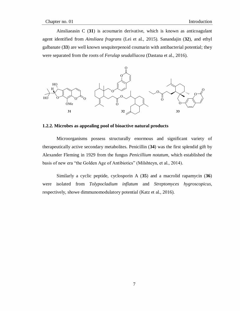
Ainsliaeasin C (31) is acoumarin derivative, which is known as anticoagulant
agent identified from Ainsliaea fragrans (Lei et al., 2015). Sanandajin (32), and ethyl
galbanate (33) are well known sesquiterpenoid coumarin with antibacterial potential; they
were separated from the roots of Ferulap seudalliacea (Dastana et al., 2016).
1.2.2. Microbes as appealing pool of bioactive natural products
Microorganisms possess structurally enormous and significant variety of
therapeutically active secondary metabolites. Penicillin (34) was the first splendid gift by
Alexander Fleming in 1929 from the fungus Penicillium notatum, which established the
basis of new era “the Golden Age of Antibiotics” (Milshteyn, et al., 2014).
Similarly a cyclic peptide, cyclosporin A (35) and a macrolid rapamycin (36)
were isolated from Tolypocladium inflatum and Streptomyces hygroscopicus,
respectively, showe dimmunomodulatory potential (Katz et al., 2016).

Chapter no. 01 Introduction
8
Bleomycin (37) is an anticancer compound, isolated from Streptomyces verticillus
(Katz et al., 2016), while polycyclic ethanones, alternethanoxin A (38) and B (39) were
identified from Alternaria sonchias cytotoxic metabolites against various cancer cells
(Evidente et al., 2014). Aspergillus aculeatus, a fungusfrom terrestrial source, produces
anti-diabetic compound rubrofusarin (40) (Dewi et al., 2016).

Chapter no. 01 Introduction
9
1.2.3. Marine source as appealing pool of bioactive natural products
Marine natural products are getting attention among the natural products due to
their versatile pharmacological properties. Terpenoids like dixiamycin A (41) and
dixiamycin B (42), were isolated from marine Actinomycete and Streptomyces sp. SCSIO
02999, possessing significant anti-bacterial activity. Alkaloids, rubrumazine B (43),
echinulin (44) and variecolorin H (45) were identified from Eurotium cristatum EN-220.
Rubrumazine B (43) possesses activity against pathogenic fungus Magnaporthe grisea
while echinulin (44) and variecolorin H (45) exhibited potential againsthuman pathogen
S. aureus (Choudhary et al., 2017). The chemical investigation of Streptomyces strain
CNH189 leads to the isolation of ansalactams B (46), ansalactams C (47) and
ansalactams D (48) which were found to be modest antibacterial agent (Blunt et al.,
2017).

Chapter no. 01 Introduction
10
Marine sources have also contributed to the bank of anti-cancer agents.
Marinomycin (49) from streptomyces sp. and chinikomycin A (50) from marinispora sp.
were found as active anti-tumor agents. Marinomycin (49) was also found active in
antibacterial assay. A secondary metabolite mechercharmycin A (51) was purified from
Thermo actinomyces sp. YM3-251 showed potent antitumor activity. Arenicolides A
(52), a 26-membered, type 1 polyketide showed modest cytotoxic effect towards HCT-
116 of human colon adenocarcinoma cell line, was isolated from Salinispora arenicola
CNR-005 (Manivasagan et al., 2014).

Chapter no. 01 Introduction
11
1.3 A Quick Glance on Some Natural Product-Based Medicines in
Near Past
Reliance of pharmaceutical drugs on natural product cannot be denied even in 21st
century. These are always endorsing source for the evolution of innovative drugs. About
one-third FDA-approved drugs during1981 to 2014 have natural product background
(Newman, 2018).They include small molecule based structures, either unaltered natural
metabolites or their derivatives and synthetic natural mimics (Li and Lou, 2018).

Chapter no. 01 Introduction
12
Following section describe the role of some natural and derived natural products recently
approved as drugs by FDA (Table 1.1).
Oritavancin (53) trade name Orbactiv (Newman et al., 2016), is semisynthetic
compound of natural precursor, chloroeremomycin (54), which was isolated from
Amycolatopsis orientalis (Domenech et al., 2009; Katz and Baltz, 2016). It is
antibacterial, oral drug, promote bacterial cell death by following three ways, interrupting
the cell wall formation through transglycosylation inhibition, cell membrane distortion by
interaction with hydrophobic 4'-chlorobiphenyl methyl (55) group and by offering more
secondary binding positions to resist bacterial strains (Markham, 2014).
Vorapaxar (56) with trade nameZontivity is derivative of himbacine (57) isolated
from plants Galbulimima baccata and G. belgraveana (Pinhey et al., 1961; Butler et al.,
2014). It is used in preventive measurement of cardiological and hematological disorders.
It involves reduction of thrombotic cardiovascular in patient with myocardial infarction
(MI) or peripheral arterial disease (PAD) history. It inhibits cardiovascular events by
inhibiting platelet aggregation related to thrombin (Poole and Elkinson 2014).

Chapter no. 01 Introduction
13
Ecteinascidin or trabectedin (58) is being marketed under brand name Yondelis as
anti-cancer, intravenous, alkaloid drug (Rinehart et al., 1990; Palanisamy et al., 2017). It
was isolated from Ecteinascidia turbinate.In its action, it binds to the guanine (59) part of
DNA at minor grooves, where it opens the double helix of DNA which eventually lead
the death of soft cancer cell (Petek et al., 2015).Xifaxan is trade name for rifaximin (60)
whichis a semisynthetic compound, derivative of rifamycin B (61) or rifamycin SV (62),
being produced by Amycolatopsis rifamycinica. It is taken orally for treatment for
digestive disorders (Bimer et al., 1972; Saxena et al., 2014).It stops the synthesis of RNA
by interrupting the steps of transcription by binding to the beta subunit of bacterial DNA-
dependent RNA polymerase. As a result controls command for protein synthesis and
inhibits cell growth (Kane and Ford 2016).
Chapter no. 01 Introduction
14
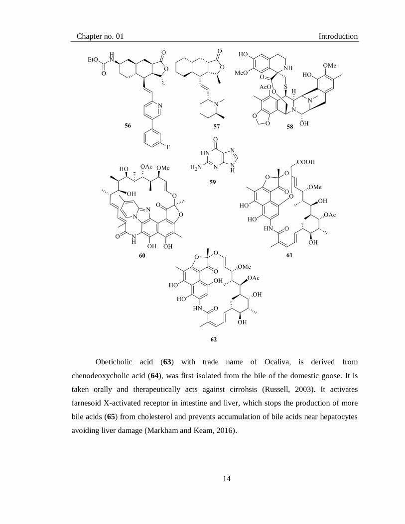
Obeticholic acid (63) with trade name of Ocaliva, is derived from
chenodeoxycholic acid (64), was first isolated from the bile of the domestic goose. It is
taken orally and therapeutically acts against cirrohsis (Russell, 2003). It activates
farnesoid X-activated receptor in intestine and liver, which stops the production of more
bile acids (65) from cholesterol and prevents accumulation of bile acids near hepatocytes
avoiding liver damage (Markham and Keam, 2016).

Chapter no. 01 Introduction
15
Midostaurin/Rydapt (66) is an alkaloidal drug derived from staurosporine (67),
which was isolated from Streptomyces staurosporeus (Omura et al., 1977; Zhou et al.,
2019).It is a prominent anticancer oral drug for the patient suffering with FLT3-positive
acute myeloid leukemia (AML) (Beatriz et al., 2018). It enriches bloodstream with white
blood cells killing cancer roots in bone marrow (US FDA 2017). It shows multikinase
inhibitor effect that works by obstruction in cell growth by controlling respective enzyme
(Kim, 2017).
Naldemedine (68), sold under the name of symproic, is derived from naltrexone
(69) (Hussar and Hussar 2018), which is semi-synthetized from oxymorphone (70)
(Sudakin, 2016) formally originatedfrom morphine (1)(Huang, Patent 2008).It was first
isolated from natural source Papaver somniferum (Sharopov et al., 2018). It is
administered orally and acts by hindering peripheral µ-opioid receptors in gastrointestinal
tract tissue which results into lesson the opioids effect on constipation (Markham, 2017).

Chapter no. 01 Introduction
16
Inotuzumab ozogamicin (71), an anticancer intravenous drug, is marketed as
Besponsa, in which inotuzumab is identified in ovary cells of Chinese hamster (Datta-
Mannan et al., 2018), which is attached to derivative cytotoxic group of “calicheamicins”
(72) isolated from Micromonospora echinospora (Song et al., 2015). Ozogamicin (73) is
cytotoxic part of Besponsal, enters into the cell by the process of endocytosis where
ozogamicin gets separated from antibody part and reached to the nucleus, starts
disintegration of DNA, consequences in cell death (Lamb, 2017).

Chapter no. 01 Introduction
17
Plazomicin (74) is an intravenous infusion, antibacterial drug traded as zemdr
(FDA Approved Drug Products 2019) It is semisynthetic aminoglycoside derivative of
sisomicin (75) (Gupta, 2017), isolated from Micromonospora inyoensis (Talukdar et al.,
2016). It impede protein production in bacterial cell by developing bonding with its
ribosomal 30S subunit, varies with drug concentration (Shaeer, 2018).
Cannabidiol (76) known as Epidiolexis a natural product isolated from Cannabis
sativa (Fairbaim and Liebmann, 1973; Dyer, 2018). It is taken orally for the treatment of
epilepsy because it has anti-seizures property which makes it valuable for treatment of
Lennox-Gastaut syndrome or Dravet syndrome in patient with age not under 2 (Elliott
and Chan 2018). Omadacycline (77) is known as Nuzyra in market (Markham and Keam
2018). It is derivative of tetracycline (78), isolated from Streptomycessp. (Petković et al.,
2017). It has antibacterial effect, administrated orally and by intravenous infusion. By
binding with protein synthesizing unit of ribosome (70S) with greater affinity it ensures
the excellent antibacterial activity (Rahman and Koh, 2018).

Chapter no. 01 Introduction
18
Eravacycline (79) trade marked as Xerava (FDA Approved Drug Products 2018)
is anti-biotic, derived from tetracycline (78) isolated from Actinomycete sp. (Butler et al.,
2014). It is taken orally and in injection form to treat infections. It hinders the
prolongation of amino acid chain by binding to responsible ribosomal (30S) unit of
bacteria to inhibit the protein synthesis (Zhanel et al., 2016).
Krintafel is the trade name for tafenoquine (80) (Elliott and Chan 2018), is a
derivative of quinoline (81) “a natural compound” was isolated initially from bark of
Cinchona sp. possess anti-malarial potential (Fernandez-Alvaro et al., 2016). Oral dosage
works to eliminate the roots of dormant parasite in the liver for the patient of age 16 or
older along with the appropriate anti-malarial therapy (Frampton, 2018). Sarecycline (82)
is generic name for seysara, an anti-inflammatory, orally taken drug, derivative of
tetracycline (77) isolated from actinomycetes (Butler et al., 2014); it is applied for skin
infection (Deeks, 2019).

Chapter no. 01 Introduction
19

Chapter no. 01 Introduction
20
Table 1.1 Some FDA approved drugs in recent years
Drug
Generic/trade
name
Lead compound/source Therapeutic
effect
Route of
administra
tion
Mode of action Manufacturer/App
roved status
Reference
Oritavancin(53)/
Orbactiv
Chloroeremomycin
(54)/Amycolatopsis orientalis
Dermatology
infaction
oral Interruption in
cell wall
synthesis
The medicine
company (2014)
(Newman et al., 2016),
Domenech et al., 2009),
(Katz, and Baltz, 2016)
(Markham, 2014)
Vorapaxar(56)/
Zontivity
Himbacine(57)/ Galbulimima
baccata and G. belgraveana
Cardic and
heamatology
oral Reduce
thrombotic
Event
Merck
(2014)
(Butler et al., 2014)
(Poole and Elkinson 2014)
Trabectedin(58)/
Yondelis
Trabectedin (58)/
Ecteinascidia turbinate
Oncology intravenous Binding with
DNA
Janssen
(2015)
Palanisamy et al., 2017).
(Petek et al., 2015)
Rifaximin
(60) /Xifaxan
Rifamycin B or Rifamycin
SV (61,62) / Amycolatopsis
rifamycinica
Gastroenterol
ogy
oral Inhibit protein
synthesis
Salix
Pharmaceuticals
(2015)
(Saxena et al., 2014).
(Kane and Ford 2016)
Obeticholic
acid(63)/ Ocaliva
chenodeoxycholic acid (64)/
domestic goose
Hepatology oral FXR activation
and control bile
acid level
Intercept
Pharmaceuticals
(2016)
(Russell, 2003) (Markham
and Keam, 2016)
Midostaurin(66) /
Rydapt
Staurosporin(67)/
Streptomyces staurosporeus
Hematology
and
Oncology
oral multikinase
inhibitor
Novartis
(2017)
Zhou et al., 2019, (Beatriz et
al., 2018). (Kim, 2017)
Naldemedine (68)/
Symproic
Morphine (1)/ Papaver
somniferous
Gastroenterol
ogy
oral Block opioid
receptor
Shionogi
(2017)
(Hussar and Hussar 2018,
(Sudakin, 2016) (Sharopov
et al., 2018). (Markham,
2017)
Inotuzumab
ozogamicin(71)/
Besponsa
Monoclonical
antibody/chinease hamster,
calicheamicins (72)/
Micromonospora
echinospora.
hematology intervenous DNA breakage
and apoptosis of
cell
Pfizer (2017) (Datta-Mannan et al., 2018)
(Lamb, 2017). (Song et al.,
2015)
Plazomicin Sisomicin (75)/ Infaction intervenous Inhibit protein Achaogen (Gupta, 2017), (Talukdar et

Chapter no. 01 Introduction
21
(74)/Zemdr Micromonospora inyoensis disesas synthesis (2018) al., 2016) (Shaeer, 2018)
Cannabidiol(76)/
Epidiolex
Cannabidiol(76)/Cannabis
sativa
Neurology oral anticonvulsant GW
Pharmaceuticals
(2018)
(Elliott and Chan 2018)
(Dyer, 2018)
Omadacycline
(77)/ NUZYRA
Tetracycline(78)/Streptomyce
ssp
antibacterial Oral and
intervenous
Inhibit protein
synthesis
Paratek
Pharmaceuticals
(2018)
(Markham and Keam 2018),
(Petković et al., 2017).
Rahman and Koh, 2018)
Eravacycline (79)
/ Xerava
Tetracycline(78)/Actinomycet
e sp
Gastroenterol
ogy
Intervenous Inhibit protein
synthesis
Tetraphase
Pharmaceuticals (2018)
(Butler et al., 2014) (Zhanel
et al., 2016)
Tafenoquine (80)
/Krintafel
Quinolin(81)/ Cinchona sp Infactious
disoreded
oral prevents the
progress of the
erythrocytic
forms of the
parasite
GlaxoSmithKline
(2018)
(Elliott and Chan 2018)
(Fernandez-Alvaro et al.,
2016) (Frampton, 2018)
Sarecycline (82) /
Seysara
Tetracycline (78) /
actinomycetes.
Dermatology oral Inhibit protein
synthesis
Paratek
Pharmaceuticals
(2018)
(Butler et al., 2014)
(Deeks, 2019)

22
CHAPTER 2
Previous Phytochemical Investigation on The
Berberis calliobotrys, Caragana ambigua And
Vincetoxicum stocksii

Chapter no. 02 Phytochemical Investigation of genus Berberis
23
Part A
2.1. Previous Phytochemical Investigation of the Genus Berberis
Main genus of family Berberidaceae is Berberis which is distributed worldwide
with its folk medicinal uses documented in Pakistan, India, Japan, China, Asia, Europe,
Africa and America. It grows as evergreen small tree or shrub with yellow flowers and
wood. Thisgenus is classified into three groups on the basis of habitat, Rocky Mountain,
Asiatic and European group.
Many species of the genus Berberis were used as source of indigenous medicines
and as modern available drugs (Khan et al., 2016). B.vulgarisis an important traditional
medicinal plant widely distributed in Iran.It has yellow wood, flowers, and red fruits.
Phytochemical investigation revealed the presence of alkaloids, phenolics, sterols, tannins
and triterpenoids compounds in B.vulgaris. It has been reported to cure inflammation,
vomiting, nausea, itching, heart diseases, diabetes, jaundice, malaria, sore throat and
block the action of acetylcholine (Zadeh et al., 2107).It is a source of isoquinoline
alkaloids including berberine (83) palmatine (84) berberamine (85), 5-
methoxyhydnocarpin (86) etc. Quantitative HPLC analysis showed that methanolic
extract of the whole plant contains 3.9% berberamine (85) and about 1.24% berberine
(83). In China berberine (83) has been used as anti-hyperglycemic agent and inhibits the
increase of blood and liver cancer (Imanshahidi and Hosseinzadeh, 2008).

Chapter no. 02 Phytochemical Investigation of genus Berberis
24
In addition, tejedine (87), baluchistanamine (88), oxyacanthine (89),
isotetrandrine (90), obaberine (91), obamegine (92), aromoline (93), and thaligrisine (94)
were also isolated from B. vulgaris.

Chapter no. 02 Phytochemical Investigation of genus Berberis
25
Some non-basic alkaloids like oxyberberine (95), thalifoline (96), chilenine (97)
and quaternary alkaloid jatrorrhizine (98) were also reported from B. vulgaris (Suau et
al., 1998).
B. aristata locally known as Rasaut, possesses several medicinal properties like in
skin diseases, ear, eye and urinary tract infections and menorrhagia, antibacterial,
antiviral, antioxidant, antifungal, antidiabetic, anti-ulcer, anti-malaria and gastric disorder
etc (Khan et al., 2016). Berberine (83) oxyberberine (95), berbamine (85), aromoline
(93), karachine (99), palmatine (84), oxyacanthine (89), taxilamine (100), epiberberine
(101), jatrorrhizine (98), 1-O-methylpakistanine (102), pakistanine (103), chitraline (104)
and kalashine (105) have been isolated from this plant (Semwal et al., 2018).
B. lyceum, which is commonly known as Ishkeen, found abundantly in Himalayan
region of Asia. In Pakistan it is found in Gilgit, Swat, and Kashmir. Its powdered root
extract was used as local medicine for diabetes in Himalaya region (Ali et al., 2015). In
addition, it has been used for treatment of fever, eye diseases, liver and kidney disorders,

Chapter no. 02 Phytochemical Investigation of genus Berberis
26
throat pain, internal wounds etc. Its roots are rich in palmatine (84) and berberine (83)
along with minor amount of jhelumine (106), baluchistanamine (88), sindamine (107),
punjabine (108), and Karakoramine (109) (Shabbir, et al., 2012). β-sitosterol (27), butyl-
3-hydroxypropyl phthalate (110), 4-methyl-7-hydroxy-coumarin (111), 4,4-
dimethylhexadeca-3-ol (112) and 3-(4’-(6-methyl-butyl)-phenyl)propan-1-ol (113) were
also obtained from this plant (Ali, et al., 2015).
B. orthobotrys, growing in Gilgit-Baltistan, is used for joint pain therapy.
Phytochemical investigation has revealed the presence of aporphine-benzylisoquinoline
alkaloids specifically berberine (83), oxyacanthine (89), berbamine (85), pakistanine
(103), chitraline (104), kalashine (105) and pakistanamine (114) in B. orthobotrys. Its
aqueous-methanolic extract along with its aqueous, n-butanol and ethyl acetate extract
were found active in various in vitro and vivo anti-arthritic protocols (Alamgeer, et al.,
2017).

Chapter no. 02 Phytochemical Investigation of genus Berberis
27
B. baluchistanicais is well known specie grown in Baluchistan province of
Pakistan and also distributed in other parts of the world. Its root extract is used to cure
cough and internal wound of humans and domestic animals. Literature studies explicate
the isolation of pakistanine (103), pakistanamine (114), (+)-baluchistine (115) and
baluchistanamine (88) from root part of B. baluchistanicais (Khan et al., 2016).B. chitria
is another famous species of genus Berberis found in Pakistan with its local use against
ulcers, eye problems, liver disorder, bleeding piles and enlarged spleen. Chlorogenic acid
(116) was identified as main constituent of B. chitria flower, magnoflorine (117) as main
metabolite in the stem portion along with berberine (83), palmatine (84), jatrorrhizine
(98), isocorydine (118), tetrahydropalmatine (119) and tetrahydroberberine (120)(Singh
et al., 2015).

Chapter no. 02 Phytochemical Investigation of genus Berberis
28
2.2. Previous Phytochemical Investigation of Berberis calliobotrys
Diversity and medicinal importance of the genus Berberis, we selected an under-
explored species B. calliobotrys to investigate for its bioactive metabolites.B. calliobotrys
is a tall shrub with brownish-yellow stem, abundantly distributedin Pakistan and
Afghanistan. In indigenous medicine system it was used for the treatment of backache,
hepatic and stomach disorder, fever pharyngitis and inflammation in Chitral valley of
Pakistan. Modern research elaborates its potentialagainst anti-inflammatory, analgesic,
and antipyretic activity of its crude methanolic extract (Alamgeer et al., 2016). Further,
its extract exhibits antipyretic, anti-convulsant and antimicrobial effects (Rasool et al.,
2015).
2.3. Classification of Berberis calliobotrys
Phylum Tracheophyta
Class Magnoliopsida
Order Magnoliopsida
Family Berberidaceae
Genus Berberis
Species Berberis calliobotrys
Previously berberine (83), aromoline (93), oxyberberine (95), karachine (99), 1-
O-methylpakistanine (102), pakistanine (103), chitraline (104) and kalashine (105),
pakistanamine (114), corydaldine (121), methylcorydaldine (122), N-methyl-6, 7-
dimethoxy-isoquinoline (123) and waziristanine (124) have been isolated from roots of B.
calliobotrys (Srivastava et al., 2015). HPLC analysisof ethyl acetate extract resulted in
the identification of quercetin (125), gallic acid (126), trans ferulic acid (127), caffeic
acid (128), chlorogenic acid (116), vanillic acid (129) and p-coumeric acid (130) (Rasool
et al., 2015).

Chapter no. 02 Phytochemical Investigation of genus Berberis
29

Chapter no. 02 Phytochemical Investigation of genus Caragana
30
Part B
2.4. Previous Phytochemical Investigation of the Genus Caragana
The sub-family Papilionoideae contributes more than 100 species to family
Fabaceae. Its relativesare abundantly distributed in the grass land, desert climate and
rarely in forest climate. Caragana, one of the important genus of this family, is rich
source of various bioactive components including terpenoids, lectin, flavonoids,
stilbenoids, and phenylpropanoids (Meng et al., 2009). Literature of traditional medicine
of Mongolian, Chinese, and Tibetan system explains the therapeutic application of
various species of genus Caragana against numerous disorders including rheumatism,
headache arthritis, hypertension, neuralgia, atherosclerosis, inflammation, cancer and
arthritis (Meng et al., 2009). Literature search further revealed that several species of
Caragana have been studied for their phytochemicals.
Caragana tangutica found inNorth-West of China is used traditionally for remedy
for trauma, hypertension, rheumatic pains injury, cardiovascular and irregular
menstruation (Qiuxia et al., 2009). Varity of components have been isolated from this
plant, they include maackiain (131), medicarpin (132), formononetin (133), cajanin
(134), melilotocarpan A (135), 2-(2', 4'-dihydroxyphenyl)- 3-methyl-6-methoxy
benzofuran (136), bolusanthinIII (137), 7,3'-dihydroxy-5-methoxy isoflavone (138) and
p-ethoxy benzoic acid (139) (Niu et al., 2013).

Chapter no. 02 Phytochemical Investigation of genus Caragana
31
Yang et alhave further reported the isolation of maackiain 3-O-β-D-
glucopyranoside (140), maackiain 3-O-6′-O-methyl malonyl-β-D-glucopyranoside (141),
(-)-maackiain 3-O-6′-O-acetyl-β-D-glucopyranoside (142) and 3-O-6′-O-acrylyl-β-D-
galactopyranoside (143) from ethyl acetate fractionof C. tangutica (Yang et al., 2017).
C. changduensis found in the region of China, traditionally used to remove lymph
in the blood by promoting blood circulation, treat hypertension and prevent elevation of
red blood cells in the blood. It has been reported to produce 6,7,2'-trihydroxy-4'-
methoxyisoflavone(144), (8S)-2,4-dihydroxy-8-hydroxymethyl-4'-methoxydeoxybenzoin
(145), 7,3'-dimethoxy-5'-hydroxy-isoflavone (146), 2,4-dihydroxybenzoate (147), ethyl

Chapter no. 02 Phytochemical Investigation of genus Caragana
32
4-hydroxybenzoate (148), methyl 4-hydroxylbenzoate (149), cararosin A (150),
piceatannol (151) and isoliquiritigenin (152) (Guo et al., 2017).
C. sinica occupies large area of China, Korea, and Japan. Dried roots of C. sinica
are used for curing heumatism, rheumatism, neuralgia, vascular hypertension,
leukorrhagia, migrane, syndrome, arthritis, and wounds. Oligostilbenes are main
constituents of C. sinica showed anti-inflammatory, anti-acetylcholinesterase, and anti-
oxidative activities. Betulinic acid (153), medicarpin (154), pallidol (155), caragasinin C
(156) (Jeong et al., 2017), (+)-α-viniferin (157) miyabenol C (158) and kobophenol (159)
have been reported from C. sinica (Menga et al., 2009).

Chapter no. 02 Phytochemical Investigation of genus Caragana
33
A perennial shrub, C. sukiensis grows insub arid and arid habitat of Eurasia.Its
ethanolic extract shows antimicrobial activity against the fungus Cryptococcus
neoformans (Mandal et al., 2015). Chromatographic separation of ethyl acetate extract of
aerial parts led to the identification of various compounds including sukienside A (160),
cyclosiversigenin (161), sukienside B (162), 3-O-β-D-xylocyclosiversigenin (163), β-
amyrin (164) and 3-β-hydroxy-2,5- melilotigenin C (165).

Chapter no. 02 Phytochemical Investigation of genus Caragana
34
C. jubataiscultivated in Himalayan state, where local people use its aerial parts to
cure cardiovascular diseases, hyper-lipidemia, blood circulation disorders and
hypertension in folkmedicine system. Its mehanolic extract was found to be active in
antioxidant assay (Mandal et al., 2016). Chomatographic analysis of stem, root and
flower parts led to the isolation of different bioactive constituents including lyoniresinol
(166), isoampelopsin F (167), caraphenol A (168), caraphenol B (169), caraphenol C
(170) (Menga et al., 2009).

Chapter no. 02 Phytochemical Investigation of genus Caragana
35
2.5. Previous Phytochemical Investigation of Caragana ambigua
Another part of my PhD project, I selected C. ambigua to be studied for its
bioactive compounds. C. ambigua is an important specie of the genus Caragana found in
Ziarat, Gilgit and Kashmir Valleys of Pakistan at about 7,000 to 12,000 feet above from
sea level. Study conducted on dried roots of C. ambigua reported the presence of
alkaloids, tannins and saponins (Kayani et al., 2007). C.ambigua, also known as Makhi,
is used as fuel wood, fodder and to increase soil fertility (Sarangzai et al., 2013).
Kingdom Plantae
Order Magnoliopsida
Class Fabales
Family Fabaceae
Genus Cagagana
Specie Caragana ambigua

Chapter no. 02 Phytochemical Investigation of genus Caragana
36
2.6. Classification of Caragana ambigua
Methanolic extract of C. ambigua contains ambiguanol (171), E-cinnamic acid
(130), β-sitosterol 3-O-β-D-glucopyranoside (172), 5-hydroxy-3', 4', 6, 7-
tetramethoxyflavone (173), 3, 3', 4', 5, 7-pentahydroxyflavane (174) and calycosin (175)
(Majida et al., 2011).

Chapter no. 02 Phytochemical Investigation of genus Vincetoxicum
37
Part C
2.7. Previous Phytochemical Investigation of the Genus Vincetoxicum
The genus Vincetoxicum of the family Asclepiadaceae, accounting for around 100
species is growing in Europe, Asia and Japan (Guzel et al., 2017). In Pakistan six species
are found namely V. hirundinaria, V. cardiostephanum, V. stocksii, V. sakesarense, V.
arnottianum and V. canescens (Ali et al., 1989). Literature search revealed the presence
of phenanthroindolizidine alkaloids, triterpenoids, flavonoids, steroidal glycosides,
volatile compounds, saponin, phenolics, acetophenone, steroids and alkanols in
Vincetoxicum species which make them responsible for antibacterial and antifungal,
antispazmodic, antimalarial antileishmanial, antidiarrheal, and cytotoxic antifeedant
properties (Guzel et al., 2017).
Phytochemical analysis of the root extract of V hirundinaria (syn: V. officinale)
(Nowaka, et al.,2000) disclosed the presence of many phenanthroindolizidine, flavonoids,
steroids and alkaloids (Lavault et al., 1999), which indicated the reason for it being used
in traditional medicinal system to cure tumor and laxative (Nowaka et al., 2000), cough,
promotes diuresis and vomiting. Compound isolated from roots, included paeonol (176),
4-hydroxyacetophenone (177), syringic aldehyde (178), myristicine (179), (Lavault et al.,
1999), β- sitosterol (27), stigmasterol (28), α-amyrin (180), hancokinol (181) (Nowaka et
al., 2000), cynatratoside E (182), cynatratoside C (183), hirundigoside B (184)
hirundigoside C (185), hirundigoside D (186) (Lavault et al.,1999).

Chapter no. 02 Phytochemical Investigation of genus Vincetoxicum
38
V. pumilum is a perennial herb commonly found in Central Asia. Methanolic
extract root and aerial part evaluation confirmed the presence of (-)-13a-α-antofine (187)
(-)-10 β, 13a-α-antofine N-oxide (188) and (-)-14 β -hydroxy-10 β, 13a-α-antofine N-
oxide (189) (Staerk et al., 2005).
V. nigrum is found in Europe mainly in France, Italy, Spain and Portugal. In
Chinese medicine system it was often used to treat rupture, scabies, scrofula and internal
fevers. (-)-13a-α-Antofine (187) which is phenanthroindolizidine alkaloids has been
found active against antibiotic activities (Nourian et al., 2016).

Chapter no. 02 Phytochemical Investigation of genus Vincetoxicum
39
2.8. Previous Phytochemical Investigation of Vincetoxicum stocksii
V. stocksii is a perennial climbing leafy, poisonous, and medicinal important vine
found in Baluchistan, Pakistan. It is used for the treatment of cancer, healing of wounds
and injuries Crude extract of V. stocksii shows antidiarrheal, antispasmodic, antibacterial
and antifungal activities (Mudassir et al., 2005). Phytochemical analysis of the plant
extract exposed the existence of glycosides and tannins while other bioactive agentswere
not found. Antidiarrheal and calcium channel blocking potential are in correspondence
with existence of tannins (Shah et al., 2011). It showed highcytotoxicity against brine
shrimps (Mudassir et al., 2012).
2.9. Classification of Vincetoxicum stocksii
Family Asclepiadaceae
Subfamily Periplocoideae
Class Asclepiadeae
Genus Vincetoxicum
Specie Vincetoxicum stocksii
Literature review revealed the presence of vincetoside (190), vincetolate (191),
vincetomine (192) vincetetrol (193), 4-hydroxy-3-methoxyphenyl 7, 8, 9-propanetriol
(194) 4-hydroxy-3,5-dimethoxybenzoic acid (195) apocynin (196) feruloyl-6-O- β-D-
glucopyranoside (197), quercetin-3-O- β-D-glucopyranoside (198) in the ethyl acetate
soluble fraction of V. stocksii (Tousif et al., 2016).

Chapter no. 02 Research Hypothesis
40
2.10. Research Hypothesis
The selected three under-explored Pakistani plants Berberis calliobotrys,
Caragana ambigua and Vincetoxicum stocksii are being used as folk medicine, and
related specis are rich in variety of bioactive secondary metabolites therefore, it is
hypothesized that these plants might also be producing certain bioactive secondary
metabolites, which contributes to their medicinal use; therefore, if they are isolated they
may provide leads for new drug development.
2.11. Research Problem and Objectives of the Study
Enriched Pakistan land and marvelous importance of folk medicine inspired us to
search out for the compounds which are making these plants important. Despite of the

Chapter no. 02 Research Hypothesis
41
presence of countless medicinal floras in Pakistan, field of natural product chemistry and
drug development is still ignored in the country. Therefore, there is crucial need to study
Pakistani indigenous plantsto getdeeper information on active principals of these natural
machineries. To address a part of this problem, we selected three Pakistani plants;
Berberis calliobotrys, Caragana ambigua and Vincetoxicum stocksii to investigate them
for their novel bioactive secondary metabolites.
The Objective Includes:
1) Collection and extraction of plant materials
2) Preparation of different extracts of all plant materials
3) Biological and chemical screening of various extracts
4) Chromatographic purification of the extracts to get pure secondary metabolites
5) Establishing structures of isolated compounds based on spectroscopy
6) Evaluation of biological potential of extract and isolated compounds

42
CHAPTER 3
Results and Discussion: Biological Screening
and Phytochemical Analyses of Berberis
Calliobotrys, Caragana Ambigua and
Vincetoxicum Stocksii;and Isolation of
Secondary Metabolites

Chapter no.03 Result and Discussion of Berberis calliobotrys
43
Part A
3.1. BiologicalScreening of Crude Extracts of Berberis calliobotrys
Different polarity solvent extracts i.e.Berberis calliobotry smethanol extractBc-
M,Berberis calliobotrys ethyl acetate extract Bc-E, Berberis calliobotrys butanol extracts
Bc-B and Berberis calliobotryswater extract Bc-W were tested for antioxidant potentials
(2, 2-diphenyl-1-picrylhydrazyl (DPPH) and 2,2'-azino-bis (3-ethylbenzothiazoline-6-
sulphonic acid (ABTS), ferric reducing antioxidant power (FRAP), cupric reducing
antioxidant capacity (CUPRAC), phosphomolybdenum and metal chelation assays(Table
3.1) selective enzyme inhibitory assays (acetylcholinesterase (AChE),
butyrylcholinesterase (BChE), α-glucosidase, α-amylase and tyrosinase) (Table 3.2).
Inclusive assessment of antioxidant potential escorted that Bc-W offered the most
convincingresults in the ABTS (451.90 mg TE/g extract), CUPRAC (778.98 mgTE/g
extract), FRAP (718.48 mgTE/g extract) and phosphomolybdenum (2.04 mmolTE/g
extract) antioxidant assays which are in accord to the highest phenolic content of Bc-W
compared to other fractions. Whereas Bc-Moffered more promising results for DPPH
(204.73 mgTE/g extract) and metal chelation (60.32 mgEDTAE/g extract) activities. It
might be because of antagonistic or synergetic and non-phenolic chelator (Marini et al.,
2018).
Table 3. 1Antioxidant properties of B. calliobotrys extracts*
Plant extracts
Radical scavenging assays Reducing power assays Total
antioxidant activity
Ferrous ion chelation
DPPH (mgTE/g extract)
ABTS (mgTE/g extract)
CUPRAC
(mgTE/g extract)
FRAP
(mgTE/g extract)
Phospho molybdenum (mmolTE/g)
Metal chelaing (mgEDTAE/g)
Bc-M 204.73±3.103a 342.25±17.34b 744.72±18.86a 566.20±3.69b 1.88±0.06b 60.32±1.53a
Bc-E 71.54±0.91d 125.24±4.45d 308.47±13.53c 273.96±4.39d 1.93±0.05b 17.03±0.61d
Bc-B 95.93±0.41c 177.76±4.46c 523.30±18.67b 410.57±8.20c 2.01±0.13a 27.36±1.78c
Bc-W 165.73±9.75b 451.90±26.90a 778.98±8.38a 718.48±9.47a 2.04±0.08a 36.44±0.57b
* Values expressed are means S.D. of three parallel measurements. TE: Trolox equivalent;
EDTAE: EDTA equivalent. Different letters indicate differences in the extracts (p< 0.05)

Chapter no.03 Result and Discussion of Berberis calliobotrys
44
All extracts of B. calliobotrys were tested for their inhibitory potential against
AChE, BChE, α-glucosidase, α-amylase, and tyrosinase (Table 3.2). In AChE, following
order was observed Bc-E>Bc-M>Bc-W>Bc-B (5.25>5.16>5.09>4.65
mgGALAE/gextract respectively), while in BChE assay, the Bc-W attributed excellent
(5.66mgGALAE/g extract) inhibition and Bc-E exhibited least anti-BChE potential
(4.11mgGALAE/g extract). Our results were in consistence with literature review which
supports the unusual response of Bc-E in both neurodegenerative assays (Kolar et al.,
2010).
Bc-E, Bc-M and Bc-B extracts contributed comparable potential for α-glucosidase
inhibitory activity while the Bc-W was found inactive. In α-amylase inhibition assay, Bc-
E showed highest inhibition power 0.76 mmolACAE/g extract, followed by Bc-B0.71
mmolACAE/g extract, Bc-M 0.53 mmolACAE/g extract and 0.29 mmolACAE/g
extract.Bc-E exhibited highest inhibitory potential in both anti-diabetic assays (Table
3.2). SimilarlyBc-E extract presented highest inhibition with value 169.92 mgKAE/g
extract, furthermore Bc-M (158.81 mgKAE/g extract) and Bc-B (153.107 mgKAE/g
extract) analysis also exposed their equally effective potential in tyrosinase inhibition.
While the Bc-W was found least active (37.06mgKAE/g extract) in the above assay.
Table 3. 2Enzyme inhibition activities of B. calliobotrys extracts*
Plant extracts
AChE
(mgGALAE/g extract)
BChE (mgGALAE/g
extract
α-Glucosidase (mmolACAE/g
extract)
α-Amylase (mmolACAE/g
extract)
Tyrosinase
(mgKAE/g extract)
Bc-M 5.16±0.11a 5.41±0.06a 1.68±0.02a 0.53±0.02b 158.81±0.59b
Bc-E 5.25±0.03a 4.11±0.29b 1.68±0.01a 0.76±0.02a 169.92±1.18a
Bc-B 4.65±0.02b 5.62±0.12a 1.63±0.03a 0.71±0.08a 153.107±0.73c
Bc-W 5.09±0.11a 5.66±0.06a Na 0.29±0.03c 37.06±0.92d
* Values expressed are means S.D. of three parallel measurements.
GALAE: Galatamine equivalent; KAE: Kojic acid equivalent; ACAE: Acarbose equivalent; na:
not active. Different letters indicate differences in the extracts (p< 0.05)

Chapter no.03 Result and Discussion of Berberis calliobotrys
45
3.2 Phytochemical Analysis of Secondary Metabolites from Berberis
calliobotrys
Different polarity solvent extracts Bc-E, Bc-M, Bc-W and Bc-B of Berberis
calliobotryswereinvestigated for their bioactive contents and UHPLCMS analyses.
3.2.1 Total phenolic and flavonoid contents estimation
The phenolic and flavonoid content investigation revealed the highest phenolic
content in Bc-W with value 94.34 mgGAE/g extracts. In flavonoid estimation assay Bc-E
exhibited 11.11 mgRE/g extract of flavonoid content, while for other extracts the contents
ranges between 1.06-1.45 mgRE/g extract (Table 3.3).
Table 3. 3Total phenolic and flavonoid contents of B. calliobotrys extracts*
Solvents Abbreviation Total phenolic content
(mgGAE/g extract)
Total flavonoid
content (mgRE/g
extract)
Methanol Bc-M 77.70±1.39b 1.06±0.34c
Ethyl acetate Bc-E 50.24±1.05d 11.11±0.37a
Butanol Bc-B 67.69±1.19c 1.08±0.18c
Water Bc-W 94.34±1.11a 1.45±0.20b
* Values expressed are means S.D. of three parallel measurements. GAE: Gallic acid equivalent;
RE: Rutin equivalent. Different letters indicate differences in the extracts (p< 0.05)
Following methods were employed for the determination of total phenolics.
Total phenolic contents were measured through Folin-Ciocalteu method, where a
mixture of 0.25 ml of extract and 1.0 ml of diluted Folin-Ciocalteu reagent (1:9 dilution
ratio) was incubated at room temperature for three min, followed by the addition of 0.75
mL of 1% Na2CO3 solution. The reaction mixture was then incubated for two hours and
the absorbance was recorded at 760 nm using gallic acid was used as standard (Aktumsek
et al., 2013).
For the determination of total flavonoids contents, aluminium trichloride method
was used for evaluation of all four crude extract (Zengin et al 2014). In this method 1.0
mL of 2% methanolic AlCl3 solution was mixed with an equal volume of extract,

Chapter no.03 Result and Discussion of Berberis calliobotrys
46
incubated for 10 min and absorbance was recorded at 415nm. In this assay, rutin was
used as standard compound (Mocan et al., 2017).
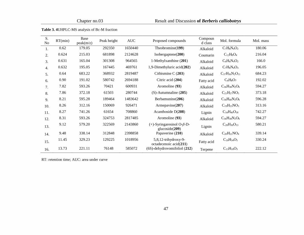
3.2.2.1 UHPLC-MS Analysis for Identification of Secondary Metabolites of B.
calliobotrys
Ultra high performance liquid chromatography mass spectrometry (UHPLC-MS)
was used for identification of secondary metabolites Bc-M and the most biological active
fractions i.e.Bc-E and Bc-W.
3.2.2.2 Secondary metabolite identification of Bc-M through UHPLC-MS analysis
UHPLC-MS negative ionization mode analysis of Bc-M conceded the presence of
16 secondary metabolites including alkaloids as major constituents (Figure 3.1) identified
compounds included 9 alkaloids, two fatty acids, two lignins, and one compound from
coumarin and terpene classes (Table 3.4). Indication of alkaloids as major constituents
was in accordance with the literature (Srivastava et al., 2015).
Figure 3. 1Total ion chromatograms (TICs) of Bc-M fraction
Chapter no.03 Result and Discussion of Berberis calliobotrys
47
Table 3. 4UHPLC-MS analysis of Bc-M fraction
S.
No RT(min)
Base
peak(m/z) Peak height AUC Proposed compounds
Compoun
d class Mol. formula Mol. mass
1. 0.62 179.05 292350 1650440 Theobromine(199) Alkaloid C7H8N4O2 180.06
2. 0.624 215.03 681898 2124628 Isobergaptene(200) Coumarin C12H8O4 216.04
3. 0.631 165.04 301308 964565 1-Methylxanthine (201) Alkaloid C6H6N4O2 166.0
4. 0.632 195.05 167445 469761 1,9-Dimethyluric acid(202) Alkaloid C7H8N4O3 196.05
5. 0.64 683.22 368932 2819487 Citbismine C (203) Alkaloid C37H36N2O11 684.23
6. 0.90 191.02 580742 2694188 Citric acid (204) Fatty acid C6H8O7 192.02
7. 7.82 593.26 70421 600931 Aromoline (93) Alkaloid C36H38N2O6 594.27
8. 7.86 372.18 61503 280744 (S)-Autumnaline (205) Alkaloid C21H27NO5 373.18
9. 8.21 595.28 189464 1483642 Berbamunine(206) Alkaloid C36H40N2O6 596.28
10. 8.26 312.16 150069 926471 Armepavine(207) Alkaloid C19H23NO3 313.16
11. 8.27 741.26 61654 708860 Acanthoside D(208) Lignin C34H46O18 742.27
12. 8.31 593.26 324753 2817485 Aromoline (93) Alkaloid C36H38N2O6 594.27
13. 9.12 579.20 322569 2143860 (+)-Syringaresinol O-β-D-
glucoside(209) Lignin
C28H36O13 580.21
14. 9.48 338.14 312848 2398858 Papaverine (210) Alkaloid C20H21NO4 339.14
15. 11.45 329.23 129225 1018956 5,8,12-trihydroxy-9-
octadecenoic acid(211) Fatty acid
C18H34O5 330.24
16. 13.73 221.11 76148 585072 (6S)-dehydrovomifoliol (212) Terpene C13H18O3 222.12
RT: retention time; AUC: area under curve

Chapter no.03 Result and Discussion of Berberis
calliobotrys
48

Chapter no.03 Result and Discussion of Berberis
calliobotrys
49
3.2.2.3Secondary metabolite estimation of Bc-E through UHPLC-MS analysis
Bc-E analysis revealed the existence of 17 secondary metabolites (Table 3.5,
Figure 3.2). It included seven flavonoids and four phenolics as major constituents;
whereas three terpenes, one saponin, propiophenone and one fatty ester were
identified as minor contributor.
Figure 3. 2Total ion chromatograms (TICs) of Bc-E fraction

Chapter no.03 Result and Discussion of Berberis calliobotrys
50
Table 3. 5UHPLC-MS analysis of Bc-E fraction
S.No RT(min) Base
peak(m/z)
Peak
height AUC Proposed compounds
Compound
class
Mol.
formula Mol.mass
1. 1.478 153.0192 22487 90608 3,4-DihydroxybenzoicAcid
(213) Phenol C7H6O4 154.0264
2. 3.085 137.0245 34358 292148 p-Salicylic acid (214) Phenol C7H6O3 138.0318
3. 8.095 193.0505 53525 194273 Kakuol (215) Propiophenone C10H10O4 194.0577
4. 8.201 193.0505 109241 438072 Ferulic acid (127) Phenol C10H10O4 194.0579
5. 8.41 197.0455 48343 197418 2-Hydroxy-3,4-
dimethoxybenzoic Acid (216) Phenol C9H10O5 198.0526
6. 9.103 361.1659 62848 241533 Hydroxyisonobilin (217) Terpene C20H26O6 362.1729
7. 9.388 431.1676 44075 187186 Agecorynin C (218) Flavonoid C22H24O9 432.1427
8. 10.052 385.0937 26167 106111 Melisimplexin (219) Flavonoid C20H18O8 386.1013
9. 10.205 449.1824 29095 122594 Neoglabrescin A (220) Terpene C23H30O9 450.1892
10. 10.313 355.0447 27799 126517 Demethyl-torosaflavone D
(221) Flavonoid C18H12O8 356.052
11. 10.626 299.0565 37211 164743 Kaempferide(222) Flavonoid C16H12O6 300.0638
12. 11.833 343.0831 182566 829314 Wightin (223) Flavonoid C18H16O7 344.0904
13. 11.834 457.076 26137 127558 Epigallocatechin 7-O-gallate
(224) Flavonoid C22H18O11 458.0833
14. 13.749 355.1196 61220 258942 Phellodensin D (225) Flavonoid C20H20O6 356.127
15. 16.714 455.3528 47344 336630 Betulinic Acid (153) Terpene C30H48O3 456.3601
16. 18.202 585.4862 54451 397558 Erythrinasinate A (226) Fatty ester C38H66O4 586.4933
17. 19.502 466.2828 36583 239994 Browniine (227) Akaloid C25H41NO7 467.2901
18. 19.503 517.2814 869799 5182485 Corchoroside B(228) Saponin C29H42O8 518.2885
RT: retention time; AUC: area under cure

Chapter no.03 Result and Discussion of Berberis
calliobotrys
51

Chapter no.03 Result and Discussion of Berberis
calliobotrys
52
3.2.2. Secondary metabolite determination of Bc-W through UHPLC-MS
analysis
UHPLC-MS analysis of Bc-Wfractionshowed the presence of 17 different
secondary metabolites (Table 3.6, Figure 3.3), comprising alkaloids as major
contributor along with phenolic glycosides, lignins, coumarin, polyphenol,
azoglycoside and epoxide glycoside.
Figure 3. 3Total ion chromatograms (TICs) of Bc-W

Chapter no.03 Result and Discussion of Berberis calliobotrys
53
Table 3. 6UHPLC-MS analysis of Bc-W fraction
s
Sr no
RT(min) Base
peak(m/z)
Peak
height AUC Proposed compounds
Class Mol.
formula
Mol.
mass
1. 3.1066 215.0344 1206736 6457920 Isobergaptene (200) Coumarine C12H8O4 216.0414
2. 3.1081 195.0527 604637 2393090 1,9-Dimethyluric acid (202) Alkaloid C7H8N4O3 196.06
3. 3.1091 179.0578 836570 4195362 Theobromine (199) Alkaloid C7H8N4O2 180.0651
4. 3.1093 165.0422 898017 3313658 1-Methylxanthine (201) Alkaloid C6H6N4O2 166.0495
5. 2.821 383.1305 771964 2901209 Macrozamin(229) Azoglycosid
e
C13H24N2O11 384.1376
6. 3.584 290.0889 971805 16903584 Sarmentosin epoxide (230) Epoxide
Glycoside
C11H17NO8 291.0962
7. 3.919 191.02 1541306 14019079 Citric acid (204) Alkaloid C6H8O7 192.0272
8. 9.825 329.0886 193706 1260922 3'-Glucosyl-2',4',6'-
trihydroxyacetophenone (231)
Phenolic
glycoside
C14H18O9 330.096
9. 10.725 711.2169 2493221 29663968 1-O-Feruloyl- β-D-
glucoside(232)
Phenolic
glycoside
C16H20O9 356.1135
10. 10.881 593.2667 121294 1320017 Aromoline (93) Alkaloid C36H38N2O6 594.2738
11. 10.891 771.2376 675048 9684476 4-O-β-D-Glucosyl-sinapate
(233)
Phenolic
glycoside
C17H22O10 386.1233
12. 11.19 595.2825 294169 1702692 Berbamunine (206) Alkaloid C36H40N2O6 596.2895
13. 11.263 777.2394 124972 1568819 Acanthoside D (208) Lignin C34H46O18 742.27
14. 11.84 312.1613 473597 2215616 Armepavine (207) Alkaloid C19H23NO3 313.1685
15. 13.775 579.209 161877 1069149 (+)-Syringaresinol O-β-D-
glucoside (209)
Lignin C28H36O13 580.2162
16. 12.937 338.1407 945834 6727954 Papaverine (210) Alkaloid C20H21NO4 339.1479
17. 17.616 293.177 269353 1933409 Gingerol (234) Polyphenol C17H26O4 294.1842

Chapter no.03 Results and Discussionof Berberies calliobotrys
54
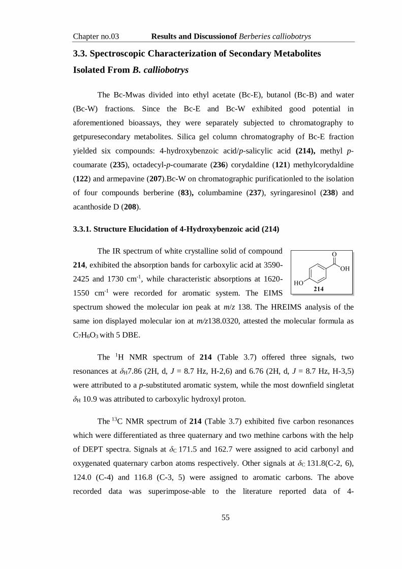
Chapter no.03 Results and Discussionof Berberies calliobotrys
55
3.3. Spectroscopic Characterization of Secondary Metabolites
Isolated From B. calliobotrys
The Bc-Mwas divided into ethyl acetate (Bc-E), butanol (Bc-B) and water
(Bc-W) fractions. Since the Bc-E and Bc-W exhibited good potential in
aforementioned bioassays, they were separately subjected to chromatography to
getpuresecondary metabolites. Silica gel column chromatography of Bc-E fraction
yielded six compounds: 4-hydroxybenzoic acid/p-salicylic acid (214), methyl p-
coumarate (235), octadecyl-p-coumarate (236) corydaldine (121) methylcorydaldine
(122) and armepavine (207).Bc-W on chromatographic purificationled to the isolation
of four compounds berberine (83), columbamine (237), syringaresinol (238) and
acanthoside D (208).
3.3.1. Structure Elucidation of 4-Hydroxybenzoic acid (214)
The IR spectrum of white crystalline solid of compound
214, exhibited the absorption bands for carboxylic acid at 3590-
2425 and 1730 cm-1, while characteristic absorptions at 1620-
1550 cm-1 were recorded for aromatic system. The EIMS
spectrum showed the molecular ion peak at m/z 138. The HREIMS analysis of the
same ion displayed molecular ion at m/z138.0320, attested the molecular formula as
C7H6O3 with 5 DBE.
The 1H NMR spectrum of 214 (Table 3.7) offered three signals, two
resonances at δH7.86 (2H, d, J = 8.7 Hz, H-2,6) and 6.76 (2H, d, J = 8.7 Hz, H-3,5)
were attributed to a p-substituted aromatic system, while the most downfield singletat
δH 10.9 was attributed to carboxylic hydroxyl proton.
The 13C NMR spectrum of 214 (Table 3.7) exhibited five carbon resonances
which were differentiated as three quaternary and two methine carbons with the help
of DEPT spectra. Signals at δC 171.5 and 162.7 were assigned to acid carbonyl and
oxygenated quaternary carbon atoms respectively. Other signals at δC 131.8(C-2, 6),
124.0 (C-4) and 116.8 (C-3, 5) were assigned to aromatic carbons. The above
recorded data was superimpose-able to the literature reported data of 4-

Chapter no.03 Results and Discussionof Berberies calliobotrys
56
hydroxybenzoic acid (Yoshioka et al., 2004). Thus compound 214 was found the
same, which is a well-known phytochemical.
Table 3. 71H- and 13C-NMR data of compound 214 (CDCl3, 400 and 125 MHz)
Position δH (J in Hz) δC
1 - 124.0
2,6 7.86 (2H, d, 8.7) 131.8
3,5 6.76 (2H, d, 8.7) 116.8
4 - 162.7
7 - 171.5
3.3.2. Structure Elucidation of Methyl p-Coumarate (235)
Compound 235was obtained as white solid, which showed absorption bands
for hydroxyl, carbonyl, and double bond at 3450, 1730 and 1650 cm-1 respectively in
theIR spectrum. The EIMS spectrum displayed
molecular ion peak at m/z 178, while the HR-EIMS
(m/z178.0641) supported the molecular formulaas
C10H10O3 with 6 DBE. The 1H-NMR spectrum of
235(Table 3.8) displayed the presence of two pairs of
doublets resonating at δH7.60 (2H, d, J = 9.0 Hz) and δH 6.90 (2H, d, J = 9.0 Hz)
contributing for the presence of p-substituted aromatic system. Furthermore two more
doublets of one proton each were seenat δH 7.59 (J = 16.0 Hz)andδH 6.45 (J = 16.0
Hz), which were attested for two hydrogens in trans relationship of an olefinic
system. A sharp singlet was observed for oxygenated methyl at δH3.54.
13C-NMR data of compound 235 (Table 3.8), displayed 10 signals which were
distinguished as three quaternary carbons (δC168.2 (C-9), 159.0 (C-4) 125.5(C-1)), six
methines (δC146.4 (C-7), 131.2 (C-2, 6), 117.0 (C-3, 5) 116.0 (C-8)) and a methyl
carbon (δC50.9, C-1'). Comparison with the reported values, compound 235 was
identified as methyl ester of p-coumarate (Daayf et al., 1997).
3.3.3.Structure Elucidation of Octadecyl-p-cumarate (236)
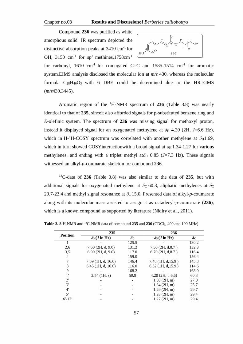
Chapter no.03 Results and Discussionof Berberies calliobotrys
57
Compound 236 was purified as white
amorphous solid. IR spectrum depicted the
distinctive absorption peaks at 3410 cm-1 for
OH, 3150 cm-1 for sp2 methines,1758cm-1
for carbonyl, 1610 cm-1 for conjugated C=C and 1585-1514 cm-1 for aromatic
system.EIMS analysis disclosed the molecular ion at m/z 430, whereas the molecular
formula C28H46O3 with 6 DBE could be determined due to the HR-EIMS
(m/z430.3445).
Aromatic region of the 1H-NMR spectrum of 236 (Table 3.8) was nearly
identical to that of 235, sinceit also afforded signals for p-substituted benzene ring and
E-olefinic system. The spectrum of 236 was missing signal for methoxyl proton,
instead it displayed signal for an oxygenated methylene at δH 4.20 (2H, J=6.6 Hz),
which in1H-1H-COSY spectrum was correlated with another methylene at δH1.69,
which in turn showed COSYinteractionwith a broad signal at δH 1.34-1.27 for various
methylenes, and ending with a triplet methyl atδH 0.85 (J=7.3 Hz). These signals
witnessed an alkyl-p-coumarate skeleton for compound 236.
13C-data of 236 (Table 3.8) was also similar to the data of 235, but with
additional signals for oxygenated methylene at δC 60.3, aliphatic methylenes at δC
29.7-23.4 and methyl signal resonance at δC 15.0. Presented data of alkyl-p-coumarate
along with its molecular mass assisted to assign it as octadecyl-p-coumarate (236),
which is a known compound as supported by literature (Nidiry et al., 2011).
Table 3. 81H-NMR and 13C-NMR data of compound 235 and 236 (CDCl3, 400 and 100 MHz)
Position 235 236
δH(J in Hz) δC δH(J in Hz) δC
1 - 125.5 - 130.2
2,6 7.60 (2H, d, 9.0) 131.2 7.50 (2H, d,8.7 ) 132.3
3,5 6.90 (2H, d, 9.0) 117.0 6.70 (2H, d,8.7 ) 116.4 4 - 159.0 - 156.4
7 7.59 (1H, d, 16.0) 146.4 7.48 (1H, d,15.9 ) 145.3
8 6.45 (1H, d, 16.0) 116.0 6.32 (1H, d,15.9 ) 114.6
9 - 168.2 - 168.0
1′ 3.54 (1H, s) 50.9 4.20 (2H, t, 6.6) 60.3
2′ - - 1.69 (2H, m) 27.0
3′ - - 1.34 (2H, m) 25.7
4′ - - 1.29 (2H, m) 29.7
5′ - - 1.28 (2H, m) 29.4
6′-17′ - - 1.27 (2H, m) 29.4
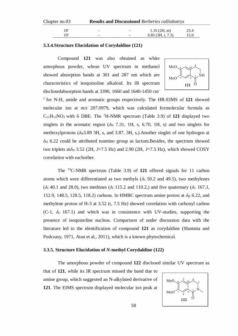
Chapter no.03 Results and Discussionof Berberies calliobotrys
58
18′ - - 1.35 (2H, m) 23.4
19′ - - 0.85 (3H, t, 7.3) 15.0
3.3.4.Structure Elucidation of Corydaldine (121)
Compound 121 was also obtained as white
amorphous powder, whose UV spectrum in methanol
showed absorption bands at 301 and 287 nm which are
characteristics of isoquinoline alkaloid. Its IR spectrum
disclosedabsorption bands at 3390, 1660 and 1640-1450 cm-
1 for N-H, amide and aromatic groups respectively. The HR-EIMS of 121 showed
molecular ion at m/z 207.0979, which was calculated formolecular formula as
C11H13NO3 with 6 DBE. The 1H-NMR spectrum (Table 3.9) of 121 displayed two
singlets in the aromatic region (δH 7.31, 1H, s, 6.70, 1H, s) and two singlets for
methoxylprotons (δH3.89 3H, s, and 3.87, 3H, s,).Another singlet of one hydrogen at
δH 6.22 could be attributed toamino group as lactum.Besides, the spectrum showed
two triplets atδH 3.52 (2H, J=7.5 Hz) and 2.90 (2H, J=7.5 Hz), which showed COSY
correlation with eachother.
The 13C-NMR spectrum (Table 3.9) of 121 offered signals for 11 carbon
atoms which were differentiated as two methyls (δC 50.2 and 49.5), two methylenes
(δC 40.1 and 28.0), two methines (δC 115.2 and 110.2,) and five quaternary (δC 167.1,
152.9, 148.5, 128.5, 118.2) carbons. In HMBC spectrum amine proton at δH 6.22, and
methylene proton of H-3 at 3.52 (t, 7.5 Hz) showed correlation with carbonyl carbon
(C-1, δC 167.1) and which was in consistence with UV-studies, supporting the
presence of isoquinoline nucleus. Comparison of under discussion data with the
literature led to the identification of compound 121 as corydaldine (Shamma and
Podczasy, 1971, Atan et al., 2011), which is a known phytochemical.
3.3.5. Structure Elucidation of N-methyl Corydaldine (122)
The amorphous powder of compound 122 disclosed similar UV spectrum as
that of 121, while its IR spectrum missed the band due to
amine group, which suggested an N-alkylated derivative of
121. The EIMS spectrum displayed molecular ion peak at

Chapter no.03 Results and Discussionof Berberies calliobotrys
59
m/z 221, while HREIMS established the molecular formula C12H15NO3 with 6 DBE
(221.1043 calculated 221.1052). The 1H-NMR spectrum (Table 3.9) of 122 was also
nearly super-imposable to that of 121 with only one difference; as the spectrum
missed signal for N-H and instead it showed an additional methyl group atδH 3.21
(s).This information substantiated the above deduction of N-alkylated derivative of
121. Furthermore the 13C-NMR spectrum (Table 3.9) of 122 also exhibited an
additional signal for N-methyl at δH 47.5 presenting HMBC correlations with
carbonyl carbon at δC165.2, thatconfirmed122 as N-methyl derivative of 121,whichis
named as is N-methylcorydaldine, another known phytochemical (Shamma and
Podczasy, 1971).
3.3.6. Structure Elucidation of Armepavine (207)
Compound 207 was isolated as white crystalline solid.The IR spectrum of this
compound showed absorption bands at 3420 cm-1 (O-H), 3120 cm-1 (=C-H) and 1649-
1466 cm-1 (Ar-C=C). EIMS spectrum afforded molecular ion peak at m/z 313, while
HREIMS analysis of the same ion (m/z313.1672) depicted the molecular formula as
C19H23NO3 with 9 DBE.Aromatic region of 1H NMR spectrum of 207 (Table 3.9)
displayed two resonances at δH 7.21 (2H, d, J = 8.8 Hz, H3', 5') and δH 6.81 (2H, d, J
= 8.8 Hz, H2', 6'), while two methoxyl proton signals and one N-methyl resonance
were seen at δH 3.88 (3H, s), 3.87, (3H, s) and 3.22 (3H, s) respectively. Two singlet
protons appeared at 6.72 (1H, s) and 7.31(1H, s) for C-5 and C-8. One downfield
triplet appeared at 4.24 (1H, t, 7.0) for proton near to nitrogen along with a doublet at
3.01 (2H, d, 6.8) for C-7. Two more triplets for methylene resonated at 3.51 (2H, t,
7.2) and 2.82 (2H, t, 7.2). This data indicated compound 207 could be also an
isoquinoline derivative having partial similarity with structure of compound
122expect an extra benzene ring. Furthermore the 13C-NMR spectrum of compound
207 also exhibited additional five signals as compared to 122 for seven carbons
(Table 3.9), which were predicted as two quaternary carbons δC 154.3 (C-1') and δC
135.4 (C-4') and two methines δC 117.3 (C-2', 6') and δC131.0 (C-3', 5') and one
methylene resonating at δC 41.0 (C-7'). The HMBC interaction (Fig 3.4) of methine
proton at δH 4.24 with δC C-8 (117.2), C-9 (119.8), -3', 5' (131.0) and with N-methyl
(δC 45.5) confirmed attachment of benzyl group at C-1 of isoquinoline part.

Chapter no.03 Results and Discussionof Berberies calliobotrys
60
Comparison with reported data confirmed that 207 is a known phytochemical
namedas armepavine (207) (Marek et al., 1997).
Figure 3. 4 Important HMBC (H→C) csorrelations of 207
Table 3. 91H-NMR and 13C-NMR data of compounds 121, 122 and 207 (CDCl3, 400 and 100 MHz)
Position 121 122 207
δH (J in Hz) δC δH (J in Hz) δC δH (J in Hz) δC
1 - 167.1 - 165.2 4.24 (1H, t, 7.0) 72.9
2 - - - - - -
3 3.52 (2H, t, 7.5) 40.1 3.49 (2H, t, 7.0) 39.7 3.51 (2H, t, 7.2) 40.1
4 2.90 (2H, t, 7.5) 28.0 2.80 (2H, t, 7.0) 29.0 2.82 (2H, t, 7.2) 30.0
5 6.70 (1H, s) 110.2 6.72 (1H, s) 111.3 6.72 (1H, s) 113.7
6 - 152.9 - 151.9 - 150.9
7 - 148.5 - 149.1 - 149.6
8 7.31(1H, s) 115.2 7.31(1H, s) 116.2 7.31(1H, s) 117.2
9 - 118.2 - 119.7 - 119.8
10 - 128.5 - 127.5 - 127.9
1' - - - - - 154.3
2', 6' - - - - 6.81 (2H, d, 8.8) 117.3
3', 5' - - - - 7.21 (2H, d, 8.8) 131.0
4' - - - - - 135.4
7' - - - - 3.01 (2H, d, 6.8). 41.0
1'-OH - - - - 4.5(1H, s) -
2-NH 6.22(1H, s) - - - - -
2-NMe - - 3.21(3H, s) 47.5 3.22 (3H, s) 45.5
6-OMe 3.89(3H, s) 50.2 3.87(3H, s) 53.8 3.88 (3H, s) 51.4
7-OMe 3.87(3H, s) 49.5 3.86(3H, s) 51.2 3.87 (3H, s) 49.2
3.3.7. Structure Elucidation of Berberine (83)
Compound 83 was isolated as yellow crystalline solid, which displayed
absorption bands in the IR spectrum at 1612-1490 cm-1 for aromatic system and at

Chapter no.03 Results and Discussionof Berberies calliobotrys
61
1041 cm-1 for methylenedioxy group. Molecular ion peak at m/z 336 was observed in
EIMS spectrum, while HREIMS analysis helped to establishing molecular formula as
C20H18NO4 with 13 DBE (336.1218 calculated 336.1230). The 1H-NMR spectrum
(Table 3.10) of 83 exhibited six signals in aromatic region presenting one AB-system
at δH 8.11 (1H, d, J = 9.2 Hz) and 7.99 (1H, d, J = 9.2
Hz), whereas, four singlets at δH 9.75(1H, s),8.70
(1H, s), 7.65 (1H, s), and 6.97 (1H, s), offered an
indication for three tetra-substituted benzene ring
systems. The same spectrum also revealed presence
of two methoxylgroups due to signals at δH 4.21 (3H,
s) and 4.10 (3H, s) and three signals for methylene
protons at δH 6.11 (2H, s), 4.93 (2H, t, J=6.4) and 3.75 (2H, t, J=6.4) suggested the
presence of three methylene protons. Downfield shift of methylene group at δH 6.11
(2H, s) revealed the presence of methylendioxy group. All aboveproton data was
inconsistent with the presence of berberine nucleus. The 13C-NMR of 83 (Table 3.10)
disclosed resonances of 20 carbon atoms, which were distinguished as nine quaternary
(δC152.1, 150.8, 150.0, 143.2, 139.4, 167.3, 163.6, 124.7, 122.0), six methines (δC
145.2, 164.4, 124.7, 121.7, 109.4, 106.4) three methylenes (δC103.6, 57.1, 28.3), and
two methoxy carbons (δC 63.8, 59.0). The down field signal for methylene (δC 103.6)
substantiated methylenedioxy group, while two other methylenes (δC 57.1, 28.3) were
connected to nitrogen atom and aromatic system respectively.
All spectroscopic data was in full agreement with the reported data of
forberberine (83) (Hsieh et al., 2004; Yu et al., 2014), which is also a known
phytochemical.
3.3.8. Structure Elucidation of Columbamine (237)
Yellow amorphous solid of compound 237
exhibited IR spectrum abosprtion bands for O-H
(3350 cm-1) and aromatic system (1620-1540 cm-
1). High resolution analysis of the molecular ion
(m/z338.1377) observed in EIMS revealed the

Chapter no.03 Results and Discussionof Berberies calliobotrys
62
molecular formula as C20H20NO4 with 13 DBE.The 1H-NMR spectrum (Table 3.10) of
237 was found barely identical to that of 83 with only one difference that the
spectrum missed signal for methylendioxy, however, an additional methoxyl signal
(δH 3.83, s) was ascertained, which indicated the hydrolyzing of methylendioxy group
of 83. The13C-NMR spectrum (Table 3.10) of 237 afforded 21 resonances with no
signal for methylenedioxy and additional signal for methoxyl group when compared
to the spectrum of 83. Thus compound 237 was found to becolumbamine 237 (Hsieh
et al., 2004), which is also a known natural product.
Table 3. 101H-NMR and 13C-NMR data of compound 83 and 237 (CDCl3, 400 and 100 MHz)
Position 83 237
δH (J in Hz) δC δH(J in Hz) δC
1 7.65 (1H, s) 106.4 7.63 (1H, s) 103.8
1a - 122.0 - 123.0 2 - 150.0 - 148.0
3 - 152.1 - 150.1
4 6.97 (1H, s) 109.4 6.97(1H, s) 104.4
4a - 163.6 - 164.6
5 3.75 (2H, t,6.4) 28.3 3.73 (2H, t,6.6) 39.3
6 4.93 (2H, t, 6.4) 57.1 4.83 (2H, t, 6.6) 56.1
7 - - - -
8 9.75 (1H, s) 145.2 9.68 (1H,s) 144.2
8a - 125.4 - 123.4
9 - 143.2 - 142.2
10 - 150.8 - 152.8 11 7.99 (1H, d, 9.2) 164.4 7.87 (1H, d, 8.7) 130.4
12 8.11 (1H, d, 9.2) 124.7 8.10 (1H, d, 8.7) 125.7
12a - 167.3 - 167.3
13 8.70 (1H, s) 121.7 8.68 (1H, s) 121.3
13a - 139.4 - 138.4
9-OMe 4.21 (3H, s) 63.8 4.12 (3H, s) 60.4
10-OMe 4.10 (3H, s) 59.0 4.12 (3H, s) 57.0
14 (-OCH2O-) 6.11 (s) 103.6 - -
2-OH - - 5.49(s) -
3-OMe - - 3.83(s) 56.2
3.3.9.Structure Elucidation of Syringaresinol (238)
Compound 238 was separated as
white amorphous solid. The IR spectrum
showed characteristics absorption bands for
OH (3340 cm-1), aromatic system (1620-
1430 cm-1) and C-O-C linkage (1380, 1650
cm-1). The EIMS exhibited molecular ion
peak at m/z 418, whereas HR-EIMS spectrum deduced the molecular formula as

Chapter no.03 Results and Discussionof Berberies calliobotrys
63
C22H26O8 with 10 DBE (418.1612 calculated 418.1628). The 1H-NMR spectrum
(Table 3.11) of 238 presented the signal for aromatic protons at δH 6.60 for methine.
One doubletresonating at δH 4.75 (dd, J = 11.2, 7.24 Hz) was attributedfor oxygenated
methineand doublet of doublets at δH 4.31 (dd, J = 11.2, 7.24Hz) and 3.94 (2H, dd,
11.2, 7.24) were deduced for oxygenated methylene. Another singlet at δH 5.54 (1H, s,
4', 4'') was assigned for phenolic proton. One singlet appeared at δH 3.93 with
integration of four methoxyl groups attached with aromatic system. This1H-NMR
spectral data for compound 238 revealed the bis-tetrahydrofuran nucleus which was
confirmed by the13C-NMR spectra that displayed the resonances for eight carbon
atoms, being attributed to three quaternary (δC 147.2, 166.4, 164.3), one methylene (δC
86.2), three methine (δC 103.8, 71.8, 54.2) and one methoxy (δC 56.4). Further dimer
skeleton was confirmed by HMBC correlation (Figure 3.5) of signal at δH 4.75 (1H,
dd, J=3.40) with δC 71.8(C-4,8) and with its own carbon at δC 86.2.Also the double
carbon atoms in molecular formula substantiated dimeric nature of 238. The whole
obtained data was similar with the reported data for compound syringaresinol (238),
which is a known lignin (Monthong et al., 2011).
Figure 3. 5Important HMBC (H→C) correlations of 238
3.3.10. Structure Elucidation of Acanthoside D (208)
Compound 208 was
found to be glucoside of
238, since most of its 1H-
and 13C-NMR spectra
(Table 3.11) displayed
similar signals to that of

Chapter no.03 Results and Discussionof Berberies calliobotrys
64
238. The only difference seen was the resonances due to hexose moiety. The aromatic
proton displayed its position at δH 5.31 (1H, d, J = 7.0 Hz) along with multiplet in the
range of δH 3.70-3.30, offered a symmetric glucoside lignin, whereas, the 13C-NMR
spectrum (Table 3.11) fully agreed with proton data because of carbon resonances
atδC 103.8, 77.3, 76.3, 70.8, 77.1 and 62.2.The sugar connectivity with aromatic
system could be determined due to HMBC (Figure 3.6) interactionof anomeric
hydrogen at δH 5.31with C-4 (δC 72.8) of aromatic system. These deductions were
substantiated due to molecular formula as C34H46O18 with 10 DBE based on HREIMS
(m/z 742.2678) analysis.Further the whole data was similar with the reported data for
compound acanthoside D (208), which is also a known lignin (Lami et al., 1991).
Figure 3. 6Important HMBC (H→C) correlations of 208

Chapter no.03 Results and Discussionof Berberies calliobotrys
65
Table 3. 111H- and 13C-NMR data of compound 238 and208 (CDCl3, 400 and 125 MHz)
Position
238 208
δH (J in Hz) δC δH (J in Hz) δC
1,5 3.12 (1H, m) 54.2 3.11 (1H, m) 53.2 2,6 4.75(1H, d, 3.40) 86.2 4.77(1H,d, 12) 87.2
4a,8a 4.31 (2H, dd, 11.2, 7.24) 71.8 4.33 (2H, dd, 12.0, 8.16) 72.8
4b,8b 3.94 (2H, dd, 11.2, 7.24) 4.09 (2H, dd, 12.0, 8.16)
1',1'' - 164.3 - 165.0
2',2'' 6.60 (1H, s) 103.8 6.63 (1H, s) 103.4
3',3'' - 147.2 - 148.2
4',4'' - 166.4 - 166.9
5',5'' - 147.2 - 148.2
6',6'' 6.60 (1H, s) 103.8 6.63 (1H, s) 103.4
3'-OCH3 3.93 (3H, s) 56.4 3.93 (3H, s) 57.4
3''-OCH3 3.93 (3H, s) 56.4 3.93 (3H, s) 57.4
5'-OCH3 3.93 (3H, s) 56.4 3.93 (3H, s) 57.4 5''-OCH3 3.93 (3H, s) 56.4 3.93 (3H, s) 57.4
4'-OH, 5.54 (1H, s) - - -
4''-OH 5.54 (1H, s) - - -
1''a, 1'b - - 5.31 (1H, d, 7.0) 103.8
2''a, 2'b - - 3.45 (1H, t, 8.4) 77.3
3''a, 3'b - - 3.52 (1H, m) 76.3
4''a, 4'b - - 3.34 (1H, t, 7.2) 70.8
5''a, 5'b - - 3.31 (1H, m) 77.3
6''a, 6'b
- - 3.70 (1H, dd, 12.2,3.9)
3.61 (1H, dd, 12.2, 3.9)
62.2

Chapter no.03 Results and Discussionof Caragana ambigua
66
Part B
3.4. Biological Screening of Crude Extracts of Caragana ambigua
Different polarity solvent extracts C. ambigua methanol extract (Ca-M), C.
ambigua hexane extract (Ca-H), C. ambigua ethyl acetateextract (Ca-E) and C.
ambigua water extract (Ca-W) were tested for antioxidant potentials (DPPH, ABTS,
FRAP, CUPRAC, phosphomolybdenum and metal chelation assays (Table 3.12) and
inhibitory assays (acetylcholinesterase (AChE), butyrylcholinesterase (BChE), α-
glucosidase, α-amylase and tyrosinase (Table 3.13)).
Comprehensive assessment of antioxidant potential revealed that Ca-E shared
most convincing result in the DPPH (83.32 mgTE/g extract), ABTS (421.94 mg TE/g
extract), CUPRAC (405.26 mgTE/g extract), FRAP (617.89 mgTE/g extract) and
phosphomolybdenum (2.70 mmolTE/g extract) antioxidant assays which are in
concurrence to its highest phenolic content while Ca-H was found least active in all
above assays except phosphomolybdenum assay(1.96 mmolTE/g extract) where it
was found next to the Ca-E. Moreover Ca-H was more potent in metal chelation
assays (74.27 mgEDTAE/g extract). It might be because of antagonistic or synergetic
and non-phenolic chelator (Marini et al., 2018; Khan at al., 2019).
Table 3. 12Antioxidant properties of C. ambigua extracts*
Plant extracts
Radical scavenging assays Reducing power assays Total antioxidant
activity
Ferrous ion
chelation
DPPH (mgTE/g extract)
ABTS (mgTE/g extract)
FRAP (mgTE/g extract)
CUPRAC (mgTE/g extract)
Phosphomolybdenum
(mmolTE/g)
Metal chelating
(mgEDTAE/g)
Ca-M 55.05±2.00b 103.12±6.10b 163.108±1.99b 204.66±9.17b 1.30±0.10a 23.82±1.24b
Ca-E 83.32±6.22d 421.94±13.93d 405.26±11.15d 617.89±7.00d 2.70±0.06a 53.96±3.15c
Ca-H 13.106±0.19a 60.10±3.75a 85.36±8.82a 161.88±8.71a 1.96±0.12a 74.27±3.71d
Ca-A 60.16±3.01c 115.52±1.22c 227.96±3.13c 236.98±2.11c 1.47±0.06a 10.32±0.21a
* Values expressed are means S.D. of three parallel measurements; means with different superscript letters in the same column are significantly (p <0.05) different TE: Trolox equivalent;
EDTAE: EDTA equivalent
All extracts of C.ambigu were tested for their inhibitory potential against
AChE, BChE, α-glucosidase, α-amylase, and tyrosinase (Table 3.13). In AChE and BChE
assays, Ca-H attributed high potential (4.81 mgGALAE/g extract and 4.95mgGALAE/g

Chapter no.03 Results and Discussionof Caragana ambigua
67
extract respectively). In α-amylase assay Ca-H (0.81 mmolACAE/g extract) showed
highest potential followed by Ca-E (0.75 mmolACAE/g extract), Ca-M (0.54
mmolACAE/g extract) and Ca-W (0.81 mmolACAE/g extract). Furthermore Ca-H
(1.68 mmolACAE/g extract) and Ca-E (1.67mmolACAE/g extract) exhibited
comparable potential for α-glucosidase followed by Ca-A (0.78 mmolACAE/g
extract) while Ca-M was found to be inactive.
Similarly in tyrosinase assays extract presented the following sequences Ca-E
>Ca-H> Ca-M > Ca-A (185.80> 176.01> 172.08> 38.57 mgKAE/g extract)
respectively.
Table 3. 13Enzyme inhibition activities of C. ambigua extracts*
Plant
extracts
AChE
(mgGALAE/g
extract)
BChE
(mgGALAE/g
extract
α-Amylase
(mmolACAE/g
extract)
α-Glucosidase
(mmolACAE/g
extract)
Tyrosinase
(mgKAE/g
extract)
Ca-M 4. 55±0.08b 2.91±0.41a 0.54±0.02b Na 172.08±1.29b
Ca-E 3.18±0.24a 3.57±0.12a 0.75±0.01b 1.67±0.01c 185.80±1.45d
Ca-H 4.81±0.32b 4.95±0.30b 0.81±0.02b 1.68±0.01b 176.01±1.10c
Ca-A Na Na 0.08±0.01a 0.78±0.10a 38.57±1.83a
* Values expressed are means S.D. of three parallel measurements; means with different superscript letters in the same column are significantly (p <0.05) different; GALAE: Galatamine
equivalent; KAE: Kojic acid equivalent; ACAE: Acarbose equivalent; na: not active.
3.5. Phytochemical Analysis of Secondary Metabolites from
Caragana ambigua
Different polarity solvent extracts Ca-M, Ca-H, Ca-E, and Ca-W were
investigated for their bioactive contents and UHPLCMS analyses.
3.5.1 Total phenolic and flavonoid contents estimation
Different polarity solvent extracts Ca-M, Ca-H, Ca-E, and Ca-W were tested
for total phenolic and flavonoid contents (Table 3.14), Analysis of results revealed the
highest phenolic and flavonoid contents in Ca-E with value 85.87mgGAE/g and 66.45
mgGAE/g extract respectively (Table 3.14).

Chapter no.03 Results and Discussionof Caragana ambigua
68
Table 3. 14Total phenolic and flavonoid content of C. ambigua extracts*
Extracts Abbreviation Total phenolic content
(mgGAE/g extract)
Total flavonoid content
(mgRE/g extract)
Methanol Ca-M 37.37±0.21b 17.36±0.27b
Ethyl acetate Ca-E 85.87±2.96d 66.45±0.37d
n-Hexane Ca-H 33.79±2.15a 15.06±0.16a
Aqueous Ca-A 44.60±0.89c 58.69±0.15c
* Values expressed are means S.D. of three parallel measurements; means with different
superscript letters in the same column are significantly (p <0.05) different; GAE: Gallic acid
equivalents; RE: Rutinequivalents
3.5.2. UHPLC-MS Analysis to IdentifySecondary Metabolites of Caragana
ambigua
UHPLC-MSwas used for identification of secondary metabolites in Ca-M and Ca-E.
3.5.2.1. Secondary metabolite estimation of Ca-M through UHPLC-MS analysis
UHPLC-MS in negative ionization mode analysis of fraction Ca-M
revealedthe presence of 16 secondary metabolites (Table 3.15,figure 3.7) including
nine-flavonoid, one compound from each class of alkaloid, saponin, terpenoids,
coumarin, glycoside, fatty acid and lignin.
Figure 3. 7Total ion chromatograms of methanol extractof C. ambigua

Chapter no.03 Results and Discussionof Caragana ambigua
69
Table 3. 15UHPLC-MS secondary metabolites profile of Fraction Ca-M
S. No RT
(min)
Base
peak(m/z)
Peak
height AUC Proposed compounds
Compound
class
Molecular
formula
Molecula
r mass
1 0.626 215.03 1091409 3957998 Isobergaptene (200) Coumarin C12H8O4 216.03
2 0.668 683.22 118383 750095 Citbismine C (203) Alkaloid C37H36N2O11 684.23
3 7.864 885.26 213028 1807731 Kaempferol-3-isorhamninoside-7-
rhamnoside (239) Flavonoid C39H50O23 886.27
4 7.921 739.20 71637 579281 Robinin (240) Flavonoid C33H40O19 740.21
5 0.868 290.08 120873 391537 Sarmentosin epoxide (230) Glycoside C11H17NO8 291.09
6 8.719 609.14 163999 1095266 Robinetin -3-rutinoside (241) Flavonoid C27H30O16 610.15
7 9.107 579.20 99994 696046 (+)-Syringaresinol O-β-D-glucoside
(209) Lignin C28H36O13 580.21
8 9.118 623.16 300142 2545922 Tricetin 7-methyl ether 3'-
glucoside-5'-rhamnoside (242) Flavonoid C28H32O16 624.17
9 9.992 1165.53 123692 1068560 Calendasaponin C (243) Terpene C54H86O25 1166.54
10 11.442 299.05 92724 709453 Kaempferide (223) Flavonoid C16H12O6 300.06
11 11.451 329.23 139562 1367030 5,8,12-trihydroxy-9-octadecenoic
acid (211) Fatty acid C18H34O5 330.24
12 11.463 517.17 90511 631280 Phellamurin (244) Flavonoid C26H30O11 518.17
13 12.166 941.51 291245 4112074 Jujubasaponin IV (245) Saponin C48H78O18 943.91
14 13.114 355.11 250807 1863271 Phellodensin D (226) Flavonoid C20H20O6 356.12
15 13.179 353.10 139571 1054198 2,3-Dehydrokievitone (246) Flavonoid C20H18O6 354.11
16 14.099 337.10 119429 901058 (-)-Glyceollin I (247) Flavonoid C20H18O5 338.11
RT: retention time; AUC: area under curve
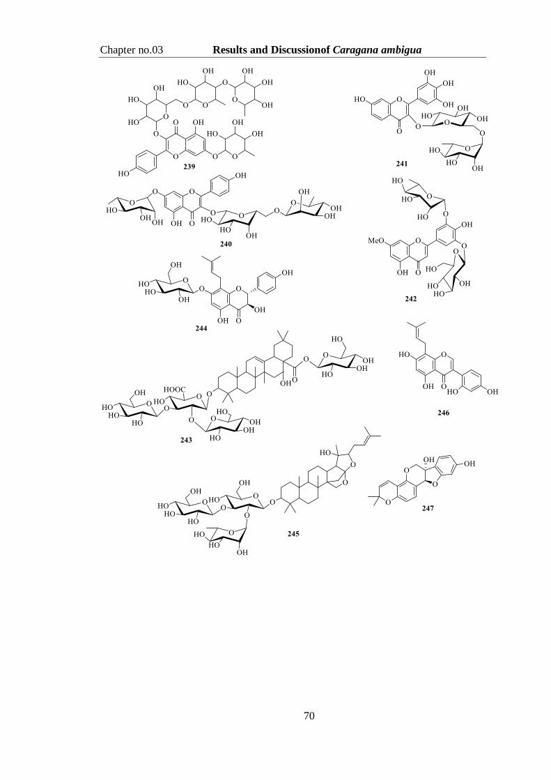
Chapter no.03 Results and Discussionof Caragana ambigua
70

Chapter no.03 Results and Discussionof Caragana ambigua
71
3.5.2.2Secondary metabolite estimation of Ca-E through UHPLC-MS analysis
UHPLC-MS analysisof Ca-E disclosed the presence of 26 secondary
metabolites including twenty flavonoids as a larger group, two fatty acids, two
phenolics and one coumarin class of compounds (Table 3.16,Figure 3.8). The
presence of aforementioned metabolites is in accord with the reported literature of
genus Caragana (Mandal et al., 2016).
Figure 3. 8Total ion chromatograms of ethyl acetate extract (Ca-E) of C.
ambigua
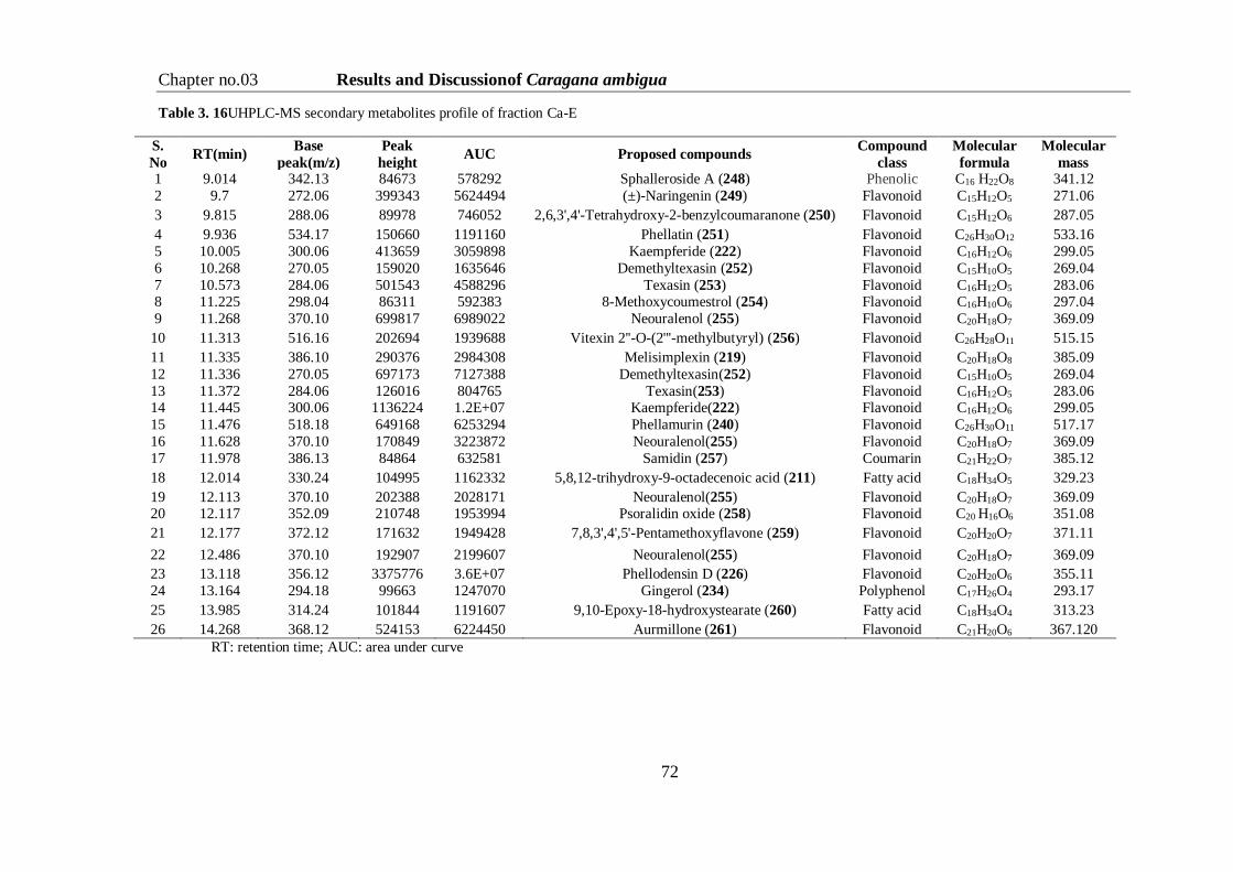
Chapter no.03 Results and Discussionof Caragana ambigua
72
Table 3. 16UHPLC-MS secondary metabolites profile of fraction Ca-E
S.
No RT(min)
Base
peak(m/z)
Peak
height AUC Proposed compounds
Compound
class
Molecular
formula
Molecular
mass 1 9.014 342.13 84673 578292 Sphalleroside A (248) Phenolic C16 H22O8 341.12
2 9.7 272.06 399343 5624494 (±)-Naringenin (249) Flavonoid C15H12O5 271.06
3 9.815 288.06 89978 746052 2,6,3',4'-Tetrahydroxy-2-benzylcoumaranone (250) Flavonoid C15H12O6 287.05
4 9.936 534.17 150660 1191160 Phellatin (251) Flavonoid C26H30O12 533.16
5 10.005 300.06 413659 3059898 Kaempferide (222) Flavonoid C16H12O6 299.05
6 10.268 270.05 159020 1635646 Demethyltexasin (252) Flavonoid C15H10O5 269.04
7 10.573 284.06 501543 4588296 Texasin (253) Flavonoid C16H12O5 283.06 8 11.225 298.04 86311 592383 8-Methoxycoumestrol (254) Flavonoid C16H10O6 297.04
9 11.268 370.10 699817 6989022 Neouralenol (255) Flavonoid C20H18O7 369.09
10 11.313 516.16 202694 1939688 Vitexin 2''-O-(2'''-methylbutyryl) (256) Flavonoid C26H28O11 515.15
11 11.335 386.10 290376 2984308 Melisimplexin (219) Flavonoid C20H18O8 385.09
12 11.336 270.05 697173 7127388 Demethyltexasin(252) Flavonoid C15H10O5 269.04
13 11.372 284.06 126016 804765 Texasin(253) Flavonoid C16H12O5 283.06 14 11.445 300.06 1136224 1.2E+07 Kaempferide(222) Flavonoid C16H12O6 299.05
15 11.476 518.18 649168 6253294 Phellamurin (240) Flavonoid C26H30O11 517.17
16 11.628 370.10 170849 3223872 Neouralenol(255) Flavonoid C20H18O7 369.09
17 11.978 386.13 84864 632581 Samidin (257) Coumarin C21H22O7 385.12
18 12.014 330.24 104995 1162332 5,8,12-trihydroxy-9-octadecenoic acid (211) Fatty acid C18H34O5 329.23
19 12.113 370.10 202388 2028171 Neouralenol(255) Flavonoid C20H18O7 369.09 20 12.117 352.09 210748 1953994 Psoralidin oxide (258) Flavonoid C20 H16O6 351.08
21 12.177 372.12 171632 1949428 7,8,3',4',5'-Pentamethoxyflavone (259) Flavonoid C20H20O7 371.11
22 12.486 370.10 192907 2199607 Neouralenol(255) Flavonoid C20H18O7 369.09
23 13.118 356.12 3375776 3.6E+07 Phellodensin D (226) Flavonoid C20H20O6 355.11
24 13.164 294.18 99663 1247070 Gingerol (234) Polyphenol C17H26O4 293.17
25 13.985 314.24 101844 1191607 9,10-Epoxy-18-hydroxystearate (260) Fatty acid C18H34O4 313.23
26 14.268 368.12 524153 6224450 Aurmillone (261) Flavonoid C21H20O6 367.120
RT: retention time; AUC: area under curve

Chapter no.03 Results and Discussionof Caragana ambigua
73
3.6. Characterization of Secondary Metabolites Isolated from the
Fraction Ca-E
Sincethe ethyl acetate soluble part (Ca-E) exhibited good potential in
biochemical assays therefore was selected for purification of itssecondary metabolites.
Silica gel column chromatographic separation yielded nine known compounds;
taraxerol (262), taraxerol acetate (263), 2-(4′-hydroxyphenyl)-ethyl stearate (264),
apigenin (265), kaempferide (222), naringinin (249), quercetin (125), quercetin 3-O-
β-D-glucopyranosideside (198) and β-sitosterol 3-O-D-glucopyranoside (172).

Chapter no.03 Results and Discussionof Caragana ambigua
74
3.6.1. Structure Elucidation of Taraxerol (262)
Crystalline solid of compound 262 exhibited absorption bands in the IR
spectrum for hydroxyl, olefinic hydrogen and
double bond at 3483, 2945 and 1655 cm-1
respectively. The EIMS spectrum showed molecular
ion peak at m/z 426, whereas the HREIMS assisted
to establish molecular formula as C30H50O with 6
DBE (m/z 426.3840). The 1H-NMR spectrum of 262
(Table 3.17) displayed eight tertiary methyl signals
atδH1.03, 0.97, 0.96, 0.88, 0.87, 0.85, 0.82, 0.81. The proton signal resonating at δH
3.70 (1H, dd, J = 9.0, 4.2 Hz) was assigned to H-3, whereas the most downfield signal
appeared at δH 5.49 (H-15, dd, J = 8.0, 3.4 Hz) was attributed todouble bond in 262.
Furthermore, various methylene resonances displayed their positionsas multiplet
between δH 2.02-1.34, which suggested compound 262 could be a triterpenoid
skeleton, which was substantiated by 13C-NMR data (Table 3.17). The 13C-NMR
broad-band spectrum showed 30 carbons signals, which were differentiated by DEPT
experiment as eight methyls, five methines, ten methylenes and seven quaternary
carbons. The olefinic carbons showed their positionsat δC 157.7 (C) and 115.9 (CH),
whereas, oxymethine appeared at δC 70.9. Comparatively downfield shift of the
olefinic quaternary carbon (δC 157.7) indicated taraxe-14-ene-3-β-ol type of
triterpenoid (Deng et al., 2004).
HMBC correlations of the olefinic proton (δH 5.49) showed HMBC correlations
(Figure 3.9) with carbons at δC 157.7 (C-14), 37.0 (C-16), 35.9 (C-13), and 31.2 (C-8)
which further confirmed the C-14 position for double bond. Furthermore HMBC
interaction of tertiary methyls of H-27 (δH 0.88) and H-18 (δH 0.81) with 157.7 (C-14)
supported the proposed structure. This data was matched with the reported data in
literature for taraxerol (Deng et al., 2004), thuscompound 262 was found to be the
same, which is a common phytochemical but isolated for the first time from our
investigated source.

Chapter no.03 Results and Discussionof Caragana ambigua
75
Figure 3. 9 Important HMBC (H→C) correlations of 262
3.6.2. Structure Elucidation of Teraxerol acetate (263)
Compound 263was also isolated as white amorphous powder. Along with
similar IR absorption bands, its spectrum afforded
strong characteristic absorption for ester at 1730 cm-1.
The EIMS spectrum showed molecular ion peak at
m/z 468, while HR-EIMS analysis (m/z 468.3962) of
the same peak depicted the molecular formula
C32H52O2 with 7 DBE. The 1H- and 13C-NMR dataof
263 (Table 3.17) was nearly identical to that of 262 with the only difference was that
it offered additional signalsfor an acetyl moiety (δH 2.02, δC 22.0 &
172.1).Aforementioned data supported the presence of acetyl group and its position
was confirmed by downfield shift of H-3 at δH 4.43, which supported the replacement
of (3-OH) with acetyl group in 263. All spectral values were foundin full agreement
with known phytochemical taraxerol acetate, already reported in literature (Deng et
al., 2004).

Chapter no.03 Results and Discussionof Caragana ambigua
76
Table 3. 171H-NMR and 13C-NMR data of compound 262 and 263 (CDCl3, 400 and 100 MHz)
Position 262 263
δH(J in Hz) δC δH(J in Hz) δC
1 1.88 (2H, dd, 13.9, 3.9) 19.1 1.90 (2H, dd, 14.3, 2.8) 36.8
2 1.55 (2H, m) 24.2 1.49 (2H, m) 23.2
3 3.70 (1H, dd, 9.0, 4.2) 70.9 4.43 (1H, dd, 9.0, 4.4) 79.9
4 - 38.1 - 38.1
5 0.82 (1H, m) 56.1 0.87 (1H, m) 55.6 6 1.49 (2H, m) 17.5 1.55 (2H, m) 18.1
7 1.32 (2H, m) 33.3 1.30 (2H, m) 33.2
8 - 31.2 - 30.2
9 1.41 (1H, t, 5.8) 49.1 1.43 (1H, t, 5.6) 49.2
10 - 37.2 - 37.0
11 1.57 (2H, m) 18.9 1.66 (2H, m) 17.1
12 1.89 (2H, m) 31.0 1.30 (2H, m) 35.2
13 - 35.9 - 37.1
14 - 157.7 - 158.2
15 5.49(1H, dd, 8.0, 3.4) 115.9 5.53(1H, dd, 8.2, 3.7) 116.6
16 3.36 (2H, t, 6.8) 37.0 3.38 (2H, t, 5.72) 36.1
17 - 35.5 - 37.9
18 0.81 (1H, m) 47.3 0.83 (1H, m) 48.2
19 1.99 (2H, m) 41.8 1.81 (2H, m) 41.7
20 - 37.7 - 38.1
21 1.41(2H, t, 5.1) 33.7 1.36 (2H, t, 5.1) 35.3
22 1.52(2H, t, 2.8) 26.9 1.65 (2H, t, 2.8) 29.0
23 0.82 (3H, s) 21.3 0.83 (3H, s) 21.3
24 0.81 (3H, s) 21.4 0.81 (3H, s) 28.6
25 1.03 (3H, s) 26.0 1.03 (3H, s) 28.9
26 0.85 (3H, s) 29.4 0.85 (3H, s) 29.0
27 0.88 (3H, s) 29.5 0.88 (3H, s) 30.3 28 0.87 (3H, s) 29.1 0.87 (3H, s) 29.3
29 0.96 (3H, s) 27.8 0.96 (3H, s) 28.1
30 0.97 (3H, s) 18.9 0.97 (3H, s) 18.8
1′ - - - 172.1
2′ - - 2.02 (3H, s) 22.0
3.6.3. Structure Elucidation of 2′-(4-Hydroxyphenyl)-Ethyl Stearate (264)
TheIR spectrum of compound 264 displayed distinguished absorption bands at
3440 (O-H), 1729 (C=O), 1520-
1469 (Ar-C=C) and 1210 cm-1(C-
O), whereas, The HREI-MS
analysis afforded molecular ion at
m/z 404.3286 attested for the molecular formula C26H44O3 with five DBE.The 1H-
NMR spectrum of compound 264 (Table 3.18) offered two doublets at δH 7.15 (2H, d,
J = 8.8 Hz) and 6.75 (2H, d, J = 8.8 Hz); the A2B2 splitting pattern indicated the

Chapter no.03 Results and Discussionof Caragana ambigua
77
presence of para-substituted benzene ring.The same spectrum showeda resonance at
δH 4.24 (2H, t, J = 6.6 Hz), which was correlated in COSY spectrum with another
triplet methylene resonance at δH 3.70 (2H, t, J = 6.6 Hz). The chemical shifts of these
two methylenes revealed their attachment with carboxylate function and phenyl ring,
respectively.Other distinct triplet methylene signal at δH 2.75 (2H, t, J = 6.7 Hz) and a
methyl signal at 0.88δH (3H, t, J = 6.8 Hz) showed their COSY relation with a broad
signal of various methylenes between δH 1.63-1.11.This data indicated that compound
264 could be a fatty ester of p-hydroxyphenylethyl.
The 13C-NMR spectrum (Table 3.18) of compound 264 showed signals for
ester carbonyl (δC 178.5), p-substituted aromatic system (δC 153.1, 164.0, 163.0,
117.0) saturated methylenes (δC 67.3, 35.1) and fatty acid chain (δC 35.0, 26.3-21.1,
14.6). The fatty acid chain length could only be fixed due to mass spectrometric data.
Thus compound 264 was finally identified as 2′-(4-hydroxyphenyl)-ethyl stearate,
which was further supported by the reported literature (Ruberto and Tringali, 2004,
Acevedo et al., 2000).
Table 3. 181H-NMR and 13C-NMR data of compound 264 (CDCl3, 500 and 125 MHz)
Position δH (J in Hz) δC
1 - 164.0
2,6 7.15 (2H, d, 8.8) 163.0
3,5 6.75 (2H, d, 8.8) 117.0
4 - 153.1
7 3.70 (2H, d, 6.6) 34.9
8 4.24 (2H, d, 6.6) 67.3
1′ - 178.5
2′ 2.75 (2H, t, 6.7) 35.0
3′ 1.63 (2H, m) 26.3
4′-16′ 1.11-1.38 (26H, br s) 24.0-27.7
17′ 1.22 (2H, m) 21.1
18′ 0.88 (3H, t, 6.8) 14.6
3.6.4. Structure Elucidation of Apigenin (265)
Yellow amorphous powder of compound 265 showed a yellow spot on TLC,
which remained yellow on heating with ceric
sulphate, which indicated a flavonoid
compound. The IR spectrum exhibited
absorption bands for O-H (3420 cm-1), C=O
(1665 cm-1), and Ar-C=C (1610-1520 cm-1).
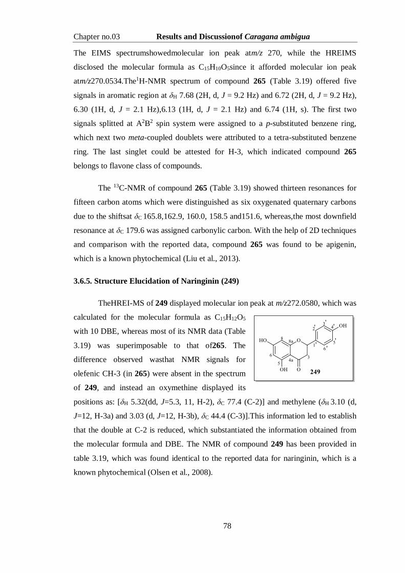
Chapter no.03 Results and Discussionof Caragana ambigua
78
The EIMS spectrumshowedmolecular ion peak atm/z 270, while the HREIMS
disclosed the molecular formula as C15H10O5since it afforded molecular ion peak
atm/z270.0534.The1H-NMR spectrum of compound 265 (Table 3.19) offered five
signals in aromatic region at δH 7.68 (2H, d, J = 9.2 Hz) and 6.72 (2H, d, J = 9.2 Hz),
6.30 (1H, d, J = 2.1 Hz),6.13 (1H, d, J = 2.1 Hz) and 6.74 (1H, s). The first two
signals splitted at A2B2 spin system were assigned to a p-substituted benzene ring,
which next two meta-coupled doublets were attributed to a tetra-substituted benzene
ring. The last singlet could be attested for H-3, which indicated compound 265
belongs to flavone class of compounds.
The 13C-NMR of compound 265 (Table 3.19) showed thirteen resonances for
fifteen carbon atoms which were distinguished as six oxygenated quaternary carbons
due to the shiftsat δC 165.8,162.9, 160.0, 158.5 and151.6, whereas,the most downfield
resonance at δC 179.6 was assigned carbonylic carbon. With the help of 2D techniques
and comparison with the reported data, compound 265 was found to be apigenin,
which is a known phytochemical (Liu et al., 2013).
3.6.5. Structure Elucidation of Naringinin (249)
TheHREI-MS of 249 displayed molecular ion peak at m/z272.0580, which was
calculated for the molecular formula as C15H12O5
with 10 DBE, whereas most of its NMR data (Table
3.19) was superimposable to that of265. The
difference observed wasthat NMR signals for
olefenic CH-3 (in 265) were absent in the spectrum
of 249, and instead an oxymethine displayed its
positions as: [δH 5.32(dd, J=5.3, 11, H-2), δC 77.4 (C-2)] and methylene (δH 3.10 (d,
J=12, H-3a) and 3.03 (d, J=12, H-3b), δC 44.4 (C-3)].This information led to establish
that the double at C-2 is reduced, which substantiated the information obtained from
the molecular formula and DBE. The NMR of compound 249 has been provided in
table 3.19, which was found identical to the reported data for naringinin, which is a
known phytochemical (Olsen et al., 2008).

Chapter no.03 Results and Discussionof Caragana ambigua
79
3.6.6Structure Elucidationof Kaempheride (222)
Compound 222was isolated as a yellow crystalline solid, and due to its
behavior on TLC was also found to be a flavonoid.Its
IR spectrum afforded absorption bands at 3410 (O-H),
1700 (C=O), 1620- 1520cm-1 (Ar-C=C),
whilethemolecular formula as C16H12O6could be
determined due to HREI-MS analysis (m/z
300.0634).The 1H NMR spectrum of 222 revealed the
presence of a p-substituted benzene ring C and two meta-coupled doublets were
attested for benzene ring B. When compared with the spectrum of 265, it missed
signal for H-3, which indicated compound 222 must be a flavonol-derivative. The
same spectrum also displayed an additional resonance due to methoxyl proton at δH
3.61.
The 13C-NMR spectrumof 222(Table 3.19) supported the mass and 1H- NMR
data. The position of methoxy group at C-4was found through HMBC correlations.
Finally the comparison of the spectroscopic data with literature values, compound
222was elucidated as kaempheride which is also a known phytochemical (Anđelković
et al.,2017).
Table 3. 191H- and 13C-NMR data of compound 265 and 249 (DMSO-d6 , 500 and 125 MHz) and 222
(CD3OD, 400 and 100 MHz)
Position
265 249 222
δH (J in Hz) δC δH (J in Hz) δC δH (J in Hz) δC
1 - - - - - -
2 - 151.6 5.32(2H, dd,5.3, 11), 77.4 - 146.9
3 6.74 (1H, s) 104.5 3.10 (d,12, H-3a)
3.03 (d,12, H-3b) 44.4 - 137.7
4 - 179.6 - 174.6 - 176.9
4a - 104.7 - 103.7 - 104.0
5 - 162.9 - 162.0 - 162.7
6 6.13 (1H, d, 2.1) 99.3 6.12 (1H, d, 2.1) 99.8 6.30 (1H, d, 2.0) 99.1
7 - 165.8 - 166.2 - 164.4
8 6.30 (1H, d, 2.1) 93.8 6.33 (1H, d, 2.2) 94.5 6.35 (1H, d, 2.0) 92.8
8a - 160.0 - 161.0 - 160.1
1' - 123.1 - 124.1 - 123.1 2' 7.68 (2H, d, 9.2) 128.8 7.17 (2H, d, 9.0) 129.1 7.65 (2H, d, 9.1) 127.8
3' 6.72 (2H, d, 9.2) 117.8 6.70 (2H, d, 9.0) 118.1 6.73 (2H, d, 9.1) 118.0
4' - 158.5 - 159.1 - 157.5
5 6.72 (2H, d, 9.2) 117.8 6.70 (2H, d, 9.0) 117.1 6.73 (2H, d, 9.1) 118.0
6' 7.68 (2H, d, 9.2) 128.8 7.17 (2H, d, 9.0) 128.7 7.65 (2H, d, 9.1) 127.8
4'-OMe - - - - 3.61(3H, s) 59.5

Chapter no.03 Results and Discussionof Caragana ambigua
80
3.6.7. Structure Elucidation of Quercetin (125)
Compound 125 was obtained as pale yellow crystalline solid that exhibited
characteristic absorption bands forO-H (3428-3369 cm-1), C=O (1708 cm-1) and C=C
(1665-1475cm-1) in the IR spectrum. HREIMS
spectrum disclosed molecular ion at m/z 302.0430,
which depicted the molecular formula as C15H10O7
with 11 DBE.The 1H NMR spectrum (Table 3.20) of
125 displayed five signals in the aromatic region at
δH7.72 (1H, d, J = 2.0 Hz), 7.59 (1H, dd, J = 8.2, 2.0
Hz), 6.75 (1H, d, J = 8.4 Hz),6.33 (1H, d, J = 2.0 Hz)
and 6.29 (1H, d, J = 2.0 Hz). The first three splitted at ABX pattern revealed a1,3,4-
trisubstituted benzene ring Bwhile the other two m-coupled resonances were
attributed to ring A of flavonoid. The 13C NMR spectra (Table 3.20) of 125 displayed
altogether fifteen carbon signals which were identified as ten quaternary carbons
(δC176.9, 165.4, 163.0, 158.5, 147.9, 147.6, 145.9, 137.1, 105.0) and five methines
(δC123.2, 121.0, 116.0, 98.9, 93.8). The data was fully fit to the NMR values reported
for quercetin (Xiuyun et al., 2006), which is a potent antioxidant natural product.
3.6.8. Structure Elucidation of Quercetin 3-O-β-D-glucopyranoside (198)
The compound 198 was also purified as yellow amorphous powder, which
absorbed in the IR spectrum at 3340-3200, 1610-1459 cm-1due tohydroxyl group,
conjugated ketone and phenyl group, respectively. The EIMS spectrum exhibited the
heaviest ion at m/z 454, which was analyzed in
HREIMS as 464.0950 for the molecular formula
as C21H20O12 with 12 DBE. The aromatic region
of the1H NMR spectrum of 198 (Table 3.20) was
identical to that of 125 indicating the same
flavonoidal skeleton. However, the same
spectrum displayed resonance of an anomeric
proton at δH 5.33 (1H, d, J = 7.0 Hz) along withother oxymethine signals at δH 3.70-
3.30 (Table 3.20). This data indicated quercetin glycoside nature of 198, which was
substantiated due to 21resonances in the 13C NMR spectrumof 198 (Table 3.20).The

Chapter no.03 Results and Discussionof Caragana ambigua
81
carbon signals due to aglycone part were similar to that of 125, while sugar signals
displayed their positions at δC 102.8, 78.3, 76.1, 70.9, 77.2 and 62.2.The attachment of
sugar moiety was established at C-3 due to HMBC correlation of anomeric proton (δH
5.33) with quaternary carbon at δC 136.4 (C-3). This data showed complete
resemblancewith the already published data for quercetin 3-O-β-D-glucopyranoside
(198) (Sukito and Tachibana, 2014).
Table 3. 201H- and 13C-NMR data of compound 125 and 198 (DMSO-d6, 400 and 100 MHz)
Position 125 198
δH(J in Hz) δC δH (J in Hz) δC
1 - - - -
2 - 147.9 - 153.8
3 - 137.1 136.4
4 - 176.9 - 180.9
4a - 105.0 - 105.0
5 - 163.0 - 163.1
6 6.29 (1H, d, 2.0) 98.8 6.17 (1H, d, 1.9) 101.2
7 - 165.4 - 165.9
8 6.33 (1H, d, 2.0) 93.8 6.34 (1H, d, 1.9) 94.8
8a - 158.5 - 157.6
1' - 123.2 - 123.7
2' 7.72 (1H, d, 2.0) 116.0 7.69 (1H, d, 2.1) 118.8
3' - 145.9 - 147.2
4' - 147.6 - 151.1
5' 6.75 (1H, d, 8.4) 116.1 6.90 (1H, d, 8.2) 117.2
6' 7.59 (1H, d, 8.2, 2.0) 121.0 7.60 (1H, dd, 8.2, 2.1) 124.4
1'' - - 5.33 (1H, d, 7.0) 102.8
2'' - - 3.45 (1H, t, 8.4) 78.3
3'' - - 3.52 (1H, m) 76.1
4'' - - 3.34 (1H, t, 7.2) 70.9
5'' - - 3.31 (1H, m) 77.2
6'' - - 3.70 (1H, dd, 12.2,3.9)
3.61 (1H, dd, 12.2, 3.9) 62.2
3.6.9. Structure Elucidation of β-Sitosterol 3-O-D-glucopyranoside (172)
Compound 172 was purified as a white amorphous powder.The IR spectrum offered
distinctive peaks for O-H (3451 cm-1) =C-
H (2922-2871 cm-1) and1655 cm-1 (C=C)
functionalities. EI-MS spectrum
displayed molecular ion peak at m/z576
with base peak at m/z 414.It aided finding
the molecular formula as C35H61O6 with
six DBE. The 1H-NMR spectrum of 172

Chapter no.03 Results and Discussionof Caragana ambigua
82
(Table 3.21) showed the presence of two singlets methyls at δH 0.91and 0.88
attributed to Me-19 and Me-18, three doublet methyls atδH 0.96 (3H, d, J = 6.6 Hz),
0.90 (d, J = 6.3 Hz), 0.82 (J = 7.2 Hz) and primary methyl resonated at δH0.89 (3H, t,
J = 7.2 Hz) assigned to Me-21, Me-26, Me-27 and Me-29, respectively. A multiplet at
δH 3.27 and a doublet of doublet at δH 5.26 were observed with characteristic coupling
constant value (J = 9.4, 4.9 Hz) typical for H-3 and H-6 of a compound having
steroidal skeleton.In additional, resonances for the sugar moiety at δH 4.22 (1H, d, J =
7.7 Hz, 1), 2.90 (1H, t, m, 2), 3.28 (1H, t, m, 3), 3.03 (1H, m, 4), 3.10 (1H, m, H-
5), 4.13 (1H,dd, J=11.5,3.10,6a) and 4.39 (1H,dd,J=11.7, 2.9, 6b).
The 13C-NMR spectrum of 172 (Table 3.21) depicted 35 carbon signals, of
which twenty nine resonances wereattributed toβ-sitosterol skeleton while remaining
signals for sugar moiety were seen in the spectrumat δC 101.0 (C-1), 72.0 (C-2), 76.0
(C-3), 70.9 (C-4), 75.5 (C-5) and 60.5 (C-6). The position of sugar moiety was
established through HMBC correlations in which the anomeric proton H-1′ (δH 4.22)
showed correlation with C-3 (δC 81.1) of sterol skeleton.Further comparative TLC and
data correlation with the reported data confirmed the compound 172 as β-sitosterol 3-
O-β-D-glucopyranoside (Akhtar et al., 2010).
Table 3. 211H-NMR and 13C-NMR data of compound 172 (CDCl3+CD3OD, 400 and 100 MHz)
Position δH (J in Hz) δC Position δH (J in Hz) δC
1 3.88 (2H, m) 38.3 19 0.91 (3H, m) 20.7
2 1.87 (2H, m) 30.2 20 1.97 (1H, m) 38.5
3 3.29 (1H, m) 81.1 21 0.96 (3H, d, 6.6) 19.3
4 3.81 (2H, m) 43.3 22 1.18 (2H, m) 41.2
5 - 141.1 23 1.90 (2H, m) 31.5
6 5.26 (1H, dd, 9.4, 4.9) 121.3 24 1.42 (1H, m) 52.0
7 2.17 (2H, m) 34.3 25 1.88 (2H, m) 27.0
8 1.22 (1H, m) 34.2 26 1.82 (3H, d, 6.3) 21.3
9 1.52 (1H, m) 49.3 27 0.82 (3H, d, 7.2) 36.0
10 - 38.3 28 1.30 (2H, m) 24.6 11 1.57 (2H, m) 23.2 29 0.89 (3H, t, 7.2) 19.3
12 1.87 (2H, m) 43.7 1' 4.22 (1H, d, 7.7) 101.0
13 - 45.3 2' 2.90 (1H, m) 72.0
14 1.26 (1H, m) 58.6 3' 3.28 (1H, m) 76.0
15 1.91 (2H, m) 23.10 4' 3.03 (1H, m) 70.9
16 1.46 (1H, m) 33.7 5' 3.10(1H, m) 75.5
17 1.51 (1H, m) 56.9
6' 4.13(1H, dd,11.5,3.10)
4.39 (1H, dd,11.7,2.9)
60.5
18 0.88(3H, s) 12.9

Chapter no.03 Results and Discussionof Vincetoxicum Stocksii
83
Part C
3.7. Biological Screening of Crude Extract of Vincetoxicum Stocksii
The crude V.stocksiihexane soluble (Vs-H) and ethyl acetate soluble (Vs-E)
extracts were evaluated for anti-oxidant, anti-urease, anti-- glucosidase and AChE
inhibition activity (Table 3.22). Vs-H showed IC50 value of 29.01±0.01 μg/mL in
antioxidant assays, while Vs-Eexhibite IC50 value of 42.00±0.03 μg/ mL, which is
more potent in antioxidant activity. Similar behavior was seen in anti-urease assay, in
which Vs-E was found more active in comparison toVs-H (IC50= 29.01±0.01,
22.00±0.03 μg/ mLrespectively).Comparable response was recorded for both the
extracts in anti-- glucosidase activity, while in AChE inhibition activity Vs-E
showed IC50 value of 16.54±0.02μg/ mL, while Vs-E exhibited 30.23±0.02μg/ mLof
IC50. Thus overall response of crude extracts towards the key enzyme revealed good
potential of Vs-E that invites further investigation.
Table 3. 22Anti-oxidant and anti-urease activities of V. Stocksii (Vs) extracts*
Plant Material/Standard
Anti-oxidant Activity (DPPH assay)
Anti-urease Activity Anti--glucosidase
Activity
AChE inhibition activity
Inhibition % at 0.5 mg/ml
IC50μg/
mL
Inhibition % at 0.5 mg/ml
IC50μg/
mL
Inhibition % at 0.5 mg/ml
IC50μg/
mL
Inhibition % at 0.5 mg/ml
IC50μg/
mL
Vs-H 82.83±0.01 29.01±0.01 92.83±0.01 29.01±0.0
1
91.52±0.14
41.51±0.11
70.47±0.08
30.23±0.02
Vs-E 70.87±0.03 42.00±0.03 80.87±0.01 22.00±0.0
3
85.23±0.16
40.38±0.12
91.47±0.08
16.54±0.02
DPPH
(Std) 92.02±0.67
37.48±0.0
1 - -
- - - -
Thiourea (Std)
- - 96.60±0.01 21.31±0.1
4 - - - -
Ascorbase (Std)
- - - - 92.23±0.1
5 37.38±0.1
2 - -
Eserine
(Std) - - - -
- - 91.27±1.1
7
0.04±0.01
* Values expressed are means S.D. of three parallel measurements

Chapter no.03 Results and Discussionof Vincetoxicum Stocksii
84
3.8. Characterization of Secondary Metabolites Isolated from V.
stocksii
SinceVs-E discloses superior prospective in biochemical assays, therefore was
preferred to be investigatedfor its secondary metabolites.It was subjected to silica gel
column chromatography to get one new: methyl 2-hydroxy-3-(2-hydroxy-5-(3-
methylbut-2-enyl)phenyl)-2-(4-hydroxyphenyl) propanoate/ stocksiloate (266), and
two rarely occuring natural products: (4-(4-(methoxycarbonyl) benzyl) phenyl)
carbamic acid (267) and bis[di-p-phenylmethane]ethyl carbamate (268), in addition to
abovefour known compounds, 4-hydroxy-3-methoxyphenyl-7,8,9 propanetriol(194),
feruloyl-6-O-β-D-glucopyranoside (197), apocynin (196) and vincetomine (192).
3.8.1. Structure Elucidationof stocksiloate (266)
Compound 266 was purified as white amorphous solid, which showed IR
absorption bands at 3505, 1737 and 1545 cm-1attestingthe presence of hydroxyl
group, ester function and aromatic ring
respectively. In the1H NMR spectrum of 266
(Table 3.23) signals splitting in aromatic region
manifested the presence of A2B2 and ABX
systems at H 7.60 (2H, d, J = 9.2 Hz), 6.85 (2H,
d, J = 8.8 Hz) and at H 6.54 (1H, dd, J = 8.4,
2.0 Hz), 6.48 (1H, d, J = 8.4 Hz), 6.42 (1H, d, J = 2.0 Hz) respectively. A methylene
resonated at H 3.07 (d, J = 7.2 Hz), whilean olefinic methine signal was seen at H
5.07 (m). The two allelic methyl protons displayed their positionsat H1.65 (d, J = 0.8
Hz) and 1.56 (brs) to disclosea prenyl moiety in the structure. In addition the spectrum
afforded signals for a methoxyl groupat H 3.75 (s), and another methylene at 3.39
(1H, d, J = 12.4 Hz) and 3.42 (1H, d, J =12.4 Hz) respectively.The 13C NMR
spectrum for 266 (Table 3.23) depicted 19 carbon resonances attributed to 21 carbon
atoms. COSY and HSQC correlations identified resonances at C 17.7 (CH3), 25.9
(CH3), 28.7 (CH2), 123.6 (CH) and 128.4 (C) for the prenyl moiety.Carboxylate
system was indicated by downfield signal at C 172.1 of quaternary carbon. Moreover

Chapter no.03 Results and Discussionof Vincetoxicum Stocksii
85
the 13C NMR spectrum also exhibited resonances at C 39.6 (-CH2), 53.6 (-OMe), and
86.7 (C) along with two benzene ring signals as shown in Table 3.23.
The HMBC data indicated correlation of methylene proton at C 3.07 with
aromatic carbons at C132.9 (C-9) and 128.4 (C-8) asserting the linkage of prenyl
group to tri-substituted benzene ring through C-8. Similarly further correlations in
HMBC shows linking of H-9 (H 6.42) with C-10 (C 28.7), H-3 (H3.39 and 3.42)
with C-2 (C 86.7) and C-4 (C 125.3) and methoxyl proton with C-1 (C 172.1).
These all allocations were substantiatedcombining information fromHSQC, HMBC
and COSY spectral data. After comparing data with the theoretically calculated values
given by ACD lab software, compound 266 was found to have given structure and
was given the name stocksiloate (266) (Khan et al., 2019).
Table 3. 231H-NMR and 13C-NMR data of compound 266 (CDCl3, 400 and 100 MHz)
Position δH(J in Hz) δC HMBC
1 - 172.1 2 - 86.7
3 3.42 (1H, d,13.8)
3.39 (1H, d,13.8)
39.6 86.7, 125.3
4 - 125.3
5 - 155.0
6 6.48 (1H, d, 8.4) 115.0 125.3, 128.4
7 6.54 (1H, dd,8.4,2.0) 129.7 128.4, 155.0
8 - 128.4
9 6.42 (1H, d, 2.0) 132.9
10 3.07 (2H, d, 7.2) 28.7 128.4, 123.6
11 5.07 (1H,m) 123.6 12 - 132.9
13 1.56 (3H, brs) 17.7
14 1.65 (3H, d,0.8) 25.9 123.6, 164.9,
17.7
1′ - 128.4 130.0
2′, 6′ 7.60 (2H, d, 9.2) 130.0 158.7
3′, 5′ 6.85 (2H, d, 8.8) 116.4 158.7
4′ - 158.7
1′′ 3.75 (3H,s) 53.6 172.1

Chapter no.03 Results and Discussionof Vincetoxicum Stocksii
86
3.8.2. Structure Elucidation of 4-(4-(methoxycarbonyl) benzyl) phenyl] carbamic
acid (267)
White amorphous powder of 267displayed IR absorption bandsat 3385-2470,
1730, 1705, 1605, 1545 and 1490 cm-1that
depicted the presence of carboxylic group,
amide group and aromatic system. The
heaviest ion peak at m/z 282 [M-H2O] was
seen in the EIMS spectrum of
267,however, the molecular formula C16H16N2O4, with 10 DBE could be determined
due to HR-FABMS (m/z301.1189) analysis in positive mode, thus indicating the
molecular mass of 267as 300 amu. The 1H NMR spectrum of 267(Table 3.24)
afforded four signals in the aromatic region at H 7.33 (2H, d, J = 8.4 Hz), 7.32 (2H,
d, J = 8.4 Hz), 7.10 (2H, d, J = 8.4 Hz) and 7.08 (2H, d, J = 8.4 Hz), attested for two
p-substituted benzene rings in the molecule. The most downfield signals at H 9.50
(1H, s) and 8.49 (1H, s) in the spectrum were designated to two secondary amines. A
singlet resonance at H 3.78 (2H, -CH2) was attributed to methylene protons and their
presence between two aromatic systems was confirmed with the help of HSQC and
HMBC correlations.The 13C NMR spectrum (Table 3.24) displayed12 carbon
resonances at C 153.9 (C), 152.5 (C), 137.6 (C), 137.0 (C), 135.5 (C), 134.8 (C),
128.8 (2CH), 118.2 (2CH), 51.4 (CH3) and 39.9 (CH2). The IR data and quaternary
carbon resonances at C 153.9 and 152.5 predicted carbamic acid moieties in 267
(Chang et al., 2011).
The mass fragmentation pattern in EIMS spectrum was observed to m/z
208, 223, 240, 256, 267 and 282) in which ion m/z 282 (100%) showed isocyanato
nature. This along with supporting the presence of carbamic acid function also gives
the reason for less stability of this function than isocyanato ion. The generation of this
ion is possibly due to dehydration of molecular ion of carbamic acid group. This leads
to the conclusion that all the fragments in this spectrum were due to dehydrated
isocyanato ion derived from 267.
Among 10 DBE, 8 were attributedto two aromatic systems, while remaining
two DBE supported the presence of two carbamic acid moieties, which was

Chapter no.03 Results and Discussionof Vincetoxicum Stocksii
87
substantiated by the HMBC correlation of most downfield carbon atoms at C 153.9
and 153.9 with two amine protons at H 9.50 and 8.49 respectively. Similarly HMBC
correlation of methoxyl proton at H 3.62 with carbon at C 153.9 indicated
methylcarbamate moiety. N-aryl linkage between two carbamic acid groups was
found by HMBC correlations of two amine protons with the carbon resonances at C
137.6, 137.0 and 118.2 respectively. The presence of methylene group between two
aromatic rings was predicted by the HMBC interactions of these aromatic ring
carbons (C 167.5, 166.8 and 128.8) with methylene proton (H 3.78). After
interpreting the data from HSQC, COSY and HMBC correlations the compound 267
depicted to have the given structure ([4-(4-(methoxycarbonyl) benzyl) phenyl]
carbamic acid) (Khan et al., 2019).
3.8.3. Structure Elucidation of Bis[di-p-phenylmethane]ethyl Carbamate (268)
Compound 268 was found to be derivative of 267, which missed IR absorption for
carboxylic acid functional group at 3385-2470 while other absorption bands were
nearly in same region as were recorded
for compound 267. The molecularion
at m/z 342 was clearly observed in
EIMS spectrum,while, the molecular
formula C19H22N2O4, with 10 DBE
could be determined due to HR-EIMS,
thus indicating the molecular mass of 268 as 342.1579 (342.1580 calcd. for
C19H22N2O4). The 1H NMR spectrum (Table 3.24) of compound 268 showed two
doublets in the aromatic region at H7.30 (4H, J = 8.4 Hz) and 7.08 (4H, J = 8.4 Hz)
presenting the possibility of twop-substituted benzene rings in the form of
symmetrical dimer. There was an imine proton signal at H8.6. A singlet of methylene
proton at H3.84 showed correlation with 13C resonance at H 41.4 in HSQC spectrum.
The proton signals at H 4.16 (4H, q, J = 7.2 Hz) and 1.29 (6H, t, J = 7.2 Hz) were
assignedto ethoxy group. The downfield shift of methylene proton was attributed to
itsattached to carboxylate function.The 1H NMR data of compound 268 was fully
supported by 13C NMR spectrum (Table 3.24) exhibiting eight signals for carbon
resonances which were identified as three quaternary carbons (C 156.2, 138.1 and

Chapter no.03 Results and Discussionof Vincetoxicum Stocksii
88
137.5), two methine (C 130.1 and 120.1), two methylene (C 41.1 and 61.8) and
one methyl (C 14.9). In accordance to the molecular formula there should be 19
carbon atoms, resulting in prediction of two symmetrical p-substituted aromatic rings
and dimeric nature of compound 265. After analyzing the data from HMBC
correlation 268 was identified to be bis[di-p-phenylmethane]ethyl carbamate, a
naturally occurring carbamate derivative (Williamand et al., 1981).
Table 3. 241H and 13C NMR data of 267 (DMSO-d6, 600 and 150 MHz, respectively), 268 (CD3OD,
600 and 150 MHz, respectively)
Position 267 268
H (J in Hz) C HMBC H (J in Hz) C HMBC
1 - 137.6 - 138.1
2 7.33 (1H, d, 8.4) 118.2 7.30 (1H, d, 8.4) 120.1
3 7.08 (1H, d, 8.4) 128.8 7.08 (1H, d, 8.4) 130.1
4 - 135.5 - 137.5
5 7.08 (1H, d, 8.4) 128.8 7.08 (1H, d, 8.4) 130.1
6 7.33 (1H, d, 8.4) 118.2 7.30 (1H, d, 8.4) 120.1
7 3.78 (2H, s) 39.9 128.8, 166.8 3.84 (2H, s) 41.1
8 - 134.8 - 137.5
9 7.10 (1H, d,8.4) 128.8 7.08 (1H, d, 8.4) 130.1
10 7.32(1H, d,8.4) 118.2 7.30 (1H, d, 8.4) 120.1
11 - 137.0 - 138.1
12 7.32 (1H, d,8.4) 118.2 7.30 (1H, d, 8.4) 130.1
13 7.10 (1H, d,8.4) 128.8 7.08 (1H, d,8.4) 120.1
1 - 153.9 - 156.2
2 - 152.5 - -
1 3.62 (3H, s) 51.4 153.9 4.16 (2H, q, 7.2) 61.8 14.9, 156.2
2 - - 1.29 (3H, t,7.2) 14.9 61.8
NH 9.50 (s) and 8.49 (s) - 137.6,118.2,1
37.0,118.2,
153.9
8.6(s) - 156.2, 130.1,
120.1
3.8.4.Structure Elucidation of 4-Hydroxy-3-Methoxyphenyl 7, 8, 9 Propanetriol
(194)
Compound 194 was scanned for IR data that disclosed hydroxyl group and
aromatic ring, whereas, the EIMS spectrum
displayed molecular ion peak at m/z 214. The
HREIMS analysis of the same ion (m/z 214.0850)
depicted the molecular formula C10H14O5 with four
DBE. The 1H-NMR spectrum (Table 3.25) of
compound 194 exhibited three signals in aromatic
region at δH7.70 (1H, d, J = 8.4 Hz), 6.77 (1H, d, J = 8.4, 2.0 Hz) and 6.95 (1H, d, J=

Chapter no.03 Results and Discussionof Vincetoxicum Stocksii
89
1.9 Hz). The ABX splitting pattern revealed a 1, 3, 4-trisubstituted benzene
ring.Among other signals in the same spectrum at δH 4.47 (1H, d, J = 6.6 Hz) and δH
3.68 (1H, q, J = 5.4) were attributed to the two oxymethine. The later methine signal
showed correlation in COSY with the first methine and in addition with an
oxymethylene resonating at δH 3.54 (1H, dd, J = 11.2, 7.0) and 3.43 (1H, dd, J =11.4,
4.2). This indicated a phenylpropanoid moiety in 194. A methoxyl proton resonance
was also seen in the same spectrum at δH3.85. The 13C-NMR spectrum (Table 3.25)
substantiated the above deduction. It displayed ten signals altogether, which were
identified as three quaternary (δC 148.4, 146.7 and 166.4),five methine (δC 120.6,
115.4, 111.4, 77.2 and 74.6), one methylene (δC 64.1) and a methoxyl carbon
resonance at δC 56.2.This data led to the structure of 194 as be 4-hydroxy-3-
methoxyphenyl-7,8,9-propanetriol/1-(4-hydroxy-3-methoxyphenyl) 1,2,3,
propanetriol (Warashina et al., 2005).
3.8.5. Structure Elucidation of Feruloyl-6-O- β-D-glucopyranoside (197)
White amorphous solid of 197displayed IR absorption bands for hydroxyl
group, carbonyl group, double bond and
aromatic moiety at 3442, 1710, 1655, 1608-
1540 cm-1respectively. The molecular ion
peak at m/z 356 was foundby EIMS
spectrum, while the HR-EIMS analysis
showed heaviest ion peak at m/z 356.1110
calculated for the molecular formula C16H20O9 with seven DBE. In addition to the
signals due to a 1,3,4-trisubstituted benzene ring, aromatic region ofthe 1H-NMR
spectrum (Table 3.25) of 197 also showed two doublets at δH 7.59 (1H, d, J = 15.4
Hz) and δH 6.37 (1H, d, J = 15.4 Hz) due to a conjugated trans double bond. This
leads to the indication of caffeoyl derived nature of 197. Further, along with methoxyl
signal (δH 3.88, s) as was found in the spectrum of 194, the spectrum displayed
resonance of an anomeric proton at δH 5.06 (1H, J = 7.2 Hz) indicatinga β-hexose
sugar in 197. The other resonances due to sugar moiety displayed their positionsat δH
3.91 (1H, d, J =6.3), 3.70 (1H, dd, J = 12.0, 5.3 Hz), 3.62 (1H, dd, J = 12.0, 2.8 Hz),
3.52 (1H, t, J = 7.2 Hz), 3.42 (1H, m), 3.12 (1H, m) the resonance at δH 3.88 was
depicted to methoxyl proton.The 13C-NMR spectrum (Table 3.25) exhibited sixteen

Chapter no.03 Results and Discussionof Vincetoxicum Stocksii
90
signals, ten out of which were foundto be methine at δC 146.5, 124.1, 116.5, 115.2,
111.3, 97.8, 77.9, 77.2, 75.1 and 71.6, one for methylene δC 64.2 and four were
attestedfor quaternary carbons at δC168.7, 150.2, 146.9 and 127.3 on the basis of
DEPT analysis. The position of sugar moiety and site of connectivity was confirmed
due to the HMBC correlations of methylene of sugar moiety (δH3.70 and 3.62) with
the carbonyl carbon at δC 165.1 (C-9), confirming the linkage of aglycone and glycon
at C-6 sugar part instead of at C1.Based onthe above discussion and comparing data
with the already reported data, compound 197 was characterized as feruloyl-6-O-
Dglucopyranoside (Dallacqua and Innocenti 2004)
Table 3. 251H and 13C NMR data of 194 and 197 (CD3OD, 600 and 150 MHz)
Position
194 197
δH (J in Hz) δC δH (J in Hz) δC
1 - 166.4 - 127.3
2 6.95 (1H, d, 1.9) 111.4 7.13 (1H, d,2.0) 111.3
3 - 148.4 - 146.9
4 - 146.7 - 150.2 5 6.70 (1H, d, 8.4) 115.4 6.81 (1H, d, 8.0) 116.5
6 6.77 (1H, dd, 8.4, 2.0) 120.6 7.02 (1H, d,8.0, 2.0) 124.1
7 4.47 (1H, d,6.6) 74.6 7.59 (1H, d, 15.4) 146.5
8 3.68 (1H, q,5.4) 77.2 6.37 (1H, d, 15.4) 115.2
9 3.54 (1H, dd, 11.2, 7.0) 64.1 - 168.7
3.43 (1H, dd,11.4, 4.2)
3-OMe 3.85 (3H, s) 56.2 3.88 (3H, s, OMe) 56.4
1 - - 5.06 (1H, d,7.2) 97.8
2 - - 3.52 (1H, t,7.2) 75.1
3 - - 3.12 (1H, m) 71.6
4 - - 3.91 (1H, d, 6.3) 77.2
5 - - 3.42 (1H, m) 77.9
6 - - 3.70 (1H, dd, 12.0, 5.3) 64.2
- - 3.62 (1H, dd, 12.0, 2.8)
3.8.6. Structure Elucidation of Apocynin (196)
1H-NMR spectrum of compound 196also showed the presence of a 1,3,4-
trisubstituted benzene ring like in previous two compounds,
as it displayed three resonances at δH 7.20 (1H, d, J = 1.8
Hz), 7.10 (1H, dd, J = 8.5, 2.0 Hz) and 6.86 (1H, d, J = 8.5
Hz). Similarly, methoxyl proton also resonated at δH 3.89.
However, the spectrum of 196 showed an addition methyl
singlet at δH 2.52, indicated the presence of an acetyl
function in 196. The acetyl function was substantiated due to characteristic absorption

Chapter no.03 Results and Discussionof Vincetoxicum Stocksii
91
and at 1715 cm-1 and by 13C-NMR spectrum, which displayed the most downfield
resonance at δC 194.6 (Table 3.26). Other carbon signals were found at their usual
positions, however, acetyl methyl showed its position at δC25.8. This data led to a
substituted acetophenone nucleus, which was further supported due to the molecular
formula as C9H10O3 based on HREIMS analysis (m/z 166.0650). The position of
methoxyl group at C-3 could be fixed due to HMBC interaction of methoxyl proton
(δH 3.89) with aromatic carbon at δC 144.6. After interpreting the spectral data and
correlating it with the reported data compound 196 was predicted to be 1-(4-hydroxy-
3-methoxyphenyl) ethanone, which is also known as apocynin (Kim et al.,2011).
Table 3. 261H and 13C NMR data of 196 (CD3OD, 500 and 125 MHz)
Position δH(J in Hz)) δC
1 - 128.6
2 7.20(1H, d, 1.8) 109.2
3 - 144.6
4 - 148.6 5 6.86 (1H, dd, 8.5) 113.3
6 7.10 (1H, dd,8.5, 2.0) 123.6
7 - 194.6
3-OMe 3.89 (3H, s) 55.6
7-Me 3.52 (3H, s) 25.8
3.8.7. Structure Elucidation of vincetomine (192)
Compound 192 was also obtained as white amorphous solid. The IR spectrum
of 192 displayed absorption bands for primary amine and
aromatic system at 3260, 1605 and 1570 cm-1 respectively.
The molecular ion peak at m/z 213 was seenin theEIMS
spectrum of 192, whereas,the HREIMS spectrum (m/z
213.1140 calcd. 213.1154) depicted the molecular formula as
C14H15NO with 08DBE.The 1H-NMR spectrum(Table 3.27)
for 192 offered the proton resonances in the aromatic region
at δH 7.35 (2H, J = 8.4 Hz), 7.28 (2H, d, J = 8.2), 7.06 (2H, d,
J = 8.4 Hz) and 7.04 (2H, d, J = 8.3 Hz), all splitted at A2B2. They were attributed to
two p-substituted benzene rings. Signals for a methylene proton and methoxy protons
were seen at δH 3.74 (s) and 3.58 (s) respectively.The most downfield signal at δH
8.45 (2H, s) was assigned to anamino group. In 13C-NMR spectrum (Table 3.27) of
192,nine carbon resonances due to14 carbons atoms were observed δC134.7,

Chapter no.03 Results and Discussionof Vincetoxicum Stocksii
92
136.1,153.5, 137.2, 128.4, 117.8, 116.0,51.0, 39.5. Based on the DEPT experiment,
these were identified as three methine (δC 128.4, 117.8 and 116.0), one methylene (δC
39.5), one methyl (δC 51.0) and four quaternary carbon atoms (δC 153.5, 137.2, 136.1,
134.7). Relatively downfield shift of methylene proton (δH 3.74) was attributed to its
presence between two benzene rings, which was substantiateddue to the HMBC
correlations of this proton signal with aromatic carbons at δC 136.1 (C1), 134.7 (C1'),
128.4 (C-2, 6, 3', 5'). The position of methoxyl group was confirmed by HMBC
correlation with carbon resonance at δC 153.5. Similarly the HMBC correlation of
amino proton with carbon resonance at δC 137.2 confirmed its attachment.
Combination of the above and comparison with the literature values, the compound
192was identified as vincetomine which is a known metabolite (Tousif et al., 2016).
Table 3. 271H and 13C NMR data of 192 (CD3OD, 500 and 125 MHz)
Position δH(J in Hz) δC HMBC
1 - 136.1 -
2 7.35 (1H, d,8.4) 128.4 116.0, 153.5
3 7.06 (1H, d,8.4) 116.0 -
4 - 153.5 -
5 7.06 (1H, d,8.4) 116.0 -
6 7.35 (1H, d, 8.4) 128.4 116.0, 153.5
7 3.74 (2H,s) 39.5 136.1, 128.4, 134.7, 117.8
1 - 134.7 -
2 7.04 (1H, d,8.3) 117.8 128.4, 137.2
3 7.28 (1H, d,8.3) 128.4 -
4 - 137.2 -
5 7.28 (1H, d,8.3) 128.4 128.4, 137.2
6 7.04 (1H, d,8.3) 117.8 -
6-OMe 3.58 (3H, s, OMe) 51.0 137.2
1-NH2 8.45 (1H, s) - -

93
CHAPTER 4
General Experimental Methods and
Techniques

Chapter no. 04 General Experiment Methods and Techniques
94
4.1. General Experimental Procedures
Phytochemical investigation of Berberis calliobotrys, Caragana ambigua and
Vincetoxicum stocksii involved various techniques including chromatography,
spectroscopy, mass spectrometry and UHPLC-MS which are described as follow;
4.1.1 Chromatographic techniques
For column chromatography, silica gel (230-400 mesh, Merck) was used as
stationary phase, while commercial grade distilled solvents were employed as mobile
phase. Gradient elution column chromatographic method was employed using n-
hexane, dichloromethane, ethyl acetate and methanol as mobile phase solvents in
single or various compositions. Fractions obtained from the column were analyzed on
pre-coated silica gel GF254 (Merck) on aluminum sheet of having 0.25mm thickness.
The separated spots of colored compounds were identified through naked eye.
Colorless UV active compounds on developed TLC plates were visualized under UV
lamp at 254 and 366 nm. UV inactive metabolites on chromatograms were visualized
with the aid of Iodine and or ceric sulphate (ceric sulphate in 10% H2SO4) solution.
4.1.2 Spectroscopic techniques
In order to get UV spectrum, UV spectrophotometer, Schimadzu UV-240 and
U-3200 Hitachi, was employed.Jasco-320-A infrared spectrophotometer was used for
obtaining IR spectra by KBr disc method to gather information about the presence of
different functional groups. 1H-NMR spectra at 400 and 500 MHz and 13C-NMR
spectra at 100 and 125 MHz were measured on Bruker AM instruments in deuterated
solvents. TMS was used as internal reference. Mass spectrometers of Finnigan MAT-
112 and MAT-113 were used in order to record HR-EI-MS, HR-FAB-MS, FAB-MS,
EI-MS spectra.
4.2. Instrumentation and Work Methodology of UHPLC-MS
The crude extract(s) was divided on solubility basis into various fractions.
Secondary metabolicpictures of the obtained fractions werescanned by RP-UHPLC-

Chapter no. 04 General Experiment Methods and Techniques
95
MS. UHPLC of Agilent 1290 Infinity LC system coupled to Agilent 6520 Accurate-
Mass Q-TOF mass spectrometer with dual ESI source was used. The UHPLC was
equipped with Agilent Zorbax Eclipse XDB-C18 column of 2.1 x 150 mm, 3.5 micron
(P/N: 930990-902), whereas, the temperature of auto-sampler and column was
maintained at 4°C and 25°C, respectively. Mobile phase A comprises 0.1% formic
acid solution in water, whereas, 0.1% formic acid solution in acetonitrile was used as
mobile phase B, and the flow rate was kept as 0.5 mL/min. 1.0 μL of methanol extract
was injected for the time of 25 min, and 5 min were used for post-run time. Nitrogen
gas with flow rate of 25 and 600 L/hour was used as a source of nebulizing and drying
gas respectively and temperature was maintained at 350°C. The fragmentation voltage
was optimized to 125 V. Analysis was performed with a capillary 1 voltage of 3500 V
(Saleem et al, 2018, khan et., 2019)
4.3. Assessment of Biological Potential of Crude Extracts of Berberis
calliobotrys and Caragana ambigua
All prepared crude extracts and their fractions were tested for total phenolic,
flavonoid content, DPPH, ABTS, FRAP, CUPRAC,phosphomolybdenum, metal
chelation, AChE, BChE, α-glucosidase, α-amylase and tyrosinase according to
protocols discussed below in Chapter No5.
4.4. Collection, Extraction and Isolation of Metabolites of Berberis
calliobotrys
4.4.1. Collection of Berberis calliobotrys
B.calliobotrys stem part was collected from Ziarat Valley Baluchistan, in
October 2013, and was authenticated by Dr. R. B. Tareen, botanist in the Botany
Department of University of Baluchistan, Quetta, Pakistan.
4.4.2. Extraction and Fractionation
Plant material (15 Kg) was shade dried (for 20 days), chopped and extracted
with methanol (20 L thrice) at room temperature. The extract was concentrated on
rotary evaporator to get dark brown mass of 105 g, which was extracted with ethyl

Chapter no. 04 General Experiment Methods and Techniques
96
acetate and butanol to get 25g and 20 g fractions leaving behind 40 g of water
fraction. All crude extracts (methanol, ethyl acetate, butanol, water) were tested for
antioxidant activity (DPPH, ABTS, FRAP, CUPRAC, phosphomolybdenum a metal
chelation assays) and enzyme inhibition activity (AChE, BChE, α-glucosidase, α-
amylase and tyrosinase).
4.4.3 Isolation and purification of secondary metabolites
The ethyl acetate soluble fraction (25g) was subjected to silica gel column
chromatography eluting with n-hexane, n-hexane-dichloromethane, dichloromethane,
dichloromethane-methanol and methanol in order of increasing polarity resulted in
further seven fractions (B1-B7). The fractions B1and B2 were not processed as they
contained low polar oily compounds. B3 fraction which was obtained from main
column with n-hexane-dichloromethane at (6:4) ratio was processedfor further
purification by repeated silica gel column chromatography, finally to get compounds
214 (16 mg) and 235 (10 mg) with n-hexane-dichloromethane at polarity (5:5) and
(3:7) respectively. Fraction B4 on repeated CC yielded compound 236 (10 mg) at n-
hexane-dichloromethane (5:5). Three sub fraction B5a-B5c were obtained from B5
fraction, out of these, B5c yielded compound 121 (12 mg) at of n-hexane:
dichloromethane (1:9).Compound 122 (14 mg) was obtained from B6, eluting
withpure dichloromethane, while compound 207 (8 mg) was isolated from sub-
fraction of B6 named as B6a; with dichloromethane and methanol (9.5:0.5) (Scheme
4.1).
Water fraction (40 g) was subjected to vacuum liquid chromatography (VIC),
using reverse phase (RP) silica gel as stationary phase, which resulted in five
fractions, BB1-BB5. Fraction BB1 obtained with methanol: water (1:9), was again
subjected to reverse phase column chromatography (RP-CC) and yielded compound
83 (7 mg) with methanol: water (1.5:8.5). Fraction BB3 obtained with methanol and
water (3:7), on further RP-CC, yielded two compounds 237 (8 mg) and 238 (10 mg)
with methanol: water (3.5:6.5) and (4:6) respectively. Compound 208(10 mg) was
purified from BB4 with methanol: water (5:5) also using RP-CC (Scheme 4.1).

Chapter no. 04 General Experiment Methods and Techniques
97
Scheme 4. 1Extraction and isolation of secondary metabolites from
Berberis calliobotrys

Chapter no. 04 General Experiment Methods and Techniques
98
4.5. Spectroscopic data of the isolated compounds
4.5.1. Spectroscopic data of 4-Hydroxybenzoic acid (214):
White Crystalline solid (16mg); IR (KBr) cm-1:3590-2425(OH), 1730 (C=O),
1620-1550 (Ar-C=C); 1H-NMR and 13C-NMR See Table3.7; EIMS: m/z 138 [M+];
HREIMS: m/z138.0320 (138.0317calcd. for C7H6O3).
4.5.2. Spectroscopic data of Methyl p-coumarate (235):
White Crystalline solid (10mg); IR (KBr) cm-1: 3450 (O-H), 1730 (C=O),
1650 (C=C); 1H-NMRand 13C-NMR See Table 3.8; EIMS: m/z 178 [M+]; HREIMS:
m/z 178.0641 (178.0630 calcd. for C30H50O).
4.5.3. Spectroscopic data of Octadecyl-p-cumarate (236):
Crystalline solid (10mg);IR (KBr) cm-1: 3410 (O-H), 3150 (H-C=C), 1610
(C=C), 1585-1514 (aromatic); 1H-NMRand13C-NMR See Table 3.8; EIMS:m/z 430
[M+]; HREIMS: m/z 430.3445 (430.3447 calcd. for C28H46O3).
4.5.4. Spectroscopic data of Corydaldine (121):
White amorphous solid (12 mg); IR (KBr) cm-1: 1660 (amide), 1640-1450
(aromatic); 1H-NMR and 13C-NMR See Table 3.9; EIMS: m/z 207 [M+]; HREIMS:
m/z 207.0979 (207.0985 calcd. for C11H13NO3).
4.5.5. Spectroscopic data of N-methyl Corydaldine (122):
White amorphous solid (14mg); IR (KBr) cm-1: 1665 (amide), 1645-1460
(aromatic); 1H-NMR and 13C-NMR See Table 3.9; EIMS: m/z 221 [M+]; HREIMS:
m/z 221.1043 (221.1052 calcd. for C12H15NO3)
4.5.6. Spectroscopic data of Armepavine (207):
White crystalline solid (10 mg); IR (KBr) cm-1: 3420 (O-H), 3120 (=C-H),
1649-1466 (Ar-C=C); 1H-NMR and 13C-NMR See Table 3.9; EIMS: m/z 313 [M+];
HREIMS: m/z 313.1670 (313.1678 calcd. for C19H23NO3).

Chapter no. 04 General Experiment Methods and Techniques
99
4.5.7. Spectroscopic data of Berberine (83):
Yellow Crystalline solid (7mg); IR (KBr) cm-1: 1612-1490 (Ph-C=C), 1041 (-
OCH2O-); 1H-NMR and 13C-NMR See Table 3.10; EIMS: m/z 336 [M+]; HREIMS:
m/z 336.1218 (336.1230 calcd. for C20H18NO4).
4.5.8. Spectroscopic data of Columbamine (237):
Yellow powdered (8mg); IR (KBr) cm-1: 3350 (O-H) 1620-1540 (Ph-
C=C);1H-NMR and 13C-NMR See Table 3.10; EIMS: m/z 338 [M+]; HREIMS: m/z
338.1377 (338.1387 calcd. for C20H20NO4).
4.5.9. Spectroscopic data of Syringaresinol (238):
White powdered (10mg); IR (KBr) cm-1: 3340 (O-H) 1620-1530 (C-O-C),
1620-1430 (aromatic), 1H-NMR and 13C-NMR See Table 3.11; EIMS: m/z 418
[M+]; HREIMS: m/z 418.1612 (418.1628 calcd. for C22H26O8).
4.5.10. Spectroscopic data of Acanthoside D (208):
White powder (10mg); IR (KBr) cm-1: 3340 (O-H), 1630-1410 (Ar-C=C),
1390, 1655 (C-O-C); 1H-NMR and 13C-NMR See Table 3.11; EIMS: m/z742 [M+];
HREIMS: m/z742.2678 (742.2684 calcd. for C34H46O18).
4.6. Collection, Extraction and Isolation of Metabolites of Caragana
ambigua
4.6.1. Collection of Caragana ambigua
Caragana ambigua whole plant was collected from Ziarat Valley Baluchistan,
in May 2014, and was authenticated by Dr. R. B. Tareen, Botanist in the Botany
Department of University of Baluchistan, Quetta, Pakistan.
4.6.2. Extraction andFractionation
Plant material (15 Kg dried) was chopped and extracted with methanol (20 L
thrice) at room temperature. Extract was concentrated on rotary evaporator to get dark

Chapter no. 04 General Experiment Methods and Techniques
100
brown mass (120 g), which was extracted with n-hexane and ethyl acetate to get 39
and 23.9 g fractions respectively. All crude extract (methanol, n-hexane, ethyl acetate,
water) were tested for antioxidant activity (DPPH, ABTS, FRAP, CUPRAC,
phosphomolybdenum a metal chelation assays) and enzyme inhibition activity
(AChE, BChE, α-glucosidase, α-amylase and tyrosinase).
The ethyl acetate fraction (23.9g) was subjected to silica gel column
chromatography and eluted with n-hexane, n-hexane-ethyl acetate, ethyl acetate, ethyl
acetate-methanol and methanol in order of increasing polarity resulting in further
twelve fractions (C1-C12). Fraction C3 was subjected for further purification resulted
in the purification of compound 262 (9mg). C4 was divide into sub-fractions C4a and
C4b on further silica gel column chromatography (CC). Sub fraction C4a on CC, led
to isolation of 263 (12mg) with n-hexane: ethyl acetate (5:5) while compound 264
(15mg) was isolated from C4b with n-hexane: ethyl acetate (4:6).
C5 fraction yielded compound 265 (10mg) with n-hexane: ethyl acetate (5:5).
Compound 222(12mg) was obtained from C8 fraction with n-hexane: ethyl acetate
(3:7). Three sub fractions (C9a-C9c) were obtained from C9 fraction, among which
fraction C9b yielded compound 249 (10mg) with n-hexane: ethyl acetate (2:8). While
fraction C9c yielded compound 125 (10mg) with n-hexane-ethyl acetate (1.5: 8.5).
Fraction C10 on CC gave compound 198 (8mg) eluting with ethyl acetate. While
compound 172 (15mg) was isolated from C11 with ethylacetate: methanol (9.5:0.5).
All this process is summarized in Scheme 4.2.

Chapter no. 04 General Experiment Methods and Techniques
101
Scheme 4. 2 Extraction and isolation of secondarymetabolites from
Caragana ambigua

Chapter no. 04 General Experiment Methods and Techniques
102
4.7. Spectroscopic Data of Isolated Compounds
4.7.1. Spectroscopic data of Teraxerol (262):
Crystalline solid (10 mg); IR (KBr) cm-1: 3483 (O-H), 2945 (=C-H), 1655
(C=C); 1H-NMR (400 MHz, CDCl3) and 13C-NMR (CDCl3, 100 MHz): See Table
3.17; EIMS: m/z 426 [M+]; HREIMS: m/z 426.3840 (426.3861 calcd. for C30H50O).
4.7.2. Spectroscopic data of teraxerol Acetate (263):
Crystalline solid (12mg); IR (KBr) cm-1: 1730 (C=O), 1655 (C=C); 1H-NMR
(400 MHz, CDCl3): and 13C-NMR (CDCl3, 100 MHz) See Table 3.17: EIMS: m/z
469 [M+]; HREIMS: m/z 468.3962 (426.3967 calcd. for C32H52O2).
4.7.3. Spectroscopic data of 2′-(4-Hydroxyphenyl)-Ethyl Stearate (264):
White amorphous solid (15mg); IR (KBr) cm-1: 3440 (O-H), 1729 (O-C=O),
1520-1469 (Ar-C=C), 1210 cm-1(C-O); 1H-NMR (500 MHz, CDCl3) and 13C-NMR
(CDCl3, 125 MHz): See Table 3.18 EIMS: m/z 404 [M+]; HREIMS: m/z 404.3286
(404.3290 calcd. for C26H44O3).
4.7.4. Spectroscopic data of Apigenin (265):
Yellow powder solid (12mg); IR (KBr) cm-1: 3302 (O-H), 1520-1610 (Ar-
C=C); 1H-NMR (DMSO-d6, 500 MHz): See Table.3.19; 13C-NMR (DMSO-d6, 125
MHz,): See Table EIMS: m/z 270 [M+]; HREIMS: m/z 270.0534 (270.0528 calcd.
for C15H10O5).
4.7.6. Spectroscopic data of Naringinin (249):
Colorless solid (12mg); IR (KBr) cm-1: 3302-3300) (O-H), 1660 (C=O), Ar-
C=C (1620-1525 cm-1); 1H-NMR (DMSO-d6, 500 MHz): 13C-NMR (DMSO-d6, 125
MHz,): See Table 3.19 EIMS: m/z 272 [M+]; HREIMS: m/z 272.0580 (272.0685
calcd. for C15H12O5).

Chapter no. 04 General Experiment Methods and Techniques
103
4.7.5. Spectroscopic data of kaempheride (222)
Yellow crystalline solid (12mg); IR (KBr) cm-1: 3410 (O-H), 1700 (C=O),
1620-1520 (Ar-C=C); 1H-NMR (DMSO-d6, 500 MHz): See Table 3.19; 13C-NMR
(DMSO-d6, 125 MHz,): See Table EIMS: m/z 300 [M+]; HREIMS: m/z 300.0634
(300.0620 calcd. for C16H12O6).
4.7.7. Spectroscopic data of Quercetin (125):
Pale yellow crystalline solid (10mg); IR (KBr) cm-1: 3428-3369 (O-H), 1708
(C=O), 1665-1475 (C=C); 1H-NMR (CD3OD, 400 MHz) and13C-NMR (CD3OD 100
MHz) See Table 3.20: EIMS: m/z 302 [M+]; HREIMS: m/z 302.0430 (302.0427
calcd. for C15H10O7).
4.7.8. Spectroscopic data of Quercetin 3-O-β-D-glucopyranoside (198):
Pale yellow crystal (8mg); IR (KBr) cm-1: 3340-3200 (O-H), 1720 (C=O),
1617-1459 (Ar-C=C); 1H-NMR (DMSO-d6, 400 MHz): 13C-NMR (DMSO-d6, 100
MHz) See Table 3.20 EIMS: m/z 270 [M+]; HREIMS: m/z 464.0950 (464.0955
calcd. for C21H20O12).
4.7.9. Spectroscopic data of β-Sitosterol 3-O-D-glucopyranoside (172):
Pale yellow crystal (15mg); IR (KBr) cm-1: 3451(O-H), 2922-2871 (=C-H),
1655 (C=C); 1H-NMR (CDCl3+CD3OD, 400MHz):.13C-NMR (CDCl3+CD3OD, 100
MHz,): See Table 3.21 EIMS: m/z 576 [M+].
4.8. Collection, Extraction and Isolation of Metabolites of V. stocksii
4.8.1. Collection of V.stocksii
Vincetoxicum stocksii was collected from Ziarat valley in September 2011,
Baluchistan, which and was identified by Prof. Dr. Rasool Bakhsh Tareen,
Department of Botany, University of Baluchistan, Quetta, Pakistan, where a voucher
specimen (RBT-VS-11) has been deposited in the herbarium.

Chapter no. 04 General Experiment Methods and Techniques
104
4.8.2. Extraction and Fractionation
The plant was dried under shade for 15 days, ground into semi-powder (20 kg)
and was extracted with methanol (18 L) for 5 days (twice). The solvent was
evaporated under vacuum to get a dark brown gummy mass (217 g). The crude
methanolic extract was suspended in water (2 L) and was extracted with n-hexane and
ethyl acetate to get 105 g and 81 g fractions respectively. The water-soluble part was
weighed after drying as 30 g.
The ethyl acetate fraction (81 g) was subjected to column chromatography over
silica gel eluting with n-hexane, n-hexane-ethyl acetate, ethyl acetate, ethyl acetate-
methanol in increasing order of polarity to get ten fractions (V1-V10).The Fraction
V5 (6 g) obtained with n-hexane:ethyl acetate (4:6) was further chromatographed on
silica gel column eluting with n-hexane: ethyl acetate to get 3 sub-fractions VS1-VS3.
The sub-fraction VS2 (3.9 g) which was subjected to repeated silica gel column
chromatography eluting with n-hexane: ethyl acetate (3.9:7.5) to get compound 266
(21 mg). The sub-fraction VS3 (1.5 g), which was also purified on silica gel column
eluting with n-hexane: ethyl acetate (2:8), provided compound 196 (12 mg).
The fraction V3 (2.8 g) obtained from the main column with n-hexane: ethyl
acetate (6:4) was further subjected to silica gel column to give compound 194 (19 mg)
and 192 (17 mg) when eluted with n-hexane:ethyl acetate (5:5). Fraction V6 (1.5 g)
which was eluted with n-hexane: ethyl acetate (3:7) on further purification with silica
gel and isocratic elution with n-hexane:ethyl acetate (3:7) yielded compound 267(6
mg). The main fraction V9 (5.0 g) from the first column eluted with pure ethyl acetate
give two sub-fractions VV1-VV2 on further silica gel column chromatography. The
sub-fraction VV2 when further purified on silica gel column eluting with ethyl
acetate:methanol (9:1) yielded compound 268 (11 mg) and 197 (15 mg) (Scheme
4.3).

Chapter no. 04 General Experiment Methods and Techniques
105
Scheme 4. 3 Extraction and isolation of secondary metabolites from
Vincetoxicum. Stocksii

Chapter no. 04 General Experiment Methods and Techniques
106
4.9. Spectroscopic Data of Isolated Compounds
4.9.1.Spectroscopic data of Stocksiloate (266):
White amorphous solid (6 mg); IR (KBr) cm-1: 3505 (O-H), 1737 (C=O),
1545 (Ph-C=C); 1H-NMR (400 MHz, CDCl3) 13C-NMR (CDCl3, 100 MHz) See
Table 3.23.
4.9.2. Spectroscopic data of 4-(4-(methoxycarbonyl)benzyl)phenyl] carbamic acid
(267):
Amorphous solid (21 mg) ;IR (KBr) cm-1: 3385-2470 (COOH), 1730 (O=C-
NH2), 1605-1490 (Ph-C=C); 1H-NMR (600 MHz, DMSO-d6and 13C-NMR ((DMSO-
d6, 150 MHz) See Table 3.24:EIMS: m/z 282 [M-H2O]; HR-FABMS: m/z 301.1189
[M+H]+ (calcd. 301.1188 for C16H17N2O4 corresponding to the formula as
C16H16N2O4).
4.9.3.Spectroscopic data of bis[di-p-phenylmethane]ethyl carbamate (268):
Amorphous solid (19 mg); IR (KBr) cm-1: 1730 (O=C-NH2), 1605-1490 (Ph-
C=C); 1H-NMR (600 MHz, DMSO-d6) and13C-NMR ((DMSO-d6, 150 MHz): See
Table 3.24EIMS: m/z 342; HR-EIMS: m/z 342.1579 (342.1580 calcd. for
C19H22N2O4,).
4.9.4 .Spectroscopic data of 4-Hydroxy-3-Methoxyphenyl 7, 8, 9 Propanetriol
(194):
White amorphous solid (11 mg); IR (KBr) cm-1: 3433 (O-H), 1610-1560 (Ph-
C=C); 1H-NMR (600 MHz, CD3OD,) and 13C-NMR (CD3OD, 125 MHz) See Table
3.25: EIMS: m/z 214; HR-EIMS: m/z 214.0850 (214.0841calcd. for C10H14O5).
4.9.5. Spectroscopic data of Feruloyl-6-O-Dglucopyranoside (197):
White amorphous solid (15 mg); IR (KBr) cm-1: 3442 (O-H), 1710 (C=O),
1655(C=C), 1608-1540 (Ph-C=C); 1H-NMR (600 MHz, CD3OD) and13C-NMR
(CD3OD, 150 MHz) See Table 3.25: EIMS: m/z 356; HR-EIMS: m/z356.1110
(356.1106 calcd. for C16H20O9).

Chapter no. 04 General Experiment Methods and Techniques
107
4.9.6. Spectroscopic data of Apocynin (196):
White amorphous powder (12 mg); IR (KBr) cm-1: 3450 (O-H),1715 (C=O),
1610-1550 (Ph-C=C); 1H-NMR (500 MHz, CD3OD) and 13C-NMR (CD3OD, 150
MHz)See Table 3.26EIMS: m/z 166; HR-EIMS: m/z166.0650 (166.0630 calcd. for
C9H10O3).
4.9.7. Spectroscopic data of Vincetomine (192):
White amorphous powder (17 mg); IR (KBr) cm-1: 3260 (-NH2), 1606-1570
(Ph-C=C); 1H-NMR (500 MHz, CD3OD,) and13C-NMR (CD3OD, 125 MHz) See
Table 3.27 EIMS: m/z 213; HR-EIMS: m/z213.1140 (213.1154 calcd. for
C14H15NO).

108
CHAPTER 5
Bioassays

Chapter no. 05 Bioassays
109
5.1. Bioassays or Protocol
Different polarity solvent extracts (methanol, hexane, ethyl acetate, and water)
of Berberis calliobotrys and C. ambigua were tested for antioxidant potentials
(DPPH, ABTS, FRAP, CUPRAC, phosphomolybdenum, metal chelation assays)
(Table 3.1, 3.12) and enzyme inhibitory assays (acetyl cholinesterase (AChE),
butyrylcholinesterase (BChE), α-glucosidase, α-amylase and tyrosinase)(Table 3.2,
3.13).
5.2. Bioassays
5.2.3. Total antioxidant capacity evaluation (Phosphomolybdenum method)
Reagent used for evaluation of total antioxidant activity of the extract was
prepared by mixing 0.6 M sulfuric acid, 4 mM ammonium molybedate and 28 mM
sodium phosphate. 3 mL of reagent solution was mixed with 0.3 mL of the sample
and incubated at 95 °C for 90 min. The absorbance were recorded at 695 nm in the
presence of trolox equivalents as standard (Grochowski et al 2017)
5.2.4. DPPH free radical scavenging assay
The stable radical 1,1-diphenyl-2-picrylhydrazyl (DPPH) was used as the
target species for the measurement of antioxidant activity. 1.0 mL of sample
concentrations was mixed with 4.0 mL of 0.004% methanolic DPPH in 96-well
plates, incubated for 30 minutes at 37 °C and absorbance was recorded at 517nm
(Zengin et al., 2016).
Another method employed for determining anti-oxidant activity is given as
follow;
The stable radical 1, 1-diphenyl-2-picrylhydrazyl l (DPPH) was used for
measurement of antioxidant activity. Different concentrations of sample were
prepared in DMSO. 5 µL of each sample concentrations and 90 µL of 100 µM DPPH
(methanolic) was added in 96-well plates of 100 µL. Mixed and incubated for 30

Chapter no. 05 Bioassays
110
minutes at 37oC. At 517nm the absorbance was recorded. Quercetin (125) was used as
reference compound. Inhibition% and IC50 value was measured (Koleva et al., 2002).
5.2.5. ABTS free radical scavenging assay
First step involved the production of ABTS by mixing ABTS solution (7 mM)
with potassium persulfate (3.85 mM) and allowed to rest at 25 °C in dark for 12-16 h.
ABTS solution was then diluted up to the absorbance of 0.700 ± 0.02 at 734 nm, and
was mixed with sample solution (ABTS: Sample,1:2), and was the mixture was
incubated at 25 °C for 30 min. The absorbance was recorded at 734 nm, while trolox
was used as reference standard (Zengin et al.,2016).
5.2.6. Metal chelating assay
2.0 mL of the sample solution was mixed with 0.05 mL of 2 mM ferrous
chloride solution followed by the addition of 0.2 mL of ferrozine (5 mM) as reaction
initiator. Absorbance was recorded at 562 nm by using disodium edentate (EDTA) as
standard (Zengin et al., 2016).
5.2.7. Cupric ion reducing assay
0.5 mL of the sample solutions were added to reaction mixture of 1.0 mL of
CuCl2 (10 mM), 1.0 mL of neocuproine (7.5 mM), and 1.0 mL of NH4Ac buffer (1.0
M, pH 7.0), incubated at 25 °C for 30 min and the absorbance was recorded at 450
nm, where trolox equivalents were used to express the measurement unit (Marini et
al., 2018).
5.2.8. Ferric reducing antioxidant assay
0.1 mL of the sample solutions were mixed with freshly prepared 2.0 mL
FRAP reagent which was prepared by 300mM of acetate buffer (pH 3.6) with TPTZ
10 mM (2,4,6-tris(2-pyridyl)-S-triazine and 20 mM of ferric chloride with ratio of
10:1:1 (v/v/v). The absorbance was recorded at 593 nm at room temperature after
incubation for 30 min, whereas, trolox was used as standard (Bahadori et al., 2017).

Chapter no. 05 Bioassays
111
5.2.9. Cholinesterase inhibition assay
Ellman’s method is used for determination of Cholinesterase inhibition
(Grochowski et al., 2017).The sample solutions, Ellman’s reagent (DTNB (5,5-dithio-
bis (2-nitrobenzoic) acid, 125 µL) and enzymes (acetylcholinesterase/
butyrylcholinesterase) are mixed in ratio 2:5:1 in the presence of buffer solution of
Tris-HCl with pH 8.0 in 96-wellplates and allowed for 15 min at 25 °C followed by
the addition of 25 µL of acetylthiocholine iodide as the substrates for AChE and
butyrylthiocholine chloride for BChE. Absorbance was measured at 405nm after 10
min in the presence of galatamine as positive control (Lazarova et al., 2015).
5.2.10. α- glucosidase inhibition assay
4-Nitrophenyl β-D-glucopyranoside was used as substrate in α-glucosidase
inhibition assay. 50 µL of the sample solutions were mixed with equal volume of 50
µL of enzyme (in phosphate buffer with pH 6.8) in the presence of 50 µL of PNPG
(10 mM) as substrate in 96-well microplate reader. After 15 min incubation at 37 °C,
The reaction was stopped by adding 50 µL of sodium carbonate. The absorbance was
recorded at 400 nm, using acarbose as positive control (Mocan et al., 2016)
5.2.11. α-Amylase inhibition assay
Caraway-Somogyi iodine/potassium iodide (IKI), commonly available
method, was used for evaluation of anti-α-amylase activity. 25 µL sample solution
was mixed with 50 µL of α-amylase solution (prepared in phosphate buffer, pH 6.9)
in 96-well-microplates and incubated for 10 min at 37 °C followed by the addition
of50 µL of starch solution (0.05%). After 10 min of incubation at 37 °C, 25 µL of 1.0
M HCl solution was used to stop the reaction. The absorbance was measured at 630
nm by adding iodine-potassium iodide with acarbose as positive inhibitor (Lazarova
et al., 2015).
5.2.12. Tyrosinase inhibition assay
In Tyrosinase inhibition measurement, L-DOPA was used as substrate.25 μL
of the extract solutions and 40 μL of tyrosinase (from mushroom, EC 1.14.18.1,
Sigma-Aldrich) solution were mixed in the presence of phosphate buffer (100 μL, pH

Chapter no. 05 Bioassays
112
6.8) in a 96-well microplate. After incubation at room temperature for 15 min the
reaction was initiated by adding 40 μL of L-DOPA. The reaction mixture was further
incubated at room temperature for 10 min; absorbance was measured at 492 nm by
using Kojic acid as reference (Zengin et al., 2016).
5.3.13. Anti-Urease assay
Berthelot assay with slightly modification is used for anti-urease activity. 10
µl of phosphate buffer (pH 7.0), 10 µl of tested sample, 25 µl of enzyme was added in
96-well plates and was incubated for 5 mints at 37ºC.After addition of 40 µl of urea
solution (20mM) incubation for 10 mints was done followed by the addition of 115 µl
of freshly prepared phenol hypochlorite reagent. For color production more
incubation for 10 mints was done. At 625 nm absorbance was recorded. Inhibition%
was calculated. EZ-Fit Enzyme Kinetics Software was used to calculate IC50 values
(Weatherburn, 1967).
List of Abbreviations
TLC Thin layer chromatography
LC liquid chromatography
HR-LC-MS High resolution liquid chromatography mass spectrometry
HSQC Heteronuclear singular quantum coherence
HMBC Heteronuclear multiple bond correlation
MS Mass Spectrometry
MN Molecular Network
DPPH 1, 1-diphenyl-2-picryl hydrazyl
EtOAc Ethyl acetate
MeOH Methanol
rha rhamnopyranoside
glu glucopyranoside
References
113
References
Ahmad, V. U. and Rehman, A. (1994). Hand book of Natural Product data:
Pentacyclic Triterpenes, Elsevier Science Publishers, Amsterdam, Vol.2.
Akhtar, M. A. P., Sharma, M. P., Farooqi, H. and Khan, H. N. (2010).
Phytochemical investigationof fruits of Corylus colurna linn.
Phytochemistry, 2, 89100.
Aktumsek, A., Zengin, G., Guler, G. O., Cakmak, Y.S. and Duran, A.
(2013). Antioxidant potentials and anticholinesterase activities of methanolic and
aqueous extracts of three endemic Centaurea L. species. Food and Chemical
Toxicology 55: 290-296.
Alamgeer, A. M. U., and Hasan, U. H. (2017). Anti-arthritic activity of
aqueous-methanolic extract and various fractions of Berberis orthobotrys Bien ex
Aitch. BMC Complementary and Alternative Medicine 17: 371.
Ali, H., Uddin, S. and Jalal, S. (2015). Chemistry and biological activities of
Berberislycium Royle. Journal of Biologically Active Products from Nature5: 295-
312.
Ali, S. I., & Khatoon, S. (1982). Genus Vincetoxicum Von Wolf
(Asclepiadaceae) in Pakistan. Pakistan Journal of Botany14: 61-68.
Alvaro, E. F., Hong, W. D., Nixon, G. L., Neill, P. M. and Calderon, F.
(2018). Antimalarial chemotherapy: Natural product inspired development of
preclinical and clinical candidates with diverse mechanisms of action:
Miniperspective. Journal of Medicinal Chemistry59: 5587-5603.
Anđelković, B., Vujisić, L., Vučković, I., Tešević, V., Vajs, V., and
Gođevac, D. (2017). Metabolomics study of Populus type propolis. Journal of
Pharmaceutical and Biomedical analysis135, 217-226
Ashtiani, F.A., Arzanlou, M., Nasehi, A., Kadir, J., Vadamalai, G. and
Damirchid, S. A. (2018). Plant tonic, a plant-derived bioactive natural product,

References
114
exhibits antifungal activity against rice blast disease. Industrial Crops and Products
112: 105-112.
Atan, M. S., Dzulkefly, K. A., Aspollah, S. M., Anuar, K. and Vijay, S.
(2011). Isolation and antibacterial activity of alkaloids from Phaeanthus opthalmicus.
Asian Journal of Chemistry 23: 3824.
Australis, R. S., Rico, R., Pez-romero, J. M. L., Jera, F. N. and Cuevas, A.
(1998). Isoquinline alkaloids from Berberis Vulgaris Subsp. Phytochemistry49: 2545-
2549.
Bahadori, M. B., Asghari, B., Dinparast, L., Zengin, G., Sarikurkcu, C.,
Mohammadi, M. A. and Bahadori, S. (2017). Salvia nemorosa L.: A novel source
of bioactive agents with functional connections. Lebensmittel Wissenschaft and
Technologie75: 42-50.
Beatriz, G. and Torr, D.L. (2018). An analysis of FDA drug approvals from
the perspective of molecules. Fernando Albericio Molecules 23: 533.
Birner, J., Hodgson, P. R., Lane, W. R. and Baxter, E. H. (1972). An
Australian isolate of Nocardia mediterranea producing rifamycin SV. The Journal of
antibiotics 25: 356-359.
Bishayee, A. and Sethi, G. (2016). Bioactive natural products in cancer
prevention an therapy: Progress and promise. Academic Press In Seminars in cancer
biology Vol. 40, pp. 1-3.
Blunt, J.W., Carroll, A.R., Copp, B.R., Davis, R.A., Keyzers, R.A. and
Prinsep, M.R. (2017). Marine natural products. Natural Product Reports 34: 235-
294.
Butler, M. S., Robertson, A. A. B. and Cooper, M. A. (2014). Natural
product and natural product derived drugs in clinical trials. Natural Product Reports
31: 1612-1661.

References
115
Choudhary, A., Naughton, L.M., Montanchez, I., Dobson, A.D.W. and
Rai, D.K. (2017). Current status and future prospects of marine natural products
(MNPs) as antimicrobials. Marine Drugs 15: 272.
Daayf, F., Rhlid, R.B. and Belanger, R.R. (1997). Methyl ester ofp-
coumaric acid: A phytoalexin-like compound from long english cucumber leaves.
Journal of Chemical Ecology 23: 1517-1526.
Dallacqua, S. and Innocenti G. (2004). Antioxidant compounds from
Chaerophyllum hirsutum extracts. Fitoterapia 75: 592-595.
Dastana, D., Salehib, P., Aliahmadic, A., Goharid, A.R., Maroofie, H. and
Ardalanb, A. (2016). New coumarin derivatives from Ferula pseudalliacea with
antibacterial activity. Natural Product Research30: 2747-2753.
Datta-Mannan, A., Choi, H., Stokell, D., Tang, J., Murphy, A., Wrobleski,
A., & Feng, Y. (2018). The Properties of Cysteine-Conjugated Antibody-Drug
Conjugates Are Impacted by the IgG Subclass.The AAPS journal20: 103.
Deeks, E. D. (2019). Sarecycline: First global approval. Drugs 79: 325-329.
Deng, Y. R., Song, A. X. and Wang, H. Q. (2004). Chemical components of
Seriphidium santolium Poljak.Journal of Chinese Chemical Society 51: 629.
Deng, Y.R., Song, A.X. and Wang, H.Q. (2004). Chemical Components of
Seriphidium santolium Poljak. Journal of the Chinese Chemical Society 51:629-636.
Dewi, R.T., Suparman, A., Mulyani, H., Darmawan, A. and Lotulung,
P.D. (2016). Identification of a new compound as α-glucosidase inhibitor from
Aspergillus aculeatus. Annales Bogorienses 20: 18-22
Domenech, O., Francius, G., Francius, P.M., Bambeke, F.V., Dufrene, Y.
and Leclercq, M.M. (2009). Interactions of oritavancin, a new lipoglycopeptide
derived from vancomycin, with phospholipid bilayers: Effect on membrane
permeability and nanoscale lipid membrane organization. Biophysica Acta 1788:
1832-1840.

References
116
Drugs@FDA: FDA Approved Drug Products".www.accessdata.fda.gov.
pp.New Drug Application (NDA): 210303. Retrieved 2019-01-22.
Drugs@FDA: FDA approves new combination treatment for acute myeloid
leukemia [media release]. 28 Apr 2017. https://www.fda.gov.
Dyer, O. (2018). US authorities recommend first approval of cannabis derived
drug.
Dzoyem, J. P., Nkuete, A. H., Ngameni, B. and Eloff, J. N. (2017). Anti-
inflammatory and anticholinesterase activity of six flavonoids isolated from
Polygonum and Dorstenia species. Archives of pharmacal research 40: 1129-1134.
Elliott, W. and Chan, J. (2018). Cannabidiol Oral Solution (Epidiolex).
Internal Medicine Alert 40:
Elliott, W. and Chan, J. (2018). Tafetoquine Tablets (Krintafel). Internal
Medicine Alert 40:134-135.
Evidente, A., Kornienko, A., Cimmino, A., Andolfi, A., Lefranc, F.,
Mathieu, V. and Kiss, R. (2014). Fungal metabolites with anticancer activity.
Natural Product Reports 31: 617-627.
Fairbairn, J. W. and Liebmann, J. A. (1973). The extraction and
estimation of the cannabinoids in Cannabis sativa L. and its products. Journal of
Pharmacy and Pharmacology25: 150-155.
Frampton, J. E. (2018). Tafenoquine: First global approval. Drugs78: 1517-
1523.
Gang, L., and Lou, H. X., (2018). Strategies to diversify natural products for
drug discovery. Medicinal Research Reviews38, 4 1255-1294.
Ghori, L. M. K., Ghaffari, M. A., Hussain, S. N., Manzoor, M., Aziz, M.
and Sarwer, W. (2016). Ethnopharmacological, phytochemical and pharmacognostic
potential of genus Heliotropium. Turkish Journal of Pharmaceutical Sciences13:
259-280.
References
117
Gong, K. K., Li, P. L., Qiao, D., Zhang, X. W., Chu, M. J., Qin, G. F. and
Li, G. Q. (2017). Cytotoxic and antiviral triterpenoids from the mangrove plant
Sonneratia paracaseolaris. Molecules 22: 1319.
Grochowski, D.M., Uysal, S., Aktumsek, A., Granica, S., Zengin, G.,
Ceylan, R., Locatelli, M. and Tomczyk, M. (2017). In vitro enzyme inhibitory
properties, antioxidant activities, and phytochemical profile of Potentilla
thuringiaca.Phytochemistry Letters20: 365-372.
Guo, L., Yang, X., Sun, X. and Wu, X. (2017). Isoflavonoids and
norneolignan from Caragana changduensis. Phytochemistry Letters 20: 32–35.
Gupta, A. (2017). Plazomicin: A step toward next generation
aminoglycosides. Review. Asian Journal of Research in Pharmaceutical Sciences7:
173-180.
Guzel, S., Pavela, R., Ilcim, A. and Kokdil, G. (2017). Phytochemical
composition and antifeedant activity of five Vincetoxicum taxa against Spodoptera
littoralis and Leptinotarsa decemlineata.Marmara Pharmaceutical Journal 21: 872-
880.
Hamid, A., Ramli, A.N.M. and Yusoff, M.M. (2017). Indole Alkaloids from
Plants as Potential Leads for Antidepressant Drugs: A Mini Review Hazrulrizawati.
Frontiers in pharmacology 8: 96.
Hortas, L.L., Larran, P.P., Munoz, M.J.G., Falque, E. and Dominguez, H.
(2018). Recent developments on the extraction and application of ursolic acid.Food
Research International 103: 130-149.
Hsieh, T. J., Chia, Y.C., Wu, Y. C. and Chen, C. Y. (2004).Chemical
Constituents from the Stems of Mahonia japonica. Journal of Chinese Chemical
Society51: 445.
https://www.centerwatch.com/drug-information /fda-approved drugs/ drug/
100298/ xerava-eravacycline

References
118
Huang, B. S. (2012).U.S. Patent No.8, 134,002. Washington, DC: U.S. Patent
and Trademark Office.
Hussar, D. A. and Hussar, E. F. (2018). New therapeutic agents marketed in
2017: Part 3. Pharmacy Today 24: 63-77.
Hussein, S. R., Latif, R. R. A., Marzouk, M. M., Elkhateeb, A.,
Mohammed, R. S., Soliman, A. A. F. and Abdel-Hameed, E. S. S. (2018).
Spectrometric analysis, phenolics isolation and cytotoxic activityof Stipagrostis
plumosa (Family Poaceae). Chemical Papers 72: 29-37.
Imanshahidi, B. M. and Hosseinzadeh, H. (2008). Pharmacological and
therapeutic effects of Berberis vulgaris and its active constituent. Phytotherapy
Research 22: 999-1012.
Jamshid, T.,Imenshahidi, M. and Hosseinzadeh, H. (2017). A review of the
effects of Berberis vulgaris and its major component, berberine, in metabolic
syndrome. Iranian Journal of Basic Medical Sciences 20: 557.
Jeong, W., Ahn, E. K., Oh, J. S., and Hong, S. S. (2017). Caragasinin C: a
new oligostilbene from the roots of Caragana sinica. Journal of Asian natural
products research 19: 1143-1147.
Kabera, J. N., Semana, E., Mussa, A. R. and He, X. (2014). Plant
Secondary Metabolites: Biosynthesis, Classification, Function and Pharmacological
Classification, Function and Pharmacological Properties. Journal of Pharmacy and
Pharmacology 2: 377-392.
Katz, L. and Baltz, R.H. (2016). Natural product discovery: past, present,
and future. Journal of Industrial Microbiology and Biotechnology 43: 155-176.
Kaur, P., Kumar, M., Singh, B., Kumar, S. and Kaur, S. (2012).
Amelioration of oxidative stress induced by oxidative mutagens and COX-2
inhibitory activity of umbelliferone isolated from Glycyrrhizaglabra. Asian Pacific
Journal of Tropical Biomedicine 2: 120-126.

References
119
Kayani, S. A., Masood, A., Achakzai, A. K. K. and Anbreen, S. (2007).
Distribution of secondary metabolites in plants of Quetta-Balochistan. Pakistan
Journal of Botany 39: 1173-1179.
Khan, I., Najeebullah, S., Ali, M. and Shinwari, Z.K. (2016).
Phytopharmacological and ethnomedicinal uses of the Genus Berberis
(Berberidaceae): A review. Tropical Journal of Pharmaceutical Research 15: 2047-
2057.
Khan, I., Najeebullah, S., Ali, M. and Shinwari.(2016).
Phytopharmacological and ethnomedicinal uses of the Genus Berberis
(Berberidaceae): A review. Tropical Journal of Pharmaceutical Research 15: 2047-
2057.
Kim, E.S. (2017). Midostaurin: First global approval. Drugs 77: 1251-1259.
Kim, K. H., Moon, E. J., Choi, S. U., Kim, S. Y. and Lee, K. R. (2011).
Biological evluation of phenolic constituents from the trunk of Berberis koreana.
Bioorganic & Medicinal Chemistry Letters, 21: 22702273.
Koleva, I. I., Beek T. A., Linssen, J. P., Groot, A and Evstatieva L. N. (2002).
Screening of plant extracts for antioxidant activity: a comparative study on three
testing methods. Phytochemical Analysis 3: 8-17.
Kumar, S., Palanisamy, N., Rajendran, M. and Marino, A. (2017). Natural
products diversity of marine Ascidians (Tunicates; Ascidiacea) and successful drugs
in clinical development. Natural Products and Bioprospecting 7: 1-111.
Lamb, Y. N. (2017). Inotuzumabozogamicin: first global approval.
Drugs77: 1603-1610.
Lami, N., Kadota, S., Kikuchi, T., and Momosa E, Y. (1991). Constituents
of the roots of Boerhaavia diffusa L. III. Identification of Ca2+ channel antagonistic
compound from the methanol extract. Chemical and Pharmaceutical Bulletin9:
1551-1555.

References
120
Lavault, M., Richomme, P. and Bruneton, J. (1999). Phytochemical
communication, Acetophenones and new pregnane glycosides from the roots of
VincetoxicumHirundinaria,Fitoterapia 70: 216-220.
Lazarova, I., Zengin, G., Bender, O., Dimitrova, D. Z., Uysal, S., Ceylan,
R. and Gunduz, M. (2015). A comparative study of Bulgarian and Turkish
Asphodelinelutea root extracts: HPLC–UV profiles, enzyme inhibitory potentials and
anti-proliferative activities against MCF-7 and MCF-10A cell lines. Journal of
Functional Foods15: 254-263.
Lei, L., Xue, Y., Liu, Z., Peng, S., He, Y., Zhang, Y., Fang, R., Wang, J.,
Luo, Z., Yao, G., Zhang, J., Zhang, G., Song, H. and Zhang, Y. (2015). Coumarin
derivatives from Ainsliaea fragrans and their anticoagulant activity. Scientific
Reports5: 135-144.
Liu, R., Sun, Q., Sun, A. and Cui, J. (2005). Isolation and purification of
coumarin compounds from Cortex fraxinus by high-speed counter-current
chromatography. Journal of Chromatography A 1072: 195-199.
Liu, R., Zhang, H., Yuan, M., Zhou, J., Tu, Q., Liu, J. J. and Wang, J.
(2013).Synthesis and biological evaluation of apigenin derivatives as antibacterial and
antiproliferative agents. Molecules18: 11496-11511.
Majida, Khan, R., Fatima, I., Karim, A., Malik, A. and Tareen, R. B.
(2011). Secondary Metabolites from Caragana ambigua. Journal of The Chemical
Society of Pakistan 33: 703-706.
Mandal, R., Shrestha, K. and Gewali, M. B. (2016). Studies on
Phytochemical Screening, Antimicrobial, Antioxidant and Cytotoxic Activities of
Caragana Jubata of Nepal. Advances in Pharmacology and Clinical Trials 1: 2474-
9214.
Mandal, R., Siva, B., Babu, V. S. P., Babu, K. S., Jagadeesh, B., Ranjit, R.,
Shrestha, K. and Gewali, M. B. (2015). Novel cycloartane triterpenoids from the
Nepal native plant Caragana sukiensis. Bioorganic and Medicinal Chemistry Letters
25: 5168–5171.

References
121
Manivasagan, P., Venkatesan, J., Sivakumar, K., Kima, S. and
Manivasagan, P. (2014). Pharmaceutically active secondary metabolites of marine
actinobacteria. Microbiological Research169: 262–278.
Mannan, A.M., Choi, H., Stokell, D., Tang, J., Murphy, A., Wrobleski, A.
and Feng, Y. (2018). The Properties of Cysteine-Conjugated Antibody-Drug
Conjugates Are Impacted by the IgG Subclass. The American Association of
Pharmaceutical Scientists20: 103.
Marek, R., Marek, J., Dostál, J., and Slavík, J. (1997) (-)-Armepavine:
Gradient-Enhanced 2D NMR Study and X-Ray Crystal Structure
Determination.Collection of Czechoslovak chemical communications62: 1623-1630.
Marek, R., Marek, J., Dostál, J., and Slavík, J. (1997). (-)-Armepavine:
Gradient-enhanced 2D NMR study and X-ray crystal structure
determination.Collection of Czechoslovak Chemical Communications62: 1623-1630.
Marini, G., Graikou, K., Zengin, G., Karikas, G.A., Gupta, M.P. and
Chinou, I.(2018). Phytochemical analysis and biological evaluation of three selected
Cordia species from Panama. Industrial Crops and Products 120: 84-89.
Markham, A. (2014). Oritavancin: First global approval. Drugs 74: 1823-
1828.
Markham, A. (2017). Naldemedine: First global approval. Drugs 77: 923-
927.
Markham, A. and Keam, S.J. (2016). Obeticholic acid: First global
approval. Drugs 76:1221-1226.
Markham, A. andKeam, S. J. (2018). Omadacycline: First global approval.
Drugs 78: 1931-1937.
Menga, Q., Niua, Y., Niua, X., Roubinb, R. H. and Hanrahanb, J. R.
(2009). Ethnobotany, phytochemistry and pharmacology of the genus Caragana used
in traditional Chinese medicine. Journal of Ethnopharmacology 124: 350–368.
References
122
Milshteyn, A., Schneider, J. S. and Brady, S. F. (2014). Mining the
Metabiome: Identifying novel natural products from microbial communities.
Chemistry and Biology 21: 1211-1223.
Mocan, A., Zengin, G., Simirgiotis, M., Schafberg, M., Mollica, A.,
Vodnar, D.C., Crişan, G. and Rohn, S. (2017). Functional constituents of wild and
cultivated Goji (L. barbarum L.) leaves: phytochemical characterization, biological
profile, and computational studies. Journal of Enzyme Inhibition and Medicinal
Chemistry 32: 153-168.
Mocan, A., Zengin, G.,Crişan, G. and Mollica, A. (2016). Enzymatic assays
and molecular modeling studies of Schisandrachinensis lignans and phenolics from
fruit and leaf extracts. Journal of Enzyme Inhibition and Medicinal Chemistry 31:
200-210.
Monthong, W., Pitchuanchom, S., Nuntasaen, N. and Pompimon, W.
(2011). (+)-Syringaresinol lignan from new species Magnolia thailandica. American
Journal of Applied Sciences 8: 1268.
Naz, H., Rasool, S., Raza, S.A., Ahmad, T., Ahsan, H., Mushtaq, M.N.,
Asif, H., Khan, Z., Noor, N., Utra, A.M. and Hassan, U.H. (2016). Anti-
inflammatory, analgesic and antipyretic activities of the aqueous methanolic extract of
Berberis calliobotrys in albino mice alamgeer. ActaPolonaePharmaceutical Drug
Research 73: 717-723.
Newman, D. J. (2018). From natural products to drugs. Physical Sciences
Reviews
Newman, D. J. and Cragg, G. M. (2012). Natural products as sources of
New Drugs over the 30 Years from 1981 to 2010. Journal of Natural Product 75:
311–335.
Newman, D. J. and Cragg, G. M. (2016). Natural Products as sources of new
drugs from 1981 to 2014. Journal of Natural Products 79: 629-661.

References
123
Nidiry, E. S., Ganeshan, G. and Lokesha, A. N. (2011). Antifungal activity
and isomerization of octadecyl p-coumarates from Ipomoea carnea subsp. fistulosa.
Natural Product Communications 6: 1889-1892.
Niu, X. F., Li, Y. M., Hu, H., Liu, X. and Qi, L. (2013). Chemical
constituents from Caragana tangutica. FengBiochemical Systematics and Ecology
51: 288-290.
Nourian, S., Sani, A. M., Golmakani, E., Feizi, P. and Roghani, K. (2016).
Determination of Antioxidant activity by High Performance Liquid Chromatography,
Phenolic and Flavonoid contents of Vincetoxicum nigrum. International Journal of
Pharm Tech Research 9: 150-157.
Nowaka, R. and Kisielb, W. (2000). Hancokinol from Vincetoxicum
officinale.Fitoterapia71: 584-586.
Olsen, H.T., Stafford, G. I. Staden, J. V., Christensen, S. B. and J ager, A.
K. (2008) Isolation of the MAO-inhibitor naringenin from Mentha aquatica L.
Journal of Ethnopharmacology117: 500–502.
Omura, S., Iwai, Y., Hirano, A., Nakagawa, A., Awaya, J., TSUCHIYA,
H., and ASUMA, R. (1977). A new alkaloid AM-2282 of Streptomyces origin
taxonomy, fermentation, isolation and preliminary characterization.The Journal of
Antibiotics 30: 275-282.
Peshin, T. and Kar H. K. (2017). Isolation and characterization of β-
sitosterol-3-O-β-D-glucoside from the extract of the flowers of Viola odorata. British
Journal of Pharmaceutical Research 16: 1-8.
Petek, B., Loggers, E., Pollack, S. and Jones, R. (2015). Trabectedin in soft
tissue sarcomas. Marine drugs13: 974-983.
Petkovic, H., Lukezic, T. and Suskovic, J. (2017). Biosynthesis of
oxytetracycline by Streptomyces rimosus: past, present and future directions in the
development of tetracycline antibiotics. Food Technology and Biotechnology55: 3-
13.

References
124
Pinhey, J. T., Ritchie, E., and Taylor, W. C. (1961). The chemical
constituents of Himantandra (Galbulimima) species. IV. The structures of himbacine,
himbeline, himandravine, and himgravine. Australian Journal of Chemistry14: 106-
134.
Poole, R. M. and Elkinson, S. (2014). Vorapaxar: first global approval.
Drugs74: 1153-1163.
Qu, Y., Safonova, O. and Luca, V.D. (2019). Completion of the canonical
pathway for assembly of anticancer drugs vincristine/vinblastine in Catharanthus
roseus.The Plant Journal74: 257-266.
Rahman, M. S. and Koh, Y. S. (2018). Omadacycline, a magic antibiotics for
bacterial infections. Journal of Bacteriology and Virology48: 109-112.
Rasool, S., Khan, F.Z., Hassan, S., Ahmed, M. and Tareen, R.B. (2015).
Anticonvulsant, antimicrobial and cytotoxic activities of Berberis calliobotrys Aitch
Ex Koehne (Berberidaceae). Tropical Journal of Pharmaceutical Research 11:
2031-2039.
Rinehart, K. L., Holt, T. G., Fregeau, N. L., Stroh, J. G., Keifer, P. A.,
Sun, F. and Martin, D. G. (1990). Ecteinascidins 729, 743, 745, 759A, 759B, and
770: potent antitumor agents from the Caribbean tunicate Ecteinascidia turbinata.
The Journal of Organic Chemistry55: 4512-4515.
Rozmer, Z. and Perjesi, P. (2016). Naturally occurring chalcones and their
biological activities. Phytochemistry 15: 87-120.
Russell, D.W. (2003). The enzyme, regukation and genetics of bile acid
synthesis. The Annual Review of Biochemistry 72: 137-174
Sarangzai, A. M., Alia, A. and Laghari, S. K. (2013). Traditional uses of
some useful medicinal plants of Ziarat district Balochistan, Pakistan.FUUAST
Journal of Biology 3: 101-107.

References
125
Saxena, A., Kumari, R., Mukherjee, U., Singh, P. and Lal, R. (2014). Draft
genome sequence of the rifamycin producer Amycolatopsis rifamycinica. Genome
Announc 2: 2-14.
Semwal, R.B., Semwal, D.K., Aswal, S. and Kumar, A. (2018). Studies and
pharmacological relevance of berberine isolated from Berberi saristata roots. Current
Medical and Drug Research 2: 181.
Shabbir, A., Shahzad, M., Arfat, Y., Arfat, L., Aziz, R.S., Murtaza, G.,
Anwar, S., Waqar and Alamgeer.(2012). A review of its traditional uses,
phytochemistry and pharmacology. African Journal of Pharmacy and
Pharmacology 6: 2346-2353.
Shaeer, K. M., Zmarlicka, M. T., Chahine, E. B., Piccicacco, N. and
Cho, J. C. (2018). Plazomicin: A next‐generation aminoglycoside.
Pharmacotherapy.The Journal of Human Pharmacology and Drug Therapy
39:77-93
Shah, A. J., Zaidi, M. A., Sajjad, H., Hamidullah and Gilani, A.
(2011).Antidiarrheal and antispasmodic activities of Vincetoxicum stocksii are
mediated through calcium channel blockade. Bangladesh Journal of
Pharmacology6: 46-50.
Shamma, M. and Podczasy, M. A. (1971). Thalictrum alkaloids—VII:
Tetrahydrothalifendine, N-methylthalidaldine and N-methylcorydaldine. Tetrahedron
27: 727-733.
Shang, Y., Du, Q., Liu, S., Staadler, M., Wang, S. and Wang, D. (2018).
Antitumor activity of isosteroidal alkaloids from the plants in the genus Veratrum and
Fritillaria. Current Protein and Peptide Science 19: 302-310.
Sharopov, F., Valiev, A., Gulmurodov, I., Sobeh, M., Satyal, P. and Wink,
M. (2018). Alkaloid content, antioxidant and cytotoxic activities of various parts of
Papaver somniferum. Pharmaceutical Chemistry Journal 52: 459-463.

References
126
Shen, B. (2015). A New Golden Age of Natural Products Drug Discovery.
Cell163: 1297-1300.
Singh, A., Bajpai, V., Kumar, S., Arya, K. R., Sharma, K. R. and Kumar,
B. (2015). Quantitative determination of isoquinoline alkaloids and chlorogenic acid
in Berberisspecies using ultra high performance liquid chromatography with hybrid
triple quadrupole linear ion trap mass spectrometry. Journal of Separation Science
38: 2007-2013.
Song, H., Xu, R. andGuo, Z. (2015). Identification and characterization of a
Methionine γ-Lyase in the Calicheamicin Biosynthetic Cluster of Micromonospora
echinospora.Chembiochem16: 100-109.
Srivastava, S., Srivastava, M., Misra, A., Pandey, G. and Rawat, A.
(2015). A review on biological and chemical diversity in Berberis (Berberidaceae).
EXCLI Journal 14: 247-267.
Staerk, D., Nezhad, K. B., Asili, J., Emami, S. A., Ahim, A.,
Sairafianpour, M. and Jaroszewski, J. W. (2005). Phenanthroindolizidine Alkaloids
from Vincetoxicum Pumilum. Biochemical Systematics and Ecology 33: 957-960.
Sudakin, J.(2016). Naltrexone: Not just for opioids anymore Daniel.The
Journal of Medical Toxicology 12: 71-75.
Sukito, A. and Tachibana, S. (2014). Isolation of hyperoside and
isoquercitrin from Camellia sasanqua as antioxidant agents. Pakistan Jornal of
Biological Sciences 17: 999-1006.
Talukdar, M., Bora, T. C. and Jha, D. K. (2016). Micromonospora: A
potential source of antibiotic. In Bioprospecting of Indigenous Bioresources of
North-East India : 195-213.
Tousif, M. I., Shazmeen, N., Riaz, N., Hussain, H., Al-Harrasi, A.,
Yaqoob, A., Nasim, F-u-H., Jabbar, A., Khatoon, T. and Tareen, R.B. (2016).
Bioactive secondary metabolites from Vincetoxicum stocksii. Journal of Chemical
Society of Pakistan 38: 294-300.

References
127
Warashina, T., Nagatani, Y. and Noro, T. (2005). Further constituents from
the bark of Tabebuia impetiginosa. Phytochemistry 66: 589-597.
Weatherburn, M. W. (1967). Phenol-hypochlorite reaction for determination
of ammonia. Analytical Chemistry 39: 971-974.
Williamand, M.S. and Krishna, P. R. (1981). Attempted Cycloaddition of
N-Ethoxycarbonyl methylen enamine to N Ethoxy carbonyl azepine: Formation of 4,
4'- Bis (ethoxycarbonylamino)-diphenylmethane. Journal of Chemical Society
Perkin 1: 447-448
Xiuyun, Z., Bogang, L., Min, Z., Xiaohong, Y. and Guolin, Z. (2006).
Chemical constituents of Buddleja brachstachya diel. Chinese Journal of Applied
and Environmental Biology12: 338-341.
Yang, X., Huang, M., Cai, J., Lv, D., Lv, J., Zheng, S., Ma, X., Zhao, P.
amd Wang, Q. (2017). Chemical profiling of anti-hepatocellular carcinoma
constituents from Caragana tangutica Maxim. by off-line semi-preparative HPLC-
NMR. Natural Product Research Formerly Natural Product Letters 31: 1150-1155.
Yoo, H. S., Lee, J. S., Kim, C. Y. and Kim, J. (2004). Flavonoids of
Crotalaria, sessiliflora. Archives of Pharmacal Research27: 544-546.
Yoshioka,T., Inokuchi, T., Fujioka, S., Kimura, Y. and Naturforsch, Z.
(2004). Phenolic compounds and flavonoids as plant growth regulators from fruit and
leaf of vitex rotundifolia. Verlag der Zeitshrift fur Naturforschung 59: 509-514
Yu, F., Takahashi, T., Moriya, J., Kawaura, K., Yamakawa, J., Kusaka,
K., Itoh, T., Morimoto, S., Yamaguchi, N. and Kanda, T. (2006). Traditional
chinese medicine and kampo: A review from the distant past for the future. The
Journal of International Medical Research34: 231-239.
Yu, L.L., Li, R.T., Ai, Y.B., Liu, W., Deng, Z.S. and Zou, Z.M. (2014).
Protoberberine isoquinoline alkaloid from Arcangelisia gusanlung. Molecule 19:
13332-13341.

References
128
Zadeh, M., Nooshin, Mehri, S. and Hosseinzadeh, H. (2017). Berberis
vulgaris and its constituent berberine as antidotes and protective agents against
natural or chemical toxicities. Iranian journal of Basic Medical sciences 20: 538.
Zaidi, M. A. and Crow, S. A. (2012).Cytotxicity of four medicinal plants of
Pakistan. Pakistan Journal of Botany 44: 395-397.
Zaidi, M. A., & Crow Jr, S. A. (2005).Biologically active traditional
medicinal herbs from Balochistan, Pakistan. Journal of ethnopharmacology96: 331-
334.
Zeb, M.A., Khan, S.U., Rehman, T.U., Sajid, M. and Selni, S. (2017).
Isolation and biological activity of β-Sitosterol and stigmasterol from the roots of
indigo feraheterantha. Pharmacy and Pharmacology International Journal 5:139
Zengin, G., Locatelli, M., Carradori, S., Mocan, A. M. andAktumsek, A.
(2016). Total phenolics, flavonoids, condensed tannins content of eight Centaurea
species and their broad inhibitory activities against Cholinesterase, Tyrosinase, α-
Amylase and α-Glucosidase. Notulae Botanicae Horti Agrobotanici Cluj-Napoca 44:
195-200.
Zengin, G., Locatelli, M., Ceylan, R. and Aktumsek, A.
(2016).Anthraquinone profile, antioxidant and enzyme inhibitory effect of root
extracts of eight Asphodeline taxa from Turkey: can Asphodeline roots be considered
as a new source of natural compounds. Journal of Enzyme Inhibition and Medicinal
chemistry 31: 754-759.
Zengin, G., Sarıkurkcu, C., Aktumsek, A. and Ceylan, R. (2016).
Antioxidant potential and inhibition of key enzymes linked to Alzheimer's diseases
and diabetes mellitus by monoterpene-rich essential oil from Sideritis galatica Bornm.
Records of Natural Product 10: 195-206.
Zhanel, G. G., Cheung, D., Adam, H., Zelenitsky, S., Golden, A.,
Schweizer, F. and Hoban, D. J. (2016). Review of eravacycline, a novel
fluorocycline antibacterial agent. Drugs 76: 567-588.

References
129
Zhou, B., Hu, Z.J., Zhang, H.J., Li, J.Q., Ding, W.J. and Ma, Z.J. (2019).
Bioactive staurosporine derivatives from the Streptomyces sp. NB-A13. Bioorganic
Chemistry 82: 33-40.

Publicatoins
130
Publications:
“Rarely occurring natural products isolated from Vincetoxicum stocksii”,Saima
Khan, Mahreen Mukhtar, Muhammad Imran Tousif, Naheed Raiz, Mamona Nazir,
Liaquat Ali, Rasool Bakhsh Tareen and Muhammad Saleem Journal of the Chemical
Society of Pakistan, Vol. No. 41 (4), 695-700 (2019).
Phytochemical profiling, in vitro biological properties and in silico studies
onCaraganaambigua stocks (Fabaceae): A comprehensive approach, Saima
Khan, Mahreen Mukhtar, MamonaNazir, NaheedRaiz, Muhammad Saleem,
Gokhan Zengin, Gazala Fazal, Hammad Saleem, Muhammad Imran Tousiff,
Rasool Baksh Tareen, Hassan H. Abdallahh, Fawzi M. MahomoodallyIndustrial
Crops & Products,131, 117–124, (2019).
“Valorization of the antioxidant, enzyme inhibition and phytochemical
propensities of Berberis calliobotrys Bien. Ex Koehne: A multifunctional
approach to probe for bioactive natural products”. Saima Khan, Mamona Nazir,
Hammad Saleem, NaheedRaiz, Muhammad Saleem, Syed Muhammad
MuneebAnjum, Gokhan Zengin, Mahreen Mukhtar, Muhammad Imran Tousif,
Fawzi M. Mahomoodally, Nafees Ahemad, Industrial Crops & Products, Vol.141,
Article 111693, (2019).
“Isolation and Enzyme Inhibitory Studies of Steroids and Alkaloidal Steroid Saponins
from Solanum surattense Burm.f.”Mahreen Mukhtar, Saima Khan MamonaNazir,
Naheed Riaz, Muhammad Imran Tousif, Muhammad Ashraf, Muhammad Shaiq
Ali, Ishtiaq Ahmad, Abdul Jabbar and Muhammad Saleem. Journal of Chemical
Society of Pakistan ·Vol. 40, No. 03, (2018).