the flp protein contacts both major and minor grooves of its
TRANSCRIPT

© 7992 Oxford University Press Nucleic Acids Research, 1992, Vol. 20, No. 22 5927-5935
The FLP protein contacts both major and minor groovesof its recognition target sequence
Gagan B.Panigrahi, Linda G.Beatty and Paul D.Sadowski*Department of Molecular and Medical Genetics, Medical Sciences Building, University of Toronto,Toronto, Ontario M5S 1A8, Canada
Received September 4, 1992; Revised and Accepted October 10, 1992
ABSTRACT
The FLP protein of the 2/xm plasmid of Saccharomycescerevisiae promotes conservative site-specificrecombination between DNA sequences that containthe FLP recognition target (FRT). FLP binds to each ofthe three 13 base pair symmetry elements in the FRTsite in a site-specific manner. We have probed bothmajor and minor groove contacts of FLP using dimethylsulphate, monoacetyl-4-hydroxyaminoquinoline1-oxide and potassium permanganate and find that theprotein displays extensive interactions with residuesof both the major and minor grooves of 10 base pairsof each symmetry element. We find no evidence thatthe FRT site assumes a single-stranded conformationupon FLP binding.
INTRODUCTION
The l/itn plasmid of yeast encodes the FLP recombinase whichcatalyses site-specific-recombination between DNA duplexescarrying FRT sites. Each FRT site consist of three, 13 base pairsymmetry elements (elements a, b and c). Symmetry elementsa and b are in inverted orientation and surround an 8 base paircore region. Element c is in the same orientation and on the sameside of the core as element b (see figure 1). Binding of FLP tothe FRT sites is the initial event in the process of recombination.Interaction of the FLP protein with the FRT site produces threedistinct protein —DNA complexes that are the result of thesuccessive interaction of FLP molecules with each of the three13 base pair symmetry elements. These complexes were namedcomplexes I, II and III (1). DNasel footprinting studies haveshown that FLP protects about 50 base pairs of DNA fromnuclease digestion (2). DMS protection and interference studieshave shown the interaction of FLP with the N7 position ofguanines in the major grooves and the N3 position of adeninesin the minor grooves of the symmetry elements (3,4).
In an attempt to increase our understanding of theprotein-DNA interaction, we have extended footprinting studiesof the symmetry elements of the FRT site. We have chosen DMS,AcHAQO and KMnO4 as footprinting reagents. DMS was usedto detect residues contacted by FLP in the minor and majorgrooves of DNA that were protected against methylation.
Protection against modification by AcHAQO was also indicativeof both major and minor groove contacts. Furthermore,modification of thymines in the major groove by KMnO4
interfered with FLP binding. Finally, we have demonstrated thatthe core of the FRT site remained double-stranded upon FLPbinding.
MATERIALS AND METHODSPlasmid fragments and FLP proteinThe plasmid fragments used in this study are shown in Figure 1.Plasmid pBAl 12 contains a single FRT site and has been describedelsewhere (2). The 80 base pair BcmW-EcoRI fragment from thisplasmid was dephosphorylated, 5'-labelled and used for allexperiments involving a complete FRT site. Plasmid pLBlcontained a single 13 base pair symmetry element (symmetryelement c) and 4 base pairs of the adjacent symmetry element b.The plasmid was digested with BamHI. The linear fragment waseither labelled at its 5'end with polynucleotide kinase or at the3'end with reverse transcriptase. A second restriction enzymedigestion was made with HaeUl and a 57 base pair fragment wasused for the missing nucleoside experiment. The FLP used wasa Sephacryl S300 fraction (85 % pure) that contained 80 units/jd (5).
DMS methylation protectionDNA fragments labelled on either the top or bottom strands weredissolved in 60/il of a buffer containing 5mM Tris-HCl, pH7.6, 12.5mM NaCl, 2.5mM MgCl2 (Buffer A) containing50mM Na Cacodylate pH 8.0, 0.5mM EDTA and 2jig calfthymus DNA. Approximately 240-360 units of FLP were addedand the mixture was incubated for 20 minutes at roomtemperature. Then, 0.5/il of DMS was added, and after oneminute at room temperature, 6/tl of tracking dye were added andthe mixture was loaded on a 5% polyacrylamide gel. The gelwas electrophoresed as described previously (6). The FLP-boundand unbound fractions of DNA were located by autoradiography,excised and eluted. DNA was depurinated at 90°C for 10 minutesand strand breaks were generated by heating with NaOH at 90°Cfor 5 minutes. The DNA was precipitated and dissolved in 2/tlof dye for denaturing gel electrophoresis (6).
: To whom correspondence should be addressed
Downloaded from https://academic.oup.com/nar/article-abstract/20/22/5927/2383276by gueston 28 March 2018

5928 Nucleic Acids Research, 1992, Vol. 20, No. 22
pBA112 eObp
? ^ ? v 9 •. -5TTTGAAGTTCCTATTCCGAAGTTCCTATTCVTCT AGAA AGT AT AGGA AC T TC A3'AA ACT TCAAGGATAAGGCTTCAAGGATAAGAGATCTTTCATATCCTTGAAGT
pLB1 57 bp
5'TT TGAAGTTCCT ATTCCGAAGT
3'AA ACTTCAAGGATAAGGCTTCA
Figure 1. Map and sequence of plasmid fragments containing FRT sites used in this study. Schematic representation of the symmetry elements is shown at the topand the sequences are at the bottom. The symmetry elements are indicated by the horizontal arrows (a,b and c) and the core by an open box. The nucleotides arenumbered above the sequence. The phosphodiester bonds cleaved by FLP are indicated by V and A .
Protection against modification with AcHAQOAcHAQO was prepared as described previously (6). DNAfragments were dissolved in 60/tl of a Buffer A containing 2/igcalf thymus DNA. 240-360 units of FLP were added and thereaction was incubated at room temperature for 20 minutes. Onemicrolitre of the AcHAQO preparation containing 40/tg ofAcHAQO was added and the reaction was incubated at 37 °Cfor 10 minutes. 6/tl of loading dye were added and the mixturewas loaded on to a 5% poly aery lamide gel, electrophoresed andfragments were isolated as described above. The major and minorgroove footprinting with AcHAQO-modified DNA was done withpiperidine and T4 polymerase exonuclease, respectively, asdescribed (6).
Thymine specific modification by KMnO4 and bindinginterference experimentThe radiolabelled DNA was dissolved in 50/il of Buffer Adenatured at 95CC for 2 minutes and cooled in ice water. 3/nlof 2mM potassium permanganate solution were immediatelyadded and the samples were incubated at 20°C for 8 minutes.The reaction was stopped by adding 100/tl of stop buffer (1.5Msodium acetate, pH 7.0/1.0 M /3-mercaptoethanol) and 350/*lwater. After two ethanol precipitations, the DNA was dissolvedin 10/tl hybridization buffer (lOmM Tris-HCl/ lmMEDTA/30mM NaCl, pH 8.0), heated to 60°C for 5 minutes andslowly cooled to room temperature. The DNA was precipitatedagain with ethanol. The modified substrate was incubated with5 — 10 units of FLP in 60/*l buffer A containing 2/tg calf thymusDNA. After 20 minutes at room temperature, 6)tl of loading dyewere added and mixture was loaded on to a 5% acrylamide gel.Various complexes were isolated after electrophoresis on a 5 %polyacry lamide gel. The DNA was cleaved at modified thy minesby heating with 1 M piperidine at 90°C, lyophilized and analyzedon a denaturing polyacry lamide gel.
KMnO4 protection experiment to search for single-strandDNA upon FLP bindingDNA fragments, 5'-labelled on either the top or bottom strand,were dissolved in 50/tl of Buffer A containing 2/jg calf thymusDNA. FLP (120 units) was added and the mixture was incubatedfor 20 minutes at room temperature. Then 1 1 of a 2mMpotassium permanganate solution was added and the incubationwas continued for 8 minutes at room temperature. Reactions wereprocessed as described above and suspended in sequencing dye.In a duplicate experiment, the DNA was analyzed on a non-denaturing 5% acrylamide gel after incubation with potassiumpermanganate. The FLP-bound DNA fragments were isolated,precipitated with ethanol and analyzed on a sequencing gel.Controls were also done in which native or denatured DNA wastreated with potassium permanganate in the absence of FLP.
Missing base probing of a substrate containing a singlesymmetry element5'labelled DNA fragments were modified with either hydrazine,to remove C and T residues or with formic acid, to remove Gand A residues, according to the standard Maxam and Gilbertprotocols (7). The modified DNA fragments were incubated with1/il of FLP protein (0.56 mg of protein/ml, 3000units per ml,(4)for 20 minutes at 30°C in a 70^1 reaction buffer (50mM Tris-HCl,pH 7.4, 33mM NaCl, 5mM MgCl2 and lOOMg/ml calfthymus DNA). The unbound and FLP-bound DNA fragmentswere purified from a 5% acrylamide gel, cleaved with hot 1 Mpiperidine and analysed on an 8% acrylamide sequencing gel.
DensitometryAutoradiograms were scanned with a LKB ultroscan XL laserdensitometer to produce profiles from which the relative intensityof each band was measured.
Downloaded from https://academic.oup.com/nar/article-abstract/20/22/5927/2383276by gueston 28 March 2018

RESULTSMethylation protection and enhancement at N7-guanine andN3-adenine bases in FLP-FRT site complexesOf the several methods that are available to probe the sequencespecific recognition of DNA by proteins, protection frommethylation by DMS has been one of the most widely used (8).DMS methylates double-stranded DNA at the N7-position ofguanine in the major groove and N3-position of adenine in theminor groove. Any protection of the DNA against methylationin the presence of protein suggests a close proximity of the proteinand the protected base. On the other hand, enhanced methylationcan be explained either by a structural alteration of the DNAinduced by binding of the protein, leading to a more exposedreaction site, or by the formation of a hydrophobic pocket bythe protein on the DNA attracting the reagent and increasing itslocal concentration.
Bruckner and Cox (3) have studied the interaction of FLPprotein with a substrate containing two symmetry elements byusing a DMS footprinting method in which the FLP-boundcomplexes were not separated from unbound substrate beforecleavage. In another study of the FLP-FRT interaction by theDMS footprinting method, only the G-specific cleavage wasperformed (4). In the present study, an attempt has been madeto gather further knowledge about the interaction of FLP by aG> A specific cleavage reaction using DNA from FLP—DNAcomplexes in which FLP was bound to each of the threesymmetry elements.
In order to determine the nature of the interaction of FLP withthe three symmetry elements, FLP was incubated with a 5' end-labelled fragment of DNA containing a wild-type FRT site andthen reacted with DMS. The complexes were then separated ona native polyacrylamide gel. Unbound DNA and the DNA fromcomplex III were isolated, depurinated at the modified G andA residues, cleaved with alkali and analyzed on a denaturingpolyacrylamide gel (Figure 2a). The autoradiograms werescanned to determine to protection and enhancement.
Significant protection of guanine and adenine residues wasobserved at several positions in both top and bottom strands ofsymmetry elements a, b and c. Cleavage of the guanines ofposition 1 of the top strand was enhanced. Although the guanineof the position —3 of bottom strand appears to be enhanced, thedensitometric measurement of intensity of this band relative toother bands in the same lane did not reveal any enhancement.These results are summarized in Figure 2(b). Adenines atpositions -12 , —13, -26 , —27 of bottom strand and positions12 and 13 of top strand did not show any protection orenhancement. Incidentally we observed non-specific modificationof thy mines in the top strand at positions - 1 3 , - 2 6 and —27.These positions were protected by bound FLP. This result wasconfirmed by the finding that modification of the same thyminesinterfered with binding of FLP (see Figure 6, below). Ourobservations agree with those of Buckner and Cox (3) exceptthat we find more protected adenines and guanines than they did(eg: adenine positions - 8 , 6, 8 and 16; guanine position 17).Our assay should be more sensitive than those of Bruckner andCox since we isolated the FLP—DNA complexes before cleavagewith alkali. Since protection of guanine and adenine residuesoccurs, we conclude that specific contact points of purine residuesidentified by methylation protection are located in both the majorand minor grooves of the DNA.
Nucleic Acids Research, 1992, Vol. 20, No. 22 5929
ao. o.
I +
o. a.
15- •16-
BOTTOM TOP
5'TT T G Ag§TTC CTgTT CCQA^I@TTCCT@T T CVT CT AGAA A©T@T@@©A AC T TC AJ3-AAACTTCA A©@®r®®goCTTCAA§g®Tg®g:AGAT CTTT.CgT®! CCT T@®@®T S
Figure 2. Protection by bound FLP against methylation at the N7 position ofguanines and the N3 position of adenines by DMS. (a) An 80 base pair fragmentcontaining an intact FRT site was 5'-end labelled either at the BanMl end (topstrand) or at the EcoRl end (bottom strand), incubated with FLP, and treatedwith DMS. The complexes were isolated, depurinated, cleaved with alkali andanalyzed on an 8% polyacrylamide denaturing gel. -FLP, cleavage of unboundsubstrates; +FLP ID, DNA isolated from complex III. Symmetry elements a,band c are shown as arrows to the right of the photograph. The 8 base pair coreregion is shown as an open box. The nucleotides are numbered as in Figure I.cl shows the position of a band resulting from FLP-induced cleavage of the targetsite, (b) The sequence of the FRT site is shown. The protected residues are markedwith circles and partially protected bases are with stippled circle. The cleavagesites are marked with V and A Enhanced cleavages are shown by enlarged letters.
Protection against modification of guanines at the C8-positionby AcHAQOThe hydroxyquinoline derivative, AcHAQO is a useful reagentfor the study of protein DNA interactions because it givesinformation about both major and minor groove contacts as wellas an indication of the single-stranded or double-strandedconformation of the DNA. AcHAQO reacts with DNA to form
Downloaded from https://academic.oup.com/nar/article-abstract/20/22/5927/2383276by gueston 28 March 2018

5930 Nucleic Acids Research, 1992, Vol. 20, No. 22
u. u.I + I +
37-35-
-5- •
-3- i
-14
-17- »»
Cl
-28- —
-31- -- '
Cl
-41-
- 4 2 - <
BOTTOM TOP
a. a.
I +
a. a.- i - iu. u.I +
-5- «•
-3-
t
14-
17-
Cl
Cl
- 2 8 - «
- 3 1 - •
- 3 6 - <
-41--42-
BOTTOM TOP
S-TTTGAA©TTCCTATT CC©AA©TTCC TATT C*T CT AGAA A©TAT A©©A AC T T C A*JAAACT TCAA©©ATAA@©CTTCAA®dJATAA©AGATCTTTACATATCCTT®AAQT5'
5TTT©AA@TTCCTATTCC©AA@TTCCTATTCXrCTAGAA/l@TATA©©AACTTCA3'3-AAACT TCAA©@ATAA©@CTTCAA@@ATAA©AGATCTTTACATATCCTT©AA@T5'
Figure 3. Protection by FLP against modification of guanines at the C8-positionby AcHAQO. (a) A 5'end-labelled fragment containing an intact FRT wasincubated with FLP, treated with AcHAQO, the complexes were isolated, cleavedwith piperidine and analyzed on an 8% denaturing polyacrylamide gel. -FLP,unbound substrate; +FLP III, DNA isolated from complex in. The position ofthe symmetry elements and core are shown in the right side of the photographs,cl represents a band generated after cleavage by the FLP. (b) The sequence ofthe FRT sites is shown. The protected guanines are circled. The cleavage sitesare marked with V and A . The enlarged letters indicates enhanced cleavages.
adducts at the C8- and Impositions of guanine and with theN6-position of adenine (9,10). Piperidine treatment cleaves theDNA at sites containing a C8-guanine adduct which is in themajor groove of DNA whereas the N2 and N6 modifications arenot cleaved. AcHAQO was used as a footprinting reagent to studythe sequence-specific interaction of the E.coli protein, IHF withits target DNA (6). These experiments supplied structuralinformation that was not apparent from DMS footprintingexperiments.
To gain further knowledge about the interactions of FLP withguanine in the major groove, 5' end-labelled DNA fragments
Figure 4. Protection by FLP against AcHAQO modification of guanines at theN position, (a) A 5'-end-labelled fragment containing an intact FRT site wasincubated with FLP and treated with AcHAQO. The complexes were isolated,digested with T4 polymerase exonuclease, and analysed on an 8% denaturingpolyacrylamide gel. -FLP, digested substrate from unbound material: +FLP,digested substrate from complex III material. On the right side of the photograph,the open box and the arrows show the position of core and symmetry elementsrespectively, cl represents the FLP-induced cleavage, (b) The protected guaninesare circled on the sequence of the symmetry elements. Enhanced bands are shownby an enlarged letter.
were incubated with FLP and modified in presence of AcHAQO.The unbound and FLP-bound DNA fragments were isolated froma polyacrylamide gel, cleaved with piperidine and analyzed ona denaturing gel (Figure 3a). The results of the experiments aresummarized in Figure 3b. The modification of several guanineswithin the symmetry elements was altered by the presence ofFLP. The Gs at positions 17, 14, - 5 , -10 , - 1 1 , - 1 8 , -19 ,—24 and —25 of the bottom strand were protected by FLP. Inthe top strand the Gs at positions 11, 10, 5, - 1 4 , - 17 and - 2 8were also protected. The reactivities of the guanines of bothstrands in the core region were enhanced (positions 1 and -3 ) .
Downloaded from https://academic.oup.com/nar/article-abstract/20/22/5927/2383276by gueston 28 March 2018
Nucleic Acids Research, 1992, Vol. 20, No. 22 5931
FLP CLEAVAGEAQO
OH
AQO AND T4 POLYMERASE EXONUCLEASE
MAXAM-GILBERT CHEMISTRY
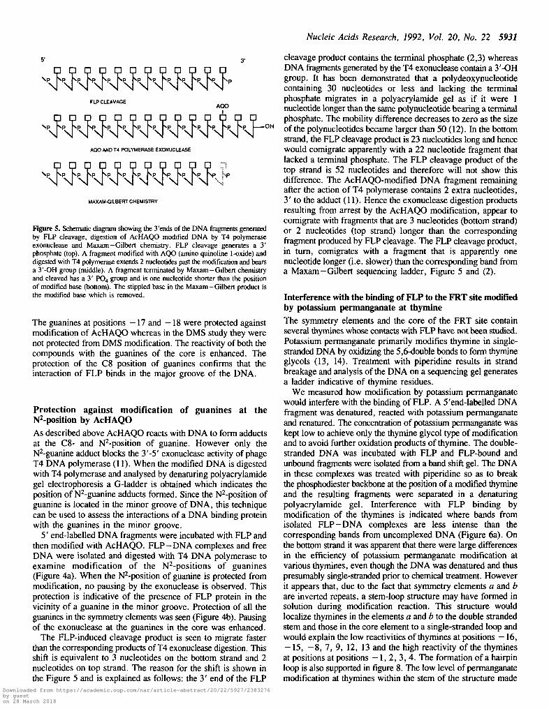
Figure 5. Schematic diagram showing the 3'ends of the DNA fragments generatedby FLP cleavage, digestion of AcHAQO modified DNA by T4 polymeraseexonuclease and Maxam—Gilbert chemistry. FLP cleavage generates a 3'phosphate (top). A fragment modified with AQO (amino quinoline 1-oxide) anddigested with T4 polymerase extends 2 nucleotides past the modification and bearsa 3'-OH group (middle). A fragment terminated by Maxam—Gilbert chemistryand cleaved has a 3' PO4 group and is one nucleotide shorter than the positionof modified base (bottom). The stippled base in the Maxam-Gilbert product isthe modified base which is removed.
The guanines at positions -17 and - 1 8 were protected againstmodification of AcHAQO whereas in the DMS study they werenot protected from DMS modification. The reactivity of both thecompounds with the guanines of the core is enhanced. Theprotection of the C8 position of guanines confirms that theinteraction of FLP binds in the major groove of the DNA.
Protection against modification of guanines at ther^-position by AcHAQOAs described above AcHAQO reacts with DNA to form adductsat the C8- and N^-position of guanine. However only thel^-guanine adduct blocks the 3'-5' exonuclease activity of phageT4 DNA polymerase (11). When the modified DNA is digestedwith T4 polymerase and analysed by denaturing polyacrylamidegel electrophoresis a G-ladder is obtained which indicates theposition of N2-guanine adducts formed. Since the N2-position ofguanine is located in the minor groove of DNA, this techniquecan be used to assess the interactions of a DNA binding proteinwith the guanines in the minor groove.
5' end-labelled DNA fragments were incubated with FLP andthen modified with AcHAQO. FLP-DNA complexes and freeDNA were isolated and digested with T4 DNA polymerase toexamine modification of the N2-positions of guanines(Figure 4a). When the ^-position of guanine is protected frommodification, no pausing by the exonuclease is observed. Thisprotection is indicative of the presence of FLP protein in thevicinity of a guanine in the minor groove. Protection of all theguanines in the symmetry elements was seen (Figure 4b). Pausingof the exonuclease at the guanines in the core was enhanced.
The FLP-induced cleavage product is seen to migrate fasterthan the corresponding products of T4 exonuclease digestion. Thisshift is equivalent to 3 nucleotides on the bottom strand and 2nucleotides on top strand. The reason for the shift is shown inthe Figure 5 and is explained as follows: the 3' end of the FLP
cleavage product contains the terminal phosphate (2,3) whereasDNA fragments generated by the T4 exonuclease contain a 3'-OHgroup. It has been demonstrated that a polydeoxynucleotidecontaining 30 nucleotides or less and lacking the terminalphosphate migrates in a polyacrylamide gel as if it were 1nucleotide longer than the same polynucleotide bearing a terminalphosphate. The mobility difference decreases to zero as the sizeof the polynucleotides became larger than 50 (12). In the bottomstrand, the FLP cleavage product is 23 nucleotides long and hencewould comigrate apparently with a 22 nucleotide fragment thatlacked a terminal phosphate. The FLP cleavage product of thetop strand is 52 nucleotides and therefore will not show thisdifference. The AcHAQO-modified DNA fragment remainingafter the action of T4 polymerase contains 2 extra nucleotides,3' to the adduct (11). Hence the exonuclease digestion productsresulting from arrest by the AcHAQO modification, appear tocomigrate with fragments that are 3 nucleotides (bottom strand)or 2 nucleotides (top strand) longer than the correspondingfragment produced by FLP cleavage. The FLP cleavage product,in turn, comigrates with a fragment that is apparently onenucleotide longer (i.e. slower) than the corresponding band froma Maxam-Gilbert sequencing ladder, Figure 5 and (2).
Interference with the binding of FLP to the FRT site modifiedby potassium permanganate at thymineThe symmetry elements and the core of the FRT site containseveral thymines whose contacts with FLP have not been studied.Potassium permanganate primarily modifies thymine in single-stranded DNA by oxidizing the 5,6-double bonds to form thymineglycols (13, 14). Treatment with piperidine results in strandbreakage and analysis of the DNA on a sequencing gel generatesa ladder indicative of thymine residues.
We measured how modification by potassium permanganatewould interfere with the binding of FLP. A 5'end-labelled DNAfragment was denatured, reacted with potassium permanganateand renatured. The concentration of potassium permanganate waskept low to achieve only the thymine glycol type of modificationand to avoid further oxidation products of thymine. The double-stranded DNA was incubated with FLP and FLP-bound andunbound fragments were isolated from a band shift gel. The DNAin these complexes was treated with piperidine so as to breakthe phosphodiester backbone at the position of a modified thymineand the resulting fragments were separated in a denaturingpolyacrylamide gel. Interference with FLP binding bymodification of the thymines is indicated where bands fromisolated FLP-DNA complexes are less intense than thecorresponding bands from uncomplexed DNA (Figure 6a). Onthe bottom strand it was apparent that there were large differencesin the efficiency of potassium permanganate modification atvarious thymines, even though the DNA was denatured and thuspresumably single-stranded prior to chemical treatment. Howeverit appears that, due to the fact that symmetry elements a and bare inverted repeats, a stem-loop structure may have formed insolution during modification reaction. This structure wouldlocalize thymines in the elements a and b to the double strandedstem and those in the core element to a single-stranded loop andwould explain the low reactivities of thymines at positions —16,- 1 5 , - 8 , 7, 9, 12, 13 and the high reactivity of the thyminesat positions at positions - 1 , 2 , 3 , 4 . The formation of a hairpinloop is also supported in figure 8. The low level of permanganatemodification at thymines within the stem of the structure made
Downloaded from https://academic.oup.com/nar/article-abstract/20/22/5927/2383276by gueston 28 March 2018

5932 Nucleic Acids Research, 1992, Vol. 20, No. 22
BOTTOM TOP
S'TT@QAAG@@CC@A@bcCGAAG@@CC@A@@CTCT AQAA AG©A© AOO A AC®QC A 33AAACT TCAAaQA@AAGGCTTCAAGQATAAGAGATCTTTACA@A@CC@®GAAGT5'
Figure 6. Interference with the binding of FLP to DNA modified by KMnO4.(a) Either top or bottom strand 5'end-labelled fragments carrying an intact FRTsite were denatured and modified with KMnO4. The modified DNA fragmentswere treated with 5 units of FLP, separated on a native polyacrylamide gel toisolate unbound and FLP-bound DNA, cleaved with piperidine and analysed ona denaturing gel. -FLP, unbound material; +FLP I, +FLP II and +FLP IIIrefer to the DNA from complex I, II and III respectively, (b) The sequence ofthe FRT site is shown. The modified thymines responsible for interference arecircled. A residue where modification appears to enhance binding is enlarged(—2 position).
it difficult to assess unambiguously the degree of bindinginterference at these sites (especially thymines of positions +13,- 1 5 and -16).
We observed no differences between unbound and bound DNAwhen high concentrations of FLP were used (data not shown).This result demonstrates that high levels of FLP could overcomeinterference with binding of FLP that was caused by themodification of a single thymine residue. For later experimentswe used subsaturating concentration of FLP. Under theseconditions, differences between complex IE and uncomplexed
30-
1
25-
20-
1
15-
10-
1
-5-
1-
5- '
10-
20-
VOG,D T y
r -—
A
^Jy\y^ A
o
yOv
VA
n T /r .—
A ,
3 y' A
AC y^
A
^" OA
A yc
o v
D T
y
yS
y*
yo
o^ y
y
T . '
VVO GA y
y
oo y^y
G r
y^~
y/~
y
^y
rc
^y
ny1
y
OG
yA
A/
/
yA
yy
c
A y*^. o
oA y
A . '
y v
V O G
AO ^y
T
y
yy
y
D T /A ^—'
/ oA
yy
^y tj
cA ^ ^
G 'y
D
o_*y^^
' A
A
D
U T .
^yD
• T .
Figure 7. Summary of the protection and interference results obtained with DMS,AcHAQO and KMnO4. Contacts are shown on a planar representation of theDNA within the FRT site. The sequence of only the top strand is shown. Thesymmetry elements are shown as arrows and core as an open box. Symbolidentifications are as follows: O , N7-guanine contacts; O, N3-adenine contacts;V, C8-guanine contacts; A, N2-guanine contacts; • , Thymine contacts. + signinside the other symbol indicates that the effects are enhanced. The base pairsin the minor grooves are shown by horizontal lines. The symbols are placed inthe groove where the modification occurs.
DNA were readily observed (Figure 6a). In particularmodification of the top strand thymines showed more interferencethan modification of the bottom strand thymines. Modificationof the thymines of the symmetry element a of the bottom strandcaused interference whereas none of the modifications of thyminesin symmetry element b had any effect on binding. In thesymmetry element c, only one modified T (position of -22)caused interference with binding. On the other hand modificationof all the thymines of the top strand interfered with FLP binding.It was not possible to ascertain from the presented data whethermodification of bases +15 and +16 on the top strand trulyinterfered with FLP binding. Modification of the thymines in thecore did not show any interference with binding. Besides
Downloaded from https://academic.oup.com/nar/article-abstract/20/22/5927/2383276by gueston 28 March 2018

Nucleic Acids Research, 1992, Vol. 20, No. 22 5933
12 3 4 1 2 3 4
II--
- 2 --4 -
- 9
-20--21 --23-
-32--33--34-
!
ti
0.0.
I +
a. a. a a. a a.
GA CTBOTTOM
GA CTTOP
BOTTOM TOP
5TTTfld
Figure 8. Assay of single-stranded DNA in the core upon FLP binding. All theDNA fragments were 5'-end labelled. Lane 1, The DNA was denatured, modifiedwith KMnO4 and then renatured, cleaved with piperidine and run on a sequencinggel. The T-specific bands are labelled at the left. Lane 2, Double-stranded DNAwas modified with KMnO4 in a double-stranded form, cleaved with piperidineand run on a sequencing gel. Note that the T specific ladder is not apparent.Lane 3, The DNA was incubated with FLP and treated with KMnO4. The DNAwas precipitated, cleaved with piperidine and run on the gel. Lane 4, Theprocessing of the material was done as described in lane 3 except that the complexIII material was gel purified.
thymines, nonspecific modifications of some bases withpermanganate interfered with FLP binding (i.e. positions -19 ,- 2 3 and -24) of bottom strand. These effects confirmed theimportance of these residues that we have already observed usingDMS (see Figure 2 above).
Modifications of thymines of symmetry elements c (positions-27 to -20) and b (positions —2 and -4 ) of the top strandinterfered with formation of complex I whereas modification ofthymines of symmetry element b (positions - 4 , - 6 , - 7 , —9,-12 , -13) interfered with formation fo complex II. Such effectswere not apparent for the bottom strand.
The above footprinting experiments are summarized in figure7 which shows the FRT site, displayed as a 2-dimensionalcylindrical plot of B-DNA. This diagram shows that FLP makesextensive contacts with each symmetry elements in both the majorand minor grooves. It further shows that FLP molecules bound
3 AA AC f tC@®©§®©gg©CiCT TC A
Figure 9. Missing contact probing of FLP interaction with a single symmetryelement, (a) The labelled top or bottom strand was either depurinated with acid(GA) or depyrimidinated with hydrazine (CT). The modified DNA was then treatedwith FLP. The unbound and FLP-bound DNAs were isolated on a gel, cleavedwith piperidine and analysed on an 8% polyacrylamide sequencing gel. The largeand small arrows show the positions of symmetry element c and part of brespectively. - : unbound DNA; + : FLP-bound DNA (b) The result of themissing contact experiment is summarized on the sequence of the symmetryelement. The bases whose absence interferes with FLP binding are circled. Thedotted circles indicate partial interference.
to symmetry elements a and b are in both the faces of the B-helix.Binding of FLP to symmetry elements a and b induces a largebend in the FRT site (15). This bend is thought to be mediatedby protein:protein interactions and may induce single-strandedform of DNA near the bend.
Search for single-stranded DNA in the FLP bound FRT site
AcHAQO reacts with native, double-stranded DNA to form threepurine adducts, two with guanine and one with adenine. Therelative amount of the N2-guanine, the C8-guanine and theN6-adenine adducts formed are 50%, 30% and 10% respectively(9,10). However, preferential formation of the C8-guanine adduct
Downloaded from https://academic.oup.com/nar/article-abstract/20/22/5927/2383276by gueston 28 March 2018

5934 Nucleic Acids Research, 1992, Vol. 20, No. 22
is observed when AcHAQO is reacted with single-stranded DNA(16). In figure 3a, the guanines at positions —3 and 1 showhypercleavage at the core. These hypercleavages might be dueto the single-stranded character of the core, which would predictthat the C8-guanine adduct would predominate. The N2- andC8-guanine adducts can be distinguished by their sensitivity tothe T4 polymerase exonuclease. The C8-guanine adduct does notimpede digestion by the exonuclease whereas the N^adductdoes. As can be seen in figure 4 (a), when the AcHAQO-modifiedDNA was treated with the exonuclease, pausing of theexonuclease at the guanines at positions —3 and 1 was enhanced.We conclude that the modification of the - 3 and 1 guanines isof the N2 type. Hence we conclude that the core region ispredominantly duplex DNA.
We also used potassium permanganate as a probe for single-stranded DNA (13,14). 5'end-labelled DNA was incubated withFLP protein and then treated with potassium permanganate (figure8). Lanes 1 and 2 show that potassium permanganate modifiesthe thymines of single-stranded but not duplex DNA. In lane 3the FLP—DNA complexes were not isolated from a gel whereasin lane 4, gel-purified complex IE material was used for cleavage.FLP remained bound to the FRT site after permanganatetreatment (i.e. complex HI material could be gel purified as shownin lane 4). Hypercleavage at thymine residues was not seen ineither lane and hence we conclude that the core remains double-stranded upon binding by FLP.
Missing base experiment with single symmetry elementWe have found previously (1,4) and have confirmed above, thatit is difficult to observe either chemical protection by FLP orinterference with FLP binding when a single symmetry elementof the intact FRT site is examined. The interference effect ofKMnO4 upon FLP binding could only be observed whenmoderate levels of FLP were used and when complex III wasexamined (see figure 6). In order to learn which nucleotides wereimportant for binding of FLP to a single symmetry element, thesubstrate containing only symmetry element c was first treatedwith either formic acid or hydrazine to remove purines orpyrimidines, respectively. The modified DNA was used todetermine whether the absence of a single base interfered withthe binding of FLP. The result showed that removal of virtuallyany base from either the top or bottom strand of a singlesymmetry element had a marked effect on binding of FLP to thatelement (figure 9a,b).
DISCUSSION
The present work identifies further interactions between FLPprotein and the FRT site. In particular we have examined majorgroove contacts with guanine and thymine residues using DMSand potassium permanganate, respectively. Minor groove contactswith guanine and adenine were studied using AcHAQO andDMS, respectively.
In the presence of FLP protein, methylation of guanines andadenines by DMS was inhibited in each of the three symmetryelements (figure 7).
In the present study and that Bruckner and Cox, the adeninesof positions —12, —13, 12 and 13 were not protected, indicatingthat these bases were not contacted closely by FLP. However,in the missing base experiment with a single symmetry element,signals were obtained at the respective adenine positions
(Figure 9 b, positions -26 and —27). In the missing nucleosideexperiment, an entire base is removed chemically (17). Themissing nucleoside signal obtained could be due to the absenceof a base contact other than the N3-position of adenine.Alternatively the signal could be due to overall structuraldeformities to DNA caused by the missing base.
When AcHAQO was used as a footprinting reagent for FLP,the guanines in the symmetry elements were protected fromAcHAQO modification at the C8-position of guanine. This resultis similar to the result obtained with DMS footprinting exceptthat two additional guanines (positions —16 and —17) wereprotected from modification by AcHAQO. The likely explanationfor this difference may reside in the different sizes of thefootprinting reagents. The two FLP molecules occupyingsymmetry elements b and c allow DMS to reach the G residueswhereas AcHAQO cannot penetrate. The guanines in the coreshow enhanced cleavage (positions - 3 and 1) upon AcHAQOmodification. A similar result is obtained with DMS (3,4 andthis work).
In addition to showing major groove contacts, AcHAQO, whencoupled with digestion by T4 DNA polymerase-exonuclease, alsodemonstrates contacts with G residues in the minor groove.Similarly, FLP protects the guanines from AcHAQO modificationin the minor groove. Only one G (position 31) which showedprotection from modification via the minor groove side was notprotected from the major groove side. Again the Gs in the coreshowed enhanced reactivity as assayed by the T4 polymerase-exonuclease. We have adduced two lines of evidence that thisenhanced activity to both DMS and AcHAQO is not due to thesingle-stranded character of the core upon FLP binding.
The conformation of the core DNA has been examined recentlywith MPE, a compound which intercalates into double-strandedDNA. Since it was observed that the MPE cleaved both strandsof the core DNA, it was concluded that core DNA was double-stranded (18). However, in the same study it was found that thecore DNA reacted with 5-0-OP-Cu as if it were single-stranded.At present we do not know the reason for the discrepancy amongthese results.
The role of thymine in FLP binding has been studied usingan interference assay. Potassium permanganate was used tomodify the thymines in single-strand DNA and the modified DNAwas renatured prior to use in an interference assay. The assayrevealed several thymine contacts of FLP protein indicating theimportance of the methyl group of thymine in the process ofsequence recognition by the FLP.
The missing base experiment can identify bases that contributeto a binding interaction. Our study with a single symmetryelement c shows that removal of virtually any base interferes withthe binding of FLP protein. Kimball (18) previously reportedmissing nucleoside experiments in which a substrate containingthree symmetry elements was used. The removal of the basesat positions T7, A8, G14, A15 of the top strand and A9, G10of the bottom strand interfered with the FLP binding. The co-operative binding of FLP to symmetry elements a and b has beenreported previously (1, 15, 19, 20, 21). The discrepancy betweenthese two studies might be due to the fact that our experimentsused a DNA fragment containing only a single symmetry element,whereas Kimball's substrate contained an intact FRT site. Thuscooperativity in binding of FLP to adjacent symmetry elementsmay have obscured some signals. It is also possible that somemissing contact signals might have been obscured if too high alevel of FLP protein was used.
Downloaded from https://academic.oup.com/nar/article-abstract/20/22/5927/2383276by gueston 28 March 2018

Nucleic Acids Research, 1992, Vol. 20, No. 22 5935
The present study adds considerable detail to our understandingof the contacts of FLP with the FRT site. We used probes thatreveal contacts with both major and minor grooves of DNA. Thecontacts are summarized in figure 7 which shows a planarrepresentation of B-DNA. It is clear that FLP shows extensivecontacts with both major and minor grooves of a 10 bp stretchof each symmetry element. Inspection of the amino acid sequenceof the FLP protein reveals no obvious DNA binding motif (22).However our studies suggest the DNA binding elements mustcover one helical turn of both major and minor grooves. It ispossible that the sequence specificity of the recognitionmechanism lies in the binding of the major groove and the minorgroove binding may be secondary. The extensive minor grooveinteractions might suggest the presence of /3-pleated sheet in theFLP protein which may bind non-specifically to the minorgroove. Alternatively, the sequence specific interactions couldalso be mediated via minor groove contacts (23). Furthermore,as the projection in figure 7 reveals, the FLP molecules boundto symmetry elements would contact both the faces of the helix.Such extensive contacts might be necessary to support the largebend (> 140°) that the FLP protein induces upon binding tosymmetry elements a and b (15).
16. Galieuge-Zouitina, S., Bailleul, B. and Loucheux-Lefbvre, M.-H. (1984)Anal. Biochem. 138, 454-457.
17. Brunelle, A. and Schleif, R (1987) Proc. Nat. Acad. Sci. U. S. A. 84,6673-6676.
18. Kimball, A. S. (1990) Ph. D. thesis, Johns Hopkins University, Maryland.19. Prasad, P. V., Horensky, D., Young, L.-J. and Jayaram, M. (1986) Mol.
Cell. Biol. 6, 4329-4334.20. Senecoff, J. F., Rossmeissl, P. J. and Cox, M. M. (1988) J. Mol. Biol.
201, 405-421.21. Parsons, R. L., Evans, B. R., Zheng, L. and Jayaram. M. (1990) J. Biol.
Chem. 265, 4527-4533.22. Amin, A. A. and Sadowski, P. (1989) Mol. and Cell. Biol. 9, 1987-1995.23. Church, G. M., Sussman, J. L. and Kim, S.-H. (1977) Proc. Natl. Acad.
Sci. USA 74, 1458-1462.
ACKNOWLEDGEMENTS
We thank Dr. I.G.Walker, University of Western Ontario, forallowing us to synthesize the AcHAQO in his laboratory. Wethank Dr. M.Gold, Dr. B.Funnel, Dr. Helena Friesen, Dr.D.Kuntz, Julie Dixon, Linda McBroom and Hua Pan for criticalreading of the manuscript. This work was supported by theMedical Research Council and the National Science andEngineering Research Council of Canada.
ABBREVIATIONS
AcHAQO, monoacetyl-4-hydroxyamino-quinoline 1-oxide;MPE, methydium propyl EDTA; 5>-OP-Cu, 5-phenyl-l,10-phenanthroline copper.
REFERENCES
1. Andrews, B. J., Beatty, L. G. and Sadowski, P. D. (1987) J. Mol. Biol.193, 345-358.
2. Andrews, B. J., Proteau, G. A., Beatty, L. G. and Sadowski, P. D. (1985)Cell, 40, 795-803.
3. Bruckner, R. C. and Cox, M. M. (1986)/ Biol. Chem. 261, 11,798-11,807.4 Beatty, L. G. and Sadowski, P. D. (1988) J. Mol. Biol. 204, 283-294.5 Pan, H., Clary, D. and Sadowski, P. D. (1991) J. Biol. Chem. 266,
11,347-11354.6. Panigrahi, G. B. and Walker, I. G. (1991) Biochemistry 30, 9761-9767.7. Maxam, A. M. and Gilbert, W. (1980) Methods Enzymol. 65, 499-560.8. Siebenlist, U. and Gilbert, W. (1980) Pro. Nat. Acad. Sci, U. S. A. 11,
122-126.9. Galiegue-Zouitina, S., Bailleul, B. and Loucheux-Lefbvre, M.-H. (1985)
Cancer Res. 45, 520-525.10. Galiegue-Zouitina, S., Bailleul, B., Ginot, Y. M., Perly, B., Vigny, P. and
Loucheux-Lefebvre, M.-H. (1986) Cancer Res. 46, 1858-1863.11. Panigrahi. G. B. and Walker, I. G. (1990) Biochemistry 29, 2122-2126.12. Tapper, D. P. and Clayton, D. A. (1981) Nucleic Acids Res. 9, 6787-6794.13. Hayatsu, H. and Ukita, T. (1967) Biochem. Biophys. Res. Commun. 29,
556-561.14. We, H., Kow, Y. W. and Wallace, S. S. (1985) Nucleic Acids Res. 13,
8035-8052.15. Schwartz, C. J. E. and Sadowski, P. D. (1990)7. Mol. Biol. 216,289-298.
Downloaded from https://academic.oup.com/nar/article-abstract/20/22/5927/2383276by gueston 28 March 2018