the fixation area of the cat superior colliculus: effects...
TRANSCRIPT

Exp Brain Res (1994) 101:109-122 9 Springer-Verlag 1994
Martin Par6 Daniel Guitton
The fixation area of the cat superior colliculus: effects of electrical stimulation and direct connection with brainstem omnipause neurons
Received: 26 July 1993 / Accepted: t February 1994
Abstract The superior colliculus has long been recog- nized as an important structure in the generation of saccadic displacements of the visual axis. Neurons with presaccadic activity encoding saccade vectors are topo- graphically organized and form a "motor map." Recent- ly, neurons with fixation-related activity have been recorded at the collicular rostral pole, at the area cen- tralis representation or fixation area. Another collicular function which deals with the maintenance of fixation behavior by means of active inhibition of orientation commands was then suggested. We tested that hypothe- sis as it relates to the suppression of gaze saccades (gaze = eye in space = eye in head + head in space) in the head-free cat by increasing the activity of the fixa- tion cells at the rostral pole with electrical microstimu- lation. Long stimulation trains applied before gaze sac- cades delayed their initiation. Short stimuli, delivered during the gaze saccades, transiently interrupted both eye and head components. These results provide further support for a role in fixation behavior for collicular fixa- tion neurons. Brainstem omnipause neurons also ex- hibit fixation-related activity and have been shown to receive a direct excitatory input from the superior col- liculus. To determine whether the collicular projection to omnipause neurons arises from the fixation area, the deep layers of the superior colliculus were electrically stimulated either at the rostral pole including the fixa- tion area or in more caudal regions where stimulation evokes orienting responses. Forty-nine neurons were ex- amined in three cats. 61% of the neurons were found to
M. Par61 . D. Guitton ([~) Montr6al Neurological Institute and Department of Neurology and Neurosurgery, McGill University, 3801 University Street, Montreal, Canada; FAX no: (514) 398-8106
Present address: Medical Research Council Group in Sensory-Motor Physiology, Queen's University, Kingston, Canada; E-mail: [email protected]
be orthodromically excited by single-pulse stimulation of the rostral pole, whereas only 29% responded to cau- dal stimulation. In addition, stimuli delivered to the ros- tral pole activated, on average, omnipause neurons at shorter latencies and with lower currents than those ap- plied in caudal regions. These results suggest that exci- tatory inputs to omnipause neurons from the superior colliculus are principally provided by the fixation area, via which the superior colliculus could play a role in suppression of gaze shifts.
Key words Omnipause neurons - Superior colliculus Fixation 9 Saccade 9 Gaze 9 Eye-head coordination 9 Cat
Introduction
The oculomotor burst generator - a premotor circuit crucial for the generation of saccadic eye movements and composed of neurons exhibiting a high-frequency burst of activity during saccades - is thought to be re- ciprocally related to inhibitory neurons located in the raphe interpositus nucleus, the omnipause neurons (OPNs), which discharge tonically during fixation be- havior (Fuchs et al. 1985; Hepp et al. 1989; Keller 1991). A complete cessation of OPN activity just prior to and during saccades releases the inhibition of the saccadic burst generator neurons. To make a saccade, it has been suggested that a motor error signal drives the burst gen- erator while an inhibitory trigger signal turns off OPNs (Robinson 1975; Keller 1977; Fuchs et al. 1985). The superior colliculus (SC) has been proposed to provide the motor error signal (for review see Sparks 1986; Guitton 1991), since some collicular efferent neurons ex- hibit presaccadic burst activity and project to premotor areas. An anatomical projection from SC to OPNs has also been reported (Bfittner-Ennever et al. 1988; Langer and Kaneko 1984, 1990), thereby suggesting that the SC may also provide the trigger signal.
The collicular projection to OPNs has been investi- gated electrophysiologically. Raybourn and Keller

110
(1977) were the first to establish, in the alert monkey, a direct excitatory connection between the SC and OPNs. Evidence for such a connection in the anesthetized cat was provided by King et al. (1980) and Kaneko and Fuchs (1982). An indirect inhibitory projection was also observed (Raybourn and Keller 1977; Kaneko and Fuchs 1982). Since the direct projection was found to be excitatory, the trigger signal that presumably silences OPNs was then considered to come from a structure other than the SC: either the rostral part of the pontine reticular formation or the mesencephalic reticular for- mation (Fuchs et al. 1985).
Recently, collicular neurons with fixation-related ac- tivity have been recorded in the area centralis represen- tation in cat and in the foveal representation in the mon- key (in cat: Munoz and Guitton 1989, 1991; Munoz et al. 1991; in monkey: Munoz and Wurtz 1992, 1993a). Their activity was found to be similar to that displayed by OPNs; they maximally discharge when the animal attentively fixates and cease firing during either saccadic eye movements or saccadic gaze shifts (gaze = eye in space = eye in head + head in space). In both cat (Guitton and Munoz 1991) and monkey (Istvan et al. 1994), these are collicular efferent neurons that send de- scending projections into the predorsal bundle. It was proposed for cat that they contact OPNs and that their activity during fixation contributes to the sustained ac- tivity observed in OPNs (Munoz and Guitton 1989, 1991; Munoz et al. 1991). The reduced firing of these collicular neurons during saccadic eye or gaze move- ments could contribute to the trigger signal that induces the cessation of discharge of OPNs. Given that micro- stimulation of the OPN area in the cat suppresses both eye and head motion (Par~ and Guitton 1989), this hy- pothesis suggests that electrical microstimulation ap- plied to the fixation area of the cat's SC should also prevent the occurrence of both eye and head movements that compose saccadic gaze shifts. The first objective of this paper was to verify this. The second objective was to investigate the direct connection between the SC and OPNs in the alert cat in order to test whether the SC fixation area projects more heavily to brainstem OPNs than other regions of the SC. A part of this study has been presented in abstract form (Par6 and Guitton 1991).
Materials and methods Three alert animals (referred to in the text as cats G, V, and Y) were used for the experiments involving OPN recording. Experiments involving SC fixation-area stimulation were conducted on the same three animals plus an additional one (cat L).
Surgical procedures
Surgery was performed under aseptic conditions. Each animal was initially given ketamine hydrochloride (10 mg/kg i.m.) and anesthesia was maintained using halothane inhaled through an endotracheal tube. A wire coil for the measurement of eye position
with the magnetic search-coil technique was sutured to the sclera of one eye. Three holes were trephined through the skull to allow access to both superior colliculi and brainstem. One stainless steel cylinder, constructed to hold a micropositioner (Kopt), was posi- tioned just rostral to the lambdoid crest, to provide access to the brainstem, and was tilted 25 ~ back from the frontal plane. The second cylinder was positioned vertically, centered over both SC. The connector for the eye coil, an attachment for a coil monitor- ing head position, a head-holding device to immobilize the head of the animal during recording sessions, and the cylinders were se- cured in place by embedding them in a dental acrylic explant that was anchored to the skull with stainless steel bolts. At the end of the surgery, the animals received an intramuscular injection of gentamicin (15 mg/kg) or cefazolin (35 mg/kg) as a prophylactic measure against infection. These antibiotics were administered on a daily basis for 10 postoperative days. To alleviate any discom- fort, animals were given analgesic medication (buprenorphine hy- drochloride 0.01 mg/kg) as needed during the postsurgical period. All surgical and experimental protocols were approved by the McGill University Animal Care Committee and complied with the Canadian Council on Animal Care policy on use of laboratory animals.
In a second procedure, the animal was slightly anesthetized (ketamine hydrochloride 10 mg/kg i.m. supplemented by 2-5 mg/ kg) for a period varying from 1 to 2 h. The movement vectors evoked at different sites in each SC were mapped out with stimu- lating electrodes (bipolar concentric electrodes Kopf NEX-25) placed in collicular layers for which electrical stimuli evoked eye movements with minimal threshold intensity. To do that, the ani- mal's head was fixed to a platform to which a stereotaxic appara- tus (Kopf) was attached and electrode carriers were employed to manipulate the stimulating electrodes. Stereotaxic coordinate ref- erences marked on the cat explant during the first surgery were used to aim the electrodes at the SC. This approach allowed us to determine the distance between the various positions of one stim- ulating electrode from one track to another, and between the different electrodes placed in each SC. The location of the stimu- lating electrodes used in the study of the SC-OPN projection is indicated in Fig. 1. In each cat, at least two stimulating electrodes were fixed in place, with dental acrylic, in the same SC: one in the fixation area (labeled 1 in Fig. 1) and the other in the caudal regions at a site where large gaze shifts could be evoked. The characteristics of the gaze shifts elicited at each caudal stimulation site were subsequently evaluated, with the animal fully alert, using parametric studies (see the accompanying paper, Par6 et al. 1994). Briefly, stimulus current strength (from 10 to 80 gA) or pulse rate (from 100 to 600 pulses/s) were varied. The maximum amplitude of the gaze shift elicited at each site, along with the direction of the movement, indicated the location of an electrode on the collicular motor map. In cats Y and V, the caudal electrode (labeled 2) evoked gaze shifts of 60 ~ and 45 ~ amplitude, respectively (Fig. 1A,B). In cat G, two caudal electrodes labeled 2 and 3 evoked gaze shifts having 50 ~ and 80 ~ of amplitude, respectively (Fig. 1C). The minimal distance between the caudal electrode labeled 2 and the one in the fixation area was about 4 mm. The placement of the stimulating electrode in the SC fixation area was assessed by: (1) its position relative to other SC sites; (2) absence of electrically evoked saccades; and (3) by the inhibition of saccadic eye move- ments and vestibular quick phases following electrical current de- livered through it. The fixation-area electrode was located in the left SC in cat L and in the right SC for cats G, V, and Y (Fig. 1). In cat Y, after the study of the collicular projection to OPNs, a second electrode was implanted in the fixation area of the left SC. Bilater- al stimulation was then used to mimic natural fixation-related activity in the SC and yielded some of the data presented in the accompanying paper (Par6 et al. 1994).
Experimental procedures
The positions of gaze and head relative to space were monitored by the search-coil-in-magnetic-field technique. Details of the coil

cat Y cat V -IY' ~ -IY
.1 ~ . -10" FB., C
"' i, f '~ i I
-2
5*
Collieular rostro-caudal axis (ram)
111
cat G
2 3
__Jl0*
2 3
Fig. 1 Locations of collicular stimulating electrodes in cats Y (A), V (B), and G (C), and gaze vectors evoked by electrical stimulation of the deep layers of the right superior colliculus (SC) at the site of each electrode. A series of stimulation sites evoking gaze vectors (arrows) in each animal are also illustrated. Their spatial location (origin of the arrows) is superimposed on the map of the right SC obtained by Mcllwain (1986). The dots (labeled with numbers) indicate the location of the stimulating electrodes used in the SC-omnipause neurons projection study
system and the calibration procedures have been reported else- where (Guitton et al. 1984). Briefly, the gain of the eye-coil and head-coil demodulators were set at the same value by rotating horizontally and vertically a reference coil at known angles with the help of a gimbal device placed at the center of the field-coil arrangement. The cat was placed on the recording table with its head at the center of the fields and held earth-fixed as the field coil was oscillated horizontally about it. A potentiometer measured the angular deviation of the field coil. Calibration of the eye- and head-coil signals was then made by comparing the potentiometer and coil signals, during saccade-free segments of data, so as to transform eye and head signals (volts) into corresponding angular deviations. In the head-fixed cat, the eye coil measured directly the eye position in the orbit. In the head-flee animal, the eye position in the orbit was obtained by subtracting the head-coil signal from the signal of the eye coil, which then measured gaze position. The head-coil calibration obtained this way could be compared direct- ly with that obtained with the gimbal. Gaze- and head-coil cali- brations were also verified by having the alert head-free cat look- ing at food targets located at known positions.
During recording sessions, the alert cats were wrapped in a loosely fitting cloth bag and placed in a restraining box. The animals were previously trained to do simple visuomotor tasks (see below). Signals of gaze and head positions and neuronal activ- ity were recorded on digital audio tape (TEAC RD-200T), the unit activity being sampled at 40 kHz and the movement at 10 kHz.
Stimulation of the colIicular fixation area
A train of cathodal pulses generated by a stimulator (Grass $88) and constant-current stimulus isolation units (Grass PSIU6), was used to stimulate the collicular fixation area. The animal was trained to fixate and track a food target manually displaced from either side of a tangent opaque barrier positioned in front of it ("barrier paradigm"; Guitton et al. 1990; Munoz and Guitton 1991). The animal quickly learned that when the target disap- peared from one side of the barrier it would reappear on the other side, and it readily made gaze shifts toward the new predicted
location of the target, as soon as the target disappeared behind the barrier. The stimulus pulse train was applied either during ongo- ing eye-head gaze saccades or simultaneously with the disappear- ance of the target, i.e., before the onset of the gaze shifts. Experi- ments were conducted either with the laboratory light constantly dimmed or with an ambient stroboscopic light that was turned off for a period of 1-2 s at the onset of the gaze shift (Munoz and Guitton 1991). The stimulus pulse train was also applied during vestibular stimulation in the dark obtained by rotating en bloc the animal in a horizontal plane about an axis centered on the head at the level of the labyrinths. Different stimulus parameters were used. The range of stimulus current strength was 20-80 pA; pulse rate varied between 100 and 600 pulses/s and train duration, be- tween 10 and 500 ms. Pulse duration was always 0.3 ms.
Collicular projection to OPNs
OPNs were recorded extracellularly using tungsten microelec- trodes (3M Mf~; Frederick Haer). Protected by a cannula, record- ing electrodes were manually advanced through the dura and cerebellum to about 5 mm above the fourth ventricle. An hy- draulic microdrive attached to the Kopf x-y micropositioner was subsequently used to advance electrodes into the brain. The refer- ence electrode consisted either of a silver wire attached to a skull bolt or of the cannula itself. Conventional filtering, amplifying, and display techniques were employed. In these recording experi- ments, the animal's head was fixed. A region rostromedial and ventral to the abducens nuclei was examined. OPNs were identi- fied by their tonic discharge during fixation and characteristic pause in activity in relation to saccadic eye movements, and their responses following collicular stimulation were tested. A diagram of the stimulation and recording setup is provided in Fig. 2A. Electrical stimulation involved a pulse of negative current of 0.2 ms duration followed by a brief positive current of 0.1 ms du- ration. Stimulus pulses were generated by a stimulator (Grass $88) connected to the stimulating electrode through constant-current photoelectric stimulus isolation units (Grass PSIU6). The level of stimulus current was determined from the voltage measured across a 10-kf2 resistor in series with the electrode and was mon- itored on an oscilloscope. Testing for activation was routinely done using stimulus currents of 500 IxA. If no response was ob- served, the current was then increased gradually up to 1 mA. A cell was considered not driven by the electrical stimulation if no consistent responses were evoked with a stimulus current strength of 1 mA. Electrical stimuli never exceeded that current strength and the animal never displayed aversive reactions. Short stimula- tion trains of two or three pulses (pulse rate between 200 and 1000 pulses/s) were also used. Once an excitable OPN was found, its excitation level for each stimulating electrode was determined.

112
A St imula t ion
Superior Collieulus
Recording
Raphe Interpositus
B
C
1
Fig. 2 A Stimulation and recording setup. Single omnipause neu- rons isolated by a microelectrode in the raphe interpositns were orthodromically excited by biphasic current pulses passed through a bipolar stimulating electrode within the deep layers of the superior colliculus. B,C Examples of excitatory responses of one neuron (cell G11) to single-pulse stimulation at just-threshold intensity, 100 gA (B) and at twice threshold, 200 gA (C). The dot- ted line marks the occurrence of stimulation pulses
The threshold intensity was defined as the current that yielded a response on 50% of stimulus presentations. For each neuron, the latency of the response (i.e., the mean time from stimulus onset to initiation of the orthodromic action potential) elicited by the elec- trical stimulus was taken as the latency observed for a current value 2 times greater than threshold (Fig. 2B,C). For some OPNs, we verified, using the collision test, whether they could be an- tidromically activated by SC stimulation. To perform such a test, a single-pulse SC stimulus was triggered by a spontaneous OPN action potential (Fuller and Schlag 1976). The maximum interval between the spontaneous spike and the electrical stimulus at which collision could occur corresponds to the response latency plus the refractory period of the axon; the latter can be function- ally defined as the minimum delay between two electrical stimuli applied at the same site and such that the second stimulus does not elicit a response.
Data analysis
ate and compare the efficacy of each stimulating electrode in elic- iting a response in a sample of OPNs, a coefficient of efficacy (CE) was calculated for each electrode and for each neuron using the following equation: CE = [L /T c x (I/Io)] 1, where L is the response latency, I/Io is the response weighted intensity, and To is the mean conduction time (0.84 ms) needed for an action potential, in axons of tecto-reticular neurons, to reach the OPN area (see Guitton and Munoz 1991). The shorter the response latency and the lower the current threshold, the higher is the CE. A C E value of zero was attributed to undriven cells. The mean value of CE for all avail- able cells was used to describe the efficacy of each SC stimulating electrode in exciting OPNs. Comparisons between the response characteristics (latency, current threshold, weighted intensity, CE) obtained for each stimulating electrode were performed using the distribution-free U-test of Wilcoxon-Mann-Whitney, because the assumption of normality of population distributions was consid- ered not tenable. Results were considered significant only if they exceeded a one-tail level of P < 0.05. Percentages were compared with the chi-square (Z 2) test.
To evaluate whether an OPN was monosynaptically driven by SC stimulation, we used the following reasoning. Guitton and Munoz (1991) measured the latencies of antidromic responses of tectoreticular neurons following stimulation near the OPN area. The shortest latency obtained was 0.4 ms. If we assume (1) 0.1 ms as the rise time for an action potential; and (2) 0.5 ms for synaptic delay, then the shortest disynaptic response should be 0.4+2(0.1+0.5)= 1.6 ms. Accordingly, OPNs showing response latencies less than 1.6 ms were probably monosynaptically acti- vated.
Blocks of records were played back and digitized by a PDP-11/73 computer for off-line analysis. A time-amplitude window discrim- inator (Bak Electronics) isolated single-unit activity and produced a logic pulse for each action potential that met amplitude and time constraints. Individual action potentials were stored with the po- sition signals. The latter were low-pass filtered at 250 Hz were sampled at 1 kHz. Stimulation pulse and single-unit signals were displayed on a storage oscilloscope to measure response latencies.
The possible different positions of each stimulating electrode tip relative to collicular efferent neurons rendered difficult a direct comparison between the current threshold necessary for each elec- trode to elicit a response in OPNs. To remedy this problem, the current threshold (I) at which an OPN was responsive was weight- ed by the current threshold (Io) needed at that site to evoke an eye movement with a train of pulses (0.2 ms pulse duration and pulse rate of 300 pulses/s). This yielded an intensity index (I/Io) for each neuron activated by each stimulating electrode. For fixation-area electrodes, the threshold intensity was weighted by the current threshold needed to interrupt saccadic eye movements. To evalu-
T i 9 i 600 I E V ! i :
Fig. 3 Effects of electrical stimulation delivered at the right supe- rior colliculus (SC) fixation area on vestibularly induced eye movements in the dark, obtained by subjecting the animal to horizontal whole body rotation. Data from cat G (right SC). From top to bottom: table position (7), vertical (Ev) and horizontal eye position (Eh), electrical stimulus (thick horizontal line). Upward deflection on traces indicates movements up and to the right, re- spectively. Stimulus current strength was 60 gA (2 x threshold); pulse rate was 300 pulses/s; train duration was 1800 ms

A
E " ~
IHS
. . ~ j r
//
B
G
20o
113
J
Results
Effects of stimulating the rostral pole of the superior colliculus
Electrical st imulation of the fixation area at the SC ros- tral pole elicited no saccades except sometimes very small and slow eye movements at high current strength. Figure 3 shows the effects on eye movements when the fixation area was unilaterally stimulated during hori- zontal sinusoidal whole body rota t ion in the dark. Vestibular quick phases were either greatly reduced in size or abolished. The inhibition of quick phases was more impor tant when these were directed ipsilaterally to the SC being stimulated. In addition, the gain of the compensatory vestibulo-ocular reflex - slow-phase eye velocity/head velocity - was significantly reduced dur- ing stimulation. In cat G, the gain of the reflex, in the dark, had a mean value of 0.91 (SD 0.10, n =2 1 ) at 0.2 Hz. It d ropped to 0.60 (SD 0.15, n=32 ) during the
Fig. 4A,B Effects of electrical stimulation delivered at the superi- or colliculus (SC) fixation area during ongoing eye-head gaze sac- cades. Data from cat G (right SC). Rightward (A) and leftward (B) gaze saccades made to visual targets. A brief stimulus (50 gA, 300 pulses/s, 30 ms) during a gaze saccade interrupts momentarily the movement. Each panel shows a single, normal eye-head gaze sac- cade (dotted traces) and three stimulation trials (solid traces). G, H, E and G, H, are the position and velocity traces of gaze, head, and eye in the horizontal plane. Thick horizontal line under eye trace indicates the period of stimulus presentation
period when the fixation area was unilaterally stimulat- ed.
The effects of stimulating the fixation area on natural gaze shifts was also investigated. In these experiments, the animal oriented to two targets situated on the hori- zontal plane. Examples of normal responses are shown in Fig. 4A,B (dotted traces). On random trials, the stim- ulation train, triggered by the gaze saccade, was deliv- ered to the fixation area during the ongoing gaze shift. These stimuli transiently interrupted both the eye and

114
,.o
r
~
9
h o
~
o0",~
r O
a ~
" 6 =
~N
.~ .~=
~N
~1 +1 I I
+l +1 +1 +1
o o p.--
+ 1 + 1 + 1 + 1
+[ +1 +1 +1
2
ee~ ,~- r"- +1 +1 +1
- ~ V
,sZ ,-!
2 8 ~ ~b2
:~ oc)
r ~ +.a
" ~ O
~ . ~
0 2
2~
2 ~
N V,.u ~
head (and thus gaze) movements (Fig. 4A,B; solid traces). Similar to the inhibition of vestibular quick phases, the effect was stronger for gaze saccades directed ipsilaterally to the SC being stimulated (compare Fig. 4A and 4B). By analyzing velocity profiles, the per- centage of gaze shifts and head movements decelerated by the stimulation was larger for ipsiversive than for contraversive ones (Table 1). Deceleration of the head movement was seen less frequently than gaze decelera- tion, perhaps due to the smoothing effects of head iner- tia. Mean latencies of the onset of deceleration of gaze and head movements following stimulation are shown in Table 1. On average, the latency of a movement's deceleration relative to the stimulus onset was about 13 ms for gaze and 32 ms for the head. In cat G, we used stimulus durations of 10, 20, 24, 30, and 50 ms (pulse rate of 300 pulses/s). The duration of the decelerating period, as measured on the gaze velocity trace, increased linearly with the stimulus duration. The head decelera- tion response was more variable. For short stimulus trains, perturbations were less noticeable in the head trace than in the gaze trace. As the stimulus duration increased the effect on the head trace was more promi- nent.
In two animals (cats G and L), a long stimulus train was delivered simultaneously with the target's disap- pearance in the barrier paradigm. Almost all gaze sac- cades directed ipsilaterally to the stimulated SC were canceled for the duration of the stimulation (Fig. 5A,D). The saccades were initiated only after the end of the stimulus train, after an interval estimated to be 94 ms (SD 45, n = 10) and 104 ms (SD 29, n = 19) in cats G and L, respectively. Saccades directed to the side contralat- eral to the stimulation site were initiated at a latency comparable with that of controls but were hypometric. Most displacements during the electrical stimulus were made by a succession of small gaze saccades (Fig. 5B), or were limited to a saccade of reduced amplitude (Fig. 5C). The dominance of one response type over an- other appeared to depend on stimulus pulse rate; the truncated saccades occurring during high-frequency stimulation. This phenomenon was not studied in detail because differences in response were also related to vari- ations in the cat's behavioral "set", a characteristic diffi- cult to control.
Responses of OPNs to collicular stimulation
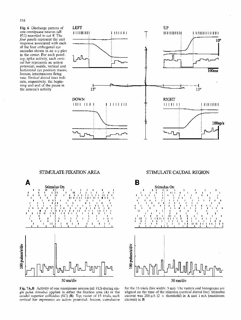
Once we had studied the collicular fixation area we pro- ceeded to evaluate its influence on OPNs. A total of 49 neurons were identified and characterized in three head- fixed animals (cats G, V, and Y). The firing behavior of this class of neuron is exemplified in Fig. 6. OPNs exhib- ited a more or less tonic rate during stationary eye posi- tion but ceased discharging prior to and for the whole duration of eye saccades made in all directions. The dis- charge characteristics of the neurons recorded in this study were similar to those described in our previous

S T I M U L A T E R I G H T SC
B
20~ !i~
I0
A
S T I M U L A T E L E F T SC
C
/.-- i
115
Fig. 5A-D Electrical stimulation delivered at the superior col- liculus (SC) fixation area suppresses eye-head gaze saccade gener- ation. A,B Data from cat G (right SC). C,D data from cat L (left SC). Rightward and leftward saccades made to visual targets (A,B) or to predictive targets in the dark (C,D). Long stimulus train delivered before the saccade suppresses ipsiversive gaze saccades (A,D) more than contraversive ones (B,C). Dotted traces, normal gaze saccades (controls); solid traces, stimulation trials. Stimulus parameters were 50 gA, 100 pulses/s, 500 ms in A,B; and 30 gA, 200 pulses/s, 200 ms in C,D. Movement traces are aligned on the same initial gaze position. Arrowheads mark the disappearance of the target from one side of the barrier, which is the cue for the animal to initiate the saccades. Light goes off at saccade onset in C,D. Thick horizontal lines under movement traces indicate stimu- lus presentation periods. Examples are horizontal gaze shifts
reports (Par6 and Gui t ton 1990, 1992, 1994). Electrical s t imulat ion of the SC was applied during fixation peri- ods when OPNs discharged tonically. The excitability of these neurons has been shown to vary with the animal 's level of arousal (Henn et al. 1984); their tonic activity decreases or completely vanishes when the animal be- comes drowsy. Also, the latency of O P N responses to SC st imulat ion is known to increase when the stimulus
D
200ms
I0 ~ !~ !~
~.- ~ ...................
is delivered during periods of reduced alertness (Ray- bourn and Keller 1977). For these reasons, the animals were kept fully alert during the administration of the electrical stimulus.
A large proportion of OPNs were activated by elec- trical microst imulat ion. Examples of responses for one cell are shown in Fig. 2B,C. Each neuron had a variable response latency, and its mean latency decreased with increasing stimulus intensity. In a few neurons, respons- es containing more than one spike were also observed. In addition, twin, closely spaced stimuli were more effec- tive than single-pulse stimuli in eliciting act ion poten- tials. These observat ions strongly suggest that O P N re- sponses to collicular s t imulat ion were the result of t ranssynapt ic mediation, i.e., of o r thodromic invasion. Ant idromic act ivat ion was ruled out by the collision test applied to ten OPNs. In all the neurons, the action potential response to the stimulus could not be made to collide with electrically evoked spikes.
In order to verify the hypothesis that SC fixation cells project p redominant ly to OPNs, the responses of OPNs to s t imulat ion by electrodes located in the collicular fix-
116
Fig. 6 Discharge pattern of one omnipause neuron (all VI2) recorded in cat V. The
four panels represent the unit response associated with each of the four orthogonal eye saccades shown in an x-y plot in the center. For each panel: top, spike activity, each verti- cal bar represents an action potential; middle, vertical and horizontal eye position traces; bottom, intantaneous firing rate. Vertical dotted lines indi- cate, respectively, the begin- ning and end of the pause in the neuron's activity
LEFT IIIIIIII I I I I I I I I
i
15 ~
DOWN IIII I I I I I I I I I I I
UP I I I I I I I I I I I I I I I I I I I I I I I I I I
U I lOOms
I t I 15 ~
RIGHT I l i l l l IIIIIIIII
lOOsp/s
STIMULATE FIXATION AREA
6. Stimulus On
I I I I I ! 1 I I | I I I I I I I i l l I I I I I I ! | I I I I I I i i i I I I I I I I I | I I I I I
I I I ::1 I I I I I I I I I I I I !1 I I I I I
I I I I : :11 I I I i I I I !1 I I I i i i
I I I I I I ::1 I I I I ! I i H I I i l l I I I I I I I I
I I I I I I I I : :1 I I I I I I I I I ~1 I I
I I I ::1 I I I I I I I I ~1 I I I
I I I I ::1 I I I I I I I I I ~1 I I I I
I I I n i l I I I
I I I i i i i i
I I I I i i i i I
I I I I I I I I I
I I I I I
I I I
STIMULATE CAUDAL REGION
B Stimulus On
I I I I ! I I I I I I I I I ~ I I I I I I I
I I I I I I i I | I I I I
I I
I I I I
I I
I I I I i I
! I
I I
I I I I I I I I I I I I I ~ I I I I I I I I I
I i i i I I i I I I I I I I I I i I I I I I I I I
I i I I II I I I I I I I I I i I I I I I I
I I : I i I I I I I I I ! I I I I I I I I I I I I I I~ I I I I I I I I I I I
I I I I I i i I I I I I I I I I I I I I I I I I I I I I I I I i
I I I i I I I I I I I
g
50 ms/div 50 ms/div
Fig. 7A,B Activity of one omnipause neuron (all VI2) during sin- for the 15 trials (bin width: 5 ms). The rasters and histograms are gle pulse stimulus applied in either the fixation area (A) or the aligned on the time of the stimulus (vertical dotted line). Stimulus caudal superior colliculus (SC) (B). Top, raster of 15 trials, each current was 200 I.tA (2 x threshold) in A and 1 mA (maximum vertical bar represents an action potential; bottom, cumulative current) in B

50tA 4O
10
00 1 2
CatY ES] CatV CatG
nB 3 4
50
o 40
30 0
lO
B
50 C 40
30
1oo. -0 200 400
STIMULATE FIXATION AREA 50
40
30
20
10
600 860 1000 00
5C
40
3C
10
O' 1 2 3 4 5 0 200 400 600 800 Latency (ms) Current Threshold (gA)
10 20 30 40 50
STIMULATE CAUDAL REGION
D 50 F 40
30
20
1o / ] 1000 00 10 20- 30 40 50
Weighted Intensity (I/Io)
8O 6of 40
20
G 6O
10C H
8c
6c
40
0 6o. D
117
, . . , , , . . . . . . . . .
iiiiiiiiiiiiiiiii!ii
ND
ation area were compared with those elicited by elec- trodes located in caudal regions of the SC. Figure 7 illustrates typical effects of single-pulse stimuli applied at two different collicular sites on the firing activity of one OPN. When the fixation area was stimulated (Fig. 7A), this neuron was transiently activated. In each trial, the neuron responded with one or two spikes only; the neuron's firing rate dropped to its baseline immedi- ately after the excitatory response. When the SC caudal region was stimulated, there was no change in this neu- ron's discharge (Fig. 7B).
The response characteristics of the OPNs recorded in each of the three cats are described in Fig. 8 and summa- rized in Table 2. In cat Y, stimulation of the fixation area and caudal electrodes activated 11 (55%) and 6 (30%) out of 20 OPNs, respectively. This difference in percent- age was not statistically significant (Z 2, P > 0.1). For neu- rons driven by each of the fixation-area and caudal elec- trodes, differences between the latencies of responses elicited at the two sites were not statistically significant (P=0.31, one-tail Mann-Whitney U-test). In contrast, important and significant differences were observed for the response current thresholds (fixation area 177 mA, caudal region 637 mA; P < 0.005) and weighted intensi- ties (fixation area 6.6, caudal region 35.4; P<0.001). Figure 9A illustrates the CE for each stimulating elec- trode and each neuron that was driven by stimulation. For all cells, the fixation-area electrode always had a higher CE than the caudal one. Furthermore, the CE values of the fixation-area electrode were significantly
Fig. 8A-H Responses of 49 omnipause neurons, studied in three cats, to fixation-area stimulation and to caudal superior colliculus (SC) stimulation. Histograms are of response latencies (A,B), cur- rent thresholds (C,D), and weighted intensities (E,F) for ortho- dromic activation. Vertical arrows indicate the mean value of each distribution. For cat G, the mean of the effects of the two caudal electrodes on each neuron is depicted. G,H Percentage of neurons driven (D) and not driven (ND) by stimulation
higher than those of the caudal electrode (P<0.001, one-tail U-test).
In cat V, the fixation-area electrode elicited responses in 7 0 P N s out of the 12 that were tested, whereas the caudal electrode did not activate any of these 12 neu- rons. The characteristics of OPN responses elicited in this animal also are described in Fig. 8 and summarized in Table 2. Figure 9B shows, for each neuron activated, the CE obtained from the stimulating electrode located in the fixation area. The CE obtained for each neuron from the caudal electrode was zero.
The results obtained in cat G were complementary to those of cats Y and V (Table 2 and Fig. 8). Of the 17 OPNs that were tested, stimulation of the fixation-area electrode elicited responses in 12 (71%). Stimulation of caudal electrodes 2 and 3 activated eight (47%) and sev- en (41%) OPNs, respectively. These latter percentages were not significantly different from that for the fixa- tion-area electrode (~2 p > 0.1). For electrodes 1 and 2, the response latencies (P<0.0005, one-tail U-test) and weighted intensities (P < 0.02) were significantly differ- ent, but not current thresholds (P = 0.057). Results were

"aO!Aeqaq uo!lex!j ut oio.I .1!aql 1.~axa plno3 ea.1e aelna!ilOa s!qt u! punoj Sliaa uo!lex D qa!q~a eta 'alod [~.qso.1 aql ~q pap!aoad gwd!au!ad s.t SNd O ol !ndu! ~.1olel.~axa ae[na.qloa oql leql lso~ns SUO!]13A.IOS -qo osoq~L "suo~oa iepnea ol p~!idde !intu!ls ueql SNdO ~u!l!oxo 1~ luo!o!jjo o.1otu 0.1~ 's~j!qs oze~ sossoaddns uo.tlelntuDs o.1oq~a 'DS oq:l jo oIod Ie.11so.1 oql u!qlD~ po.1o -g!IO p !Intu!ls leql polg.1lsuotuop oaeq OA~ '.1odgd s!ql uI
uo!ssn~s!Q
"(~sal-~/ I~.e~-auo 'I000"0 > d) luea!jm~!s Xllea!]s!le:~s ~l@!q OSle se~ sanleA ~ID ~ql u~mDq ~3uaa~jj!p aq,L "g0"0 pue 9I'0 'gpa.qaadsa.~ 'o~aaa sapoal3010 ~u.qeintu!ls Iepneo pue e02e uotlex~ oql aoj sonleA ~D ueztu zq.l. "0s~ -~176 'I000"0> d) :lueo!j!u~!s ,{iq~.tq SeA~ ZOUZ -azjj!p oqj~ '~ioA.qaadsoa '8"17I T- g'~E pue ~'g T ~'L ZJOA~ sonleA ueotu oqs "s '8"~g~'~ pue L'~g-L'I sum DS FP ngo pug iealsoa jo uo!]elnmtAs aoj s -u! pm, qgpA~ jo ~gm3a oqj~ '(lsm-D I!m-ouo '~000"0> d) lueo!j!u~!s ~(ipzo!ls!l~zls S~ZA~ suo.qnq!a~s.~p OAq osoql UOOAClOq 03uo.~oJJ!p OqSL ",(DA.q~ads~:t 'V fl L~ T- L817 pue V fl ~9IT~;fg s~z,a ploqsaaq~ luoaana uso~ "s -ZA.tlazdsa.1 'Vfl 0~;6 ol 08 pue V fl 0179 ol g17 tuoaj pa~uea uo~Saa ispnsa pus saae uoDsx!j oql jo uop, slntu!ls Xq uo.~leA.u, ae a.~ttto.tpoql.1o .toj ploqso.tql luo.tan D "(g'0 < d '~Z) lueo!j.m~!s alleO!ls!lels lou seta a~m, uaaaad m aaua.1ajj.tp s.tqk (spoqlatu pue sle.taaaelA I aas) stu 9"/ueql ssa I jo Xauale I asuodsaa e peq .~aql "0"! 'UaA!ap ,qlea!ldeu -Xsouotu paaap!suoa aq xpjss plnoa SNdO amsuodsaa aql jo (17I/6) %179 pue (0~/~:) %LL '.~ph!laodsa.t 'uo.qeln -tu!ls DS FP nea pue 13&te-UO!l13X.tj .1oq "(1sol-/'/ aotIl!qA~ -uuelAI IFI-ouo 'g0'0>d) luea!j!u~!s se~ suo!mq!als!p ~auale[ oaal aql u0z~alaq oauaaajj!p aq,L "sin 0"I T0"g jo ueatu e ql!~ 'sin 17 ol 8"0 tuoaj pa~uea uo.qelnm!ls iepnea ao 3 pzu!mqo asuodsaa aql jo ,~auzle I aq~L "sin 6"0-T-g'I jo ueatu e qlpa 'stu 17 ol 8"0 tuoaj pa~uea uoDelntu!ls iea1 -soa aoj pautelqo asuods0a 0ql jo dauale I aq,L "(g00"0 > d) lSOl-zZ Kq lu13o!j!l,t~!s .~ileO!lsDels se~a o~muaoaad u! aaua~ajj!p s.tqz '(%6E) suo.1nau 17I Xtuo palea.tlae DS Iepneo aql u! pa.1aa!pp uo!lelntu!ls ieo!.11aap 'asmluoa X~ "snlntu!ls aql Xq pal!axa aaa~ (%I9) 0~ 'ea.1e no D -ex!j DS 0ql jo uo.qelntu!as ol asuodsaa e aoj polsal a.tata leql SNdO 617 jo mO "paaop!suoa se~ uoanau qaea no sapo.11aap Fpnea o~1 0ql jo slaajja oql jo ueatu aql 'D lea aod '~ alqeL u! paz!.1etutuns aae SletU!US oaaql aql ut pzlsal SNd O aql II e jo 8 "~!d u! u~oqs sasuodsaa aq~L
"apoa:l -aala eaae-uo!lex~ 0ql ueql HD aoq~!q e OAet I sapoalaap [epnea 0ql p!p 8 IP a aoj ,quo (lsaa-O [.tel-auo 'g00 > d) suoi~aa iepnea aql u! paaem!s asoqa tt-eql SNd O ~tlqea -DOS U! luo!a.tJJz o.totu s se~ goae uo.tlex~ oql u! pale3o I apoalaala oql leql saleatpu! lua.mtjjao3 s.tqa jo uos!aedtuoD "D6 "~.~d u! uaxoqs s! opoalaolo ~u!lelntu!as qaea aoj pue uo:mau qaeo aoj anleA ~D aqz "(9I"0 = d) sploqsaaql lua.1ana aou anq '!uaaojj.tp s paaap.~s -uoa aaaa~ (~;0"0 > d) sa.tl!sualu! polq~!ota pue (g0"0> d) so.~auale I asuodsaa aql '.f pue I sopo-qaala aoj Je[!ttI!S
o'~ ~g"
e'~
0
0
?
A t~
ta,a t,O bO~ b-~ ~-""
O
bo to -,,,.a Oo kD ~ C.~ ..~
b...~ =r"
t"a g::a, ~
g~ r.~
g~ ~g
o
m-:
r
D r
g~
O
::e
E
Z g 9
v~ Ge2
=.. g~
9
P~
8H

119
A 0.6~
~ 0 . 4
@
0.2 O L~
cat Y
fixation area (1) caudal region (2) ................ caudal region (3) ..........
l m I
I I I I I I I I t l l 2 3 4 5 6 7 8 9 1 0 1 1
0.3
0.2-
0.1
B - c a t V
m] I
_ , , , , , , 0 ~ l : : : : ; ........... .......... .......... ........... .......... .......... 1 2 3 4 5 6 7
Neurons
0.3
0.2
0.1
C
F 1 ]
cat G
!_____J I
-:::4 F i . . . . . i ...... i :: ........... ! ~ ...........
L I I I I I I I I I I I
2 3 4 5 6 7 8 9 10 11 12
Fig. 9A-C Comparison of the efficacy in inducing a response following stimulation in superior colliculus (SC) fixation area and caudal regions. Values of the coefficient of efficacy for each stimu-
lating electrode and each neuron in the three cats Y (A), V (B) and G (C). Solid lines, fixation-area electrodes; dotted lines and dashed line, caudal SC electrodes
Table 3 Characteristics of omnipause neuron responses to superior colliculus stimulation. (SCfsuperior colliculus fixation area, SCc superior colliculus caudal region, see Table 2, Msyn neurons monosynaptically driven by collicular stimulation)
CE (ms. g A ) - n D Msyn ND Latency (ms) I (~tA) I/Io
n % n % n % Mean SD Range Mean SD Range Mean SD Range Mean SD Range
SCf 49 30 61 23/30 77 19 39 1.5 0.9 0.8-4.0 235 165 45-640 7.3 5.2 1.7 23.7 0.16 0.15 0.02-0.62 SCc 49 14 29** 9/14 64 35 71 2.0* 1.0 0 .84.0 487* 237 80-950 22.5* 14.8 3.0-52.8 0.04* 0.06 0.01-0.22
* Distributions significantly different from that found by stimulating the fixation area (one-tail U-test, P < 0.05). ** A percentage significantly different from the fixation area (Z2-test, P < 0.01). In calculating these response characteristics we used for cat G the mean of the effects of the two caudal electrodes
Evidence for a fixation area at the rostral pole of the cat's SC
Recent recordings from cat collicular output neurons have revealed that those emanating from the area cen- tralis representation at the rostral pole of the SC have discharge properties quite different from those else- where (Munoz and Guit ton 1989, 1991; Munoz et al. 1991). They discharge tonically when the animal is fixat- ing, but their firing ceases during gaze shifts. This dis- charge pattern is similar to that of brainstem OPNs (Keller 1974; Evinger et al. 1982; Par6 and Guit ton 1990, 1992, 1994). Cells with similar discharge proper- ties also have been found in the rostral pole of the mon- key SC (Munoz and Wurtz 1992, 1993a).
Since attentive fixation hyperpolarizes presaccadic SC efferent neurons located outside the fixation area (Guitton and Munoz 1991), Munoz et al. (1991) pro- posed that stimulating the collicular fixation area should suppress eye and gaze saccades or interrupt them if stimulation occurred during the movement. The authors speculated that this could be implemented via
both a reduced drive from the collicular motor map and an excitatory projection of fixation neurons onto OPNs, which in turn inhibit a gaze saccade burst generator. Recently, it has been reported that activation of the monkey collicular fixation area suppresses ocular sac- cades (Munoz and Wurtz 1993b) and inhibits the activi- ty of SC saccade-related cells (Munoz and Wurtz 1993c). The present results complement these two studies by supporting: (1) the inhibitory role of the SC fixation area on both eye and head motion in gaze saccade gen- eration; and (2) the speculated projection from the SC fixation area onto OPNs which, themselves, have been shown to be implicated in gaze control (Par6 and Guit- ton 1989, 1990).
Unilateral electrical stimulation of the SC fixation area decelerated ipsiversive saccades more than con- traversive saccades. In other words, saccades generated by the SC whose own fixation area was being stimulated were less affected than those generated by the other SC, the one contralateral to the stimulated fixation area. Munoz and Wurtz (1993b) have reported the same effect in the monkey and have explained this asymmetrical

120
effect by intracollicular interactions. However, a brain- stem mechanism cannot be rejected. According to the anatomy, the population of OPNs is segregated into two subpopulations lying on each side of the brainstem mid- line (Bfittner-Ennever et al. 1988; Langer and Kaneko 1990). Efferent tectal axons that constitute the predorsal bundle cross the midline and thus may contact con- tralateral OPNs (Grantyn and Grantyn 1982; Ito et al. 1984; Olivier et al. 1993). Furthemore, OPNs appear to project predominantly to elements of the saccadic burst generator located on the contralateral side (Ohgaki et al. 1987). Thus activation of one fixation area, say of the right SC, may excite strongly the contralateral left OPN population, which in turn inhibits more specifically the right burst generator responsible for the generation of rightward saccades. Saccades directed ipsilaterally to the SC being stimulated will then be more affected.
OPN responses to SC stimulation
OPNs were excited at short latencies by electrical stimu- lation within the deep layers of the SC. Given the un- stable latency of the responses and the negative results of the collision tests, we conclude that the responses of these cells were orthodromically evoked by SC stimula- tion. Indeed, there is no anatomical indication of a pro- jection from OPNs to the SC (Langer and Kaneko 1983). Based on the calculations given in the Materials and methods section, the majority of the neurons that could be activated by SC stimulation were monosynap- tically driven: 77% for fixation area stimulation and 64% for stimuli delivered to the caudal SC. Stimuli deli- vered within the fixation area were found to be signifi- cantly more efficient in activating OPNs than were those applied to caudal regions. First, stimulation of the fixa- tion area excited more neurons than stimulation applied to caudal regions: 61% versus 29% for the three cats. Second, the values of the CE for the fixation-area elec- trode were significantly higher than those of the caudal electrodes. This was due primarily to differences in stim- ulus intensities and secondarily to response latencies. Thus our results support the hypothesis that the SC fix- ation area projects more heavily onto OPNs than other regions of the SC. These physiological results are cor- roborated by recent anatomical evidence (J.A. Btittner- Ennever, private communication).
The mean current threshold needed to evoked action potentials in OPNs by stimulating the rostral and cau- dal SC sites was 236 gA (range 45-640 ~tA) and 487 i.tA (range 80-9501aA), respectively. At these current strengths, it can be estimated that collicular tissue at distances of about 1 and 2 mm from the stimulation site, respectively, could have been excited by the stimulus (Ranck 1977; Yeomans 1990). The stimulating elec- trodes were located at least 4 mm apart. Thus, OPN responses elicited by stimulation of SC caudal regions with the mean current (487 gA) were not likely to be caused by direct fixation-area activation. However, it is
possible that OPN activation by strong caudal SC stim- ulation (e.g., 950 gA) might have been caused by direct excitation of projecting cells in the fixation area, which is evaluated to be about 3 mm in diameter in the cat (Munoz and Guitton 1991) and 1.8 mm in monkey (D.P. Munoz, private communication).
Apart from current spread, there is no evidence sug- gesting that stimulation of a SC site activates fibers orig- inating from areas distant from the one being directly activated by the electrical stimulus. The axons of SC efferent neurons that enter the predorsal bundle do not travel along the rostrocaudal axis within the SC (Grantyn and Grantyn 1982; Moschovakis and Kara- belas 1985). They exit the SC deep layers at about the same anteroposterior level as the location of their cell bodies. Furthermore, activation of collaterals of distant efferent neurons can also be rejected. Some efferent neu- rons do possess collaterals (Grantyn and Grantyn 1982; Moschovakis and Karabelas 1985). However, the ma- jority of these collaterals are connected to neurons lo- cated in the other SC through commisural projections. The others are recurrent collaterals projecting in the close vicinity of the parent cell body. Thus, there is no evidence suggesting that SC efferent neurons make con- tact with distant neurons within the same colliculus. Electrophysiology also supports this view. Stimulation of sites in caudal SC regions has been shown to ortho- dromically inhibit fixation neurons, and vice versa (Munoz and Wurtz 1993c). However, in either case, an- tidromic activation was not observed, suggesting that the inhibitory effect was mediated by local interneurons.
One explanation for the activation of OPNs by cau- dal SC stimulation is that this was caused by exciting collicular efferent neurons that, overall, have weak pro- jections onto the OPN area (Olivier et al. 1993). These neurons control orienting eye saccades and gaze shifts and it is not clear why they project onto OPNs which, themselves, suppress orienting movements. Because this projection is weak, electrical stimuli applied in the cau- dal regions would necessarily need to excite a larger area of neural tissue to become as effective as stimuli delivered at the rostral pole in recruiting SC cells that project onto OPNs.
Comparison with previous studies
Without considering rostrocaudal variations in the SC- OPN projection, Raybourn and Keller (1977) reported that 85% (23/27) of primate OPNs are activated by elec- trical stimulation of the SC. Assuming a maximum con- duction velocity of 50 m/s for SC output fibers, as found in the cat by Grantyn and Grantyn (1976), these authors estimated the percentage of responsive OPNs that were monosynaptically excited by SC stimulation to be 52% (12/23). Although monkey OPNs seemed to be activated by stimulation of any collicular site, there was some degree of variation in the SC-OPN projection similar to what we have found; stimulation of the rostral SC elicit-

121
ed a much higher percentage of monosynaptic responses than the rest of the SC.
In the cat, King et al. (1980) also studied the latency distribution of OPN responses to SC stimulation. Their results showed that OPNs had responses with latencies ranging from 1.0 to 4.0 ms (mean 2.6, SD 1.0). Based on the calculations that we have used in this paper, their results indicate that only 12% (2/17) of their responsive OPNs most probably received a monosynaptic projec- tion from SC efferents. This very low percentage could be explained by the fact that their data were obtained in the anesthetized cat (ketamine) and that reduced alert- ness increases response latency and threshold intensity. Consistent with this explanation, these authors reported that SC stimulation failed to activate OPNs in many of their experiments; positive results were obtained in only three cats out of eight. With the same animal prepara- tion, Kaneko and Fuchs (1982) reported that 66% (23/ 35) of OPNs were activated by SC stimulation. The re- maining 34% (12/35) did not respond at stimulating currents of 1 mA or more. Unfortunately, no indication of the response latencies was provided by these authors.
Concluding remarks on the SC-OPN excitatory connection
It has been reported that some OPNs are phasically excited by visual stimuli (in cat: Evinger et al. 1982; Par6 and Guitton 1992; in monkey: Fuchs et al. 1991) and that the pathway mediating that response appears to involve the SC (King et al. 1980). We suggest that this is mediated via an excitatory projection onto OPNs from fixation neurons, the efferent collicular neurons known to be active during fixation behavior. Indeed, the visual receptive field of cat OPNs are located in the central visual field (Evinger et al. 1982) and are therefore very similar to those exhibited by SC fixation neurons (Munoz and Guitton 1991). These observations further support the hypothesis of Munoz and colleagues (Munoz and Guitton 1989, 1991; Munoz et al. 1991) that the excitatory drive from the SC onto OPNs origi- nates primarily from the area centralis representation by means of fixation neurons. In addition, activation of fixation neurons by either natural attentive fixation (see accompanying paper, Par6 et al. 1994) or, as shown here, by electrical stimulation suppresses orienting behavior just like microstimulation of the OPN area does (Par6 and Guitton 1989).
Acknowledgements This work was supported by the Medical Research Council of Canada (MRC), Le fonds de la Recherche en Sant6 du Qu6bec, and by a grant from the National Eye Institute (EY-08216).
References
Biittner-Ennever JA, Cohen B, Pause M, Fries W (1988) Raphe nucleus of the pons containing omnipause neurons of the ocu- lomotor system in the monkey, and its homologue in man. J Comp Neurol 267:307-321
Evinger C, Kaneko CRS, Fuchs AF (1982) Activity of omnipause neurons in alert cats during saccadic eye movements and visu- al stimuli. J Neurophysiol 47:827-44
Fuchs AF, Kaneko CRS, Scudder CA (1985) Brainstem control of saccadic eye movements. Annu Rev Neurosci 8:307-337
Fuchs AF, Ling L, Kaneko CRS, King WM, Usher SD (1991) The timing of the response of brainstem omni-pause neurones rela- tive to saccadic eye movements in rhesus monkeys. Soc Neu- rosci Abstr 17:462
Fuller JH, Schlag JD (1976) Determination of antidromic excita- tion by the collision test: problems of interpretation. Brain Res 112:283-298
Grantyn A, Grantyn R (1976) Synaptic action of tecto-fugal path- ways on abducens motoneurons. Brain Res 81:543-551
Grantyn A, Grantyn R (1982) Axonal patterns and sites of termi- nation of cat superior colliculus neurons projecting in the tec- to-bulbo-spinal tract. Exp Brain Res 46:243-256
Guitton D (1991) Control of saccadic eye and gaze movements by the superior colliculus and basal ganglia. In: Carpenter RHS (ed) Vision and visual dysfunction, eye movements. Macmil- lan, London, vol 8, pp 244-276
Guitton D, Munoz DP (1991) Control of orienting gaze shifts by the tectoreticulospinal system in the head-free cat. I. Identifica- tion, localization, and effects of behavior on sensory responses. J Neurophysiol 66:1605-1623
Guitton D, Douglas RM, Volle M (1984) Eye-head coordination in cats. J Neurophysiol 52:1030-1049
Guitton D, Munoz DP, Galiana HL (1990) Gaze control in the cat: studies and modeling of the coupling between eye and head movements in different behavioral tasks. J Neurophysiol 64:509-531
Henn V, Baloh RW, Hepp K (1984) The sleep-wake transition in the oculomotor system. Exp Brain Res 54:166-176
Hepp K, Henn V, Vilis T, Cohen B (1989) Brainstem regions relat- ed to saccade generation. In: Wurtz RH, Goldberg ME (eds) The neurobiology of saccadic eye movements (Reviews of ocu- lomotor research, vol 3). Elsevier, North Holland, pp 105-212
Istvan P J, Dorris MC, Munoz DP (1994) Functional identification of neurons in the monkey superior colliculus that project to the paramedian pontine reticular formation. Soc Neurosci Abstr (in press)
Ito J, Markham CH, Curthoys IS (1984) Projections to eye move- ment-related pause neuron region in cat using HRP. Exp Neu- rol 86:93-104
Kaneko CRS, Fuchs AF (1982) Connections of cat omnipause neurons. Brain Res 241:166 170
Keller EL (1974) Participation of medial pontine reticular forma- tion in eye movement generation in monkey. J Neurophysiol 37:316-332
Keller EL (1977) Control of saccadic eye movements by midline brain stem neurons. In: Baker R, Berthoz A (eds) Control of gaze by brain stem neurons. Elsevier/North-Holland, Amster- dam, pp 327-336
Keller EL (1991) The brainstem. In: Carpenter RHS (ed) Vision and visual dysfunction, eye movements, vol 8. Macmillan, London, pp 200-222
King WM, Precht W, Dieringer N (1980) Afferent and efferent connections of cat omnipause neurons. Exp Brain Res 38: 395- 403
Langer TP, Kaneko CRS (1983) Efferent projection of the cat oculomotor reticular omnipause neuron region: an autoradio- graphic study. J Comp Neurol 217:288-306
Langer TP, Kaneko CRS (1984) Brainstem afferents to the omni- pause region in the cat: a horseradish peroxidase study. J Comp Neurol 230:444-458

122
Langer TP, Kaneko CRS (1990) Brainstem afferents to the oculo- motor omnipause neurons in monkey. J Comp Neurol 295: 413427
Mcllwain JT (1986) Effects of eye position on saccades evoked electrically from superior colliculus of alert cats. J Neurophys- iol 55:97-112
Moschovakis AK, Karabelas AB (1985) Observations on the so- matodendritic morphology and axonal trajectory of intracel- lularly HRP-labeled efferent neurons located in the deeper layers of the superior colliculus of the cat. J Neurophysiol 239:276-308
Munoz DP, Guitton D (1989) Fixation and orientation control by the tecto-reticulo-spinal system in the cat whose head is unre- strained. Rev Neurol (Paris) 145:567-579
Munoz DP, Guitton D (1991) Control of orienting gaze shifts by the tectoreticulospinal system in the head-free cat. II. Sus- tained discharges during motor preparation and fixation. J Neurophysiol 66:1624-1641
Munoz DP, Wurtz RH (1992) Role of the rostral superior col- liculus in active visual fixation and execution of express sac- cades. J Neurophysiol 67:1000-1002
Munoz DP, Wurtz RH (1993a) Fixation cells in monkey superior colliculus. I. Characteristics of cell discharge. J Neurophysiol 70:559-575
Munoz DP, Wurtz RH (1993b) Fixation cells in monkey superior colliculus. II. Reversible activation and deactivation. J Neuro- physiol 70:576-589
Munoz DP, Wurtz RH (1993c) Interactions between fixation and saccade neurons in primate superior colliculus. Soc Neurosci Abstr 19:787, 1993
Munoz DP, Guitton D, P61isson D (1991) Control of orienting gaze shifts by the tectoreticulospinal system in the head-free cat. III. Spatio-temporal characteristics of phasic motor dis- charges. J Neurophysiol 66:1642-1666
Ohgaki T, Curthoys IS, Markham CH (1987) Anatomy of physio- logically identified eye-movement-related pause neurons in the cat: pontomedullary region. J Comp Neurol 266:56-72
Olivier E, Grantyn A, Chat A, Berthoz A (1993) The control of slow orienting eye movements by tectoreticulospinal neurons in the cat: behavior, discharge patterns and underlying con- nections. Exp Brain Res 93:435449
Par6 M, Guitton D (1989) Gaze-related activity of brainstem om- nipause neurons recorded in the alert head-free cat. Soc Neu- rosci Abstr 15:239
Par6 M, Guitton D (1990) Gaze-related activity of brainstem om- nipause neurons during combined eye-head gaze shifts in the alert cat. Exp Brain Res 83:21(~214
Par~ M, Guitton D (1991) The direct connection between the superior colliculus and omnipause neurons in the cat. Soc Neurosci Abstr 17:457
Par6 M, Guitton D (1992) Discharge characteristics of omnipause neurons in the alert cat. Soc Neurosci Abstr 18:705
Par6 M, Guitton D (1994a) Discharge behavior of omnipause neurons in the cat. In: d'YdewaUe G, Van Rensbergen J (eds) Visual and oculomotor functions: advances in eye movement research (Studies in visual information processing, vol 5). El- sevier, North Holland, pp 271-283
Par6 M, Crommelink M, Guitton D (1994) Gaze shifts evoked by stimulation of the superior colliculus in the head-free cat con- form to the motor map but also depend on stimulus strength and fixation activity. Exp Brain Res 101 : 123-139
Ranck JB (1977) Which elements are excited in electrical stimula- tion of mammalian central nervous system: a review. Brain Res 98:417440
Raybourn MS, Keller EL (1977) Colliculoreticular organization in primate oculomotor system. J Neurophysiol 40:861-878
Robinson DA (1975) Oculomotor control signals. In: Bach-y-Rita P, Lennerstrand G (eds) Basic mechanisms of ocular motility and their clinical implications. Pergamon, Oxford, pp 337-374
Sparks DL (1986) Translation of sensory signals into commands for control of saccadic eye movements: role of the superior colliculus. Physiol Rev 66:118-171
Yeomans JS (1990) Principles of brain stimulation. Oxford Uni- versity Press, New-York.