the evolution of organellar coat complexes and...
TRANSCRIPT

BI86CH26-Rout ARI 31 May 2017 8:4
The Evolution of OrganellarCoat Complexes andOrganization of the EukaryoticCellMichael P. Rout1 and Mark C. Field2
1The Rockefeller University, New York, NY 10021; email: [email protected] Trust Centre for Anti-Infectives Research, School of Life Sciences, University ofDundee, Dundee DD1 5EH, United Kingdom; email: [email protected]
Annu. Rev. Biochem. 2017. 86:637–57
First published as a Review in Advanceon May 3, 2017
The Annual Review of Biochemistry is online atbiochem.annualreviews.org
https://doi.org/10.1146/annurev-biochem-061516-044643
Copyright c© 2017 by Annual Reviews.All rights reserved
Keywords
membrane trafficking, nucleocytoplasmic transport, nuclear pore complex,coated vesicle, coatomer, clathrin, intraflagellar transport, molecularevolution, protocoatomer, eukaryogenesis
Abstract
Eukaryotic cells possess a remarkably diverse range of organelles that pro-vide compartmentalization for distinct cellular functions and are likely re-sponsible for the remarkable success of these organisms. The origins andsubsequent elaboration of these compartments represent a key aspect inthe transition between prokaryotic and eukaryotic cellular forms. The pro-tein machinery required to build, maintain, and define many membrane-bound compartments is encoded by several paralog families, including smallGTPases, coiled-bundle proteins, and proteins with β-propeller andα-solenoid secondary structures. Together these proteins provide the mem-brane coats and control systems to structure and coordinate the endomem-brane system. Mechanistically and evolutionarily, they unite not only secre-tory and endocytic organelles but also the flagellum and nucleus. The ancientorigins for these families have been revealed by recent findings, providingnew perspectives on the deep evolutionary processes and relationships thatunderlie eukaryotic cell structure.
637
Click here to view this article'sonline features:
• Download figures as PPT slides• Navigate linked references• Download citations• Explore related articles• Search keywords
ANNUAL REVIEWS Further
Ann
u. R
ev. B
ioch
em. 2
017.
86:6
37-6
57. D
ownl
oade
d fr
om w
ww
.ann
ualr
evie
ws.
org
Acc
ess
prov
ided
by
Roc
kefe
ller
Uni
vers
ity o
n 07
/19/
17. F
or p
erso
nal u
se o
nly.
BI86CH26-Rout ARI 31 May 2017 8:4
Contents
INTRODUCTION . . . . . . . . . . . . . . . . . . . . . . . . . . . . . . . . . . . . . . . . . . . . . . . . . . . . . . . . . . . . . . . 638GENERAL PRINCIPLES OF COMPARTMENT CONSTRUCTION . . . . . . . . . . . 639EUKARYOGENESIS AND HOW COMPARTMENTALIZATION
WAS ACHIEVED . . . . . . . . . . . . . . . . . . . . . . . . . . . . . . . . . . . . . . . . . . . . . . . . . . . . . . . . . . . . . 640COATS AND TETHERS: THE CHIEF PLAYERS? . . . . . . . . . . . . . . . . . . . . . . . . . . . . . . 642COMMON ARCHITECTURES WITHIN MEMBRANE COAT
AND TETHER COMPLEXES . . . . . . . . . . . . . . . . . . . . . . . . . . . . . . . . . . . . . . . . . . . . . . . . 643β-α Protocoatomer Architecture . . . . . . . . . . . . . . . . . . . . . . . . . . . . . . . . . . . . . . . . . . . . . . . . 643Coiled-Bundle Complexes . . . . . . . . . . . . . . . . . . . . . . . . . . . . . . . . . . . . . . . . . . . . . . . . . . . . . . 643Amphipathic Lipid Packing Sensor Motifs . . . . . . . . . . . . . . . . . . . . . . . . . . . . . . . . . . . . . . . 645GTPases . . . . . . . . . . . . . . . . . . . . . . . . . . . . . . . . . . . . . . . . . . . . . . . . . . . . . . . . . . . . . . . . . . . . . . . 645Longins . . . . . . . . . . . . . . . . . . . . . . . . . . . . . . . . . . . . . . . . . . . . . . . . . . . . . . . . . . . . . . . . . . . . . . . . 645
ARCHITECTURE AND MORPHOLOGY . . . . . . . . . . . . . . . . . . . . . . . . . . . . . . . . . . . . . . . 646THE COAT AND TETHER COMPLEXES . . . . . . . . . . . . . . . . . . . . . . . . . . . . . . . . . . . . . . 646
Adaptin/Coat Protein Complex I (COPI)/Clathrin . . . . . . . . . . . . . . . . . . . . . . . . . . . . . . . 646Coat Protein Complex II (COPII) . . . . . . . . . . . . . . . . . . . . . . . . . . . . . . . . . . . . . . . . . . . . . . . 648The Nuclear Pore Complex . . . . . . . . . . . . . . . . . . . . . . . . . . . . . . . . . . . . . . . . . . . . . . . . . . . . . 649The Intraflagellar Transport System . . . . . . . . . . . . . . . . . . . . . . . . . . . . . . . . . . . . . . . . . . . . . 649Multi-Subunit Tethering Complexes . . . . . . . . . . . . . . . . . . . . . . . . . . . . . . . . . . . . . . . . . . . . 650
HOW TO BUILD THE EUKARYOTIC CELL . . . . . . . . . . . . . . . . . . . . . . . . . . . . . . . . . . 651CONCLUSIONS: WHY ONE SYSTEM DOMINATES
THE EUKARYOTIC CELL . . . . . . . . . . . . . . . . . . . . . . . . . . . . . . . . . . . . . . . . . . . . . . . . . . . 652
INTRODUCTION
The transition from prokaryotic to eukaryotic cells occurred over a billion and a half years ago andrepresents one of the most important and spectacular changes to cellular structure in all of evolu-tion (1, 2). Although many prokaryotes possess some internal organization that may even includemembranous structures (3–5), in the eukaryotic cell this has become elevated to a far greater levelof sophistication and includes the multiple organelles of the endomembrane system, the nucleus,and the flagellum. The elaborate internal membrane compartments of modern eukaryotes are atestament to functional flexibility, which we presume evolved to respond to changing environ-mental conditions and new requirements by a cell in a multitude of ways (i.e., controlling nuclearegress and ingress, entry and exit of bulk materials across the plasma membrane, and sorting ofproteins into specific compartments).
Intracellular organelles originated either from the acquisition of some preexisting externalbiological structure, as is the case for the endosymbiotically derived mitochondrion and plastid/chloroplast, or via adaptation and elaboration of preexisting intrinsic cellular structures andmolecules, which require duplication and neofunctionalization of existing genes and their prod-ucts (6–10). The origins of these endogenous organelles, the order in which they arose, and theirsubsequent adaptation within modern lineages are central to understanding eukaryotic evolution-ary cell biology and also their impact on mechanisms of development, disease and pathogenesis.Over the last two decades or so, many of the paralog protein families that constitute the speci-ficity machinery and facilitate the evolution of new compartments have been identified through
638 Rout · Field
Ann
u. R
ev. B
ioch
em. 2
017.
86:6
37-6
57. D
ownl
oade
d fr
om w
ww
.ann
ualr
evie
ws.
org
Acc
ess
prov
ided
by
Roc
kefe
ller
Uni
vers
ity o
n 07
/19/
17. F
or p
erso
nal u
se o
nly.

BI86CH26-Rout ARI 31 May 2017 8:4
biochemical, comparative genomic, and structural analyses; as we will detail below, these familiesinclude GTPases, longins, SNAREs (soluble NSF attachment protein receptors), tethering com-plexes, and a group of proteins that includes the vesicle coating complexes central to intracellulartransport (11–14).
GENERAL PRINCIPLES OF COMPARTMENT CONSTRUCTION
The material from which internal compartments are constructed includes a membrane—a lipidbilayer, comprised chiefly of various phospholipids and sterols, in which peripheral and transmem-brane proteins are embedded. Depending on lipid composition, membranes are usually planar andthus require stretching and/or deformation to mold them into tubules, sacs, cisternae, or morecomplex structures (15). As a result, the eukaryotic endomembrane system is assembled and dy-namically maintained by protein complexes, many of which induce membrane deformation togenerate compartments and the vesicles that ferry membrane and lumenal cargoes among thesecompartments. Other complexes mediate interactions between endomembrane compartments andvesicles or with the cytoskeleton, regulating fission, budding movement, docking, as well as thesubcellular positioning of the endomembrane compartments themselves. As a consequence of spe-cific transport, these complexes define the identity of each membrane compartment and traffickingvesicle. Membrane manipulations require several factors. First, local compartmentalization in theplane of the membrane is required to recruit specific factors to the site of bending, such as where avesicle is to form. Second, energy is needed, both to increase the local concentration of recruitedfactors and to overcome the membrane’s own resistance to deformation. Third, an asymmetryacross the lipid bilayer is also essential to define the direction of bending (15).
Because the mechanisms cells employ for membrane bending are constrained by these archi-tectural, biophysical, and functional requirements, at a fundamental level all bending machineriesfollow similar operating parameters (16). To initiate the process of generating a compartment,curvature must be induced at a site on the target membrane. Local compositions, including asym-metric conical lipids, are likely contributors (17). However, recent work has underscored theimportance of amphipathic protein helices to this process; the insertion of these helices into oneleaflet of the lipid bilayer likely assists in initiating curvature, and because they also preferentiallyinteract with the already-curved membrane, they also create a positive feedback cascade (15, 16).There is synergy between lipid and protein recruitment in this initial step—for example, PI(4,5)P2(phosphatidylinositol 4,5-bisphosphate) is required to recruit epsin, CALM, and amphiphysin, allof which have N-terminal amphipathic helices for membrane insertion, to initiate clathrin-coatedvesicle formation (18–20). However, this initial induced curvature appears to be insufficient forthe degree of membrane deformation needed for compartmentalization and vesiculation, and soa crucial next step involves recruitment of extrinsic coat-forming proteins. Even as monomers,the shape alone of these coat proteins can help further drive membrane bending (21, 22), butpolymerization into larger curved scaffolds, with the considerable energy expenditure involvedand a precurved structure being assembled, truly drives membrane curvature and creates a scaf-fold to support and stabilize continued membrane deformation; most coat proteins oligomerizeto form a roughly spherical lattice, whose tight interaction with underlying proteins ensures themembrane remains in its deformed state and ultimately likely contributes to the thermodynamicenergy required to achieve fission (23). Thus, many examples of eukaryotic membrane manipu-lation ultimately involve a coating event. If the eventual goal is vesiculation, this coating step isfollowed by a scission event, once again mediated by recruitment of specific proteins, and possiblylipids, to create a transient scission complex and a membrane environment conducive to fission.However, the dynamics of these different events can vary greatly. For example, if the goal of the
www.annualreviews.org • Evolution of Membrane Coating Complexes 639
Ann
u. R
ev. B
ioch
em. 2
017.
86:6
37-6
57. D
ownl
oade
d fr
om w
ww
.ann
ualr
evie
ws.
org
Acc
ess
prov
ided
by
Roc
kefe
ller
Uni
vers
ity o
n 07
/19/
17. F
or p
erso
nal u
se o
nly.

BI86CH26-Rout ARI 31 May 2017 8:4
process is trafficking between compartments, then, as with clathrin-mediated endocytosis (CME),the coating complexes are maintained for milliseconds to seconds (24, 25); conversely, if the goalof the process is to assemble a stable endomembrane compartment, then, as with the nuclear porecomplex (NPC), the assembled coat complex persists for days or even years (26).
EUKARYOGENESIS AND HOW COMPARTMENTALIZATIONWAS ACHIEVED
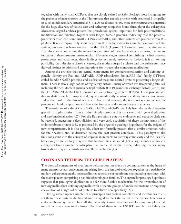
The revolution in genomics has allowed an unprecedented level of detailed information on thecomponents of any eukaryotic cell to be obtained and has allowed for comparisons among essen-tially all the major eukaryotic lineages to be made. Moreover, advances in structural biology havealso propelled comparisons among the detailed architectures of many of the protein complexesassociated with endomembranes. Together, these insights allow exploration of the different waysthat endomembrane systems are potentially modified in cells and the evolutionary origins of thesemembrane-manipulating machineries. Such studies have likely identified the major families ofprotein players, and it has emerged that the overall configuration of the endomembrane systemwas established very early in the evolutionary history of eukaryotes (27–29). By the time of the lasteukaryotic common ancestor (LECA), approximately one and one half billion years ago, a finalconsensus endomembrane arrangement and associated complement of components had becomeestablished. Remarkably, reconstructions from comparative genomic studies established that theLECA’s endomembrane system was actually extraordinarily complex (Figure 1). As well as thecore exocytic, endocytic, and phagocytic apparatuses, these systems were, according to the comple-ment of genes predicted as present, already differentiated into multiple endosomal and recyclingpathways, such that elaborate transport and tethering systems were functioning. The majority ofmodern eukaryotes, including the plants, protists, amoeba, and opisthokonts (animals and fungi),have retained much of this complexity along with the components that established them.
Peering beyond the LECA to events surrounding the first eukaryotic common ancestor (FECA)and events leading up to eukaryogenesis has been significantly more challenging (Figure 1).However, connections between mechanisms of intracellular vesicle and intraflagellar transport(IFT) and their dependence on Rab and Rab-like proteins and further similarities with Ran, theGTPase principally involved in nucleocytoplasmic transport, have been obvious for a considerabletime, and all of these GTPases are comparatively closely related within the greater Ras superfamily(30–34).
Eukaryotic forms emerged most likely within the Archaea bacteria and close to the Thau-marchaeota, Aigarchaeota, Crenarchaeota, and Korarchaeota (TACK) clade, based on extensivewhole genome sequencing and greater similarity among many genes shared between TACK ar-chaea and eukaryotes (35–37). Recently, the predicted gene complement of a metagenome from aLokiarchaeum, which itself is closely related to the TACK clade and was isolated from hydrother-mal vent sediments, has strengthen this paradigm further, as the reconstructed genome containsclose relatives to hallmark eukaryotic genes (i.e., genes so far considered restricted to eukaryoticgenomes). Even more recently, additional relatives to Lokiachaea have been identified and termedthe Asgard Achaea (38, 39). Significantly, these are derived from geographically widely dispersedlocations, suggesting this is a major branch of previously uncharacterized Archaea. Taken together,the Asgard archaea host homologs to many trafficking genes within their predicted metagenomes.
The reconstructed genomes of these organisms notably contain predicted protein domains thatin eukaryotes are associated with intracellular trafficking and secretion. These include ESCRT(endosomal sorting complexes required for transport), TRAPP (transport protein particle), andhomologs of the Sec23/24 COPII (coat protein complex II) vesicle coatomer protein complex,
640 Rout · Field
Ann
u. R
ev. B
ioch
em. 2
017.
86:6
37-6
57. D
ownl
oade
d fr
om w
ww
.ann
ualr
evie
ws.
org
Acc
ess
prov
ided
by
Roc
kefe
ller
Uni
vers
ity o
n 07
/19/
17. F
or p
erso
nal u
se o
nly.

BI86CH26-Rout ARI 31 May 2017 8:4
FECA
LECA
Small GTPasesSec23/24-likeLonginsβ-Propeller/α-Solenoid gene clustersESCRTsActin, tubulin
α-Proteobacteria
Archaebacteriaand TACK EubacteriaAsgard Eukaryota
?
Small GTPases (~30)SNAREs (~15)Sec23/24Longinsβ-Propeller/α-Solenoid proteins (~10)ESCRT I, II, IIIBAR domainsActin, tubulin
?
Figure 1The pathway to eukaryotic structure. According to the now well-accepted two domains of life model,eukaryotes arose from within the Archaea, with the closest known lineage being the TACK/Asgard archaea.These organisms share a number of clear similarities to the eukaryotic state, for example, a role for ESCRTin cytokinesis, together with small GTPases, longins, homologs of the COPII transport system (resemblingSec23 and Sec24), and other potential distant homologs to trafficking genes. This lineage gave rise to theorganism that was the FECA, which is assumed to have already acquired a nucleus and possibly someinternal structure. However, uncertainty in the order of events of evolution of the internal membrane (e.g., ifphagocytosis or secretory structures arose first) has made a definition of FECA elusive. By contrast, theLECA is well defined and contained a clear, complex cell architecture with fully differentiated internalsystems. At some point in the transition from FECA to LECA the mitochondrion was acquired, and despitethe clear importance of the mitochondrion for bioenergetics, the genetic contribution of this endosymbiontto cellular complexity appears minor. LECA was in fact more complex than many well-studied modeleukaryotes, such as Saccharomyces, Chlamydomonas, and Trypanosoma. Abbreviations: BAR,Bin/amphiphysin/Rvs domains; COPII, coat protein complex II; ESCRT, endosomal sorting complexesrequired for transport; FECA, first eukaryotic common ancestor; LECA, last eukaryotic common ancestor;SNAREs, soluble NSF attachment protein receptors; TACK, Thaumarchaeota, Aigarchaeota,Crenarchaeota, and Korarchaeota. The question mark indicates cellular forms for which the precise internalconfiguration is uncertain. Archaeal lineages are shown in magenta, eubacteria in blue, and eukaryotes ingray. Thorarchaeal image courtesy of Jack Kirby.
www.annualreviews.org • Evolution of Membrane Coating Complexes 641
Ann
u. R
ev. B
ioch
em. 2
017.
86:6
37-6
57. D
ownl
oade
d fr
om w
ww
.ann
ualr
evie
ws.
org
Acc
ess
prov
ided
by
Roc
kefe
ller
Uni
vers
ity o
n 07
/19/
17. F
or p
erso
nal u
se o
nly.
BI86CH26-Rout ARI 31 May 2017 8:4
together with many small GTPases that are closely related to Rabs. Perhaps most intriguing arethe presence of gene clusters in the Thorarchaea that encode proteins with predicted β-propelleror α-solenoid secondary structures (38–41). As we discuss below, these architectures are signaturesfor the huge diversity of vesicle coat and tethering complexes found throughout the eukaryotes.Moreover, Asgard archaea possess the prenylation system important for Rab posttranslationalmodification and function, together with longin domain proteins, indicating that the potentialprecursors to at least some small GTPases, SNAREs, and other systems are present within thisphylum. It is a comparatively short step from this configuration to a simple eukaryotic cellularsystem, envisaged as being on board in the FECA (Figure 1). However, given the absence ofany information concerning the internal organization of these fascinating organisms, the precisefunctions of these proteins remain unclear. Nevertheless, in terms of establishing the link betweenprokaryotes and eukaryotes, these findings are extremely provocative. Indeed, it is an excitingpossibility that, despite a shared ancestor, the modern Asgard archaea and the eukaryotes havederived distinct solutions and configurations for intracellular compartmentalization.
Among the proteins that are central components for compartmentalization and defining or-ganelle identity are Rab and ARF/ARL (ADP-ribosylation factor/ARF-like) family GTPases,coiled-bundle SNARE proteins, and a subset of these and related proteins possessing a longin do-main. There is also a large cohort of regulatory factors—many of which are also paralog families,including the Sec7 domain guanosine triphosphate (GTP) guanosine exchange factors (GEFs) andthe Tre-2/Bub2/Cdc16 (TBC) domain GTPase-activating proteins (GAPs). These protein fam-ilies mediate vesicular transport and, equally significantly, control specificity. As a consequence,and as the result of the flux of vesicular delivery and removal, the transport system dictates theprotein and lipid composition and hence the function of donor and target organelles.
The evolution of Rabs, ARFs, SNAREs, GEFs, and GAPs has likely been the result of stepwisegrowth in sophistication from a rather simple system and is consistent with gene duplicationand neofunctionalization (27). For the Rab proteins a putative endocytic and exocytic clade canbe resolved, suggesting a deep division and very early acquisition of these distinct arms of theendomembrane system (13), as proposed by the organelle paralogy hypothesis for the origins ofnew compartments. It is also possible, albeit not formally proven, that a similar situation holdsfor the SNAREs and, as discussed below, the coat protein complexes. This paradigm is alsofully consistent with the concept of stepwise increments to pathway complexity and the idea of abasic exocytic and endocytic system that has become elaborated (42); a large number of moderneukaryotes have a simpler cellular plan than predicted for the LECA, indicating that secondaryloss is also a frequent contributor to cellular evolution (43).
COATS AND TETHERS: THE CHIEF PLAYERS?
The physical constraints of membrane deformation, mechanistic commonalities at the heart ofmany transport steps, and constraints arising from the limits of evolution together may explain whymodern eukaryotes actually possess a limited repertoire of membrane manipulating machines, withthe major players comprising a handful of paralogous families. The organellar paralogy hypothesissuggests that paralogous duplication is a far more flexible mechanism for the diversification ofnew organelles than defining organelles with disparate groups of unrelated proteins or requiringcoevolution of a large cohort of proteins to achieve new specificity (27).
Having settled upon a simple set of principles and protein complexes and membranes to en-act them, these systems duplicated and diverged to meet the needs of the diverse functions ofendomembrane systems. Thus, all the currently known membrane-deforming complexes fallinto three major structural classes. The first of these is the ESCRT complex, including the
642 Rout · Field
Ann
u. R
ev. B
ioch
em. 2
017.
86:6
37-6
57. D
ownl
oade
d fr
om w
ww
.ann
ualr
evie
ws.
org
Acc
ess
prov
ided
by
Roc
kefe
ller
Uni
vers
ity o
n 07
/19/
17. F
or p
erso
nal u
se o
nly.
BI86CH26-Rout ARI 31 May 2017 8:4
membrane-deforming Snf7 domain subunits. ESCRT chiefly functions in late endosomal sortingand cytokinesis, although numerous additional roles have now been described (44). The secondis a group of Bin/amphiphysin/Rvs (BAR) domain–containing proteins, with roles mainly at theGolgi and endosomal interface, in phagocytosis, and once more in cytokinesis (20, 22, 45).
However, the third group, the protocoatomer-derived complexes, has come to dominate, interms of both the number of processes that they facilitate and the range of architectures that thesecomplexes are able to accommodate (11). Protocoatomer complexes all consist of four classes ofcomponents: coat-forming proteins, small GTPases, coiled-coil–containing proteins, and longindomain–containing proteins (Figure 2). These complexes are mostly thought to act in exocytosis,endocytosis, and intracellular vesicular transport, such as the COPI, COPII, and clathrin com-plexes. However, membership of this family extends to proteins that do not form transport vesiclesbut instead build stable membrane-associated coats, such as the NPC; that form tethering com-plexes, such as the HOPS (homotypic fusion and vacuole protein sorting)/CORVET (class C corevacuole-endosome tethering) and SEA (Seh1-associated) tethering complexes; or that, similarlyto the NPC, associate with membranes to direct protein transport, such as the IFT complexesthat mediate trafficking inside flagella (46–50).
COMMON ARCHITECTURES WITHIN MEMBRANE COATAND TETHER COMPLEXES
Several domains are key features of membrane deforming systems.
β-α Protocoatomer Architecture
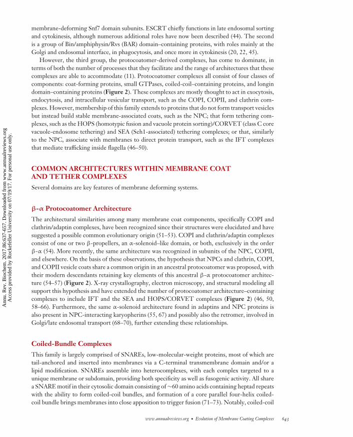
The architectural similarities among many membrane coat components, specifically COPI andclathrin/adaptin complexes, have been recognized since their structures were elucidated and havesuggested a possible common evolutionary origin (51–53). COPI and clathrin/adaptin complexesconsist of one or two β-propellers, an α-solenoid–like domain, or both, exclusively in the orderβ-α (54). More recently, the same architecture was recognized in subunits of the NPC, COPII,and elsewhere. On the basis of these observations, the hypothesis that NPCs and clathrin, COPI,and COPII vesicle coats share a common origin in an ancestral protocoatomer was proposed, withtheir modern descendants retaining key elements of this ancestral β-α protocoatomer architec-ture (54–57) (Figure 2). X-ray crystallography, electron microscopy, and structural modeling allsupport this hypothesis and have extended the number of protocoatomer architecture–containingcomplexes to include IFT and the SEA and HOPS/CORVET complexes (Figure 2) (46, 50,58–66). Furthermore, the same α-solenoid architecture found in adaptins and NPC proteins isalso present in NPC-interacting karyopherins (55, 67) and possibly also the retromer, involved inGolgi/late endosomal transport (68–70), further extending these relationships.
Coiled-Bundle Complexes
This family is largely comprised of SNAREs, low-molecular-weight proteins, most of which aretail-anchored and inserted into membranes via a C-terminal transmembrane domain and/or alipid modification. SNAREs assemble into heterocomplexes, with each complex targeted to aunique membrane or subdomain, providing both specificity as well as fusogenic activity. All sharea SNARE motif in their cytosolic domain consisting of ∼60 amino acids containing heptad repeatswith the ability to form coiled-coil bundles, and formation of a core parallel four-helix coiled-coil bundle brings membranes into close apposition to trigger fusion (71–73). Notably, coiled-coil
www.annualreviews.org • Evolution of Membrane Coating Complexes 643
Ann
u. R
ev. B
ioch
em. 2
017.
86:6
37-6
57. D
ownl
oade
d fr
om w
ww
.ann
ualr
evie
ws.
org
Acc
ess
prov
ided
by
Roc
kefe
ller
Uni
vers
ity o
n 07
/19/
17. F
or p
erso
nal u
se o
nly.

BI86CH26-Rout ARI 31 May 2017 8:4
Longinβ/α-Coatomer α-Solenoid RINGβ-Propeller Sec13
1W63 2PM93MKQ 4ZY85DNG
AP4
AP5
AP3
TSET
COPII
HOPSCORVET
SEA
COPI family
COPII family
Clathrin
COPI
AP1
AP2
IFTNPC
Figure 2Major relationships among protocoatomer architecture–containing complexes. At least twelve complexes possessprotocoatomer-related subunits, but the precise relationships among these systems remain difficult to characterize. This difficulty islikely in part due to the presence of multiple complexes within some of the structures (e.g., IFT and the NPC), but subunit sharing andcoevolutionary constraints are also important factors. Overall data suggest the presence of two groups in the protocoatomerevolutionary history, a major adaptin/COPI/TSET cluster (red ) and a COPII cluster (blue), with IFT remaining refractory to accurateplacement owing to the lack of discriminatory architectural information. The adaptin/COPI cluster shares the common architecture ofa heteromeric complex containing α-solenoid and longin domain subunits that interact with a β-propeller/α-solenoid–containingmembrane coat system. The second COPII cluster either shares Sec13 or, in the case of HOPS/CORVET, is a clear relative to SEA.Several of these complexes also integrate a RING domain within their protocoatomer subunits and are perhaps notable by the absenceof longin domain–containing subunits. Significantly, the expansion of the adaptins could be explained as a recent event, as thesecomplexes retain high degrees of sequence identity and several share a coating complex, whereas all other systems have distinctprotocoatomer subunits. Abbreviations: AP, adaptor protein; COPI, coat protein complex I; COPII, coat protein complex II;CORVET, class C core vacuole-endosome tethering; HOPS, homotypic fusion and vacuole protein sorting; IFT, intraflagellartransport; NPC, nuclear pore complex; RING, really interesting new gene; SEA, SEh1 associated; TSET, TPLATE complex. TheProtein Data Bank numbers for the various domains are provided below each structure.
644 Rout · Field
Ann
u. R
ev. B
ioch
em. 2
017.
86:6
37-6
57. D
ownl
oade
d fr
om w
ww
.ann
ualr
evie
ws.
org
Acc
ess
prov
ided
by
Roc
kefe
ller
Uni
vers
ity o
n 07
/19/
17. F
or p
erso
nal u
se o
nly.

BI86CH26-Rout ARI 31 May 2017 8:4
complexes are also found at the heart of the NPC (74, 75), but though suggestive, their relationshipto SNAREs is currently unclear.
Amphipathic Lipid Packing Sensor Motifs
Amphipathic lipid packing sensor (ALPS) motif sequences are 20–40 amino acids in length andcontain regular bulky hydrophobic residues spaced by 3–4 small polar uncharged residues, suchthat all the hydrophobic residues align at one face of an α-helix (76). ALPS motifs are intrinsicallysoluble but bind efficiently to positively curved membranes; however, rather than recognize thecurved surface geometry of membranes per se, ALPS motifs recognize defects in lipid packing thatarise from curvature. ALPS motifs are present in coat-forming proteins, membrane tethers, nucle-oporins, and lipid transporters in which they essentially act as detectors of membrane curvature.This is certainly the case for the ALPS domain of ArfGAP1, which detects packing defects in thecurved bilayers of COPI vesicles, specifically inserting into membranes that are under curvaturestress. As a result, GTPase stimulation activity is restricted to curved membrane regions (77).ArfGAP1 ALPS motifs help to organize two reactions: the assembly/disassembly cycle of COPIand the attachment of vesicles to long coiled-coil tether proteins (78, 79).
GTPases
As discussed above, GTPases and their GEFs and GAPs are central to membrane-associatedtransport processes. GTPases of the ARF family control cargo selection by coat complexes, whereasGTPases of the Rab family are required for the fusion of vesicles with the appropriate targetmembrane. Coat assembly is initiated by the activation of an Arf protein, and Arf1 regulatesformation of COPI vesicles and of clathrin-coated vesicles that contain the adaptor protein (AP)complexes AP1, AP3, and AP4 (80). Likewise, COPII vesicle budding involves Sar1, an Arf relative,for initiation (81). When activated by a cognate GEF, localized to the target membrane, theARF/Sar-GDP (guanosine diphosphate) becomes ARF/Sar-GTP and then recruits the coatomerthrough direct interactions.
Longins
The longin or uDENN domain is a regulatory element in many SNAREs but is also present as acomponent of several coat systems, including Npr1/2 of SEA, δ-COP (delta-subunit of COPI) ofCOPI, and adaptin subunits, and also many GTPase interacting proteins, including the prokary-otic MlgA (12, 71). The importance of the vast contributions of longin domains to cell structureand their potentially ancient origin has come to the fore only recently. The ∼15 kDa longindomain is a conserved α-β-α sandwich fold (a very common architecture) that in traffickingcontributes to regulation of assembly and fusion, including budding, tethering, and regulationof Rab GTPases. The longin domain regulates the fusogenic activity of SNAREs by mediatingintramolecular interactions with their coiled bundle domain. Longin domain–containing pro-teins are classified into seven superfamilies: longin SNAREs, adaptins, sedlins, SANDs (Sp100,AIRE-1, NucP41/75, DEAF-1), targetins, DENNs (differentially expressed in normal and neo-plastic cells), and APL2 VPS1 synthetic lethal proteins (AVLs) (12, 71). A subset of SNAREs, theVAMPs (vesicle-associated membrane proteins), have a highly conserved longin domain, and theseSNAREs act to mediate intracellular membrane fusion and define localization through associationwith coat proteins, as in longin Sec22b with the Sec23/24 subunits of COPII. Longin proteinsalso bring membranes together without initiating their fusion—for example, Sec22b brings the
www.annualreviews.org • Evolution of Membrane Coating Complexes 645
Ann
u. R
ev. B
ioch
em. 2
017.
86:6
37-6
57. D
ownl
oade
d fr
om w
ww
.ann
ualr
evie
ws.
org
Acc
ess
prov
ided
by
Roc
kefe
ller
Uni
vers
ity o
n 07
/19/
17. F
or p
erso
nal u
se o
nly.
BI86CH26-Rout ARI 31 May 2017 8:4
endoplasmic reticulum (ER) and plasma membranes into close proximity, stabilizing close con-tacts between these two membranes. The small σ and μ adaptin subunits and their homologs σ
and ζ in COPI are all longin-like domain proteins and important mediators of function throughsignaling and protein–protein interaction pathways (12, 82). Furthermore, longin domains are in-volved in regulating Rab GEFs and tethering complexes, including DENN, SAND, and TRAPPcomplexes, and also in the TOR (target of rapamycin) pathway through nutrient sensing and au-tophagy via their incorporation in the SEA complex. Longin domains appear to be very ancient,being present in Archaea and possibly also related specific prokaryotic GTPase-activating pro-teins, with some indication that prokaryotic GTPase circuits have persisted into eukaryotic forms(12, 40).
ARCHITECTURE AND MORPHOLOGY
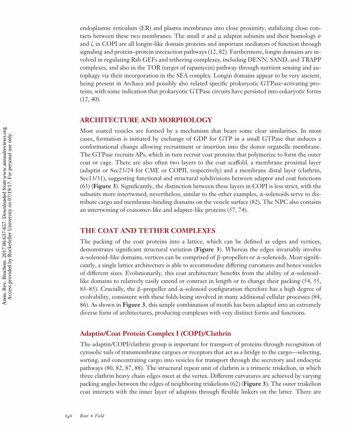
Most coated vesicles are formed by a mechanism that bears some clear similarities. In mostcases, formation is initiated by exchange of GDP for GTP in a small GTPase that induces aconformational change allowing recruitment or insertion into the donor organelle membrane.The GTPase recruits APs, which in turn recruit coat proteins that polymerize to form the outercoat or cage. There are also often two layers to the coat scaffold, a membrane proximal layer(adaptin or Sec23/24 for CME or COPII, respectively) and a membrane distal layer (clathrin,Sec13/31), suggesting functional and structural subdivisions between adaptor and coat functions(63) (Figure 3). Significantly, the distinction between these layers in COPI is less strict, with thesubunits more intertwined; nevertheless, similar to the other examples, α-solenoids serve to dis-tribute cargo and membrane-binding domains on the vesicle surface (82). The NPC also containsan intertwining of coatomer-like and adapter-like proteins (57, 74).
THE COAT AND TETHER COMPLEXES
The packing of the coat proteins into a lattice, which can be defined as edges and vertices,demonstrates significant structural variation (Figure 3). Whereas the edges invariably involveα-solenoid–like domains, vertices can be comprised of β-propellers or α-solenoids. Most signifi-cantly, a single lattice architecture is able to accommodate differing curvatures and hence vesiclesof different sizes. Evolutionarily, this coat architecture benefits from the ability of α-solenoid–like domains to relatively easily extend or contract in length or to change their packing (54, 55,83–85). Crucially, the β-propeller and α-solenoid configuration therefore has a high degree ofevolvability, consistent with these folds being involved in many additional cellular processes (84,86). As shown in Figure 3, this simple combination of motifs has been adapted into an extremelydiverse form of architectures, producing complexes with very distinct forms and functions.
Adaptin/Coat Protein Complex I (COPI)/Clathrin
The adaptin/COPI/clathrin group is important for transport of proteins through recognition ofcytosolic tails of transmembrane cargoes or receptors that act as a bridge to the cargo—selecting,sorting, and concentrating cargo into vesicles for transport through the secretory and endocyticpathways (80, 82, 87, 88). The structural repeat unit of clathrin is a trimeric triskelion, in whichthree clathrin heavy chain edges meet at the vertex. Different curvatures are achieved by varyingpacking angles between the edges of neighboring triskelions (62) (Figure 3). The outer triskelioncoat interacts with the inner layer of adaptins through flexible linkers on the latter. There are
646 Rout · Field
Ann
u. R
ev. B
ioch
em. 2
017.
86:6
37-6
57. D
ownl
oade
d fr
om w
ww
.ann
ualr
evie
ws.
org
Acc
ess
prov
ided
by
Roc
kefe
ller
Uni
vers
ity o
n 07
/19/
17. F
or p
erso
nal u
se o
nly.

BI86CH26-Rout ARI 31 May 2017 8:4
Nic96
ClathrinAP2 α2 AP2 β1
Sec31and Sec13
COP α COP β'COP γ COP β
Nup85and Seh1
Nup145Cand Sec13
Nup84
Nup133Nup120
Nup192
Cse1
Nup170
Clathrin
COPI
COPII
NPC core scaffold
Karyopherins
Adaptor CoatomerCoatomerAssemblageLattice
Endocytosisand secretion
Recycling/sortingendosomes
Golgi complex
Endoplasmicreticulum
NPC andkaryopherins
Architecture COPI-like COPII-likeTrafficking process
Figure 3Molecular anatomy of coat complexes. Extreme left, idealized endomembrane system, emphasizing transport events and thecompartments with which they are associated. Locations of protocoatomer architecture–contacting complexes are shown in purple.Middle left, architectures of coats showing the lattice ( purple) and its association with the membrane ( gray). The assemblage details athigher resolution and the position of subunits forming the lattices with approximate structures and β-propeller (cyan) and α-solenoid( pink) are indicated. Middle right, detailed architectures of monomers constituting COPI and COPI-related complexes with atomicresolution models compared with the schematic structures. Extreme right, detailed architectures of monomers constituting COPII andCOPII-related complexes with atomic resolution models compared with the schematic structures. Protein Data Bank accessionnumbers for the shown structures are 2JKR, 3IYV, 5A1X, 4BZJ, 1WA5, 4XMN, 2RFO, and 4IFQ. Abbreviations: AP, adaptor protein;COPI, coat protein complex I; COPII, coat protein complex II; NPC, nuclear pore complex.
now known to be five adaptin complexes, all of which share a common heterotetrameric structure,with two large subunits (γ1, α2, δ3, ε4, ζ5, and β1–5) and two smaller subunits (μ1–5 and σ1–5).Both the μ and σ subunits contain a longin domain, which also suggests an ancient duplicationfrom a simpler heterodimer—a theme one can also clearly see in the NPC (see below) (57, 74).These are a clear example of paralogous expansion generating new complexes, with evidence that,for AP1 and AP2, this expansion is still ongoing (89). A recently discovered divergent member ofthis group, TSET (TPLATE complex), despite a patchy distribution across eukaryotes, is likely aLECA component and contains all the adaptin-like protein equivalents as well as coat-like proteins.TSET is likely more closely related to COPI than the other adaptins (Figure 2) (90).
COPI comprises an F-COPI adaptor and a B-COPI coat. F-COPI comprises two large subunits(γ-COP and β-COP), a medium subunit (δ-COP), and a small subunit (ζ-COP) (82), which arelikely distant paralogs to the equivalent adaptin subunits. The B-COPI coat complex consistsof α, β′, and ε subunits (91). The F-COPI adaptor and B-COPI coat combine in a solubleheteroheptameric complex that is recruited in its entirety to the membrane. Though the subunitstructures are broadly similar to clathrin, F-COPI does not assemble into triskelions. Instead, the
www.annualreviews.org • Evolution of Membrane Coating Complexes 647
Ann
u. R
ev. B
ioch
em. 2
017.
86:6
37-6
57. D
ownl
oade
d fr
om w
ww
.ann
ualr
evie
ws.
org
Acc
ess
prov
ided
by
Roc
kefe
ller
Uni
vers
ity o
n 07
/19/
17. F
or p
erso
nal u
se o
nly.

BI86CH26-Rout ARI 31 May 2017 8:4
COPI coat is built of a repetition of building blocks, termed triads, that contain all the importantfunctional elements organized in a precise three-dimensional structure such that F+B COPIproteins all intertwine together into one large layer (Figure 3) (82). These “triads” are connectedby flexibly attached domains, to propagate curvature over larger areas while allowing a malleablearchitecture to eventually form buds, and may have arisen to facilitate accommodation of a diverserange of transport vesicle geometries.
A further candidate member of this group is the exomer, a divergent adaptin-like complexthat interestingly does not recruit a coat but bends the membrane directly (80, 88). Finally, theretromer is a heteropentameric complex involved in recycling transmembrane receptors fromendosomes to the trans-Golgi network. The largest subunit, Vps35, is an α-solenoid protein andresembles AP complexes—however, membrane curvature and vesiculation are induced by BARdomain–containing proteins within the complex, and currently no clear evidence indicates theretromer is part of the protocoatomer class of membrane-deforming systems (68–70, 88).
Together these data suggest a great deal of similarity between COPI, TSET, and clathrin/adaptin complexes. Although the combined coat and adaptor of TSET have been suggested as amissing link in the evolution of this family, recent data indicates instead a common ancestor forTSET and COPI (47). Significantly, this may also reflect mechanistic difference in the assemblyprocesses of the adaptin and TSET/COPI subfamilies, whereby adaptin and clathrin recruitmentoccurs as two distinct steps that are well documented (92, 93), but the entire COPI coatomer, andpossibly TSET (47), is recruited to the membrane en bloc. Significantly, there are separate coatsfor AP5 (SPG11) (94) and TSET (TTRAY) (90), underscoring the surprising complexity withinthe adaptin/COPI family. Presumably stepwise assembly facilitates flexibility in cargo recruitmentand may be a distinct solution to the roles of different coat proteins.
Coat Protein Complex II (COPII)
This complex is primarily involved in anterograde transport from the ER to the Golgi appara-tus. COPII appears at first glance to be rather different from the adaptin/COPI/clathrin group,but closer inspection demonstrates both mechanistic and structural similarities. The Sec23/24subunits form a bow tie–shaped structure, with a concave positively charged membrane proximalsurface to interact with the acidic phospholipid composition of the COPII vesicle (60, 66). Sec23and Sec24 are clearly ancient duplicates of each other, following the theme of generation of com-plexity through paralogous duplications and subsequent divergence within a complex (see sectionabove). Sec23/24 possess a smorgasbord of features including a β-barrel, a zinc finger, an α/βvWA (von Willebrand A domain) or so-called trunk domain, an all-helical region, and a carboxy-terminal gelsolin module (60, 66, 95)—another ancient domain found in the Asgard archaealsuperphylum (38, 39). The inner Sec23/24 coat can also form a regular lattice independent ofthe outer Sec31/13 coat, suggesting it may not only function to link cargo and membrane to theouter coat but also play a structural role in determining vesicle morphology (66, 95). There is aconsiderable degree of flexibility in the geometry of the COPII coat. At least three properties con-tribute to outer coat adaptability: variability of intersection angles at the vertices, flexibility withinthe central rod hinge, and the absence of any inherent asymmetry in the Sec13/31 rods, allowingthem to make head-to-head, head-to-tail, and tail-to-tail contacts. Together this versatility allowscoating of not only spherical, but also tubular membranes and therefore accommodation of largeelongated cargoes such as procollagen. This configuration also suggests that the unstructuredC-terminal region of Sec31, which connects the inner and outer coats, constrains the coat layersbut does not fix their absolute positions relative to one another (66, 95).
648 Rout · Field
Ann
u. R
ev. B
ioch
em. 2
017.
86:6
37-6
57. D
ownl
oade
d fr
om w
ww
.ann
ualr
evie
ws.
org
Acc
ess
prov
ided
by
Roc
kefe
ller
Uni
vers
ity o
n 07
/19/
17. F
or p
erso
nal u
se o
nly.

BI86CH26-Rout ARI 31 May 2017 8:4
The Nuclear Pore Complex
This structure is the largest structurally characterized member of the protocoatomer complexes.It appears to be in essence a supercomplex of COPI-like and COPII-like systems and indicatesa rather complex relationship with the other protocoatomer members (Figure 3) (11, 54, 55),although for now we have placed it for convenience with other COPII-like systems due to thedefining presence of Sec13 (Figure 2). The structural diversity within the NPC suggests thatthere must have been a progenitor NPC with duplications that gave rise to a complicated corescaffold, itself made up from a pair of outer rings flanking a pair of inner rings. It seems possiblethat the COPI + COPII architecture of the core scaffold arose through a merging of differentcoat complexes when the endomembrane was less differentiated (11, 57, 67, 96).
The architecture of the NPC itself does not currently provide clear answers—the core scaffoldcarries both COPII-like and COPI-like protein architectures; additionally, the inner ring alsoincludes the adaptin-like proteins Nup188 and Nup192, which share similar architectures withthe major karyopherin family of soluble cargo-carrying transport factors (57, 74). These transportfactors interact with the Rab-like GTPase, Ran, to drive bidirectional macromolecular transportacross the NPC (58). Flexible connectors, analogous to the unstructured C-terminal region ofSec31 and flexible connectors of the adaptins in COPI and clathrin/adaptin complexes, connectbetween the outer and inner rings (97). Whereas longin domains seem absent from the NPC,coiled-bundle domains are present in numerous subassemblies throughout the NPC (74, 75).
Valuable evidence in support of a closer relationship between COPII and the NPC can beinferred from the presence of a shared component, Sec13 (56). However, this shared componentis evidence only for a relationship between the outer ring Nup84 NPC subcomplex and COPIIand not additional NPC elements, and it would perhaps imply that the split between COPII andthe NPC was at the stage of a very primitive proto-NPC. Complicating this is the late endosomalSEA complex, which shares both Sec13 and Seh1 with the NPC (at least in the yeast Saccharomyces)in addition to possessing several β-α protocoatomer subunits. One of these, SEA4, is similar toSec31, which suggests an additional connection with COPII, and is possibly also shared withVps39, a component of the HOPS/CORVET complex (46, 50, 98). Although this remains rathertentative, these pieces of evidence suggest a close relationship among HOPS/CORVET, COPII,SEA, and the NPC (Figure 2).
The Intraflagellar Transport System
The flagellum (cilium) is also an ancient eukaryotic structure predicted to be present in the LECA.Flagellum assembly requires a specialized IFT system, which comprises three complexes: IFT-A,IFT-B, and the BBsome. These complexes are loosely associated biochemically and assemble incells into IFT trains, structures several hundred nanometers in length that are closely engaged withthe axoneme, and bind directly to tubulin (99, 100). IFT is powered by kinesin 2 and cytoplasmicdynein, to facilitate bidirectional transport. IFT-A contains 7 subunits and IFT-B together withthe BBsome 17 subunits, with the BBsome contributing 10 more, to achieve a compositionalcomplexity rivaling the NPC. IFT complexes A and B are invariably present to some degree inorganisms that possess conventional flagella and/or cilia, whereas the association is less strong forthe BBsome, as some flagellates lack this complex. Several subunits of each complex (WDR19,WDR35, IFT140, IFT122, IFT172, and IFT80) are clearly members of the protocoatomer family,containing the hallmark β-α architecture. IFT subunits are also remarkably well conserved at thesequence level across eukaryotes, substantially more than for the NPC. In evolutionary terms, itis likely that IFT-B evolved first from a simpler system pre-LECA and then duplicated to formthe BBsome and finally IFT-A. Significantly, the modern IFT system possesses several GTPases
www.annualreviews.org • Evolution of Membrane Coating Complexes 649
Ann
u. R
ev. B
ioch
em. 2
017.
86:6
37-6
57. D
ownl
oade
d fr
om w
ww
.ann
ualr
evie
ws.
org
Acc
ess
prov
ided
by
Roc
kefe
ller
Uni
vers
ity o
n 07
/19/
17. F
or p
erso
nal u
se o
nly.

BI86CH26-Rout ARI 31 May 2017 8:4
that are closely related to Rab8 (IFT22, IFT27), whereas the BBsome contains an ARL protein(ARL6/BBS3) (101, 102).
Clearly the presence of all these elements indicates a fundamental connection to the proto-coatomer, with the presence of GTPases controlling both vesicle fusion (Rabs) and coat assembly(ARLs). A short region of ∼150 residues that sits between the β-propeller– and α-solenoid–likesegments in all β-α IFT subunits possesses some similarity toward the α and β′ subunits of theCOPI complex, whereas TTC21, IFT88, TTC26, TTC30A/B, BBS4, and BBS8 also exhibit ar-chitectural similarity to the COP-ε subunit, which together suggests an evolutionary relationshipbetween IFT and COPI (49). The presence of coat proteins and GTPases is potentially a furtherexample of a coat system, together with fusion and control element equivalents, remaining inbiochemical association, but the sheer number of potential coating proteins in the IFT systemindicates considerable complexity. Significantly, a novel means of association between proto-coatomer subunits not so far described elsewhere was revealed from the structure of a complexof Chlamydomonas reinhardtii IFT70/IFT52, in which the IFT70 solenoids wrap tightly arounda proline-rich and mainly hydrophobic fragment of IFT52 that runs through the center of thesolenoid tube (103). Interaction maps and partial structures are available for much of the IFTsystem but currently remain insufficient to fully understand how the quaternary structure of IFTresembles other members of the protocoatomer family, if at all.
The IFT complexes thus remain difficult to place evolutionarily, at least in part owing to theabsence of full structural information, which raises the possibility that IFT is, similarly to theNPC, a chimera of COPI-like and COPII-like components, such that placing the entire complexis in truth impossible and that subcomplexes should be the unit of consideration (Figure 2).Although some evidence links several IFT components to COPI (49), the presence of some NPCproteins at the base of the cilium may indicate more complex level of interactions and evolutionaryrelationships (104).
Multi-Subunit Tethering Complexes
Although SNAREs are sufficient for fusion in in vitro reconstitutions, it is a rather slow reactionwith poor specificity. Additional components are often recruited to assist with the docking andfusion events and are referred to as tethering complexes. Mechanistically these complexes allinteract with SNAREs and/or GTPases to control the specificity of vesicle fusion and, in manyinstances, nucleotide exchange reactions. Multi-subunit tethering complexes (MTCs) can alsointeract with coat complexes: for example, COPI with the Dsl1 (CATCHR family) tether, COPIIwith the TRAPP I tether, and AP3 with the HOPS tether. Significantly, the HOPS complexcontains protocoatomer-like subunits, strongly suggesting that HOPS is derived from primordialcoat complexes. In ancestral eukaryotic cells, both donor and acceptor membranes may havebeen covered by different bona fide coats, and fusion may have been initiated by the direct contactbetween them. During evolution, one of these coats acquired and improved its capability to inducemembrane curvature, whereas the other, with a preference for flat membranes, developed into atethering factor and lost some coat-forming abilities (47, 105).
MTCs are categorized into three groups that also share some domains with the protocoatomercomplexes. Recent findings have blurred the distinction between true coat complexes and tetheringcomplexes, traditionally considered to be involved in vesicle docking. HOPS/CORVET is classedas a tethering complex but possesses β/α domain subunits, recognizes curved membranes, acts inlate endocytic targeting (106, 107), and is a near-universal eukaryotic feature (29). It is unknown ifHOPS/CORVET forms a lattice contributing to formation of transport intermediate complexes orHOPS is important for homotypic fusion and vacuole protein sorting and later recruits CORVET
650 Rout · Field
Ann
u. R
ev. B
ioch
em. 2
017.
86:6
37-6
57. D
ownl
oade
d fr
om w
ww
.ann
ualr
evie
ws.
org
Acc
ess
prov
ided
by
Roc
kefe
ller
Uni
vers
ity o
n 07
/19/
17. F
or p
erso
nal u
se o
nly.

BI86CH26-Rout ARI 31 May 2017 8:4
for endosomal transport. Similar to the adaptins, a degree of subunit expansion to create at leastthree variant complexes, but retaining the core Vps11, 16, 18, and 33 protocoatomer subunits, hasextended the functionality of this system and facilitates a switch between being controlled by Rab5or Rab7; most clearly, these complexes were derived from a single ancestral complex (47, 105). Atsome level HOPS/CORVET appears as an intermediate in structural sophistication between thesimple adaptins and NPC/IFT supercomplexes.
Further, the SEA complex, which interacts with the TOR pathway at vacuolar/late endosomalmembranes, is very clearly HOPS/CORVET-related and significantly shares two subunits, Seh1and Sec13, with the NPC as well as a structural analog of Sec31 (as discussed above). This indicatesthat not all members of the protocoatomer system likely are true coats and suggests a degree ofsubunit promiscuity that may have facilitated the evolution of additional complexes by a mixingof preexisting components. Hence, the protocoatomer family has functions that have divergedfrom the core process of membrane deformation. Whether membrane deformation or tetheringrepresents the ancestral role is unclear, as all of these complexes are sufficiently retained acrosseukaryotes to support a presence for them in the LECA. It is unclear if the presence of thesecomplexes within the endosomal system, rather than other transport routes, has any significance,but it could be the case that all of these tethers are derived from a single ancestral complex thathas come to populate the multiple steps between endocytosis and vacuole delivery. It has yet tobe determined if the newly characterized SEA complex, with some structural similarities to theHOPS/CORVET complex, is involved in tethering, coat formation, or another function (98).
HOW TO BUILD THE EUKARYOTIC CELL
Several distinct groups of protocoatomer-containing complexes can be discerned, based on com-mon architectural principles, and with differing levels of complexity (Figure 2). When consideredindividually, distinct evolutionary mechanisms are clearly responsible for this variability.
For the adaptin/COPI group, the possibility that these complexes arose by sequential paralo-gous expansion is high. This is well established for the adaptins (51, 89, 108) and, given the levelof architectural similarity, likely includes the COPI and TSET complexes as well. Significantly,TSET and COPI each have a unique coat, whereas most of the adaptins (AP1, AP2, and AP3)share the clathrin coat and AP5 has a distinct coat. This suggests a possible scenario in whichCOPI/TSET arose as a Golgi complex trafficking system that was later duplicated to TSET forpost-Golgi transport and COPI for intra-Golgi transport. Similarly, for the AP complexes, paral-ogous expansion from a progenitor adaptin complex became associated with endosomal/recyclingsystems, and presumably the progenitor fulfilled a similar but less differentiated role. This modelsuggests that a basic Golgi trafficking system existed that subsequently gave rise to post-Golgiand sorting systems for additional routes but that the differentiation between COPI/TSET andadaptins took place quite early in establishing this configuration. It may be significant in this regardthat adaptin complexes and TSET are not that infrequently lost (108–110), suggesting continuallineage-specific sculpting.
Complexes within the second major group are united by their shared Sec13 subunit (i.e., NPC,COPII, and SEA). Clear structural similarities among additional subunits indicate that COPIIand SEA are related, and although the functions of these two complexes are distinct, they are bothpotentially involved in ER-related functionality. The NPC also shares elements of the COPII-type architecture, and its association with the NE (nuclear envelope) indicates that potentialER functions connect all three of these complexes, once more suggesting a route of paralogousexpansion.
www.annualreviews.org • Evolution of Membrane Coating Complexes 651
Ann
u. R
ev. B
ioch
em. 2
017.
86:6
37-6
57. D
ownl
oade
d fr
om w
ww
.ann
ualr
evie
ws.
org
Acc
ess
prov
ided
by
Roc
kefe
ller
Uni
vers
ity o
n 07
/19/
17. F
or p
erso
nal u
se o
nly.
BI86CH26-Rout ARI 31 May 2017 8:4
The final group includes the HOPS/CORVET complexes. In this case, the mechanism ofevolution is very clear for the complexes within the cluster, as all share a heterotetrameric corebut bolt on additional subunits to differentiate the individual complexes. A connection to theSEA complex is evidenced by the presence of a RING (really interesting new gene) domain andadditional features (46, 98). Hence the HOPS group could be viewed as equivalent to the adaptinexpansion, with the exception that the majority of subunits in HOPS are shared, rather than fullydifferentiated into distinct genes as in the adaptins.
In summary, current data suggest Golgi- and ER-associated complexes are possible ancestralforms, existing in the transition between the FECA and LECA. Expansions and differentiationof these complexes generated over a dozen distinct complexes in the LECA, which have come todifferentiate the endosomal and other transport systems.
CONCLUSIONS: WHY ONE SYSTEM DOMINATESTHE EUKARYOTIC CELL
To allow cells to grow, membranous cellular structures must be dynamic and moldable—forexample, in both postmitotic separation of daughter nuclei and cytokinesis. Cell division representsa fundamental need for the ability to bend membranes, as even the most primitive cell must replicateand hence complete fission. Significantly, the ESCRT complex is required for cytokinesis in botharchaeal and eukaryotic cells, and emerging evidence suggests a role in NE maintenance, perhapshinting at a unified role in cell division (111).
However, overall the eukaryotic membrane coat repertoire is dominated by the protocoatomersignature β-propeller/α-solenoid proteins. As the core role of the protocoatomer is forming scaf-folds, we propose that some aspect of their molecular architecture is exceptionally flexible, allowingscaffolding of many membranes and complexes ranging in sophistication from simple adaptins tothe NPC and IFT (11). This implies a level of evolvability and architectural flexibility apparentlyabsent from the BAR domain and ESCRT systems in which, essentially, the same complex orsubsections of that complex may operate in more than one context, but paralog expansion andneofunctionalization do not occur. For ESCRT, essentially only a single set of coat proteins ispresent (112), and hence, only one architecture is possible.
In conclusion, reconstructing the order and relationships among membrane coating systemscurrently represents the best path to unravel the order of the majority of events in eukaryogenesis.In viewing the proteins that make up the coating complexes (Figure 3), one is struck not only bytheir basic similarities but also by the remarkable diversity in form, size, and architecture of theprotein networks that they form. This provides a spectacular example of how biological complexitycan arise from simplicity.
DISCLOSURE STATEMENT
The authors are not aware of any affiliations, memberships, funding, or financial holdings thatmight be perceived as affecting the objectivity of this review.
ACKNOWLEDGMENTS
We thank Joel Dacks for critical reading of the manuscript. Work in the authors’ laboratories issupported by the Wellcome Trust, the Medical Research Council UK, and the National Institutesof Health (M.P.R.: R21 AI096069, U54 GM103511, U01 GM098256, P41 GM109824, and
652 Rout · Field
Ann
u. R
ev. B
ioch
em. 2
017.
86:6
37-6
57. D
ownl
oade
d fr
om w
ww
.ann
ualr
evie
ws.
org
Acc
ess
prov
ided
by
Roc
kefe
ller
Uni
vers
ity o
n 07
/19/
17. F
or p
erso
nal u
se o
nly.

BI86CH26-Rout ARI 31 May 2017 8:4
R01 GM112108; M.C.F.: MR/N010558/1, MR/K008749/1, MR/P009018/1 from the MRC and204697/Z/16/Z from the Wellcome Trust).
LITERATURE CITED
1. Dacks JB, Field MC, Buick R, Eme L, Gribaldo S, et al. 2016. The changing view of eukaryogenesis—fossils, cells, lineages and how they all come together. J. Cell Sci. 129:3695–703
2. Szathmary E, Smith JM. 1995. The major evolutionary transitions. Nature 374:227–323. Stolz JF. 1998. Bacterial intracellular membranes. In Nature Encyclopedia of Life Sciences, ed. A Mitchell,
J Trapnell, S Hadfield, V Kerguelen, F Richmond, pp. 1–5. Chichester, UK: Wiley4. Fuerst JA. 2005. Intracellular compartmentation in planctomycetes. Annu. Rev. Microbiol. 59:299–3285. Seufferheld M, Lea CR, Vieira M, Oldfield E, Docampo R. 2004. The H+-pyrophosphatase of Rhodospir-
illum rubrum is predominantly located in polyphosphate-rich acidocalcisomes. J. Biol. Chem. 279:51193–202
6. Margulis L. 1981. Symbiosis in Cell Evolution. New York: W.H. Freeman7. Wilson RJ, Denny PW, Preiser PR, Rangachari K, Roberts K, et al. 1996. Complete gene map of the
plastid-like DNA of the malaria parasite Plasmodium falciparum. J. Mol. Biol. 261:155–728. Funes S, Davidson E, Reyes-Prieto A, Magallon S, Herion P, et al. 2002. A green algal apicoplast ancestor.
Science 298:21559. McFadden GI. 2011. The apicoplast. Protoplasma 248:641–50
10. McFadden GI. 2014. Apicoplast. Curr. Biol. 24:R262–6311. Field MC, Sali A, Rout MP. 2011. Evolution: On a bender—BARs, ESCRTs, COPs, and finally getting
your coat. J. Cell Biol. 193:963–7212. De Franceschi N, Wild K, Schlacht A, Dacks JB, Sinning I, Filippini F. 2014. Longin and GAF domains:
structural evolution and adaptation to the subcellular trafficking machinery. Traffic 15:104–2113. Elias M, Brighouse A, Gabernet-Castello C, Field MC, Dacks JB. 2012. Sculpting the endomembrane
system in deep time: high resolution phylogenetics of Rab GTPases. J. Cell Sci. 125:2500–814. Gabernet-Castello C, O’Reilly AJ, Dacks JB, Field MC. 2013. Evolution of Tre-2/Bub2/Cdc16 (TBC)
Rab GTPase-activating proteins. Mol. Biol. Cell 24:1574–8315. Stachowiak JC, Brodsky FM, Miller EA. 2013. A cost-benefit analysis of the physical mechanisms of
membrane curvature. Nat. Cell Biol. 15:1019–2716. Jarsch IK, Daste F, Gallop JL. 2016. Membrane curvature in cell biology: an integration of molecular
mechanisms. J. Cell Biol. 214:375–8717. McMahon HT, Gallop JL. 2005. Membrane curvature and mechanisms of dynamic cell membrane
remodelling. Nature 438:590–9618. Ford MG, Mills IG, Peter BJ, Vallis Y, Praefcke GJ, et al. 2002. Curvature of clathrin-coated pits driven
by epsin. Nature 419:361–6619. Miller SE, Mathiasen S, Bright NA, Pierre F, Kelly BT, et al. 2015. CALM regulates clathrin-coated
vesicle size and maturation by directly sensing and driving membrane curvature. Dev. Cell 33:163–7520. Peter BJ, Kent HM, Mills IG, Vallis Y, Butler PJ, et al. 2004. BAR domains as sensors of membrane
curvature: the amphiphysin BAR structure. Science 303:495–9921. Mim C, Unger VM. 2012. Membrane curvature and its generation by BAR proteins. Trends Biochem. Sci.
37:526–3322. Simunovic M, Voth GA, Callan-Jones A, Bassereau P. 2015. When physics takes over: BAR proteins and
membrane curvature. Trends Cell Biol. 25:780–9223. McMahon HT, Boucrot E. 2015. Membrane curvature at a glance. J. Cell Sci. 128:1065–7024. Doyon JB, Zeitler B, Cheng J, Cheng AT, Cherone JM, et al. 2011. Rapid and efficient clathrin-mediated
endocytosis revealed in genome-edited mammalian cells. Nat. Cell Biol. 13:331–3725. Kaksonen M, Toret CP, Drubin DG. 2005. A modular design for the clathrin- and actin-mediated
endocytosis machinery. Cell 123:305–2026. Toyama BH, Savas JN, Park SK, Harris MS, Ingolia NT, et al. 2013. Identification of long-lived proteins
reveals exceptional stability of essential cellular structures. Cell 154:971–82
www.annualreviews.org • Evolution of Membrane Coating Complexes 653
Ann
u. R
ev. B
ioch
em. 2
017.
86:6
37-6
57. D
ownl
oade
d fr
om w
ww
.ann
ualr
evie
ws.
org
Acc
ess
prov
ided
by
Roc
kefe
ller
Uni
vers
ity o
n 07
/19/
17. F
or p
erso
nal u
se o
nly.

BI86CH26-Rout ARI 31 May 2017 8:4
27. Dacks JB, Field MC. 2007. Evolution of the eukaryotic membrane-trafficking system: origin, tempo andmode. J. Cell Sci. 120:2977–85
28. Field MC, Gabernet-Castello C, Dacks JB. 2007. Reconstructing the evolution of the endocytic system:insights from genomics and molecular cell biology. Adv. Exp. Med. Biol. 607:84–96
29. Koumandou VL, Dacks JB, Coulson RM, Field MC. 2007. Control systems for membrane fusion in theancestral eukaryote; evolution of tethering complexes and SM proteins. BMC Evol. Biol. 7:29
30. Eguether T, San Agustin JT, Keady BT, Jonassen JA, Liang Y, et al. 2014. IFT27 links the BBSome toIFT for maintenance of the ciliary signaling compartment. Dev. Cell 31:279–90
31. Elias M, Klimes V, Derelle R, Petrzelkova R, Tachezy J. 2016. A paneukaryotic genomic analysis of thesmall GTPase RABL2 underscores the significance of recurrent gene loss in eukaryote evolution. Biol.Direct 11:5
32. Huet D, Blisnick T, Perrot S, Bastin P. 2014. The GTPase IFT27 is involved in both anterograde andretrograde intraflagellar transport. eLife 3:e02419
33. Ribbeck K, Lipowsky G, Kent HM, Stewart M, Gorlich D. 1998. NTF2 mediates nuclear import ofRan. EMBO J. 17:6587–98
34. Wang Z, Fan ZC, Williamson SM, Qin H. 2009. Intraflagellar transport (IFT) protein IFT25 is aphosphoprotein component of IFT complex B and physically interacts with IFT27 in Chlamydomonas.PLOS ONE 4:e5384
35. Guy L, Ettema TJ. 2011. The archaeal ‘TACK’ superphylum and the origin of eukaryotes. TrendsMicrobiol. 19:580–87
36. Raymann K, Brochier-Armanet C, Gribaldo S. 2015. The two-domain tree of life is linked to a new rootfor the Archaea. PNAS 112:6670–75
37. Saw JH, Spang A, Zaremba-Niedzwiedzka K, Juzokaite L, Dodsworth JA, et al. 2015. Exploring micro-bial dark matter to resolve the deep archaeal ancestry of eukaryotes. Philos. Trans. R. Soc. B 370:20140328
38. Spang A, Saw JH, Jorgensen SL, Zaremba-Niedzwiedzka K, Martijn J, et al. 2015. Complex archaea thatbridge the gap between prokaryotes and eukaryotes. Nature 521:173–79
39. Zaremba-Niedzwiedzka K, Caceres EF, Saw JH, Backstrom D, Juzokaite L, et al. 2017. Asgard archaeailluminate the origin of eukaryotic cellular complexity. Nature 541(7637):353–58
40. Klinger CM, Spang A, Dacks JB, Ettema TJ. 2016. Tracing the archaeal origins of eukaryotic membrane-trafficking system building blocks. Mol. Biol. Evol. 33:1528–41
41. Surkont J, Pereira-Leal JB. 2016. Are there Rab GTPases in Archaea? Mol. Biol. Evol. 33:1833–4242. Dacks JB, Peden AA, Field MC. 2009. Evolution of specificity in the eukaryotic endomembrane system.
Int. J. Biochem. Cell Biol. 41:330–4043. Koumandou VL, Wickstead B, Ginger ML, van der Giezen M, Dacks JB, Field MC. 2013. Molecular
paleontology and complexity in the last eukaryotic common ancestor. Crit. Rev. Biochem. Mol. Biol.48:373–96
44. Christ L, Raiborg C, Wenzel EM, Campsteijn C, Stenmark H. 2016. Cellular functions and molecularmechanisms of the ESCRT membrane-scission machinery. Trends Biochem. Sci. 42:42–56
45. McDonald NA, Gould KL. 2016. Linking up at the BAR: oligomerization and F-BAR protein function.Cell Cycle 15:1977–85
46. Algret R, Fernandez-Martinez J, Shi Y, Kim SJ, Pellarin R, et al. 2014. Molecular architecture andfunction of the SEA complex, a modulator of the TORC1 pathway. Mol. Cell Proteom. 13:2855–70
47. Chou HT, Dukovski D, Chambers MG, Reinisch KM, Walz T. 2016. CATCHR, HOPS and CORVETtethering complexes share a similar architecture. Nat. Struct. Mol. Biol. 23:761–63
48. Spang A. 2012. The DSL1 complex: the smallest but not the least CATCHR. Traffic 13:908–1349. van Dam TJ, Townsend MJ, Turk M, Schlessinger A, Sali A, et al. 2013. Evolution of modular intraflag-
ellar transport from a coatomer-like progenitor. PNAS 110:6943–4850. Spang A. 2016. Membrane tethering complexes in the endosomal system. Front. Cell Dev. Biol. 4:3551. Boehm M, Bonifacino JS. 2001. Adaptins: the final recount. Mol. Biol. Cell 12:2907–2052. Duden R, Griffiths G, Frank R, Argos P, Kreis TE. 1991. β-COP, a 110 kd protein associated with
non-clathrin-coated vesicles and the Golgi complex, shows homology to β-adaptin. Cell 64(3):649–65
654 Rout · Field
Ann
u. R
ev. B
ioch
em. 2
017.
86:6
37-6
57. D
ownl
oade
d fr
om w
ww
.ann
ualr
evie
ws.
org
Acc
ess
prov
ided
by
Roc
kefe
ller
Uni
vers
ity o
n 07
/19/
17. F
or p
erso
nal u
se o
nly.
BI86CH26-Rout ARI 31 May 2017 8:4
53. Schledzewski K, Brinkmann H, Mendel RR. 1999. Phylogenetic analysis of components of the eukaryoticvesicle transport system reveals a common origin of adaptor protein complexes 1, 2, and 3 and the Fsubcomplex of the coatomer COPI. J. Mol. Evol. 48(6):770–78
54. Devos D, Dokudovskaya S, Alber F, Williams R, Chait BT, et al. 2004. Components of coated vesiclesand nuclear pore complexes share a common molecular architecture. PLOS Biol. 2:e380
55. Devos D, Dokudovskaya S, Williams R, Alber F, Eswar N, et al. 2006. Simple fold composition andmodular architecture of the nuclear pore complex. PNAS 103:2172–77
56. Field MC, Dacks JB. 2009. First and last ancestors: reconstructing evolution of the endomembranesystem with ESCRTs, vesicle coat proteins, and nuclear pore complexes. Curr. Opin. Cell Biol. 21:4–13
57. Alber F, Dokudovskaya S, Veenhoff LM, Zhang W, Kipper J, et al. 2007. The molecular architecture ofthe nuclear pore complex. Nature 450:695–701
58. Cook A, Bono F, Jinek M, Conti E. 2007. Structural biology of nucleocytoplasmic transport. Annu. Rev.Biochem. 76:647–71
59. Brohawn SG, Partridge JR, Whittle JR, Schwartz TU. 2009. The nuclear pore complex has entered theatomic age. Structure 17:1156–68
60. Fath S, Mancias JD, Bi X, Goldberg J. 2007. Structure and organization of coat proteins in the COPIIcage. Cell 129:1325–36
61. Fotin A, Cheng Y, Grigorieff N, Walz T, Harrison SC, Kirchhausen T. 2004. Structure of an auxilin-bound clathrin coat and its implications for the mechanism of uncoating. Nature 432:649–53
62. Fotin A, Cheng Y, Sliz P, Grigorieff N, Harrison SC, et al. 2004. Molecular model for a complete clathrinlattice from electron cryomicroscopy. Nature 432:573–79
63. Lee C, Goldberg J. 2010. Structure of coatomer cage proteins and the relationship among COPI, COPII,and clathrin vesicle coats. Cell 142:123–32
64. Graham SC, Wartosch L, Gray SR, Scourfield EJ, Deane JE, et al. 2013. Structural basis of Vps33Arecruitment to the human HOPS complex by Vps16. PNAS 110:13345–50
65. Guo Z, Johnston W, Kovtun O, Mureev S, Brocker C, et al. 2013. Subunit organisation of in vitroreconstituted HOPS and CORVET multisubunit membrane tethering complexes. PLOS ONE 8:e81534
66. Stagg SM, LaPointe P, Razvi A, Gurkan C, Potter CS, et al. 2008. Structural basis for cargo regulationof COPII coat assembly. Cell 134:474–84
67. Sampathkumar P, Gheyi T, Miller SA, Bain KT, Dickey M, et al. 2011. Structure of the C-terminaldomain of Saccharomyces cerevisiae Nup133, a component of the nuclear pore complex. Proteins 79:1672–77
68. Collins BM. 2008. The structure and function of the retromer protein complex. Traffic 9:1811–2269. Hesketh GG, Perez-Dorado I, Jackson LP, Wartosch L, Schafer IB, et al. 2014. VARP is recruited on
to endosomes by direct interaction with retromer, where together they function in export to the cellsurface. Dev. Cell 29:591–606
70. Hierro A, Rojas AL, Rojas R, Murthy N, Effantin G, et al. 2007. Functional architecture of the retromercargo-recognition complex. Nature 449:1063–67
71. Daste F, Galli T, Tareste D. 2015. Structure and function of longin SNAREs. J. Cell Sci. 128:4263–7272. Dubuke ML, Munson M. 2016. The secret life of tethers: the role of tethering factors in SNARE complex
regulation. Front. Cell Dev. Biol. 4:4273. Bombardier JP, Munson M. 2015. Three steps forward, two steps back: mechanistic insights into the
assembly and disassembly of the SNARE complex. Curr. Opin. Chem. Biol. 29:66–7174. Kosinski J, Mosalaganti S, von Appen A, Teimer R, DiGuilio AL, et al. 2016. Molecular architecture of
the inner ring scaffold of the human nuclear pore complex. Science 352:363–6575. Fernandez-Martinez J, Kim SJ, Shi Y, Upla P, Pellarin R, et al. 2016. Structure and function of the
nuclear pore complex cytoplasmic mRNA export platform. Cell 167:1215–2876. Antonny B. 2011. Mechanisms of membrane curvature sensing. Annu. Rev. Biochem. 80:101–2377. Hanna MG IV, Mela I, Wang L, Henderson RM, Chapman ER, et al. 2016. Sar1 GTPase activity is
regulated by membrane curvature. J. Biol. Chem. 291:1014–2778. Vanni S, Vamparys L, Gautier R, Drin G, Etchebest C, et al. 2013. Amphipathic lipid packing sensor
motifs: probing bilayer defects with hydrophobic residues. Biophys. J. 104:575–8479. Drin G, Morello V, Casella JF, Gounon P, Antonny B. 2008. Asymmetric tethering of flat and curved
lipid membranes by a golgin. Science 320:670–73
www.annualreviews.org • Evolution of Membrane Coating Complexes 655
Ann
u. R
ev. B
ioch
em. 2
017.
86:6
37-6
57. D
ownl
oade
d fr
om w
ww
.ann
ualr
evie
ws.
org
Acc
ess
prov
ided
by
Roc
kefe
ller
Uni
vers
ity o
n 07
/19/
17. F
or p
erso
nal u
se o
nly.

BI86CH26-Rout ARI 31 May 2017 8:4
80. Paczkowski JE, Richardson BC, Fromme JC. 2015. Cargo adaptors: structures illuminate mechanismsregulating vesicle biogenesis. Trends Cell Biol. 25:408–16
81. Haucke V. 2003. Vesicle budding: a coat for the COPs. Trends Cell Biol. 13:59–6082. Dodonova SO, Diestelkoetter-Bachert P, von Appen A, Hagen WJ, Beck R, et al. 2015. A structure of
the COPI coat and the role of coat proteins in membrane vesicle assembly. Science 349:195–9883. Kobe B, Kajava AV. 2000. When protein folding is simplified to protein coiling: the continuum of
solenoid protein structures. Trends Biochem. Sci. 25:509–1584. Yoshimura SH, Hirano T. 2016. HEAT repeats—versatile arrays of amphiphilic helices working in
crowded environments? J. Cell Sci. 129:3963–7085. Forwood JK, Lange A, Zachariae U, Marfori M, Preast C, et al. 2010. Quantitative structural analysis
of importin-β flexibility: paradigm for solenoid protein structures. Structure 18:1171–8386. Chaudhuri I, Soding J, Lupas AN. 2008. Evolution of the β-propeller fold. Proteins 71:795–80387. Robinson MS. 2015. Forty years of clathrin-coated vesicles. Traffic 16:1210–3888. Bonifacino JS. 2014. Adaptor proteins involved in polarized sorting. J. Cell Biol. 204:7–1789. Dacks JB, Poon PP, Field MC. 2008. Phylogeny of endocytic components yields insight into the process
of nonendosymbiotic organelle evolution. PNAS 105:588–9390. Hirst J, Schlacht A, Norcott JP, Traynor D, Bloomfield G, et al. 2014. Characterization of TSET, an
ancient and widespread membrane trafficking complex. eLife 3:e0286691. Beck R, Rawet M, Wieland FT, Cassel D. 2009. The COPI system: molecular mechanisms and function.
FEBS Lett. 583:2701–992. Kaksonen M, Sun Y, Drubin DG. 2003. A pathway for association of receptors, adaptors, and actin
during endocytic internalization. Cell 115:475–8793. Liu J, Sun Y, Drubin DG, Oster GF. 2009. The mechanochemistry of endocytosis. PLOS Biol.
7:e100020494. Hirst J, Borner GH, Edgar J, Hein MY, Mann M, et al. 2013. Interaction between AP-5 and the hereditary
spastic paraplegia proteins SPG11 and SPG15. Mol. Biol. Cell 24(16):2558–6995. Zanetti G, Prinz S, Daum S, Meister A, Schekman R, et al. 2013. The structure of the COPII transport-
vesicle coat assembled on membranes. eLife 2:e0095196. Kosinski J, Mosalaganti S, von Appen A, Teimer R, DiGuilio AL, et al. 2016. Molecular architecture of
the inner ring scaffold of the human nuclear pore complex. Science 352(6283):363–6597. Fischer J, Teimer R, Amlacher S, Kunze R, Hurt E. 2015. Linker Nups connect the nuclear pore complex
inner ring with the outer ring and transport channel. Nat. Struct. Mol. Biol. 22:774–8198. Dokudovskaya S, Waharte F, Schlessinger A, Pieper U, Devos DP, et al. 2011. A conserved coatomer-
related complex containing Sec13 and Seh1 dynamically associates with the vacuole in Saccharomycescerevisiae. Mol. Cell Proteom. 10:M110 006478
99. Kubo T, Brown JM, Bellve K, Craige B, Craft JM, et al. 2016. Together, the IFT81 and IFT74 N-terminiform the main module for intraflagellar transport of tubulin. J. Cell Sci. 129:2106–19
100. Taschner M, Weber K, Mourao A, Vetter M, Awasthi M, et al. 2016. Intraflagellar transport proteins172, 80, 57, 54, 38, and 20 form a stable tubulin-binding IFT-B2 complex. EMBO J. 35:773–90
101. Bhogaraju S, Taschner M, Morawetz M, Basquin C, Lorentzen E. 2011. Crystal structure of the in-traflagellar transport complex 25/27. EMBO J. 30:1907–18
102. Hemsworth GR, Price HP, Smith DF, Wilson KS. 2013. Crystal structure of the small GTPaseArl6/BBS3 from Trypanosoma brucei. Protein Sci. 22:196–203
103. Taschner M, Kotsis F, Braeuer P, Kuehn EW, Lorentzen E. 2014. Crystal structures of IFT70/52 andIFT52/46 provide insight into intraflagellar transport B core complex assembly. J. Cell Biol. 207:269–82
104. Kee HL, Verhey KJ. 2013. Molecular connections between nuclear and ciliary import processes. Cilia2:11
105. Balderhaar HJ, Ungermann C. 2013. CORVET and HOPS tethering complexes—coordinators of en-dosome and lysosome fusion. J. Cell Sci. 126:1307–16
106. Nickerson DP, Brett CL, Merz AJ. 2009. Vps-C complexes: gatekeepers of endolysosomal traffic. Curr.Opin. Cell Biol. 21:543–51
656 Rout · Field
Ann
u. R
ev. B
ioch
em. 2
017.
86:6
37-6
57. D
ownl
oade
d fr
om w
ww
.ann
ualr
evie
ws.
org
Acc
ess
prov
ided
by
Roc
kefe
ller
Uni
vers
ity o
n 07
/19/
17. F
or p
erso
nal u
se o
nly.
BI86CH26-Rout ARI 31 May 2017 8:4
107. Cabrera M, Langemeyer L, Mari M, Rethmeier R, Orban I, et al. 2010. Phosphorylation of a membranecurvature-sensing motif switches function of the HOPS subunit Vps41 in membrane tethering. J. CellBiol. 191:845–59
108. Barlow LD, Dacks JB, Wideman JG. 2014. From all to (nearly) none: tracing adaptin evolution in Fungi.Cell. Logist. 4:e28114
109. Lee LJ, Klute MJ, Herman EK, Read B, Dacks JB. 2015. Losses, expansions, and novel subunit discoveryof adaptor protein complexes in haptophyte algae. Protist 166:585–97
110. Manna PT, Kelly S, Field MC. 2013. Adaptin evolution in kinetoplastids and emergence of the variantsurface glycoprotein coat in African trypanosomatids. Mol. Phylogenet. Evol. 67:123–28
111. Hurley JH. 2015. ESCRTs are everywhere. EMBO J. 34:2398–407112. Leung KF, Dacks JB, Field MC. 2008. Evolution of the multivesicular body ESCRT machinery; reten-
tion across the eukaryotic lineage. Traffic 9:1698–716
www.annualreviews.org • Evolution of Membrane Coating Complexes 657
Ann
u. R
ev. B
ioch
em. 2
017.
86:6
37-6
57. D
ownl
oade
d fr
om w
ww
.ann
ualr
evie
ws.
org
Acc
ess
prov
ided
by
Roc
kefe
ller
Uni
vers
ity o
n 07
/19/
17. F
or p
erso
nal u
se o
nly.

ANNUAL REVIEWSConnect With Our Experts
New From Annual Reviews:Annual Review of Cancer Biologycancerbio.annualreviews.org • Volume 1 • March 2017
Co-Editors: Tyler Jacks, Massachusetts Institute of Technology Charles L. Sawyers, Memorial Sloan Kettering Cancer Center
The Annual Review of Cancer Biology reviews a range of subjects representing important and emerging areas in the field of cancer research. The Annual Review of Cancer Biology includes three broad themes: Cancer Cell Biology, Tumorigenesis and Cancer Progression, and Translational Cancer Science.
TABLE OF CONTENTS FOR VOLUME 1:• How Tumor Virology Evolved into Cancer Biology and
Transformed Oncology, Harold Varmus• The Role of Autophagy in Cancer, Naiara Santana-Codina,
Joseph D. Mancias, Alec C. Kimmelman• Cell Cycle–Targeted Cancer Therapies, Charles J. Sherr,
Jiri Bartek• Ubiquitin in Cell-Cycle Regulation and Dysregulation
in Cancer, Natalie A. Borg, Vishva M. Dixit• The Two Faces of Reactive Oxygen Species in Cancer,
Colleen R. Reczek, Navdeep S. Chandel• Analyzing Tumor Metabolism In Vivo, Brandon Faubert,
Ralph J. DeBerardinis• Stress-Induced Mutagenesis: Implications in Cancer
and Drug Resistance, Devon M. Fitzgerald, P.J. Hastings, Susan M. Rosenberg
• Synthetic Lethality in Cancer Therapeutics, Roderick L. Beijersbergen, Lodewyk F.A. Wessels, René Bernards
• Noncoding RNAs in Cancer Development, Chao-Po Lin, Lin He
• p53: Multiple Facets of a Rubik’s Cube, Yun Zhang, Guillermina Lozano
• Resisting Resistance, Ivana Bozic, Martin A. Nowak• Deciphering Genetic Intratumor Heterogeneity
and Its Impact on Cancer Evolution, Rachel Rosenthal, Nicholas McGranahan, Javier Herrero, Charles Swanton
• Immune-Suppressing Cellular Elements of the Tumor Microenvironment, Douglas T. Fearon
• Overcoming On-Target Resistance to Tyrosine Kinase Inhibitors in Lung Cancer, Ibiayi Dagogo-Jack, Jeffrey A. Engelman, Alice T. Shaw
• Apoptosis and Cancer, Anthony Letai• Chemical Carcinogenesis Models of Cancer: Back
to the Future, Melissa Q. McCreery, Allan Balmain• Extracellular Matrix Remodeling and Stiffening Modulate
Tumor Phenotype and Treatment Response, Jennifer L. Leight, Allison P. Drain, Valerie M. Weaver
• Aneuploidy in Cancer: Seq-ing Answers to Old Questions, Kristin A. Knouse, Teresa Davoli, Stephen J. Elledge, Angelika Amon
• The Role of Chromatin-Associated Proteins in Cancer, Kristian Helin, Saverio Minucci
• Targeted Differentiation Therapy with Mutant IDH Inhibitors: Early Experiences and Parallels with Other Differentiation Agents, Eytan Stein, Katharine Yen
• Determinants of Organotropic Metastasis, Heath A. Smith, Yibin Kang
• Multiple Roles for the MLL/COMPASS Family in the Epigenetic Regulation of Gene Expression and in Cancer, Joshua J. Meeks, Ali Shilatifard
• Chimeric Antigen Receptors: A Paradigm Shift in Immunotherapy, Michel Sadelain
ANNUAL REVIEWS | CONNECT WITH OUR EXPERTS
650.493.4400/800.523.8635 (us/can)www.annualreviews.org | [email protected]
ONLINE NOW!
Ann
u. R
ev. B
ioch
em. 2
017.
86:6
37-6
57. D
ownl
oade
d fr
om w
ww
.ann
ualr
evie
ws.
org
Acc
ess
prov
ided
by
Roc
kefe
ller
Uni
vers
ity o
n 07
/19/
17. F
or p
erso
nal u
se o
nly.
BI86-FrontMatter ARI 1 June 2017 10:4
Annual Review ofBiochemistry
Volume 86, 2017ContentsAt the Intersection of Chemistry, Biology, and Medicine
Christopher T. Walsh � � � � � � � � � � � � � � � � � � � � � � � � � � � � � � � � � � � � � � � � � � � � � � � � � � � � � � � � � � � � � � � � � � � � � � � � � � 1
Protein Misfolding DiseasesF. Ulrich Hartl � � � � � � � � � � � � � � � � � � � � � � � � � � � � � � � � � � � � � � � � � � � � � � � � � � � � � � � � � � � � � � � � � � � � � � � � � � � � � � �21
Protein Misfolding, Amyloid Formation, and Human Disease:A Summary of Progress Over the Last DecadeFabrizio Chiti and Christopher M. Dobson � � � � � � � � � � � � � � � � � � � � � � � � � � � � � � � � � � � � � � � � � � � � � � � � �27
Structural Studies of Amyloid Proteins at the Molecular LevelDavid S. Eisenberg and Michael R. Sawaya � � � � � � � � � � � � � � � � � � � � � � � � � � � � � � � � � � � � � � � � � � � � � � � �69
Mechanisms and Functions of Spatial Protein Quality ControlEmily Mitchell Sontag, Rahul S. Samant, and Judith Frydman � � � � � � � � � � � � � � � � � � � � � � � � � �97
The Ubiquitin System, Autophagy, and Regulated Protein DegradationAlexander Varshavsky � � � � � � � � � � � � � � � � � � � � � � � � � � � � � � � � � � � � � � � � � � � � � � � � � � � � � � � � � � � � � � � � � � � � � � 123
Ubiquitin Ligases: Structure, Function, and RegulationNing Zheng and Nitzan Shabek � � � � � � � � � � � � � � � � � � � � � � � � � � � � � � � � � � � � � � � � � � � � � � � � � � � � � � � � � � � 129
Mechanisms of Deubiquitinase Specificity and RegulationTycho E.T. Mevissen and David Komander � � � � � � � � � � � � � � � � � � � � � � � � � � � � � � � � � � � � � � � � � � � � � � � 159
Proteasomal and Autophagic Degradation SystemsIvan Dikic � � � � � � � � � � � � � � � � � � � � � � � � � � � � � � � � � � � � � � � � � � � � � � � � � � � � � � � � � � � � � � � � � � � � � � � � � � � � � � � � � � � 193
Mechanisms of Autophagy InitiationJames H. Hurley and Lindsey N. Young � � � � � � � � � � � � � � � � � � � � � � � � � � � � � � � � � � � � � � � � � � � � � � � � � � 225
Systems Biology of MetabolismJens Nielsen � � � � � � � � � � � � � � � � � � � � � � � � � � � � � � � � � � � � � � � � � � � � � � � � � � � � � � � � � � � � � � � � � � � � � � � � � � � � � � � � � 245
Metabolite Measurement: Pitfalls to Avoid and Practices to FollowWenyun Lu, Xiaoyang Su, Matthias S. Klein, Ian A. Lewis, Oliver Fiehn,
and Joshua D. Rabinowitz � � � � � � � � � � � � � � � � � � � � � � � � � � � � � � � � � � � � � � � � � � � � � � � � � � � � � � � � � � � � � � 277
Isocitrate Dehydrogenase Mutation and (R)-2-Hydroxyglutarate:From Basic Discovery to Therapeutics DevelopmentLenny Dang and Shin-San Michael Su � � � � � � � � � � � � � � � � � � � � � � � � � � � � � � � � � � � � � � � � � � � � � � � � � � � 305
v
Ann
u. R
ev. B
ioch
em. 2
017.
86:6
37-6
57. D
ownl
oade
d fr
om w
ww
.ann
ualr
evie
ws.
org
Acc
ess
prov
ided
by
Roc
kefe
ller
Uni
vers
ity o
n 07
/19/
17. F
or p
erso
nal u
se o
nly.

BI86-FrontMatter ARI 1 June 2017 10:4
Conceptual and Experimental Tools to Understand Spatial Effects andTransport Phenomena in Nonlinear Biochemical NetworksIllustrated with Patchy SwitchingRebecca R. Pompano, Andrew H. Chiang, Christian J. Kastrup,
and Rustem F. Ismagilov � � � � � � � � � � � � � � � � � � � � � � � � � � � � � � � � � � � � � � � � � � � � � � � � � � � � � � � � � � � � � � � 333
Biochemistry of Catabolic Reductive DehalogenationMaeva Fincker and Alfred M. Spormann � � � � � � � � � � � � � � � � � � � � � � � � � � � � � � � � � � � � � � � � � � � � � � � � � 357
Electric Fields and Enzyme CatalysisStephen D. Fried and Steven G. Boxer � � � � � � � � � � � � � � � � � � � � � � � � � � � � � � � � � � � � � � � � � � � � � � � � � � � � 387
Eukaryotic DNA Replication ForkPeter M.J. Burgers and Thomas A. Kunkel � � � � � � � � � � � � � � � � � � � � � � � � � � � � � � � � � � � � � � � � � � � � � � � 417
Telomerase Mechanism of Telomere SynthesisR. Alex Wu, Heather E. Upton, Jacob M. Vogan, and Kathleen Collins � � � � � � � � � � � � � � � � 439
Site-Specific Self-Catalyzed DNA Depurination: A BiologicalMechanism That Leads to Mutations and Creates SequenceDiversityJacques R. Fresco and Olga Amosova � � � � � � � � � � � � � � � � � � � � � � � � � � � � � � � � � � � � � � � � � � � � � � � � � � � � � � 461
A New Facet of Vitamin B12: Gene Regulation by Cobalamin-BasedPhotoreceptorsS. Padmanabhan, Marco Jost, Catherine L. Drennan,
and Montserrat Elıas-Arnanz � � � � � � � � � � � � � � � � � � � � � � � � � � � � � � � � � � � � � � � � � � � � � � � � � � � � � � � � � � 485
Engineering and In Vivo Applications of RiboswitchesZachary F. Hallberg, Yichi Su, Rebekah Z. Kitto, and Ming C. Hammond � � � � � � � � � � � � 515
Cyclic GMP–AMP as an Endogenous Second Messenger in InnateImmune Signaling by Cytosolic DNAKazuki Kato, Hiroki Omura, Ryuichiro Ishitani, and Osamu Nureki � � � � � � � � � � � � � � � � � � 541
A Bright Future for Antibiotics?Donna Matzov, Anat Bashan, and Ada Yonath � � � � � � � � � � � � � � � � � � � � � � � � � � � � � � � � � � � � � � � � � � 567
Molecular Characteristics and Biological Functions of Surface-Activeand Surfactant ProteinsMargaret Sunde, Chi L.L. Pham, and Ann H. Kwan � � � � � � � � � � � � � � � � � � � � � � � � � � � � � � � � � � � 585
How α-Helical Motifs Form Functionally Diverse Lipid-BindingCompartmentsLucy Malinina, Dinshaw J. Patel, and Rhoderick E. Brown � � � � � � � � � � � � � � � � � � � � � � � � � � � � � 609
The Evolution of Organellar Coat Complexes and Organization of theEukaryotic CellMichael P. Rout and Mark C. Field � � � � � � � � � � � � � � � � � � � � � � � � � � � � � � � � � � � � � � � � � � � � � � � � � � � � � � � 637
vi Contents
Ann
u. R
ev. B
ioch
em. 2
017.
86:6
37-6
57. D
ownl
oade
d fr
om w
ww
.ann
ualr
evie
ws.
org
Acc
ess
prov
ided
by
Roc
kefe
ller
Uni
vers
ity o
n 07
/19/
17. F
or p
erso
nal u
se o
nly.
BI86-FrontMatter ARI 1 June 2017 10:4
Endoplasmic Reticulum–Plasma Membrane Contact SitesYasunori Saheki and Pietro De Camilli � � � � � � � � � � � � � � � � � � � � � � � � � � � � � � � � � � � � � � � � � � � � � � � � � � � 659
Mitochondrial Machineries for Protein Import and AssemblyNils Wiedemann and Nikolaus Pfanner � � � � � � � � � � � � � � � � � � � � � � � � � � � � � � � � � � � � � � � � � � � � � � � � � � � 685
Oxidative StressHelmut Sies, Carsten Berndt, and Dean P. Jones � � � � � � � � � � � � � � � � � � � � � � � � � � � � � � � � � � � � � � � � 715
Multiple Functions and Regulation of Mammalian PeroxiredoxinsSue Goo Rhee and In Sup Kil � � � � � � � � � � � � � � � � � � � � � � � � � � � � � � � � � � � � � � � � � � � � � � � � � � � � � � � � � � � � � � 749
Redox-Based Regulation of Bacterial Development and BehaviorAbigail J. Sporer, Lisa J. Kahl, Alexa Price-Whelan, and Lars E.P. Dietrich � � � � � � � � � 777
Extracellular Heme Uptake and the Challenge of Bacterial CellMembranesWeiliang Huang and Angela Wilks � � � � � � � � � � � � � � � � � � � � � � � � � � � � � � � � � � � � � � � � � � � � � � � � � � � � � � � � 799
Teaching Old Dyes New Tricks: Biological Probes Built fromFluoresceins and RhodaminesLuke D. Lavis � � � � � � � � � � � � � � � � � � � � � � � � � � � � � � � � � � � � � � � � � � � � � � � � � � � � � � � � � � � � � � � � � � � � � � � � � � � � � � � 825
Microbial Rhodopsins: Diversity, Mechanisms, and OptogeneticApplicationsElena G. Govorunova, Oleg A. Sineshchekov, Hai Li, and John L. Spudich � � � � � � � � � � � 845
Cellular Electron Cryotomography: Toward Structural Biology In SituCatherine M. Oikonomou and Grant J. Jensen � � � � � � � � � � � � � � � � � � � � � � � � � � � � � � � � � � � � � � � � � � � 873
Errata
An online log of corrections to Annual Review of Biochemistry articles may be found athttp://www.annualreviews.org/errata/biochem
Contents vii
Ann
u. R
ev. B
ioch
em. 2
017.
86:6
37-6
57. D
ownl
oade
d fr
om w
ww
.ann
ualr
evie
ws.
org
Acc
ess
prov
ided
by
Roc
kefe
ller
Uni
vers
ity o
n 07
/19/
17. F
or p
erso
nal u
se o
nly.