the estimation of free formaldehyde by … · the estimation of free formaldehyde by ... solution...
TRANSCRIPT

THE ESTIMATION OF FREE FORMALDEHYDE BY DIFFUSION*
BY M. JOHN BOYD AND MILAiX A. LOGAN
@Torn the Department of Biological Chemistry, University of Cincinnati College of Medicine, Cincinnati)
(Received for publication, July 12, 1945)
The procedure to be described was devised for the purpose of determining the amount of free or uncombined formaldehyde in bacterial toxins during the process of rendering them non-toxic. The determination of formalde- hyde is made without changing the pH and without diluting the sample to be tested. Disturbance of amino acid-formaldehyde equilibria existing in solution is therefore minimum.
The following principle is employed in the diffusion procedure. Formal- dehyde vapor is allowed to diffuse from the surface of a solution to be analyzed into a membrane containing phenylhydrazine hydrochloride. A red color is developed in the membrane by adding ferricyanide and acid and compared with colors developed at the same time in a similar manner from standard solutions of formaldehyde.
The outstanding merit of the phenylhydrazine-ferricyanide reaction for formaldehyde as applied in this procedure is it,s remarkable sensitivity. Notwithstanding the very low volatility of formaldehyde in dilute solutions, samples containing 2 to 1000 y of formaldehyde per rc. are suitable for analysis. Presumably only a small proportion of t,he formaldehyde in or near the surface of the solution takes part in the reaction.
Rimini (I) evidently first used phenylhydrazine in a calorimetric test for formaldehyde. As modified by Schryver (2) and others (3, 4), the reagents have been used to test for free formaldehyde. In these methods the reagents (one of which was fairly concentrated HCl or NaOII) were added to the solution to be analyzed. The color that, was developed de- pended, in part, on the amount of formaldehyde which remained dissociated from combinations with amino acids after the addition of the reagents.
The procedure and apparatus are designed so that the formaldehyde can be determined to f10 y per cc. in solutions containing less than 200 y per cc. and to f5 per cent for higher concentrati0ns.l The procedure is performed with apparatus which is readily constructed and a technique
* The work described in this paper was done under a rontract, recommended by the Committee on Medical Research, between the Ofire of Scientific Researchand Devel- opment and the rniversity of Cincinnati.
* From 0 to 20 y results (f2 y) can be obtained with a cellulose nitrate film. With the Visking membrane, roncentrations higher than 1000 y per cc. can be determined with slight sarrifice in accuracy.
571
by guest on June 14, 2018http://w
ww
.jbc.org/D
ownloaded from

572 ESTIMATION OF FREE FORMALDEHYDE
which is easily mastered. Significant changes in formaldehyde concentra- tions during the detoxication process are easily and quickly determined on small samples of toxin-toxoid.
The method has been applied to the investigation of combinations of formaldehyde with amino acids over a wide range of pH. It has been found that the rate at which formaldehyde diffuses from pure dilute solu- tions is dependent on the pH of the solution. These variations with pH are recorded and discussed.
EXPERIMENTAL
Apparatus- Test block (Fig. 1). Twenty holes, 0.25 inch apart, are drilled with a
0.75 inch drill in two rows equidistant from the center line on a block of resin (such as Lucite, Plexiglas, or Formica) measuring 2.25 X 1 X 12 inches. The large diameter of the drill is allowed to penetrate the block 0.3 of an inch. A block drilled to a depth of 0.5 inch is satisfactory and may be preferable when the concentrations of formaldehyde to be determined are 500 to 1000 y per cc. (Holes drilled to a depth of 1 inch have not been satisfactory.)
The block is now paraffined. The holes are filled with molten paraffin and the excess paraffin is removed by vigorously shaking the block upside down. Lucite was selected from the currently available resins because the surfaces of the blocks were plane. Formica blocks have the same advantage.2 A drilled Formica block should be heated to 100” until it gives a negative test for formaldehyde when exposed to the membrane containing phenylhydrazine hydrochloride. It should be paraffined fre- quently.
Glass plate for Visking membrane. Photographic plates 14 X 3 X 0.06 inches are excellent for this purpose. The plates should be cut 1 inch wider than the test block.
Glass container for phenylhydrazine solution. A cuboidal glass container approximately 4 X 5 X 3 inches deep, with cover. A refrigerator dish is suitable. . White opaque glass plate. A white opaque glass plate 4 X 14 X 0.25 inches thick to be used as a background for matching color.
Visking cellulose sausage casing. A roll of Visking cellulose sausage casing 1.875 inches wide is used for the membrane (Visking Corporation, Chicago). This should be stored at room temperature.
Reagents- 1 per cent phenylhydraxine hydrochloride. 3 gm.of recrystallized phenyl-
2 Formica was kindly supplied by the Formica Insulation Company, Cincinnati, Ohio.
by guest on June 14, 2018http://w
ww
.jbc.org/D
ownloaded from

M. J. BOYD AND M. A. LOGAN 573
hydrazine hydrochloride are dissolved in 300 cc. of distilled water (prepare fresh daily).
10 per cent potassium ferricyanide. 20 gm. of solid ferricyanide are dis- solved in 200 cc. of distilled water.
Approximately 3.5 N HCl. 300 cc. of concentrated HCI are diluted to 1 liter.
Standard Formaldehyde Solution-A procedure for the standardization of concentrated (1 to 12 M) formaldehyde solutions has been previously described (5). Standard solutions (0.001 to 0.03 M) suitable for use in this diffusion procedure can be prepared by diluting standardized formalin solutions. Identical results are obtained by standardizing solutions pre- pared by hydrolysis and distillation of paraformaldehyde as described by Weinberger (6), or from commercial formaldehyde, the methyl alcohol of which is removed by refluxing, as described by Blair and Ledbury (7).
FIG. 1. Test block
If the pH of a solution to be tested lies between 3.0 and 7.0, the dilution of the standards can be made with distilled water. Addition of phthalate or acetate buffer in the range of pH 3.0 to 7.0 makes no difference in the results. Variations of salt concentrations to the extent of 1.0 M are without influence.
If the pH of a solution to be tested is above 7.0 or below 3.0, the pH of the standards must be adjusted to correspond with that of the unknown within 0.1 pH unit. Phosphate or borate buffers or sulfuric or hydrochloric acid added to make the solution 0.1 M may be employed to aid in maintaining a suitable pH.
The addition of 0.001 per cent sodium dodecyl sulfate to the standards facilitates pipetting the solutions so that they lie evenly in the depressions of the test block and with a meniscus approximating in shape that of many toxoids. Omission of dodecyl sulfate makes no difference in the results.
For testing toxoids dilute standards containing from 20 to 1000 y per cc., differing serially by 20 y per cc., may be employed.
by guest on June 14, 2018http://w
ww
.jbc.org/D
ownloaded from

574 ESTIMATION OF FREE FORMALDEHYDE
Solutions buffered at pII 1.0 t’o 6.0 are stable for more than a week at 4”. Solutions buffered at pII 7.0 to 9.5 do not change for 24 hours at, 4”.
Procedure- -A small roll of Visking membrane (a sufficient amount for 1 day’s use) is soaked in 1.0 per cent phenylhydrazine-hydrochloric acid solution in the glass container for at least a half hour before use. (The membrane may be left in the solution for t’he remainder of the day.) When the membrane is ready for use, it is pulled over the edge of the container onto a glass plate, so as to cover the entire length of the plate. The mem- brane is cut at the end of the plate and its surface is wiped with a clean cloth, thereby removing any adherent liquid, wrinkles, or bubbles of air which appear between the glass and the cellophane. The membrane is now ready for use.
1 cc. quantities of standard formaldehyde solutions and 1 cc. quantities of the unknown containing 20 to 1000 y per cc. are introduced into the holes of the test block. Standards are prepared so as to contain 20, 40, 60, 80, 100 y, et’c., of formaldehyde per cc.
The plate membrane is placed over t.he holes in the test block. A small weight is put on the plate to hold it firmly against the block. The mem- brane is exposed for about 30 minut,es for formaldehyde concentrations between 20 to 100 y per cc., 15 minutes between 100 and 200 y per cc., 5 minutes between 200 and 300 y, and 2 minutes for higher concentrations up to 1000 y per cc.
At the end of the exposure to the formaldehyde the plate membrane is removed from the test block and approximately 200 CC. of ferricyanide solution are poured on the exposed surface. The same quantity of 3.5 N IICl is poured on the membrane, and the red color develops almost im- mediately. In order to remove all excess ferricyanide the plate membrane is given a second washing with the same quantity of 3.5 N IICl. Excess reagent is wiped off and the glass plate with the membrane uppermost is placed against the white opaque glass background t’o match the unknown colors with the standard ones.
When many samples are to be t’ested of which the concentrations are not known even approximately, the following procedure saves time. All the samples are t,ested at the same time against standards which differ serially by 50 to 100 y per cc., depending on the probable spread of the concentra- tions of the unknowns. The membrane is exposed t,o the formaldehyde solutions in the block for 5 minutes. Samples of which the concentrations must be determined with greater accuracy are again tested against suitable standards which differ serially by 20 y per cr. and are exposed for the times suggested above.
Preparation of Cellulow Nitrate Plate Films- -The following procedure for the preparation of plate films was employed in this laboratory for more than 9 months. The apparent intensity of color produced on these films
by guest on June 14, 2018http://w
ww
.jbc.org/D
ownloaded from

M. J. BOYD AND M. A. LOGAN 575
is at least twice as great as that produced on the cellophane membrane. 1 to 2 y of formaldehyde produces a readable color on the films. The procedure is therefore to be preferred for the estimation of concentrations of formaldehyde from 0 to 20 y per cc. An exposure time 50 to 75 per cent of that recommended for the cellophane plate is adequate and the color is developed as described for that procedure.
For the determination of free formaldehyde, in many toxoids the cello- phane membrane is preferred because a minimum of skill is required in its preparation.
Apparatus- Turntable (optional). A smooth board 14.5 inches long and 3.5 inches
wide is mounted at its center on a vertical shaft in a bearing. The device is equipped with a brake. The surface of the board should be capable of rotation in a horizontal plane.
Reagents- i0 per cent nitrocellulose. A 10 per cent solution of nitrocellulose is pre-
pared by dissolving 143 gm. of nitrocellulose (type 7720 + second PXP con- taining 43 gm. of alcohol) in 457 cc. of aldehyde-free ethyl alcohol and 500 cc. of ether. This stock solution is stored in the cold room.
0.6 per cent phenylhydrazine hydrochloride. The phenylhydrazine hydro- chloride is prepared by dissolving 3 gm. of recrystallized phenylhydrazine hydrochloride in 450 cc. of aldehyde-free ethyl alcohol and 50 cc. of ether. This solution is also kept in the cold room and should be prepared fresh each week.
Nitrocellulose-phenylhydrazine mixture. Equal quantities of 10 per cent nitrocellulose and 0.6 per cent phenylhydrazine hydrochloride are mixed in a graduat*e cylinder by inverting the cylinder several times. Approxi- mately 30 cc. of this mixture are used in preparing each plate film. This mixture is to be prepared fresh each day.
Potassium ferricyanide solution and 3.5 N hydrochloric acid are prepared as described for use with the cellophane membrane.
Preparation of Plate Film-Either of the following methods may be used satisfactorily.
1. A clean thin glass plate is placed in a horizontal position on a large level sheet of glass. With one stroke, the nitrocellulose-phenylhydrazine mixture is poured on the plate so as to cover it completely. The plate is immediately tipped upon its edge with the long axis still horizontal and allowed to drain 5 seconds. The plate is then laid flat on the level glass surface and permitted to dry for 3 minutes at 30” or 5 minutes at 25” in still air. When the plate film is made in this manner the unknowns and formaldehyde standards must be placed in the same row of holes in the test block.
2. A thin glass plate is placed with its cegter opposite the center of a
by guest on June 14, 2018http://w
ww
.jbc.org/D
ownloaded from

576 ESTIMATION OF FREE FORMALDEHYDE
level turntable. A streak of beeswax at each end of the board will keep Ihe glass in place. With one stroke the plate is covered with the nitro- cellulose-phenylhydrazine mixture. The turntable is immediately spun so as to make two complete revolutions in approximately 1 second. Plate films made by spinning at double this speed are not satisfactory. After three or four revolutions the brake is applied. The plate is immediately placed on a level surface and dried for 3 minutes at 30” or 5 minutes at 25”. The colored spots produced by the same standards placed in both rows of holes in the test block will be identical for plate films prepared in this manner.
The prime objective in pouring the plate is to prepare a film which is uniform in thickness over the area which is to be exposed to the solutions. The technique employed should be tested by preparing films containing a dye, such as methyl red, and noting the uniformity of the film or by developing the color of the same standard placed in various holes in the test block.
The directions given above have worked satisfactorily at room tempera- tures from 25-30” and relative humidities of 60 to 75 per cent. Unusual atmospheric conditions might necessitate changes in the drying time. If the plate is too moist (too much solvent present), the color developed will be spotty. If the plate is dried too long, less than the usual amount of color will be developed. When the film is properly dried, the surface is still tacky and the odor of ether has for the most part disappeared.
E$ect of Added Substances on Diffusion of Formaldehyde-It is shown (Table I) that redistilled acetone added to make the concentration 0.025 M,
redistilled ethyl or methyl alcohol added to the extent of 0.5 M, or sodium chloride added to make the concentration 1 .O M did not influence the amount of color produced by a solution 0.01 M in respect to formaldehyde at pII 5.0 when compared with a 0.01 M formaldehyde solution at pH 5.0 without the additions. Similarly 0.01 per cent sodium dodecyl sulfate did not influence the results. Higher concentrations of the volatile substances decreased the color obtained. Concentrations of acetaldehyde greater than 0.001 M appreciably decreased the amount of color obtained. 20 cc. portions of solutions which were 0.1 M in respect to acetaldehyde and 0.01 M in respect to formaldehyde were aerated with a rapid current of air for 30 minutes and the solution made to its original volume. The original concentration of formaldehyde remained unchanged.
When the Schryver reagents are added to the solution to be tested, the resulting reaction is far from specific for formaldehyde (8). As used in this diffusion procedure, the reaction is more nearly specific. Compounds in- cluding lactic, pyruvic, malic, and tartaric acids, glycerol, and glycine, which are said to give rise t’o color when the reagents are added to the
by guest on June 14, 2018http://w
ww
.jbc.org/D
ownloaded from

M. J. BOYD AND M. A. LOGAN 577
solution (8), produce no coloration in the membrane used in the diffusion procedure.
E$ect of pH on Di$usion of Formaldehyde from Solution-As shown in Fig. 2 the amount of color produced in the membranes in a given time by 0.01 M formaldehyde solution is constant if the pH is between 3.0 and 7.0. As the pH of the solutions is increased above 7.0 or decreased below 3.0, the amount of color produced in the membrane is increased. Similar re- sults are given by 1.0 and (3.005 M solutions of formaldehyde.
To determine the influence of pH on the diffusion of formaldehyde, buffer solutions made according to Clark (9) were added to standard formaldehyde solutions so that the concentration of formaldehyde would be 1.0, 0.01,
TABLE I E$ect of Added Substances on Diffusion of O.Ot M Formaldehyde Solution
Substance added
0. . . . . . . . . . . . . .
Acetone...................... “ . . . . . . . . . . . . . . . . . . . .._
Methyl alcohol. . . . . “ “ . . . . . . . .
Ethyl “ : :. . . . . . “ “
Acetaldehyde. . . “ . . . . . . . . . . . . . . ‘I . . . . . . . . . . . . . . . . .
Sodium chloride. “ “ .._........_
Concentration Formaldehyde found
M y per CC.
300 0.025 300 0.5 280-290 0.5 300 1.0 280 0.5 300 1.0 290 0.005 240 0.001 290-300 0.1 (Aerated 30 min.) 300 1.0 300 0.5 300
per cent Sodium dodecyl sulfate. . . , . 0.01 300
or 0.005 M in 0.1 M buffer solution. Comparable tests in 0.05 M buffers (above pH 1.0) gave similar results. 0.1 N hydrochloric acid, 0.1 N sulfuric acid (pH l.O), phthalate (pH 2.4 to 5.6), acetate (pH 4.0 to S.O), phosphate (pH 6.4 to 7.4 and 11.6), and borate (pH 8.6 to 9.6) buffers were used.
The color produced by the formaldehyde solutions at different pH values was compared with standard formaldehyde solutions buffered at pH 4.0, which differed serially by 20 y of formaldehyde per cc.
If the pH of a formaldehyde solution is changed from 1.0 or 9.0 to 4.0 or vice versa, the formaldehyde rapidly attains a rate of diffusion corre- sponding t,o the new pH.
The phenomenon is obviously a consequence of the dipolar characteristics of formaldehyde (10-15). The change in rate of depolymerization with the
by guest on June 14, 2018http://w
ww
.jbc.org/D
ownloaded from

578 ESTIMATION OF FREE FORMALDEHYDE
change in pH found by Wadano, Trogus, and Hess (13) is similar to that which we have found in respect to the rate of diffusion.
It may be presumed that t’he change in diffusion with changes in pH is dependent on the extent of anionic or cationic dissociation of the formal- dehyde. Whether or not that dissociation manifests itself by increased concentration of the ions in the surface layers or decreased polymerization in the surface layers, or both, has yet to be shown.
Combinalion of Dilute Formaldehyde with Amino Acids--The extent to which amino acids combine with dilute formaldehyde at different pH values was compared in the following way.
z
g 40-
0 I 35- ” I z 30-
; 25-
2 s 20-
t;I 15-
2 2 IO-
%
2 5
2 O 2
II I III I III I 2 3 4 5 6 7 8 9 IO
PH
FIG. 2. Effect of pH on the diffusion of formaldehyde from solution
A series of formaldehyde standards which differed serially by 20 y per cc. from 20 to 300 y of formaldehyde was prepared in 0.1 M buffers at several pH values. Phthalate (pH 3.4 and 5.0), phosphate (pII 6.6, 7.4, and ll.S), and borate (pH 9.0) buffers were employed and prepared accord- ing to Clark (9). 0.1 N IIZSO* was used for pH 1.0. The solutions con- tained 0.004 per cent of suitable indicators (thymol blue, brom-phenol blue, brom-cresol green, brom-thymol blue, phenol red, or tropaeolin 00).
Each amino acid-formaldehyde solution was prepared so that the con- centration of amino acid was 0.1 M (or 0.01 M for the least soluble ones), formaldehyde 0.01 M (300 y per cc.), and.buffer and indicator similar to that
by guest on June 14, 2018http://w
ww
.jbc.org/D
ownloaded from

M. J. BOYD AND M. A. LOGAN 579
in a series of formaldehyde standards. The pH of each was adjusted with NaOH or II804 to correspond, as judged calorimetrically, with that of the appropriate series of buffered formaldehyde standards.
The amino acid-formaldehyde solutions were compared against standards 1 hour and 24 hours after mixing. These reactions were carried out at room temperature (25”).
TABLE II
Combination of 0.01 M Formaldehyde with Amino Acids at 25” during Period of 1 Hour after Mixing
Each amino acid solution was 0.1 M with the exception of tryptophane and tyro- sine. The total formaldehyde concentration of each solution was 0.01 M (300 7
per cc.).
The results are expressed in micrograms per cc.
Amino acids
Glyoine ............................ dl-Alanine ......................... dl-p-Phenylalanine. ................ dl-Valine .......................... .Z(-)-Proline ....................... dl-Norleucine. ..................... I(-)-Leucine. ..................... I(-)-Tyrosine (0.01 M) .............
Z(f)-Glutamic acid. ............... I(+)-Aspartic “ ................ dl-Methionine ..................... Z(+)-Lysinet. ..................... I(+)-Argininet. ................... dl-Serine .......................... dl-Threonine. ...................... dl-Tryptophane (0.01 M) ........... Z(-)-IIistidineJy .................... I(-)-Cysteinet .................... __---- -__
-
:I
:I .I .I :/ --
pH 1.0 -I-
280 280 280 280 270 270 250 250 280 280 240 240 240 240 1 240 240
1-2 1-2 I O
0 -- -- __- I_- -
CHzO remaining in solution __- pH 3.4
-- pH5.0 1 pH7.4 1 pH9.0 pH11.6 -.I-- m:---_+__
I 300 1 300 I 20-40 300 j 300 ( 6Q-80 300 300
/ 300 1280
’ 300 1150
300 I 300
1
220% (
300 200* ’ / 300 ‘280
300
280 240 140 150
0 --
300 300 300* 300 300 240
go* 240
0
220% ) 220” 150 !
‘0 I 70 50 1
5- 10 1
\
I I
.-- - * Identical results were obtained 24 hours after mixing. t Added as the monohydrochloride.
As is shown in Table II, a combination of formaldehyde and several amino acids which are not substituted in the 0 position is evident with the concentrations employed only at a pH above 9.0 or in some cases above pH 7.4 The reaction which occurs goes t,o completion promptly.
The extent of the reaction between formaldehyde and serine, threonine, and cysteine increased in the order given (Fig. 3; Table II). All three amino acids show some combination with formaldehyde below pH 7.0
by guest on June 14, 2018http://w
ww
.jbc.org/D
ownloaded from

580 580 ESTIMATION OF FREE FORMALDEHYDE ESTIMATION OF FREE FORMALDEHYDE
and in the case of cysteine essentially all formaldehyde was bound at pH and in the case of cysteine essentially all formaldehyde was bound at pH 1.0. 1.0.
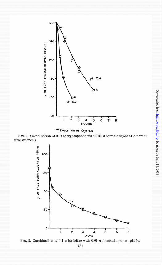
The combination of tryptophane3 with formaldehyde shows a maximum The combinat’ion of tryptophane3 with formaldehyde shows a maximum at pH 5.0 (Fig. 4; Table II). at pH 5.0 (Fig. 4; Table II). The reaction proceeds at a finite rate over The reaction proceeds at a finite rate over several hours and finally results in the deposition of crystals. several hours and finally results in t’he deposition of crystals. Histidine Histidine reacts rapidly with formaldehyde at pH 7.4. reacts rapidly with formaldehyde at pH 7.4. At pH 5.0 the reaction pro- At pH 5.0 the reaction pro-
3002 3002
250- 250-
50- 50-
o- o- I 2 3 4 5 6 7 8 9 IO
PM
FIG. 3. C’ombination of amino acids with 0.01 M formaldehyde at different pII FIG. 3. C’ombination of amino acids with 0.01 M formaldehyde at different pII values 1 hour after mixing. values 1 hour after mixing.
ceeds slowly for many days (Fig. 5) until with the concentrations employed ceeds slowly for many days (Fig. 5) until with the concentrations employed the formaldehyde completely disappears from solution. the formaldehyde completely disappears from solution.
The results are consistent with and extend previous findings with serine The results are consistent with and extend previous findings with serine (16), rysteine (17), tryptophnne (IS-20), and histidine (21). (16), ryskine (17), tryptophane (IS-20), and histidine (21).
In Fig. 6 is shown the disappearance of formaldehyde from a bacterial In Fig. 6 is shown t,he disappearance of formaldehyde from a bacterial
3 We are indebted to The Dow Chemical Company, Midland, Michigan, for a gen- 3 We are indebted to The Dow Chemical Company, Midland, Michigan, for a gen- erous gift of synthetic dl-tryptophane. erous gift of synthetic dl-trypt’ophane.
by guest on June 14, 2018http://w
ww
.jbc.org/D
ownloaded from
250 -
s
%
ki g 200-
x
d
f :: 150- 3.4
ti c
b *
x IOO-
pn 5.0
50- I I I I I I , I
12345678 HOURS
* Deposition of Crystals
FIG. 4. Combination of 0.01 & tryptophane with 0.01 M formaldehyde at different time intervals.
0-L I I I I I I 1
I 2 3 4 5 6 7
DAYS
FIG. 5. Combination of 0.1 M histidine with 0.01 M formaldehyde at pfI 5.0 581
by guest on June 14, 2018http://w
ww
.jbc.org/D
ownloaded from

582 ESTIMATION OF FREE FORMALDEHYDE
filtrate as determined by this procedure. The filtrate was prepared from an enzymic digest of muscle which contained 0.4 per rent nitrogen. The conditions illustrated are typical but are not necessarily those found best suited for any partirular detoxication. The loss of formaldehyde repre- sents combination with constituents of the medium. Only an infinitesimal portion of formaldehyde can be presumed to combine with the toxin in these filtrates. In our hands the procedure has been of great vaIue in controlling the concentrations of formaldehyde in bacterial filtrates from
I 2 3 4 5
DAYS
FIG. 6. Disappearance of formaldehyde from a bacterial fiRrate
three different species in order t’o accomplish detoxication with minimum loss of antitoxin combining power.
SUMMARY
1. A procedure has been developed for the estimation of free formalde- hyde in solution. The following principle is employed. Formaldehyde vapor is allowed to diffuse from solution into a membrane containing phenylhydrazine hydrochloride. A red color is developed in the membrane by adding ferricyanide and acid and is compared with colors developed at
by guest on June 14, 2018http://w
ww
.jbc.org/D
ownloaded from

M. J. BOYD AND M. 4. LOGAN 583
the same time in the same manner from standard solutions of formaldehyde. 2: The effect of pH on the diffusion of formaldehyde from solution has
been studied. 3. The reaction of dilute formaldehyde with amino acids at different pH
values has been investigated. The results show that amino acids with SH or OH groups in the @ position combine immediat#ely to a greater extent with formaldehyde than those not) so substituted.
4. The procedure is useful for the control of formaldehyde concentration during detoxication of bacterial filtrates.
BIBLIOGRAPHY
1. Rimini, E., Ann. furmacoterap. e chim., 97, 101 (1898). 2. Schryver, S. B., Proc. Roy. Sot. London, Series B, 82, 226 (1910). 3. Wadsworth, A. B., Standard methods of the Division of Laboratories and Re-
search of the Pu’ew York State Department of Health, Baltimore, 2nd edition, 65 (1939).
4. Matsukawa, D., J. Biochem., Japan, 30, 385 (1939). 5. Boyd, M. J., and Logan, M. A., J. Biol. Chem., 146, 279 (1942). 6. Weinberger, W., Ind. and Eng. Chem., Anal. Ed., 3, 357 (1931). 7. Blair, E. W., and Ledbury, W., J. Chem. Sot., 127,26 (1925). 8. Young, E. G., and Conway, C. F., J. Biol. Chem., 142, 839 (1942). 9. Clark, W. M., The determination of hydrogen ions, Baltimore, 3rd edition, 200,
216 (1928). 10. Hibben, J. II., J. Am. Chem. Rot., 63, 2418 (1931). 11. vonHenri, V., and Schou, S. A., Z. Phys., 49, 774 (1928). 12. Auerbach, F., Arb. AT. Gsndhtsamte., 22, 584 (1905). 13. Wadano, M., Trogus, C., and Hess, I<., Ber. them. Ges., 67, 174 (1934). 14. Levy, M,, J. Biol. Chem., 106, 157 (1934). 15. Wadano, M., Ber. them. Ges., 67, 191 (1934). 16. Levene, P. A., and Schormiiller, A., J. Biol. Chem., 106, 547 (1934). 17. Ratner, S., and Clarke, II. T., J. Am. Chem. Sot., 69, 200 (1937). 18. Homer, A., Biochem. J., 7, 7 (1913). 19. Jacobs, W. A., and Craig, L. C., J. Biol. Chem., 113, 759 (1936). 20. Wadsworth, A., and Pangborn, M. C., J. Biol. Chem., 116.423 (1936). 21. Holden, F. II., and Freeman, M., Australian J. Exp. Biol. and Med. SC., 8, 189
(1931).
by guest on June 14, 2018http://w
ww
.jbc.org/D
ownloaded from

M. John Boyd and Milan A. LoganFORMALDEHYDE BY DIFFUSION
THE ESTIMATION OF FREE
1945, 160:571-583.J. Biol. Chem.
http://www.jbc.org/content/160/2/571.citation
Access the most updated version of this article at
Alerts:
When a correction for this article is posted•
When this article is cited•
to choose from all of JBC's e-mail alertsClick here
ml#ref-list-1
http://www.jbc.org/content/160/2/571.citation.full.htaccessed free atThis article cites 0 references, 0 of which can be
by guest on June 14, 2018http://w
ww
.jbc.org/D
ownloaded from