the emerin-binding transcription factor lmo7 is regulated by
TRANSCRIPT
Submitted 14 April 2013Accepted 29 July 2013Published 20 August 2013
Corresponding authorKatherine L. Wilson,[email protected]
Academic editorTokuko Haraguchi
Additional Information andDeclarations can be found onpage 14
DOI 10.7717/peerj.134
Copyright2013 Wozniak et al.
Distributed underCreative Commons CC-BY 3.0
OPEN ACCESS
The emerin-binding transcription factorLmo7 is regulated by association withp130Cas at focal adhesionsMichele A. Wozniak1,2, Brendon M. Baker2, Christopher S. Chen2 andKatherine L. Wilson1
1 Department of Cell Biology, Johns Hopkins University School of Medicine, Baltimore,MD, USA
2 Department of Bioengineering, University of Pennsylvania, Philadelphia, PA, USA
ABSTRACTLoss of function mutations in the nuclear inner membrane protein, emerin, causeX-linked Emery-Dreifuss muscular dystrophy (X-EDMD). X-EDMD is character-ized by contractures of major tendons, skeletal muscle weakening and wasting, andcardiac conduction system defects. The transcription factor Lmo7 regulates muscle-and heart-relevant genes and is inhibited by binding to emerin, suggesting Lmo7misregulation contributes to EDMD disease. Lmo7 associates with cell adhesionsand shuttles between the plasma membrane and nucleus, but the regulation andbiological consequences of this dual localization were unknown. We report endoge-nous Lmo7 also associates with focal adhesions in cells, and both co-localizes andco-immunoprecipitates with p130Cas, a key signaling component of focal adhesions.Lmo7 nuclear localization and transcriptional activity increased significantly inp130Cas-null MEFs, suggesting Lmo7 is negatively regulated by p130Cas-dependentassociation with focal adhesions. These results support EDMD models in whichLmo7 is a downstream mediator of integrin-dependent signaling that allows tendoncells and muscles to adapt to and withstand mechanical stress.
Subjects Biochemistry, Cell Biology, Molecular BiologyKeywords Lmo7, p130Cas, Focal adhesions, Emery-Dreifuss muscular dystrophy, Laminopathy,LEM-domain, Nuclear envelope, Nucleoskeleton, Tendon, Emerin
INTRODUCTIONLim domain only 7 (Lmo7) is a transcription factor with major roles in muscle, heart
and other tissues (Ott et al., 2008) including lung epithelium, where Lmo7 is proposed
to function as a tumor suppressor (Ott et al., 2008; Tanaka-Okamoto et al., 2009). Lmo7
regulates breast cancer cell migration by acting synergistically with the small GTPase RhoA
to reduce G:F-actin ratios, leading to the activation of myocardin-related transcription
factor (MRTF; also known as MAL or MKL1), a serum response factor (SRF) cofactor
that activates cytoskeletal genes (Hu et al., 2011; Ho et al., 2013). Lmo7 shuttles into
and out of the nucleus (Holaska, Rais-Bahrami & Wilson, 2006), and positively regulates
many muscle- and heart-relevant genes (Holaska, Rais-Bahrami & Wilson, 2006; Ott et
al., 2008). Among these genes is EMD which encodes a conserved nuclear membrane
protein named emerin (Berk, Tifft & Wilson, 2013). Emerin, a LEM-domain protein,
How to cite this article Wozniak et al. (2013), The emerin-binding transcription factor Lmo7 is regulated by association with p130Cas atfocal adhesions. PeerJ 1:e134; DOI 10.7717/peerj.134
directly binds to membrane components of ‘LINC’ complexes (SUN-domain proteins,
nesprins; Mislow et al., 2002; Zhang et al., 2005; Haque et al., 2010) and to the nucleoskeletal
proteins lamin A and actin (Lee et al., 2001; Holaska et al., 2003; Holaska, Kowalski &
Wilson, 2004). However emerin also directly binds transcription regulators including
β-catenin (Markiewicz et al., 2006) and—notably—Lmo7 itself (Holaska, Rais-Bahrami
& Wilson, 2006). In emerin-downregulated cells, Lmo7 nuclear localization is decreased
or undetectable (Holaska, Rais-Bahrami & Wilson, 2006). In cells subjected to external
mechanical force, emerin is required to activate specific ‘mechano-sensitive’ genes in
response to force (Lammerding et al., 2005).
Loss of emerin, mutations in emerin-associated proteins (A-type lamins, nesprin-1,
nesprin-2, LUMA) or mutations in transcription factor FHL1 are all genetically linked to
Emery-Dreifuss muscular dystrophy (EDMD) (Meinke, Nguyen & Wehnert, 2011). These
‘EDMD genes’ suggest proper functioning of the affected tissues (heart, cardiac conduction
system, specific skeletal muscles, major tendons) requires an emerin-containing multi-
protein complex at the nuclear envelope (Simon & Wilson, 2011). Lmo7 is required for
heart development in zebrafish, including development of the cardiac conduction system
(Ott et al., 2008). The transcription-activator role of Lmo7 is inhibited by binding emerin,
suggesting the emerin protein negatively feedback-regulates Lmo7 activity in the nucleus
(Holaska, Rais-Bahrami & Wilson, 2006; Dedeic et al., 2011).
As a binding partner for emerin, Lmo7 was of particular interest because it localizes
at cell–cell adhesions (Ooshio et al., 2004; Yamada et al., 2004), and might therefore
transmit adhesion signals to the nucleus. Previous work suggested Lmo7 might also
localize at focal adhesions, since a polypeptide comprising the C-terminal half of human
Lmo7 (‘hLmo7C’; residues 888–1683) was detected both at the nuclear envelope and
cell surface, where it co-localized with paxillin, when expressed transiently in HeLa cells
(Holaska, Rais-Bahrami & Wilson, 2006). We report that endogenous Lmo7 associated
with focal adhesions in both HeLa cells and mouse embryonic fibroblasts (MEFs), and
co-immunoprecipitated with p130Cas, a major scaffolding and signaling component
of focal adhesions (Defilippi, Di Stefano & Cabodi, 2006) that also influences myogenic
differentiation (Kawauchi et al., 2012). The nucleocytoplasmic distribution of Lmo7,
and the expression of six (of nine tested) Lmo7-regulated genes, were significantly
altered in p130Cas-null MEFs. These results suggest Lmo7 activity is regulated by
p130Cas-dependent association with focal adhesions. These findings are discussed in
the light of a Drosophila study that showed A-type lamins exert their critical function
in tendon cells (Uchino et al., 2013), which connect to muscle cells via the extracellular
matrix, and evidence that integrin-dependent signaling is important for cells to respond to
and withstand mechanical stress (Pines et al., 2012).
MATERIALS AND METHODSCell culture and transfectionsHeLa cells, wildtype MEFs and p130Cas-/- MEFs were maintained in 10% FBS in
DMEM. HeLa cells were transiently transfected to express GFP (eGFP-C2; Clontech),
Wozniak et al. (2013), PeerJ, DOI 10.7717/peerj.134 2/18
GFP-rLmo7a, pcDNA3.1 myc, or myc-p130Cas using Lipofectamine PLUS (Invitrogen)
per manufacturer instructions. The GFP-rLmo7a construct was a gift from Y. Takai (Osaka
University). HeLa cells were obtained from C. Machamer (Johns Hopkins School of
Medicine). Wildtype and p130Cas-/- MEFs and Myc constructs were gifts from P. Keely
(University of Wisconsin-Madison). Mammalian cells were used under Johns Hopkins
University Institutional Review Board approval (#B0807070104).
Indirect immunofluorescence and microscopyTo stain focal adhesions, cells were plated on fibronectin-coated coverslips for 30 min,
two hours or six hours, then fixed (3.7% formaldehyde, 15 min), quenched (0.15 M
glycine, 10 min), made permeable (0.2% Triton X-100; 10 min), blocked (1% bovine
serum albumin in PBS; 1 h, 22–25◦C) and incubated with primary antibodies (30 min,
22–25◦C) specific for Lmo7 (H00004008-A01 from Novus, or HPA020923 from Sigma),
vinculin (h-VIN-1; Sigma) or pFAK (4424G; Invitrogen), each at 1:100 dilution, or
antibodies specific for p130Cas (06-500; Millipore; 1:20 dilution). Cells were rinsed with
PBS and incubated 30 min (22–25◦C) with DAPI (1:4000) plus either Alexa Fluor488-
or Alexa Fluor555-conjugated anti-mouse or anti-rabbit secondary antibodies (1:200)
from Invitrogen. Coverslips were rinsed in PBS and mounted on glass slides in PBS. For
conventional epifluorescence imaging, coverslips were mounted in PBS with ProLong Gold
Antifade reagent (Molecular Probes/Life Technologies, Grand Island, NY), and images
were acquired using a Nikon Eclipse E600 microscope equipped with a Q-imaging Retiga
EXi CCD camera and IPLab v3.9 software, or a Nikon TE200 microscope equipped with a
Spot CCD camera and Spot software (Diagnostic Instruments). Total internal reflectance
fluorescence (TIRF) microscopy was performed using a Nikon Eclipse Ti equipped with a
CFI Apo TIRF 60x oil (N.A. 1.49) objective and Evolve EMCCD camera (Photometrics). To
highlight colocalization, correlation images were created using a custom Matlab script: the
intensities of two corresponding image channels at each pixel location were multiplied, and
the resulting image was rescaled to its minimum and maximum values.
Generation of GST-fused domains of rLmo7aThe cDNA encoding each rLmo7a domain was amplified from a FLAG-rLmo7a construct
(Ooshio et al., 2004; a gift from Y. Takai [Osaka University]) using the following primers,
which included BamH1 and Xho1 restriction sites: CH domain primers were GGATCC-
GAGGCTCAGAGATGGGTGGAG and CTCGAGTTGTGCTTTTCTTCCCAGCCAGTA;
F-Box primers were GGATCCCTACCTCCAGAAATCCAAGCGAAATTTCTC and
CTCGAGAGTCAACATGTCGTCTTTCTTCAGTCG; PDZ domain primers were GGATC-
CCCCGGGACCAAACATGACTTTGG and CTCGAGTCCGTAGCGCCTGACATCC;
and the LIM domain primers were GGATCCGTGTGCTCCTACTGTAATAGCATT and
CTCGAGAGATTTGAATCGGAGATAGCAGTC. The resulting PCR products were ligated
into the pGEM-T Easy vector (Promega) per manufacturer instructions. Ligation products
were transformed into E. coli, and plasmids were purified. The pGEM-T-CH, -FBox, -PDZ
or -LIM constructs were then excised by restriction with BamH1 and Xho1, ligated into the
Wozniak et al. (2013), PeerJ, DOI 10.7717/peerj.134 3/18
BamH1/Xho1-restricted pGEX 4T-3 vector for fusion to GST, and transformed into E. coli.
Positive clones were identified by restriction analysis and verified by DNA sequencing.
Purification of GST-fused proteinsTransfected bacteria treated four hours with isopropyl β-D-1-thiogalactopyranoside
(IPTG) were pelleted, resuspended in Tris-buffered saline (TBS) and sonicated. Triton
X-100 was added to 1% (v/v) and lysates were rotated (15 min, 4◦C). After centrifugation
(12,000 rpm, 15 min, 4◦C) we added 75 µl glutathione sepharose (Sigma) to the
supernatant, then rotated (1 h, 4◦C), briefly centrifuged to collect the beads, and washed
three times in TBS.
ImmunoprecipitationsCells that transiently expressed GFP- or myc-tagged constructs for two days were lysed
in Triple Detergent Lysis Buffer (50 mM Tris-Cl pH 8, 150 mM NaCl, 0.1% SDS, 1%
NP40, 1% Triton X-100, Roche protease inhibitor complete mini tablet), and cleared by
centrifugation. To immunoprecipitate GFP, lysates were incubated with 15 µl Protein A
sepharose plus 2.5 µl GFP antibody (A6455, Molecular Probes) and rotated overnight
(4◦C). To immunoprecipitate Myc, cell lysates were incubated with 10 µl GammaBind G
sepharose (Amersham Biosciences) plus 4 µg myc antibody (9E10, Santa Cruz) and rotated
two hours (4◦C). Bound proteins were collected by centrifugation, washed three times with
Triple Detergent Lysis Buffer and eluted with SDS sample buffer. Proteins were resolved
by SDS-PAGE, transferred to PVDF membrane and probed with antibodies against Lmo7
(CO5 or NO2, gifts from Y. Takai [Osaka University]; 1:5000), p130Cas (clone 21, BD
Transduction Laboratories; 1:1000), talin (05-385, Millipore; 1:1000), emerin (serum 2999;
Lee et al., 2001; 1:5000) or Myc (A-14, Santa Cruz; 1:1000). HRP-conjugated secondary
antibodies (Jackson ImmunoResearch Laboratories; 1:5000) were detected by horseradish
peroxidase chemiluminescence (Amersham).
GST pulldownsHeLa cells were lysed in GST Lysis Buffer (100 mM HEPES pH 7.4, 150 mM NaCl, 2 mM
EDTA, 0.1% SDS, 1% Triton X-100, 1 mM DTT, Roche protease inhibitor complete mini
tablet), and cleared by centrifugation (12,000 g, 12 min, 4◦C). The supernatant (lysate) was
incubated (1.5 h, 4◦C) with 15 µg recombinant GST, GST-CH, GST-F-Box, GST-PDZ or
GST-LIM (see above). Beads were washed three times (GST Lysis Buffer); proteins were
eluted with SDS sample buffer, resolved by SDS-PAGE, transferred to PVDF and probed
with antibodies to GST (Santa Cruz Biotechnology, 1:1000), p130Cas (see above), or
paxillin (BD Transduction Laboratories, 1:1000) as described above.
Cell fractionationCells were plated on fibronectin-coated petri dishes overnight, then fractionated using the
NE-PER Kit (Thermo Scientific) per manufacturer instructions. Equal protein amounts
(10 µg) of each fraction were resolved by SDS-PAGE and immunoblotted with antibodies
Wozniak et al. (2013), PeerJ, DOI 10.7717/peerj.134 4/18

to Lmo7 (Novus, 1:1000), β-tubulin (Sigma, 1:1000) or lamins A/C (Novacastra, 1:1000)
as described above.
Quantitative real-time PCRMEFs were rinsed with PBS and total RNA was extracted using the RNeasy mini kit
(Qiagen). RNA (0.5 µg) was reverse transcribed using the high-capacity cDNA reverse
transcription kit (Applied Biosystems) and the resulting cDNAs were amplified in
an ABI 7300 system (Applied Biosystems). Results were analyzed using the ddCT
method and normalized to 18S. Primers used were: 18s, GTAACCCGTTGAACCCCATT
and CCATCCAATCGGTAGTAGCG; mef2D, CCTCAACAGTGCTAATGGAGCC and
CCAAGTATCCAGCCGCATCC; Rbl1, GAATGCCTCTTGGATCTTTCC and GT-
GAACTTTCGAGGTGTTCCA; mef2C, ATGGGGAGAAAAAAGATTCAGATTACG and
GCATGCGACTCTCTGAAGGATGGGC; Id2, ATGAAAGCCTTCAGTCCGGTGAGG
and GCAAAGTACTCTGTGGCTAA; crebbp, TCCAGGGCGAGAATGTGACC and
CCCTGTGCAGTCTCCACGGC; pcaf, AGCTGAACCCTCAGATCCCA and CACTTGT-
CAATCAACCCTGC; mbnl, ATGGCTGTTAGTGTCACACC and CTACATCTGGGTAA-
CATACTTGTGGC; mef2B, ATGGGGAGGAAAAAAATCCAGATCTCAC and ACCGA-
CATTGCGGGGGCCACGG; emerin, GTTTGCCTGCAATGGTACTGT and CAAGCACT-
TAAACCCATGAGC.
RESULTSTo test potential focal adhesion localization of endogenous Lmo7, HeLa cells were
cultured two hours on fibronectin-coated coverslips to allow focal adhesions to form, then
double-stained by indirect immunofluorescence using antibodies specific for either Lmo7,
vinculin or activated (Y397-phosphorylated) focal adhesion kinase (‘pFAK’). Because
nuclear signals (typically∼0.2 µm from the cell surface) are not reliably detected by TIRF
microscopy, cells were imaged using either epifluorescence microscopy (Fig. 1A) or TIRF
microscopy (Fig. 1B). Colocalization signals were further visualized using heat maps
generated by multiplying intensities across fluorescent channels (Fig. 1, ‘cross-correlation’;
see Methods). Lmo7 was detected in the nucleus (Fig. 1A) as expected. Lmo7 also localized
at pFAK- and vinculin-positive focal adhesion sites (Figs. 1A and 1B), and at structures
near the cell surface that resembled actin stress fibers (Fig. 1B). To determine if Lmo7
focal adhesion localization changed over time, we also used TIRF microscopy to image
HeLa cells cultured on fibronectin-coated coverslips for thirty minutes or six hours
(Fig. 1C). Lmo7 focal adhesion localization signals were highest at thirty minutes, when
Lmo7 colocalized strongly with vinculin (MA Wozniak, unpublished data) and with pFAK
at focal adhesion puncta located distal to the cell edge (Fig. 1C). Lmo7 did not co-localize
perfectly with either pFAK or vinculin, consistent with many other focal adhesion proteins
(Kanchanawong et al., 2010). These results supported the hypothesis that endogenous
Lmo7 can localize at focal adhesions.
Wozniak et al. (2013), PeerJ, DOI 10.7717/peerj.134 5/18
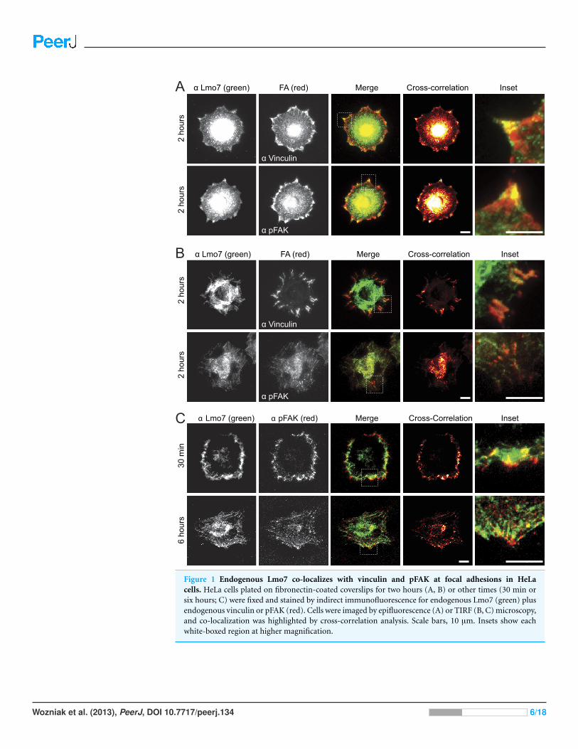
Figure 1 Endogenous Lmo7 co-localizes with vinculin and pFAK at focal adhesions in HeLacells. HeLa cells plated on fibronectin-coated coverslips for two hours (A, B) or other times (30 min orsix hours; C) were fixed and stained by indirect immunofluorescence for endogenous Lmo7 (green) plusendogenous vinculin or pFAK (red). Cells were imaged by epifluorescence (A) or TIRF (B, C) microscopy,and co-localization was highlighted by cross-correlation analysis. Scale bars, 10 µm. Insets show eachwhite-boxed region at higher magnification.
Wozniak et al. (2013), PeerJ, DOI 10.7717/peerj.134 6/18
GFP-Lmo7a association with candidate focal adhesion proteinsThe full Lmo7 polypeptide includes four homology domains (CH, F-box, PDZ and LIM
domains; Fig. 2A) (Ooshio et al., 2004), some of which were candidate mediators of
binding to focal adhesions. For example, LIM domains in other proteins can form homo-
or hetero-dimers (Dawid, Breen & Toyama, 1998), and several resident focal adhesion
proteins either have a LIM domain (e.g., paxillin (Turner & Miller, 1994) and zyxin (Sadler
et al., 1992)), or bind to LIM-domain proteins (e.g., talin, which binds the LIM-domain
of muscle protein NRAP (Luo, Herrera & Horowits, 1999), and p130Cas, which binds the
LIM-domain of zyxin (Yi et al., 2002)). We tested potential Lmo7 association with three
candidate endogenous focal adhesion proteins—talin, paxillin, and p130Cas—in HeLa
cells that transiently expressed either GFP or GFP-fused full-length rat Lmo7 splicing
variant a (GFP-rLmo7a; Fig. 2A), which is 71.8% identical to human Lmo7. Whole cell
protein lysates were immunoprecipitated using GFP antibodies, resolved by SDS-PAGE
and immunoblotted with antibodies specific for endogenous talin, paxillin or p130Cas.
The endogenous proteins were each detected in input lysates (“I”; Fig. 2B; 5% loaded; talin
not shown), and did not co-immunoprecipitate with GFP alone (Fig. 2B). GFP-rLmo7a
showed no detectable association with talin (unpublished observations); however it
co-immunoprecipitated weakly with paxillin and robustly with endogenous p130Cas
(Fig. 2B, “P”; 80% loaded; n= 4), suggesting Lmo7 associates with p130Cas in vivo.
Paxillin and p130Cas association with specific Lmo7 domainsTo independently test candidate interactors, and potentially map binding region(s)
within Lmo7, we fused GST to the N-terminus of the predicted CH, F-Box, PDZ or
LIM domains of Lmo7 as depicted in Fig. 2A. Each purified recombinant polypeptide
(Fig. 2C) was incubated with HeLa cell protein lysates, and glutathione-bound proteins
were eluted with SDS-sample buffer and resolved in duplicate SDS-PAGE gels, which
were either coomassie-stained (Fig. 2D) or immunoblotted for either endogenous
p130Cas (130 kD; Fig. 2E) or paxillin (∼68 kDa; Fig. 2F). Qualitative inspection of
coomassie-stained gels showed large amounts of each GST-fused ‘bait’, a low amount of
each corresponding GST-dimer band (Fig. 2D, black squares), and additional unidentified
bands; these included bands consistent with p130Cas (e.g., Fig. 2D, PDZ lane) and
paxillin (e.g., Fig. 2D, F-box lane). Three different regions of Lmo7 (GST-CH, GST-PDZ,
and GST-LIM) each consistently retained endogenous p130Cas (Fig. 2E, n = 3), but
one—GST-PDZ—consistently retained the highest p130Cas signals. Paxillin was retained
weakly by GST-LIM, and at high levels by GST-F-box (Fig. 2F; n = 3). These assays were
qualitative, and did not distinguish between direct versus indirect binding to each Lmo7
fragment. Nevertheless, specific retention of paxillin by the F-box of Lmo7, and retention
of p130Cas by three other domains (CH, PDZ, LIM), suggested that Lmo7 association with
focal adhesions is mediated by association (direct or indirect) with paxillin and p130Cas.
Further studies focused on p130Cas because it is a major focal adhesion scaffolding protein
(Defilippi, Di Stefano & Cabodi, 2006) that binds zyxin, which (like Lmo7) shuttles to the
nucleus (Nix et al., 2001).
Wozniak et al. (2013), PeerJ, DOI 10.7717/peerj.134 7/18

Figure 2 Lmo7 association with focal adhesion protein p130Cas. (A) Schematic of the rat Lmo7apolypeptide and GFP- or GST-fused constructs used in this study. CH, predicted Calponin Homologydomain; F-BOX, predicted F-box domain; PDZ, predicted PSD95/Dlg1/Zo-1 domain; NLS, predictednuclear localization signal; LIM, LIM-domain. Boxes above rLmo7a indicate regions sufficient for directbinding to α-actinin (Ooshio et al., 2004), afadin (Ooshio et al., 2004) or emerin (Holaska, Rais-Bahrami &Wilson, 2006). (B) Whole cell protein lysates from HeLa cells that expressed GFP or GFP-rLmo7a for twodays were immunoprecipitated with GFP antibodies, resolved by SDS-PAGE and immunoblotted withantibodies specific for paxillin or p130Cas. I, input (5% loaded); P, pellet (80% loaded). (C) Purifiedrecombinant GST-fused Lmo7 polypeptides, resolved by SDS-PAGE and stained with Coomassie. (D–E)GST-pulldowns from whole HeLa cell lysates. Each GST-fused Lmo7 polypeptide (GST-CH, GST-F-box,GST-PDZ, GST-LIM), or GST alone, was incubated with HeLa cell lysates, then bound to glutathione,washed, eluted and resolved by SDS-PAGE. Bound proteins were detected in separate gels that were eitherstained with Coomassie (D), or immunoblotted for p130Cas (E) or paxillin (F). The black boxes in (D)indicate GST-fused proteins that migrated as SDS-resistant dimers.
Endogenous Lmo7 associates with p130Cas-myc and colocalizeswith endogenous p130Cas in vivoLysates from HeLa cells that transiently expressed the empty Myc vector, or C-terminally
Myc-tagged p130Cas (Cary et al., 1998), were precipitated using Myc antibodies and
immunoblotted for endogenous Lmo7 (Fig. 3A). A large (∼200 kD) endogenous Lmo7
isoform co-immunoprecipitated consistently with Myc-p130Cas (Fig. 3A; n = 3).
Furthermore, in both HeLa cells (n = 2) and MEFs (n = 3) plated on fibronectin-coated
coverslips for two hours, indirect immunofluorescence double-staining showed a subset
of endogenous Lmo7 co-localized with endogenous p130Cas in discrete puncta at focal
adhesions, as visualized by epifluorescence microscopy (Fig. 3B) and TIRF imaging
(Fig. 3C). We concluded Lmo7 associates with p130Cas at focal adhesions.
Wozniak et al. (2013), PeerJ, DOI 10.7717/peerj.134 8/18
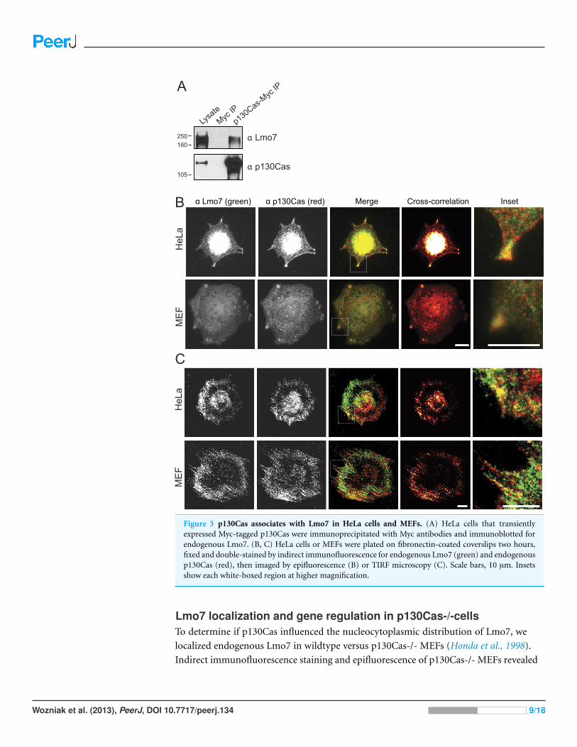
Figure 3 p130Cas associates with Lmo7 in HeLa cells and MEFs. (A) HeLa cells that transientlyexpressed Myc-tagged p130Cas were immunoprecipitated with Myc antibodies and immunoblotted forendogenous Lmo7. (B, C) HeLa cells or MEFs were plated on fibronectin-coated coverslips two hours,fixed and double-stained by indirect immunofluorescence for endogenous Lmo7 (green) and endogenousp130Cas (red), then imaged by epifluorescence (B) or TIRF microscopy (C). Scale bars, 10 µm. Insetsshow each white-boxed region at higher magnification.
Lmo7 localization and gene regulation in p130Cas-/-cellsTo determine if p130Cas influenced the nucleocytoplasmic distribution of Lmo7, we
localized endogenous Lmo7 in wildtype versus p130Cas-/- MEFs (Honda et al., 1998).
Indirect immunofluorescence staining and epifluorescence of p130Cas-/- MEFs revealed
Wozniak et al. (2013), PeerJ, DOI 10.7717/peerj.134 9/18

little or no detectable Lmo7 at the cell surface, and substantially higher nuclear signals,
compared to wildtype MEF controls (Fig. 4A; n = 3). There were greatly reduced, but
detectable, signals for endogenous activated (Y397-phosphorylated) FAK (‘pFAK’) at the
cell surface and cytoplasm (Fig. 4A, α-pFAK). This suggested p130Cas was important, but
not essential, for pFAK localization. The altered subcellular distribution of Lmo7 observed
by epifluorescence was independently verified by cell fractionation and immunoblotting.
Wildtype and p130Cas-/- MEFs were fractionated to separate nuclei from cytoplasm, and
protein lysates were resolved by SDS-PAGE and probed with antibodies specific for Lmo7,
A-type lamins (nuclear marker) or β-tubulin (cytoplasmic marker; Fig. 4B, n = 3). The
nuclear and cytoplasmic markers were each enriched in the appropriate fraction (Fig. 4B).
Densitometry and quantification of the nuclear-to-cytoplasmic signal ratio for Lmo7,
relative to wildtype controls, confirmed the predominantly nuclear distribution of Lmo7 in
p130Cas-/- cells (Fig. 4C). The difference in signal ratios was significant (p < 0.05 by the
paired t-test; n = 3). We concluded that p130Cas regulates the subcellular distribution of
Lmo7, and might normally retain Lmo7 outside the nucleus.
p130Cas influences Lmo7-dependent gene regulationTo determine if p130Cas-dependent Lmo7 localization was biologically relevant to genes
regulated by Lmo7, we used quantitative real-time PCR to measure the mRNA levels of two
genes (encoding Mef2D and Rbl1) that are negatively regulated by Lmo7, and seven genes
(encoding Id2, Crebbp, PCAF, Mbnl, Mef2B, Mef2C, Emerin) positively regulated by Lmo7
(Holaska, Rais-Bahrami & Wilson, 2006). Relative to wildtype MEF controls, the mRNA
levels of five Lmo7-activated genes (Id2, Crebbp, Pcaf, Mbnl and Mef2B) were significantly
higher in p130Cas-/- MEFS (Fig. 4D; n = 4; p < 0.05 and Fig. S1). The magnitude of this
increase ranged from 40% (Crebbp) to 70% (Pcaf, Mef2B) to 350%–400% (Id2, Mbnl).
Of the Lmo7-inhibited genes, mRNA levels of one (Rbl1) were unaffected, whereas the
other (Mef2D) decreased significantly (by 70%) relative to wildtype MEFs (Fig. 4D; n= 4;
p< 0.05 by paired t-test and Fig. S1). Thus, six of nine tested genes responded in a manner
consistent with higher Lmo7 activity in the nucleus of p130Cas-/- MEFs. These results
demonstrated p130Cas is biologically relevant to the nuclear activity of Lmo7.
DISCUSSIONFocal adhesion signaling regulates gene expression through mechanisms that remain
unclear. Some focal adhesion proteins activate MAP kinase signaling and downstream gene
expression (Howe, Aplin & Juliano, 2002). By contrast, other focal adhesion components
transmit signals by directly translocating to the nucleus. These ‘direct translocators’
include zyxin, paxillin, Crp, FHL3 and Abl, all of which have a LIM domain(s) (Hervy,
Hoffman & Beckerle, 2006), and most of which influence transcription (Krcmery et al.,
2010). Some including nTrip6, paxillin and Hic5 are transcriptional co-activators. Nuclear
Abl, a nonreceptor tyrosine kinase, has many roles in the nucleus (Hervy, Hoffman &
Beckerle, 2006) and can also phosphorylate emerin directly in vitro (Tifft, Bradbury &
Wilson, 2009). Our finding that endogenous Lmo7 localizes at focal adhesions, and is
Wozniak et al. (2013), PeerJ, DOI 10.7717/peerj.134 10/18
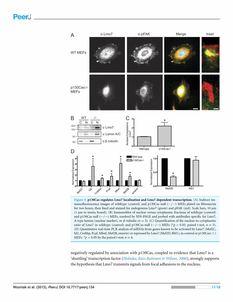
Figure 4 p130Cas regulates Lmo7 localization and Lmo7-dependent transcription. (A) Indirect im-munofluorescence images of wildtype (control) and p130Cas null (−/−) MEFs plated on fibronectinfor two hours, then fixed and stained for endogenous Lmo7 (green) and pFAK (red). Scale bars, 10 µm(1 µm in insets; boxed). (B) Immunoblot of nuclear versus cytoplasmic fractions of wildtype (control)and p130Cas null (−/−) MEFs, resolved by SDS-PAGE and probed with antibodies specific for Lmo7,A-type lamins (nuclear marker), or β-tubulin (n= 3). (C) Quantification of the nuclear-to-cytoplasmicratio of Lmo7 in wildtype (control) and p130Cas-null (−/−) MEFs (*p < 0.05, paired t-test, n = 3).(D) Quantitative real-time PCR analysis of mRNAs from genes known to be activated by Lmo7 (Mef2C,Id2, Crebbp, Pcaf, Mbnl, Mef2B, emerin) or repressed by Lmo7 (Mef2D, Rbl1), in control or p130Cas(-/-)MEFs. *p< 0.05 by the paired t-test; n= 4.
negatively regulated by association with p130Cas, coupled to evidence that Lmo7 is a
‘shuttling’ transcription factor (Holaska, Rais-Bahrami & Wilson, 2006), strongly supports
the hypothesis that Lmo7 transmits signals from focal adhesions to the nucleus.
Wozniak et al. (2013), PeerJ, DOI 10.7717/peerj.134 11/18
Lmo7 might resemble other transcription-regulating PDZ-LIM proteins, for which
sequestration in the cytoplasm is important to ‘fine-tune’ cell- and tissue-specific
activity in neurons (Kurooka & Yokota, 2005; Lasorella & Iavarone, 2006) and the heart
(Camarata et al., 2010; Krcmery et al., 2010). Since most Lmo7 distributes throughout
the cytoplasm and can also associate with cell adhesions, our findings suggest Lmo7 is
dynamically regulated by a p130Cas-scaffolded focal adhesion kinase(s) or other signaling
component(s) as it ‘cycles’ on and off focal adhesions.
Epifluorescence images (Fig. 3B) suggested p130Cas also localizes in the nucleus.
However this localization is debated. For example GFP-p130Cas localizes at high levels
in the nucleus (Kim et al., 2004), and polyclonal antibodies raised against GST-fused
p130Cas residues 318–486 or 670–896 also stain the nucleus (Harte et al., 1996).
Monoclonal antibody 4F4 detected nuclear signals in primary chicken embryonic cells;
after Src-mediated transformation this signal localized at the cell surface (Kanner et
al., 1991). Two studies suggest nuclear-localized p130Cas is less phosphorylated, hence
potentially inactive (Kanner et al., 1991; Petch et al., 1995). However other antibodies do
not detect p130Cas in the nucleus (e.g., Harte et al., 1996). Thus the nuclear localization
and potential function of nuclear p130Cas are open questions.
How might p130Cas-dependent focal adhesion regulation of Lmo7 relate to Emery-
Dreifuss muscular dystrophy (EDMD)? The major clinical aspect of EDMD is its affect
on the heart (cardiomyopathy with potentially lethal cardiac conduction system defects),
with major tendons and a subset of skeletal muscles also affected (Emery, 1987). Lmo7 is
required for mouse C2C12 myoblasts to differentiate (Dedeic et al., 2011). Lmo7 activates
muscle-specific genes such as myoD and pax3 early in differentiation; later (after myotube
formation) Lmo7 localizes predominantly in the cytoplasm (Dedeic et al., 2011). Because
sustained activation of pax3 inhibits myogenic differentiation (Boutet et al., 2007), we
speculate that Lmo7 localization during muscle differentiation might be ‘fine-tuned’ by
focal adhesion/p130Cas-dependent sequestration in the cytoplasm.
Our study did not reveal which domain(s) of p130Cas associate (directly or indirectly)
with Lmo7, or, more importantly, how their association is regulated. However, our
findings are consistent with a recent focal adhesion proteome study that reported myosin
II-dependent recruitment of Lmo7 to focal adhesions in human foreskin fibroblasts
(Kuo et al., 2011). Since focal adhesions grow and integrin-cytoskeleton connections
are strengthened in response to myosin-mediated contractility and mechanical force
(Chrzanowska-Wodnicka & Burridge, 1996; Choquet, Felsenfeld & Sheetz, 1997), we
propose Lmo7 is involved in mechanical force-induced signaling. Supporting this idea,
cell migration—which is strongly regulated by mechanically-induced signals (Lo et al.,
2000)—is disrupted by loss of emerin (Emerson et al., 2009), p130Cas (Honda et al., 1998)
or Lmo7 (Hu et al., 2011).
The proposed mechanotransduction function of Lmo7 might, we speculate, provide
physiological feedback regulation of muscle contraction. Interestingly, p130Cas is one of
several proteins that undergo physical extension in response to mechanical force (Sawada
et al., 2006; Johnson et al., 2007; Grashoff et al., 2010). In fibroblasts, mechanical stretch is
Wozniak et al. (2013), PeerJ, DOI 10.7717/peerj.134 12/18
proposed to extend the substrate domain of p130Cas to expose fifteen YXXP motifs for
phosphorylation by Src family kinases (Sawada et al., 2006). Tyrosine phosphorylation
recruits signaling proteins such as Crk (Sakai et al., 1994), Nck (Schlaepfer, Broome &
Hunter, 1997), and PTP-PEST (Garton et al., 1997), which then trigger downstream
pathways involved in cell adhesion, migration and proliferation. Whether Lmo7 is
influenced by stretch-induced phosphorylation of p130Cas is a new question raised by
our findings.
p130Cas-dependent regulation of Lmo7 may be relevant to EDMD heart defects, since
both proteins are important in the heart. In zebrafish, Lmo7 knockout leads to cardiac
conduction system defects, including arrhythmia (Ott et al., 2008). In mice, p130Cas
knockout is lethal during embryogenesis, with defects in cardiovascular development;
cardiomyocytes have disorganized myofibrils and disorganized Z-disks (Honda et al.,
1998), the cell surface structures that mechanically link the sarcomeres (contractile
networks) of neighboring cardiomyocytes. Our discovery that p130Cas negatively regulates
Lmo7 suggests the cardiovascular phenotypes of p130Cas-null mice arise at least in
part from excess nuclear Lmo7 and consequent misregulation of Lmo7-dependent
genes. In our study, loss of p130Cas affected six (of nine tested) Lmo7-regulated genes
in a manner consistent with excess Lmo7 transcriptional activity in the nucleus. The
three exceptions were mRNAs encoding Mef2C and emerin (which failed to increase in
p130Cas-null MEFs) and Rbl1, which remained high in p130Cas-null MEFs. Further
work is needed to understand why these genes ‘resisted’ transcriptional control by excess
Lmo7 in MEFs. However we speculate that Lmo7 control over these ‘resistant’ genes might
require a second p130Cas-dependent focal adhesion signaling event; possibilities include
emerin phosphorylation by activated Abl or Src (Tifft, Bradbury & Wilson, 2009). Indeed
Src phosphorylates a region of emerin required to bind Lmo7 and other transcription
regulators (Tifft, Bradbury & Wilson, 2009).
The third EDMD-affected tissue, tendons, was recently studied in a Drosophila EDMD
model with mutations in the A-type lamin (lamin ‘C’). Remarkably, the critical function of
lamin C is exerted in tendon cells, not muscle, and involves the spectraplakin-dependent
stabilization of the cytoskeleton (Uchino et al., 2013). Mammalian tendons are maintained
by fibroblasts, which actively migrate and proliferate in response to injury (Arnesen &
Lawson, 2006). Bone marrow mesenchymal stem cells can be triggered to differentiate into
tendon cells (‘tenocytes’) by mechanical stretching, through a pathway that requires focal
adhesion kinase and RhoA/ROCK (Xu et al., 2012). Stretch drives both focal adhesion
growth (Shikata et al., 2005) and ROCK-mediated contractility (Xu et al., 2012), and stabi-
lizes integrin-dependent force signaling at sites of muscle-tendon attachment (Pines et al.,
2012). Hence our results identify Lmo7 as a downstream mediator of integrin-dependent
signaling, and suggest that defects in focal adhesion signaling contribute to EDMD disease,
particularly in tendons and muscle. Interestingly these results raise the possibility that a
second p130Cas-dependent signal, speculated to involve Src or Abl, is required for Lmo7 to
control a subset of genes, including those that encode emerin, Rbl1 and mef2C.
Wozniak et al. (2013), PeerJ, DOI 10.7717/peerj.134 13/18

ACKNOWLEDGEMENTSThe authors are grateful to Yoshimi Takai (Osaka University, Japan) for the rLmo7
constructs and Lmo7 antibodies, Patricia Keely (University of Wisconsin) for the wildtype
and p130Cas(-/-) MEFs and myc constructs, and the Wilson Lab for helpful discussions.
ADDITIONAL INFORMATION AND DECLARATIONS
FundingThis work was funded by the National Institutes of Health grants to MAW (postdoc-
toral fellowship NRSA F32 AR054219-01), BMB (postdoctoral fellowship NRSA F32
EB014691), CSC (RO1 GM74048 and RO1 EB00262) and KLW (RO1 GM048646). The
funders had no role in study design, data collection and analysis, decision to publish, or
preparation of the manuscript.
Grant DisclosuresThe following grant information was disclosed by the authors:
National Institutes of Health grants: postdoctoral fellowship NRSA F32 AR054219-01,
postdoctoral fellowship NRSA F32 EB014691, RO1 GM74048 and RO1 EB00262, RO1
GM048646.
Competing InterestsKatherine L. Wilson is an Academic Editor for PeerJ.
Author Contributions• Michele A. Wozniak conceived and designed the experiments, performed the experi-
ments, analyzed the data, wrote the paper.
• Brendon M. Baker conceived and designed the experiments, analyzed the data,
contributed reagents/materials/analysis tools, wrote the paper.
• Christopher S. Chen and Katherine L. Wilson conceived and designed the experiments,
contributed reagents/materials/analysis tools, wrote the paper.
EthicsThe following information was supplied relating to ethical approvals (i.e., approving body
and any reference numbers):
The Institutional Review Board is Johns Hopkins University, and the IRB approval # (for
HeLa, HEK293 and fibroblasts) is JHU IRB #B0807070104 (expires 6/30/2013).
Supplemental InformationSupplemental information for this article can be found online at http://dx.doi.org/
10.7717/peerj.134.
Wozniak et al. (2013), PeerJ, DOI 10.7717/peerj.134 14/18
REFERENCESArnesen SM, Lawson MA. 2006. Age-related changes in focal adhesions lead to altered cell
behavior in tendon fibroblasts. Mechanisms of Ageing and Development 127(9):726–732DOI 10.1016/j.mad.2006.05.003.
Berk JM, Tifft KE, Wilson KL. 2013. The nuclear envelope LEM-domain protein emerin. Nucleus4(4):1–17 DOI 10.4161/nucl.25751.
Boutet SC, Disatnik MH, Chan LS, Iori K, Rando TA. 2007. Regulation of Pax3 by proteasomaldegradation of monoubiquitinated protein in skeletal muscle progenitors. Cell 130(2):349–362DOI 10.1016/j.cell.2007.05.044.
Camarata T, Krcmery J, Snyder D, Park S, Topczewski J, Simon H-G. 2010. Pdlim7 (LMP4)regulation of Tbx5 specifies zebrafish heart atrio-ventricular boundary and valve formation.Developmental Biology 337(2):233–245 DOI 10.1016/j.ydbio.2009.10.039.
Cary LA, Han DC, Polte TR, Hanks SK, Guan JL. 1998. Identification of p130Cas as a mediator offocal adhesion kinase-promoted cell migration. Journal of Cell Biology 140(1):211–221DOI 10.1083/jcb.140.1.211.
Choquet D, Felsenfeld DP, Sheetz MP. 1997. Extracellular matrix rigidity causes strengthening ofintegrin-cytoskeleton linkages. Cell 88(1):39–48 DOI 10.1016/S0092-8674(00)81856-5.
Chrzanowska-Wodnicka M, Burridge K. 1996. Rho-stimulated contractility drives the formationof stress fibers and focal adhesions. Journal of Cell Biology 133(6):1403–1415DOI 10.1083/jcb.133.6.1403.
Dawid IB, Breen JJ, Toyama R. 1998. LIM domains: multiple roles as adapters and functionalmodifiers in protein interactions. Trends in Genetics 14(4):156–162DOI 10.1016/S0168-9525(98)01424-3.
Dedeic Z, Cetera M, Cohen TV, Holaska JM. 2011. Emerin inhibits Lmo7 binding to the Pax3 andMyoD promoters and expression of myoblast proliferation genes. Journal of Cell Science124(Pt 10):1691–1702 DOI 10.1242/jcs.080259.
Defilippi P, Di Stefano P, Cabodi S. 2006. p130Cas: a versatile scaffold in signaling networks.Trends in Cell Biology 16(5):257–263 DOI 10.1016/j.tcb.2006.03.003.
Emerson LJ, Holt MR, Wheeler MA, Wehnert M, Parsons M, Ellis JA. 2009. Defects in cellspreading and ERK1/2 activation in fibroblasts with lamin A/C mutations. Biochimica etBiophysica ACTA 1792(8):810–821 DOI 10.1016/j.bbadis.2009.05.007.
Emery AE. 1987. X-linked muscular dystrophy with early contractures and cardiomyopathy(Emery-Dreifuss type). Clinical Genetics 32(5):360–367 DOI 10.1111/j.1399-0004.1987.tb03302.x.
Garton AJ, Burnham MR, Bouton AH, Tonks NK. 1997. Association of PTP-PEST with the SH3domain of p130cas; a novel mechanism of protein tyrosine phosphatase substrate recognition.Oncogene 15(8):877–885 DOI 10.1038/sj.onc.1201279.
Grashoff C, Hoffman BD, Brenner MD, Zhou R, Parsons M, Yang MT, McLean MA, Sligar SG,Chen CS, Ha T, Schwartz MA. 2010. Measuring mechanical tension across vinculin revealsregulation of focal adhesion dynamics. Nature 466(7303):263–266 DOI 10.1038/nature09198.
Haque F, Mazzeo D, Patel JT, Smallwood DT, Ellis JA, Shanahan CM, Shackleton S. 2010.Mammalian SUN protein interaction networks at the inner nuclear membrane and theirrole in laminopathy disease processes. Journal of Biological Chemistry 285(5):3487–3498DOI 10.1074/jbc.M109.071910.
Harte MT, Hildebrand JD, Burnham MR, Bouton AH, Parsons JT. 1996. p130Cas, a substrateassociated with v-Src and v-Crk, localizes to focal adhesions and binds to focal adhesion kinase.Journal of Biological Chemistry 271(23):13649–13655 DOI 10.1074/jbc.271.23.13649.
Wozniak et al. (2013), PeerJ, DOI 10.7717/peerj.134 15/18

Hervy M, Hoffman L, Beckerle MC. 2006. From the membrane to the nucleus and backagain: bifunctional focal adhesion proteins. Current Opinion in Cell Biology 18(5):524–532DOI 10.1016/j.ceb.2006.08.006.
Ho CY, Jaalouk DE, Vartiainen MK, Lammerding J. 2013. Lamin A/C and emerinregulate MKL1-SRF activity by modulating actin dynamics. Nature 497(7450):507–511DOI 10.1038/nature12105.
Holaska JM, Kowalski AK, Wilson KL. 2004. Emerin caps the pointed end of actin filaments:evidence for an actin cortical network at the nuclear inner membrane. PLoS Biology2(9):E231 (1354–1362)DOI 10.1371/journal.pbio.0020231.
Holaska JM, Lee KK, Kowalski AK, Wilson KL. 2003. Transcriptional repressor germ cell-less(GCL) and barrier to autointegration factor (BAF) compete for binding to emerin in vitro.Journal of Biological Chemistry 278(9):6969–6975 DOI 10.1074/jbc.M208811200.
Holaska JM, Rais-Bahrami S, Wilson KL. 2006. Lmo7 is an emerin-binding protein that regulatesthe transcription of emerin and many other muscle-relevant genes. Human Molecular Genetics15(23):3459–3472 DOI 10.1093/hmg/ddl423.
Honda H, Oda H, Nakamoto T, Honda Z-I, Sakai R, Suzuki T, Saito T, Nakamura K, Nakao K,Ishikawa T, Katsuki M, Yazaki Y, Hirai H. 1998. Cardiovascular anomaly, impaired actinbundling and resistance to Src-induced transformation in mice lacking p130Cas. NatureGenetics 19(4):361–365 DOI 10.1038/1246.
Howe AK, Aplin AE, Juliano RL. 2002. Anchorage-dependent ERK signaling–mechanismsand consequences. Current Opinion in Genetics and Development 12(1):30–35DOI 10.1016/S0959-437X(01)00260-X.
Hu Q, Guo C, Li Y, Aronow BJ, Zhang J. 2011. LMO7 mediates cell-specific activation of theRho-myocardin-related transcription factor-serum response factor pathway and plays animportant role in breast cancer cell migration. Molecular and Cellular Biology 31(16):3223–3240DOI 10.1128/MCB.01365-10.
Johnson CP, Tang HY, Carag C, Speicher DW, Discher DE. 2007. Forced unfolding of proteinswithin cells. Science 317(5838):663–666 DOI 10.1126/science.1139857.
Kanchanawong P, Shtengel G, Pasapera AM, Ramko EB, Davidson MW, Hess HF,Waterman CM. 2010. Nanoscale architecture of integrin-based cell adhesions. Nature468(7323):580–584 DOI 10.1038/nature09621.
Kanner SB, Reynolds AB, Wang HC, Vines RR, Parsons JT. 1991. The SH2 and SH3 domains ofpp60src direct stable association with tyrosine phosphorylated proteins p130 and p110. EmboJournal 10(7):1689–1698.
Kawauchi K, Tan WW, Araki K, Abuı̈ Bakar FB, Kim M, Fujita H, Hirata H, Sawada Y. 2012.p130Cas-dependent actin remodelling regulates myogenic differentiation. Biochemical Journal445(3):323–332 DOI 10.1042/BJ20112169.
Kim W, Kook S, Kim DJ, Teodorof C, Song WK. 2004. The 31-kDa caspase-generated cleavageproduct of p130cas functions as a transcriptional repressor of E2A in apoptotic cells. Journal ofBiological Chemistry 279(9):8333–8342 DOI 10.1074/jbc.M312026200.
Krcmery J, Camarata T, Kulisz A, Simon H-G. 2010. Nucleocytoplasmic functions of thePDZ-LIM protein family: new insights into organ development. Bioessays 32(2):100–108DOI 10.1002/bies.200900148.
Wozniak et al. (2013), PeerJ, DOI 10.7717/peerj.134 16/18
Kuo JC, Han X, Han C-T, Hsiao JR, Yates III CM, Waterman. 2011. Analysis of themyosin-II-responsive focal adhesion proteome reveals a role for beta-Pix in negative regulationof focal adhesion maturation. Nature Cell Biology 13(4):383–393 DOI 10.1038/ncb2216.
Kurooka H, Yokota Y. 2005. Nucleo-cytoplasmic shuttling of Id2, a negative regulator of basichelix-loop-helix transcription factors. Journal of Biological Chemistry 280(6):4313–4320DOI 10.1074/jbc.M412614200.
Lammerding J, Hsiao J, Schulze PC, Kozlov S, Stewart CL, Lee RT. 2005. Abnormal nuclearshape and impaired mechanotransduction in emerin-deficient cells. Journal of Cell Biology170(5):781–791 DOI 10.1083/jcb.200502148.
Lasorella A, Iavarone A. 2006. The protein ENH is a cytoplasmic sequestration factor for Id2in normal and tumor cells from the nervous system. Proceedings of the National Academy ofSciences of the United States of America 103(13):4976–4981 DOI 10.1073/pnas.0600168103.
Lee KK, Haraguchi T, Lee RS, Koujin T, Hiraoka Y, Wilson KL. 2001. Distinct functionaldomains in emerin bind lamin A and DNA-bridging protein BAF. Journal of Cell Science 114(Pt 24):4567–4573.
Lo C-M, Wang H-B, Dembo M, Wang Yu-li. 2000. Cell movement is guided by the rigidity of thesubstrate. Biophysical Journal 79(1):144–152 DOI 10.1016/S0006-3495(00)76279-5.
Luo G, Herrera AH, Horowits R. 1999. Molecular interactions of N-RAP, a nebulin-related proteinof striated muscle myotendon junctions and intercalated disks. Biochemistry 38(19):6135–6143DOI 10.1021/bi982395t.
Markiewicz E, Tilgner K, Barker N, van de Wetering M, Clevers H, Dorobek M, Hausmanowa-Petrusewicz I, Ramaekers FCS, Broers JLV, Blankesteijn WM, Salpingidou G, Wilson RG,Ellis JA, Hutchison CJ. 2006. The inner nuclear membrane protein emerin regulatesbeta-catenin activity by restricting its accumulation in the nucleus. Embo Journal25(14):3275–3285 DOI 10.1038/sj.emboj.7601230.
Meinke P, Nguyen TD, Wehnert MS. 2011. The LINC complex and human disease. BiochemicalSociety Transactions 39(6):1693–1697 DOI 10.1042/BST20110658.
Mislow JMK, Holaska JM, Kim MS, Lee KK, Segura-Totten M, Wilson KL, McNally EM. 2002.Nesprin-1alpha self-associates and binds directly to emerin and lamin A in vitro. FEBS Letters525(1–3):135–140 DOI 10.1016/S0014-5793(02)03105-8.
Nix DA, Fradelizi J, Bockholt S, Menichi B, Louvard D, Friederich E, Beckerle MC. 2001.Targeting of zyxin to sites of actin membrane interaction and to the nucleus. Journal of BiologicalChemistry 276(37):34759–34767 DOI 10.1074/jbc.M102820200.
Ooshio T, Irie K, Morimoto K, Fukuhara A, Imai T, Takai Y. 2004. Involvement of LMO7 inthe association of two cell–cell adhesion molecules, nectin and E-cadherin, through afadinand alpha-actinin in epithelial cells. Journal of Biological Chemistry 279(30):31365–31373DOI 10.1074/jbc.M401957200.
Ott EB, van den Akker NMS, Sakalis PA, Gittenberger-de Groot AC, Te Velthuis AJW,Bagowski CP. 2008. The lim domain only protein 7 is important in zebrafish heartdevelopment. Developmental Dynamics 237(12):3940–3952 DOI 10.1002/dvdy.21807.
Petch LA, Bockholt SM, Bouton A, Parsons JT, Burridge K. 1995. Adhesion-induced tyrosinephosphorylation of the p130 src substrate. Journal of Cell Science 108(Pt 4):1371–1379.
Pines M, Das R, Ellis SJ, Morin A, Czerniecki S, Yuan L, Klose M, Coombs D, Tanentzapf G.2012. Mechanical force regulates integrin turnover in Drosophila in vivo. Nature Cell Biology14(9):935–943 DOI 10.1038/ncb2555.
Wozniak et al. (2013), PeerJ, DOI 10.7717/peerj.134 17/18

Sadler I, Crawford AW, Michelsen JW, Beckerle MC. 1992. Zyxin and cCRP: two interactive LIMdomain proteins associated with the cytoskeleton. Journal of Cell Biology 119(6):1573–1587DOI 10.1083/jcb.119.6.1573.
Sakai R, Iwamatsu A, Hirano N, Ogawa S, Tanaka T, Mano H, Yazaki Y, Hirai H. 1994. A novelsignaling molecule, p130, forms stable complexes in vivo with v-Crk and v-Src in a tyrosinephosphorylation-dependent manner. Embo Journal 13(16):3748–3756.
Sawada Y, Tamada M, Dubin-Thaler BJ, Cherniavskaya O, Sakai R, Tanaka S, Sheetz MP. 2006.Force sensing by mechanical extension of the Src family kinase substrate p130Cas. Cell127(5):1015–1026 DOI 10.1016/j.cell.2006.09.044.
Schlaepfer DD, Broome MA, Hunter T. 1997. Fibronectin-stimulated signaling from a focaladhesion kinase-c-Src complex: involvement of the Grb2, p130cas, and Nck adaptor proteins.Molecular and Cellular Biology 17(3):1702–1713.
Shikata Y, Rios A, Kawkitinarong K, Depaola N, Garcia J, Birukov K. 2005. Differential effects ofshear stress and cyclic stretch on focal adhesion remodeling, site-specific FAK phosphorylation,and small GTPases in human lung endothelial cells. Experimental Cell Research 304(1):40–49DOI 10.1016/j.yexcr.2004.11.001.
Simon DN, Wilson KL. 2011. The nucleoskeleton as a genome-associated dynamic ‘network ofnetworks’. Nature Reviews Molecular Cell Biology 12(11):695–708 DOI 10.1038/nrm3207.
Tanaka-Okamoto M, Hori K, Ishizaki H, Hosoi A, Itoh Y, Wei M, Wanibuchi H, Mizoguchi A,Nakamura H, Miyoshi J. 2009. Increased susceptibility to spontaneous lung cancer inmice lacking LIM-domain only 7. Cancer Science 100(4):608–616 DOI 10.1111/j.1349-7006.2009.01091.x.
Tifft KE, Bradbury KA, Wilson KL. 2009. Tyrosine phosphorylation of nuclear-membraneprotein emerin by Src, Abl and other kinases. Journal of Cell Science 122(Pt 20):3780–3790DOI 10.1242/jcs.048397.
Turner CE, Miller JT. 1994. Primary sequence of paxillin contains putative SH2 and SH3 domainbinding motifs and multiple LIM domains: identification of a vinculin and pp125Fak-bindingregion. Journal of Cell Science 107(Pt 6):1583–1591.
Uchino R, Nonaka YK, Horigome T, Sugiyama S, Furukawa K. 2013. Loss of DrosophilaA-type lamin C initially causes tendon abnormality including disintegration ofcytoskeleton and nuclear lamina in muscular defects. Developmental Biology 373(1):216–227DOI 10.1016/j.ydbio.2012.08.001.
Xu B, Song G, Ju Y, Li X, Song Y, Watanabe S. 2012. RhoA/ROCK, cytoskeletal dynamics, andfocal adhesion kinase are required for mechanical stretch-induced tenogenic differentiationof human mesenchymal stem cells. Journal of Cellular Physiology 227(6):2722–2729DOI 10.1002/jcp.23016.
Yamada A, Irie K, Fukuhara A, Ooshio T, Takai Y. 2004. Requirement of the actin cytoskeletonfor the association of nectins with other cell adhesion molecules at adherens and tight junctionsin MDCK cells. Genes Cells 9(9):843–855 DOI 10.1111/j.1365-2443.2004.00768.x.
Yi J, Kloeker S, Jensen CC, Bockholt S, Honda H, Hirai H, Beckerle MC. 2002. Members of theZyxin family of LIM proteins interact with members of the p130Cas family of signal transducers.Journal of Biological Chemistry 277(11):9580–9589 DOI 10.1074/jbc.M106922200.
Zhang Q, Ragnauth CD, Skepper JN, Worth NF, Warren DT, Roberts RG, Weissberg PL,Ellis JA, Shanahan CM. 2005. Nesprin-2 is a multi-isomeric protein that binds lamin andemerin at the nuclear envelope and forms a subcellular network in skeletal muscle. Journal ofCell Science 118(Pt 4):673–687 DOI 10.1242/jcs.01642.
Wozniak et al. (2013), PeerJ, DOI 10.7717/peerj.134 18/18