the egyptian journal of fertility and sterilityefss-egypt.com/pdf/publication/the egyption journal...
TRANSCRIPT

THE EGYPTIAN JOURNALOF FERTILITY AND STERILITY
Issn 1110 -6352
Volume 17 Number 1 January 2013
EFSSEDITOR : MOHAMED YEHIA

Egypt.J.Fertil.Steril. Volume17 - number 1 January 2013 i
The Egyptian Journal Of Fertility And SterilityThe Official Journal of the Egyption Fertility and Sterility Society ( EFSS)
Editor in Chief : Moamed Yehia . Ain ShamsAssistant Editor : Hosam Thabet Salem . Assiut – Ahmed Badawy . MansouraBotros Rizk : Associate Editor for North America
Editorial Board
M. Abulghar . CairoA. Assaf . BanhaA. Badawy . MansouraH. Badawy . CairoI. Fahmy . Cairo UniversityH. A. Hassan . AlexandriaA. El-Karaksy . CairoR. T. Mansour . IVF Center CairoM. Sammour . Ain ShamsG. I. Serour . Al AzherO. Shawky . CairoK. Z. Shoerir . CairoH. Sallam . AlexandiraH. T. Salem . AssiutM. Shaaban . AssiutA. El-Tagy . Al Azhar
International Advisory Board
H. Abdalla . U.K.S. Badawi . USAI. Cook . U.K.P. Devroey. BelgiumM. Fathalla . EgyptV. Gomel . USAL. Hamburger . SwedenY. Khalaf . UKB. Tarlatzis . GreeceS. Silber . USAS. l. Tan. CanadaP. Rizk . USA
President : G. I. Serour. Al AzharVice President : M. Toppozada. AlexandriaSecretary General : A. El-Shalakany. Ain ShamsTreasurer : E. Darwish. AlexandriaBoard Members : H. Thabet Salem. Assiut M. Yehia. Ain Shams I. Fahmy. Cairo M. El-Sherbini. Dammitta I. Mahrous. Al Azhar
The Egyptian Society of Fertility and Sterility:

Egypt.J.Fertil.Steril. Volume17 - number 1 January 2013ii
SUBMISSION OF PAPERSManuscripts should be written in English, typed with double spacing, submitted and, where possible, on a disk. Figures and diagrams should, if possible be used instead of tables. The work shall not be published elsewhere in any language without the written consent of the editor in chief. The articles published in this journal are protected by copy-right. Contributors should submit their papers and disk to:
Editor in chiefProf dr. Mohamed YahiaProf. ob & gynecology, Ain Shams University Email: [email protected]
Asst. Editor:Prof. Ahmed Badawy,Prof. ob & gynecology, Mansoura University.Email: [email protected]
Preparation of manuscripts
• Papers should be typed double- spaced, on white paper, size A4 (210 x 297 mm). up-per, lower, right and left margins should have a minimum of 25 mm.
•• The pages should be numbered consecutively, beginning with the title page, each sec-
tion of the manuscript should commence on a new page, in the following sequence: title page; abstract, synopsis, and key words, main text ( ending with acknowledg-ments); references; tables; and legends for illustrations.
Title pageThe title page should contain:
1- The title itself, and subtitle if any.
2- The number(s) of the author(s), first name(s)mentioned and highest academicdegree).
3- The number(s) of the department(s) and/ or institution(s) from which the study originated.
4- The name and full address (including telephone and tele-fax numbers) of the “cor-responding” author.
5- A “running title” of maximum 40 characters, including word spaces.
Abstract, Synopsis and Key words• Page 2 of the manuscript. shou’d carry an Abstract not exceeding 250 words. A struc-
tured abstract is required for original research articles; excluded are case reports and brief communications. The structured abstract should contain the following headings (each of them beginning a new paragraph): Background and aim: (main question or hypothesis ), Methods (Study design, number and type of subjects, treatment, and typeofstatisticalanalysis),Results(outcomeofstudyandstatisticalsignificance,ifappropriate ). Conclusions (those directly supported by data, along with any clinical implications).
• The abstract should be followed by 3 - 7 key words or short phrases for Indexing purposes. Key words should be separated by semicolons.
• Synopsis: A ~ummary of the abstract in maximum of 30 words to be printed in the table of contents mainly describing the conclusions.
Instrucions To Authors

Egypt.J.Fertil.Steril. Volume17 - number 1 January 2013 iii
Main Text• The text is conventionally divided into sections headed; In-
troduction, Material and Methods, Results, and Discussion. Lengthy papersmay require sub-headings for clarification,particularly in the Results and Discussion sections.
• When reporting research on human beings, the authors must include an assurance that the work was approved by a medical ethics committee and that the subjects gave their informed consent to participate. do not repeat in the text all the data displayed in the tables or illustrations, do not repeat detailed data (numbers) of results in the discussion section. Avoid unqualified statements and conclusions that are notsupported by the data.
•
AcknowledgmentsAcknowledgments should only be made to funding institutions and organizations and, if to persons, only to thosewho have made substantial contributions to the study.
References
• References should be numbered consecutively (Arabic n merals) in the order in which they appear in the text. In the text section, the reference numbers should be given in paren-theses. References within tables or legends should be num-bered in accordance with the order in which they appear in the text.
• Avoid abstracts as references. Unpublished observations and personal communications -may not be used as refer - ences, but may be cited within parentheses in the text. Only papers published or in press should be numbered and .included in the reference list. Use the form of references adopted in index Medicus i.e., the Vancouver Style
Examples of correct form of references:1- Standard journal article
List all authors when six or less. When seven or more, list only firstsixandaddetal.ToppozadaMK,GaafarAA,ShaalaSA.In - vivo inhibition of the human non pregnant uterus by pros-taglan din E2. Prostaglandins, 1974; 8: 401 - 406.
2- Books:(a)Personalauthor:SperoffL,GlassRH,KaseNO.clinicalgynecologic endocrinology and infertility. 4th edition, Balti-more, Williams & Wilkins; 1988: 105(b)Chapterinbook;WilhelmssonL,NorstromA,Tjugum1,Hamberger L. Interaction between prostaglan dins and cat-echolamines on cervical collagen. In: Toppozada M., Bygde-man‘.M.,HafezESE,Eds.Prostaglandinsandfertilityregula-tion. Advances in reproductive health care. Lancaster, England, MTP Press Ltd., 1985 : 75 - 80.
3- Agency publicationNational Center for Health Statistics. Acute conditions: in-cidences and associated disability, United States July 1908 - June1909.Rockville.MD.:NationalCenterforHealthStatis-tics, 1972.
Tables
Tables should be typed on separate sheets. They should be num-bered consecutively (in Roman numerals) and should be provided with a brief title. Vertical and horizontal lines should not be used within the body of the table.
Illustrations
• Allfiguresmustbeclearandsubmittedeitherasglossyblackand white photographs or as graphic reproductions (Two complete sets); freehand or typewritten lettering is unaccep-table. Roentgenograms and similar material should be sub-mitted as photographic prints. Letters, numbers and symbols must be clear and large enough to remain visible after size-reduction for printing.
• Eachfigure shouldhaveon its reverse side, lightlywrittenby pencil, the numerical order (Fig. #), the name(s) of the author(s), and the correct orientation, e.g., an arrow pointing to the top. Do not mount it on cardboard, or use clips or tapes.
• Photomicrographs must have an internal scale marker (or themagnification factormustbegiven in the legend).Anysymbols, arrows or letters used should be in strong contrast with the background. Previously published illustrations must be acknowledged, giving the original source; with a written permission from the copyright-holder to reproduce the mate-rial.Nopermissionisrequiredfordocuments in thepublicdomain.
• For illustrations in colour, colour negatives or positive tran parencies must be supplied . .Legends for illustrations should be typed on a separate page, using Arabic numerals corre-sponding to the illustrations.
Proofs
• Proofs will be sent for the correction of typographic errors only.Nochangeinmake-upcanbeaccepted.Proofsnotre-turned within 10 days will be considered approved by the author.
• The Egyptian Journal of Fertility and Sterility has no page charges and offers no free reprints. The cost of printing illus-trationsincolourwillbechargedtotheauthor(s).Significantchanges in the printed proofs will also be charged to authors.

Egypt.J.Fertil.Steril. Volume17 - number 1 January 2013
Contents :
12
7
11
16
20
24
2832
Letter from the Editor ...................................................................................................................................................................................................................................................
Interstitial Ectopic Pregnancies: Laparoscopy Vs. Laparotomy ..................................................................................................
Botros Rizk, Candice Holliday and Mostafa Abuzeid,
Dimethylsulfoxide vs. 1, 2 Propandiol as Cryoprotective Additives during Vitrification in Cryopreservation of Human Embryos .........................................................................................................
Norhan El. Saadany, M.H.M. Khalil, Waleed A.S. Ahmed,
Elham H. Madany, Mohamed M Shaaban and Mohammed M. Farrag
Follicular Fluid Activin A and Leptin Are Not Correlated With IVF Outcome Measures ...........................................................................................................................................................................................................................
Mohamed M Shaaban, Waleed S Ahmed, Iman Keshk and Hossam El Bahaie
Cesarean Section Scar Endometriomas: Immunohistochemical Staining of Estrogen Receptor-alpha, and CD34 .....................................................................................................................................................
Abdelaziz A. Elrefaeey, Anas M. Gamal, Iman Yassein and Khaled R. Zalata
Prevalence of Polycystic Ovary Syndrome among Fertile and Infertile Women in Minia Governorate, Egypt ..............................................................................................................................................................
Ahmad Sameer Sanad
Comparison between Hysterosalpingography and Saline Infusion Sonography in Patients with Recurrent Failed Implantation in Intra-Cytoplasmic Sperm Injection ....................................................................................................................................................................................................
Sherif F. Hendawy
Value of Oral Contraceptive Pill Pretreatment before GnRH Antagonist Ovarian Stimulation Protocol on The Outcome of IVF/ICSI ..................................................................
Ahmed T. Soliman,
News & Views ................................................................................................................................................................................................................................................................................
Tarek Shokeir

1Egypt.J.Fertil.Steril. Volume17 - number 1 January 2013
Letter from the Editor:
Dear Colleagues, As this issue comes up to your hands, I hope you notice the changes we have made. There will be more emphasis on review article, and we will start to publish some of the interesting papers that were delivered during the annual meetings.Wewelcomeyourcommentsaswellasthescientificactivitytobeincorporatedintheupcomingissues.
The society has been active and we were invited to participate in many conferences such as Mehala OB/GYN Department Annual meeting, Damietta Governorate Annual meeting as well as the upcoming Ismailia Medical Syndicate meeting.WearealwaysreadytosupportanyscientificactivityalloverEgypt.
All over the news, were a health warning about contraceptive pills containing drospirenone, while by no means we should incriminate it till harder proof is found, I would imagine that a body should be present in our specialty to look at this warnings and to contact producers to judge if it should be in the market or not, your feedback on this subject is appreciated.
As always we pray to god to keep Egypt safe and to grant its prosperity.
Chief Editor, Prof. Mohamed Yehia

2 Egypt.J.Fertil.Steril. Volume17 - number 1 January 2013
Botros Rizk
AbstractAs used in the literature, an interstitial ectopic pregnancy can refer to three different situations. A true interstitial ectopic pregnancy occurs in the Fallopian tube’s interstitial or intramural segment. When a woman has a single uterine horn, a bicornuate uterus, or a septate uterus, her ectopic pregnancy is a cornual pregnancy. When an ectopic pregnancy occurs in one of the uterine angles, but outside the Fallopian tube, a rare angular pregnancy has occurred.
In the past, an interstitial pregnancy was managed conservatively until over 12 weeks on the premise that the uterine muscle protected against early rupture. Recently, however, evidence contradicts this belief as early rupture is more common than initially thought. With the advances in laparoscopic surgery, laparoscopy is accomplished with great suc-cess. That said, if the physician deems it safer to do laparotomy, patient safety is key to management of an interstitial ectopic pregnancy.
Introduction Management of ectopic pregnancies has progressed in recent years (1-10). In 1% to 2% of all pregnancies, an ectopic pregnancy occurs (7). Of those ectopic pregnancies, 2% to 4% of them are interstitial (11-24). When an embryo implants in the interstitial or intramural portion of the Fallopian tube, an interstitial ectopic pregnancy has occurred (Figs 1.1-1.5).
In order to manage an interstitial ectopic pregnancy, the physician must assess the patient’s stability and whether the ectopic pregnancy has ruptured. For patients who must be immedi-ately diagnosed for reasons such as a positive urine pregnancy test, rebound tenderness, and/or hemodynamic instability, diagnostic laparoscopy is preferred (6). If the ectopic pregnancy has ruptured or the patient is otherwise unstable, diagnosis requires laparotomy (15).
LaparotomyIn the 1800s, laparotomy for a diagnosis of ectopic pregnancy was developed (6) (Figs. 1.2, 1.6, 1.7). Over the last 2 decades, and despite the rise of laparoscopic management, laparotomy with cornual resection or cornuostomy has remained a popular surgical man-agement technique in the following circumstances: (1) for hemodynamically unstable patients; (2) for patients where laparoscopy would be complex (obesity, hemoperitoneum, or multiple dense adhesions); and (3) for patients with physicians uncomfortable with laparoscopy (Figs. 1.8-1.9 (A)-(B)) (9).
Statistical Analysis
In the 1970s and 1980s, laparoscopy began to replace laparotomy because, for most pa-tients, laparoscopy was considered more conservative, more safe, and less costly than open surgery (6) (Figs. 1.3-1.5, 1.10-1.13). A wide variety of hemostatic techniques have been used laparoscopically, including tourniquet purse string suture or endoloop (25) or stay sutures (19), intramyometrial injection of diluted pitressin (Figs. 1.3, 1.4, 1.10 - 1.13) (26, 27), electrocauterization, ultrasonic cutting and coagulating surgical device (harmon-icscalpel)andfibringlue.Additionally,manycasesoflaparoscopiccornuostomyhavebeen undertaken (17, 26, 28-35) (Figs. 1.11-1.13).
ConclusionOutof the312womenwithPCOS,122casesfulfilledtheNCEPandthemodifiedATPIII for the diagnosis of MS making the prevalence of MS among PCOS women to be 39.11%.TheprevalenceofMSincreasedsignificantlywithage,BMI,IRandfreetestoster-one plasma level (table II). The most dominant component of MS was the increased waist circumference (>88 cm) being present in 77.87% of PCOS women, followed by lowered
Interstitial Ectopic Pregnancies: Laparoscopy Vs. Laparotomy
Botros Rizk, MD, MA, FRCOG, FACOG, FRCSC, FACS, HCLD, Professor and Head of the Division of Reproductive Endocrinology and Infertility of the Department of Obstetrics & Gynecology at the University of South Alabama, [email protected]
Candice Holliday, JD, Research Assistant, Division of Reproductive Endocrinology and Infertility of the Department of Obstetrics & Gynecology at the University of South Alabama, [email protected]
Mostafa Abuzeid, MD, FRCOG, FACOG, Professor of Reproductive Endocrinology at Michigan State University, Director of Reproductive Technology at Hurley Medical Center, Medical Director of IVF Michigan, [email protected]
Correspondence to Botros Rizk, [email protected] (251) 415-1491

3Egypt.J.Fertil.Steril. Volume17 - number 1 January 2013
Botros Rizk
HDL-CLplasmalevel(75.41%).Hypertriglyceridemiawaspresentin 28.69% and BP elevation was present in only 20.5%. Elevated FPGwasdetectedinonly17.21%ofwomenwithPCOS(tableIII).
AmongthestudiedCVvariables,QTcandQTdweresignificantlyhigher among the MS PCOS cases compared to non- MS PCOS: sig-nificantincreaseinIVSD,insignificantdecreaseinEFandincreasein cIMT in PCOS cases with MS. On the other hand, the non- MS PCOScases showedsignificantprolongationofQTc interval, sig-nificantincreaseinIVSDandsignificantincreaseincIMT(P0.018)when compared to the healthy control women (table IV). In the pre-sent study higher left ventricular mass (LVM), higher left atrium size, andlowerLVEFandearlytolatemitralflowvelocitywereobservedin both groups of PCOS in comparison to control (table IV).
In this manuscript we have addressed the surgical management of interstitial pregnancies. Rupture of an interstitial pregnancy can occur before 12 weeks. Before a rupture occurs, laparoscopic management is the preferred management option. That said, if laparoscopicmanagementisdifficultfortheclinician,laparotomyis a valid alternative.
Figure 1.1: Left interstitial ectopic pregnancy. Courtesy of Botros Rizk.
Figure 1.2: Interstitial ectopic pregnancy. Reproduced with per-mission from Rizk B, Owens A, Abuzeid M. Ectopic pregnancy
ultrasonographic diagnosis and management. In: Rizk B, Pus-check E (Eds). Ultrasonography in Gynecology. Cambridge:Cambridge University Press, in press for 2012. Ch 9.
Figure 1.3: Laparoscopic view of interstitial ectopic pregnancy. Reproduced with permission from Rizk B, Abuzeid M, et al. Ec-topic pregnancy. In: Rizk B (Ed.). Ultrasonography in Reproduc-tive Medicine and Infertility. Cambridge: Cambridge University Press, 2010. Chapter 31, 264.
Figure 1.4: Laparoscopic view of interstitial ectopic pregnancy after pitressin injection. Reproduced with permission from Rizk B, Abuzeid M, et al. Ectopic pregnancy. In: Rizk B (Ed.). Ultra-sonography in Reproductive Medicine and Infertility. Cambridge: Cambridge University Press, 2010. Chapter 31, 264.

4 Egypt.J.Fertil.Steril. Volume17 - number 1 January 2013
Botros Rizk
Figure 1.5: Laparoscopic view of interstitial ectopic pregnancy. Reproduced with permission from Rizk B, Abuzeid M, et al. Ec-topic pregnancy. In: Rizk B (Ed.). Ultrasonography in Reproduc-tive Medicine and Infertility. Cambridge: Cambridge University Press, 2010. Chapter 31, 264.
Figure 1.6: Management of interstitial ectopic pregnancy at laparot-omy. Reproduced with permission from Rizk B, Owens A, Abuzeid M. Ectopic pregnancy ultrasonographic diagnosis and management. In: Rizk B, Puscheck E (Eds). Ultrasonography in Gynecology.Cambridge: Cambridge University Press, in press for 2012. Ch 9.
Figure 1.7: Management of interstitial ectopic pregnancy at laparot-omy. Reproduced with permission from Rizk B, Owens A, Abuzeid M. Ectopic pregnancy ultrasonographic diagnosis and management. In: Rizk B, Puscheck E (Eds). Ultrasonography in Gynecology.Cambridge: Cambridge University Press, in press for 2012. Ch 9.
Figure 1.8: Cornual resection of left interstitial pregnancy and stay sutures in place. Courtesy of Botros Rizk.
Figures 1.9 (A) and (B): Suturing of uterine cornu after removal of left interstitial ectopic pregnancy. Courtesy of Botros Rizk.

5Egypt.J.Fertil.Steril. Volume17 - number 1 January 2013
Botros Rizk
5 Egypt.J.Fertil.Steril. Volume 16 number 1 January 2012
Figure 1.10: Laparoscopic view of right interstitial ectopic preg-nancy after pitressin injection. Reproduced with permission from Rizk B, Owens A, Abuzeid M. Ectopic pregnancy ultrasonograph-ic diagnosis and management. In: Rizk B, Puscheck E (Eds). Ul-trasonographyinGynecology.Cambridge:CambridgeUniversityPress, in press for 2012. Ch 9.
Figure 1.11: Laparoscopic view of cornuostomy incision in right interstitial ectopic pregnancy. Reproduced with permission from Rizk B, Owens A, Abuzeid M. Ectopic pregnancy ultrasonograph-ic diagnosis and management. In: Rizk B, Puscheck E (Eds). Ul-trasonographyinGynecology.Cambridge:CambridgeUniversityPress, in press for 2012. Ch 9.
Figure 1.12: Laparoscopic view of cornuostomy in right intersti-tial ectopic pregnancy after trimming the edges of the incision. Products of conception seen on anterior surface of the right broad ligament (arrow). Reproduced with permission from Rizk B, Ow-ens A, Abuzeid M. Ectopic pregnancy ultrasonographic diagnosis and management. In: Rizk B, Puscheck E (Eds). Ultrasonography inGynecology.Cambridge:CambridgeUniversityPress,inpressfor 2012. Ch 9.
Figure 1.13: Laparoscopic view of cornuostomy in right intersti-tialectopicpregnancyafter repairwitha fewinterruptedfigureof 8 sutures with 0-Vicryl sutures. Reproduced with permission from Rizk B, Owens A, Abuzeid M. Ectopic pregnancy ultra-sonographic diagnosis and management. In: Rizk B, Puscheck E (Eds).Ultrasonography inGynecology.Cambridge:CambridgeUniversity Press, in press for 2012. Ch 9.

6 Egypt.J.Fertil.Steril. Volume17 - number 1 January 2013
Botros Rizk
References1. Rizk B, Abuzeid M, Rizk C, Owens S, LaFleur J, Simaika
Y. Ectopic Pregnancy. In: Rizk B (Ed). Ultrasound in Re-productive Medicine and Infertility. Cambridge: Cambridge University Press, 2010. Chapter 31, 259-270.
2. Rizk B, Owens A, Abuzeid M. Ectopic pregnancy ultrasono-graphic diagnosis and management. In: Rizk B, Puscheck E (Eds). Ultrasonography in Gynecology. Cambridge: Cam-bridge University Press, in press for 2012. Ch 9.
3. Rizk B, Owens A, Abuzeid M. Interstitial, cornual and an-gular pregnancy. In: Rizk B, Puscheck E (eds). Ultrasonog-raphy in Gynecology. Cambridge: Cambridge UniversityPress, in press for 2012. Ch 10.
4. LaingFC.EctopicPregnancy. In:Timor-Tritsch IE,Gold-steinSR(Eds).UltrasoundinGynecology,SecondEdition.Churchill, Livingstone, Elsevier: Philadelphia, PA. 2007 ; chapter 13, pp. 161-175.
5. Dimitry ES, Rizk B. Ectopic pregnancy: Epidemiology, advances in diagnosis and management. Brit J Clin Pract 1992;46(1):52-4.
6. VanMelloNM,MolF,AnkumWM,MolBW,etal.Ectopicpregnancy: how the diagnostic and therapeutic management has changed. Fertil Steril 2012; 98(5): 1066-1073.
7. Barnhart KT. Early pregnancy failure: beware of the pitfalls of modern management. Fertil Steril 2012; 98(5): 1061-1065.
8. RubalL,ChungK.Doyouneedtodefinitivelydiagnosethelocation of a pregnancy of unknown location? The case for “yes.” Fertil Steril 2012; 98(5): 1078-1084.
9. ReidS,CondousG.Isthereaneedtodefinitivelydiagnosethe location of a pregnancy of unknown location? The case for “no.” Fertil Steril 2012; 98(5): 1085-1090.
10. Bourne T, Bottomley C. When is a pregnancy nonviable and what criteria should be used to define miscarriage? FertilSteril. 2012; 98(5): 1091-1096.
11. Tulandi T, Al-Jaroudi D. Interstitial pregnancy: results gen-erated from the Society of Reproductive Surgeons Registry. ObstetGynecol2004;103:47-50.
12. Milanowski A , Bates SK. Semantics and pitfalls in the di-agnosis of cornual interstitial pregnancy. Fertil Steril 2006; 86(8):1764.e11–1764.e14.
13. Timor-Tritch IE, Monteagudo A, Matera C, et al. Sonograph-ic evolution of cornual pregnancies treated without surgery. ObstetGynecol1992;79:1044-1049.
14. Ackerman TE, Levi CS, Dashefky SC, et al. Interstitial line: sonographicfindingininterstitial(cornual)ectopicpregnan-cy. Radiology 1993;189:83-87.
15. Lawrence A, Jurkovic D. Three-dimensional ultrasound di-agnosisofinterstitialpregnancy.UltrasoundObstetGynecol1999;14:292-3.
16. TanakaT,HayashiH,KutsuzawaT,etal.Treatmentofinter-stitial ectopic pregnancy with methotrexate: report of a suc-cessful case. Fertil Steril 1982;37:851-2.
17. Reich H, Johns DA, DeCaprio J, et al. Laparoscopicmanagement of 109 ectopic pregnancies. J Reprod Med 1988;33:885-890.
18. MeyerW, Mitchell DE. Hysteroscopic removal of an in-terstitial ectopic gestation: a case report. J Reprod Med 1989;34:928-929.
19. Kulkarni K, Ashraf M, Abuzeid M. Interstitial ectopic preg-nancy: management and subsequent reproductive outcome. American Association of Gynecologic Laparoscopists(AAGL)37thGlobalcongressofminimallyinvasivegyne-cologyLasVegas,NV,Oct30–Nov1,2008.
20. Morito Y, Tsutsumi O, Momoeda M. Cornual pregnancy successfullytreatedlaparoscopicallywithfibringlue.ObstetGynecol1997;90:685-690.
21. Choi Ys, Eun DS, Cho J, et al. Laparoscopic cornuotomy using a temporary tourniquet suture and a diluted vaso-pressin injection in interstitial pregnancy. Fertil Steril 2009;91(5):193-1937.
22. Marcus S, et al. Heterotopic Pregnancy. In: Rizk B (ed).Ultrasound in Reproductive Medicine and Infertility. Cam-bridge University Press, Cambridge, UK. 2010. Chapter 31. pp. 271-275.
23. Fylstra DL. Ectopic pregnancy not within the (distal) fal-lopian tube: etiology, diagnosis, and treatment. Amer Jour ObstetGynecol.2012;206(4):p.289-299.
24. Lau S, Tulandi T. Conservative medical and surgical man-agement of interstitial ectopic pregnancy. Fertil Steril 199;72:207-215.
25. TawfiqA,AgameyaAF,ClamanP.Predictorsof treatmentfailure for ectopic pregnancy treated with single dose metho-trexate. Fertil Steril 2000; 74: 877-880.
26. Katz Z, Lurie S. Laparoscopic cornuostomy in the treatment of interstitial pregnancy with subsequent hysterosalpingog-raphy.BritishJObstetGynecol1997;104:955-956.
27. Potter MB, Lepine LA, Jamieson DJ. Predictors of success with methotrexate treatment of tubal ectopic pregnancy at Grady Memorial Hospital.Amer J Obstet Gynecol 2003;188: 1192-1194.
28. Matsuzaki S, Fukaya T, Murakami T, Yajima A. Laparo-scopic cornuostomy for interstitial pregnancy. A case report. J Reprod Med 1999; 44(11):981-2.
29. Sagiv R, Golan A, Arbel-Alon S, Glezerman M, Threeconservative approaches to treatment of interstitial preg-nancy. J Amer Association Gynecologic Laparoscopists.2001;8(1):154-8.
30. PasicRP,HammonsG,GardnerJS,HainerM.Laparoscopictreatment of corneal heterotopic pregnancy. J Amer Associa-tionGynecologicLaparoscopists.2002;9(3):372-5.
31. Chan LY, Yuen PM. Successful treatment of ruptured inter-stitial pregnancy with laparoscopic surgery. A report of 2 cases. J Reprod Med 2003;48(7):569-71.
32. SuCF,TsaiHJ,ChenGD,ShihYT,LeeMS.Uterineruptureat scar of prior laparoscopic cornuostomy after vaginal deliv-eryofafull-termhealthyinfant.JObstetGynaecolReseach.2008;34(4 Pt 2):688-91.
33. MoonHS,ChoiYJ,ParkYH,KimSG.Newsimpleendo-scopic operations for interstitial pregnancies. Amer J Obstet Gynecol2000;182(1Pt1):114-21.
34. WardaH,SalemH,AbuzeidM.Asimplified techniqueoflaparoscopy cornuostomy for interstitial ectopic pregnancy. JMinInvasivGynecol2011;18(6):SupplPageS114.
35. Warda H, Abuzeid M. Laparoscopic Cornuostomy for aLargeInterstitialEctopicpregnancy.The41stAAGLGlobalCongress onMIGS,November 5-9, 2012 LasVegas, Ne-vada,USAVideopresentationNovember9,2012.

Egypt.J.Fertil.Steril. Volume17 - number 1 January 2013
Norhan El. Saadany
7
Dimethylsulfoxide vs. 1, 2 Propandiol as Cryoprotective Additives during Vitrification in Cryopreservation of Human Embryos
Norhan El. Saadany 1, M.H.M. Khalil 1, Waleed A.S. Ahmed 2,Elham H. Madany 2, Mohamed M Shaaban 2 Mohammed M. Farrag 21 Department of Chemistry, Faculty of Science, Ain Shams University, Cairo, Egypt2 Obstetrics and Gynecology Department; Faculty of Medicine; Suez Canal University
Corresponding Author: Mohamed M Shaaban, M.DAssociate Professor of Obstetrics and Gynecology. Suez Canal [email protected]: 01005153911
AbstractObjective:Assessment of the survival rate of human embryos after vitrification/thawprocess using two different freezing (vitrification)mediawith two different solvents;Dimethylsulfoxide(DMSO)basedmediumand1,2Propandiol(PROH)basedmedium.These two solvents act as cryoprotective additives (CPA) that provide a protective effect forhumanembryosvitrification.
Subject & Methods: Day2 or day3 divided human embryos were exposed to DMSO or PROHbasedmediabeforeplungingintoLiquidNitrogen(LN2)storage.Lateron,dur-ing replacement cycles, the embryos were thawed and their survival rate was evaluated.
Results:Twohundredsandtwelveembryosinvitrificationprocesswereintotwogroups.GroupA (114 embryos) were treated with sequential vitrification medium based onDMSO andGroupB (98 embryos)were treatedwith PROHbasedmedium.Thawedembryos were transferred to physiological media designed for cleavage stage embryos andkeptintheCO2incubatorfor2to8hours.Aftervitrification/thawingprocedure,thesurvivalratesofgroupA(DMSO)andgroupB(PROH)were85survivedembryosof114(74.6%) and 52 survived embryos of 98 (52%) respectively (P value<0.05).
Conclusion:DMSObasedmediumused forvitrification/thawprocessofday2orday3dividedembryosdemonstratedasignificanthigherembryosurvivalrate thanthatofPROHbasedvitrificationmedium.
Keywords: Embryo cryopreservation; Dimethylsulfoxide; DMSO; 1,2Propandiol;PROH;Vitrification.
IntroductionThe use of controlled ovarian stimulation protocols during ART programs led to the pro-duction of large numbers of human oocytes and consequently embryos. In a routine IVF practice, 60 % of stimulated IVF cycles may yield surplus embryos suitable for storage by freezing (Cryopreservation) [1]. Cryopreservation now plays a pivotal role in clinical assistedreproductionandhasaprofoundimpactontreatmentstrategy[2].Thefirstsuc-cessful pregnancy after transfer of a frozen-thawed embryo was achieved in 1983 by [3] using a slow freezing protocol with dimethylsulfoxide (DMSO). Since then, thousands of other babies have been born as a result of the transfer of frozen embryos.
Cryopreservation is the technique of freezing cells and tissues at very low temperatures (-196°C) at which the biological material remains genetically stable and metabolically inert, while minimizing ice crystal formation. In general, when a tissue is subjected to low temperatures, ice crystals will eventually form. These crystals may disrupt the cell mem-brane leading to the death of the cell. The goal of cryopreservation is to replace some of the water with other compounds that will not form large crystals when frozen. These com-pounds are known as the cryoprotective additives (CPA) [4]. The most common methods ofhumanembryoscryopreservationaretheSlowFreezingandtheVitrification:
1.Slow FreezingSlow-freezing protocol means a slow, controlled rate of cooling, after exposing the embryos stepwise to low concentrations of CPA. During slow-cooling, cells dehy-drate, shrink and the concentration of solutes increases as water freezes in the me-dium. Slow freezing method is consuming more time up to 3 hours and requires a spe-cial freezing programming machine to provide the controlled freezing procedure [5].
2.VitrificationVitrificationisaprocesswhich,bycombiningtheuseofconcentratedsolutionswithrapid cooling, avoids the formation of ice. Samples reach low temperatures in a glassy state, which has the molecular structure of a viscous liquid and is not crystalline. This method has the potential advantages of being rapid to carry out and does not requirecontrolledratecoolingapparatus.Goodsurvivalofhumanembryoshasbeendemonstratedbyvitrificationinanumberoflaboratories[6].Ithasbeensuggested

8 Egypt.J.Fertil.Steril. Volume17 - number 1 January 2013
Norhan El. Saadany
thatvitrificationimpartslesstraumatocellsandis,therefore,a more effective means of cryopreservation of the human em-bryo than conventional slow freezing [7],
The general properties of cryopreservation media (CPAs) are they are mostly chemical solvents of a low molecular weight, non-toxic and canpermeate cells [4]. Nowadaysdimethyl-sulfoxide(DMSO)andpropandiol(PROH)arethemostsuc-cessfully used, FDA approved CPAs. At the same time, they are the most widely studied CPAs for decades. In general, it is widelyacceptedthatPROHisbestusedforpronucleatestage(day 1) - based cryopreservation programs, while DMSO is superior in dividing embryo stage (day 2 or 3) –based cryo-preservation programs [8-10].
However, these studieswerewidely based on slow freezingprotocolsandaspreviouslymentioned;thevitrificationtech-nique is now gaining wide popularity and is suggested to be su-perior for cleavage-stage embryos [11]. We hypothesized that the rule of superiority of DMSO based slow freezing program appliestoDMSObasedvitrificationprogram.However,lackof comparative studies supporting this hypothesis for human embryos has led us to design this study to investigate this point.
Subjects and MethodsThe study was conducted during the period from March 2009 till January 2011. The study was carried out in a private setting in Cairo, Egypt. Study protocol was approved by ethical committee of Suez Canal University. All eligible women who had undergone IVF/ICSI cycles with supernumerary good quality embryos dur-ing this period were included in the study after signing initial rel-evant consents. All women had long protocol IVF or ICSI cycles. TheyreceivedGnRHaintheformof0.1mgtriptorelinacetateas daily subcutaneous injection starting on day 21 to 23 of the menstrual cycle. Triptorelin acetate administration was continued until loss of follicular activity by transvaginal ultrasonography. At this stage exogenous gonadotropins was initiated and triptorelin acetate was decreased to half. When the largest 3 follicles reached 18 mm diameter, a single 10,000 IU intramuscular dose of human chorionicgonadotropins(hCG)wereadministered.Transvaginalfollicular aspiration took place 35-36 hours later. The fertilization procedure was applied either by IVF or ICSI. Embryo transfer was performed two or three days later by replacement of morpho-logically best three embryos.
Supernumerary good quality embryos (class A or B [10]) were subjected to cryopreservation under the local ruling conditions. Cryopreservation procedure was applied on day 2 or 3 at 4-8 cell stage embryos. Patient-based randomization allocated frozen embryos into two groups; group A; embryos were cryopreserved with ready-to-use commercially available DMSO vitrificationmedia & group B; embryos were cryopreserved with ready-to-use commerciallyavailablePROHvitrificationmediausingthefol-lowing protocol:
1. After exposure to equilibrium solution for 5 to 15 min em-bryos in group A and B were exposed to ready to use com-merciallyavailableDMSOorPROHvitrificationmediafor 60-90 seconds based on simple randomization table.
2. EmbryoswerethenloadedinHSVStrawswithtwoparts;inner one where the embryos were loaded and external cover to protect the inner part.
3. After loading the embryos inHSV straws, the externalpart of the straws were sealed by heating sealer and then immediatelystoredinLN2tankswheretheywerecom-pletelycoveredwithLN2tobestoredat-196˚C.
4. Later on, during the replacement cycle, thawing proce-dure was applied to the embryos of each group using ready-to-use commercially available thawing solutions. Thawing solutions are applied in 3 steps with different su-crose concentrations; 1M, 0.5M, 0M. Embryos were ex-posed to the media at each step for 2-3 minutes. At the last step, the embryos were exposed to physiological sucrose freesolutionasafinalwashstep.Thesestepsallowthewithdrawing of the intracellular CPA gradually and not in a sudden manner to minimize the cell shock and maintain the cell viability.
5. Embryos were then incubated in CO2 incubators, cultured in physiological cleavage media for 2 to 8 hours. Then examination for survival was performed.
The main outcome measure was embryo survival rate during thawing in replacement cycles. Survival was indicated by com-plete return of all embryo blastomeres to the cytoplasmic and morphologicalconditionsexistingpriortovitrification.SPSS19package (SPSS, Chicago, IL, USA) was used for statistical analy-sis. Data was expressed as means ± SD. Student t, chi square and factorialANOVAtestswereusedwhenappropriate.Significantvalues were set at p<0.05 level.
ResultsOverall, 66 women were eligible for the study and had undergone thawing of embryos in 66 replacement cycles. All participants wereundergoingareplacementcycleafterafailedfirstfreshcy-cle. A total of 212 embryos were included with 114 embryos in group A and 98 embryos in group B.
Table 1 shows a comparison between the two groups as regards to female age, number of oocytes retrieved, oocyte maturity, the fertilizationrateandthedivisionrate.Theonlysignificantdiffer-ence was shown in fertilization rate (56.2% vs. 47.6% for group A and group B respectively, p=0.04).
Table 1: Cycle characteristics of the two groups
DMSO(n=36) PROH(n=30) P
Age 28.9 ± 4.6 29.67±5.2 0.51
Retrieved oocytes 16.77±4.3 18.55±3.6 0.40
Mature oocytes 14.43±2.3 14.08±3.1 0.83
Fertilized oocytes 10.9±1.8 (65.2%) 8.86 ±2.3 (47.6%) 0.04 (S)
Divided embryos 10.7±2.4 (64%) 8.83±1.4 (47.5%) 0.05
S:Significantat0.05
After thawing survival rate was examined in the two groups. 85 out of 114 embryos (74.6%) survived group A, in contrast to 51 out of 98 embryos (52%) for group B, the difference was highly significant,p<0.005.Figure1

9Egypt.J.Fertil.Steril. Volume17 - number 1 January 2013
Norhan El. Saadany
Figure 1: Post-thawing survival rate in the two groups.
100%
80%
60%
40%
20%
0%
74.60%
DMSO
Embryos survival rates and cryoprotectant
POH
52%
To avoid bias, the following factors: age, number of retrieved oo-cytes, number of mature oocytes, number of fertilized oocytes and number of divided oocytes that were retrieved at fresh cycles, were tested in relation to number of survived embryos in replacement cyclesviafactorialANOVAtest.Noneof thementionedfactorssignificantlyaffectedthenumberofsurvivedembryos(table2).
Table 2: ANOVA test relating different influencing factors to number of survived embryos in the two groups
Group A Group B
F P F P
Age 0.549 0.892 1.128 0.413
No. retrieved oocytes 0.791 0.69 0.889 0.609
No. mature oocytes 0.439 0.952 1.46 0.243
No. fertilized oocytes 1.54 0184 2.043 0.087
No. divided oocytes 1.626 0.157 2.007 0.092
We investigated embryo survival per stage of embryo develop-ment. For 4-cell stage , 29 out of 43 embryos (67.4%) survived in group A, in contrast to 25 out of 41 survivor embryos in group B, (60.9%), p=0.13. For 6-cell stage, 34 out of 44 embryos (77.2%) survived in group A, in contrast to 19 out of 35 survivor embryos in group B (54.2%), p=0.02. For 8-cell stage, 22 out of 27 embry-os (81.4%) survived in group A, in contrast to 7 out of 22 survivor embryos in group B (31.8%), p=0.01. Table 3
Table 3.Survival rate per stage of embryo development
Group A Group B p
4-cell stage 67.4% 60.9% 0.13
6-cell stage 77.2% 54.2% 0.02 S
8-cell stage 81.4% 31.8% 0.01 S
Fertilized oocytes 10.9±1.8 (65.2%) 8.86 ±2.3 (47.6%) 0.04 (S)
S:Significantat0.05
Disscussion
The role of cryopreservation in assisted reproduction has long been a milestone in IVF structure, with 60% of cycles having ex-tra embryos suitable for freezing and up to 18% of total IVF cy-cles in USA are non-donor frozen cycles [12]. IVF centers usually adopt one of two strategies for embryo replacement upon having a good number of fertilized oocytes. The choice is principally based on local expertise and preference of individual centers. The 1st strategy is based on 1st day post oocyte collection (pronucleate stage)embryofreezing.Itusuallyentailskeepingfive2PNem-bryos for fresh replacement and freezing the extra embryos for future replacement. The 2nd strategy is based on divided embryo (day 2, 3 or 5) freezing. It usually entails choosing the best 1-3 embryos for fresh embryo transfer and freezing the extra good quality dividing embryos for further replacement cycles. This strategy is more widely accepted by most of the centers. Tradi-tionally, with the use of slow freezing protocols, the two most widelyusedcryoprotectiveagentswere1,2propandiol(PROH)and Dimethylsulfoxide (DMSO), with plethora of evidence sug-gestingsuperiorityofPROHforpronucleatestagefreezingandDMSO for dividing stage embryo freezing programs [8-10]. Van der Elst et al [10]in 1995 have conducted a randomized controlled trial on different protocols of slow freezing, two of them were using each of the two cryoprotective additives solely and the third was a combination of the two. The study proved superiority of Dimethylsulfoxide based freezing protocol on the other two protocols as regards to embryo survival as well as pregnancy per transfer rates. Their suggested explanation was a change of mem-brane permeability and surface to volume ratio in multicellular as opposed to unicellular embryos, in addition to difference in freez-ing temperature favor Dimethylsulfoxide for freezing of divided embryos. Other explanations were suggested by Brian Wowk [13] thatunnecessaryformationofhydrogenbondbetweenPOHmol-ecules and proteins less active sites causing the risk of a complete lethaldehydrationandoverconsumptionofPOHbutDMSOisleaving these sites for some water molecules preventing the le-thal complete dehydration. Also, crowding occurs due to over-reaction between 1,2 propandiol and protein molecules weakens the hydrogen bonds that in return minimizes the activity of 1,2 propandiol as a cryoprotective agent , thus, allowing the protein molecules to regain its chemical activity leading to a lethal effect..
Vitrification,asamethodofcryopreservation,hasbeengainingwide acceptance during the last decade. It entails rapid cooling after emersion with concentrated cryoprotective agents, avoiding the formation of ice and possible cell trauma related to over dehy-drationorpossiblerehydration.Goodevidencehasdemonstratedsurvival of human or animal embryos comparable or even better to conventional slow freezing method [6, 7, 14].
As the technique entails use of high concentration of cryopro-tective agent, it is expected that type of cryoprotectant agent is of utmost importance to achieve this tissue equilibrium. Several agents, including dimethylsulfoxide, acetamide, and 1,2propan-diol propylene glycol have been used without evidence of superi-ority of one agent on the others [7, 15]. To our knowledge, this is thefirststudycomparingtwocryoprotectiveagentsduringvitri-fication.Asapragmaticstudy,ourresultsconcentrateonembryosurvival, rather than pregnancy rate in order to avoid the need to control for many confounding factors that could affect pregnancy, especially as we adopt a strategy of dividing embryo freezing with all patients enrolled in this study having failed their fresh cycles.Ourresultsshowasignificantlyhighersurvivalrateinem-bryos subjected to dimethylsulphoxide as a cryoprotective agent in comparison to those subjected to 1,2 propandiol (74.6% vs.

10 Egypt.J.Fertil.Steril. Volume17 - number 1 January 2013
Norhan El. Saadany
52% respectively). Interestingly, the difference was more appar-ent with advancement of stage of embryo development. It is logic to think that the superior protective effect of dimethylsulphoxide over 1,2 propandiol on dividing cells using slow freezing protocol appliesalsowiththehigherconcentrationusedforvitrification.This appears to be more evident with higher stages of embryo development.However,thispointneedsfurthervalidation.
As it was unpractical to randomize embryos, we opted to patient-based randomization at the outset of the study. This has led to inability to control for all possible influencing factors from thestart. As a result, we ended with slightly unequal arms of the study (114 vs. 98 embryos for dimethylsulphoxide and 1, 2 propandiol groups) and slightly different fertilization rate for the two groups. However, it isourpolicy to freezeonlygoodquality embryos.When all confounding factors, including patient age and fertili-zation rate were tested for effect on embryo survival, none had shownanysignificanteffect.
In conclusion: Dimethylsulphoxide based medium used for vit-rification/thawprocessofday2orday3dividedembryosdem-onstrated a significant higher embryo survival rate than that of1,2propandiolbasedvitrificationmedium.
References1. CamusM.Humanembryocryopreservation:reviewofclini-
cal issues related to the success rate”, Proceedings of Sym-posiumon “Cryobiology andCryopreservation onHumanGametes and Embryos” ESHRE Campus 2004. Brussels,Belgium, 12th to 13th March; 24–26.
2. EdgarDH,ArcherJandBourneH.Theapplicationandim-pact of cryopreservation of early cleavage stage embryos in assistedreproduction.HumFertil,2004;8(4):225–230
3. TrousonAandMohrL.Humanpregnancyfollowingcryo-preservation, thawing and transfer of an eight-cell embryo. Nature,1983;305(5936):707-9.
4. Mazur P, Rall WF and Leibo SP. Kinetics of water loss and the likelihoodofintracellularfreezinginmouseova.Influenceofthe method of calculating the temperature dependence of wa-ter permeability. Cell Biophys, 984; 6 (3):197–213.
5. Ashwood-SmithMJ,MorrisGW,FowlerR,AppletonTC,Ashorn R. Physical factors are involved in the destruction of embryos and oocytes during freezing and thawing proce-dures.HumReprod,1988;3(6):795-802.
6. Elder K and Brian D. Cryopreservation In: In vitro fertiliza-tion”, 2nd edition. Cambridge University Press; 2000: 192-228.
7. BalabanB,UrmanB,AtaB,IsiklarA,LarmanMG,Ham-ilton R, Gardner DK.A Randomized controlled study ofhuman Day 3 embryo cryopreservation by slow freezing or vitrification:vitrificationisassociatedwithhighersurvival,metabolism and blastocyst formation.HumReprod, 2008;23 (9): 1976–1982.
8. Quinn P. Success of oocyte and embryo freezing and its ef-fect on outcome with in-vitro fertilization. Semin Reprod Endocrinol, 1990; 8:272-80.
9. Veek LL, Amundson CH, Brotman LJ, DeScisciolo C,MaloneyMK,MuasherSJ,JonesHWJr.Significantlyen-hanced pregnancy rates per cycle through cryopreservation and thaw of pronuclear stage oocytes. Fertil Steril, 1993; 59:1202-7.
10. Van der Elst J. Camus M, Van den Abbeel E, Maes R, Dev-roey P, Van Steirteghem AC. Prospective randomized study on the cryopreservation of human embryos with dimethyl-sulfoxide or 1, 2-propanediol protocols. Fertil Steril, 1995; Jan 63(1):92-100
11. LinTK,SuJT,LeeFK,LinYR,LoHC.Cryotopvitrifica-tion as compared to conventional slow freezing for human embryos at the cleavage stage: survival and outcomes. Tai-wanJObstetGynecol,2010;49(3):272–278
12. Center for Disease Control. Assisted Reproductive Technol-ogy national summary. Fertility Clinic Report, 2009; P 16.
13. Wowk, B. “Revolutionary breakthroughs in the cryobiol-ogy”, 21st century of medicine seminar, CRYOCARE UP-DATE,Alcor'sCRYONICS,1999issueoftheCryonicsIn-stitute'sTHEIMMORTALIST;Jan-Feb
14. Gautam SK, Verma V, Palta P, Chauhan MS, Manik RS.Effect of type of cryoprotectant on morphology and devel-opmental competence of in vitro-matured buffalo (Bubalus bubalis)oocytessubjectedtoslowfreezingorvitrification.Reprod Fertil Dev, 2008; 20(4):490-6.
15. Kasai M and Mukaida T. Cryopreservation of animal and hu-manembryosbyvitrification.ReprodBiomedOnline,2004;Aug: 9(2):164-70.

11Egypt.J.Fertil.Steril. Volume17 - number 1 January 2013
Mohamed M Shaaban
Follicular Fluid Activin A and Leptin Are Not Correlated With IVF Outcome Measures
Mohamed M Shaaban1, Waleed S Ahmed1, Iman Keshk1 and Hossam El Bahaie21 Department of Obstetrics and Gynecology, 2 Department of clinical pathology, Faculty of Medicine - Suez Canal University
Corresponding AuthorMohamed M Shaaban. M.DAssociate Professor of Obstetrics and Gynecology. Suez Canal University, [email protected]: 01005153911
ABSTRACTObjective: Thestudyisdesignedtoevaluatetherelationshipbetweenfollicularfluid(FF)Leptin, Activin A and IVF outcome. Subjects & Methods: Prospective observational study measuring FF Leptin and Activin A in 90 patients un-dergoing ICSI excluding women having PCOS. FF samples collected at oocyte retrieval from follicles > 18 mm in diameter were analyzed for Leptin and Activin A using ELISA and results were correlated to ICSI outcome.
Results: For the whole study population, mean FF Leptin level was 45.2 ng/ml and mean BMI was 25.3 kg/m². FF Leptin had a positive correlation with BMI. The mean FF Ac-tivin A was 880.8 pg/ml. 29/90 patients (32.2%) achieved pregnancy, 25 of them (86.2%) had more than 50% grade A embryos on Day 2 in contrast to 33 patients (54.1%) in the non-pregnant group (P <.02). FF Leptin and Activin A did not correlate to day 2 embryo quality or pregnancy outcome FF total Activin A level did not relate to FF Leptin level or number of oocytes collected. Low or high FF Activin A or Leptin levels did not affect fertilization, pregnancy or embryo quality.
Conclusion:NeitherFFActivinAnorFFLeptinlevelscanbeusedtopredictIVFout-come measures.
Key Words:ActivinA,Leptin,follicularfluid,intracytoplasmicsperminjection,preg-nancy.
IntroductionAssisted reproduction is a complicated process involving multiple stages of ovarian stim-ulation, ovum pick up, fertilization, embryo cleavage and implantation. The ultimate goal of all these procedures is achievement of a viable intra uterine pregnancy as a step of achievementofahealthybaby.Goodqualityofallreproductivecomponentsandprimar-ilyembryoshasapositiveimpactonsuccessrates.Definitely,betterselectionofembryosis one of the greatest challenges in IVF [1]. A morphological approach of choosing good quality embryos at 2-8 cell stage based on number, equality of size and percent fragmen-tation has been the method of choice of day 2 or 3 embryo transfer [2].
Activinsaredisulphide-linkeddimericglycoproteinsbelongingtotheTGF-bsuperfam-ily. Other members include inhibin and follistatin. Dimerization of b subunits alone gives rise to three forms of activin referred to as activin A (bA-bA), activin AB (bA-bB) and activin B (bB-bB) [3]. While inhibin and activin exert their actions mainly through a negativefeedbackeffectonpituitaryFSH,activins,mainlyactivinAexerttheireffectsthroughlocalautocrineandparacrineeffectsongranulosacellsthroughactiononspecificreceptors [4]. Evidence from in vitro and animal studies suggests that activin effects are mainly stimulant to granulosa cell proliferation and maturation, steroidogenesis and oo-cytematuration[5].ActivincanpromoteFSHreceptorexpressiononundifferentiatedratgranulosa cells, evidence that can explain the transformation of follicle from late pre-an-traltoearlyantralstage[6].OncegranulosacellshaveacquiredfunctionalFSHreceptors,theirproliferationanddifferentiationwouldbedrivenmainlybyFSH,butmodulatedbyother extrinsic and locally produced factors including insulin, growth hormone and leptin [7].ActivinAwasshowntobehigherinfollicularfluidoffolliclescontaininghighqual-ity oocytes of IVF cycles [8].
Leptin, a protein secreted from adipose and many other tissues of mammals was found to a greater extent in people with a high body mass index [9]. Leptin was shown to be involved in various reproductive aspects including initiation of puberty, fertility and preg-

12 Egypt.J.Fertil.Steril. Volume17 - number 1 January 2013
Mohamed M Shaaban
nancy. Oestrogen was shown to induce leptin secretion. Obesity per se was found to have detrimental effects on conception wheth-er in natural or assisted reproduction cycles [10]. Serum leptin at variousstagesofIVFcycles,inadditiontofollicularfluidleptinweresuggestedaspredictorsforIVFoutcomewithsomeconflict-ingresults.Serumandfollicularfluidleptinwereconsistentlyris-ing during IVF cycles with consistent correlation between serum leptinlevelondayofoocyteretrievalandfollicularfluidleptin.Anifandisetal.reportedthatelevatedfollicularfluidleptincon-centration was associated with reduced ovarian response, follicle maturation, embryo quality and response [11]; however, others failed to prove those associations [12, 13]. The aim of this work is tostudyacorrelationbetweenfollicularfluidactivinAandleptinand the various IVF outcome measures.
Subjects & MethodsThe study was carried out in a private IVF center setting in Cairo, Egypt.Patientslessthan42yearswithday2-3FSH<10IU/Lwereincluded in the study excluding patients with polycystic ovarian syndrome to nullify its effect on follicular development and IVF outcome.Ninetywomenwereincludedinthestudyaftersigninginitial relevant consents and after obtaining the ethical approval from Suez Canal University ethical committee. All women had long protocol ICSI cycles as per the center protocol. They received GnRHaintheformof0.1mgtriptorelinacetateasdailysubcutane-ous injection starting on day 21 to 23 of the menstrual cycle. Trip-torelin acetate administration was continued until loss of follicular activity by transvaginal ultrasonography. At this stage exogenous gonadotropins were initiated and triptorelin acetate was decreased tohalf.When≥3folliclesreached18mmdiameterormore,asin-gle 10,000 IU intramuscular dose of human chorionic gonadotro-pins (hCG) was administered. Transvaginal follicular aspirationtook place 35-36 hours later under sedation/general anesthesia.
All large mature follicles (>18 mm) were aspirated into empty ster-ile tubes and oocytes noted carefully to follow them up after ferti-lization and embryo development. Samples with blood contamina-tionorwithflushingfluidwereexcluded.Thefluidwasusedonlyifitcontainsgoodquality(gradeI)oocyteandfollicularfluidwascentrifuged at 1500 r.p.m. for 15 minutesand the supernatants were frozen at -70 C for future analysis [11]. ELISA tests were used for measuring leptin and activin A as described elsewhere [12, 14]. Body mass index was evaluated on day of oocyte recovery. Stand-ard ICSI procedure for all cases as per the IVF center protocolwas carried out only to metaphase II (M II) oocyteswith daily evalua-tion of fertilization and embryo grading. Embryos were graded on day 2 and 3 according to the number of blastomeres (<5 or >5), equality of size and degree of fragmentation, giving a score of 1 or 2 for each item. Embryos were scored ‘A’ for those achieving a score of 5-6 and ‘B’ for those scoring less.
For statistical purposes, Day 2 embryo quality was considered good when the majority of embryos were grade A and poor when the majority was grade B. Two embryos (or 3 embryos in women over 40 years) were transferred in early (day 2or 3) and late (day 5) ET respectively.ETwasperformedusinglabotectcatheter(Germany)under ultrasound guidance. Luteal phase support was achieved by 400mgvaginalprogesterone.Pregnancywasconfirmedbyquan-titativeβ-hCGafter2weeksofoocyteretrievalfollowedbysono-graphicconfirmationofcardiacactivity2weekslater.
SPSS 19 package (SPSS, Chicago, IL, USA) was used for statisti-cal analysis. Data was expressed as means ± SD. Student t, chi square and Pearson correlation test were used when appropriate. Significantvaluesweresetatp<0.05level.
ResultsA total of 90 women were counseled, consented, and enrolled into the study from October 2010 till April 2011. There were no patient drop-outs or cycle cancellation. The indications for treat-ment were: Male factor: 34 patients (37.7%), tubal factor: 14 pa-tients (15.5%), endometriosis: 12 patients (13.3%), unexplained infertility: 18 patients (20%) and combined factors: 12 patients (13.3%).The whole study group age ranged between 23 to 41 years with a mean of 29.6 ± 5.1 years. All women were nullipa-rous.Nodifferenceinthetotaldoseofgonadotropinswasfoundbetween the pregnant and the non-pregnant groups.
Forthewholegroup,meanfollicularfluid(FF)leptinlevelwas45.2 ± 26.7 ng/ml and BMI was 25.3 ±3.3 kg/m2. FF leptin had a positive correlation with body mass index (r=0.4, p<0.005) (Fig 1).Meanfollicularfluidactivinwas880.8±954pg/ml.Nocor-relationbetweenfollicularfluidactivinandoocytenumber(Pear-son correlation: 0.089 p=0.4) or follicular fluid leptin (Pearsoncorrelation: 0.15 p=0.15).
120.00
100.00
80.00
60.00
40.00
20.00
.0020.00 22.50 25.50 27.50 30.00 32.50
BMI kg/m2
Follicular fluid leptin (ng/m1)
O Observes _ Linear
Pregnancy rate for the whole group was 32.2% (29/90). Table 1 shows patient characteristics in pregnant and non-pregnant groups. No significant differences were shown regarding age,durationofinfertility.Bodymassindex,follicularfluidleptinoractivinaswellasfertilizationratewerenotsignificantlydiffer-entinthetwogroups.Theonlysignificantdifferencewasinthepregnantgrouphavingsignificantlybetterqualityembryosonday2 (p=0.02).

13Egypt.J.Fertil.Steril. Volume17 - number 1 January 2013
Mohamed M Shaaban
Table (1): The demographic parameters of the patients before Table 1: patient characteristics according to pregnancy state
Pregnant (N=29) Not pregnant (N=61) All patients (N=90) *P value
Age (years)** 28.9 ±4.6 30.3 ±5.3 29.6± 5.1 0.61
Months of Infertility ** 54.6 ± 23.6 57.8 ± 29.5 56.7 ± 27.6 0.64
BMI (kg/m²)** 24.9 ± 3.6 25.7 ± 3.1 25.5 ± 3.3 0.25
FF leptin (ng/ml)** 43.5 ± 24.8 45.7 ± 23.1 45.2 ± 26.7 0.62
Activin A (pg/ml)** 782.2 ± 368.2 925.2 ± 338.3 880.8 ± 354.6 0.53
Day 2 good emb. quality 25 (86.2%) 33 (54.1%) 58 (64.4%) 0.02 (S)
Fertilization rate ** 65.7 ± 12.7 65.8 ± 22.2 65.7 ± 19.7 0.90
**mean±SDS:Significantat0.05levelBMI=BodyMassIndex;FF=FollicularFluid
Lookingattheembryoquality,follicularfluidleptinandactivinwerenotsignificantlydifferentinthetwogroupsof‘good’or‘bad’embryo quality on day 2; (34.4 ± 21.5 vs. 37.2 ± 20.7ng/ml for leptin and 825.2 ± 725.4 and 895.4 ± 912.3 for activin A, P = 0.5 and 0.62 respectively).
Thedatawereclassifiedaccordingtofollicularfluidleptinof≤and>60ng/mlintolowandhighFFleptinrespectively(table2).Therewerenosignificantdifferencesbetweenthetwosubgroupsasregardstopatients’ageordurationofinfertility,numberofoocytesre-trieved,fertilizationrate,goodqualityembryorateandpregnancyrate.Also,meanFFactivinwasnotsignificantlydifferentbetweenthe two subgroups.
Similarly,thedatawereclassifiedaccordingtofollicularfluidactivinof≤and>1000pg/mlintolowandhighFFactivinrespectively(table2).Therewerenosignificantdifferencesbetweenthetwosubgroupsasregardstopatients’ageordurationofinfertility,numberofoocytesretrieved,fertilizationrate,goodqualityembryorateandpregnancyrate.Also,meanFFLeptinwasnotsignificantlydifferentbetween the two subgroups.
Table 2: Low and high leptin and activin A in the study group.
Leptin (ng/ml) Total Activin A (pg/ml)
Low (no = 74) High (no = 16) *P value Low (no = 65) High (no = 25) *P value
Age (years)** 30.3 ±5.7 29.6 ±5.1 0.7 31.6 ±5.7 28.9 ±5.1 0.5
Months of Infertility ** 50.2 ± 22.3 55.4 ± 24.3 0.4 52.2 ± 23.6 54.8 ± 21.3 0.4
No. of oocytes collected ** 10.09 ± 5.5 11.88 ±6.5 0.9 12.95 ± 5.3 11.8 ±7.2 0.67
Fertilization rate ** 65.6 ± 20.7 62.9 ± 23.5 0.6 64.6 ± 21.7 65.7 ± 16 0.83
Day 2 good emb. quality 50 (67.5%) 8 (50%) 0.1 41 (63%) 17 (68%) 0.63
Pregnancy 25(33.8%) 4 (25%) 0.1 23 (35.3%) 6 (24%) 0.09
FF level ** FF Activin904.3 ± 442
FF Activin796 ± 491 0.2 FF Leptin
36.9 ± 28.5FF Leptin
27.3 ± 14.1 0.1
*Significantat0.05level**mean±SD

14 Egypt.J.Fertil.Steril. Volume17 - number 1 January 2013
Mohamed M Shaaban
Figures 2 and 3 show the distribution of pregnancy among pa-tients with different levels of Leptin and Activin A respectively. The occurrence of pregnancy did not correlate to the level of the hormones.
Figure 2: Low and high follicular fluid Leptin and pregnancy
0
30
60
90
120
01 00
Leptinlevelof≤60ng/mlisconsideredlow•=Nonpregnant•=pregnant
Figure 3: Low and high follicular fluid Activin A and pregnancy
400
600
800
1000
1200
1400
1600
01 00
ActivinAlevelof≤1000pg/mlisconsideredlow•=NonpregnantX=pregnant
Discussion
Leptin and activin are two hormones that do have some role in folliculogenesis and the local regulations of follicular develop-ment and therefore were studied to try to prove or not any associa-tionbetweenthemandtheARTparametersandoutcome.Ninetypatients were enrolled, of them 29 (32.2%) achieved pregnancy. Patients’ characteristics were similar in the pregnant and non-pregnant groups as regards to age, duration of infertility and body mass index.Theonly significant differencewas in the pregnantgrouphavingsignificantlybetterqualityembryosonday2whichwasinaccordancewithotherstudies[15,16].Wedefinedgoodembryo quality at day 2 as those patients having 50% or more of their embryos of grade A.
Consistentwithotherstudies[17,18],follicularfluidleptinwaspositivelycorrelatedwithbodymassindex.ObesitywasdefinedbyWHOas having aBMI ofmore than 30 km/m2 and obese
women are almost three times more likely than non-obese women to have some degree of infertility [19]. This could be either related to obesity itself or due to associated polycystic ovarian syndrome that is associated with obesity in more than 50% of its population [20]. For that reason women with PCOS were excluded from the study.
Follicularfluidleptinwasnotfoundtobecorrelatedwithpreg-nancy or indeed any other ART outcome measures. This result was observed in other studies [21, 21] although other investiga-tors reported an association between FF leptin and IVF/ICSI out-come [10, 11]. Leptin can affect follicular development through a central (hypothalamic-pituitary) and end organ (ovary and endo-metrium) effects [22]. When the group was further divided to those with high and low FFleptin(≤and>60ng/ml)–thiscutofpointwasbasedontheobservation by Anifandis et al, 2005 [10] that below this level poor embryo quality and IVF failure are expected –there was no significantdifferencesbetweenthe twosubgroupsasregards tothe outcome measures namely fertilization, pregnancy or day 2 good embryo quality rates. This was consistent with other reports [12, 13]. Lau et al (1999) found that level of Activin A was higher in fol-licles containing good quality oocytes, however this increase did notreachsignificantdifferenceandfailedtoshowanassociationwith the fertilizing ability of the oocytes [8]. We tested this hy-pothesis by measuring the level of Activin A in large follicles con-taininggoodqualityoocytes.Therewasnosignificantdifferencein Activin A levels in pregnant and non-pregnant groups.
The group was further classified into low and highActivinAlevelsubgroupsfor thosehavingFFactivinA≤and>1000pg/ml respectively – the choice of the cut of point 1000 pg/ml was arbitrary-,again,therewerenosignificantdifferencesinanyofthe outcome measures namely fertilization, pregnancy or day 2 good embryo quality rate. This could be explained by the fact that available Activin A is tightly bound to follistatin, a cysteine rich glycoprotein locally secreted by theca cells that was found to haveahighbindingaffinitytoActivinA[7]andwassuggestedtoneutralize its biological activity in distant target tissues [23]. Our resultssuggestthatthisbindingaffinityisalsofunctioninginlo-calfollicularfluidmedium.
AhypothesisthatfollicularfluidActivinAaffectsfollicularfluidLeptin synthesiswas tested.We failed tofinda correlationbe-tween FF Activin A and Leptin levels. Whether there was actual no relation or an effect is neutralized by follistatin binding re-quiresfurthervalidationbytestingtheeffectofpurifiedFFAc-tivin A on FF Leptin. In conclusion, the local hormonal milieu in the ovary can only be examinedbymeasurementoffollicularfluidhormones.Thiscangiveusanideaaboutthehormonalinfluenceonthedevelopingfollicles subjected to the controlled ovarian stimulation. In our study, there was no correlation between FF Activin A, Leptin and the IVF outcome measures as well as no correlation between FF Activin A and Leptin.

15Egypt.J.Fertil.Steril. Volume17 - number 1 January 2013
Mohamed M Shaaban
References1. Plachot M. Choosing the right embryo: the challenge of the
nineties [editorial]. J In Vitro Fert Embryo Transf, 1989; 6(4) 193-4.
2. LundqvistM,RovaK,SimbergNandLundkvistO.Embryotransfer after 2 or 5 days of IVF culture: a retrospective com-parison.ActaObstetGynecolScand;2002;81(2):126-32.
3. Ying SY. Inhibins, activins and follistatins: gonadal proteins modulating the secretion of follicle-stimulating hormone. Endocrine Reviews, 1988; 9(2):267–93.
4. De Kretser DM, Meinhardt A, Meehan T, Phillips DJ, O’Bryan MK and Loveland KA. The roles of inhibin and related peptides in gonadal function. Mol Cell Endocrinol, 2000; 161(1-2):43–46.
5. Sidis Y, Fujiwara T, Leykin L, Issacson K, Toth T and Sch-neyer. Characterization of inhibin/activin subunit, activin re-ceptor, and follistatin messenger ribonucleic acid in human and mouse oocytes: evidence for activin’s paracrine sign-aling from granulosa cells to oocytes. Biol Reprod, 1998; 50(4):807–12.
6. Xiao S, Robertson DM and Findlay JK. Effects of activin andfolliclestimulatinghormone(FSH)-suppressingprotein/follistatinonFSHreceptorsanddifferentiationofculturedrat granulosa cells Endocrinology, 1992;131(3):1009–1016.
7. KnightPGandGlisterC.Potentiallocalregulatoryfunctionsof inhibins, activins and follistatin in the ovary. Reproduc-tion, 2001; 121(4):503-512.
8. Lau CP, LedgerWL, Groome NP, Barlow DH and Mut-tukrishna S. Dimeric inhibins and activin A in human follicu-larfluidandoocyte-cumulusculturemedium.HumReprod,1999; 14 2525-2530.
9. Garcia-MayorRV,AndradeMA,RiosM,LageM,DieguezC and Casanueva FF. Serum leptin levels in normal children: relationship to age, gender, body mass index, pituitary-go-nadal hormones, and pubertal stage. J Clin Endocrinol Me-tab, 1997; 82(9):2849–55.
10. Anifandis G, Koutselini E, Louridas K, Liakopoulos V,Leivaditis K, Mantzavinos T, et al. Estradiol and leptin as conditional prognostic IVF markers. Reproduction, 2005; 129(4):531–4.
11. Anifandis G, Koutselini E, Stefnidis L, Liakoppoulos V,LeivaditisC,MantzvinosTetal.Serumandfollicularfluidleptin levels are correlated with human embryo quality. Re-production, 2005;130(6):917–21
12. ChenR, FischB,Ben-HaroushA,KaplanB,HodM andOrvietoR.Serumandfollicularfluidleptinlevelsinpatientsundergoing controlled ovarian hyperstimulation for in vitro fertilization.ClinExperObstetGynecol,2004;31(2):103–6.
13. HillMJ,UyeharaCF,HashiroGM and Frattarell JL.Theutilityof serum leptin and follicularfluid leptin, estradiol,and progesterone levels during an in vitro fertilization cycle. JAssistReprodGenet,2007;24(5):183-88.
14. Muttukrishna S, Groome N and LedgerW. Gonadotropiccontrol of secretion of dimeric inhibins and activin A during the human menstrual cycle and pregnancy. J Clin Endocrinol Metab, 1997; 81:3328-3334.
15. Puissant F, Van Rysselberge M, Barlow P, Deweze J and Leroy F. Embryo scoring as a prognostic tool in IVF treat-ment.HumReprod,1987;2(8):705-8.
16. Ziebe S, PetersenK, Lindenberg S,AndersenAG,Gabri-elsenAandAndersenAN.Embryomorphologyorcleavagestage: how to select the best embryos for transfer after in vitrofertilization.HumReprod,1997;12(7):1545–9.
17. CioffiJ,VanBlerkomJ,AntczakM,ShaferA,WittmorSandSnodgrassHR.Theexpressionofleptinanditsreceptorsinpreovulatoryhumanfollicles.MolHumReprod1997;3467–472.
18. Fedorcsak P, Storeng R, Dale PO, Tanbo T, Torjesen P, Ur-bancsek J et al. Leptin and leptin binding activity in the preovulatory follicle of polycystic ovary syndrome patients. Scand J Clin Lab Invest, 2000; 60(8):649–655.
19. GesinkLawDC,MaclehoseRFandLongnecker.MPObe-sityandtimetopregnancy.HumReprod,2007;22414–20.
20. NormanRJ,DewaillyD,LegroRandHickeyTE.Polycysticovary syndrome. Lancet, 2007; 370 685–97.
21. TakikawaS,IwaseA,GotoM,HarataT,UmezuT,NakaharaTetal.Assessmentofthepredictivevalueoffollicularfluidinsulin, leptin and adiponectin in assisted reproductive cy-cles.GynecolEndocrinol,2010;26(7):494-9.
22. Moschos S, Chan JL, Mantzoros CS. Leptin and reproduc-tion: a review. Fertil Steril, 2002; 77: 433-444.
23. Woodruff TK. Regulation of cellular and system function by activin. Biochem Pharmacol, 1998; 55(7):953–963.

16 Egypt.J.Fertil.Steril. Volume17 - number 1 January 2013
Abdelaziz A. Elrefaeey
Cesarean Section Scar Endometriomas: Immunohistochemical Staining of Estrogen Receptor-alpha, and CD34
Abdelaziz A. Elrefaeey, Anas M. Gamal, Iman Yassein1, Khaled R. Zalata21Department of Obstetrics and Gynecology, 2Department of Pathology,Faculty of Medicine, Mansoura University, Egypt
AbstractObjective: Cesarean section scar endometriomas (CSSEs) are believed to be the result of direct inoculation of the subcutaneous tissues or abdominal fascia with endometrial cells during surgery as well as higher immune tolerance during pregnancy. The ectopic endometrium exhibits multiple subtle, but biologically important, molecular abnormali-ties favoring increased production of estrogen, cytokines, prostaglandins, and metallo-proteinases.
Patients and Methods: The present study was undertaken to immunohistochemically study the expression of the nuclear estrogen receptors (ERs) alpha and cytoplasmic en-dothelial cell markers CD34 in 34 cases of CSSEs, 27 cases of ovarian endometriomas (OEs) compared with 18 cases with late proliferative endometrium (PE) as control group
Results: The incidence of CSSEs is 0.39% in Mansoura university hospitals, the mean total score (TS) of ERs-alpha and CD34 assessed by mean vascular density (CD-34 MVD) were significantly increased (p<0.001) in cases of CSSEs andOEs versus the controlgroup.TherewasastatisticallysignificantincreaseintheTSofERs-alpha(p<0.001)incasesofOEsversusCSSEs.Ontheotherhand,therewasnostatisticallysignificantdiffer-ence in CD-34 MVD between cases of CSSEs and OVs. In cases with CSSEs, there was nosignificantcorrelation(r0.212,p0.229)betweenTSofERs-alphaandCD-34MVD.NosignificantcorrelationswerenotedbetweeneitherERsorCD-34MVDandageofthepatients, parity, number of prior CSs, duration since the last CS(s) and size of CSSEs.
Conclusion: CSSEs are a multifactorial disease, both ERs alpha and CD34 may play a role in the pathogenesis and maintenance of endometriosis. Obstetricians should keep in mind measures to prevent transmission of endometrial cells during CS.
Key words: Cesarean section scar endometriomas, ovarian endometriomas, immunohis-tochemical staining, CD34, ERs-alpha.
IntroductionRobertMayerin1903wasthefirst,whodescribedthepresenceofendometriosisinthepostoperative scar (1). Cesarean section scar endometriomas (CSSEs) was believed to be the result of direct inoculation of the abdominal fascia or subcutaneous tissues with en-dometrial cells during surgery (2). Other proposed theories are: higher immune tolerance during pregnancy and autoantibody formation (3,4). Circulating blood cells originating from bone marrow can differentiate into endometriotic tissues at various sites (5).
Although endometriosis is a nonmalignant disorder, the ectopic endometrium has the capacity to adhere, attach, and implant. It exhibits multiple subtle, but biologically im-portant, molecular abnormalities, including the activation of oncogenic pathways or bio-synthetic cascades favoring increased production of estrogen, cytokines, prostaglandins, and metalloproteinases(6).
Estradiol enhances the survival or persistence of endometriotic tissues. Moreover, it ag-gravatesthepathologicalprocesses(e.g., inflammationandgrowth)andthesymptoms(e.g., pain) associated with endometriosis. The predominant expression of ERs-alpha may be essential for the development and growth of peritoneal and ovarian endometriosis (7). There are scarce informations about ERs alpha in cases of CSSEs. Direct and indirect evidences have suggested that angiogenesis is a prerequisite for the development of en-dometriosis, and activation of angiogenesis for adequate blood supply is essential for the survival of the normal as well as ectopic endometrium (8-10)
CorrespondenceAbdelaziz A. Elrefaeey,Department of Obstetrics and Gynecology, Mansoura University, Mansoura, [email protected]

17Egypt.J.Fertil.Steril. Volume17 - number 1 January 2013
Abdelaziz A. Elrefaeey
CD34, a sialomucin-type glycophosphoprotein, has potentially important roles in blood vessel formation in both embryos and adults. Under all circumstances, CD34 have been shown to fa-cilitate cell migration, it seems to act as a signaling molecule. In endometriosis, it appears that, up to 37% of the micro vascular endothelium of the ectopic endometrial tissue originates from en-dothelial progenitor cells (11). CD34 were elevated in cases of endometrial carcinoma, and endometrial hyperplasia, than benign endometrium (12), yet it has not been studied before in cases of OEs and CSSEs.
The present study was undertaken to study the expression of ERs-alpha and CD34 in tissue biopsies from CSSEs and ovarian endo-metriomas (OEs) compared with late proliferative endometrium as control.
Subject & Methods Inthepresentstudy,61formalin-fixed,paraffin-embeddedtissuebiopsies obtained from Egyptian women during the period from June 2007 till May 2012, were immunohistochemically studied. Of these, 34 cases were CSSEs their data were retrieved from the archival materials of department of pathology and department of gynecology, Mansoura university hospitals. The clinico-patholog-ical parameters (e.g. age, parity, symptoms as cyclic or non cyclic pain, size of endometriomas, number of prior CSs and the time in-terval between CS and the surgical excision) were retrieved from thehospitalpatientfiles,aswellasthehistopathologicalreports.It was not possible to check CS status whether it was selective or elective at delivery. Also, the presence of associated pelvic endo-metriosis in cases of CSSEs was not possible to assess.
The other 27 ones were patients with OEs underwent ovarian cys-tectomy or oophorectomy via either laparotomy or laparoscopy at Mansoura university hospitals. The control group included 18 age-matched cases with late proliferative endometrium, they have regular menstrual cycles and had not received any hormone ther-apy in the prior 6 months.
Exclusion criteria included: Patients with previous uterine sur-gery (e.g. myomectomy, hysterotomy, ectopic pregnancy, and previous tubal surgery), other abdominal operations as appendec-tomy, other extra-pelvic sites for endometriomas (e.g. episiotomy scar), and prior hormonal therapy 3-6 months before surgery and recurrent endometrioic lesions. Mansoura ethical committee ap-proved the study.
Immuno-histochemical staining From each paraffin block, 3 sections each of 4 µm-thicknessesweretaken.OnesectionwasstainedwithHematoxylinandEosinfor revision of the histopathological diagnosis; the criterion for di-agnosis of endometrioma was the presence of endometrial glands and/or stromal cells in the tissues. The other 2 sections were de-waxed using xylol for 15 min, rehydrated in an alcohol-raw, and subjected to antigen retrieval on a high setting for 10 min in a pres-surecookerinsodiumcitratebuffer(pH6.0)containingcitricacid0.1 M and sodium citrate 0.1 M in distillate aqua. After cooling, the slides were washed twice in phosphate buffer solution (PBS). Endogenous peroxidase activity was blocked by immersion in 3% hydrogenperoxide(Merck)inmethanolfor20min.Non-specificbinding of the primary antibodies was blocked by incubating the sections with “diluted normal serum” for 20 min at room temper-ature. Sections were evaluated by two expert pathologists, who were blinded about the clinical data of patients. Results from both were collected and the mean of the results was used.
ERs-alpha was diluted in diluting medium (Dako, Glostrup,Denmark) for a further 30 min and repeated washing with PBS, visualization was performed with DAB for 8-10 min. The slides were further counterstained with Mayer’s acidic Hematoxylinandwashedinanalcohol-raw(50-98%).Negativecontrolswereperformed by replacing the primary antibody with normal mouse serum.Positivecellsshowedabrownishnuclearreaction(fig1A,2A), ERs-alpha was evaluated by the total score (TS) (13). The TS is the sum of intensity score (IS) and proportion score (PS). IS was graded as 0 point = no staining reaction, 1 point = weak staining, 2 points = moderate and 3 = strong and PS was graded as 0 point = cell nuclei completely negative, 1 point = 1% positive nuclei, 2 points = >1-10% positive nuclei, 3 points = >10 -33% positive nuclei, 4 points = >33-66% positive nuclei, 5 points = > 66-100% positive nuclei.
CD34 Primary CD34 antibodies were prediluted. The slides were then incubated for 1 hour at 37°C and kept at 4°C in a humid chamber, after washing the sections with PBS, biotinylated anti-mouseIgGwasappliedtoslides,followedbyincubationandrins-ing with a stream of PBS. Conjugated antibodies were visualized with Diaminobenzidine (DAB) chromogen stain. Sections were counterstainedwithMayer’sHematoxylinfor1–2minutes,dehy-drated and mounted. For each case, a negative control was applied by replacing the antibody by PBS or nonimmune serum. Brown cytoplasmic staining of CD34+cells was considered positive reac-tion(fig1B,2B).Eachbrownstainedcellorcellclusterthatwasclearly separated from adjacent microvessels, endometrial cells and other connective tissue elements were considered as a single countable microvessel. Initially, the most vascularised tumor are-as containing the greatest number of capillaries and small venules (so called neovascular hot spots) were selected under low power (x40 and x100) using a light microscope. Five hot spots were tak-en,high-power(x400)fieldswerethenchosenrandomly,andthenumberofmicrovesselsineachhighpowerfieldwascountedineach sample (14). Vessels characterized by thick muscular walls orwithlumengreaterthan20μmindiameterwereexcludedfromthe count. Mean vascular count (MVC) was calculated as the mean of the 5 values obtained. CD-34 MVD was calculated by dividingtheMVCoftheexaminedfieldsonthehigh-powerfieldarea which is 0.74 mm2.
Statistical analysis was carried out via Statistical package for so-cial Science (SPSS) version 17 program on windows XP. Qualita-tive data were represented in the form of number and frequency, while quantitative data were represented in the form of mean ± standard deviation (mean±SD). Kolmogrov-smirnov test was used to test normality of quantitative data. Student’s t test was used to compare groups. Whereas, Pearson’s correlation test was used to determine correlation between variables. Results were consideredsignificantifpvaluelessthanorequal0.05.
ResultsDuringthestudyperiod,37caseswithhistologicallyconfirmedabdominal wall endometriomas, 3 cases were excluded: one case following ectopic pregnancy, and one case following abdominal hysterectomy, and one was recurrent lesion and 34 cases were di-agnosed with CSSEs, among 10,136 women underwent CS, giv-ing an incidence of CSSEs about 0.34% (table 1). The mean age of the patients at the time of surgical excision in CSSEs was (28.8 + 6.09) years range (22-42), in patients with OEs, it was (29.11 + 5.19) years range (20 – 41), while in women with PE, it was (29.72+7.31) years range (18-44) years. There was no statistically significantdifferencebetweenallgroups.

18 Egypt.J.Fertil.Steril. Volume17 - number 1 January 2013
Abdelaziz A. Elrefaeey
In CSSEs, the mean number of parity was (2.56 ± 1.11), with a range from 1 to 6, the mean number of prior CSs was (2.12 ± 0.91), with a range from one to four, 25 patients (61%), had one prior CS. All patients complained of scar nodule(s), cyclic pain in the affected area, relating to the menstrual period was present in23patients(67.7%).Noncyclicpaininthescararea,withoutany relationship with the men¬strual period, was reported by 11 patients (32.3%). The mean duration of between CS(s) and surgi-cal excision was (30.88± 12.07) with a range from (9-58) months.
ThemeansizeofCSSEs,definedthelargestsinglediameter,(5.36±1.39)rangedfrom(2.5-6.7)cm.Histopathologicallyin30cases(91%), the endometrial glands and stroma were within in a back-groundoffibroadipose tissue (subcutaneous),while in3casesskeletalmusclefiberswerepresent(subaponeurotic)(table1).
ERs-alpha were expressed in all tissue biopsies both in the en-dometrial glandular and stromal cells in all groups. The mean TSofER-alphawas increasedsignificantly (p<0.001) from theOEs (5.52 + 1.40), than CSSEs (4.53 + 1.26), versus the control group (2.85+ 1.38). Interestingly, there was a statistically sig-nificant increase(p<0.005)amongcasesofOEsversusCSSEs(table2).ThemeanCD-34MVDwas increasedsignificantly (p<0.001) in cases with CSSEs (45.50+ 19.67) than control group (28.50+13.81).Also,itwasincreasedsignificantly(p<0.001)incaseswithOEs(41.30+17.57)thancontrolgroup.However,therewasnostatisticallysignificantdifference(p<0.44)betweencasesof CSSEs and OEs (table 2).
In caseswith CSSEs, therewas no significant correlation (r =0.212, p =0.229) between TS of ERs-alpha and CD-34 MVD. Also, no significant correlationswere noted between eitherTSof ERs-alpha or CD-34 MVD, age of the patients, size of CSSEs, number of prior CS(s) (table 3). There was no statistically sig-nificantchangebetweeneitherTSofERs-alphaorCD-34MVDin relation the type of pain or size of CSSEs (data not shown). In CSSEs, scar lesion(s) were excised > 20 months in 28 patients, andin6patients<20monthsafterpriorCS(s),therewasasignifi-cant increase (P < 0.05) in both CD-34 MVD and TS of ERs-alpha and duration > 20 months (table 4).
DiscussionTheincidenceofCSSEsinthepresentstudywas0.34%,Nomi-nato et al (15) reported the incidence of scar endometrioma in 0.2% women submitted former to CS, others reported an inci-dence of 0.29%, and a relative risk of 27.3 for the occurrence of surgical scar endometriosis following CS(4). Our results were higher probably due to different selection criteria as we selected only cases with CSSEs. The average age of the patients in the present study was 28 years, it was younger than that reported by (16), this may be due to tendency toward earlier marriages and consequently earlier deliveries in our patients.We did not findany correlation between the number of parity and the occurrence of CSSEs, these results were in agreement with (16). On contrary low parity may increase the risk of CSSEs (17). The number of prior CSs did not increase the incidence of CSSEs, these were in agreement with (17, 18). In 23 patients (67.7%) cyclic pain was present, this was concordant with others (16, 19).
ERs alpha in CSSEs and OVs ERs-alpha were immunohisto-chemically expressed both in the glandular and stromal cells in all studied groups. The mean TS of ER-alpha was increased significantly(p<0.001) in thecasesofOEs, thancontrolgroup.These results were in agreement with others (7,17). Also, it was increasedsignificantly(p<0.001)inthecasesofCSSEs,thancon-trolgroup.OurstudywasamongthefirstonestoaddressERs-
alphainCSSEs.Interestingly,therewasastatisticallysignificantincrease (p <0.001) among cases of OEs versus CSSEs. The exact causesof thesefindingswereunknown itmay indicate thates-tradiol may be more important in the pathogenesis OVs rather than CSSEs, other factors may contribute to the development of CSSEs.Also,therewasalackofliteraturedataconfirmingrela-tionofhyperestrogenemiatoscarendometriomas.Thesefindingshighlight the importance of estradiol both in the pelvic and CSS-Es. It is well known estradiol enhances the survival or persistence of endometriotic tissues, it aggravates the pathological processes (e.g.,inflammationandgrowth)andthesymptoms(e.g.,pain)as-sociated with endometriosis. Moreover, ERs are ligand-depend-enttranscriptionalfactors,whichcanbindtodifferentDNAsitesto initiate theexpressionofspecificgenes. Inaddition, indirectmechanismsthroughcontactswithDNA-boundtranscriptionfac-tors have reported (7).
CD-34 MVD in CSSEs and OVs Vascular density was quite heterogeneous in any given tissue sections of the various histo-logical types, some areas being vascular and other areas remain-ing relatively avascular. The mean CD-34 MVD was increased significantly(p<0.001)incaseswithOEsthancontrolgroup.Invitrostudyconfirmedtheseresultsinmice(20).ThemeanCD-34MVDwasincreasedsignificantly(p<0.001)incaseswithCSSEsthancontrolgroup.Therewasnostatisticallysignificantdiffer-ence (p >0.05) between cases of CSSEs and OEs. The present studyisamongthefirstonesthatstudytheexpressionofCD34in CSSEs. The potential causes of increase in CD34 in both OEs and CSSEs may be due to chronic overstimulation of endothelial cells leading to cell activation and proliferation leading to angio-genesis. Regardless of its possible causes, angiogenesis is of para-mount importance in the growth and survival of endometriotic lesions as ectopic lesions require nutritional supply to maintain proliferation and to invade into ectopic sites within the host. Thus, CD34 may appear to be part of the complex interplay of sialomu-cins contributing to the maintenance of endometriosis.
Thepotential significanceof angiogenesis, itmay allow for theidentificationofpatientsathighriskofrecurrenceaftersurgicalexcisionwhomaybenefitfromaggressivesurgicalproceduresaswell as postoperative therapy. Also, the potential antiangiogenetic therapy as a method of treatment of endometriosis. In cases with CSSEs,therewasnosignificantcorrelationbetweenTSofERs-alpha and CD-34 MVD. This highlight the concept that both pelvic and CSSEs are a multifactorial disease. Also, there was no sig-nificantcorrelationsbetweeneitherofCD-34MVDnorERs-alphaand the size of CSSEs. Increased expression of both and CD-34 MVD and ERs-alpha in tissue biopsies of CSSEs more than 20 months after prior CS, may indicate that both factors are important both in the pathogenesis and progression of OEs and CSSEs and may explain the aggressiveness nature of endometriosis.
Since the results of the present study supports the theory of iatro-genic cell transportation, as well as in the face of increasing rates of cesarean deliveries, obstetricians should adopt measures to pre-ventCSSEs,firstofallunnecessaryCS(s)shouldbeavoidedand,if CS is performed careful surgical techniques as (1) Perform se-lective CSs after the onset of spontaneous labor, whenever possi-ble, instead of elective CSs as the onset of labor marks the termi-nation of pregnancy- induced immune tolerance to the implanted endometrial cells. Wicherek et al.,(4) stated that performing CS(s) without the presence of labor conditions more than doubles the risk in relation to situations in which cervical ripening and uterine contractions are present. (2) Shielding the wound by a quadran-gular bandage during placental extraction and during curettage of the uterine cavity and immediately discard swabs or sponges used for cleaning the uterine cavity (21). (3) Avoid penetration of the endometrium during suturing the myometrium as reported by (22). (4) Failure to close the parietal and visceral peritoneum in the CS may be related to greater rates of CSSEs (23), although an

19Egypt.J.Fertil.Steril. Volume17 - number 1 January 2013
Abdelaziz A. Elrefaeey
evidence based obstetrics recommend during cesarean section to leave the visceral peritoneum unsutured. We advise multicenter randomized control to substantiate or refute these steps. (4) Thor-ough washing the abdominal wall via irrigation with a salt solu-tionbeforedefinitiveclosure (24). (5)Also, it is recommendednot to use the same surgical material and the same in¬struments as used in hysterorraphy, when suturing other abdominal wall layers as stated by(25). (6) Ongoing use of high doses of progesterone duringthefirstsixmonthsafterCSsinordertodecreasetheoc-currence of endometriosis at the surgical site (26). (7) Prolonged breast feeding is well known protecting factor because of causing hypoestrogenic status that does not support endometriosis devel-opment (26). Although, there are no randomized controlled trials that can support these maneuvers, we advise adopting these steps during the surgical procedures.
We can conclude that CSSEs is a multifactorial disease, both ERs alpha and CD34 may play a role in its pathogenesis and main-tenance. Obstetrician should keep in mind measures to prevent transmission of endometrial cells during CS.
References1- Olejek A, Zamłyński J, Podwińska E, Houk S, Paliga-
ŻytniewskaM,Kellas-lęczka S:Abdominalwall endome-trioma in thecesarean section scar.GinekolPol2008;79:612-5.
2- GunesM, Kayikcioglu F, Ozturkoglu E, HaberalA. Inci-sional endometriosis after cesarean section, episiotomy and othergynecologicprocedures.JObstetGynaecolRes.2005;31(5): 471-5.
3- Brenner C,Wohlgemuth S. Scar endometriosis. Surg Gy-necol Obstet 1990; 170:538-40.
4- Wicherek L, Klimek M, Skret-Magierlo J, et al., The ob-stetrical history in patients with Pfannenstiel scar endome-triomas--ananalysisof81patients.GynecolObstetInvest.2007;63(2):107-13
5- Sasson IE,TaylorHS. Stem cells and the pathogenesis ofendometriosis.AnnNYAcadSci2008;1127:106-15
6- Tseng JF, Ryan IP, Milam TD, et al. Interleukin-6 secretion in vitro is up-regulated in ectopic and eutopic endometrial stromal cells from women with endometriosis. J Clin Endo-crinol Metab 1996;81:1118-22
7- MatsuzakiS,MurakamiT,UeharaS,CanisM,SasanoH,Okamura K. Expression of estrogen receptor alpha and beta in peritoneal and ovarian endometriosis. Fertil Steril. 2001; 75(6):1198-205.
8- Inan S,KuscuNK,Vatansever S,OzbilginK, SayhamS.Increased vascular surface density in ovarian endometriosis. GynecolEndocrinol2003;17:143-50.
9- Maas JWM, Groothuis PG, Dunselman GAJ, De GoeijAFPM, Struijker Boudier HAJ, Evers JLH. Endometrialangiogenesis throughout the humanmenstrual cycle.HumReprod 2001; 16:1557-61.
10- LaschkeMW;GiebelsC;Menger,M.D“Vasculogenesis:Anewpieceoftheendometriosispuzzle”.HumanReproduc-tion Update. 2011; 17 (5): 628–36.
11- NairA,NairH,LucidiR,etal.modelingtheearlyendometri-otic lesion: mesothelium-endometrial cell co-culture increas-es endometrial invasion and alters mesothelial and endome-trial gene transcription. Fertil Steril 2007; 90:1487-95
12- Czekierdowski A, Czekierdowska S, Czuba B, Cnotaw, Sodowskik J, kotarski j, Zwirska-korczala K. Microvessel density assessment in benign and malignant endometrial changes. J of Physiology and Pharmacology 2008; 59 (Suppl 4): 45–51
13- AllredDC,HarveyJM,BerardoM,ClarkGM.Prognosticand predictive factors in breast cancer by immunohisto-chemical analysis. Mod Pathol 1998; 11: 155-68.
14- WeidnerN,CarrollPR,FlaxJ,BlumenfeldW,FolkmanJ.Tumor angiogenesis correlates with metastasis in invasive prostate carcinoma. Am J Pathol 1993; 143:401-9.
15-Nominato N.S., Prates L.F.V.S., Lauar I., Morais J., andMaiaL.&GeberS:Cesareansectiongreatlyincreasesriskof scar endometriosis. Eur J Obstet Gynecol Reprod Biol2010; 152:83-5.
16- LeiteG.K.C.,deCarvalhoL.F.P.,KoreksH.,GuazzelliT.F.,KenjG.&deToledoV.:Scarendometriomafollowingob-stetric surgical incisions: retrospective study on 33 cases and review of the literature. Sao Paulo Med J 2009; 127: 270-7.
17- deOliveiraM.A, deLeonAC, Freire EC, deOliveiraHC& Study SO: Risk factors for abdominal scar endometriosis after obstetrics hysterotomies: a case control study. Acta Ob-stetGynecolScand2007;86:73-80.
18- Kazakov DV, Ondič O, Zumečnuk M, Shelekhova KV,MukenšnublP,HesO,DvoukV,MichalM:Morphologicalvariations of scar-related and spontaneous endometriosis of theskinandsuperficialsofttissue:astudyof71caseswithemphasis on atypical features and types of müllerian differ-entiations. J Am Acad Dermatol 2007; 57:134-46.
19- Oehler MK, Rees MC and Bicknell R: Steroids and the endo-metrium. Curr Med Chem. 2000;7: 543-5618-22.
20- FanghuaShen,XishiLiu,Jian-GuoGeng,andSun-WeiGuo.Increased Immunoreactivity to SLIT/ROBO1 in Ovarian En-dometriomas. Am J Pathol. 2009 August; 175(2): 479–488.
21- Zhu Z, Al.-Beiti MAM, Tang L, Liu X & Lu X: Clinical characteristic analysis of 32 patients with abdominal inci-sionendometriosis.JObstetGynaecol2008;28;742-5.
22- Minaglia S, Mishell DR Jr, Ballard CA. Incisional endome-triomas after Cesarean section: a case series. J Reprod Med. 2007; 52(7):630-4
23- WasfieT,GomezE,SeonS,ZadoB.Abdominalwallendo-metrioma after cesarean section: a preventable complication. Int Surg. 2002; 87(3):175-7.
24- Esquivel-EstradaV,Briones-Garduño JC,Mondragón-Bal-lesteros R. Implante de endome¬triosis en cicatriz de oper-acióncesárea.CirugíaeCirujanos.2004;72(2):113-5.
25- Cárdenas-Lailson L, Berlanga-Ramírez F, Athié-Athié A,Gonzáles-Parada F,Villanueva-EganL. et al.,:Abdominalwall endometrioma: clinical characteristics and results of surgicaltre¬atment.CirujanoGeneral.2002;24(4):295-9.
26- DingD.C.&HsuS.:Scarendometriosisatthesiteofcesar-eansection.TaiwanJObstetGynecol2006,45247-9.

20 Egypt.J.Fertil.Steril. Volume17 - number 1 January 2013
Ahmad Sameer Sanad
Ahmad Sameer Sanad, MD Department of Obstetrics and Gynaecology, Faculty of Medicine, Minia University, El-Minia, Egypt
AbstractObjective: To demonstrate the prevalence of polycystic ovary syndrome PCOS in fertile andinfertilewomeninMiniaGovernorate,Egypt.
Patients & Methods: 1450 patients visiting outpatients’ clinics of Minia Maternity Uni-versityHospitalwereclassifiedintotwogroups;group(I),included620fertilewomenand group (II) included 830 infertile women. All patients were searched for ovulatory disorders and manifestations of hyperandrogenism. Trans-vaginal ultrasound was done for all patients. Free testosterone (free T) was done only for patients with hyperandrogen-ism (336).
Results: The prevalence of PCOS was 27.4%. The prevalence was 14% and 37.5% in fertile and infertile women respectively. The percentages of ovulatory disorder, hirsutism and PCO in infertile patients with POCS were 73.3%, 60.4% and 79.4% respectively. There was significant correlation between prevalence of PCOS and increased BMI(r=0.221 and P = 0.001).
Conclusions: PCOS represents a major health and reproductive problems in women of the reproductive age.
Key words: prevalence, PCOS, hirsutism, free testosterone and anovulation.
IntroductionPCOS is one of the most common endocrine disorders affecting women of reproductive age. Epidemiological studies have reported that the prevalence ranged from 6.5% to 8% using biochemical and/or clinical criteria, (1, 2) and this prevalence increased to 20% or more in ultrasound-based studies. (3, 4)
There is evidence that the prevalence of PCOS differs in populations with increased risks of insulin resistance and metabolic disease (5, 6). Other studies in Australia have con-cluded that this prevalence increased in women with obesity, hyperinsulinism, diabetes, dyslipidemia and a history of low birth weight. (7, 8) The most frequent presentations of women with PCOS are infertility, menstrual irregularity, hirsutism, and/or other outward signs of androgen excess such as acne or alopecia. A guide to the diagnosis also include metabolic disturbances such as obesity insulin resistance, dyslipidemia, and hyperten-sion. Due to these adverse clinical and metabolic complications, considerable effort re-mains regarding what collection of symptoms constitutes a diagnosis of PCOS. (9)
This condition should promote early diagnosis and management because there is strong evidence that women with PCOS may suffer from infertility, dysfunctional uterine bleed-ing, metabolic syndrome, type II diabetes, and cardiovascular disease. There are also some studies concluded that women with PCOS are at increased risk of obstructive sleep apnea, depression, nonalcoholic fatty liver disease, and certain cancers. (10, 11) The aim of this study was to demonstrate the prevalence of polycystic ovary syndrome in fertile andinfertilewomeninMiniaGovernorate,Egypt.
Patients and methodsThis cross sectional observational analytic study was conducted on 1450 women visited theoutpatientclinicsofMiniaMaternityUniversityHospitalintheperiodbetweenJanu-ary2010andApril2011.Patientswereclassifiedinto2groups;groupIincludedfertilepatients while group II included infertile patients.
Inclusion criteria for fertile group were as follow: middle aged female, on IUCD for contraception and her last delivery was for at least 2 years. An inclusion criterion for
Prevalence of Polycystic Ovary Syndrome among Fertile and Infertile Women in Minia Governorate, Egypt
Corresponding AuthorAhmad Sameer Sanad, MD, Department of Obstetrics & Gynaecology, Faculty of Medicine, Minia University, El-Minia, EgyptFax: +2-0862332666, Telephone: +2-01000222994E-Mail: [email protected]
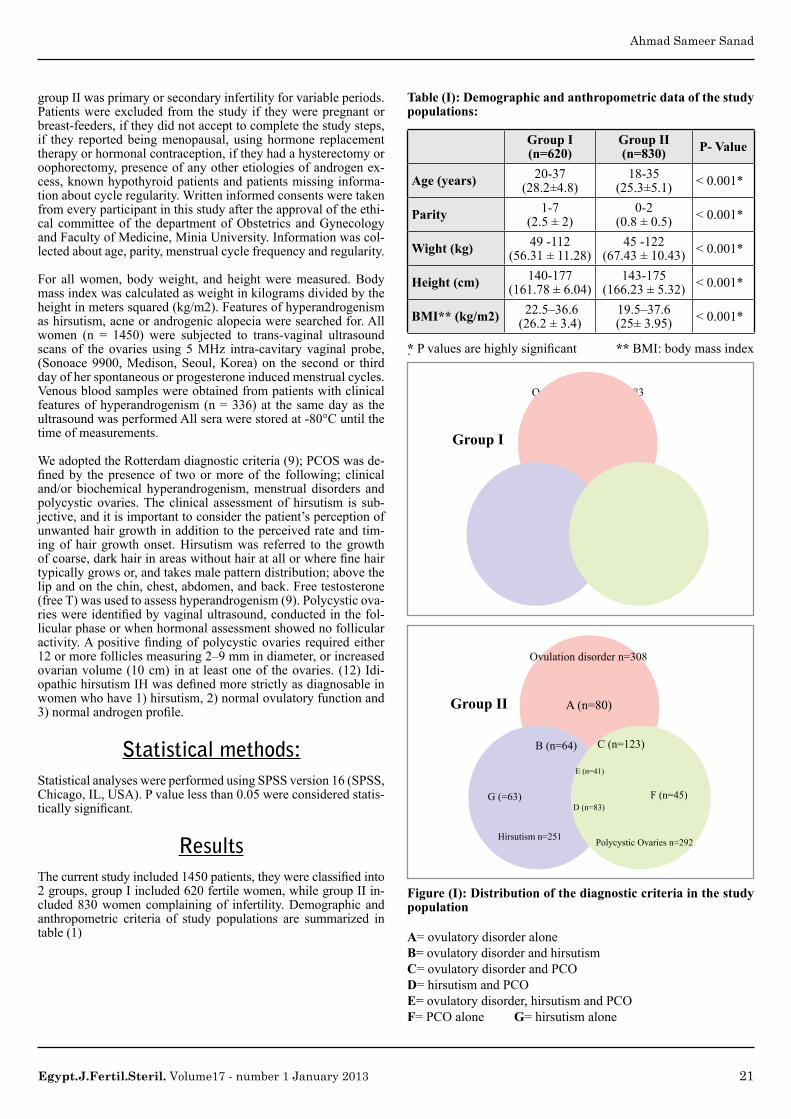
21Egypt.J.Fertil.Steril. Volume17 - number 1 January 2013
Ahmad Sameer Sanad
group II was primary or secondary infertility for variable periods. Patients were excluded from the study if they were pregnant or breast-feeders, if they did not accept to complete the study steps, if they reported being menopausal, using hormone replacement therapy or hormonal contraception, if they had a hysterectomy or oophorectomy, presence of any other etiologies of androgen ex-cess, known hypothyroid patients and patients missing informa-tion about cycle regularity. Written informed consents were taken from every participant in this study after the approval of the ethi-cal committeeof thedepartmentofObstetrics andGynecologyand Faculty of Medicine, Minia University. Information was col-lected about age, parity, menstrual cycle frequency and regularity.
For all women, body weight, and height were measured. Body mass index was calculated as weight in kilograms divided by the height in meters squared (kg/m2). Features of hyperandrogenism as hirsutism, acne or androgenic alopecia were searched for. All women (n = 1450) were subjected to trans-vaginal ultrasound scans of the ovaries using5MHz intra-cavitary vaginal probe,(Sonoace 9900, Medison, Seoul, Korea) on the second or third day of her spontaneous or progesterone induced menstrual cycles. Venous blood samples were obtained from patients with clinical features of hyperandrogenism (n = 336) at the same day as the ultrasound was performed All sera were stored at -80°C until the time of measurements.
We adopted the Rotterdam diagnostic criteria (9); PCOS was de-finedby thepresenceof twoormoreof the following;clinicaland/or biochemical hyperandrogenism, menstrual disorders and polycystic ovaries. The clinical assessment of hirsutism is sub-jective, and it is important to consider the patient’s perception of unwanted hair growth in addition to the perceived rate and tim-ingofhair growthonset.Hirsutismwas referred to thegrowthofcoarse,darkhairinareaswithouthairatallorwherefinehairtypically grows or, and takes male pattern distribution; above the lip and on the chin, chest, abdomen, and back. Free testosterone (free T) was used to assess hyperandrogenism (9). Polycystic ova-rieswereidentifiedbyvaginalultrasound,conductedinthefol-licular phase or when hormonal assessment showed no follicular activity.Apositivefindingofpolycysticovaries requiredeither12 or more follicles measuring 2–9 mm in diameter, or increased ovarian volume (10 cm) in at least one of the ovaries. (12) Idi-opathichirsutismIHwasdefinedmorestrictlyasdiagnosableinwomen who have 1) hirsutism, 2) normal ovulatory function and 3)normalandrogenprofile.
Statistical methods:Statistical analyses were performed using SPSS version 16 (SPSS, Chicago, IL, USA). P value less than 0.05 were considered statis-ticallysignificant.
Results Thecurrentstudyincluded1450patients,theywereclassifiedinto2 groups, group I included 620 fertile women, while group II in-cluded 830 women complaining of infertility. Demographic and anthropometric criteria of study populations are summarized in table (1)
Table (I): Demographic and anthropometric data of the study populations:
Group I (n=620)
Group II (n=830) P- Value
Age (years) 20-37 (28.2±4.8)
18-35 (25.3±5.1) < 0.001*
Parity 1-7 (2.5 ± 2)
0-2 (0.8 ± 0.5) < 0.001*
Wight (kg) 49 -112 (56.31 ± 11.28)
45 -122 (67.43 ± 10.43) < 0.001*
Height (cm) 140-177 (161.78 ± 6.04)
143-175 (166.23 ± 5.32) < 0.001*
BMI** (kg/m2) 22.5–36.6 (26.2 ± 3.4)
19.5–37.6 (25± 3.95) < 0.001*
*Pvaluesarehighlysignificant** BMI: body mass index
Ovulation disorder n=308
Group II
Hirsutismn=251 Polycystic Ovaries n=292
A (n=80)
B (n=64)
G(=63)
C (n=123)
F (n=45)
E (n=41)
D (n=83)
Ovulation disorder n=83
Group I
Hirsutismn=73 Polycystic Ovaries n=85
A (n=18)
B (n=24)
G(=20)
C (n=34)
F (n=22)
E (n=7)
D (n=22)
Figure (I): Distribution of the diagnostic criteria in the study population
A= ovulatory disorder alone B= ovulatory disorder and hirsutismC= ovulatory disorder and PCO D= hirsutism and PCOE= ovulatory disorder, hirsutism and PCO F= PCO alone G= hirsutism alone

22 Egypt.J.Fertil.Steril. Volume17 - number 1 January 2013
Ahmad Sameer Sanad
Table II: Distribution of the diagnostic criteria in the study population:
Results Group I (n=620)
Group II (n=830) P- Value*
Oligo/ anovulation 83 (13.4%) 308 (37.1%) < 0.001
Hirsutism 73 (11.7%) 263 (31.7%) < 0.001
Free T (pmol/L) 49 ± 7.7(18-60) 51 ± 9.6 (20-63) 0.0657**
PCO 85 (13.7%) 292 (35.25%) < 0.001
PCOS 87 (14%) 311 (37.5%) < 0.001
*pvalue<0.05issignificantandpvalue<0.001ishighlysignificant** t-test with unequal variance
From Figure (1), the prevalence of PCOS in fertile group (B+C+D+E) was 87/620 (14%), while in infertile group; the prevalence was 311/830 (37%). Analysis of the data of the in-fertile group showed that the percentage of ovulation disorders (B+C+E) in patients with PCOS was 73.3% (228/311), while the percentage of hirsutism (B+D+E) was 60.4% (188) and percent-age of PCO (C+D+E) was 79.4% (247). 12 Patients (16%) pre-sented with hirsutism only of the fertile group had normal free T theywerediagnosedasidiopathichirsutismIH,while30outof263 patients (11.4%) presented with hirsutism had normal free T intheinfertilegroupanddiagnosedasIH
BMI Vs PCOS
C1
390
34.0
29.0
24.0
19.0
70.0 130.0 190.0 205.0 310.00.0
C2
Figure (II): Correlation between BMI and number of patients with PCOS
C1 represent PCOS cases in group II, C2 represent BMI (body mass index) r=0.221 and P = 0.001.The number of women with PCOS increased with higher BMI (correlation coefficient was0.221andP=0.001)asshowninfigure2
Discussion In the current study the prevalence of PCOS was 14% among fertile women, while in patients complaining of infertility the prevalence was 37% using the Rotterdam diagnostic criteria in a random sample of an Egyptian population. The reported preva-lence of PCOS in various geographic regions varies between 2.2% and 26%. (2, 6, 14-17). The prevalence was 2.4% among 915 Southern Chinese women recruited through the offer of a free
medical examination using Rotterdam diagnostic criteria (15); the prevalence was 6.5% in 154 white Spanish women during blood donationusingnationalinstituteofhealth(NIH)criteria.(18)ThecumulativeprevalenceofPCOSwas6.6%using theNIHdefi-nition among women undergoing pre-employment physical ex-aminations in the United States, (2); the prevalence of PCOS was reported to be 17.8% among 978 South Australian women, who were recruited in a retrospective birth cohort study using Rotter-dam diagnostic criteria (14). Among 157 women with type II dia-betes in Esfahan-Iran, the prevalence of PCOS was 8.2%. (19) It was clear that, the PCOS prevalence depends on the recruitment processofthestudypopulationandcriteriausedforitsdefinition;the Rotterdam diagnostic criteria increased its prevalence by 2 timesversusNIHcriteria,asreportedbefore.(20-22)
The main bewildering questions emerged in the 14 % of fertile group who had full picture of PCOS, were about health complica-tions that may be concealed in those patients, their past reproduc-tive history; pregnancy might occur during episodes of occasional ovulation and their future reproductive performance; they may be the substrate of future secondary infertility.
Inthepresentstudy,therewassignificantpositivecorrelationbe-tween women with PCOS and their BMI (r=0.221 & P = 0.001). This is in agreement with previous studies (14, 16, 23) that re-ported women with PCOS had the highest median BMI.
Hirsutism is the commonest clinicalmanifestation of androgenexcess in PCOS. In the present study, 73 patients (11.7%) of the fertile women had hirsutism, only 32 % of those women (24/73) had cycle irregularities while the rest of them presented with reg-ularcycles.12(16%)patientshadIH.Whileintheinfertilegroup,hirsutismwasdiagnosed in31.7%andIHwasdiagnosed in30cases out of 263 women. It is important to note that hirsutism was found in 60.9 % (53/87) and 60.4% (188/311) of patients with PCOS in fertile and infertile groups respectively.
The reported prevalence of idiopathic hirsutism IH varies from5-29%. (24). Overall 60–75% of patients with PCOS will have hirsutism (25) but there is wide variation based on ethnicity and degree of obesity. Its assessment should therefore be ethnic specif-ic. Most studies have examined Caucasian and African-American women.(26) East Asian women have a lower prevalence of hir-sutism (27) while the prevalence and severity of hirsutism in wom-en with PCOS of Southern Asian origin is greater when compared to Caucasians.(28) The prevalence of PCOS in women with hir-sutism is 75–80%, whereas 20–40% with acne alone have PCOS. About 10% of women with alopecia only will have PCOS.(29, 30)
In the current study, menstrual irregularities were found in 13.4 % in the fertile group compared to 37.1% in the infertile group (P < 0.001) but ovulatory disorder constituted 74.7% and 73.3% of patients with PCOS in fertile and infertile groups respectively. Approximately 75% of women with PCOS have menstrual ir-regularities suggestive of anovulation. This includes oligomen-norhea and amenorrhea.However 20–30%of oligoanovulatorywomen with PCOS can present with apparent eumenorrhoea (i.e. subclinical oligoanovulation). Therefore eumenorrhoeic women with other features of PCOS should have multiple determinations of serum progesterone (drawn between Day 20–24 of their men-strual cycle) to accurately categorize their ovulatory status. (31) In the present study PCO morphology (PCOM) was diagnosed in 13.7% and 35.2% of both study groups.
The term polycystic ovary to describe this morphology is a mis-nomer because there are no dominant cysts or follicles larger than 10 mm because of anovulation. A study from Cape Town demon-strated no correlation between the severity of ovarian morphol-ogy and the endocrine or metabolic manifestations of PCOS. (32) PCOS is a functional disorder that does not depend on the pres-

23Egypt.J.Fertil.Steril. Volume17 - number 1 January 2013
Ahmad Sameer Sanad
ence of polycystic ovaries, and the absence of PCOM does not exclude the diagnosis. Approximately 20–30% of asymptomatic women < 35 years of age will have PCOM; many studies proved thatabout20%ofthesewomenwillactuallyhavePCOSbyNIHdefinition.Conversely,10–25%ofpatientswithPCOSbyNIHdefinitionwillnothavePCOMonultrasonography.(9,25,29,31)
Up to 30% of females with normal androgens and normal menses can have PCOM. (12, 33) It has also been suggested that some women with PCOM and normal ovulatory cycles may have high-erLH,androgenandinsulinlevelsaswellaslowerSHBGlevelswhen compared with control women without PCOM. (34) This phenotype may therefore represent the mildest form of PCOS. PCOS represents a major health and reproductive problems in womenofthereproductiveageinMiniaGovernorate. Werec-ommend follow up of fertile patients with PCOS to answer the above listed questions.
References 1. Asuncion M CR, San Millan JL, Sancho J, Avila S, Escobar-
MorrealeHF.Aprospectivestudyoftheprevalenceofthepoly-cystic ovary syndrome in unselected Caucasian women from Spain. J Clin Endocrinol Metab. 2000;85(7):2434–8.
2. Azziz R, Woods KS, Reyna R, Key TJ, Knochenhauer ES, Yildiz BO. The prevalence and features of the polycystic ovary syn-drome in an unselected population. J Clin Endocrinol Metab. 2004;89(6):2745-9.
3. Cresswell JL BD, Osmond C, Egger P, Phillips DI, Fraser RB. Fetal growth, length of gestation, and polycystic ovaries in adult life. Lancet. 1997;350(9085):1131–5.
4. Michelmore K, Ong K, Mason S, Bennett S, Perry L, Vessey M, et al. Clinical features in women with polycystic ovaries: re-lationships to insulinsensitivity, insulingeneVNTRandbirthweight. Clin Endocrinol (Oxf). 2001;55(4):439-46.
5. ZhangHY, Zhu FF,Xiong J, ShiXB, Fu SX.Characteristicsof different phenotypes of polycystic ovary syndrome based on the Rotterdam criteria in a large-scale Chinese population. BJOG:aninternationaljournalofobstetricsandgynaecology.2009;116(12):1633-9.
6. GoodarziMO,QuinonesMJ,Azziz R, Rotter JI, HsuehWA,Yang H. Polycystic ovary syndrome in Mexican-Americans:prevalence and association with the severity of insulin resist-ance. Fertil Steril. 2005;84(3):766-9.
7. LawsPJHL.Australia’smothersandbabies2006.Sydney:Aus-tralian Institute ofHealth andWelfareNational Perinatal Sta-tisticsUnit.2008;(AIHWCat.No.PER46;PerinatalStatisticsSeriesNo.22.).
8. Vos T, Barker B, Begg S, Stanley L, Lopez AD. Burden of dis-ease and injury in Aboriginal and Torres Strait Islander Peoples: the Indigenous health gap. International journal of epidemiol-ogy. 2009;38(2):470-7.
9. GroupREA-SPCW.Revised2003consensusondiagnosticcri-teria and long-term health risks related to polycystic ovary syn-drome. Fertil Steril. 2004;81((1)):19–25.
10. Cerda C P-AR, Riquelme A, Soza A, Villaseca P, Sir-Petermann T,etal.Nonalcoholicfattyliverdiseaseinwomenwithpolycys-ticovarysyndrome.JHepatol.2007;47((3)):412–7.
11. HollinrakeEAA,MaifeldM,VanVoorhisBJ,DokrasA. In-creased risk of depressive disorders in women with polycystic ovary syndrome. Fertil Steril. 2007;87((6)):1369–76.
12. BalenAH,LavenJS,TanSL,DewaillyD.Ultrasoundassess-ment of the polycystic ovary: international consensus defini-tions.HumReprodUpdate.2003;9(6):505-14.
13. Berkeley AS, DeCherney AH, Polan ML. Abdominal my-omectomy and subsequent fertility. Surg Gynecol Obstet.1983;156(3):319-22.
14. MarchWAMV,WillsonKJ,PhillipsDI,NormanRJ,DaviesMJ.The prevalence of polycystic ovary syndrome in a community sampleassessedundercontrastingdiagnosticcriteria.HumRe-prod. 2010;25:544-51.
15. ChenX,YangD,MoY,LiL,ChenY,HuangY.Prevalenceofpolycystic ovary syndrome in unselected women from southern
China.EurJObstetGynecolReprodBiol.2008;139(1):59-64.16. Knochenhauer ES, Key TJ, Kahsar-Miller M, Waggoner W,
Boots LR, Azziz R. Prevalence of the polycystic ovary syn-drome in unselected black and white women of the southeast-ern United States: a prospective study. J Clin Endocrinol Metab. 1998;83(9):3078-82.
17. KumarapeliV,SeneviratneRdeA,WijeyaratneC.Health-relatedquality of life and psychological distress in polycystic ovary syn-drome:ahiddenfacetinSouthAsianwomen.BJOG:aninterna-tional journal of obstetrics and gynaecology. 2011;118(3):319-28.
18. Asuncion M, Calvo RM, San Millan JL, Sancho J, Avila S, Escobar-MorrealeHF.Aprospectivestudyoftheprevalenceofthe polycystic ovary syndrome in unselected Caucasian women from Spain. J Clin Endocrinol Metab. 2000;85(7):2434-8.
19. TehraniFR,SimbarM,TohidiM,HosseinpanahF,AziziF.Theprevalence of polycystic ovary syndrome in a community sam-ple of Iranian population: Iranian PCOS prevalence study. Re-productive biology and endocrinology : RB&E. 2011;9:39.
20. KumarapeliV,SeneviratneRdeA,WijeyaratneCN,YapaRM,Dodampahala SH.A simple screening approach for assessingcommunity prevalence and phenotype of polycystic ovary syn-drome in a semi-urban population in Sri Lanka. American jour-nal of epidemiology. 2008;168(3):321-8.
21. HsuMI, LiouTH,Chou SY,ChangCY,HsuCS.Diagnosticcriteria for polycystic ovary syndrome in Taiwanese Chinese women: comparison betweenRotterdam2003 andNIH 1990.Fertil Steril. 2007;88(3):727-9.
22. Broekmans FJ, Knauff EA, Valkenburg O, Laven JS, Eijkemans MJ, Fauser BC. PCOS according to the Rotterdam consensus criteria:ChangeinprevalenceamongWHO-IIanovulationandassociationwithmetabolicfactors.BJOG:aninternationaljour-nal of obstetrics and gynaecology. 2006;113(10):1210-7.
23. Boyle JA, Cunningham J, O’Dea K, Dunbar T, Norman RJ.Prevalence of polycystic ovary syndrome in a sample of Indig-enous women in Darwin, Australia. The Medical journal of Aus-tralia. 2012;196(1):62-6.
24. Diamanti-Kandarakis E, Kouli CR, Bergiele AT, Filandra FA, TsianateliTC,SpinaGG,etal.Asurveyofthepolycysticovarysyndrome in theGreek islandofLesbos: hormonal andmeta-bolicprofile.JClinEndocrinolMetab.1999;84(11):4006-11.
25. NormanRJ,DewaillyD,LegroRS,HickeyTE.Polycysticova-ry syndrome. Lancet. 2007;370(9588):685-97.
26. Ferriman D, Gallwey JD. Clinical assessment of body hairgrowth in women. J Clin Endocrinol Metab. 1961;21:1440-7.
27. DeUgarte CM, Woods KS, Bartolucci AA, Azziz R. Degree of facial and body terminal hair growth in unselected black and whitewomen: towardapopulationaldefinitionofhirsutism. JClin Endocrinol Metab. 2006;91(4):1345-50.
28. Wijeyaratne CN, BalenAH, Barth JH, Belchetz PE. Clinicalmanifestations and insulin resistance (IR) in polycystic ovary syndrome (PCOS) among South Asians and Caucasians: is there a difference? Clin Endocrinol (Oxf). 2002;57(3):343-50.
29. Azziz R, Carmina E, Dewailly D, Diamanti-Kandarakis E, Esco-bar-MorrealeHF,FutterweitW,etal.TheAndrogenExcessandPCOS Society criteria for the polycystic ovary syndrome: the complete task force report. Fertil Steril. 2009;91(2):456-88.
30. CelaE,RobertsonC,RushK,KoustaE,WhiteDM,WilsonH,et al. Prevalence of polycystic ovaries in women with andro-genic alopecia. European journal of endocrinology / European Federation of Endocrine Societies. 2003;149(5):439-42.
31. Azziz R, Carmina E, Dewailly D, Diamanti-Kandarakis E, Es-cobar-MorrealeHF,FutterweitW,etal.Positionsstatement:cri-teriafordefiningpolycysticovarysyndromeasapredominantlyhyperandrogenic syndrome: an Androgen Excess Society guide-line. J Clin Endocrinol Metab. 2006;91(11):4237-45.
32. FranksS,AdamsJ,MasonH,PolsonD.Ovulatorydisordersinwomen with polycystic ovary syndrome. Clinics in obstetrics and gynaecology. 1985;12(3):605-32.
33. Polson DW, Adams J, Wadsworth J, Franks S. Polycystic ovaries--acommonfindinginnormalwomen.Lancet.1988;1(8590):870-2.
34. Carmina E, Wong L, Chang L, Paulson RJ, Sauer MV, Stanczyk FZ, et al. Endocrine abnormalities in ovulatory women with pol-ycysticovariesonultrasound.HumReprod.1997;12(5):905-9.

24 Egypt.J.Fertil.Steril. Volume17 - number 1 January 2013
Sherif F. Hendawy
Comparison between Hysterosalpingography and Saline Infusion Sonography in Patients with Recurrent Failed Implantation in Intra-Cytoplasmic Sperm Injection
Sherif F. HendawyDepartment of Obstetrics and Gynecology Ain Shams UniversityCairo - Egypt
AbstractObjective:Abnormaluterinefindingsarereportedinupto50%ofwomenwithrecurrentimplantation failure.Hysterosalpingography is commonly used in evaluation of thesepatients. The introduction of saline infusion sonohysterography has improved diagnosis ofendometrialpathologies.AimofworkwastocompareaccuracyofHysterosalpingo-graphy and Saline infusion sonohysterography in diagnosing uterine pathologies among infertile women with failed intra-cytoplasmic sperm injection.
Subject & Methods: 118 women with recurrent implantation failure underwent hystero-salpingography and saline infusion sonohysterography. The reports were reviewed and findings including submucousfibroids, intrauterine septum, synechia andpolypswerecomparedtothoseobtainedbyhysteroscopy.Sensitivity,specificity,andaccuracyofpro-cedures were measured.
Results:Regardingsubmucousfibroids,Hysterosalpingographyhadsensitivity,specific-ity and accuracy 80%, 96% and 92.9% respectively, whereas, Saline infusion sonohys-terographyhadsensitivity,specificityandaccuracy95.2%,98%and97.5%respectively.Regardingintrauterineseptum,Hysterosalpingographyhadsensitivity,specificityandac-curacy 85%, 95.3% and 93.7% respectively whereas Saline infusion sonohysterography hadsensitivity,specificityandaccuracy89.5%,97.1%,and95.9%respectively.Regard-ingintrauterinesynechia,Hysterosalpingographyhadsensitivity,specificityandaccuracy75.5%, 94.4% and 88.1% respectively. Saline infusion sonohysterography had sensitivity, specificityandaccuracy70.8%,97.7%,and88.1%respectively.Regardingendometrialpolyps,Hysterosalpingographyhad sensitivity, specificityandaccuracy64.7%,97.7%and 85.5% respectively. Saline infusion sonohysterography had sensitivity, specificityand accuracy 66% and 97.7% and 86.1% respectively.
Conclusion: Salineinfusionsonohysterographyiscomparable,insensitivity,specificityandaccuracy,toHysterosalpingographyinevaluationofuterineabnormalities.
Key word: Recurrent ICSI, hysterosalpingography, saline infusion sonohysterography
IntroductionApproximately 15% of couples are affected with subfertility, of which up to 20% remain unexplained. Uterine cavity abnormalities can be a contributing cause of subfertility and recurrent implantation failure. Uterine cavity assessment has been suggested as a routine investigation in the evaluation of subfertile women. (1)
The success of in-vitro fertilization (IVF) treatment is low. Failure of IVF treatment is generally due to embryonic, uterine or transfer factors, but remains unexplained in most cases. A number of interventions have been proposed to improve IVF outcome, many of whichmaynotbeevidence-basedand theirefficacy isuncertain.Oneof thecommoninvestigations proposed following IVF failure is to re-evaluate the uterine cavity. (2)
Traditionally,hysterosalpingography(HSG)hasbeenthemostcommonlyusedtechniquein the evaluation of uterine cavity. The introduction of saline infusion sonohysterography (SIS)hassignificantlyimprovedsonographicdiagnosisofvariousendometrialpatholo-gies. This procedure entails instillation of warm saline into the uterine cavity transcervi-cally to provide enhanced visualization of the endometrium during transvaginal ultra-sound examination. (3)
WeaimedtocomparetheaccuracyofHSGandSISfordiagnosinguterinepathologiesamong infertile women with recurrent failed intra-cytoplasmic sperm injection (ICSI).

25Egypt.J.Fertil.Steril. Volume17 - number 1 January 2013
Sherif F. Hendawy
The sensitivity, specificity, positive, negative predictive valuesandaccuracyforHSGandSISweredeterminedforthediagnosisof endometrial pathology.
Subjects & Methods
A prospective interventional study was introduced to the emer-Thisstudyincludedonehundredandfiftyinfertilewomenwithunexplained recurrent implantation failure (defined as at leasttwo failed previous ICSI cycles, during which good quality em-bryos were transferred (4)). A written consent was obtained from all candidates and the study was approved by the medical ethics committee of Ain Shams University. All patients were subjected toHSG,SISanddiagnostichysteroscopy(DH)whichisthegoldstandard investigation.
TheHSGwasperformedunderfluoroscopyinanoutpatientof-ficesettingatleast48hoursaftermenseshadceased.ThepatientswereroutinelypremedicatedwithHyoscineButylbromide10mg(Buscopan®, Boehringer Ingelheim) prior to the procedure. The patientwasplacedina lithotomyposition,andasterileGravesspeculum was inserted to expose the ectocervix. Using a tenacu-lum,tofixandapplytractiononthecervix,thecervicaloswascannulated with a Leech Wilkinson Uterine Canula of suitable size.Inordernottoobscuretheloweruterinesegment,theGravesspeculum was withdrawn slowly and carefully, not to dislodge the uterine canula. Ten cc of urographin contrast were injected in-trauterinewithfluoroscopiccontrol(OEC9800,GeneralElectricCompany,Fairfield,CT).Acombinationofpulsefluoroscopy(8framesper second)andcontinuousfluoroscopywereusedwithautomated exposure control. Static image capture was achieved byuseofthefluoroscopiclastimageholdfeature.Imagesofearlyandmaximalopacificationoftheuterinecavity,fallopiantubes,and peritoneal contrast spillage were obtained.
SIS was performed by the same operator, during the follicular phase of the cycle. In lithotomy position, the ectocervix was exposed using a sterile warm Collin speculum (to facilitate its removal during the procedure). After cleaning with povidone-io-dine,asterile5-Fcatheter,withanocclusiveballoon,wasflushedwith sterile saline solution before being inserted through the cer-vical os. A ring forceps was used for advancement of the catheter approximately 5–10 cm to position the tip beyond the endocervi-cal canal and not touching the uterine fundus. The speculum was removedwhilethecatheterwasleftinplace.Next,transvaginalsonographic sagittal and coronal or transverse scanning of the pelvis, adnexae, and uterus was performed during instillation of sterile saline solution. Various amounts (5–20 ml) of saline solu-tion were used; only 2–5 ml are needed to distend the uterine cav-ity adequately. A study was considered normal when serial sagit-tal and coronal views of the distended endometrial cavity failed to reveal any distortion, cavitary defect, or undistended regions. Intracavitary defects were described and a likely diagnosis was suggested.
DHwasperformedusingrigidhyteroscope5.2mm(KarlStorz,Germany).The scopewas advanced under direct visualization.The uterine cavity was distended with normal saline, installed from a 500 ml bag wrapped in a pressure bag connected to a ma-nometer and pumped to 120–200mmHg.(5) Either positive ornegativefindingswererecorded,andapplicabletherapeuticpro-cedureswerecompleted.Thepresenceoffibroids,anintrauterineseptum, intrauterine synechia, or endometrial polyps were report-ed.Intheabsenceofthesefindings,thecavitywasdescribedasnormal.
Statistical analysis:All data were transferred to IBM cards using IBM personal com-puter, analyzed with statistical program for social science “SPSS V11.0, SPSS Inc., Chicago, IL, USA”. The data obtained were ex-pressed as descriptive statistics (mean ± standard deviation, range and percentage). Chi-squared test was used for the analysis. A P-valueof<0.05wasconsidered statistically significant.Consider-inghysteroscopicfindingsasthegoldstandardconfirmatorytest,sensitivity,specificity,positivepredictivevalue,negativepredictivevalue and accuracy were calculated.
ResultsOf the 150 patients, only 118 patients continued the study. Their age ranged between 19 and 42 years with a mean±SD 32.81±4.32. Five patients had previous pregnancies while 113 were nulligrav-ida. Seventy nine patients had two failed ICSI attempts while the other 39 patients had more than two failed ICSI procedure.
Figure (1): Comparison between DH, HSG and SIS as regards detection of specific intrauterine lesions:
Perc
enta
ge
Submucous Fibroids
17 1720
29 28
1614 1412 1313 13
Intrauterine Septum
Intrauterine synechia
Endometrial Polyps
302520151050
DH HSG SIS
Table (1): Comparison between HSG and SIS as regards sensitivity and specificity for intrauterine lesions:
Sensitivity Specificity
HSG (%)
SIS (%) P HSG
(%)SIS (%) P
Submucous Fibroids 80 95.2 >0.05 96 98 >0.05
Intrauterine Septum 85 89.5 >0.05 95.3 97.1 >0.05
Intrauterine synechia 75.5 70.8 >0.05 94.4 97.7 >0.05
Endometrial Polyps 64.7 66 >0.05 97.7 97.7 >0.05

26 Egypt.J.Fertil.Steril. Volume17 - number 1 January 2013
Sherif F. Hendawy
Table (2): Comparison between HSG and SIS as regards posi-tive predictive value and negative predictive value for intrauterine lesions:
Positive predictive value
Negative predictive value
HSG (%)
SIS (%) P HSG
(%)SIS (%) P
Submucous Fibroids 83.1 91 >0.05 95.1 99 >0.05
Intrauterine Septum 77.3 85 >0.05 97.1 98 >0.05
Intrauterine Synechia 87.2 94.4 >0.05 88.4 85.7 >0.05
Endometrial Polyps 94.3 94.3 >0.05 82.5 83.3 >0.05
Table (2): Comparison between HSG and SIS as regards posi-tive predictive value and negative predictive value for intrauterine lesions:
HSG (%) SIS (%) P
Submucous Fibroids 92.9 97.5 >0.05
Intrauterine Septum 93.7 95.9 >0.05
Intrauterine Synechia 88.1 88.1 >0.05
Endometrial Polyps 85.5 86.1 >0.05
DiscussionStructural abnormalities of the uterus may affect the reproduc-tive outcome by interfering with implantation and causing spon-taneousmiscarriage.Abnormaluterinefindingsarereportedinasmany as 50% of women with recurrent implantation failure (6)
Hysteroscopyisgenerallyconsideredtobethegoldstandardinthe diagnosis of intrauterine pathologies, including endometrial polyps, submucous myomas, intrauterine adhesions and uterine septa.(7) Recently, the use of contrast media such as saline with transvaginal sonography is increasingly used to improve the de-lineation of uterine cavity abnormalities.(3)In thisstudywecomparedHSGandSISas regards rateofde-tection,sensitivity,specificity,positivepredictivevalue,negativepredictive value and accuracy of the test in diagnosing uterine abnormalities.Hysteroscopicfindingswerethegoldstandardandreference.
This study showed that hysteroscopy had a better diagnostic capa-bilitycomparedtoHSGandSIS,diagnosingsubmucousfibroid,intrauterine septum, intrauterine synechia and endometrial polyps with a percentage of 17%, 14%, 29% and 28% respectively, while HSGshowedpercentagesof13%,12%,20%and13%whereas,SIS showed percentages of 16%, 13%, 17% and 14% for the same lesions respectively (Figure 1).
Several studies have comparedDH,HSG andSIS in detectinguterineabnormalities.OnestudycomparingHSGwithhysteros-copy reportedsensitivityof81%anda specificityof80%witha false negative rate of 9% and a false-positive rate of 22%.(8) Another study conducted to assess the diagnostic reliability of hysteroscopyandHSG,demonstratedHSGtohaveasensitivity
of79%andaspecificityof82%,withan18%falsepositiverateand a 19% false-negative rate. They concluded that even though HSGismainlyusedfortheassessmentoftubalpatency,ithasasecondary role in the assessment of the uterine cavity.(9) Furthermore,SIShasbeenfoundtobesensitive,specificandac-curate in identifying intrauterine abnormalities such as polyps, submucosalfibroids,adhesions,septaanduterineanomalies.Onestudy even showed that SIS had the same diagnostic accuracy as hysteroscopy for endometrial polyps.(10) Another study com-paring SIS with hysteroscopy reported 87.5% sensitivity, 100% specificity, 100%positive predictive value and 91.6%negativepredictive value for the detection of any cavity abnormality with SIS as compared with hysteroscopy.(11)
On the other hand, the accuracy of HSG in assessment of theuterine cavity integrity in infertile patients has been reported to beratherdisappointing.Thesensitivityandspecificitywerede-scribed to be 79–98% and 15–82%, respectively (12) and similar studies have also shown that hysteroscopy had a better diagnostic accuracy than SIS.(13)
Inthisstudy,asregardssubmucousfibroids,HSGhadasensitiv-ity and specificity80%and96%respectively. Positivepredic-tive value, negative predictive value and accuracy were 83.3%, 95.1% and 92.9% respectively, whereas, SIS had a sensitivity andspecificity95.2%and98%respectively,positivepredictivevalue, negative predictive value and accuracy of 91%, 99% and 97.5% respectively. These results are in agreement with Erdem et al. were SIS had a Positive predictive value, negative predictive value and accuracy of 91%, 99% and 97.5% respectively.(14)
Inthisstudyasregardintrauterineseptum,HSGhadasensitivityandspecificityof85%and95.3%respectively.Positivepredic-tive value, negative predictive value and accuracy were 77.3%, 97.1% and 93.7% respectively whereas SIS had a sensitivity and specificity 89.5% and 97.1% respectively. Positive predictivevalue, negative predictive value and accuracy were 85%, 98% and 95.9% respectively.As regard intrauterine synechia, HSGhadasensitivityandspecificityof75.5%and94.4%respectively.Positive predictive value, negative predictive value and accuracy were 87.2%, 88.4% and 88.1% respectively. SIS had a sensitivity andspecificity70.8%and97.7%respectively.Positivepredictivevalue, negative predictive value and accuracy were 94.4%, 85.7% and88.1%respectively.SimilarstudieshaveshownHSGandSISto have a sensitivity of 75% in the detection of intrauterine adhe-sions.(10)
Inthisstudyasregardendometrialpolyps,HSGhadasensitivityandspecificity64.7%and97.7%respectively.Positivepredictivevalue, negative predictive value and accuracy were 94.3%, 82.5% and85.5%respectively.SIShadasensitivityandspecificity66%and 97.7% respectively, Positive predictive value, negative pre-dictive value and accuracy of 94.3%, 83.3% and 86.1% respec-tively, which was in accordance with several studies, one showing sensitivityandspecificityof100%and91.8%respectively,(14)and another study which showed that SIS had a sensitivity and negative predictive value 100% for endometrial polyps.(15)
Finally,inconclusionSISiscomparabletoHSGindiagnosisofuterine abnormalities, but with the advantage of lack of ionizing radiation and of lower cost, more feasibility, and outpatient pro-cedurewithbetter tolerability; itcanreplaceHSGasaprimarydiagnostictestforuterineanomaliespriortoDH.

27Egypt.J.Fertil.Steril. Volume17 - number 1 January 2013
Sherif F. Hendawy
References1. Jyotsna Pundir and Tarek El touky: uterine cavity assessment
priortoIVF.Women’sHealth2010;6:841-848.2. Society for Assisted Reproductive Technology and the Amer-
ican Society for Reproductive Medicine: Assisted reproduc-tive technology in the United States: 2001 results generated from the American Society for Reproductive Medicine/So-ciety for Assisted Reproductive Technology registry. Fertil. Steril. 2007; 87:1253–1266.
3. Elsayes KM, Pandya A, Platt JF et al: Technique and diag-nosticutilityofsalineinfusionsonohysterography.IntJGy-naecol Obstet. 2009 A; 105(1):5-9.
4. SharifKWandHgunaimS:Managementof273casesofre-current implantation failure: results of a combined evidence-based protocol. Reprod Biomed Online. 2010; 21(3):373-80.
5. Nagele,F.,O’Connor,H.,Davies,A.etal:Outpatientdiag-nostichysteroscopies.Obstet.Gynecol.1996;88:87–92.
6. Brown SE, Coddington CC, Schnorr J et al.: Evaluation of outpatient hysteroscopy, saline infusion hysterosonography, and hysterosalpingography in infertile women: a prospective, randomized study. Fertil. Steril. 2000;74(5): 1029–1034.
7. BozdagG,AksanG,EsinlerIetal:Whatistheroleofof-ficehysteroscopyinwomenwithfailedIVFcycles?ReprodBiomed Online 2008; 17:410–415.
8. RomaDalfóA,UbedaB,UbedaAetal.:Diagnosticvalueof hysterosalpingography in the detection of intrauterine ab-normalities: a comparison with hysteroscopy: AJR Am. J. Roentgenol. 2004; 183(5): 1405–1409.
9. GaglioneR,ValentiniAL,PistilliEetal.:AcomparisonofhysteroscopyandHysterosalpingography. Int. J.Gynaecol.Obstet. 1996; 52(2): 151–153.
10. Soares SR, Barbosa dos Reis MM et al: Diagnostic accuracy of sonohysterography, transvaginal sonography, and hystero-salpingography in patients with uterine cavity diseases. Fer-til Steril. 2000; 73(2):406-11.
11. AyidaG,ChamberlainP,BarlowDetal:Uterinecavityas-sessment prior to in vitro fertilization: comparison of trans-vaginal scanning, saline contrast hysterosonography and hysteroscopy.UltrasoundObstetGynecol1997;10:59–62.
12. GolanA,EilatE,Ron-ElRetal.:Hysteroscopyissuperiortohysterosalpingography in infertility investigation. Acta Ob-stet.Gynecol.Scand.1996;75(7):654–656
13. Soguktas S, Cogendez E, Kayatas SE et al: Comparison of saline infusion sonohysterography and hysteroscopy in di-agnosis of premenopausal women with abnormal uterine bleeding.EurJObstetGynecolReprodBiol.2012;161(1):66-70.
14. ErdemM,BilginU,BozkurtNetal:Comparisonoftrans-vaginal ultrasonography and saline infusion sonohysterogra-phy in evaluating endometrial cavity in pre- and postmeno-pausal women with abnormal uterine bleeding. Menopause. 2007; 14(5):846-52.
15. BingolB,GunencZ,GedikbasiAetal:Comparisonofdiag-nostic accuracy of saline infusion sonohysterography, Trans-vaginal sonography and hysteroscopy. J Obstet gynaecol. 2011; 31 (1): 54-8.

28 Egypt.J.Fertil.Steril. Volume17 - number 1 January 2013
Ahmed T. Soliman
Value of Oral Contraceptive Pill Pretreatment before GnRH Antagonist Ovarian Stimulation Protocol on The Outcome of IVF/ICSI
Ahmed T. Soliman, M.DDepartment of Obstetrics and Gynecology, Faculty of Medicine, Tanta University
AbstractObjective: To evaluate the effect of oral contraceptive pill (OCP) pretreatment in gonad-otropin-releasinghormone(GnRH)antagonistovarianstimulationprotocolontheout-come of IVF/ICSI regarding number of retrieved oocyte, oocyte maturation rate, fertili-zation rate, good quality embryo rate, cycle cancellation rate, pregnancy rate and clinical abortion rate.
Subjects & Methods: Atotalof84patients, inaprospectivecontrolledclinical trial,had ICSIusingGnRHantagonist protocol during the period from February 1st, 2009 to September 30th, 2011 were included in this prospective randomized trial. We compared the IVF outcomes be-tween OCP pretreated (n=43) and no pretreatment group (n=41) in gonadotropin-releas-inghormone(GnRH)antagonistovarianstimulationprotocol.
Results: The mean duration of ovarian stimulation and mean amount of gonadotropins in OCPpretreatedgroupwassignificantlyhigherthanthatofnopretreatmentgroup(11.7±2.3 vs. 9.8 ± 1.8 days and 2720.4 ± 1165.0 IU vs. 2295.8 ± 1121.1 IU). The mean number of retrieved oocytes and oocytematuration ratewas significantly higher inOCP pre-treated group (10.9 ± 5.3 vs. 7.5 ± 5.2 and 90.8% vs. 73.3%). The number of total gained embryosandthegoodqualityembryoratewasalsosignificantlyhigherinOCPpretreatedgroup (7.9 ± 0.9 vs. 4.2 ± 1.0 and 69.9 % vs. 48.7%). Fertilization rate was also higher in OCP pretreated group (84.9 ± 0.2% vs. 70.5 ± 0.3%). The implantation and pregnancy ratewerehigher,althoughnotreachingstatisticallysignificantlevel,inOCPpretreatedgroup (11.6% vs. 10.7% and 45.4% vs. 36.2%).
Conclusion: OCPpretreatmentbeforeGnRHantagonistprotocolforIVFappearstohavereliablebenefitsintermsofIVFoutcomesregardingnumberofretrievedoocytes,oocytematuration rate, fertilization rate, and good quality embryo rate. But, it also has a weak point in respect to longer stimulation duration and increased gonadotropin consumption. TheOCPpretreatedcycle inGnRHantagonistprotocol ismoreadvantageous,despitenotreachingstatisticalsignificantlevel,inrespecttopregnancyrate,andcyclecancella-tion rate. Well controlled, large scaled studies are needed to support effectiveness of OCP pretreatmentbeforestartingGnRHantagonistovarianstimulationprotocolforIVF/ICSI.
Keywords: Gonadotropin-releasinghormoneantagonist;Oralcontraceptivepillpretreat-ment; Ovarian stimulation; In vitro Fertilization
IntroductionGonadotropin-releasing hormone (GnRH) antagonists have beenwidely used after itsfirstintroductioninassistedreproductivetechnologiestopreventaprematureluteiniza-tion[1].GnRHantagonistprotocolsarepreferredforpoorrespondersbecauseofshorterduration and use of lower amount of gonadotropins for ovarian stimulation as compared withtraditionalGnRHlongagonistprotocols[2].However,itinducesinsufficientsyn-chronizationoffollicularcohortdevelopmentandlackofflexibilityinthestartingdayofovarianstimulation,whichislesslikelyinGnRHagonistlongprotocols[3].Forgettingover these limitations, several pretreatments have been applied [4-7]. Among them, oral contraceptive (OCP) pretreatment has been reported to induce higher numbers of oocytes retrievedcomparedtonopretreatmentgroupinGnRHantagonistcycles[8].
The effect of this intervention on the probability of pregnancy has so far been examined onlyinasmallrandomizedcontrolledtrial(RCT)[4].However,priortoadoptingamodi-ficationinanalreadyestablishedprotocoloftreatmentsuchasthedailyGnRHantagonistprotocol [5], its effect on the probability of pregnancy needs to be evaluated.

29Egypt.J.Fertil.Steril. Volume17 - number 1 January 2013
Ahmed T. Soliman
In this study, we aimed to evaluate the effect of OCP pretreatment ininvitrofertilization(IVF)cyclesusingGnRHantagonistsbycomparison between OCP-pretreated and non-treated groups.
Subjects & MethodsAtotalof84patientsindicatedforICSIusingGnRHantagonistprotocol during the period from February 1st, 2009 to September 30th, 2011 were included in this prospective randomized trial. Pa-tients were prospectively selected from our IVF center
Inclusioncriteriawere:age<39years;≤3previousassisted re-production (ART) attempts; body mass index (BMI) of 18–29 kg/m2; regular menstrual cycles; no polycystic ovaries according to Rotterdamdefinition;noendometriosis>stageII;basalhormonallevelsofFSH(<10IU/l)andLH(<10IU/l)atinitiationofstimu-lation for the non-OCP group and at initiation of OCP in the OCP group; and no previous poor response to ovarian stimulation. Poor ovarian response was characterized either by cancellation of the cycle due to poor follicular development after at least 10 days of gonadotropinstimulation,orbyretrievaloflessthanfivecumu-lus–oocyte– complexes (COCs) at oocyte retrieval.
The study population were divided into 43 patients used OCP pre-treatmentinpreviousmenstrualcyclebeforestartingGnRHan-tagonist protocol for IVF (OCP pretreated group), and 41 patients usednomedicationbeforeGnRHantagonistprotocolforIVF(nopretreatment group). Randomization was done using computer-generated program and all IVF cycles carried out in our center
In OCP pretreated group, daily OCP (Yasmin®, Bayer Schering PharmaAG,Berlin,Germany)wasappliedfromthefirstdayofprevious menstrual cycle. after OCP discontinuation, the ovar-ian stimulation was done with gonadotropin from the second day ofmenstrualcycleasusualasextensivelydescribed.Briefly,thedose of gonadotropins was determined on an individual basis ac-cordingtotheage,Day3FSHvalueandechographiccharacter-
istics of the ovaries. Patients underwent serial transvaginal ultra-sound starting on Day 6 of ovarian hyperstimulation. The patients underwentpituitarydownregulationwithdailyGnRHantagonist(Cetrotide®,Merck-Serono,Geneva,Switzerland) frommidorlate follicular period of this cycle applied when dominant follicle reached to 12 or 13 mm. When two or more follicles reached 16 -18mmindiameter10,000IUofhCG(Choriomon,IBSA,Luga-no3, Suisse) was administered. Trans-vaginal ultrasound guided oocyte pick-up (OPU) was performed 34-36 hours later and then, maturity and quality of retrieved oocytes was evaluated. Embryo transfer was performed 48–72 h after the oocyte collection.
The IVF outcomes such as retrieved oocyte number, oocyte mat-uration rate, fertilization rate, good quality embryo rate, cycle cancellation rate, pregnancy rate and clinical abortion rate were compared between OCP pretreated and no pretreatment group. Clinicalpregnancywasdefinedas theultrasonographicdemon-stration of an intrauterine gestational sac 4 weeks after embryo transfer.
Statistical analysis was performed using SPSS ver. 16.0 (SPSS Inc., Chicago, IL, USA). Each variable was presented as mean ± standard deviation. Student’s t -test and Chi-square test were used wherever appropriate. P -value of < 0.05 was considered statisti-callysignificant.
ResultsThe mean age and body mass index in OCP pretreated and non-pretreated groups were similar (34.6 ± 3.1 vs. 35.1 ± 3.5 years and 22.3 ± 2.6 kg/m2 vs. 21.7 ± 1.9 kg/m2). The basal follicle stimulatinghormone(FSH)level(10.2±3.7IU/mLvs.9.1±2.9IU/mL) was also similar between two groups. The primary in-fertility rate tended to be higher in non-pretreated group (66.1% vs.55.4%)butnotstatisticallysignificant.Themeandurationofinfertility was longer in control group (5.1 ± 2.7 years vs. 4.7 ± 2.3years)but,also,notstatisticallysignificant(Table1).
OCP pretreated group (n = 43) No pretreatment group (n = 41) P - value
Age of female (yr) 34.6 ± 3.1 35.1 ± 3.5 NS
BMI (kg/m2) 22.3 ± 2.6 21.7 ± 1.9 NS
Duration of infertility (yr) 4.7 ± 2.3 5.1 ± 2.7 NS
Primary infertility (%) 55.4 66.1 NS
Secondary infertility (%) 44.6 33.9 NS
Basal serum FSH (mIU/mL) 10.2 ± 3.7 9.1 ± 2.9 NS
Table 1: Comparison of baseline characteristics in both study groups

30 Egypt.J.Fertil.Steril. Volume17 - number 1 January 2013
Ahmed T. Soliman
OCP pretreated group (n = 43) No pretreatment group (n = 41) P - valueDuration of COH (day) 11.7 ± 2.3 9.8 ± 1.8 0.0001Dosage of gonadotropin (IU) 2720.4 ± 1165.0 2295.8 ± 1121.1 0.054E2 on hCG day (pg/mL) 1170.8 ± 1267.3 1086.0 ± 877.5 NSEM thickness on hCG day (mm) 10.4 ± 2.6 10.0 ± 2.4 NSNumber of retrieved oocyte 10.9 ± 5.3 7.5 ± 5.2 0.053Number of matured oocyte 9.9 ± 4.3 5.5 ± 4.1 0.052Oocyte maturation rate (%) 90.8 73.3 0.052Number of total gained embryo 7.9 ± 0.9 4.2 ± 1.0 0.053Good quality embryo rate (%) 69.9 48.7 0.054Number of transferred embryo 2.6 ± 0.9 2.3 ± 1.0 NSFertilization rate (%) 84.9 ± 0.2 70.5 ± 0.3 0.017Implantation rate (%) 11.6 10.7 NSPregnancy rate/embryo transfer (%) 45.4 36.2 0.304Clinical abortion rate (%) 41.7 42.1 NSCycle cancellation rate (%) 13.5 17.5 NS
Table 2: Comparison of IVF/ICSI outcomes in both study groups The mean duration of ovarian stimulation in OCP pretreated group wassignificantlylongerthanthatofnopretreatmentgroup(11.7±2.3vs.9.8±1.8days).Meanamountofgonadotropinsforcontrolledovarian stimulation in OCP pretreated group was higher than that of control group (2720.4 ± 1165.0 IU vs. 2295.8 ± 1121.1 IU). The meannumberofretrievedoocytesandoocytematurationratewassignificantlyhigherinOCPpretreatedgroupthanthatofnopretreat-ment group (10.9 ± 5.3 vs. 7.5 ± 5.2 and 90.8% vs. 73.3%). The number of total gained embryos and the good quality embryo rate was alsosignificantlyhigherinOCPpretreatedgroupthanthatofcontrolgroup(7.9±0.9vs.4.2±1.0and69.9%vs.48.7%).Fertilizationrate was also higher in OCP pretreated group (84.9 ± 0.2% vs. 70.5 ± 0.3%). The implantation and pregnancy rate were higher, although notreachingstatisticallysignificantlevel,inOCPpretreatedgroup(11.6%vs.10.7%and45.4%vs.36.2%).Theclinicalabortionratealsoshowednosignificantdifferencebetweentwogroups.ThecyclecancellationratetendedtobelowerinOCPpretreatedgroupthanOCP non-treated group but not statistically different.
DiscussionThe present study was scheduled to evaluate the effect of oral contraceptive pill (OCP) pretreatment in gonadotropin-releasing hormone(GnRH)antagonistovarianstimulationprotocolsontheoutcome of IVF/ICSI regarding number of retrieved oocytes, oo-cyte maturation rate, fertilization rate, good quality embryo rate, cycle cancellation rate, pregnancy rate and clinical abortion rate.
The retrieval of good quality oocyte is a very important factor to achieve pregnancy in infertile women undergoing IVF/ICSI. Togaingoodquality embryo, growthoffinelymaturedoocyteisfirstlyneeded.Togetmorematuredoocytes,thesynchronizedgrowingoffolliclesisoneofimportantfactor.DuringCOH,mostof the early antral follicles are required to grow coordinately in response to exogenous gonadotropins thus accomplishing simul-taneous functional and morphological maturation [10].
Marked discrepancies of follicular size at the end of COHmaybe counterproductive since they imply that a substantial fraction ofFSH-sensitivefolliclesfail toundergosatisfactorymaturation.This phenomenon potentially reduces the number of viable oocytes and embryos and the probability of conception. Selection of good embryos for transfer depends on embryo cohort size: implications for the `mild ovarian stimulation’ debate [10]. The number of em-bryos available for transfer predicts successful pregnancy outcome, especially in older women with normal ovarian hormonal reserve testing[11].LowmaturationrateofoocytesinGnRHantagonistcycles was thought to be due to older ages of patients receiving the antagonist protocol, or asynchronous follicular development and a limited number of dominant follicles due to ovarian stimulation withoutpituitarysuppressioninGnRHantagonistprotocols.
Asynchronousmulti-folliculargrowthduringCOHmaybeadi-rect consequence of size heterogeneities of early antral follicles during the early follicular phase [12]. Luteal estradiol administra-tionstrengthenstherelationshipbetweenday3FSHandinhibinB levels and ovarian follicular status [13]. Some follicles are able torespondtolowerFSHlevelsthanothersbytheirintrinsicsen-sitivitytoFSH,andstarttheirdevelopmentduringthelatelutealphase[14].SincelargerfolliclesaremoreFSHresponsivethanaresmaller follicles, exogenous gonadotropin administration is likely to intensify further size discrepancies of growing follicles during COH[15].Folliculardevelopmentbeginsduringthelutealphaseof the human menstrual cycle. Hence, COH protocols such asmidluteallongprotocol,suppressionoflutealFSHsecretioncouldpreventuntimelyanduncoordinateddevelopmentofFSHsensi-tive follicles during the luteal-follicular transition and faster fol-liculargrowthsynchronizationduringCOHcanbeobtained[16].
However, this luteal suppression of FSH cannot be achieved inGnRHantagonistCOHprotocols.Therefore,markedfollicularsizediscrepancieswouldbeoccurredinGnRHantagonistCOHcycles.
OCP pretreatment might exert a suppressive effect on the cohort of existing follicles. Fanchin et al. (2003) showed that luteal E2 adminis-tration synchronizes the follicular cohort and is associated with more folliclesandoocytesretrieved[13].AsdemonstratedbyVanHeus-den et al. (1999), OCP is able to suppress the luteofollicular transition andtheendogenousFSHriseoccurs3daysafterOCPwithdrawal[17]. The same effect is described by De Ziegler et al. (1998) after E2 withdrawal[18].Inanotherstudy,OCPpretreatmentinGnRHan-tagonist cycles in low responders, also, resulted in improving ovarian responsebyintrinsicgonadotropinsbeforeCOH[19].

31Egypt.J.Fertil.Steril. Volume17 - number 1 January 2013
Ahmed T. Soliman
In the present study, the baseline characteristics of IVF cycles between both groups were comparable. In OCP pretreated group it presented improvement of fertilization rate and gained more number of fertilized embryo than that of OCP non-treated group even if longer duration and larger used dose of gonadotropin for ovarian stimulation.
According to previous meta-analysis regarding OCP pretreatment inGnRHantagonistcycles[20],OCPpretreatmentwasassociatedwith an increased gonadotropin consumption and increased dura-tion of stimulation without improvement of ongoing pregnancy rate. There were many other studies which concerned to OCP pretreat-mentandIVFoutcomesinGnRHantagonistcycles.Amongthem,Kolibianakisetal.[21],reportedthatOCPpretreatedGnRHantago-nistCOHcycleshavenosignificantbenefitinongoingpregnancyratesandmoreoverresultsinasignificantlyhigherearlypregnancyloss of compared to non-OCP cycles. In another systemic review andmeta-analysisanalyzedbyGriesingeretal.[22],OCPpretreat-ment inGnRHantagonist forCOHhavenosignificantbenefit inincreasing ongoing pregnancy rates. A recent study focused on com-promised group like as low responders [23]. The study showed high-er number of retrieved and matured oocytes, and fertilized oocytes inOCPpretreatmentgroupinlowresponderswhichwasdefinedaselevatedbasalFSHlevel(>8.5mIU/mL),and/orantralfolliclecount<5. In the present study, the number of gained embryo and oocyte fertilization rate were higher in cycles of OCP pretreatment.
Inconclusion,OCPpretreatmentbeforeGnRHantagonistprotocolforIVFappearstohavereliablebenefitsintermsofIVFoutcomesregarding number of retrieved oocytes, oocyte maturation rate, fertilization rate, and good quality embryo rate. But, it also has a weak point in respect to longer stimulation duration and increased gonadotropin consumption.TheOCPpretreated cycle inGnRHantagonist protocol is more advantageous, despite not reaching statisticalsignificantlevel,inrespecttopregnancyrate,andcyclecancellation rate. Well controlled, large scaled studies are need-ed to support effectiveness of OCP pretreatment before starting GnRHantagonistovarianstimulationprotocolforIVF/ICSI.
References1. Martinez-SalazarJ,CerrilloM,QueaG,PachecoA,Garcia-
VelascoJA.GnRHantagonistganirelixpreventsprematureluteinization in IUI cycles: rationale for its use. Reprod Bi-omed Online 2009;19:156-61.
2. Berin I, Stein DE, Keltz MD. A comparison of gonadotropin-releasinghormone(GnRH)antagonistandGnRHagonistflare protocols for poor responders undergoing in vitro fertili-zation. Fertil Steril 2010;93:360-3.
3. Depalo R, Lorusso F, Palmisano M, Bassi E, Totaro I, Vacca M,etal.FolliculargrowthandoocytematurationinGnRHagonist and antagonist protocols for in vitro fertilisation and embryotransfer.GynecolEndocrinol2009;25:328-34.
4. OrvietoR,RabinsonJ,MeltzerS,ZohavE,AntebyE,Hom-burgR.SubstitutingHCGwithGnRHagonisttotriggerfinalfollicular maturation: a retrospective comparison of three different ovarian stimulation protocols. Reprod Biomed On-line 2006;13:198-201.
5. Nogueira D, Friedler S, Schachter M, Raziel A, Ron-ElR, Smitz J. Oocyte maturity and preimplantation develop-ment in relation to follicle diameter in gonadotropin releas-ing hormone agonist or antagonist treatments. Fertil Steril 2006;85:578-83.
6. FanchinR,SchonنuerLM,Cunha-FilhoJS,MéndezLozanoDH,FrydmanR.Coordinationofantralfolliclegrowth:ba-sis for innovative concepts of controlled ovarian hyperstimu-lation. Semin Reprod Med 2005;23:354-62.
7. Fanchin R, Méndez Lozano DH, Schonنuer LM, Cunha-FilhoJS,FrydmanR.Hormonalmanipulationsintheluteal
phase to coordinate subsequent antral follicle growth during ovarian stimulation. Reprod Biomed Online 2005;10:721-8.
8. OehningerS.OvulationinductioninIVF.MinervaGinecol2011;63:137-56.
9. ArslanM, Bocca S,Mirkin S, Barroso G, Stadtmauer L,Oehninger S. Controlled ovarian hyperstimulation protocols for in vitro fertilization: two decades of experience after the birth of Elizabeth Carr. Fertil Steril 2005;84:555-69.
10. Devreker F, Pogonici E, De Maertelaer V, Revelard P, Van den Bergh M, Englert Y. Selection of good embryos for transfer depends on embryo cohort size: implications for the ‘mild ovarianstimulation’debate.HumReprod1999;14:3002-8.
11. OpsahlMS,BlauerKL,BlackSH,LincolnSR,ThorsellL,Sherins RJ. The number of embryos available for transfer predicts successful pregnancy outcome in women over 39 years with normal ovarian hormonal reserve testing. J Assist ReprodGenet2001;18:551-6.
12. FanchinR,Cunha-FilhoJS,SchonنuerLM,KadochIJ,Co-hen- Bacri P, Frydman R. Coordination of early antral fol-licles by luteal estradiol administration provides a basis for alternative controlled ovarian hyperstimulation regimens. Fertil Steril 2003;79:316-21.
13. Fanchin R, Cunha-Filho JS, Schonنuer LM, Righini C,de Ziegler D, Frydman R. Luteal estradiol administration strengthens the relationship between day 3 follicle-stimu-lating hormone and inhibin B levels and ovarian follicular status. Fertil Steril 2003;79:585-9.
14. KleinNA,BattagliaDE,FujimotoVY,DavisGS,BremnerWJ, Soules MR. Reproductive aging: accelerated ovarian follicular development associated with a monotropic folli-cle-stimulating hormone rise in normal older women. J Clin Endocrinol Metab 1996;81:1038-45.
15. McNattyKP,HillierSG,vandenBoogaardAM,Trimbos-KemperTC,ReichertLEJr,vanHallEV.Folliculardevelop-ment during the luteal phase of the human menstrual cycle. J Clin Endocrinol Metab 1983;56:1022-31.
16. Fanchin R, Salomon L, Castelo-Branco A, Olivennes F, Frydman N, Frydman R. Luteal estradiol pre-treatmentcoordinates follicular growth during controlled ovar-ianhyperstimulationwithGnRHantagonists.HumReprod2003;18:2698-703.
17. vanHeusdenAMandFauserBC(1999)Activityofthepitui-tary-ovarian axis in the pill-free interval during use of low-dose combined oral contraceptives. Contraception 59,237–243.
18. de Ziegler D, Jaaskelainen AS, Brioschi PA, Fanchin R and Bulletti C (1998) Synchronization of endogenous and exog-enous FSH stimuli in controlled ovarian hyperstimulation(COH).HumReprod13,561–564.
19. KimCH,JeonGH,CheonYP,JeonI,KimSH,ChaeHD,etal.ComparisonofGnRHantagonistprotocolwithorwithoutoralcontraceptivepillpretreatmentandGnRHagonistlow-dose long protocol in low responders undergoing IVF/intra-cytoplasmic sperm injection. Fertil Steril 2009;92:1758-60.
20. BodriD, Sunkara SK,CoomarasamyA.Gonadotropin-re-leasing hormone agonists versus antagonists for controlled ovarian hyperstimulation in oocyte donors: a systematic re-view and meta-analysis. Fertil Steril 2011;95:164-9.
21. KolibianakisEM,PapanikolaouEG,CamusM,TournayeH,Van Steirteghem AC, Devroey P. Effect of oral contraceptive pill pretreatment on ongoing pregnancy rates in patients stimu-latedwithGnRHantagonistsandrecombinantFSHforIVF.Arandomizedcontrolledtrial.HumReprod2006;21:352-7.
22. GriesingerG,VenetisCA,MarxT,DiedrichK,TarlatzisBC,Kolibianakis EM. Oral contraceptive pill pretreatment in ovar-ianstimulationwithGnRHantagonistsforIVF:asystematicreview and meta-analysis. Fertil Steril 2008;90:1055-63.
23. FrancoJGJr,BaruffiRL,MauriAL,PetersenCG,FelipeV,CornicelliJ,etal.GnRHagonistversusGnRHantagonistinpoor ovarian responders: a meta-analysis. Reprod Biomed Online 2006;13:618-27.

32 Egypt.J.Fertil.Steril. Volume17 - number 1 January 2013
Tarek Shokeir
Live Operative Workshops In Gynecologic Endoscopy: Issues And ProblemsDuring the last quarter of the 20th century, and especially during the last decade, there has been a paradigm shift in the methods for performance surgery. For many procedures, the “invasiveness” involved has been dramatically reduced resulting in superior outcomes manifested as improved survival, fewer complications, and quicker return to functional healthandproductive life.However, thehighly technicalnatureofgynecologicendo-surgery and the rapid rate with which new instruments are introduced, require a high intensity of surgical training. One way to accomplish this issue is the implementation of “live operative workshops”.
Thepopularprograminanysurgicalmeetingespeciallyinthefieldofgynecologicendos-copy is the live operative workshop. Live workshops allow the audience to observe surgi-cal anatomy and technique. They provide an opportunity for interaction and an excellent platform to learn newer surgical skills. Advancements in video technology have made it possible to transmit good quality images and this is even truer for minimally invasive endoscopic gynecologic procedures. It is therefore not surprising that these workshops play an important role in surgical education. It has become easier to demonstrate new pro-cedures, display newer instruments and popularize recent treatments. Many hospitals and surgeons are increasingly using this medium as an advertisement to build a reputation. Transcontinental video demonstrations are possible and one need not go across seven seas to observe a new surgical technique. But in this enthusiasm, are we truely compromise patient safety, privacy and patient human rights? Is a true consent available? Is there an element of subtle coercion? Do the patients get honest and unbiased information? And finally,whowillberesponsibleforcomplicationswhichmayariseoutoftheseproce-dures, especially when they are performed by “master surgeons” who come from foreign countries?. It is equally distressing to see patients from the underdevloped world, being subjected to “experimental procedures”. It is not uncommon to see surgeons from the developed world to try out a new surgical technique before performing it in their own institution and on their countrywomen. Is it not unethical to allow our population to be used as guinea pigs?. I am not questioning the usefulness of live workshops but I believe we should all be concerned about these issues. It is important to remember that the American College of SurgeonsandtheAmericanCollegeofObstetriciansandGynecologistshavebannedliveprocedures during their meetings.
It is true that unless we take adequate steps to council our patients adequately, ensure their safety and protect their privacy, we risk unethical censure. The patients should have an absolute indication for the procedure and no compromization should be permissible just to accommodate the “impatient surgeon and an industry driven procedure”. The faculty should impress upon the enthusiastic audience that the workshop serve a limited purpose, therebeingnosubstituteforin-servicetraining.Self-propagation,financialgainandad-vertisement should not be the motive behind these workshops. As a policy, we must pro-vide guidelines and device a mechanism which can audit all workshops. It is imperative that the “concept of see one, do one and teach one” be discarded.
We do believe that the degree of training and experience strongly correlate with com-plication rates and the success of gynoendosurgery is very much dependent on the sur-geon’sskills.However,futuretrainingprogramsmustfindawaytoincludeparticipationin a substantial number of surgical procedures which is essential for acquiring proper technical skills and for gaining adequate knowledge of patient selection, preparation for surgery, and postoperative care. Perhaps the use of recent computer programs, graphics, animations, and recent multimedia virtual systems will facilitate the development of vir-tual simulator trainers to enhance the ability to learn and master new complex endoscopic operations.
Tarek Shokeir, M.D
News & Views
Tarek Shokeir, M.DDepartment of OB/GYN, Mansoura University Hospital, Mansoura Faculty of Medicine, Mansoura, Egypt
Corresponding author :.Tarek Shokeir,M.DProfessor of OB/GYN,Dept. of OB/GYN,Mansoura Faculty of Medicine,Mansoura, EgyptE.mail: [email protected]
