the effects of prenatal long-duration exposure to 900-mhz
TRANSCRIPT

1
http://journals.tubitak.gov.tr/medical/
Turkish Journal of Medical Sciences Turk J Med Sci(2015) 45: © TÜBİTAKdoi:10.3906/sag-1404-168
The effects of prenatal long-duration exposure to 900-MHz electromagnetic field on the 21-day-old newborn male rat liver
Zehra TOPAL1, Hatice HANCI1, Tolga MERCANTEPE2, Hüseyin Serkan EROL3, Osman Nuri KELEŞ2,Haydar KAYA4, Sevdegül MUNGAN5, Ersan ODACI1,*
1Department of Histology and Embryology, Faculty of Medicine, Karadeniz Technical University, Trabzon, Turkey2Department of Histology and Embryology, Faculty of Medicine, Recep Tayyip Erdoğan University, Rize, Turkey
3Department of Biochemistry, Faculty of Veterinary, Atatürk University, Erzurum, Turkey4Department of Electrical and Electronic Engineering, Faculty of Engineering, Karadeniz Technical University, Trabzon, Turkey
5Department of Pathology, Faculty of Medicine, Karadeniz Technical University, Trabzon, Turkey
* Correspondence: [email protected]
1. IntroductionDevices such as mobile phones, wireless internet modems, and radios and televisions, which occupy an important place in social life, produce electromagnetic fields (EMFs). Widespread use of these devices in daily life increases the intensity of exposure to EMFs on a day-to-day basis. Investigation of the effects on health of devices such as mobile phones used in close proximity to the body is attracting considerable interest from scientists. Mobile phones manufactured using the latest technology operate in a high frequency range (300–3000 MHz). This further heightens concerns regarding the effect of mobile phones on human health (1). Most Global System for Mobile Communications (GSM) operators in Europe, Asia, and Africa use a frequency of 900 MHz (2). Mobile phones for a 900-MHz GSM system and their base stations establish EMF in the range of 890–960 MHz. Studies have reported
that EMF of such intensity leads to irreversible oxidative damage in the lymphoid organs of rats that have not yet reached adulthood as compared to adult rats (3).
Examination of studies on the subject shows that the results of EMF applied at different frequencies and for different durations vary in different tissues. For example, morphological changes such as an increase in cell size in lymphocytes exposed to a 1.8-GHz EMF and injury in the nucleus and organelles with long-term exposure have been reported (4). In another study, a 50-Hz EMF led to an increase in apoptosis and lipid peroxidation (LPO) in the adult rat heart (5). An EMF of 940 MHz (15 V/m, specific absorption rate [SAR] = 40 mW/kg) has been reported to lead to conformational changes in DNA structure in the thymus (6). Another study reported an increase in malondialdehyde (MDA), an indicator of oxidative stress, in the brain of mice exposed to low-frequency EMF (7).
Background/aim: To determine what effect a 900-MHz electromagnetic field (EMF) applied in the prenatal period would have on the liver in the postnatal period.
Materials and methods: At the start of the study, adult pregnant rats were divided into two groups, control and experimental. The experimental group was exposed to a 900-MHz EMF for 1 h daily during days 13–21 of pregnancy. After birth, no procedure was performed on either mothers or pups. Male rat pups (n = 6) from the control group mothers (CGMR) and male rat pups (n = 6) from the experimental group mothers (EGMR) were sacrificed on postnatal day 21.
Results: Biochemical analyses showed that malondialdehyde and superoxide dismutase values increased and glutathione levels decreased in the EGMR pups. Marked hydropic degeneration in the parenchyma, particularly in pericentral regions, was observed in light microscopic examination of EGMR sections stained with hematoxylin and eosin. Examinations under transmission electron microscope revealed vacuolization in the mitochondria, expansion in the endoplasmic reticulum, and necrotic hepatocytes.
Conclusion: The study results show that a 900-MHz EMF applied in the prenatal period caused oxidative stress and pathological alterations in the liver in the postnatal period.
Key words: Electromagnetic field, liver, male rat, prenatal period
Received: 28.04.2014 Accepted: 10.06.2014 Published Online: 00.00.2015 Printed: 00.00.2015
Research Article

2
TOPAL et al. / Turk J Med Sci
EMF has also been reported to lead to oxidative injury in testis and kidney tissues (8). Rises in stress-related metallothioneins and hematological changes have been observed in the kidney and liver even with subchronic application of EMF (9).
Although several studies have been performed in order to determine the effects of exposure to EMF in living tissue and organs, the amount of information available concerning the effects of prenatal exposure to EMF on developing organs and tissues is limited. Additionally, we encountered no studies in the literature investigating the effect of a 900-MHz EMF applied in the prenatal period on the liver in the postnatal period. The purpose of this study was therefore to investigate the effect of a 900-MHz EMF applied in the prenatal term on the newborn rat pup liver in the postnatal period.
2. Materials and methods2.1. Experimental protocolEthical approval was obtained from the Karadeniz Technical University (KTU) Animal Care and Ethics Committee. The Sprague Dawley rats used were obtained from the KTU Faculty of Medicine Surgical Research Center (KTUSRC). Rats were housed and fed at the KTUSRC throughout the study. Rats were kept at room temperature (22 ± 2 °C) and humidity (50 ± 10%) in a controlled (12/12 h) light/dark cycle. Standard laboratory chow and tap water were provided. At the start of the study, six pregnant Sprague Dawley rats weighing 150–200 g were equally divided into a control group (CG) and experimental group (EG). No procedure was performed on the CG rats. Rats in the EG were exposed to the effect of a 900-MHz EMF for 1 h daily at the same time each day (1100–1200 hours) inside a specially manufactured Plexiglas jar on days 13–21 of pregnancy. We used an EMF application protocol already described in previous studies to expose EG pregnant rats to a 900-MHz EMF (10–13). During EMF application, the frequency on the EMF inside the jar with rats inside the jar was measured in order to determine the intensity of electrical field distribution. We calculated that pregnant EG rats were exposed to a mean electrical field intensity of 14.22 V/m inside the jar (0.54 W/m2). The whole-body SAR was calculated as 0.027 W/kg (RadHaz SAR Equivalency Calculator Version 1.0, Richard Tell Associates, Inc., USA). No procedure was performed on mothers or pups after birth, and rat pups were left to feed naturally with their mothers. The study then continued with male rat pups obtained from the pregnant rats. On postnatal day 21, male rat pups obtained from control group mothers (CGMR, n = 6) and male rat pups obtained from experimental group mothers (EGMR, n=6) were sacrificed and their livers were removed. Half of the liver tissues were used in biochemical analyses and half
in light microscopy and transmission electron microscopy (TEM) analyses.2.2. Histopathological analysesHistopathological evaluations were performed using light and electron microscopy. Tissue procedures for light microscopic analysis were performed at the KTU Faculty of Medicine’s Department of Histology and Embryology. Sections 5 µm in thickness were taken using an automatic microtome (Leica RM 2255, Leica Instruments, Germany) from tissue fixed in paraffin after routine histological tissue procedures. Sections were stained with hematoxylin and eosin (H&E) and examined under a light microscope (Olympus BX-51, Japan) by a pathologist. Liver tissues of 1 mm in thickness were used for ultrastructural investigations. Liver tissue samples underwent sampling, prefixation, washing, postfixation, washing, dehydration, saturation, embedding, polymerization, ultrathin sectioning, observation, and photographic image capture for TEM analyses. Images were obtained on a JEOL 100SX TEM (JEOL Ltd., Akishima, Japan) and photographic image capture equipment (Kodak 4489, Eastman Kodak Company, USA).2.3. Biochemical analysesSuperoxide dismutase (SOD) and catalase (CAT) enzyme activities and concentrations of glutathione (GSH) and LPO in liver tissues were determined. Measurement of SOD activity was performed as described by Sun et al. (14) and expressed as mmol min–1 mg–1 of tissue. Decomposition of H2O2 in the presence of CAT was observed at 240 nm, and CAT results were expressed as mmol min–1 mg–1 of tissue (15). The total amount of GSH in the tissues was measured using the method described by Sedlak and Lindsay (16), with some modifications. Absorbance was measured at 412 nm, and the GSH level in the liver was expressed as nmol/g tissue. The level of LPO in the liver tissues was determined by estimating MDA using the thiobarbituric acid test and was expressed as nmol MDA/g tissue (17). 2.4. Statistical analysesKruskal–Wallis analysis of variance and Mann–Whitney U-test with corrected Bonferroni test were used for statistical analysis of biochemical results using the SPSS 13.1 (SPSS Inc., USA). P < 0.05 was considered statistically significant. Statistical data are presented as mean ± standard deviation.
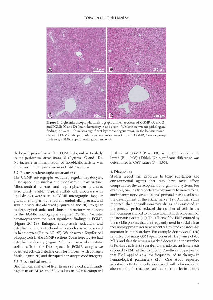
3. Results3.1. Light microscopic observationsThe H&E liver sections were observed under light microscopy. No pathological finding was observed in the CGMR liver specimens, and the parenchyma and portal areas had a normal appearance (Figures 1A and 1B). However, marked hydropic degeneration was present in
3
TOPAL et al. / Turk J Med Sci
the hepatic parenchyma of the EGMR rats, and particularly in the pericentral areas (zone 3) (Figures 1C and 1D). No increase in inflammation or fibroblastic activity was determined in the portal areas in EGMR sections.3.2. Electron microscopic observationsThe CGMR micrographs exhibited regular hepatocytes, Disse space, and nuclear and cytoplasmic ultrastructure. Mitochondrial cristae and alpha-glycogen granules were clearly visible. Typical stellate cell processes with lipid droplet were seen in CGMR micrographs. Regular granular endoplasmic reticulum, endothelial process, and sinusoid were also observed (Figures 2A and 2B). Irregular nuclear, cytoplasmic, and sinusoid structures were seen in the EGMR micrographs (Figures 2C–2F). Necrotic hepatocytes were the most significant findings in EGMR (Figure 2C–2F). Enlarged endoplasmic reticulum and cytoplasmic and mitochondrial vacuoles were observed in hepatocytes (Figure 2C–2F). We observed Kupffer cell phagocytosis in the EGMR sections. Some hepatocytes had cytoplasmic density (Figure 2F). There were also mitotic stellate cells in the Disse space. In EGMR samples we observed activated stellate cells for fibrosis (with collagen fibrils; Figure 2E) and disrupted hepatocyte cord integrity.3.3. Biochemical resultsBiochemical analysis of liver tissues revealed significantly higher tissue MDA and SOD values in EGMR compared
to those of CGMR (P = 0.08), while GSH values were lower (P = 0.08) (Table). No significant difference was determined in CAT values (P = 1.00).
4. DiscussionStudies report that exposure to toxic substances and environmental agents that may have toxic effects compromises the development of organs and systems. For example, one study reported that exposure to nonsteroidal antiinflammatory drugs in the prenatal period affected the development of the sciatic nerve (18). Another study reported that antiinflammatory drugs administered in the prenatal period reduced the number of cells in the hippocampus and led to dysfunction in the development of the nervous system (19). The effects of the EMF emitted by the mobile phones that are frequently used in social life as technology progresses have recently attracted considerable attention from researchers. For example, Sonmez et al. (20) reported that many GSM operators used a frequency of 900 MHz and that there was a marked decrease in the number of Purkinje cells in the cerebellum of adolescent female rats exposed to EMF at that frequency. Another study reported that EMF applied at a low frequency led to changes in hematological parameters (21). One study reported genotoxic effects in cells associated with chromosomal aberration and structures such as micronuclei in mature
Figure 1. Light microscopic photomicrograph of liver sections of CGMR (A and B) and EGMR (C and D) (stain: hematoxylin and eosin). While there was no pathological finding in CGMR, there was significant hydropic degeneration in the hepatic paren-chyma of EGMR rats, particularly in pericentral areas (zone 3). CGMR, Control group male rats; EGMR, experimental group male rats.

4
TOPAL et al. / Turk J Med Sci
Figure 2. Transmission electron microscopic (TEM) photomicrographs of the liver in CGMR (A and B) and EGMR (C–F). In CGMR, normal hepatocytes with regular nu-clear structure (n in 2A) and cytoplasmic ultrastructure (h in 2B) and Disse space (d in 2B), clear mitochondrial cristae (m in 2A), alpha-glycogen granules (spiral arrow in 2A and 2B), stellate cell (arrow in 2A and 2B), process (arrow head in 2B) with lipid droplet (asterisk in 2B), and microvilli (arrow in 2A and 2B) can be seen. In EGMR, necrotic hepatocytes (x in 2F) with irregular nuclear structure (n in 2C) and dense cytoplasm (asterisk in 2F) with vacuoles (v in 2E) can be seen. Cytoplasmic (v in 2C) and mito-chondrial (m in 2C and 2F; arrow head in 2D and 2E) vacuoles (spiral arrow in 2C and 2D), enlarged endoplasmic reticulum (arrow in 2C and 2D), alpha-glycogen (tail arrow in 2C and 2D; spiral arrow in 2F), stellate cell (asterisk in 2E) in Disse space, microvilli (tail arrow in 2E), part of fragmented hepatocyte (arrow in 2E), and bile canaliculi (b in 2F) with microvilli (arrow in 2F) were also seen in EGMR. CGMR, Control group male rats; EGMR, experimental group male rats (TEM: 10,000× for A; 15,000× for B and D; 6000× for C and E; 25,000× for F).
Table. Biochemical analysis results from liver tissues from CGMR and EGMR.
Biochemical parameters CGMR (n = 6)(mean ± SD)
EGMR (n = 6)(mean ± SD) P
MDA (nmol/mg tissue) 13.11 ± 0.83 18.31 ± 1.26 a 0.008*
SOD (mmol min–1 mg–1 tissue) 1.49 ± 0.05 1.77 ± 0.01a 0.008*
GSH (nmol/mg tissue) 1.20 ± 0.07 0.76 ± 0.02b 0.008*
CAT (mmol min–1 mg–1 tissue) 0.017 ± 0.001 0.018 ± 0.003c 1.00
*P < 0.05. Values with different superscripted letters are significantly different.SD, Standard deviation; CGMR, control group male rats; EGMR, experimental group male rats; MDA, malondialdehyde; SOD, superoxide dismutase; GSH, glutathione; CAT, catalase
5
TOPAL et al. / Turk J Med Sci
and immature rats exposed to long-term application of an 1800-MHz EMF (22). Şekeroğlu et al. (22) showed the irreversible effects of cytogenotoxic damage, especially in immature rats.
In growing rats, EMF at a frequency of 900 MHz has been reported to lead to moderate desquamation and vacuolization in the epithelium of testicular seminiferous tubules, while at a frequency of 1800 MHz it led to pathological findings such as severe vacuolization, necrosis, and desquamation in the seminiferous tubule epithelium (23). Odacı et al. (24) determined a decrease in granular cell numbers in the dentate gyrus of rat pups exposed to a 900-MHz EMF in the prenatal period. Another study investigating the effects of EMF in the prenatal period reported a decrease in testicular seminiferous tubule diameter (25).
All these studies clearly show that EMFs applied in the prenatal period have adverse effects on various organs (26,27). However, the effects on the liver of EMF applied in the prenatal period are still unknown, due to the lack of sufficient studies on the subject. Gokcimen et al. (28) reported in their study of young male rats that a magnetic field led to sinusoidal dilatation in the parenchyma and periportal area of liver tissue. Sinusoidal expansion and vacuoles surrounded by membrane in light microscopy and TEM findings were reported in rabbit liver sections exposed to a 650-MHz EMF for 12 months in another study. In the group exposed to EMF for 18 months in that study, irregularity was observed in sinusoidal lumen diameters, the cell cytoplasm was empty and replaced by granules, chromatin was less condensed, and the space between the interior and exterior nuclear membrane expanded (29). Another study referred to EMF altering mitochondrial structures, particularly in hepatocytes (30).
EMF applied to chicken embryos was reported to lead to cytoplasmic degenerations in liver cells (31). Similar to Lahijani et al.’s (31) findings, necrotic hepatocytes with an irregular nucleus and irregular cytoplasm structure were also observed in EGMR pups in our study. Histopathological findings such as expansions in endoplasmic reticulum, vacuoles in mitochondria, and active stellate cell fibrosis observed in EGMR sections in our study show the adverse effects on the liver in the postnatal period of 900-MHz EMF applied in the prenatal period. Our microscopic evaluations also confirm our TEM findings, because significant hydropic degeneration was determined in the pericentral area (zone 3) in slides from the EGMR group.
One study involving an adult male rat model established that a 128-mT static magnetic field increased MDA, an indicator of oxidative stress, in the testes, and that this stress increased levels of 8-oxo-dG, a determinant of DNA injury (32). MDA and SOD levels have been reported to increase and GSH to decrease in rats exposed to EMF at a frequency of 945 MHz and with a SAR value of 11.3 mW/kg (31). High-frequency 4.7-T EMF has been shown to lead to LPO in mice by increasing TBARS (30).
Studies have reported that exposure to EMF increases free radical production in rats and leads to DNA damage and oxidative stress in the liver (33,34). Koyu et al. (35) reported that the oxidative stress established in the liver by a 900-MHz EMF led to hepatic injury and lipid peroxidation in association with P-450-mediated organic hydroperoxide metabolism activation of hydroxyl radicals. Another study reported that EMF applied to the liver caused a rise in bilirubin, MDA, and SOD levels and suggested that this increase in SOD activity might be associated with a rise in anion structures (36). Güler et al. (37) reported that a 50-kHz electric field led to a rise in TBARS levels in plasma, liver, lung, and kidney tissues and that this increase showed tissue damage. They also reported that a rise in SOD levels was an indicator of ROS production against this damage and that this showed the physiopathological dimension of the exposure (37). GSH is an important ROS scavenger, represents the first line in antioxidant defense, and protects the cells against oxidative damage. However, elevated oxidative stress compromises the adaptive mechanism by suppressing GSH. Studies have reported that a rise in SOD is a response to that suppression (38). Another study stated that GSH protects hepatic cells against the chemical and enzymatic effects of oxidative damage (28). The biochemical findings from our study are similar to those from these studies. The rise in MDA levels in our study shows the presence of oxidative stress. On the basis of our biochemical data, we think that the 900-MHz EMF that we applied in the prenatal period led to a decrease in levels of GSH, a good scavenger of ROS in the enzymatic antioxidant defense system, and increased SOD levels as a response to that inhibition. We therefore report that EMF applied in the prenatal period causes oxidative damage and changes in the antioxidant defense system in the rat pup liver.
In conclusion, a 900-MHz EMF applied in the prenatal period led to oxidative stress in the rat pup liver and increased SOD levels as a response to oxidative stress, and hepatocytes may appear as significant pathological findings and this may affect the development of the liver.

6
TOPAL et al. / Turk J Med Sci
References
1. Ozguner F, Oktem F, Ayata A, Koyu A, Yilmaz HR. A novel antioxidant agent caffeic acid phenethyl ester prevents long-term mobile phone exposure-induced renal impairment in rat. Mol Cell Biochem 2005; 277: 73–80.
2. Maaroufi K, Had-Aissouni L, Melon C, Sakly M, Abdelmelek H, Poucet B, Save E. Spatial learning, monoamines and oxidative stress in rats exposed to 900 MHz electromagnetic field in combination with iron overload. Behav Brain Res 2014; 258: 80–89.
3. Aydin B, Akar A. Effects of a 900-MHz electromagnetic field on oxidative stress parameters in rat lymphoid organs, polymorphonuclear leukocytes and plasma. Arch Med Res 2011; 42: 261–267.
4. Esmekaya MA, Aytekin E, Ozgur E, Güler G, Ergun MA, Omeroğlu S, Seyhan N. Mutagenic and morphologic impacts of 1.8 GHz radiofrequency radiation on human peripheral blood lymphocytes (hPBLs) and possible protective role of pre-treatment with Ginkgo biloba (EGb 761). Sci Total Environ 2011; 410–411: 59–64.
5. Kiray A, Tayefi H, Kiray M, Bagriyanik HA, Pekcetin C, Ergur BU, Ozogul C. The effects of exposure to electromagnetic field on rat myocardium. Toxicol Ind Health 2013; 29: 418–425.
6. Hekmat A, Saboury AA, Moosavi-Movahedi AA. The toxic effects of mobile phone radiofrequency (940 MHz) on the structure of calf thymus DNA. Ecotoxicol Environ Saf 2013; 88: 35–41.
7. Deng Y, Zhang Y, Jia S, Liu J, Liu Y, Xu W, Liu L. Effects of aluminum and extremely low frequency electromagnetic radiation on oxidative stress and memory in brain of mice. Biol Trace Elem Res 2013; 156: 243–252.
8. Ozturk A, Baltaci AK, Mogulkoc R, Oztekin E. Zinc prevention of electromagnetically induced damage to rat testicle and kidney tissues. Biol Trace Elem Res 2003; 96: 247–254.
9. Salem A, Hafedh A, Rached A, Mohsen S, Khémais BR. Zinc prevents hematological and biochemical alterations induced by static magnetic field in rats. Pharmacol Rep 2005; 57: 616–622.
10. Hancı H, Odacı E, Kaya H, Aliyazıcıoğlu Y, Turan İ, Demir S, Çolakoğlu S. The effect of prenatal exposure to 900-MHz electromagnetic field on the 21-old-day rat testicle. Reprod Toxicol 2013; 42: 203–209.
11. İkinci A, Odacı E, Yıldırım M, Kaya H, Akça M, Hancı H, Aslan A, Sönmez OF, Baş O. The effects of prenatal exposure to a 900 megahertz electromagnetic field on hippocampus morphology and learning behavior in rat pups. NeuroQuantology 2013; 11: 582–590.
12. Baş O, Sönmez OF, Aslan A, İkinci A, Hancı H, Yıldırım M, Kaya H, Akça M, Odacı E. Pyramidal cell loss in the cornu ammonis of 32-day-old female rats following exposure to a 900 megahertz electromagnetic field during prenatal days 13–21. NeuroQuantology 2013; 11: 591–599.
13. Odacı E, İkinci A, Yıldırım M, Kaya H, Akça M, Hancı H, Sönmez OF, Aslan A, Okuyan M, Baş O. The effects of 900 megahertz electromagnetic field applied in the prenatal period on spinal cord morphology and motor behavior in female rat pups. NeuroQuantology 2013; 11: 573–581.
14. Sun Y, Oberley LW, Li Y. A simple method for clinical assay of superoxide dismutase. Clin Chem 1988; 34: 497–500.
15. Aebi H. Catalase. Medhods Enzymol 1984; 105: 121–126.
16. Sedlak J, Lindsay RH. Estimation of total, protein-bound, and non- protein sulfhydryls groups in tissue with Ellman’s reagent. Anal Biochem 1968; 25: 192–205.
17. Ohkawa H, Ohishi H, Yagi K. Assay for lipid peroxide in animal tissues by thiobarbituric acid reaction. Anal Biochem 1979; 95: 351–358.
18. Canan S, Aktaş A, Ulkay MB, Colakoglu S, Ragbetli MC, Ayyildiz M, Geuna S, Kaplan S. Prenatal exposure to a non-steroidal anti-inflammatory drug or saline solution impairs sciatic nerve morphology: a stereological and histological study. Int J Dev Neurosci 2008; 26: 733–738.
19. Gokcimen A, Rağbetli MC, Baş O, Tunc AT, Aslan H, Yazici AC, Kaplan S. Effect of prenatal exposure to an anti-inflammatory drug on neuron number in cornu ammonis and dentate gyrus of the rat hippocampus: a stereological study. Brain Res 2007; 1127: 185–192.
20. Sonmez OF, Odaci E, Bas O, Kaplan S. Purkinje cell number decreases in the adult female rat cerebellum following exposure to 900 MHz to electromagnetic field. Brain Res 2010; 1356: 95–101.
21. Cakir DU, Yokus B, Akdag MZ, Sert C, Mete N. Alterations of hematological variations in rats exposed to extremely low frequency magnetic fields (50 Hz). Arch Med Res 2009; 40: 352–356.
22. Şekeroğlu V, Akar A, Atlı Şekeroğlu Z. Cytotoxic and genotoxic effects of high-frequency electromagnetic fields (GSM 1800 MHz) on immature and mature rats. Ecotoxicol Environ Saf 2012; 80: 140–144.
23. Nisbet HO, Nisbet C, Akar A, Cevik M, Karayigit MO. Effects of exposure to electromagnetic field (1.8/0.9 GHz) on testicular function and structure in growing rats. Res Vet Sci 2012; 93: 1001–1005.
24. Odaci E, Bas O, Kaplan S. Effects of prenatal exposure to a 900 MHz electromagnetic field on the dentate gyrus of rats: a stereological and histopathological study. Brain Res 2008; 1238: 224–229.
25. Tenorio BM, Jimenez GC, Morais RN, Torres SM, Albuquerque Nogueira R, Silva Junior VA. Testicular development evaluation in rats exposed to 60 Hz and 1 mT electromagnetic field. J Appl Toxicol 2011; 31: 223–230.
26. Koç A, Ünal D, Çimentepe E, Bayrak Ö, Karataş ÖF, Yıldırım ME, Bayrak R, Aydın M. The effects of antioxidants on testicular apoptosis and oxidative stress produced by cell phones. Turk J Med Sci 2013; 43: 131–137.

7
TOPAL et al. / Turk J Med Sci
27. Demir T, Gültürk S, Çançalar AD, Durmuş N. Investigation of the effects of magnetic field exposure on febrile seizure latency, seizure duration, and electroencephalographic recordings in a rat febrile convulsion model. Turk J Med Sci 2014; 44: 295–304.
28. Gökcimen A, Ozgüner F, Karaöz E, Ozen S, Aydin G. The effect of melatonin on morphological changes in liver induced by magnetic field exposure in rats. Okajimas Folia Anat Jpn 2002; 79: 25–31.
29. Tarantino P, Lanubile R, Lacalandra G, Abbro L, Dini L. Post-continuous whole body exposure of rabbits to 650 MHz electromagnetic fields: effects on liver, spleen, and brain. Radiat Environ Biophys 2005; 44: 51–59.
30. Watanabe Y, Nakagawa M, Miyakoshi Y. Enhancement of lipid peroxidation in the liver of mice exposed to magnetic field. Ind Health 1997; 35: 285–290.
31. Lahijani MS, Tehrani DM, Sabouri E. Histopathological and ultrastructural studies on the effects of electromagnetic fields on the liver of preincubated white leghorn chicken embryo. Electromagn Biol Med, 2009; 28: 391–413.
32. Amara S, Abdelmelek H, Garrel C, Guiraud P, Douki T, Ravanat JL, Favier A, Sakly M, Ben Rhouma K. Effect of subchronic exposure to static magnetic field on testicular function in rats. Arch Med Res 2006; 37: 947–952.
33. Yurekli AI, Ozkan M, Kalkan T, Saybasili H, Tuncel H, Atukeren P, Gumustas K, Seker S. GSM base station electromagnetic radiation and oxidative stress in rats. Electromagn Biol Med 2006; 25: 177–188.
34. Amara S, Abdelmelek H, Garrel C, Guiraud P, Douki T, Ravanat JL, Favier A, Sakly M, Ben Rhouma K. Zinc supplementation ameliorates static magnetic field-induced oxidative stress in rat tissues. Environ Toxicol Pharmacol 2007; 23: 193–197.
35. Koyu A, Ozguner F, Yilmaz H, Uz E, Cesur G, Ozcelik N. The protective effect of caffeic acid phenethyl ester (CAPE) on oxidative stress in rat liver exposed to the 900 MHz electromagnetic field. Toxicol Ind Health 2009; 25: 429–434.
36. Emre M, Cetiner S, Zencir S, Unlukurt I, Kahraman I, Topcu Z. Oxidative stress and apoptosis in relation to exposure to magnetic field. Cell Biochem Biophys 2011; 59: 71–77.
37. Güler G, Seyhan N, Aricioğlu A. Effects of static and 50 Hz alternating electric fields on superoxide dismutase activity and TBARS levels in guinea pigs. Gen Physiol Biophys 2006; 25: 177–193.
38. Ghodbane S, Amara S, Garrel C, Arnaud J, Ducros V, Favier A, Sakly M, Abdelmelek H. Selenium supplementation ameliorates static magnetic field-induced disorders in antioxidant status in rat tissues. Environ Toxicol Pharmacol 2011; 31: 100–106.