the effects of con a on cell surface shedding in cell cultures
TRANSCRIPT

J. Cell Sci. 46, 221-234 (1980) 221Printed in Great Britain © Company of Biologists Limited 1980
THE EFFECTS OF CON A ON CELL SURFACE
SHEDDING IN CELL CULTURES
THOMAS C.DOETSCHMAN*Biological Sciences Group, Genetics and Cell Biology Section, U-iz$,University of Connecticut, Storrs, CT. 06268 U.S.A.
SUMMARY
A cell surface immobilizing concentration of Con A inhibits the shedding of [*H]fucose-containing glycoproteins from the surface of chick embryonic leg and breast muscle cellcultures and cultures of the rat skeletal muscle cell line L6. The Con A-induced inhibition ofshedding is much less in the mouse fibroblast cell line 3T3. In all 4 cell types the lectin inhibitsthe shedding of some fucosyl-glycoproteins more than others, especially those of a lipid-containing fraction which is excluded in Biogel A-51T1 chromatography. This differentialnature of the Con A effect is not changed by the cytoskeletal disrupters cytochalasin B orcolchicine. Con A causes an increase in the amount of trypsin-sensitive surface fucosyl-glycoprotein in the cell surface and appears to decrease the overall amount of cell surfacedegradation suggesting that the inhibition of shedding caused by Con A is not due to anincrease in internalization and degradation. The data suggest that some shedding may occur atspecific cell surface sites to which surface materials must laterally migrate.
INTRODUCTION
The effects of Con A on cell surface shedding in cell cultures
The mechanisms involved in cell surface shedding are poorly understood. Cellsurface proteolytic activity is one of the mechanisms by which shedding can occur(Kapeller, Gal-Oz, Grover & Doljanski, 1973; Doljanski & Kapeller, 1976; Hynes,1974, 1976; Baumann & Doyle, 1978; Mosher & Vaheri, 1978; Parry, 1978). Sheddingcan also occur by the release of plasma membrane vesicles as suggested by somestudies (Peterson & Rubin, 1969; Vitetta & Uhr, 1972; Nowotny et al. 1974; Doetsch-man, 1980a) and demonstrated by others (Stanbridge & Weiss, 1978). Cell surfacecaps (Karnovsky, Unanue & Leventhal, 1972; Leonard, 1973; Stanbridge & Weiss,1978; Nicolson, 1979) can be the site of shedding, and shedding may also occur atcoated endocytotic pits (Brown, Yeh & Holley, 1979). With the exception of proteo-lysis most of these processes would require that the molecules to be shed have somelateral mobility within the plasma membrane. Consequently, agents known to interferewith cell surface mobility would be useful in probing the mechanisms of cell surfaceshedding.
At high concentrations Con A loses its mitogenic properties and inhibits thepatching and capping of many cell surface receptors (Yahara & Edelman, 1975).
• Present address: ETH-Ziirich, Institut fur Zellbiologie, HOnggerberg, CH-8093 Zurich,Switzerland.

222 T. C. Doetschman
Con A has been shown to decrease the lateral diffusion of Con A receptors(Schlessinger et al. 1976) and of other receptors as well (Schlessinger et al. 1977).
Because of the wide range of its effects on cell surface mobility, Con A was used inthis study to investigate the relationship between shedding and cell surface mobility.Con A is shown to have an inhibitory effect on shedding and this does not appear tobe the result of increased internalization and degradation of surface molecules. Sincethe modulation of surface mobilities by Con A can be partially reversed in someinstances by colchicine (Yahara & Edelman, 1975; Schlessinger et al. 1977), andpatching and capping of cell surface receptors can be inhibited by cytochalasin B(Edelman, 1976), these drugs were also used simultaneously with Con A and werefound to have little effect on the Con A inhibition of shedding. The results suggestthat at least some surface glycoproteins are shed in the form of membranous vesiclesat specific cell surface sites.
MATERIALS AND METHODS
Cells were mechanically dissociated from leg or breast muscles of 11 -day chick embryos aspreviously described (Tepperman, 1972; Tepperman, Morris, Essien & Heywood, 1975). Therat muscle cell line L6 was a gift from Dr David Yaffe (Weizmann Institute, Israel). All musclecells were cultured on Falcon plastic dishes coated with 3 % gelatin (Difco). The culturemedium was prepared as previously described (Herrmann, Havaranis & Doetschman, 1975),except that no HEPES-TES buffer was used for the L6 cells since it is toxic to them. Themedium for the 3T3 cells consisted of 0 2 % NaHCO3-buffered DME nutrient mixture,10% foetal calf serum (Gibco), 200 units/ml penicillin, and 200 fig/ml streptomycin. The3T3 cells were at subconfluent densities throughout the experimental periods.
The cultures were labelled with L-[6-3H]fucose (i3'4 Ci/mmol, New England Nuclear) withthe amounts indicated in the figure legends and the footnotes to the tables. Before addition tothe cultures the labelled fucose was concentrated to dryness and redissolved in 1/20 volume95 % ethanol in order to keep the ethanol concentration of the medium at a low level. Thelabel was removed by rinsing the cultures 4 times with nutrient mixture. The medium releasedmaterial was then collected in nutrient mixture with 50 fig/ml unlabelled fucose. During therelease period Con A (Miles, 3 times crystallized, lyophilized), cytochalasin B (Sigma, Lot. No.C 6267), and colchicine (Sigma) were added as described in the figure legends and footnotes tothe tables. At the end of the release period phenylmethyl sulphonyl fluoride (Sigma) was addedto all medium released fractions to a final concentration of 16 fig/ml in order to inhibit pro-teolytic activity. Cell debris was removed by centrifugation at 900 g for 10 min. The releasedfraction was dialysed extensively against distilled water, analysed for radioactive content, con-centrated to dryness in a Speed Vac Concentrator (Savant), and dissolved and boiled in electro-phoresis sample buffer containing 2-3 % SDS and 5 % /J-mercaptoethanol for 3 min. Thesamples were electrophoresed according to Laemmli (1970) on slab gels with a 3-step acryla-mide gradient: 3/8 at 12-5 %, 3/8 at 10%, and 1/4 at 7-5 % with a 3 % acrylamide stackinggel. Gels were stained for protein (Heywood & Kennedy, 1979) and then photographed.Fluorograms of the gels were prepared as described previously (Bonnor & Laskey, 1974;Laskey & Mills, 1975).
Effects of Con A on shedding 223
RESULTS
Con A infiibition of shedding
The shedding of fucosyl-glycoprotein is inhibited by Con A (Table 1). This isthe case with all of the muscle cell types tested (leg, breast, and L6) and it occurs inthe presence of cytochalasin B, colchicine, or both. Alone, cytochalasin B has noconsistent effect on the amount shed, and colchicine always inhibits shedding toa lesser degree than Con A. The effect of Con A and colchicine are generally additive.The only exception to these results is found in 3T3 cell cultures where Con A doesnot inhibit shedding as much as in the muscle cells. In one experiment it did notinhibit at all. In the 3T3 cells Con A inhibits shedding less than does colchicine.
In experiment 3 the shed material was collected from 4 to 10 h after removal andchase of the label to provide a control experiment in which the released fraction is notcomplicated by the presence of secreted labelled material. There is evidence that L6cells (Doetschman, 1980&) as well as other cells (Yurchenco & Atkinson, 1977;Doyle et al. 1978; Gottesman, 1978) secrete proteins and glycoproteins within thefirst 4 h after being synthesized. Consequently, by eliminating the presence ofsecreted labelled material from the released fraction, the effects of Con A, cyto-chalasin B, and colchicine could be compared to those found during a time when somelabelled-glycoprotein was being secreted. A comparison of the results of experiments1 and 2 with experiment 3 shows little difference. Consequently, the various effectsdescribed above are primarily on shedding and not secretion.
Differential nature of shedding inhibition
Since Biogel A-51T1 gel exclusion chromatography separates the shed fucosyl-glycoproteins into large (greater than 5 x io6 Daltons) lipid-containing and smallerlipid-free materials (Doetschman, 1980 a) it was of interest to know if Con A inhibitsthe shedding of one of these fractions more than the other. This was found to be thecase in leg muscle and 3T3 cell cultures. The shedding of the lipid-containing fractionwas inhibited by 24±8% (standard error, 4 experiments) in leg and by 32±8%(6 experiments) in 3T3 cultures. The amount shed in the lipid-containing fraction isgiven as the percent of the total amount shed.
Evidence for the shedding inhibition and its differential nature is also found influorograms of the 3H-fucosylated glycoproteins that are shed into the medium(Fig. 1). Con A inhibits the shedding of some glycoproteins more than others. Thoseaffected the most migrate slower than the albumin marker. In fact, the differentialCon A effect is more evident in the 3T3 cultures where Con A has less of an inhibitoryeffect on the total amount of shedding than it is in the muscle cultures (Fig. 1 c). This isexpected because of the greater inhibition of shedding of the lipid-containing gelexclusion fraction in the 3T3 cells. The differential nature of the Con A inhibition isunaltered even if cytochalasin B and/or colchicine are present with the Con A duringthe release period.
In a similar experiment the differential effects of Con A were more strongly evident.The results are shown in Fig. 2. There are several intense bands in the fluorogram of

Tab
le I
. Q
uant
itat
ive
effe
cts
of Con A
, cy
toch
alas
in B
, and
cok
hine
on
the
shed
ding
of f
ucos
yl-g
lyco
prot
eim
Am
ount
rel
ease
d as
per
cent
of
cont
rola
Add
itio
ns
Exp
. I
Exp
. 2
Exp
. 3
Exp
. I
Exp
. z
Exp
. 3
Exp
. I
Exp
. 2
Ex
p.
3
Exp
. I
Exp
. 2
Exp
.3
Non
e (c
ontr
ol)
Con
-A
CB
C
OL
C
on-A
& C
B
Con
-A &
CO
L
Con
-A &
CB
& C
OL
C
B &
CO
L
a O
ne c
ultu
re p
er d
eter
min
atio
n.
Par
enth
eses
ind
icat
e th
e to
tal
dp
m x
lo
-= in
the
con
trol
cul
ture
s.
Exp
erim
ent
I.
Cel
ls w
ere
plat
ed a
t 5.
0 x
106 li
ve c
ells
/~m
-mm
plat
e (l
eg),
4.0
x
lo6
live
cel
ls/r
oo-m
m p
late
(br
east
), 1
.0 x
~
o~
ce
lls
/~m
-rn
m
plat
e (U),
and
1.5
x 10'
cell
s/~
oo
-mm
plat
e (3
T3)
. A
ll c
ultu
res
wer
e la
bell
ed w
ith
20
0 p
Ci
[WJf
ucos
e fr
om 4
5 to
69
h of
cu
ltu
re (
leg,
bre
ast,
L6
) or
fro
m 1
8 to
42
h of
cu
ltu
re (
3T3)
in c
ompl
ete
med
ium
aft
er w
hich
the
lab
el w
as r
emov
ed.
Th
e sh
ed f
ract
ion
was
col
lect
ed i
n c
hase
med
ium
for
th
e ne
xt 6
h i
n th
e pr
esen
ce o
f va
riou
s co
mbi
nati
ons
of C
on
A (
10 p
g/m
l),
cyto
chal
asin
B (
CB
, 5
,ug/
rnl)
, an
d c
olch
icin
e (C
OL
, 5
pg/m
l).
Th
ese
sam
e co
ncen
trat
ions
wer
e us
ed i
n e
xper
imen
ts 2
and
3.
Exp
erim
ent 2. C
ells
wer
e pl
ated
at
1.5
x 10'
live
cell
s/b
-mm
pla
te (
leg,
bre
ast)
, 5.
0 x
rob
cell
s/&
-mm
pl
ate
(L6)
, an
d 3
.0 x
10'
celh
/6o
-mm
pl
ate
(3T
3).
Cul
ture
s w
ere
labe
lled
wit
h 100 p
Ci
[SH
]fuc
ose
from
24 to
48
h of
cul
ture
in
com
plet
e m
ediu
m.
Th
e s
hed
fra
ctio
n w
as c
olle
cted
in
th
e pr
esen
ce o
f th
e va
riou
s ad
diti
ons
from
48
to 5
4 h.
E
xper
imen
t 3.
Cel
ls w
ere
plat
ed a
t 4.0
x
roe
live
cel
ls/r
m-m
m p
late
(br
east
), 3
.0 x
ro
o ce
lls/
~o
o-m
m pl
ate (U),
and
1.5
x
106 ce
lls/
rm-m
m
plat
e (3
T3)
. Cul
ture
s w
ere
labe
lled
wit
h 18
0 p
Ci
[SH
]fuc
ose f
rom
19
to 2
9 h
of c
ultu
re (
brea
st)
and
fro
m 2
4 t
o 4
4 h
(M, 3
T3
) in
com
plet
e m
ediu
m.
Th
e m
ater
ials
rel
ease
d in
to t
he m
ediu
m f
rom
the
nex
t 4
h of
cul
ture
wer
e di
scar
ded
and
tho
se s
hed
in
th
e pr
esen
ce o
f th
e ad
diti
ons
du
rin
g t
he
foll
owin
g 6
h w
ere
coll
ecte
d.

Effects of Con A on shedding 225
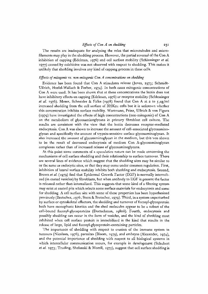
that gel which are in all of the samples except those in which Con A was present duringthe release period. Most of these bands can be found between the top of the gel andthe albumin marker. Inversely, there are bands present in Con A-treated sampleswhich are more intense relative to other bands in the same sample than are thecorresponding bands in the samples without Con A. The differential effect of Con A,regardless of the presence or absence of cytochalasin B or colchicine is also evident inFig. 2. In the culture treated with Con A plus cytochalasin B (well 6) there are 3 bandsof about 70000, 44000, and less than 44000 Daltons which are not as intense in theother Con A-treated cultures presented in Fig. 2. This result occurred in only 1 experi-ment and will be discussed later. It does not, however, detract from the conclusionthat Con A has a differential effect on shedding.
Con A inhibition of internalization and degradation
The overall reduction of the amount of pHJfucose-containing glycoprotein shedin the presence of Con A could be caused by variations in 2 parameters: the amountof surface fucose-labelled glycoproteins capable of being shed and the rate of shedding.The degree to which each of these parameters is involved in the Con A effect wasinvestigated next in breast muscle cell cultures. Because of the possibilities of cellsurface recycling (Schneider, Tulkens, de Dure & Trouet, 1979) and cytoplasmicstorage forms of plasma membrane (Doyle et al. 1978), it is not known for certainhow much of the fucosyl-glycoprotein in the cells is at the surface or in the cytoplasmat any given time. Trypsin is usually thought to act only at the surface of viable cells.For this reason the Con A effect on the trypsin-sensitive and total cell bound fucosyl-glycoprotein was measured. The results are shown in Table 2. Con A increasesdramatically the trypsin-sensitive surface fucosyl-glycoprotein. The reduction inshedding, however, does not completely account for the increased trypsin-sensitivesurface glycoprotein.
There are 3 possible interpretations of these results. First, if the trypsin-removablefucosyl-glycoproteins are assumed to represent the major portion of the fucosyl-glycoproteins in the cell surface and the trypsin-insensitive material consists ofinternalized recycling or precursor plasma membrane, then the increase of approxi-mately 22 points in the percentage of total label in the culture that is trypsin-sensitiveis much more than what can be accounted for by the decrease in shedding which isfrom 8 to 12 % of total label. This interpretation suggests that endocytosis is inhibitedby Con A as well. Second, there is some evidence that trypsin can cause the release ofcytoplasmic glycoprotein destined for secretion (Jett & Jamieson, 1973). This isprobably not occurring here because the trypsin was added after 6 h of release ina fucose chase medium. It has already been shown that little labelled glycoprotein issecreted at this time (Doetschman, 19806). Third, it is possible that the cell-boundfraction represents the cell surface fucosyl-glycoprotein and that the Con A effect ontrypsin sensitivity is due to a change in distribution, orientation, or configuration ofsurface materials as occurs in the microredistribution and cooperative binding effectsof Con A (Bornens, Karsenti & Avrameas, 1976; Karsenti, Bornens & Avrameas,1977; Prujanski, Ravid & Sharon, 1978; Gordon & Young, 1979). A decrease in

226
a -
o-
6 7- 8^iL 10
T. C. Doetschman
B7 8 9 10
It
W
I I-a
-o
2 3 4 5 6 7 8 9 1 0
•liii-i

Effects of Con A on shedding 227
intemalization and degradation is not necessarily consistent with this interpretation ofthe Con A effect on shedding. However, since the total amount of fucosyl-glycoproteinper culture is greater by 13 + 9% in the Con A-treated cultures (last column inTable 2), intemalization and degradation are necessarily decreased.
2 3 4 5 6 7 8 9
a -
o-
Fig. 2. Electrophoretic profiles of the PHJfucosyl-glycoproteins shed from leg musclecell cultures in the presence of combinations of Con A, cytochalasin B, and colchicine.The samples were obtained from Experiment 1 of Table 1 and were prepared as des-cribed in the footnotes to Table 1. The amount of radioactivity in each sample canbe calculated from Table 1. No additions (control), 2; Con A, 3; cytochalasin B, 4;colchicine, 5; Con A and cytochalasin B, 6; Con A and colchicine, 7; Con A, cyto-chalasin B, and colchicine, 8; and cytochalasin B and colchicine, 9. The molecularweights of the markers are 200000 for myosin (m), 68000 for albumin (a), and44000 for ovalbumin (o).
Fig. 1. Electrophoretic profiles of the [3H]fucosyl-glycoproteins shed in the presenceof combinations of Con A, cytochalasin B, and colchicine. The samples were obtainedfrom Experiment 2 of Table 1 and were prepared as described in the footnotes toTable 1. The amount of radioactivity in each sample can be calculated from Table 1.Leg (A), breast (B), 3T3 (c), L6 (D); Unlabelled molecular weight markers, 1 and 10;no additions (control), 2; Con A, 3; cytochalasin B, 4; colchicine, 5; Con A andcytochalasin B, 6; Con A and colchicine, 7; Con A, cytochalasin B, and colchicine, 8;cytochalasin B and colchicine, 9. The molecular weights of the markers are 200000for myosin (m), 68000 for albumin (a), and 44000 for ovalbumin (o).

Tab
le 2
. E
flect
of
Co
n A
on
the
cell
ulm
dis
trib
utio
n of
fuco
syl-
glyc
opro
tkns
-
Lab
el f
ound
in
each
cel
lula
r co
mpa
rtm
ent
as p
erce
nt o
f to
tal
.A I
> T
reat
men
t C
ell-
boun
d S
hed
T
ryps
in-s
ensi
tive
T
ota
l, d
pm
/cu
ltu
re
Tim
e, h
, W
&
&
of
rel
ease
C
on A
T
ryp
sin
E
xp
.1
Ex
p.2
E
xp
.1
Ex
p.2
E
xp
.1
Ex
p.2
E
xp.
I E
xp.
2
0
-
-
o
o 6
1260
00
6800
0 .Y
97
94
3 0
-
+ 73
65
0
0
27
3 5
1470
00
log
00
0 9
6 -
-
75
63
24
34
I 3
~m
wo
65
000
b
6 -
+ 57
40
2
2
29
21
3 I
113-
7
60
00
%
6 +
-
82
73
I7
25
I 2
-, 11
6000
72
000
6 +
+ 42
31
I4
I7
44
5 2
I
95-
f C
hick
em
bryo
nic
brea
st m
uscl
e ce
lls
wer
e pl
ated
at
5.0
x ro
e li
ve c
ells
/~o
o-m
m pl
ate
and
lab
elle
d w
ith
20
pC
i [*
H]f
ucos
e fr
om 2
4 to
42
h of
cu
ltur
e. T
he
labe
l was
rem
oved
and
th
e cu
ltur
es r
inse
d 4
tim
es i
n m
acro
mol
ecul
e-fr
ee m
ediu
m.
At
42 h
of
cult
ure
2 s
ets
of c
ultu
res
wer
e tr
eate
d w
ith
or w
itho
ut 0
.05
% tr
ypsi
n in
Cae
+-,
Mg8
+-f
ree P
BS
for
15
min
. The
cell
s w
ere
then
scr
aped
off
the
pla
tes
and
cen
trif
uged
at
900
g f
or 1
0 m
in.
Por
tion
s of
the
sup
erna
tant
s (t
ryps
in-s
ensi
tive
fu
cosy
l-gl
ycop
rote
in)
and
the
cel
l pe
llet
s (c
ell-
boun
d fu
cosy
l-gl
ycop
rote
in)
wer
e m
easu
red
for
radi
oact
ivit
y. T
he
rem
aini
ng c
ultu
res
wer
e in
cuba
ted
eith
er i
n th
e pr
esen
ce o
r ab
senc
e of
10 p
g/m
l C
on A
du
rin
g a
6-h
rel
ease
per
iod
(42
to 4
8 h
of c
ultu
re).
Th
e s
hed
frac
tion
was
th
en r
emov
ed a
nd t
he
cell
s tr
eate
d w
ith
or
wit
hout
try
psin
as
desc
ribe
d ab
ove.
Th
e c
ell-
boun
d, s
hed,
an
d
tryp
sin-
sens
itiv
e fr
acti
ons
wer
e th
en a
naly
sed
for
radi
oact
ivit
y. D
ata
from
2 c
ultu
res
wer
e av
erag
ed f
or e
ach
dete
rmin
atio
n.

Effects of Con A on shedding 229
DISCUSSION
The important findings of this study are that (1) io/tg/ml of Con A inhibitsshedding in general; (2) Con A differentially inhibits the shedding of fucosyl-glyco-proteins that are in large lipid-containing particles; (3) even when combined withcytochalasin B or colchicine, Con A maintains its differential inhibitory effect onshedding; (4) the inhibition is due to a decreased rate of shedding rather than anincreased internalization and degradation; and (5) internalization and degradation arealso inhibited by Con A.
Con A effect is not due to cell death
It is unlikely that the shedding inhibition caused by Con A is a cell death-relatedphenomenon. First, the inhibition is of the same magnitude in the L6 cells as in theprimary cultures (Table i), yet the amount of cell death in these 2 culture systems isquite different, 2 and 15%, respectively (Doetschman & Jewett, 1980). Similarly,the inhibitory effect is quite different in the L6 and 3T3 cultures (Table 1) whereasthe amount of cell death is the same (Doetschman & Jewett, 1980). Second, the31 to 55 % decrease in shedding caused by Con A (Table 1) cannot be quantitativelyaccounted for by a maximum of 18% contamination due to cell death (Doetschman& Jewett, 1980). Third, if the differential effect of Con A on shedding were dueto cell death then one would expect less of this effect in the L6 and 3T3 than in theprimary cultures. This is definitely not the case (Fig. 1). Fourth, no cell-deathdifferences due to Con A were found in another study (Dunlap & Donaldson, 1978).
Con A effect is on shedding
The Con A inhibition of the release of fucosyl-glycoprotein into the culture mediumis shown to be an effect on shedding rather than on secretion. This is consistent withprevious results which showed that under the same labelling and release conditionsused here, nearly all of the labelled, released fucosyl-glycoproteins are shed and notsecreted. The reason for this is that after a prolonged labelling period the cell surfacebecomes so heavily labelled that the amount of label released by secretion is nearlyundetectable (Doetschman, 19806). The methods used do not indicate whethersecretion is also inhibited by Con A.
Shedding at cell surface sites
The inhibition of shedding caused by Con A at a concentration that inhibits themobility of some cell surface materials (Schlessinger et al. 1976) suggests that there isa shedding site(s) to which surface glycoprotein must migrate (Bretscher, 1976;Stern & Bretscher, 1979; Ukena, Borysenko, Karnovsky & Berlin, 1974; Edelman,1976; Bourguignon & Singer, 1977) before shedding can occur. Cell surface caps(Karnovsky et al. 1972; Leonard, 1973; Stanbridge & Weiss, 1978; Nicolson, 1979)and coated pits (Brown et al. 1979) are possible sites for cell surface shedding. Sincecell surface sites are also involved in receptor-mediated endocytosis via capping(Unanue & Karnovsky, 1973; Albertini & Anderson, 1977) or coated pits (Goldstein,
230 T. C. Doetschman
Anderson & Brown, 1979), Con A would be expected to have an inhibitory effect onendocytosis as well. The evidence presented here is consistent with this possibility.
If there are shedding sites on the cell surface it is possible that the shed materialsoccur in the form of membrane vesicles as in endocytosis. Suggestive evidence fornaturally occurring shed vesicles has been found in several cell culture systems(Peterson & Rubin, 1969; Vitetta & Uhr, 1972; Nowotny et al. 1974; Doetschman,1980a).
Proteolysis is not a major shedding mechanism
Cell surface proteolytic activity has been suggested as one of the mechanisms ofcell surface shedding (Kapeller et al. 1973; Hynes, 1974, 1976; Doljanski & Kapeller,1976; Baumann & Doyle, 1978; Mosher & Vaheri, 1978; Parry, 1978). If a significantamount of shedding is caused by proteolytic activity, then Con A, by increasing theexposure of cell surface glycoproteins to proteolytic attack (Table 2), should increaseshedding. On the contrary, Con A decreases shedding. This does not rule out thepossibility that some shedding occurs by proteolysis, but it does suggest that serineproteases contribute at most only a small degree to shedding. Another study has alsoshown that the serine protease inhibitor phenylmethyl sulphonyl fluoride has noinhibitory effect on shedding (Doetschman & Jewett, 1980).
Roles of microfilaments and microtubules on cell shedding
By using the cytoskeletal disruptors cytochalasin B and colchicine, evidence wasobtained which suggests that microtubules and perhaps microfilaments are involvedin the shedding process. Alone and in conjunction with Con A colchicine inhibitsshedding. There is some evidence that cytochalasin B may stimulate shedding but theeffect is very small. With one exception (Fig. 2) these effects are not differential in thesense that some shed materials are affected to different degrees. Likewise, these drugsdo not change the differential nature of the inhibition of shedding caused by Con A.In other words, although microtubules and microfilaments may affect the rate atwhich surface materials are shed, they confer little selectivity on the major portion ofshed materials. This does not seem to be the case for the shedding of IgM and IgDwhich are inhibited by cytochalasin B and colchicine, respectively (Emerson & Cone,1979). This suggests that cell surface receptors with specific functions may requirespecific regulation mediated by cytoskeletal elements.
It is not uncommon for cytoskeletal perturbants to alter Con A effects in some casesand not in others. Carraway et al. (1979) point out that it may be important to distin-guish the global effects of Con A, where the lectin localized to one small area of thecell surface immobilizes cell surface proteins over the entire surface (Edelman, 1976),from the short-range (Bornens et al. 1976); Karsenti et al. 1977; Gordon & Young,1979) effects of Con A. Applying this distinction to the results presented here, cyto-skeletal elements may not interfere with the global protein-immobilizing effect ofCon A but at the short-range level they may alter the shedding of individual surfacemolecules. This may be the reason for the exception to the cytochalasin B resultsfound here.
Effects of Con A on shedding 231
The results are inadequate for analysing the roles that microtubules and micro-filaments may play in the shedding process. However, the partial reversal of the Con Ainhibition of capping (Edelman, 1976) and cell surface mobility (Schlessinger et al.1977) caused by colchicine was not observed with respect to shedding. This makes itunlikely that shedding involves any kind of capping process in these cells.
Effects of mitogenic vs. non-mitogenic Con A concentrations on shedding
Evidence has been found that Con A stimulates release (Jones, 1973; Schmidt-Ullrich, Hoelzl-Wallach & Ferber, 1974). In both cases mitogenic concentrations ofCon A were used. It has been shown that at these concentrations the lectin does nothave inhibitory effects on capping (Edelman, 1976) or receptor mobility (Schlessingeret al. 1976). Moser, Schneider & Falke (1978) found that Con A at 2 to 5 /ig/mlincreased shedding from the cell surface of BHK21 cells but it is unknown whetherthis concentration inhibits surface mobility. Wortmann, Prinz, Ullrich & von Figura(1979) have investigated the effects of high concentrations (non-mitogenic) of Con Aon the metabolism of glycosaminoglycans in primary fibroblast cell culture. Theresults are consistent with the view that the lectin decreases receptor-mediatedendocytosis. Con A was shown to increase the amount of cell-associated glycosamino-glycan and specifically the amount of trypsin-sensitive surface glycosaminoglycan. Italso increased the amount of glycosaminoglycan in the medium, but this was shownto be the result of decreased endocytosis of medium Con A-glycosaminoglycancomplexes rather than of increased release of glycosaminoglycans.
At this point some comments of a speculative nature can be made concerning themechanisms of cell surface shedding and their relationship to surface turnover. Thereare several lines of evidence which suggest that the shedding sites may be similar toor the same as endocytic sites, or that they may come under common regulation. First,inhibition of lateral surface mobility inhibits both shedding and endocytosis. Second,Brown et al. (1979) find that Epidermal Growth Factor (EGF) is normally internali-zed (in coated vesicles) by fibroblasts, but when antibody to EGF is present the factoris released rather than internalized. This suggests that some kind of a filtering systemmay exist at coated pits which selects some surface materials for endocytosis and somefor shedding. A cell surface site with some of these properties has been hypothesizedpreviously (Bretscher, 1976; Stern & Bretscher, 1979). Third, in a system unperturbedby surface or cytoskeletal effectors, the shedding and turnover of fucosyl-glycoproteinboth have monophasic kinetics and the shed molecules appear to be a subset of thecell-bound fucosyl-glycoproteins (Doetschman, 19806). Fourth, endocytosis andpossibly shedding can occur in the form of vesicles, and the kind of shedding mostinhibited when cell surface protein is immobilized is the kind that results in therelease of large, lipid and fucosyl-glycoprotein-containing particles.
The importance of shedding with respect to evasion of the immune system intumours (Nicolson, 1976), parasites (Bloom, 1979), and embryos (Alexander, 1974),and the potential importance of shedding with respect to all biological systems inwhich intercellular communication occurs, for example in development (Schubertet al. 1973; Truding, Shelanski & Morell, 1975), suggest that cell surface shedding is

232 T. C. Doetschman
worthy of further attention. It is hoped that the study presented here provides someinsights into the phenomenon of cell surface shedding and raises some questions uponwhich future studies can be built.
The author thanks Dr Heinz Herrmann (University of Connecticut) for his support andadvice on all aspects of this work. The L6 cell line was a gift from Dr David Yaffe (WeizmannInstitute, Israel). This research benefited from use of the Cell Culture Facility supported byNational Cancer Institute Grant no. CA 14733. This research was supported by a MuscularDystrophy Association Grant to Dr Heinz Herrmann, by American Cancer Society Grantno. RDP 8, and was submitted in partial fulfilment of a Ph.D. degree at the University ofConnecticut.
REFERENCESALBERTINI, D. F. & ANDERSON, E. (1977). Microtubule and microfilament rearrangements
during capping of Con A receptors on cultured ovarian granulosa cells. J. Cell Biol. 73,m-127.
ALEXANDER, P. (1974). Escape from immune destruction by the host through shedding ofsurface antigens: Is this a characteristic shared by malignant and embryonic cells? CancerRes. 34, 2077-2082.
BAUMANN, H. & DOYLE, D. (1978). Turnover of plasma membrane glycoproteins and glyco-lipids of Hepatoma Tissue Culture cells, J. biol. Chem. 353, 4408-4418.
BLOOM, B. R. (1979). Games parasites play: How parasites evade immune surveillance. Nature,Lond. 279, 21-26.
BONNOR, W. M. & LASKEY, R. A. (1974). A film detection method for tritium-labelled proteinsand nucleic acids in polyacrylamide gels. Eur. J. Biochem. 46, 83-88.
BORNENS, M., KARSENTI, E. & AVRAMEAS, S. (1976). Cooperative binding of concanavalin A tothymocyte8 at 4 °C and micro-redistribution of concanavalin A receptors. Eur. J. Biochem.65, 61-69.
BOURGUIGNON, L. Y. W. & SINGER, S. J. (1977). Transmembrane interactions and the mech-anism of capping of surface receptors by their specific ligands. Proc. natn. Acad. Sci. U.S.A.74, 5031-5035-
BRETSCHER, M. S. (1976). Directed lipid flow in cell membranes. Nature, Lond. 260, 21-23.BROWN, K. D., YEH, Y-C. & HOLLEY, R. W. (1979). Binding, internalization, and degradation
of Epidermal Growth Factor by Balb 3T3 and BP3T3 cells: Relationship to cell density andthe stimulation of cell proliferation. J. cell. Physiol. 100, 227-238.
CARRAWAY, K. L., DOSS, R. C, HUGGINS, J. W., CHESNUT, R. W. & CARRAWAY, C. A. (1979).Effects of cytoskeletal perturbant drugs on ecto s'-nucleotidase, a concanavalin A receptor.J. Cell Biol. 83, 529-543-
DOETSCHMAN, T. C. (1980a). Cell Surface Shedding and Translational Control as Aspects ofMuscle Differentiation. Ph.D. Thesis, University of Connecticut. Storrs, C.T.
DOETSCHMAN, T. C. (19806). Cell surface shedding and turnover in L6 cells. Cell Biol. int.Rep- 4, 379-39O.
DOETSCHMAN, T. C. & JEWETT, J. (1980). The contribution of cell death to medium releasedfractions of cell cultures. In Vitro (In Press.)
DOLJANSKI, F. & KAPELLER, M. (1976). Cell surface shedding - The phenomenon and itspossible significance. .7. theor. Biol. 62, 253-270.
DOYLE, D., BAUMANN, H., ENGLAND, B., FRIEDMAN, E., HOU, E. & TWETO, J. (1978). Bio-genesis of plasma membrane glycoproteins in Hepatoma Tissue Culture cells. Jf. biol. Chem.
DUNLAP, M. K. & DONALDSON, D. J. (1978). Inability of colchicine to inhibit newt epidermalcell migration or prevent concanavalin A-mediated inhibition of migration. Expl Cell Res.116, 15-19.
EDELMAN, G. M. (1976). Surface modulation in cell recognition and cell growth. Science, N.Y.192, 218-226.
Effects of Con A on shedding 233
EMERSON, S. G. & CONE, R. E. (1979). Differential effects of colchicine and cytochalasins onthe shedding of murine B cell membrane IgM and IgD. Proc. natn. Acad. Sci. U.S.A. 766582-6586.
GOLDSTEIN, J. L., ANDERSON, R. G. W. & BROWN, M. S. (1979). Coated pits, coated vesicles,and receptor-mediated endocytosis. Nature, Land. 279, 679-685.
GORDON, J. A. & YOUNG, R. K. (1979). The role of concanavalin A dissociation on positivecooperativity of binding with native and fixed erythrocytes. J. biol. Chem. 254, 1932-1937.
GOTTESMAN, M. M. (1978). Transformation-dependent secretion of a low molecular weightprotein by murine fibroblasts. Proc. natn. Acad. Sci. U.S.A. 75, 2767-2771.
HERRMANN, H., HAVARANIS, A. S. & DOETSCHMAN, T. C. (1975). Incorporation of fucose andglucosamine into cell bound and medium released macromolecules. jf. cell. Pliysiol. 85,SS7-S68.
HEYWOOD, S. M. & KENNEDY, D. S. (1979). Messenger RNA affinity column fractionation ofeukaryotic initiation factor and the translation of myosin messenger RNA. Arc/is Biocliein.Biophys. 192, 270-281.
HYNES, R. O. (1974). Role of surface alterations in cell transformation: The importance ofproteases and surface proteins. Cell I, 147-156.
HYNES, R. O. (1976). Cell surface proteins and malignant transformation. Biochim. biophys.Ada 458, 73-107-
JETT, M. & JAMIESON, G. A. (1973). Secretory and surface components released by proteolysisof lymphoid cells and their isolated membranes. Biocliem. biophys. Res. Commun. 55, 1225-1233-
JONES, G. (1973). Release of surface receptors from lymphocytes. J. Immun. n o , 1526-1531.KAPELLER, M., GAL-OZ, R., GROVER, N. B. & DOLJANSKI, F. (1973). Natural shedding of
carbohydrate-containing macromolecules from cell surfaces. Expl Cell Res. 79, 152-158.KARNOVSKY, M. J., UNANXJE, E. R. & LEVENTHAL, M. (1972). Ligand-induced movement of
lymphocyte membrane macromolecules II . Mapping of surface moieties, jf. exp. Med. 136,907-930.
KARSENTI, E., BORNENS, M. & AVRAMEAS, S. (1977). Control of density and microredistributionof concanavalin-A receptors in rat thymocytes at 4 °C. Eur. J. Biochem. 75, 251-256.
LAEMMLI, U. K. (1970). Cleavage of structural proteins during the assembly of bacteriophageT4. Nature, Lond. 227, 680-685.
LASKEY, R. A. & MILLS, A. D. (1975). Quantitative film detection of 3H and " C in polyacryl-amide gels by fluorography. Eur. J. Biochem. 56, 335-341.
LEONARD, E. J. (1973). Cell surface antigen movement: Induction in hepatoma cells byantitumour antibody. J. Immun. n o , 1167-1169.
MOSER, H., SCHNEIDER, D. & FALKE, D. (1978). Influence of concanavalin-A on protein
synthesis and protein release in BHK 21 cells. Biochim. biophys. Acta 507, 445-458.MOSHER, D. F. & VAHERI, A. (1978). Thrombin stimulates the production and release of
a major surface-associated glycoprotein (Fibronectin) in cultures of human fibroblasts. ExplCell Res. 112, 323-334.
NICOLSON, G. L. (1976). Trans-membrane control of the receptors on normal and tumourcells. II . Surface changes associated with transformation and malignancy. Biochim. biophys.Acta 458, 1-72.
NICOLSON, G. L. (1979). Topographic display of cell surface components and their role intransmembrane signalling. Curr. Top. dev. Biol. 13, 305-338.
NOWOTNY, A., GROHSMAN, J., ABDELINOOR, A., ROTE, N., YANG, C. & WALTERSDORFF, R.(1974). Escape of TA3 tumors from allogenetic immune rejection: Theory and experiments.Eur. J. Immun. 4, 73-78.
PARRY, G. (1978). Membrane assembly and turnover. In Subcellular Biochemistry, vol. 5 (ed.D. B. Roodyn), pp. 261-326. New York: Plenum Press.
PETERSON, J. A. & RUBIN, H. (1969). The exchange of phospholipids between cultured chicleembryo fibroblasts and their growth medium. Expl Cell Res. 58, 365-378.
PRUJANSKY, A., RAVID, A. & SHARON, N. (1978). Cooperativity of lectin binding to lymphocytes,and its relevance to mitogenic stimulation. Biochim. biophys. Acta 508, 137-146.
SCHLESSINGER, J., ELSON, E. L . , WEBB, W. W., YAHARA, I., RUTISHAUSER, U . & EDELMAN,
16 CBL 46

234 T.C. Doetschman
G. M. (1977). Receptor diffusion on cell surfaces modulated by locally bound concanavalin A.Proc. natn. Acad. Sci. U.S.A. 74, 1110-1114.
SCHLESSINGER, J., KOPPEL, D. E., AXELROD, D., JACOBSON, K., WEBB, W. W. & ELSON, E. L.(1976). Lateral transport on cell membranes: Mobility of concanavalin A receptors onmyoblasts. Proc. natn. Acad. Set. U.S.A. 73, 2409-2413.
SCHMIDT-ULLRICH, R., HOELZL-WALLACH, D. F. & FERBER, E. (1974). Concanavalin Aaugments the turnover of electrophoretically defined thymocyte plasma membrane proteins.Biochim. biophys. Ada 356, 288-299.
SCHNEIDER, Y-J., TULKENS, P., DE DUVE, C. & TROUET, A. (1979). Fate of plasma membraneduring endocytosis II. Evidence for recycling (shuttle) of plasma membrane constituents.J. Cell Biol. 82, 466-474.
SCHUBERT, D., TARIKAS, H., HUMPHREYS, S., HEINEMANN, S. & PATRICK, J. (1973). Proteinsynthesis and secretion in a myogenic cell line. Devi Biol. 33, 18-37.
STANBRIDGE, E. J. & WEISS, R. L. (1978). Mycoplasma capping on lymphocytes. Nature, Lond.276, 583-587-
STERN, P. L. & BRETSCHER, M. S. (1979). Capping of exogenous Forssman glycolipid on cells.J. Cell Biol. 82, 829-833.
TEPPERMAN, K. (1972). Macromolecular Synthesis in Cultured Muscle Cells. Ph.D. Thesis,University of Connecticut, Storrs, CT.
TEPPERMAN, K., MORRIS, G., ESSIEN, F. & HEYWOOD, S. M. (1975). A mechanical dissociationmethod for preparation of muscle cell cultures. J. cell. Physiol. 86, 561-566.
TRUDING, R., SHELANSKI, M. L. & MORELL, P. (1975). Glycoproteins released into the culturemedium of differentiating murine neuroblastoma cells. J. biol. Chem. 250, 9348-9354.
UKENA, T. E., BORYSENKO, J. Z., KARNOVSKY, M. J. & BERLIN, R. D. (1974). Effects of col-chicine, cytochalasin B, and 2-deoxyglucose on the topographical organization of surface-bound concanavalin A in normal and transformed fibroblasts. J. Cell Biol. 61, 70-82.
UNANUE, E. R. & KARNOVSKY, M. J. (1973). Redistribution and fate of Ig complexes on surfaceof B lymphocytes: Functional implications and mechanisms. Transplantation Rev. 14,184-210.
VITETTA, E. S. & UHR, J. W. (1972). Cell surface immunoglobulin V. Release from murinesplenic lymphocytes. J. exp. Med. 136, 676-696.
WORTMANN, J., PRINZ, R., ULLRICH, K. & VON FIGURA, K. (1979). Effects of lectins on themetabolism of sulfated glycosaminoglycans in cultured fibroblasts. Biochim. biophys. Ada588, 26-34-
YAHARA, I. & EDELMAN, G. M. (1975). Modulation of lymphocyte receptor mobility by locallybound concanavalin A. Proc. natn. Acad. Sci. U.S.A. 72, 1579—1583.
YURCHENCO, P. D. & ATKINSON, P. H. (1977). Equilibration of fucosyl glycoprotein pools inHeLa cells. Biocliemistry, N.Y. 16, 944—953.
{Received 17 March 1980)