the effect of varying concentrations of sambong …
TRANSCRIPT

www.wjpps.com Vol 5, Issue 2, 2016.
1099
Satyam et al. World Journal of Pharmacy and Pharmaceutical Sciences
THE EFFECT OF VARYING CONCENTRATIONS OF SAMBONG
LEAVES (Blumea balsamifera) DECOCTION ON WOUND HEALING
Krishna Deo Das1, Satyam Prakash
2* and Khushbu Yadav
3
1Assistant Professor, Department of Community Medicine, Janaki Medical College Teaching
Hospital , Janakpur, Nepal.
2*Assistant Professor, Department of Biochemistry, Janaki Medical College Teaching
Hospital, Janakpur, Nepal.
3Medical Microbiologist, Department of Microbiology, National Institute of Science and
Technology, Kathmandu, Nepal.
ABSTRACT
Background: Blumeae balsamifera (Sambong) is an ancient medicinal
herb with a rich constituents of essential oils and widely used in the
Philippines for a long period of time before the introduction of modern
medicine for the treatment of septic wounds and other infections. The
dominance of commercially manufactured products is increasing day
by day due to the rapid development and progress. Drugs are first and
foremost need to cure ailments and different diseases for human
beings. Almost all the country has always been dependent on highly
priced commercial drugs giving little chance for the utilization of
locally grown medicinal herbs and plants. Therefore, the present study
was focussed to determine the effect of Blumea balsamifera on wound
healing as an alternative to expensive medicine such as betadine. Methods: The descriptive
method was used to find out the active constituents of the Sambong leaves through
phytochemical screening whereas experimental method was used to determine the
characteristics of the experimental mice for their treatment of wounds using Sambong leaves
decoction. Results: The treatments with 50, 100 and 150 grams of Sambong leaves decoction
in all three replications range from 11-12, 7-9 and 5-6 days for deep wound which was found
fair, good and very good respectively and with betadine from 3-4 days was very good to
excellent as positive control. The effect of Sambong leaves decoction and betadine in the
number of days and redness and swelling of wound healing in mice was found to be
WORLD JOURNAL OF PHARMACY AND PHARMACEUTICAL SCIENCES
SJIF Impact Factor 5.210
Volume 5, Issue 2, 1099-1116 Research Article ISSN 2278 – 4357
Article Received on
08 Nov 2015,
Revised on 28 Dec 2015,
Accepted on 17 Jan 2016
*Correspondence for
Author
Satyam Prakash
Assistant Professor,
Department of
Biochemistry, Janaki
Medical College Teaching
Hospital, Janakpur, Nepal.

www.wjpps.com Vol 5, Issue 2, 2016.
1100
Satyam et al. World Journal of Pharmacy and Pharmaceutical Sciences
statistically insignificant. Conclusion: The varying concentrations of Sambong leaves
decoction produce different effect on deep wounds of mice. The greater the concentration of
Sambong leaves decoction, the faster the wound healing was ensured.
KEYWORDS: Wound healing, Blumea balsamifera, Phytochemistry, Betadine, Decoction.
INTRODUCTION
Medicinal plants and herbal medicines have been used for many centuries as a source of
people’s drugs for the treatment and prevention of diseases, disorders and the promotion of
good health and still provide the first line of primary health-care even in the present age to
major segments of the population worldwide.[1]
Nowadays, herbal medicines are widely
consumed and their sales have been rising significantly all over the world. According to the
reports of the World Health Organization (WHO), to treat diseases over 80% of the
populations in developing countries mainly rely on herbs, which are considered to be safer
and more effective than synthetic drugs.[2,3]
Blumeae balsamiferae, also named as Sambong in some tropical countries, is an ancient
medicinal herb with a rich constituents of essential oils and is a remarkable medicinal plant
that grows wild in India to Southern China and throughout Southeast Asian nations such as
the Philippines.[4]
Sambong, the herbal medicines have been widely used in the Philippines
for a long period of time before the introduction of modern medicine. Traditionally, medicine
practitioners have described the therapeutic efficacies of many traditional and indigenous
plants against diseases.[5]
Wound healing is a process of restoring damaged cells and tissues.[6]
The phases of wound
healing occur in a precise and regulated order. The wound-healing process consists of four
highly integrated and overlapping phases: hemostasis, inflammation, proliferation and tissue
remodeling or resolution.[7,8]
These phases and their biophysiological functions must occur in
the proper sequence, at a specific time and continue for a specific duration at an optimal
intensity.[9]
The wound also undergoes physical contraction, which might be mediated by
contractile fibroblasts.[10]
Neuropeptide Substance P (SP) is a pro-inflammatory neuropeptide,
and modulates inflammatory responses of skin wounds. SP also promotes the synthesis and
metabolism of fibroblast and increases accumulation of collagen in the proliferative phase of
mesenchymal cell growth and dynamics.[11]
In addition, SP is an important medium in the
process of wound repair and scar healing.[12,13]

www.wjpps.com Vol 5, Issue 2, 2016.
1101
Satyam et al. World Journal of Pharmacy and Pharmaceutical Sciences
With a consistent efficacy with B. balsamifera medicinal materials, which could induce
resuscitation, clear heat and relieve pain. Recently, extracts of its leaves have been verified to
display various new physiological activities, such as antitumor, antifungal[14,15]
, radical-
scavenging and anti-obesity properties.[16]
The main active compound is L-borneol, which
was characterized by a high volatility. Besides, essential oils, flavonoids and terpenoids with
several different biological activities were also reported.[17]
It’s leaves have been used for
healing many conditions including eczema, dermatitis, skin injury, skin bruises, beriberi,
lumbago, menorrhagia, rheumatism and some other diseases.[18]
Recently, the extracts of the
leaves have been verified to display physiological activities on plasmin-inhibitory,[19,20]
anti-
fungal,[14]
free radical scavenging and anti-obesity functions.[21,16]
Blumeae balsamiferae was used to treat snake bite injury and skin wounds and itch. It is
documented that external application of the mashed fresh leaves or leaf water washings
decoction could treat traumatic injury, carbuncle and skin pruritus.[22]
Natural products that
are safe and attain physiological properties are tremendous sources of new-fangled
therapeutics for the treatment of conditions like mechanical damage of the skin.[23]
As the world rapidly progresses and develops, there seems to be a dominance of
commercially manufactured products and one of which is drugs that man primarily need to
cure ailments and different diseases. The country has always been dependent on highly priced
commercial drugs giving little chance for the utilization of locally grown medicinal herbs and
plants. Therefore, the present study was focussed to determine the effect of a locally grown
plant, Sambong (Blumea balsamifera) on wound healing as an alternative to expensive
medicine such as betadine. The output of the study is the effect of varying concentrations of
Sambong leaves decoction on the wound healing of mice. The findings of this study could be
used as reference on the preparation of Sambong leaves as decoction when used to treat
wounds. Thus, this study was designed to create awareness of the importance of medicinal
Sambong plant and provide vital information regarding its utilization that can be used as an
alternative to high-cost manufactured drugs and preparation in the treatment of wounds.
MATERIALS AND METHODS
The descriptive and experimental research based study was conducted at Virgen Milagrosa
University Foundation (VMUF) Veterinary Laboratory in collaboration with pharmacy
laboratory at the Faculty of Graduate School, VMUF, San Carlos City, Pangasinan,
Phillipines from August 2013 to March 2014. The descriptive method was used to find out

www.wjpps.com Vol 5, Issue 2, 2016.
1102
Satyam et al. World Journal of Pharmacy and Pharmaceutical Sciences
the active constituents of the Sambong leaves through phytochemical screening whereas
experimental method was used to determine the characteristics of the experimental mice for
their treatment of wounds using Sambong leaves decoction.
Study Population
This study comprises 24 mice, among them 6 white mice served as control and 18 mice
served as experimental groups.
Plant material
Sambong is a half woody, strongly aromatic shrub, densely and softly hairy, 1 to 4 meters
high. Stems grow up to 2.5 centimeters in diameter. Leaves are simple, alternate, elliptical to
oblong lanceolate, 7 to 20 centimeters long, toothed at the margins, pointed or blunt at the tip,
narrowing to a short petiole which are often appendaged.
Figure 1: Blumea balsamifera.
Sample collection
Fresh leaves of Blumea balsamifera as shown in fig. 1 were collected from the plants grown
in the campus of VMUF, San Carlos City, Pangasinan, Phillipines.
Preparation of the Extract
Extract of the dried plant material reduced to a moderate coarse powder was prepared by
refluxing 50 gms of the powdered plant material in a 500 ml Erlenmeyer flask with 300 ml of
80% ethanol for 1 hour in a boiling water bath. The flask was removed and the contents were
allowed to cool at room temperature and were then filtered. Sufficient ethanol was added
through the residue on the filter paper to make 500 ml of the extract.

www.wjpps.com Vol 5, Issue 2, 2016.
1103
Satyam et al. World Journal of Pharmacy and Pharmaceutical Sciences
PHYTOCHEMICAL SCREENING
Screening for Alkaloids
Seventy milliliters of the ethanolic extract was evaporated via steam bath to get the residue.
This was dissolved in seven to fifteen milliliters of hydrochloric acid, aided by warming on
the steam bath for 1 or 2 minutes. It was then cooled, filtered and adjusted to a volume of
only seven milliliters by washing the residue on the filter paper with a sufficient quantity of
one percent hydrochloric acid. A few drops of grains of the powdered sodium chloride was
added to the filtrate, it was shaken and was filtered. One milliliter of the filtrate was placed
into each small test tube. To the first test tube, three drops of Modified Mayer’s Reagent was
added; to the next, 3 drops of Wagner’s Reagent (Iodine and Potassium iodide T.S.); then
three drops of Valser’s and to the last test tube, three drops Bouchardat’s reagent. Positive
indication was observed for the production of Precipitate.
Screening for Unsaturated Sterols and Triterpenes
Thirty milliliters of the ethanolic extract were evaporated via water bath. The residue was
allowed to cool at room temperature and fifteen milliliters of light petroleum ether were
added, mixed well and filtered. More volumes of petroleum ether were added as needed until
the last volume of petroleum ether became colorless then ethereal filtrates were all combined.
The defatted residue was set aside for screening for flavonoids and leucoanthocyanins. The
combined ethereal filtrates were evaporated and then the residue was dissolved in 15 ml of
chloroform. The chloroformic solution was dried over anhydrous sodium sulfate and filtered.
Lieberman – Burchard Test and Salkowski tests were essentially dehydration reactions and
therefore moisture was excluded in each of the experimental steps.
Screening for Flavonoids
The defatted residue was dissolved in 30 ml of 50% ethanol and was filtered. Bate – Smith –
Metcalf Test and Wilstater – Cyanidin Test were performed and observed for color
change.[24]
Screening for Steroid (Cardioactive) Glycosides
Libermann-burchard test, kedde reaction tests and keller-killiani test were done for screening
of steroids.[24]
Screening for Saponin
Screening of saponin was done by Froth and Hemolysis test which are as follows.

www.wjpps.com Vol 5, Issue 2, 2016.
1104
Satyam et al. World Journal of Pharmacy and Pharmaceutical Sciences
Froth Test
Ten milliliters distilled water were added in 2 separate test tubes, test tube 1 containing two
milliliters of 10% gugo extract (control) and test tube 2 containing two milliliters of the
ethanolic extract. Both tubes were shaken vigorously for 30 seconds. It was observed over a
period of 30 minutes for the formation of Honeycom.
Hemolysis Test
A blood agar was obtained using small test tube; a mini cup of blood sugars which are
equidistant from one another, each agar cups was numbered from the bottom of the inverted
plate with a marking pencil, with a small pipette, enough plant extract and distilled water was
added in the agar cups. The plate was allowed to stand undisturbed after an hour. Then agar
plate was observed for the formation of halozone in the three agar cups.
Screening for Tannins and Polyphenolic Compounds
About 100 ml of the plant extract were taken and evaporated to incipient dryness over a
steam bath. It was cooled to room temperature; the residue was extracted with 25 ml of hot
distilled water. This was centrifuged for several minutes and the upper half from each tube
used was decanted. 3-4 drops of 10% sodium chloride solution were added to salt out
undesirable constituents through precipitation. Precipitates were filtered off. Gelatin Test,
Gelatin Block Test and Ferric chloride Test was applied for screening of Tannins and
Polyphenolic Compounds.[24]
Screening for Anthraquinones
Borntrager’s Test and Modified Borntrager’s Test were performed for anthraquinones
screening.[24]
Experimental Procedures
In gathering data regarding the effect of varying concentration of Sambong leaves extract on
the wound healing on the mice, mice’s legs were incised. After the incisions of wound,
treatment of the wound was done with three different treatments of Sambong leaves
decoction for the experimental groups. The treatment was done twice a day, every morning
and afternoon at 6:00 pm. After every treatment, each mice wound was covered with the use
of gauze and plaster to prevent foreign materials from contaminating the wound. The
observation was done before every treatment of the wound (Figure 2).

www.wjpps.com Vol 5, Issue 2, 2016.
1105
Satyam et al. World Journal of Pharmacy and Pharmaceutical Sciences
Figure 2: Flowchart scheme of Experimental procedure.
Treatment of Experimental Group using varying concentrations of Sambong leaves
extract
The experimental group was divided into three groups as T1, T2 and T3. The fresh leaves of
Bulmea balsamifera were taken for T1, T2 and T3 groups. Fifty grams for T1, 100 grams for
T2 and 150 grams for T3 of Sambong leaves were weighed and cut into small pieces. About
150 ml of water was measured and was then put it in a pot together with the sambong leaves.
Moderate heat was applied until the leaves become tender. After cooling it was filtered using
filter paper. The filtrate was placed in a clean bottle to be ready for use. Different amounts of
Sambong leaves decoction on the replicates of mice legs wound by swabbing it on the
affected area.
Incisison of wound (2cm wide; 7 mm deep was
made vertically) on the mice To Betadine
Application of the different concentration of sambong leaves decoction on the
wound of mice (Betadine for the control group)
Application of the Gauze on the treated area
Gathering of Data
Treatment of Data
Observation
Replicate
www.wjpps.com Vol 5, Issue 2, 2016.
1106
Satyam et al. World Journal of Pharmacy and Pharmaceutical Sciences
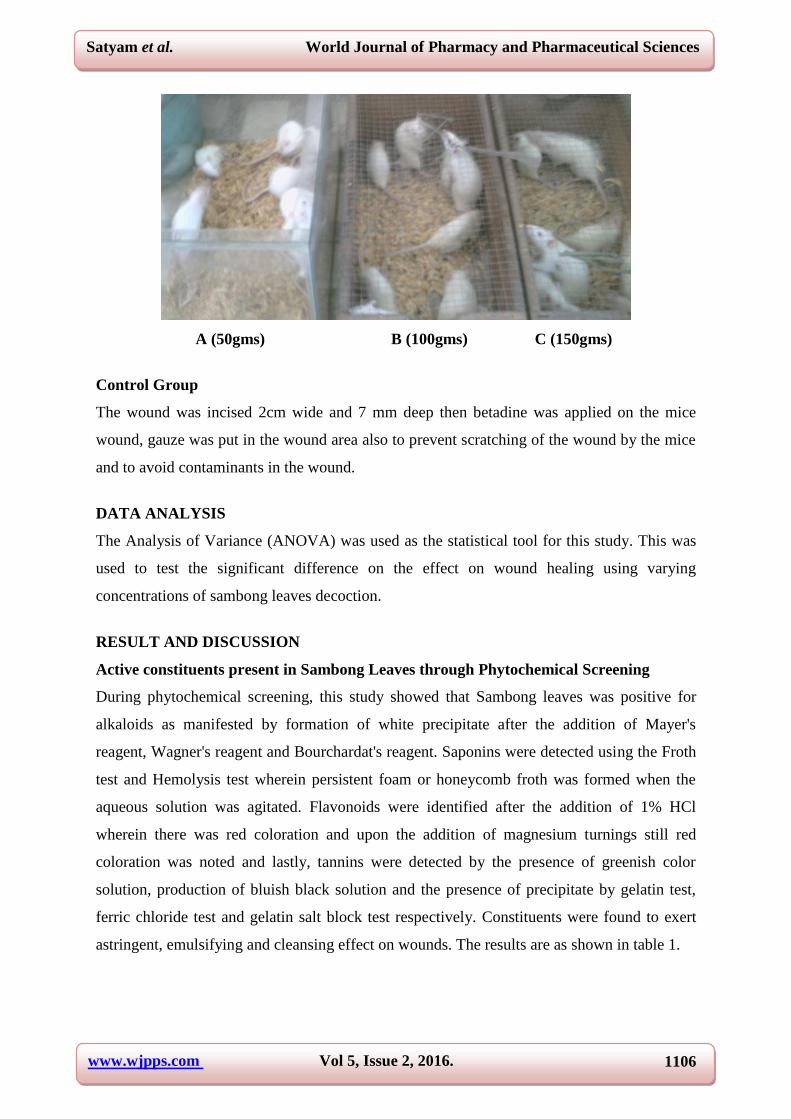
A (50gms) B (100gms) C (150gms)
Control Group
The wound was incised 2cm wide and 7 mm deep then betadine was applied on the mice
wound, gauze was put in the wound area also to prevent scratching of the wound by the mice
and to avoid contaminants in the wound.
DATA ANALYSIS
The Analysis of Variance (ANOVA) was used as the statistical tool for this study. This was
used to test the significant difference on the effect on wound healing using varying
concentrations of sambong leaves decoction.
RESULT AND DISCUSSION
Active constituents present in Sambong Leaves through Phytochemical Screening
During phytochemical screening, this study showed that Sambong leaves was positive for
alkaloids as manifested by formation of white precipitate after the addition of Mayer's
reagent, Wagner's reagent and Bourchardat's reagent. Saponins were detected using the Froth
test and Hemolysis test wherein persistent foam or honeycomb froth was formed when the
aqueous solution was agitated. Flavonoids were identified after the addition of 1% HCl
wherein there was red coloration and upon the addition of magnesium turnings still red
coloration was noted and lastly, tannins were detected by the presence of greenish color
solution, production of bluish black solution and the presence of precipitate by gelatin test,
ferric chloride test and gelatin salt block test respectively. Constituents were found to exert
astringent, emulsifying and cleansing effect on wounds. The results are as shown in table 1.

www.wjpps.com Vol 5, Issue 2, 2016.
1107
Satyam et al. World Journal of Pharmacy and Pharmaceutical Sciences
Table 1. Constituents present in Sambong Leaves through Phytochemical Screening.
Screening Result Inference Indications
Alkaloids
Mayer’s reagent
Formation of white precipitate Presence of alkaloids
Valser’s reagent Formation of white precipitate Presence of alkaloids
Wagner’s reagent Formation of white precipitate Presence of alkaloids
Bouchardat’s reagent Formation of white precipitate Presence of alkaloids
Unsaturated Sterols &
Triterpenes
Leiberman- Burchard
No immediate change in the
color of the solution to blue-
green.
Absence of unsaturated
sterol and triterpenes
Salkowski test
No immediate change in the
color of the solution to blue-
green.
Absence of unsaturated
sterols and triterpenes
Flavonoid 1% HCl No strong red coloration Presence of flavonoids
1% HCl w/ Mg turnings No red coloration Persence of Flavonoids
Steroid (CARDIOACTIVE
Glycosides:Liebermann –
Burchard
No reddish brown precipitate Absence of steroids
Kedde Reaction No blue coloration of the
solution Absence of Steroids
Keller- Killiani test No precipitate Absence of Steroids
Saponins
Hemolysis test No Zone of hemolysis Presence of saponins
Emulsifying
effect
Froth test No Honeycomb froth
formation Absence of Saponins
Cleansing
property
Tannins
Gelatin test
Ferric chloride Test
Gelatin salt block test
Greenish blue color solution
Production of bluish black
solution
Production of precipitate
Presence of Tannin Astringent
effect
Anthraquinone Heterosides
Borntrager test
No Color change in the
solution
Absence of Anthraquinone
Heterosides
Effect on the number of days that wound heals
Treatments with 50% Sambong leaves decoction in all three replications range from 11 to 12
days for wound healing. Categorically described, this range of number of days it takes for
wound in mice to heal was labeled 2, indicating that the length of time for the deep wound to
heal was fair. The number of days it takes for deep wounds in mice to heal completely ranges
from 7 to 9 days, implying that the number of days of healing of wounds treated with 100
grams of Sambong leaves decoction was good throughout. And for deep wounds in mice
treated with 150 grams of Sambong leaves decoction, 5-6 days was needed for complete
healing. This number of days needed for complete healing of wounds in mice was considered
very good. The results are shown in table 2. This may be due to the concentration of

www.wjpps.com Vol 5, Issue 2, 2016.
1108
Satyam et al. World Journal of Pharmacy and Pharmaceutical Sciences
Sambong leaves decoction increases, the number of days needed for healing of deep wounds
in mice decreases.
Meanwhile, the use of positive control Betadine shows the least number of days needed for
deep wound healing, ranging only from 3-4 days or from very good to excellent in terms of
the rates of wound healing. The healing of wounds in mice with the use either of the
experimental drugs in different concentrations or control drugs may be explained by the
antiseptic effects of both types of medication in wounds.
The healing of wounds brought about by the use of either types of treatment is rather indirect.
Either medication induces healing of wounds by preventing bacteria and other microorganism
from thriving and multiplying. Without microorganisms invading the wound areas, the faster
the healing takes place because the surge of hematological materials in the area will only
concentrate on replacing dead tissues or at least repairing damaged yet viable tissues and the
need to protect the wound from further damage by bacterial or other opportunistic infection
by sending more non-specific and immune defense cells are minimized, if not totally
eliminated.
Table 2. Effect of Sambong leaves decoction at various concentrations and of Betadine
on the period of healing of the wound in mice in number of days.
RE
PL
ICA
TIO
N
SAMBONG CONCENTRATIONS BETADINE
(CONTROL) 50 gm. 100 gm. 150 gm.
TREATMENT TREATMENT TREATMENT TREATMENT
1 2 3 4 5 6 1 2 3 4 5 6 1 2 3 4 5 6 1 2 3 4 5 6
Act
ual
Valu
es
1 12 12 12 11 12 12 8 8 7 9 7 8 6 5 6 6 6 5 3 3 4 3 3 3
2 11 12 11 12 12 11 9 8 8 7 8 7 5 5 5 5 6 5 4 3 3 4 3 3
3 12 12 12 11 12 11 7 7 8 8 9 9 6 6 5 5 5 6 4 4 3 3 4 3
Cate
gori
cal
Valu
es*
1 2 2 2 2 2 2 3 3 3 3 3 3 3 4 4 4 4 4 5 5 4 5 5 5
2 2 2 2 2 2 2 3 3 3 3 3 3 4 4 4 4 4 4 4 5 5 4 5 5
3 2 2 2 2 2 2 3 3 3 3 3 3 4 4 4 4 4 4 4 4 5 5 4 5
*Legend: 5 – Excellent; the deep wound takes 1- 3 days to heal; 4 – Very good; the deep
wound takes 4 – 6 days to heal; 3 – Good; the deep wound takes 7 – 9 days to heal; 2 – Fair;
the deep wound takes 10 -12 days to heal; 1 – Poor; the deep wound takes 13-15 days to heal.

www.wjpps.com Vol 5, Issue 2, 2016.
1109
Satyam et al. World Journal of Pharmacy and Pharmaceutical Sciences
Difference in the Effect of Sambong Leaves as to Number of Days
The difference in the effect of Sambong leaves as to number of days takes for wounds to heal
was found to be significantly different. ANOVA was calculated. The result of this analysis is
presented in Table 3.
Table 3. Differences in Means in the number of days of Healing of wound in mice as to
treatment applied.
Source of Variation Sum of
Squares Df
Mean
Square F p-value
Between Groups 72.708 3 24.236
333.315 0.000 Within Groups 4.944 68 7.271E-02
Total 77.653 71
The ANOVA computation revealed a significant difference in the number of days deep
wounds heal with the use of Sambong leaves decoction at different concentrations and
Betadine. The computed p-value (0.000) was lower than the set alpha (0.01). The results are
shown in table 4. Pairwise comparison using Scheffe’s test showed that for those deep
wounds treated with 50 grams of Sambong leaves decoction, the number of days needed for
deep wound healing to occur was significantly different from those treated with 100 grams of
Sambong leaves decoction (p=.000). Likewise, when 50 gm of Sambong leaves decoction
was compared to 150 gm of Sambong decoction, the number of days of healing was also
significantly different (p=.000). Furthermore, when 50 grams of Sambong leaves treatment
was compared to Betadine in terms of the effect in wound healing measured in number of
days, significant difference was also evident (p=.000).
Comparing the other Sambong leaves concentrations and the positive control (Betadine) with
each other revealed similar significant differences (p=.000). This means that Betadine was
significantly different to the three concentration of Sambong leaves decoction in terms of
effecting deep wound healing in mice measured in number of days, none of the Sambong
leaves was comparable to its effect on wound healing.
Table 4. Post hoc test for the differences in Means in the Days of Healing of wound in
mice Classified as to their treatment applied (with sambong in three different
concentrations and Betadine as positive control) using Scheffe test.
Treatment I Treatment J Mean Difference (I-J) Sig.
50gm sambong 100 -1.06 .000
150 -2.00 .000

www.wjpps.com Vol 5, Issue 2, 2016.
1110
Satyam et al. World Journal of Pharmacy and Pharmaceutical Sciences
Betadine(control) -2.67 .000
100gm sambong
50 1.06 .000
150 -0.94 .000
Betadine(control) -1.61 .000
150gm sambong
50 2.00 .000
100 0.94 .000
Betadine(control) -0.67 .000
Betadine (Control)
50 2.67 .000
100 1.61 .000
150 0.67 .000
Effect on varying concentrations of Sambong leaves decoction and using betadine as
control
The use of Sambong leaves decoction with 50 grams concentration, the number of days
needed for the resolution of redness takes 11-12 days and this period of regression of
swelling was considered fair in all replications. Using 100 gm concentration of Sambong
leaves decoction applied on deep wound resulted in resolution of redness in 8-9 days,
described as good all throughout the treatments in all three replications. As for the use of 150
gm of Sambong leaves decoction, the redness resolved between 4-6 days, which was a very
good indication as it was consistent all throughout the treatments in all three replications.
The number of days needed for swelling to subside with the use of 50 gm Sambong leaves
decoction was within 10-11 days, considered as fair in terms of its speed all throughout the
treatment in all three replications. Seven to eight days was needed for swelling to subside
with the use of 100 grams of Sambong leaves decoction and this rate of resolution of swelling
was considered good as it was consistent all throughout the treatments in all three
replications. It would take 3-5 days for swelling to subside with the use of 150 grams of
Sambong decoction, as evidenced by such occurrence in all treatments and replications and
this rate of resolution of swelling was considered 3-5 days, crossing midway between being
described as very good and excellent.
With the use of positive control (Betadine), 2-3 days was needed for swelling to subside and
this rate of resolution was considered excellent as it was consistent all throughout the
treatments in all its three replications. The results are shown in table 5.

www.wjpps.com Vol 5, Issue 2, 2016.
1111
Satyam et al. World Journal of Pharmacy and Pharmaceutical Sciences
Table 5. The effects of Sambong leaves decoction at various concentrations and of
Betadine as control on the redness and swelling in the wound of mice.
VA
LU
ES
RE
PL
ICA
TIO
NS
PA
RA
ME
TE
RS
SAMBONG CONCENTRATIONS BETADINE
(CONTROL) 50 gm. 100 gm. 150 gm.
TREATMENT TREATMENT TREATMENT TREATMENT
1 2 3 4 5 6 1 2 3 4 5 6 1 2 3 4 5 6 1 2 3 4 5 6
AC
TU
AL
VA
LU
ES
1 Redness 12 11 12 11 11 12 8 9 9 8 8 9 4 6 5 5 6 4 3 4 3 4 3 3
Swelling 11 10 11 10 10 11 7 8 8 7 7 8 3 5 4 4 5 3 2 3 2 3 2 2
2 Redness 11 12 12 12 11 12 9 8 8 8 8 9 6 6 5 5 4 5 4 4 3 3 4 3
Swelling 10 11 11 11 10 11 8 7 7 7 7 8 5 5 4 3 4 3 3 3 2 2 3 2
3 Redness 12 11 11 12 12 11 8 8 9 9 8 9 5 4 6 6 5 3 3 3 4 3 4 3
Swelling 11 10 10 11 11 10 7 7 8 8 7 8 4 3 5 5 4 4 2 2 3 2 3 2
CA
RT
EG
OR
ICA
L V
AL
UE
S* 1
Redness 2 2 2 2 2 2 3 3 3 3 3 3 4 4 4 4 4 4 5 4 5 4 5 5
Swelling 2 2 2 2 2 2 3 3 3 3 3 3 5 4 4 4 4 5 5 5 5 5 5 5
2 Redness 2 2 2 2 2 2 3 3 3 3 3 3 4 4 4 4 4 4 4 4 5 5 4 5
Swelling 2 2 2 2 2 2 3 3 3 3 3 3 4 4 4 5 4 5 5 5 5 5 5 5
3 Redness 2 2 2 2 2 2 3 3 3 3 3 3 4 4 4 4 4 5 5 5 4 5 4 5
Swelling 2 2 2 2 2 2 3 3 3 3 3 3 4 5 4 4 4 4 5 5 5 5 5 5
*Legend: 5 – Excellent; the deep wound takes 1- 3 days to heal; 4 – Very Good; the deep
wound takes 4 – 6 days to heal; 3 – Good; the deep wound takes 7 – 9 days to heal; 2 – Fair;
the deep wound takes 10 -12 days to heal;1 – Poor; the deep wound takes 13-15 days to heal.
Resolution of Redness and Swelling
Although the relative descriptions of the effects of various concentrations of Sambong leaves
as compared with a positive control in terms of resolution of both redness and swelling were
concerned, the range of days involved was not quite similar. For instance, it can be noted that
while the redness subsides in 11-12 days with the use of 50 grams of Sambong leaves
decoction, its swelling resolved in 10-11 days, or that swelling starts resolving one day earlier
than redness. The same observation can be made with the other concentrations of Sambong
leaves extract and even with the use of Betadine as positive control.
Whenever there is cell and tissue damage, repair is initiated by the onset of inflammation.
There are four cardinal signs of inflammation: redness (rubor), heat (calor), swelling (tumor)
and pain (dolor).[25,26]
The onset of pain is due to the release of prostaglandins upon the
damage on plasma membranes of cells of tissues damaged by infliction of wound on the
mice. Damaged cells will also trigger the release of clotting factors to stop leakage of blood

www.wjpps.com Vol 5, Issue 2, 2016.
1112
Satyam et al. World Journal of Pharmacy and Pharmaceutical Sciences
as well as of chemotactic factors that will attract platelets and thrombocytes in the area of cell
damage.[27]
These chemotactic substances will also trigger release of serotonin by the
attracted platelets or thrombocytes that will, in turn, cause vasoconstriction that will attempt
to prevent blood loss.[28]
Later, vasodilation occurs. Dilation of blood vessels in the wound
area results in increased flow of blood to the area, thus explaining for the redness. Cells in the
area of damage become more permeable to virtually all substances needed for cellular and
histological repair during these times, accounting for the swelling.[29]
Vasodilation will be
constant until repair of damaged cells and tissues is complete. Since mice blood is warm, the
surge of blood flow to the area results also in increased local temperature, explaining for the
occurrence of calor.[27]
This study found that swelling subsides before redness occurs as shown in table 6. This may
be swelling is caused directly by the increased permeability of the tissue damage area, while
the redness is directly caused by dilation of the blood capillaries in the area of tissue damage.
Therefore, serotonin secretion persists a little longer than the increased permeability of cells
and tissues in the damaged area.
Table 6. ANOVA Results for the Differences in Means in the number of days of
resolution of redness and swelling in mice as to treatment applied.
Parameter Sources of
Variation
Sum of
Squares Df
Mean
Square F Sig.
Days of Resolution of
Redness
Between Groups 71.042 3 23.681
376.429 .000 Within Groups 4.278 68 6.291E-02
Total 75.319 71
Days of Resolution of
Swelling
Between Groups 94.667 3 31.556
689.714 .000 Within Groups 3.111 68 4.575E-02
Total 97.778 71
Pairwise comparison using Scheffe’s test showed that for those deep wounds treated with 50
grams of Sambong leaves decoction, the number of days needed for resolution of redness and
swelling to occur was significantly different from those treated with 100 grams of Sambong
leaves decoction (p=.000). Likewise, when 50gm of Sambong leaves decoction was
compared to 150 gm of Sambong decoction, the number of days for redness and swelling to
subside was also significantly different (p=.000). Furthermore, when 50 grams of Sambong
leaves treatment was compared to Betadine in terms of the effect in resolution of redness and
swelling measured in number of days, significant difference was also evident (p=.000).
Comparing the other Sambong leaves concentrations and the positive control (Betadine) with

www.wjpps.com Vol 5, Issue 2, 2016.
1113
Satyam et al. World Journal of Pharmacy and Pharmaceutical Sciences
each other reveal similar significant differences (p=.000). The results are shown in table 7.
This may be due to Betadine was significantly different to the three concentration of
Sambong leaves decoction in terms of resolution of redness in mice measured in number of
days, none of the Sambong leaves was comparable to its effect on wound healing in terms of
rate or speed of redness resolution.
Table 7. Post hoc test for the differences in Means in the resolution of redness and
swelling of wound in mice classified as to their treatment applied (with Sambong in
three different concentrations and Betadine as positive control) using Scheffe test.
(I) Concentration (J) Concentration Mean Difference
(I-J) Std. Error
Sig.
(pvalue)
50
100 -1.00 8.36E-02 .000
150 -2.00 8.36E-02 .000
Betadine (control) -2.61 8.36E-02 .000
100
50 1.00 8.36E-02 .000
150 -1.00 8.36E-02 .000
Betadine (control) -1.61 8.36E-02 .000
150
50 2.00 8.36E-02 .000
100 1.00 8.36E-02 .000
Betadine (control) -.61 8.36E-02 .000
Betadine control
50 2.61 8.36E-02 .000
100 1.61 8.36E-02 .000
150 .61 8.36E-02 .000
50
100 -1.00 7.13E-02 .000
150 -2.22 7.13E-02 .000
Betadine (control) -3.00 7.13E-02 .000
100
50 1.00 7.13E-02 .000
150 -1.22 7.13E-02 .000
Betadine (control) -2.00 7.13E-02 .000
150
50 2.22 7.13E-02 .000
100 1.22 7.13E-02 .000
Betadine (control) -.78 7.13E-02 .000
Betadine (control)
50 3.00 7.13E-02 .000
100 2.00 7.13E-02 .000
150 .78 7.13E-02 .000
CONCLUSION
The present study concluded that varying concentrations of Sambong (Blumea balsamifera)
leaves decoction bent different effects on deep wounds of mice. The greater the concentration
of Sambong leaves decoction, the faster the wound healing was found. There was significant
difference in the effect of Sambong leaves decoction and Betadine in the number of days of

www.wjpps.com Vol 5, Issue 2, 2016.
1114
Satyam et al. World Journal of Pharmacy and Pharmaceutical Sciences
wound healing in mice. Also significant difference in the effect of the Sambong leaves
decoction and Betadine in the resolution of redness and swelling was found.
Wound healing in general and the cardinal signs of inflammation are resolved not solely by
application of antiseptics or by antiobiotic treatment, because the use of these are meant to
augment healing as well as prevent further complication of the pathological processes and do
not absolutely determine wound healing and resolution of cardinal signs of inflammation.
Moreover, wound healing and its signs and symptoms are not solely dependent on the use of
antiseptics and antibiotics, but also on other factors like food taken i.e. some nutrients found
in food speed up the healing process in several ways and metabolic rate among others.
In the light of the foregoing conclusions, parallel studies and clinical trials adding more
parameters on wound healing should be conducted to determine the predictors of wound
healing, redness and swelling resolution of the cardinal signs of inflammation together with
the use of Sambong leaves decoction.
ACKNOWLEDGEMENT
Authors extend their deepest gratitude and cordial thanks to Dr. Leo B. Solis, Advisor,
Virgen Milagrosa University Foundation, San Carlos City, Pangasinan, Phillipines for his
valuable suggestions, supervision, guidance and support for the completion of this research
work.
REFERENCES
1. Singh KN, Lal B. Ethnomedicines used against four common ailments by the tribal
communities of Lahaul-Spiti in western Himalaya. J of Ethnopharm, 2008; 115: 147–159.
2. Singh A, Singh DK. Molluscicidal activity of Lawsonia inermis and its binary and tertiary
combinations with other plant derived molluscicides. Indian J Exp Biol, 2001; 39(3):
263-268.
3. Canter PH, Thomas H, Ernst E. Bringing medicinal plants into cultivation: Opportunities
and challenges for biotechnology. Trends Biotechno, 2005; 23: 180–185.
4. Hatusima S. Detailed flora list of the Batan Island, An enumeration of the plants of Batan
Island, N. Philippines. Memoirs of the Faculty of Agriculture, Kagoshima University.,
1966; 5: 13–70.
5. Natarajan V, Venugopal PV, Menon T. Effect of Azadirachta indica (neem) on the
growth pattern of dermatophytes. Indian J Med Micro, 2003; 21(2): 98-101.

www.wjpps.com Vol 5, Issue 2, 2016.
1115
Satyam et al. World Journal of Pharmacy and Pharmaceutical Sciences
6. Diegelmann RF, Evans MC. Wound healing: an overview of acute, fibrotic and delayed
healing. Front Biosci, 2004; 9: 283-289.
7. Peppa M, Stavroulakis P, Raptis SA. Advanced glycoxidation products and impaired
diabetic wound healing. Wound Repair Regen, 2009; 17(4): 461-472.
8. Gosain A, Dipietro LA. Aging and wound healing. World J Surg, 2004; 28(3): 321-326.
9. Mathieu D, Linke JC, Wattel F. Non-healing wounds. In: Handbook on hyperbaric
medicine, Mathieu DE, editor., editor. Netherlands: Springer., 2006; 401-427
10. Guo S, DiPietro LA. Factors affecting wound healing. J Dent Res, 2010; 89(3): 219-229.
11. Jia ZG, Fang Y, Yao M and et al. Molecular mechanisms of substance P-induced
monocyte chemoattractant protein-1 secretion in fibroblasts under high glucose culture
condition. Shanghai Jiao Tong Da Xue Xue bao, 2012; 10(32): 1302-1306.
12. Lai X, Wang Z, Wei L, Wang L. Effect of substance P released from peripheral nerve
ending on endogenous expression of epidermal growth factor and its receptor in wound
healing. Zhong Hua Chuang Shang Za Zhi, 2002; 5(3): 176-179.
13. Muangman P, Tamura RN, Muffley LA, et al. Substance P enhances wound closure in
nitric oxide synthase knockout mice. J Surg Res, 2009; 153(2): 201-209.
14. Li J, Zhao GZ, Chen HH, et al. Antitumour and antimicrobial activities of endophytic
streptomycetes from pharmaceutical plants in rainforest. Lett Appl Microbiol, 2008;
47(6): 574-580.
15. Ragasa CY, Co AL, Rideout JA. Antifungal metabolites from Blumea balsamifera. Nat
Prod Res, 2005; 19(3): 231-237.
16. Kubota H, Kojima-Yuasa A, Morii R, Huang X, Norikura T, Rho SN, Matsui-Yuasa I.
Anti-obesity effect of Blumea balsamifera extract in 3T3–L1 preadipocytes and
adipocytes. Am J Chin Med, 2009; 37: 843–854.
17. Chen M, Jin HZ, Zhang WD, Yan SK, Shen YH. Chemical constituents of plants from the
genus Blumea Chem Biodiver, 2009; 809–817.
18. Chen M, Qin J, Fu J, et al. Blumeaenes A–J, sesquiterpenoid esters from Blumea
balsamifera with NO inhibitory activity. Planta Med, 2010; 76(9): 897-902.
19. Noor Rain A, Khozirah S, Mohd Ridzuan MA, et al. Antiplasmodial properties of some
Malaysian medicinal plants. Trop Biomed, 2007; 24(1): 29-35.
20. Osaki N, Koyano T, Kowithayakorn T, Hayashi M, Komiyama K, Ishibashi M.
Sesquiterpenoids and plasmin- inhibitory flavonoids from Blumea balsamifera. J Nat
Prod, 2005; 68(3): 447-449.

www.wjpps.com Vol 5, Issue 2, 2016.
1116
Satyam et al. World Journal of Pharmacy and Pharmaceutical Sciences
21. Nessa F, Ismail Z, Mohamed N, Haris MRHM. Free radical- scavenging activity of
organic extracts and of pure flavonoids of Blumea balsamifera DC leaves. Food Chem,
2004; 88(2): 243-252.
22. State Administration of Traditional Chinese Medicine ed. Volumn 21 in: Chinese Materia
Medica (Book 7). Shanghai, China: Scientific and Technical Publishers, 1999; 738-740.
23. Bhathena SJ, Velasquez MT. Beneficial role of dietary phytoestrogens in obesity and
diabetes. Am J Clin Nutr, 2002; 76(6): 1191-1201.
24. Aguinaldo A, Espeso E, Guevara B, Nonato M. A guidebook to plant screening:
phytochemical and biological: botany section. Research Center for the Natural Sciences,
University of Santo Tomas, Philippines., 2004; 24-50.
25. Norikura T, Kojima-Yuasa A, Shimizu M., Huang XD, Xu SH, Kametani S, Rho SN,
Kennedy DO, Matsui-Yuasa I. Anticancer activities and mechanisms of Blumea
balsamifera extract in hepatocellular carcinoma Cells. Am J Chin Med, 2008; 36:
411–424.
26. Kennedy DO, Matsui-Yuasa I. Anticancer activities and mechanisms of Blumea
balsamifera extract in hepatocellular carcinoma Cells. Am J Chin Med, 2008; 36:
411–424.
27. Xu SB, Zhao JH. Protective actions of Blumea flavanones on experimental liver injury.
Chin Pharm Bull, 1998; 14: 191–192.
28. Pang, Y.X.; Wang, D.; Fan, Z.W.; Chen, X.L.; Yu, F.L.; Hu, X.; Wang, K.; Yuan, L.
Blumea balsamifera—A phytochemical and pharmacological review. Molecules, 2014;
19: 9453–9477.
29. Fishel R, Barbul A, Wasserkrug HL, Penberthy LT, Rettura G, Efron G. Cyclosporine a
impairs wound healing in rats. J Surg Res, 1983; 34: 572–575.