the effect of meditation on regulation of internal body states
TRANSCRIPT

ORIGINAL RESEARCHpublished: 07 July 2015
doi: 10.3389/fpsyg.2015.00924
Frontiers in Psychology | www.frontiersin.org 1 July 2015 | Volume 6 | Article 924
Edited by:
Catherine Kerr,
Brown University, USA
Reviewed by:
Neil Gerald Muggleton,
National Central University, Taiwan
Norman Farb,
Baycrest, Canada
*Correspondence:
Sahib S. Khalsa,
Laureate Institute for Brain Research,
6655 S. Yale Ave, Tulsa, OK 74136,
USA
Specialty section:
This article was submitted to
Consciousness Research,
a section of the journal
Frontiers in Psychology
Received: 27 December 2014
Accepted: 22 June 2015
Published: 07 July 2015
Citation:
Khalsa SS, Rudrauf D, Davidson RJ
and Tranel D (2015) The effect of
meditation on regulation of internal
body states. Front. Psychol. 6:924.
doi: 10.3389/fpsyg.2015.00924
The effect of meditation onregulation of internal body states
Sahib S. Khalsa 1, 2, 3*, David Rudrauf 1, 4, Richard J. Davidson 5 and Daniel Tranel 1, 6
1Department of Neurology, University of Iowa, Iowa City, IA, USA, 2 Laureate Institute for Brain Research, Tulsa, OK, USA,3 Faculty of Community Medicine, University of Tulsa, Tulsa, OK, USA, 4 Laboratory of Functional Imaging, Institut National de
la Santé et de la Recherche Médicale U678s/Université Pierre et Marie Curie, Paris, France, 5Department of Psychology,
University of Wisconsin-Madison, Madison, WI, USA, 6Department of Psychology, University of Iowa, Iowa City, IA, USA
Meditation is commonly thought to induce physiologically quiescent states, as evidenced
by decreased autonomic parameters during the meditation practice including reduced
heart rate, respiratory rate, blood pressure, skin conductance, and increased alpha
activity in the electroencephalogram. Preliminary empirical support for this idea was
provided in a case report by Dimsdale and Mills (2002), where it was found that
meditation seemed to regulate increased levels of cardiovascular arousal induced by
bolus isoproterenol infusions. In that study, while meditating, a self-taught meditator
exhibited unexpected decreases in heart rate while receiving moderate intravenous
doses of the beta adrenergic agonist isoproterenol. This effect was no longer observed
when the individual received isoproterenol infusions while not meditating. The current
study was designed to explore this phenomenon empirically in a group of formally trained
meditators. A total of 15 meditators and 15 non-meditators individually matched on age,
sex, and body mass index were recruited. Participants received four series of infusions
in a pseudorandomized order: isoproterenol while meditating (or during a relaxation
condition for the non-meditators), isoproterenol while resting, saline while meditating
(or during a relaxation condition for the non-meditators), and saline while resting. Heart
rate was continuously measured throughout all infusions, and several measures of heart
rate were derived from the instantaneous cardiac waveform. There was no evidence
at the group or individual level suggesting that meditation reduced the cardiovascular
response to isoproterenol, across all measures. These results suggest that meditation is
not associated with increased regulation of elevated cardiac adrenergic tone.
Keywords: meditation, adrenergic, regulation, sympathetic, parasympathetic, isoproterenol, body
Introduction
Meditation is a form of mental training that has been practiced for thousands of years, andthat can be conceptualized as a family of complex emotional and attentional regulatory trainingregimens developed for various ends, including the cultivation of well-being and emotional balance(Davidson et al., 1976; Ekman et al., 2005; Brefczynski-Lewis et al., 2007). Meditation has alsobeen defined as involving a process of intentional self-regulation of attention, in which attentionis directed from a combination of external and internal stimuli to a primarily internally perceptivestate (Astin et al., 2003; Bonadonna, 2003). Traditional philosophies emphasize that anyonecan learn to meditate (Taimni, 1961), and that through repeated practice meditation provides

Khalsa et al. Meditation and body regulation
long-term effects that outlast the confines of individualmeditative states (Nyanaponika, 1969; Ahir, 1999; Burley, 2000).
A central tenet of early investigations into the effects ofmeditation has been that meditation induces a physiologicallyquiescent bodily state. This was based on initial reportsof decreases in autonomic parameters such as heart rate,respiratory rate, blood pressure, skin conductance, andadrenergic reactivity, as well as increased levels of alpha activityin the electroencephalogram during the practice of meditation[mostly Transcendental Meditation (TM)] (Wallace et al.,1971; Orme-Johnson, 1973; Beary and Benson, 1974; Farrowand Hebert, 1982; Hoffman et al., 1982; Jevning et al., 1992;Travis and Wallace, 1997; Aftanas and Golocheikine, 2002;Aftanas and Golosheykin, 2005). Early researchers describedthis pattern of autonomic responses as an integrated “relaxationresponse” (Benson et al., 1974). More recently, investigationshave continued finding reductions in physiological parameterssuch as heart rate and blood pressure occur during the acutemeditation practice (Telles et al., 1995; Barnes et al., 1999;Solberg et al., 2004) as well as following practice over longerperiods of time (Barnes et al., 2004; Harinath et al., 2004). Similareffects have also been reported for more secular and abbreviatedprotocols such as an 8 week training in Mindfulness Based StressReduction, which incorporates instruction in meditation as wellas teaches cognitive and other approaches to enhance stressreduction (Hughes et al., 2013).
Although the notion that meditation results in physiologicallyquiescent states is well established, the extent to which meditativepractices exert such effects is unknown. Early investigations ofyogis in India who claimed to be able to exert considerablevoluntary control of the heart (some claimed to be able tostop the heart) revealed limited support for this idea. Atbest, some individuals were able to exert transient bradycardia,or reductions in heart rate by engaging in combinations ofposture manipulation, muscular contraction and breath holding(including the Valsalva maneuver, or “bearing down,” whichelicits a complex pattern of reflexes including tachy/bradycardiaand hyper/hypotension) (Wenger et al., 1961). Subsequentstudies have reported mixed results, primarily for different yogaadepts (Fenz and Plapp, 1970; Kothari et al., 1973).More recently,shorter term changes in the ability to reduce the resting heartrate have been reported (Telles et al., 2004). The perceptionthat meditation and yoga confers an increased cardiac regulatorycapability has persisted, despite this heterogeneous literature.
Preliminary empirical support via a case study approachsuggested a novel effect of meditation on the ability to regulateincreased levels of cardiovascular arousal. Dimsdale and Mills(2002) reported a case study in which a female meditator wasrandomly recruited to participate in a standardized isoproterenolchallenge as a control subject in their studies of sympatheticnervous system regulation in hypertension (Dimsdale et al., 1988;Mills et al., 1998). Isoproterenol stimulates peripheral beta 1and beta 2 adrenergic receptors equally, and when administeredintravenously results in rapid and transient increases in heartrate, contractility and bronchodilation, as well as decreases indiastolic blood pressure. The protocol involved administrationof a standard isoproterenol sensitivity test, involving sequentially
increasing doses of intravenous isoproterenol. Halfway into theinfusions the woman spontaneously started meditating, andcontinued to meditate during the infusions. At the time of theisoproterenol challenge the investigators did not know she wasa meditator, but noticed afterwards that after the fourth dose (1mcg) her heart rate had begun to decrease, in stark contrast to theexpected increase. They report that at the highest dose (4 mcg),when the expected heart rate response was a 21 beats per minute(bpm) increase above baseline, they observed a 17 bpm decreasebelow baseline. When this unexpected result was mentioned,the participant stated that she had decided to start meditatinghalfway into the experiment. Suspecting that her meditationpractice had interfered with the isoproterenol response, she wasasked to return 2 weeks later and repeat the challenge, withexplicit instructions to avoid meditating during the infusions.Her heart rate response to the subsequent infusion protocolappeared entirely consistent with the typical laboratory responseto isoproterenol at rest (i.e., an approximately 20 bpm increaseabove baseline at the highest dose). This individual reported aself-taught meditation practice for many years, and that she didnot practice any particular tradition of meditation.
The current study was designed to investigate whether thereis an effect of meditation on attenuating adrenergically mediatedincreases in sympathetic arousal, using an empirical group studyapproach. We recruited formally trained meditators with at leastseveral years of experience, and examined autonomic parametersof sympathetic arousal during meditation and resting conditions,during the application of isoproterenol and saline infusions.To examine whether such a putative effect was specific to ameditation practice or was a generic ability inherent to humansmore generally, we also recruited a group of individually matchednon-meditators, and tested them under relaxation and restingconditions.
We hypothesized that the practice of meditation would bespecifically associated with an enhanced ability to regulate bodystates occurring in the context of acute physiological arousal. Totest this hypothesis, the impact of a meditation practice on thecardiovascular response to isoproterenol was assessed. Severalspecific predictions derive from our hypothesis: (1) In the face ofisoproterenol infusions, during a meditation practice meditatorswould display lower isoproterenol induced heart rate increasesthan during a non-meditation condition. (2) Meditators woulddemonstrate lower heart rate increases during a meditationpractice than non-meditators during a relaxation practice. (3)Heart rate reductions in the meditators would be specific tomeditation, i.e., meditators and non-meditators would haveequivalent heart rate responses to isoproterenol at rest (when notengaging in meditation or relaxation strategies).
Methods
ParticipantsFifteen meditators and fifteen non-meditators participated inthe study. Each non-meditator was individually matched to acorresponding meditator based on three criteria: age, gender,and body mass index (see Table 1). Meditators were consideredeligible for participation if they reported a continuous (daily or
Frontiers in Psychology | www.frontiersin.org 2 July 2015 | Volume 6 | Article 924

Khalsa et al. Meditation and body regulation
TABLE 1 | Meditator and non-meditator demographic data.
Meditators (M) Non-meditators (NM)
Sex 10 Men, 5 Women 10 Men, 5 Women
Age (yrs) 44.7±13.2 44.0±13.7
Body Mass Index 24.5±4.6 25.5±4.0
Race 15 Caucasian
American
14 Caucasian American,
1 Asian American
Education (years) 17.3±2.2 15.9±2.3
Beck Anxiety Inventory score 5.1±3.4 3.5±2.9
Beck Depression Inventory score 4.3±4.6 3.7±5.3
Meditation practice (years)* 10.8±10.8 0±0
Cumulative meditation practice
(hours)*
4947±6251 0±0
Retreat experience (days)** 19±14 0±0
CD25 (micrograms) 4.48±1.5 4.72±2.2
Practice similarity: iso +
meditation/relaxation
3.5±1.1 3.4±1.1
Practice similarity: saline +
meditation/relaxation
3.7±1.1 3.8±0.8
Chronotropic Dose 25 (CD25): isoproterenol dose (mcg) required to increase the heart
rate by 25 beats per minute. Mean ± SD. *p < 0.01, **p < 0.05. Means ± standard
deviation. Iso, isoproterenol.
near daily) meditation practice during the previous 2 years, and ifthey had also attended one or more weeklong meditation retreatswithin the previous year. Using this criteria, 11 of the recruitedmeditators were Vipassana practitioners, and the other fourmeditators were Kundalini practitioners. Non-meditators wereconsidered eligible for participation if they had never receivedformal meditation training in meditation or yoga and did notpractice self-taught meditation.
All participants were screened for the presence of anyneurological, psychiatric, cardiac or respiratory disease duringa detailed phone interview, and were excluded if they reporteda history of disease in any of these categories. None of thestudy participants were smokers, and none of the women tookoral contraceptives or were pregnant, as assessed via urinepregnancy test. Each participant demonstrated a normal 12lead electrocardiogram (EKG), as assessed by a board certifiedcardiologist or neurologist.
This study was approved by the University of Iowa’sInstitutional Review Board, and all participants providedinformed consent prior to participation.
TasksParticipants were informed that they would be asked to restquietly with their eyes open on two occasions, and to meditate(or alternatively, to relax) with their eyes closed on two occasions.The instruction to keep the eyes open or closed was chosen toreflect the manipulation reported in Dimsdale and Mills (2002).It was also chosen to improve the face validity of the meditationand relaxation conditions, as these activities are typically taughtand practiced with the eyes closed. Meditators were asked toengage in their usual daily meditation practice for a period of15–20min during the meditation condition. Since the Kundalini
tradition offers many different types of meditations derivingdifferent types of effects, Kundalini meditators were asked toselect a specific meditation practice that they had found helpedthem decrease their level of bodily arousal in the past. Sincethe Vipassana tradition does not provide such an approach,Vipassana meditators were simply asked to engage in theirusual meditation practice. Non-meditators were instructed in theperformance of a cognitive relaxation strategy for a period of 15–20min during the relaxation condition. Specifically, they wereasked to engage in a relaxation practice they had found usefulfor themselves in the past, one that would help them to “relaxyour mind and body, help you slow your thoughts, slow yourbreathing, and slow your heart rate.” If a non-meditator reportedno such a strategy, they were given the option to perform oneor any combination of several relaxation strategies including(1) reliving a pleasant memory, such as a warm day at thebeach, (2) replaying their favorite song internally, (3) completelyrelaxing their musculature and/or (4) slowing their breathing.These strategies were selected with the aim of facilitatingelicitation of the relaxation response (Benson et al., 1974). Non-meditators were specifically instructed to avoid falling asleepduring the relaxation practice, despite any inclinations that mightarise.
During the rest conditions both groups were instructedto avoid engaging in their meditation/relaxation practice, andinstead, to explore their everyday thoughts (for example,thoughts about activities from the recent past such as what theyhad done the day before, or thoughts about potential futureactivities such as what they would do once the study had beenconcluded).
All participants were informed that they would be receivingisoproterenol or saline at some points during the entire session,but that neither they nor the experimenter would know whenthe nurse administered a particular agent. The room in whichthe study took place was kept quiet and was dimly lit throughoutduration of the study to minimize distractions. Participants wereseated in a padded chair, with a curtain draped over the armcontaining the intravenous line, in order to reduce distractionand to preserve the blinding.
To assess task validity, after each meditation condition wascomplete, participants were asked to “rate how similar yourmeditation practice just now compares to your meditationpractice in general,” on a scale from 1 (“not at all the same”)to 5 (“completely the same”). Non-meditators were also askedto rate their relaxation practice in a similar fashion, usingthe same scale. If they reported never having practiced thespecific instructed relaxation condition, they were asked to relatetheir experience to other events where they had felt a state ofrelaxation.
Infusion ProtocolTwo sets of standard bolus isoproterenol infusion protocols andtwo sets of matched saline infusion protocols were administered.The isoproterenol protocols consisted of sequentially increasingbolus isoproterenol doses of 0.1, 0.5, 1.0, 2.0, and 4.0 (mcg),delivered 3.5min apart. The saline protocols consisted of fiveidentically delivered bolus infusions of saline.
Frontiers in Psychology | www.frontiersin.org 3 July 2015 | Volume 6 | Article 924

Khalsa et al. Meditation and body regulation
Infusion DeliveryEach infusion (isoproterenol and saline) consisted of two 3milliliter (ml) bolus infusions delivered sequentially throughan intravenous catheter. During isoproterenol infusions, a 3mlbolus containing the specified dose was delivered, immediatelyfollowed by a 3ml bolus of saline to flush the line. Duringsaline infusions, a 3ml bolus of saline was delivered, immediatelyfollowed by an additional 3ml bolus of saline. Both bolusvolumes were administered in entirety within a 15 s period by anurse from the General Clinical Research Center. This method ofdelivery minimized the participant’s ability to use external cuesto distinguish between the different infusion types, and ensuredrapid and standardized systemic introduction of isoproterenol.
ProcedureThe infusion protocol order was pseudorandomized and singleblinded. This was necessitated by practical considerations: inorder to ensure the safety of isoproterenol administration,a physician was required to be present during the firstround of infusions. Due to constraints in the physician’sschedule, a majority of the time the first condition consisted ofisoproterenol infusions. However, the remaining task selectionprocess (for example, isoproterenol plus rest vs. isoproterenolplus meditation) remained randomized, and was determinedby the nurse administering the infusions. The remaining threeinfusion conditions were completely randomized.
Psychophysiological MeasuresHeart rate was continuously measured throughout all infusions,from a lead II electrocardiogram (MP100 acquisition unit, BiopacSystems, Inc.), at a sampling rate of 200Hz. All artifacts affectingthe cardiac waveform (e.g., movement related and cardiac, suchas premature ventricular contractions) were visually identifiedand manually removed.
Several measures of heart rate were derived from theinstantaneous cardiac waveform. These measures were dividedacross three epochs of specific relevance to the timeline of heartrate changes induced by isoproterenol (Figure 1). The first epochconsisted of a 30 s interval starting immediately after the onsetof each infusion. This reflects a period when bolus isoproterenolinduced heart rate changes are unlikely to occur. The secondepoch consisted of a 90 s interval beginning 30 s after infusionadministration. This reflects a period when bolus isoproterenolinduced heart rate changes are most likely to occur. The thirdepoch consisted of a 60 s period beginning immediately afterthe end of the second epoch. This interval reflects a periodwhen the heart rate is nearing baseline or has already returnedback to baseline. These three epochs combine to represent the3min following each infusion onset, when the probability ofisoproterenol induced changes in heart rate are maximal. Becausethe participant could hear the nurse was preparing the nextinfusion during the final 30 s period (of the 3.5min separatingeach infusion), this period was not included in the analysis toremove any potential influence of this preparatory period on theheart rate.
Within each epoch, four measures were obtained. (1) Meanheart rate change. This was determined for each participant by
FIGURE 1 | Epochs used to derive different measures of heart rate. This
example shows a typical heart rate response to a 2.0 mcg dose of
isoproterenol in a single subject.
subtracting the average heart rate during epoch 1 from epoch2. This controls for any potential residual elevations in baselineheart rate that might occur following increasing doses, andprovided a reliable estimate of changes induced by isoproterenoladministration. (2) Mean heart rates for each participant weredetermined across epochs 1, 2, and 3. This provided a measureof average heart rate over the entire window of response toisoproterenol. (3) The lowest heart rates occurring for eachparticipant within a 3 s period throughout epochs 1, 2, and 3. Thisprovided a measure of the floor effect for the heart rate responseto isoproterenol. (4) The highest heart rates occurring for eachparticipant within a 3-s period were identified across epochs 1, 2,and 3. This provided a measure of the ceiling effect of the heartrate response to isoproterenol.
Finally, heart rate and blood pressure were measured withan automated non-invasive blood pressure monitoring device(inflatable cuff wrapped around the dominant arm) similar towhat was likely used during the Dimsdale and Mills (2002)study1. These latter measures were initiated 30 s after the start ofeach infusion, in an effort tomirror the type ofmeasurement usedby Dimsdale andMills (2002). Due to variability in the amount oftime taken for the machine to generate a blood pressure reading,each measure with this device was obtained approximately 60–80 s after each infusion onset (maximum range observed: 58–93 safter initiating measurement).
Data AnalysisContinuous data were analyzed using general linear models(GLM) with repeated measures, with group (meditator, non-meditator) as the between subjects factor and with condition(isoproterenol plus meditation or relaxation, isoproterenol plusrest, saline plus meditation or relaxation, saline plus rest) anddose (isoproterenol, saline) as the within subjects factors. Inthe GLM analysis, the meditation vs. rest contrast is explicitlyevaluated in the condition term. Therefore, the pertinentmeasures determining whether meditators show differences intheir autonomic responses during meditation vs. rest were testedby the group x condition, and by the group x condition xdose interactions. All repeated measures tests were assessed for
1These measures were incorporated after several participants had been tested. As
a result, data for the full sample are unavailable (measures were collected from 11
meditators and 13 non-meditators).
Frontiers in Psychology | www.frontiersin.org 4 July 2015 | Volume 6 | Article 924

Khalsa et al. Meditation and body regulation
violations of the sphericity assumption, and when violated, werecorrected with the Huynh-Feldt method. In these instances thecorrected p-values are reported, along with the Huynh-Feldtepsilon (ε) correction.
Results
ParticipantsMeditators reported significantly more years of meditationpractice t(28) = 3.90, p = 0.002, hours of cumulative meditationpractice t(28) = 3.07, p = 0.008, and days of retreat experiencet(28) = 2.31, p = 0.037, than non-meditators. The groups did notdiffer with respect to age t(28) = 0.15, p = 0.88, body mass indext(28) = −0.63, p = 0.53, or education t(28) = 1.64, p = 0.11. Thegroups also did not differ with respect to baseline levels of anxiety(assessed via the Beck Anxiety Inventory) t(28) = 1.39, p =
0.18, or depression (assessed via the Beck Depression Inventory)t(28) = 0.33, p = 0.74.
Mean Heart Rate ChangeAs expected, we observed a significant effect of conditionF(1, 3) = 140.22, p < 0.0001, η
2p = 0.83, ε = 0.780,
observed power = 1.00, and dose F(1, 4) = 68.5, p < 0.0001,η2p = 0.71, ε = 0.554, observed power = 1.00, on the mean
heart rate response to isoproterenol. There was a significantinteraction between condition and dose F(1, 12) = 68.54, p <
0.0001, η2p = 0.59, ε = 0.737, observed power = 1.00, such
that increases in heart rate occurred at increasing doses ofisoproterenol (but not saline) administration. However, despitethese changes, there were no group differences in the heart rateresponse to isoproterenol. There was no effect of group F(1, 28) =2.97, p = 0.10, η2
p = 0.10, observed power = 0.384, and therewere no interactions between condition and group F(1, 3) = 0.21,p = 0.84, η
2p = 0.01, observed power = 0.088, between dose
and group F(1, 4) = 0.21, p = 0.84, η2p = 0.01, observed
power = 0.093, or between condition and group and doseF(1, 12) = 0.82, p = 0.60, η2
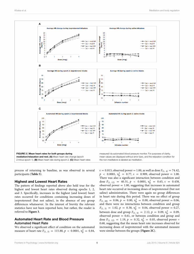
p = 0.03, observed power = 0.482,suggesting that the heart rate increases induced by isoproterenolwere not statistically different between the groups (Figure 2A).Mean average heart rate changes and associated 95% confidenceintervals are displayed in Table 2.
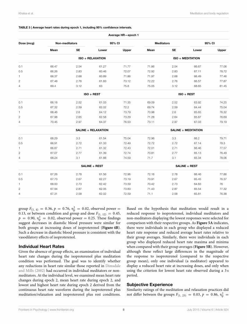
Mean Heart RateEpoch 1We did not find an effect of condition on the average heartrate during epoch 1, F(1, 3) = 0.39, p = 0.76, η
2p = 0.01,
observed power= 0.12, but did observe a significant effect of doseF(1, 4) = 4.74, p = 0.008, η2
p = 0.15, ε = 0.616, observed power=0.80. There was also a significant interaction between conditionand dose F(1, 12) = 4.41, p < 0.0001, η
2p = 0.14, ε = 0.772,
observed power = 0.99, suggesting that increases in mean heartrate during epoch 1 occurred at increasing doses of isoproterenol(but not saline) administration. However, despite these changes,there were no group differences in heart rate during this period.There was no effect of group F(1, 28) = 1.20, p = 0.28, η2
p = 0.04,observed power = 0.19, and there were no interactions betweencondition and group F(1, 3) = 2.00, p = 0.12, η
2p = 0.07,
observed power = 0.50, between dose and group F(1, 4) = 0.33,p = 0.76, η
2p = 0.01, observed power = 0.12, or between
condition and group and dose F(1, 12) = 0.74, p = 0.68, η2p =
0.03, observed power= 0.43, suggesting that the mean heart rateincreases observed for increasing doses of isoproterenol duringepoch 1 were not statistically different between groups. Averageheart rate changes and associated 95% confidence intervals aredisplayed in Table 3. The finding of dose related increases duringthis epoch reflects that fact that some of the heart rate changesinduced by isoproterenol (particularly at the higher doses) beganoccurring early, as was observed in several participants.
Epoch 2We observed a significant effect of condition on the average heartrate during epoch 2, F(1, 3) = 84.19, p < 0.0001, η
2p = 0.75,
observed power = 1.00, as well as for dose F(1, 4) = 84.19,p < 0.0001, η
2p = 0.75, ε = 0.402, observed power = 1.00.
There was also a significant interaction between condition anddose F(1, 12) = 72.91, p < 0.0001, η
2p = 0.72, ε = 0.330,
observed power = 1.00, suggesting that increases in mean heartrate during epoch 2 occurred at increasing doses of isoproterenol(but not saline) administration. However, despite these changes,there were again no group differences in heart rate during thisperiod. There was no effect of group F(1, 28) = 0.65, p =
0.43, η2p = 0.02, observed power = 0.12, and there were no
interactions between condition and group F(1, 3) = 2.00, p =
0.12, η2p = 0.07, observed power = 0.37, between dose and
group F(1, 4) = 1.44, p = 0.24, η2p = 0.05, observed power
= 0.16, or between condition and group and dose F(1, 12) =
0.70, p = 0.59, η2p = 0.02, observed power = 0.41, suggesting
that the mean heart rate increases observed for increasing dosesof isoproterenol during epoch 2 were not statistically differentbetween groups (Figure 2B and Table 4).
Epoch 3We observed a significant effect of condition on the average heartrate during epoch 3, F(1, 3) = 8.30, p < 0.0001, η
2p = 0.23,
observed power = 0.98, as well as for dose F(1, 4) = 30.66,p < 0.0001, η
2p = 0.52, ε = 0.440, observed power = 1.00.
There was also a significant interaction between condition anddose F(1, 12) = 20.72, p < 0.0001, η
2p = 0.43, ε = 0.418,
observed power = 1.00, suggesting that increases in mean heartrate during epoch 3 occurred at increasing doses of isoproterenol(but not saline) administration. However, despite these changes,there were no group differences in heart rate during this period.There was no effect of group F(1,28) = 0.92, p = 0.35, η2
p = 0.03,observed power = 0.15, and there were no interactions betweencondition and group F(1, 3) = 1.68, p = 0.18, η
2p = 0.06,
observed power = 0.43, between dose and group F(1, 4) = 0.93,p = 0.39, η
2p = 0.03, observed power = 0.29, or between
condition and group and dose F(1, 12) = 0.65, p = 0.67, η2p =
0.02, observed power= 0.38, suggesting that the mean heart rateincreases observed for increasing doses of isoproterenol duringepoch 3 were not statistically different between groups. Thefinding of dose related increases during this epoch reflects thatfact that some of the heart rate changes induced by isoproterenol(particularly at the higher doses) were still present and in the
Frontiers in Psychology | www.frontiersin.org 5 July 2015 | Volume 6 | Article 924
Khalsa et al. Meditation and body regulation
FIGURE 2 | Mean heart rates for both groups during
mediation/relaxation and rest. (A) Mean heart rate change (epoch
2minus epoch 1). (B) Mean heart rate during epoch 2. (C) Mean heart rates
measured via automated blood pressure monitor. For purposes of clarity,
mean values are displayed without error bars, and the relaxation condition for
the non-meditators is labeled as meditation.
process of returning to baseline, as was observed in severalparticipants (Table 5).
Highest and Lowest Heart RatesThe pattern of findings reported above also held true for thehighest and lowest heart rates observed during epochs 1, 2,and 3. Specifically, increases in the highest (and lowest) heartrates occurred for conditions containing increasing doses ofisoproterenol (but not saline), in the absence of any groupdifferences whatsoever. In the interest of brevity the relevantstatistics have not been reported here, but rather, the reader isreferred to Figure 3.
Automated Heart Rate and Blood PressureAutomated Heart RateWe observed a significant effect of condition on the automatedmeasure of heart rate F(1, 3) = 115.88, p < 0.0001, η2
p = 0.84,
ε = 0.813, observed power= 1.00, as well as dose F(1, 4) = 74.42,p < 0.0001, η
2p = 0.77, ε = 0.909, observed power = 1.00.
There was also a significant interaction between condition anddose F(1, 12) = 40.31, p < 0.0001, η
2p = 0.65, ε = 0.438,
observed power = 1.00, suggesting that increases in automatedheart rate occurred at increasing doses of isoproterenol (but notsaline) administration. There were again no group differencesin heart rate during this period. There was no effect of groupF(1, 28) = 0.06, p = 0.80, η
2p = 0.00, observed power = 0.06,
and there were no interactions between condition and groupF(1, 3) = 1.02, p = 0.38, η
2p = 0.04, observed power = 0.27,
between dose and group F(1, 4) = 2.12, p = 0.09, η2p = 0.09,
observed power = 0.61, or between condition and group anddose F(1, 12) = 1.19, p = 0.32, η
2p = 0.05, observed power =
0.68, suggesting that the mean heart rate increases observed forincreasing doses of isoproterenol with the automated measurewere similar between the groups (Figure 2C).
Frontiers in Psychology | www.frontiersin.org 6 July 2015 | Volume 6 | Article 924
Khalsa et al. Meditation and body regulation
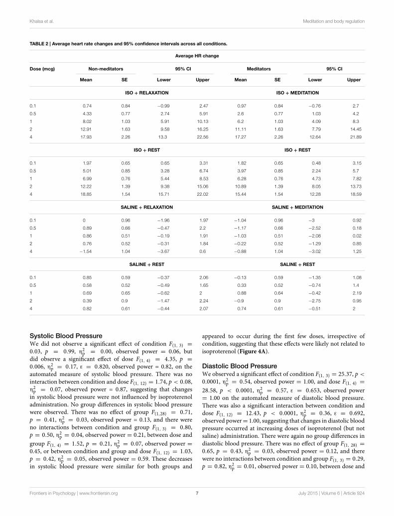
TABLE 2 | Average heart rate changes and 95% confidence intervals across all conditions.
Average HR change
Dose (mcg) Non-meditators 95% CI Meditators 95% CI
Mean SE Lower Upper Mean SE Lower Upper
ISO + RELAXATION ISO + MEDITATION
0.1 0.74 0.84 −0.99 2.47 0.97 0.84 −0.76 2.7
0.5 4.33 0.77 2.74 5.91 2.6 0.77 1.03 4.2
1 8.02 1.03 5.91 10.13 6.2 1.03 4.09 8.3
2 12.91 1.63 9.58 16.25 11.11 1.63 7.79 14.45
4 17.93 2.26 13.3 22.56 17.27 2.26 12.64 21.89
ISO + REST ISO + REST
0.1 1.97 0.65 0.65 3.31 1.82 0.65 0.48 3.15
0.5 5.01 0.85 3.28 6.74 3.97 0.85 2.24 5.7
1 6.99 0.76 5.44 8.53 6.28 0.76 4.73 7.82
2 12.22 1.39 9.38 15.06 10.89 1.39 8.05 13.73
4 18.85 1.54 15.71 22.02 15.44 1.54 12.28 18.59
SALINE + RELAXATION SALINE + MEDITATION
0.1 0 0.96 −1.96 1.97 −1.04 0.96 −3 0.92
0.5 0.89 0.66 −0.47 2.2 −1.17 0.66 −2.52 0.18
1 0.86 0.51 −0.19 1.91 −1.03 0.51 −2.08 0.02
2 0.76 0.52 −0.31 1.84 −0.22 0.52 −1.29 0.85
4 −1.54 1.04 −3.67 0.6 −0.88 1.04 −3.02 1.25
SALINE + REST SALINE + REST
0.1 0.85 0.59 −0.37 2.06 −0.13 0.59 −1.35 1.08
0.5 0.58 0.52 −0.49 1.65 0.33 0.52 −0.74 1.4
1 0.69 0.65 −0.62 2 0.88 0.64 −0.42 2.19
2 0.39 0.9 −1.47 2.24 −0.9 0.9 −2.75 0.95
4 0.82 0.61 −0.44 2.07 0.74 0.61 −0.51 2
Systolic Blood PressureWe did not observe a significant effect of condition F(1, 3) =
0.03, p = 0.99, η2p = 0.00, observed power = 0.06, but
did observe a significant effect of dose F(1, 4) = 4.35, p =
0.006, η2p = 0.17, ε = 0.820, observed power = 0.82, on the
automated measure of systolic blood pressure. There was nointeraction between condition and dose F(1, 12) = 1.74, p < 0.08,η2p = 0.07, observed power = 0.87, suggesting that changes
in systolic blood pressure were not influenced by isoproterenoladministration. No group differences in systolic blood pressurewere observed. There was no effect of group F(1,28) = 0.71,p = 0.41, η
2p = 0.03, observed power = 0.13, and there were
no interactions between condition and group F(1, 3) = 0.80,p = 0.50, η2
p = 0.04, observed power = 0.21, between dose and
group F(1, 4) = 1.52, p = 0.21, η2p = 0.07, observed power =
0.45, or between condition and group and dose F(1, 12) = 1.03,p = 0.42, η2
p = 0.05, observed power = 0.59. These decreasesin systolic blood pressure were similar for both groups and
appeared to occur during the first few doses, irrespective ofcondition, suggesting that these effects were likely not related toisoproterenol (Figure 4A).
Diastolic Blood PressureWe observed a significant effect of condition F(1, 3) = 25.37, p <
0.0001, η2p = 0.54, observed power = 1.00, and dose F(1, 4) =
28.58, p < 0.0001, η2p = 0.57, ε = 0.653, observed power
= 1.00 on the automated measure of diastolic blood pressure.There was also a significant interaction between condition anddose F(1, 12) = 12.43, p < 0.0001, η
2p = 0.36, ε = 0.692,
observed power= 1.00, suggesting that changes in diastolic bloodpressure occurred at increasing doses of isoproterenol (but notsaline) administration. There were again no group differences indiastolic blood pressure. There was no effect of group F(1, 28) =0.65, p = 0.43, η
2p = 0.03, observed power = 0.12, and there
were no interactions between condition and group F(1, 3) = 0.29,p = 0.82, η2
p = 0.01, observed power = 0.10, between dose and
Frontiers in Psychology | www.frontiersin.org 7 July 2015 | Volume 6 | Article 924
Khalsa et al. Meditation and body regulation
TABLE 3 | Average heart rates during epoch 1, including 95% confidence intervals.
Average HR—epoch 1
Dose (mcg) Non-meditators 95% CI Meditators 95% CI
Mean SE Lower Upper Mean SE Lower Upper
ISO + RELAXATION ISO + MEDITATION
0.1 66.47 2.54 61.27 71.77 71.86 2.54 66.67 77.06
0.5 66.26 2.83 60.46 72.07 72.92 2.83 67.11 78.72
1 66.37 2.68 60.89 71.86 71.97 2.68 66.49 77.46
2 67.48 2.76 61.83 73.12 72.22 2.76 66.57 77.87
4 69.4 3.12 63 75.8 75.05 3.12 68.65 81.45
ISO + REST ISO + REST
0.1 66.18 2.52 61.03 71.35 69.09 2.52 63.92 74.25
0.5 67.32 2.59 62.02 72.2 69.74 2.59 64.44 75.04
1 68.46 2.6 64.12 73.79 70.98 2.6 65.65 76.32
2 67.98 2.65 62.58 73.29 71.28 2.64 65.87 76.69
4 70.45 2.97 64.37 76.53 73.11 2.97 67.03 79.19
SALINE + RELAXATION SALINE + MEDITATION
0.1 68.29 3.3 61.54 75.04 72.96 3.3 66.2 79.71
0.5 66.91 2.72 61.33 72.49 72.72 2.72 67.14 78.3
1 66.87 2.71 61.32 72.43 72.01 2.71 66.46 77.57
2 67.07 2.77 61.39 72.74 70.81 2.77 65.13 76.49
4 68.24 3.1 61.88 74.59 71.7 3.1 65.34 78.06
SALINE + REST SALINE + REST
0.1 67.26 2.78 61.56 72.96 72.16 2.78 66.46 77.86
0.5 67.73 2.67 62.27 73.19 70.91 2.67 65.45 76.37
1 68.00 2.73 62.42 73.59 70.42 2.73 64.83 76
2 67.94 2.87 62.05 73.83 71.43 2.87 65.54 77.32
4 67.31 2.58 62.02 72.59 71.1 2.58 65.81 76.38
group F(1, 4) = 0.36, p = 0.76, η2p = 0.02, observed power =
0.13, or between condition and group and dose F(1, 12) = 0.45,p = 0.90, η
2p = 0.02, observed power = 0.25. These findings
suggest decreases in diastolic blood pressure were similar forboth groups at increasing doses of isoproterenol (Figure 4B).Such a decrease in diastolic blood pressure is consistent with thevasodilatory effects of isoproterenol.
Individual Heart RatesGiven the absence of group effects, an examination of individualheart rate changes during the isoproterenol plus meditationcondition was performed. The goal was to identify whetherany reductions in heart rate similar those reported in Dimsdaleand Mills (2002) had occurred in individual meditators or non-meditators. At the individual level, we examined mean heart ratechanges during epoch 2, mean heart rate during epoch 2, andlowest and highest heart rate during epoch 2 derived from thecontinuous heart rate waveform during the isoproterenol plusmeditation/relaxation and isoproterenol plus rest conditions.
Based on the hypothesis that meditation would result in areduced response to isoproterenol, individual meditators andnon-meditators displaying the lowest responses were selected forcomparison with their respective groups. As Figure 5A indicates,there were individuals in each group who displayed a reducedheart rate response and reduced average heart rates relative totheir group averages. Similarly, there were individuals in eachgroup who displayed reduced heart rate maxima and minimawhen compared with their group averages (Figure 5B). However,although these reflect large differences in the magnitude ofthe response to isoproterenol (compared to the respectivegroup mean), only one individual (a meditator) appeared todisplay a reduced heart rate at increasing doses, and only whenusing the criterion for lowest heart rate observed during a 3 speriod.
Subjective ExperienceSimilarity ratings of the meditation and relaxation practices didnot differ between the groups F(1, 25) = 0.03, p = 0.86, η
2p =
Frontiers in Psychology | www.frontiersin.org 8 July 2015 | Volume 6 | Article 924

Khalsa et al. Meditation and body regulation
TABLE 4 | Average heart rates during epoch 2, including 95% confidence intervals.
Average HR—epoch 2
Dose (mcg) Non-meditators 95% CI Meditators 95% CI
Mean SE Lower Upper Mean SE Lower Upper
ISO + RELAXATION ISO + MEDITATION
0.1 67.21 2.55 61.98 72.43 72.84 2.55 67.61 78.06
0.5 70.59 2.71 65.04 76.13 75.53 2.71 69.98 81.08
1 74.39 2.95 68.35 80.43 78.17 2.95 72.13 84.21
2 80.39 3.5 73.22 87.55 83.34 3.5 76.17 90.5
4 87.33 4.14 78.87 95.8 92.32 4.14 83.85 100.79
ISO + REST ISO + REST
0.1 68.17 2.51 63.03 73.31 70.9 2.51 65.76 76.04
0.5 72.34 2.64 66.93 77.74 73.71 2.64 68.31 79.12
1 75.44 2.84 69.62 81.26 77.26 2.84 71.44 83.08
2 80.21 3.19 73.66 86.75 82.17 3.19 75.63 88.71
4 89.31 3.51 82.13 96.49 88.55 3.51 81.37 95.73
SALINE + RELAXATION SALINE + MEDITATION
0.1 68.39 2.9 62.36 74.23 71.92 2.9 65.98 77.85
0.5 67.79 2.69 62.23 73.3 71.55 2.69 66.04 77.06
1 67.73 2.8 62.01 73.45 70.99 2.8 65.26 76.71
2 67.83 2.75 62.2 73.46 70.59 2.75 65.96 76.22
4 66.7 2.73 61.11 72.29 70.82 2.73 65.23 76.4
SALINE + REST SALINE + REST
0.1 68.10 2.77 62.44 73.77 72.03 2.77 66.36 77.7
0.5 68.31 2.63 62.93 73.7 71.24 2.63 65.86 76.63
1 68.69 2.69 63.19 74.19 71.3 2.66 65.8 76.8
2 68.33 2.66 62.89 73.77 70.53 2.66 65.09 75.97
4 68.12 2.8 62.38 73.86 71.84 2.8 66.1 77.58
0.001, observed power = 0.0542. The meditation and relaxationpractices appeared to be rated as more similar to the usualpractice during the saline infusion condition, but this differencewas not statistically significant F(1, 1) = 2.18, p = 0.15, η
2p =
0.08, observed power= 0.30.
Post AnalysisSince a heterogeneously recruited sample of meditators couldpotentially result in differential heart rate responses duringmeditation, we subsequently evaluated the responses of theKundalini and Vipassana practitioners separately, and incomparison to each other.
Vipassana vs. Healthy ComparisonDuring the meditation condition there was no effect of groupF(1, 24) = 1.31, p = 0.26, η
2p = 0.05, observed power = 0.20,
2Data were missing from 3 healthy comparison participants.
and there were no interactions between condition and groupF(1, 1) = 0.002, p = 0.98, η
2p = 0.00, observed power =
0.05, or between condition and group and dose F(1, 4) = 0.10,p = 0.98, η2
p = 0.004, observed power= 0.07. This suggests thatduring both meditation conditions (saline and isoproterenol)the Vipassana meditators displayed similar average heart rateincreases as healthy comparisons.
Kundalini vs. healthy ComparisonDuring the meditation condition there was no effect of groupF(1, 17) = 2.1, p = 0.17, η
2p = 0.11, observed power = 0.27,
and there were no interactions between condition and groupF(1, 1) = 0.006, p = 0.94, η
2p = 0.00, observed power =
0.05, or between condition and group and dose F(1, 4) = 1.58,p = 0.19, η2
p = 0.09, observed power = 0.46. This suggests thatduring both meditation conditions (saline and isoproterenol)the Kundalini meditators displayed similar average heart rateincreases as healthy comparisons.
Frontiers in Psychology | www.frontiersin.org 9 July 2015 | Volume 6 | Article 924

Khalsa et al. Meditation and body regulation
TABLE 5 | Average heart rates during epoch 3, including 95% confidence intervals.
Average HR—epoch 3
Dose (mcg) Non-meditators 95% CI Meditators 95% CI
Mean SE Lower Upper Mean SE Lower Upper
ISO + RELAXATION ISO + MEDITATION
0.1 66.58 2.58 61.29 71.87 71.57 2.58 66.28 76.85
0.5 66.28 2.71 60.73 71.82 71.7 2.71 66.16 77.25
1 68.1 2.74 62.49 73.71 72.5 2.74 66.89 78.11
2 70.59 3.15 64.13 77.05 75.11 3.15 68.66 81.57
4 73.94 3.67 66.43 81.45 81.28 3.67 73.77 88.79
ISO + REST ISO + REST
0.1 67.88 2.55 62.66 73.1 71.8 2.55 66.57 77.02
0.5 68.38 2.63 62.99 73.77 70.67 2.63 65.28 76.06
1 68.75 2.65 63.32 74.19 70.86 2.65 65.43 76.3
2 71.47 3.04 65.25 77.69 75.57 3.04 69.35 81.79
4 77.07 3.33 70.24 83.9 80.35 3.33 73.52 87.18
SALINE + RELAXATION SALINE + MEDITATION
0.1 67.93 2.74 62.31 73.54 71.97 2.74 66.34 77.57
0.5 68.06 2.67 62.6 73.52 71.85 2.67 66.39 77.31
1 68.58 2.7 63.04 74.12 71.2 2.7 65.66 76.74
2 67.89 2.78 62.2 73.58 70.39 2.78 64.7 76.08
4 66.48 2.69 60.97 72 70.09 2.69 65.57 75.6
SALINE + REST SALINE + REST
0.1 68.12 2.6 62.8 73.44 71.78 2.6 66.46 77.1
0.5 68.67 2.67 63.19 74.14 71.24 2.67 65.77 76.72
1 69.73 2.53 64.54 74.93 71.04 2.53 65.85 76.23
2 68.38 2.66 62.94 73.83 71.83 2.66 66.39 77.27
4 69.81 2.77 64.14 75.47 72.15 2.77 66.49 77.82
Kundalini vs. VipassanaDuring the meditation condition there was no effect of groupF(1, 13) = 0.11, p = 0.75, η2
p = 0.008, observed power = 0.06,and there were no interactions between condition and groupF(1, 1) = 0.001, p = 0.98, η2
p = 0.00, observed power = 0.05, orbetween condition and group and dose F(1, 4) = 0.88, p = 0.48,η2p = 0.06, observed power= 0.26. This suggests that during both
meditation conditions (saline and isoproterenol) the two groupsof meditators displayed similar average heart rate increases.
Discussion
As expected, bolus isoproterenol infusions produced dose-dependent increases in heart rate in both groups duringa condition of rest. However, meditators did not displaylowered heart rate responses to isoproterenol while practicingmeditation. Both meditators and non-meditators displayedsimilar dose dependent increases in heart rate during conditionsof isoproterenol plus rest, and during isoproterenol plus
meditation/relaxation. The lack of group differences in heart ratewas reliable. It was observed for five different measures of heartrate (mean heart rate change, mean heart rate, lowest heart rateduring a 3 s period, highest heart rate during a 3 s period, andvia automated heart rate monitor), and throughout three epochsthat captured the entire time span over which isoproterenolinduced changes occur. Equivalent decreases in diastolic bloodpressure were also observed in both groups following increasingdoses of isoproterenol. These decreases are consistent with theknown vasodilatory effects of isoproterenol on lowering diastolicblood pressure. We do not consider these to be related to themeditation intervention, as they occurred in both isoproterenolconditions. Non-meditators did not display lowered heart rateswhen practicing a relaxation condition, which was predicted.
Since the Dimsdale and Mills (2002) study reported afinding in a single individual, we examined individual heart rateresponses within in each group of participants. Of the measuresutilized, there was only one participant who demonstrated anappreciable reduction in heart rate during the meditation plus
Frontiers in Psychology | www.frontiersin.org 10 July 2015 | Volume 6 | Article 924

Khalsa et al. Meditation and body regulation
FIGURE 3 | Highest and lowest heart rates for both groups during
epoch 2. (A) Highest heart rates observed during a 3 s period, averaged
across each group (ceiling effect). (B) Lowest heart rates observed during a
3 s period, averaged across each group (floor effect). For purposes of clarity,
mean values are displayed without error bars, and the relaxation condition for
the non-meditators is labeled as meditation.
isoproterenol condition similar to that reported by Dimsdale andMills (2002) (i.e., a 17 bpm decrease in heart rate from the lowestto the highest dose). This finding occurred in a meditator, but itwas only observed with one measure, the lowest heart rate, whichis also the least reliable index of the response to isoproterenol.This finding was no longer present when more robust measuresof the heart rate response to isoproterenol, such as the averageheart rate and average heart rate change, were examined in thesame individual. Thus, it seems unlikely that the individual effectreported by Dimsdale and Mills (2002), although intriguing,generally applies to individuals practicing meditation.
Several potential factors might explain the discrepancy infindings between the two studies. First, individual variability inheart rate responses might explain their observation of loweredheart rate in one subject. We observed considerable variabilityin heart rate responses to isoproterenol even when using acontinuous measure, with select individuals from both groupsexhibiting attenuated heart rate responses to isoproterenol. Asecond and related reason could bemeasurement error. Dimsdaleand Mills (2002) did not utilize a continuous measure of heartrate in their original study, and it is unclear when the automatedmonitor they utilized actually captured their meditator’s heartrate. Although they report that heart rate was measured 60 s afterthe infusion was delivered, it is possible that there was enoughmeasurement variability to result in missing the heart rate epochof interest. In the present study, we observed large variabilityin the output of the automated heart rate measurement whenusing the blood pressure monitoring device (e.g., 58–93 s after
initiatingmeasurement), which is long enough to potentiallymisscapturing an individual’s heart rate response. It is also possiblethat the window of measurement of the automated heart ratemonitor was too brief. In the current study, we observed thelargest reductions in heart rate response during a momentarymeasure, the lowest heart rate during a 3 s period, which providessome plausibility to this theory. Third, their meditator was notformally trained, and indicated that she would often enter suchdeep states that she would need to set an alarm clock to rouseher from meditation. Thus, it is possible that this individuallikened sleep (or some other altered state of consciousness) withmeditation, and that the observed effect had nothing to do withmeditation but a different process worth understanding further.However, despite these potential methodological and conceptualconsiderations, the participant in Dimsdale and Mills’ study diddemonstrate a heart rate increase at the 1.0 mcg dose, followed bytwo successive decreases below her resting heart rate. Assumingthis was not a spurious finding, such a profound decrease inheart rate could also occur during an episode of increased vagaloutput (as suggested by the authors). Neurocardiogenic syncopeis one example of such an increase in vagal output. These episodesare usually preceded by an increase in sympathetic tone (aswas observed in the meditator at the 1.0 mcg dose) and caneven be triggered by isoproterenol (Kikushima et al., 1999).These episodes are often foreshadowed by symptoms such aslightheadedness, nausea, warmth, pallor and/or sweating, andalthough the meditator denied experiencing a subset of these(nausea, dizziness or fainting), it is possible that she was not
Frontiers in Psychology | www.frontiersin.org 11 July 2015 | Volume 6 | Article 924

Khalsa et al. Meditation and body regulation
FIGURE 4 | Blood pressure for both groups during
meditation/relaxation and rest. (A) Systolic blood pressure measured via
automated blood pressure monitor. (B) Diastolic blood pressure measured
via automated blood pressure monitor. For purposes of clarity, mean values
are displayed without error bars, and the relaxation condition for the
non-meditators is labeled as meditation.
aware of these symptoms given the fact that she was in ameditative state described as so deep as to require rousing withan alarm clock. Since heart rate was the only autonomic measurereported in that study, it is difficult to determine whether such areflex occurred. Evaluating this particular meditator’s (or othersthe future) cardiovascular responses to upright tilt table testing(perhaps even incorporating isoproterenol) could be useful toexclude this as a possibility. Another possibility is that respiratorymodulation could have played a role in the observed heart ratechanges, as respiratory induced changes in heart rate variabilityhave been observed in meditators (Peng et al., 2004). We did notexamine this possibility as it was outside the scope of inquiry inour study.
There are several limitations associated with the presentstudy. First, the meditation practice was investigated underhighly artificial circumstances. Meditators were meditating ina novel environment, with an intravenous line placed, wereattached to various recording devices and were seated nextto two individuals who were observing them throughout thestudy. Despite efforts to facilitate the practice, such as a curtainbetween the investigators and participant, and a dimly lit,quiet room, it cannot be claimed that the meditation andrelaxation conditions took place in the usual environment.Second, the current study does not take into account the potentialinfluence of testing anxiety, that is, concern on the part ofthe meditators about “performing well.” Although meditatorsdid not voice any such concerns, and did not differ fromthe non-meditators in questionnaire measures of anxiety, this
possibility cannot be ruled out. A third limitation is the factthat the meditation plus saline conditions did not appear tolower the heart rate substantially. This could again be due to theartificiality of the environment, thus preventing meditators fromauthentically engaging in their practice and displaying increasedparasympathetic indices. However, retrospective ratings of thequality of the meditation practice indicated that meditatorsfound their meditation practice during the infusions to bepredominantly similar to their daily practice, reducing this asa possibility. The lack of heart rate reductions during salinecould also be related to the type of meditation participantspracticed. For example, some meditations are suggested toincrease cardiovascular states whereas others are suggested todecrease them (Amihai and Kozhevnikov, 2014). Since mostmeditators were not instructed to use their meditation practiceto reduce their heart rate (this would have been seen by theVipassana practitioners as constraining and interfering withthe meditation practice), it is possible that the mediators wereperforming the former type of meditation. Reports from themeditators about the nature and quality of their practice did notseem to indicate this as a possibility.
It is also possible that the lack of attenuated autonomicresponses in the current study could be related to a non-ideal sample of meditators and/or a non-ideal meditationpractice. For example, since the majority of the meditatorscame from a Vipassana background, their default meditationpractice may not be intended to have effects on autonomicstate. This argument is somewhat limited by the fact that
Frontiers in Psychology | www.frontiersin.org 12 July 2015 | Volume 6 | Article 924

Khalsa et al. Meditation and body regulation
FIGURE 5 | Global and individual outlier heart rate changes
during isoproterenol conditions. (A) Global mean heart rate
change (epoch 2minus epoch 1), and global mean heart rate
observed during epoch 2 for all participants. (B) Global maximum
heart rate change and global minimum heart rate change observed
during epoch 2 for all participants. The green lines indicate the
meditator who displayed the lowest response during the isoproterenol
plus meditation condition. The blue lines indicate the non-meditators
who displayed the lowest response during the isoproterenol plus
relaxation condition. For purposes of clarity, the relaxation condition
for the non-meditators is labeled as meditation. Error bars represent
standard error of the mean.
Vipassana meditation techniques emphasize the cultivationof quiescent and tranquil states of mind, and show someevidence of altered sympatho-vagal balance (Krygier et al.,2013). Alternatively, it is theoretically possible that the defaultbody scanning meditation practice practiced by the Vipassanameditators in this study could have been similar to a formof “open presence” rather than “focused attention” meditation.Since the former type of meditation has been associatedwith heightened arousal instead of relaxation, this might beexpected to induce an increased autonomic response duringmeditation instead of a quiescent one. We observed a fewmarginal interactions between condition and group on heart ratewith the meditators potentially exhibiting increased heart ratesduring meditation, that might potentially support this possibility.However, these non-significant effects were not consistentlyobserved across the numerous heart rate measures employed.Further complicating this theoretical interpretation is evidencesuggesting that open presence meditation can be associated withattenuated autonomic responses to startle challenges that probesympathetic reactivity (Levenson et al., 2012). We conclude thereis no direct evidence in the current study to support the notionof differentially decreased or increased autonomic indices in themeditators.
Another question about the current sample is whether theabsence of effect could have been due to an inexperiencedsample of meditators relative to the meditator in Dimsdale
and Mills (2002), as the minimum requirement was only 2years of meditative experience. Direct comparison betweenthe studies is not possible, as the years of practice were notprovided in that report. However, the notion that duration ofpractice is synonymous with meditative expertise is a matter ofongoing debate. Previous investigations of long term meditatorshave not always yielded evidence of increased ability, evenfor aspects of internal sensory experience that are routinelycultivated in Vipassana and Kundalini traditions (Khalsa et al.,2008; Daubenmier et al., 2013). A larger issue is the factthat the meditator described in that report was not formallytrained in any particular tradition, making it impossible toselect an appropriately representative group of meditators. Inthe current study we recruited formally trained meditators frommultiple traditions, used extensive measurements of heart rateresponse, and examined individual outlier responses in bothgroups to determine whether there was specific evidence thatmeditation was associated with a lowered heart rate. Thoughsome individuals displayed attenuated heart rate responses toisoproterenol, we found no evidence differentiating meditatorsfrom non-meditators.
A general point is that while these limitations warrantconsideration, many of them would be similarly imposed by anyempirical study of meditation and thus cannot be easily obviated.If further investigations of the current topic were continued, onehelpful strategy would be to screen large samples of meditators
Frontiers in Psychology | www.frontiersin.org 13 July 2015 | Volume 6 | Article 924

Khalsa et al. Meditation and body regulation
to identify those individuals who can reliably demonstrateenhanced autonomic regulatory capabilities, and then performdetailed investigation into the cognitive and neurophysiologicalmechanisms underlying such abilities.
We feel it is also important to note that the current findings donot necessarily negate previously reported findings of decreasedautonomic tone following the practice of meditation. Althoughour study appeared sufficiently powered to detect effects relatedto the isoproterenol vs. saline conditions, and the impact ofdifferent doses, there was lower power at the group level.While replicating this approach with a larger sample wouldhelp to address this limitation, the current results suggestthat any effects, even if found reliable at the population level,would be small and would have limited consequence at theindividual level. The current findings also have limited bearingon the effects of meditation on emotional experience or emotionregulation, effects consistently perceived at the individual levelwith training, as these constructs were not investigated. Highlevels of adrenergic hormones are often associated with acuteanxiety, stress, anger, fear and hostility, and different formsof meditation are routinely practiced by some individuals tohelp ameliorate such experiences (Shirey, 2007; Robins et al.,2012; Serpa et al., 2014). Determining the biological mechanismssupporting those beneficial effects remains an important andactive area of inquiry, regardless of whether they are derived
from changes in autonomic reactivity or from changes in thecentral nervous system. Overall, these results simply suggestthat the formal act of meditation may not be sufficient tophysiologically counteract certain forms of elevated levels ofperipheral cardiovascular adrenergic arousal or engender quickerrecovery from such arousal.
Support
This research was supported by a grant from the National Centerfor Complementary and Alternative Medicine (NCCAM F31AT003061-01A1), the National Center for Research Resources,General Clinical Research Centers Program (NIH M01-RR-59), a McDonnell Foundation Collaborative Award to DT(#220020387), and the William K. Warren Foundation.
Acknowledgments
We would like to thank Shinzen Young for assistance withparticipant recruitment, Erik St. Louis, Brian Olshansky, andChirag Sandesara for assistance with participant screening, SoniaSchubert for nursing assistance with administration of theisoproterenol protocol, Becky Triplett and the IV ads pharmacystaff for isoproterenol preparation, and James Martins for safetymonitoring.
References
Aftanas, L., and Golosheykin, S. (2005). Impact of regular meditation practice on
EEG activity at rest and during evoked negative emotions. Int. J. Neurosci. 115,
893–909. doi: 10.1080/00207450590897969
Aftanas, L. I., and Golocheikine, S. A. (2002). Non-linear dynamic complexity
of the human EEG during meditation. Neurosci. Lett. 330, 143–146. doi:
10.1016/S0304-3940(02)00745-0
Ahir, D. C. (1999). Vipassana: A Universal Buddhist Meditation Technique. Delhi:
Sri Satguru Publications.
Amihai, I., and Kozhevnikov, M. (2014). Arousal vs. relaxation: a comparison
of the neurophysiological and cognitive correlates of vajrayana and theravada
meditative practices. PLoS ONE 9:e102990. doi: 10.1371/journal.pone.0102990
Astin, J. A., Shapiro, S. L., Eisenberg, D. M., and Forys, K. L. (2003). Mind-body
medicine: state of the science, implications for practice. J. Am. Board Fam.
Pract. 16, 131–147. doi: 10.3122/jabfm.16.2.131
Barnes, V. A., Davis, H. C., Murzynowski, J. B., and Treiber, F. A. (2004).
Impact of meditation on resting and ambulatory blood pressure and heart
rate in youth. Psychosom.Med. 66, 909–914. doi: 10.1097/01.psy.0000145902.91
749.35
Barnes, V. A., Treiber, F. A., Turner, J. R., Davis, H., and Strong, W. B. (1999).
Acute effects of transcendental meditation on hemodynamic functioning in
middle-aged adults. Psychosom. Med. 61, 525–531. doi: 10.1097/00006842-
199907000-00017
Beary, J. F., and Benson, H. (1974). A simple psychophysiologic technique which
elicits the hypometabolic changes of the relaxation response. Psychosom. Med.
36, 115–120. doi: 10.1097/00006842-197403000-00003
Benson, H., Beary, J. F., and Carol, M. P. (1974). The relaxation response.
Psychiatry 37, 37–46.
Bonadonna, R. (2003). Meditation’s impact on chronic illness. Holist. Nurs. Pract.
17, 309–319. doi: 10.1097/00004650-200311000-00006
Brefczynski-Lewis, J. A., Lutz, A., Schaefer, H. S., Levinson, D. B., and
Davidson, R. J. (2007). Neural correlates of attentional expertise in long-term
meditation practitioners. Proc. Natl. Acad. Sci. U.S.A. 104, 11483–11488. doi:
10.1073/pnas.0606552104
Burley, M. (2000). Hatha-yoga. Its Context, Theory and Practice. New Delhi: Shri
Jainendra Press.
Daubenmier, J., Sze, J., Kerr, C. E., Kemeny, M. E., and Mehling, W. (2013). Follow
your breath: respiratory interoceptive accuracy in experienced meditators.
Psychophysiology 50, 777–789. doi: 10.1111/psyp.12057
Davidson, R. J., Goleman, D. J., and Schwartz, G. E. (1976). Attentional and
affective concomitants of meditation: a cross-sectional study. J. Abnorm.
Psychol. 85, 235–238. doi: 10.1037/0021-843X.85.2.235
Dimsdale, J. E., and Mills, P. J. (2002). An unanticipated effect of meditation on
cardiovascular pharmacology and physiology. Am. J. Cardiol. 90, 908–909. doi:
10.1016/S0002-9149(02)02726-1
Dimsdale, J., Ziegler, M., and Graham, R. (1988). The effect of hypertension,
sodium, and race on isoproterenol sensitivity. Clin. Exp. Hypertens. A 10,
747–756.
Ekman, P., Davidson, R. J., Ricard, M., and Wallace, A. B. (2005). Buddhist and
psychological perspectives on emotions and well-being. Curr. Dir. Psychol. Sci.
14, 59–63. doi: 10.1111/j.0963-7214.2005.00335.x
Farrow, J. T., and Hebert, J. R. (1982). Breath suspension during the transcendental
meditation technique. Psychosom. Med. 44, 133–153. doi: 10.1097/00006842-
198205000-00001
Fenz, W. D., and Plapp, J. M. (1970). Voluntary control of heart rate in a
practitioner of yoga: negative findings. Percept. Mot. Skills 30, 493–494. doi:
10.2466/pms.1970.30.2.493
Harinath, K., Malhotra, A. S., Pal, K., Prasad, R., Kumar, R., Kain, T.
C., et al. (2004). Effects of Hatha yoga and Omkar meditation on
cardiorespiratory performance, psychologic profile, and melatonin secretion.
J. Altern. Complement. Med. 10, 261–268. doi: 10.1089/107555304323062257
Hoffman, J. W., Benson, H., Arns, P. A., Stainbrook, G. L., Landsberg,
G. L., Young, J. B., et al. (1982). Reduced sympathetic nervous system
responsivity associated with the relaxation response. Science 215, 190–192. doi:
10.1126/science.7031901
Hughes, J. W., Fresco, D. M., Myerscough, R., van Dulmen, M. H., Carlson, L.
E., and Josephson, R. (2013). Randomized controlled trial of mindfulness-
based stress reduction for prehypertension. Psychosom. Med. 75, 721–728. doi:
10.1097/PSY.0b013e3182a3e4e5
Frontiers in Psychology | www.frontiersin.org 14 July 2015 | Volume 6 | Article 924

Khalsa et al. Meditation and body regulation
Jevning, R., Wallace, R. K., and Beidebach, M. (1992). The physiology of
meditation: a review. A wakeful hypometabolic integrated response. Neurosci.
Biobehav. Rev. 16, 415–424. doi: 10.1016/S0149-7634(05)80210-6
Khalsa, S. S., Rudrauf, D., Damasio, A. R., Davidson, R. J., Lutz, A., and Tranel,
D. (2008). Interoceptive awareness in experiencedmeditators. Psychophysiology
45, 671–677. doi: 10.1111/j.1469-8986.2008.00666.x
Kikushima, S., Kobayashi, Y., Nakagawa, H., and Katagiri, T. (1999). Triggering
mechanism for neurally mediated syncope induced by head-up tilt test: role of
catecholamines and response to propranolol. J. Am. Coll. Cardiol. 33, 350–357.
doi: 10.1016/S0735-1097(98)00567-1
Kothari, L. K., Bardia, A., and Gupta, O. P. (1973). The yogic claim of voluntary
control over the heart beat: an unusual demonstration. Am. Heart J. 86,
282–284. doi: 10.1016/0002-8703(73)90260-3
Krygier, J. R. J., Heathers, A. J., Shahrestani, S., Abbott, M., Gross, J. J., and
Kemp, A. H. (2013). Mindfulness meditation, well-being, and heart rate
variability: a preliminary investigation into the impact of intensive Vipassana
meditation. Int. J. Psychophysiol. 89, 305–313. doi: 10.1016/j.ijpsycho.2013.
06.017
Levenson, R. W., Ekman, P., and Ricard, M. (2012). Meditation and the
startle response: a case study. Emotion 12, 650–658. doi: 10.1037/a00
27472
Mills, P. J., Dimsdale, J. E., Ancoli-Israel, S., Clausen, J., and Loredo, J. S. (1998).
The effects of hypoxia and sleep apnea on isoproterenol sensitivity. Sleep 21,
731–735.
Nyanaponika, T. (1969). The Heart of Buddhist Meditation. A Handbook of Mental
Training Based on the Buddha’sWay of Mindfulness. New York, NY: The Citadel
Press.
Orme-Johnson, D. W. (1973). Autonomic stability and Transcendental
Meditation. Psychosom. Med. 35, 341–349. doi: 10.1097/00006842-197307000-
00008
Peng, C. K., Henry, I. C., Mietus, J. E., Hausdorff, J. M., Khalsa, G., Benson, H., et al.
(2004). Heart rate dynamics during three forms of meditation. Int. J. Cardiol.
95, 19–27. doi: 10.1016/j.ijcard.2003.02.006
Robins, C. J., Keng, S. L., Ekblad, A. G., and Brantley, J. G. (2012).
Effects of mindfulness-based stress reduction on emotional experience and
expression: a randomized controlled trial. J. Clin. Psychol. 68, 117–131. doi:
10.1002/jclp.20857
Serpa, J. G., Taylor, S. L., and Tillisch, K. (2014). Mindfulness-based
stress reduction (MBSR) reduces anxiety, depression, and suicidal
ideation in veterans. Med. Care 52 (Suppl. 5), S19–S24. doi:
10.1097/MLR.0000000000000202
Shirey, M. R. (2007). An evidence-based solution for minimizing stress and anger
in nursing students. J. Nurs. Educ. 46, 568–571.
Solberg, E. E., Ekeberg, O., Holen, A., Ingjer, F., Sandvik, L., Standal, P. A., et al.
(2004). Hemodynamic changes during long meditation. Appl. Psychophysiol.
Biofeedback 29, 213–221. doi: 10.1023/B:APBI.0000039059.20738.39
Taimni, I. K. (1961). The Science of Yoga: The Yoga-sutras of Patanjali in
Sanskit with Transliteration in Roman, Translation and Commentary in English.
Wheaton, IL: Theosophical Publication House.
Telles, S., Joshi, M., Dash, M., Raghuraj, P., Naveen, K. V., and Nagendra, H.
R. (2004). An evaluation of the ability to voluntarily reduce the heart rate
after a month of yoga practice. Integr. Physiol. Behav. Sci. 39, 119–125. doi:
10.1007/BF02734277
Telles, S., Nagarathna, R., and Nagendra, H. R. (1995). Autonomic changes during
“OM” meditation. Indian J. Physiol. Pharmacol. 39, 418–420.
Travis, F., and Wallace, R. K. (1997). Autonomic patterns during respiratory
suspensions: possible markers of Transcendental Consciousness.
Psychophysiology 34, 39–46. doi: 10.1111/j.1469-8986.1997.tb02414.x
Wallace, R. K., Benson, H., and Wilson, A. F. (1971). A wakeful hypometabolic
physiologic state. Am. J. Physiol. 221, 795–799.
Wenger, M. A., Bagchi, B. K., and Anand, B. K. (1961). Experiments in India
on “voluntary” control of the heart and pulse. Circulation 24, 1319–1325. doi:
10.1161/01.CIR.24.6.1319
Conflict of Interest Statement: The authors declare that the research was
conducted in the absence of any commercial or financial relationships that could
be construed as a potential conflict of interest.
Copyright © 2015 Khalsa, Rudrauf, Davidson and Tranel. This is an open-access
article distributed under the terms of the Creative Commons Attribution License (CC
BY). The use, distribution or reproduction in other forums is permitted, provided the
original author(s) or licensor are credited and that the original publication in this
journal is cited, in accordance with accepted academic practice. No use, distribution
or reproduction is permitted which does not comply with these terms.
Frontiers in Psychology | www.frontiersin.org 15 July 2015 | Volume 6 | Article 924