the e ling tra tmnt minimally invasive lumbar...
TRANSCRIPT

www.neurosurgicalreview.com © Southern Academic Press
29
JNR 2011; 1(S1): 29-32
IntroductIon
The current treatment algorithm for LSS consists of con-servative management (physical therapy, rest, medica-tions, exercise, etc.), followed by epidural steroid injec-tions (ESI), and finally opens surgical decompression and fusion (Fig. 1). The mild® procedure (Vertos Medical) is a minimally invasive alternative to open or endoscopic surgery for lumbar decompression in the treatment of lumbar spinal stenosis (LSS). mild is typically performed using intravenous (IV) sedation monitored anesthesia care (MAC) and consists of partial removal of inter-lam-inar bone (laminotomy), and partial excision (debulking) of the ipsilateral ligamentum flavum and fatty tissue from the posterior aspect the lumbar spinal canal.
Decompression procedures primarily differ accord-ing to the access and guidance components. For exam-ple, a percutaneous approach is defined by a 5-10 mm surgical incision, an endoscopic approach requires a 15-20 mm incision, and a typical open laminotomy requires a 4-6 cm incision. The mild procedure utilizes a 5.1 mm portal for percutaneous approach and employs continu-ous epidurographic lateral oblique fluoroscopic image guidance throughout the procedure [2].
Methods
LSS is generally described as compression of the neu-ral elements related to a variety of degenerative changes including: facet hypertrophy, hypertrophic ligamentum flavum, and bulging of the intervertebral disc. In LSS, the space within the spinal canal narrows, which leads to asymptomatic compression of nerves and ultimately symptomatic neurogenic claudication. The goal of any surgical treatment of LSS is relief of symptoms by ad-equate neural decompression while preserving as much of the anatomy, stability, and biomechanics of the lum-
bar spine as possible. Endoscopic and traditional open surgical treatment of LSS may not only require the 1.5-6 cm incisions mentioned above, but also involve a wide laminectomy and undercutting of the medial facet with fo-raminotomy; leading to local tissue trauma, scarring, and potential postoperative spinal instability [1].
The mild procedure offers a minimally invasive alter-native to a standard laminotomy-laminectomy. Typically, mild is performed using local anesthesia and IV sedation to keep the patient comfortable. This procedure treats LSS by removing small, but adequate, portions of laminar bone and debulking the ligamentum flavum.
The evolving TreaTmenT of Pain
Minimally Invasive Lumbar Decompression for Spinal StenosisTimothy R. Deer1, Nagy Mekhail2, Gabriel Lopez3, & Kasra Amirdelfan4
Abstract: Over 1.2 million patients are diagnosed with, and in active treatment for lumbar spinal stenosis (LSS) at any given time. Typically, these patients experience discomfort while walking or standing, for any length of time. The LSS treatment algorithm contains a gap of under-treated patients between epidural steroid injections (ESI) and open surgery. With the introduction of a minimally invasive lumbar decompression, or mild®, procedure (Vertos Medical) for spinal stenosis, the LSS patient no longer needs to suffer under-treatment or undergo major lumbar surgery. mild® makes it is possible to address this progressive disease with a progressive option.
Keywords: Decompression, lumbar, MiDAS, mild®, percutaneous, stenosis.
Address correspondence to: Timothy R. Deer, M.D., 400 Court Street, Suite 100,Charleston, WV 25301, Telephone (304) 347-6141, Facsimile (304) 347-6855. E-mail: [email protected].
1 The Center for Pain Relief, Inc., Charleston, WV USA;2 Cleveland Clinic, Pain Management, Cleveland, OH USA;3 Corpus Christi Pain Medicine, Corpus Christi, TX USA;4 IPM Medical Group, Inc., Walnut Creek, CA USA.
Figure 1. Current Treatment Algorithm.

www.neurosurgicalreview.com © Southern Academic Press
30
JNR 2011; 1(S1): 29-32
This improves the space in the spinal canal while minimizing trauma to the surrounding tissue and bony structures. The restoration of space is confirmed dur-ing the procedure utilizing the continuous contralateral oblique epidurogram (Fig. 2).
Purposeful design elements of the surgical instru-ments used in the mild procedure include built-in safety features such as blunt tips to protect structures at the posterior approach and special top cutting surfaces for precision cutting at the desired angle (Fig. 3). The dis-posable mild kit also includes a portal stabilizer to mini-mize medial and lateral movement during the procedure, and an instrument depth guide.
The typical mild patient is elderly, often 65 years of age or older, and presents with lumbar spine neurogenic claudication symptoms thought to be concordant with findings of lumbar stenosis verified through imaging (MRI or CT) studies. Conservative measures have not allevi-ated symptoms satisfactorily. Neurogenic claudication is triggered by axial loading activities and unlike intermittent
or vascular claudication, is relieved by flexion and not by mere cessation of walking as in vascular claudications. Use of a simple standing test or, historically the Van Gel-deren bicycle test, can assist when distinguishing be-tween vascular and neurogenic claudication. The clinical symptoms from LSS result from a constrained cross sec-tional area of the canal either due to direct compression of the nerve roots that comprise the cauda equina, or due to reduction of venous outflow resulting in nerve root ischemia (or some combination of the two). The presence of a hypertrophied ligamentum flavum, compromising the anterior-posterior and lateral dimensions of the spinal ca-nal identifies optimal candidates for the procedure [2, 3].
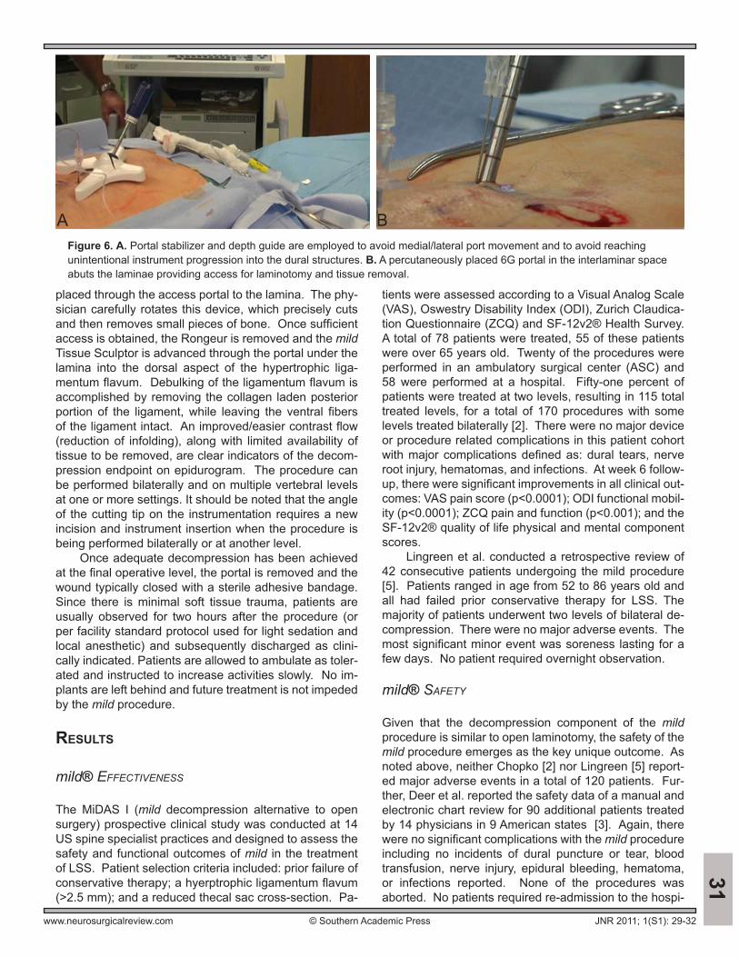
At the beginning of the procedure the patient is placed in the prone position on a radiolucent operating table, draped, and sterilely prepped. Appropriate bol-stering is used as needed (Fig. 4). An epidurogram is then performed for the purpose of identifying the hyper-trophic ligament flavum. Utilization of the contralateral oblique view presents the thickest cross-section of the lamina, providing optimal ligamentum infolding imaging. Next, the interlaminar space is identified through fluo-roscopic visualization (Fig. 5). After the trajectory has been planned and the patient’s skin marked, the mild 6G portal and 7G trocar are inserted percutaneously. These devices are advanced along the desired trajectory un-der fluoroscopic guidance. Once positioned, the trocar is removed, leaving the access portal in the interlaminar space (Figs. 6, 7). The mild Bone Sculptor Rongeur is
Figure 2. mild Devices: a. mild Tissue Sculpter; b. mild Portal Stabilizer; c. mild Bone Sculpter Rongeur; d. mild Surgical Clamp; e. mild Portal; f. mild Depth Guide; g. mild Trocar and Handle.
Figure 3. Epidurogram allows visualization for safe and adequate decompression of the target area.
Figure 4. Patient is kept comfortable in prone position with appropriate bolstering to open interlaminar space exposing target hemilaminar region.
Figure 5. Epidurogram is performed with low volume intermittent injections using flow patterns to determine decompression and stopping point.
www.neurosurgicalreview.com © Southern Academic Press
31
JNR 2011; 1(S1): 29-32
placed through the access portal to the lamina. The phy-sician carefully rotates this device, which precisely cuts and then removes small pieces of bone. Once sufficient access is obtained, the Rongeur is removed and the mild Tissue Sculptor is advanced through the portal under the lamina into the dorsal aspect of the hypertrophic liga-mentum flavum. Debulking of the ligamentum flavum is accomplished by removing the collagen laden posterior portion of the ligament, while leaving the ventral fibers of the ligament intact. An improved/easier contrast flow (reduction of infolding), along with limited availability of tissue to be removed, are clear indicators of the decom-pression endpoint on epidurogram. The procedure can be performed bilaterally and on multiple vertebral levels at one or more settings. It should be noted that the angle of the cutting tip on the instrumentation requires a new incision and instrument insertion when the procedure is being performed bilaterally or at another level.
Once adequate decompression has been achieved at the final operative level, the portal is removed and the wound typically closed with a sterile adhesive bandage. Since there is minimal soft tissue trauma, patients are usually observed for two hours after the procedure (or per facility standard protocol used for light sedation and local anesthetic) and subsequently discharged as clini-cally indicated. Patients are allowed to ambulate as toler-ated and instructed to increase activities slowly. No im-plants are left behind and future treatment is not impeded by the mild procedure.
results
mild® EffEctivEnEss
The MiDAS I (mild decompression alternative to open surgery) prospective clinical study was conducted at 14 US spine specialist practices and designed to assess the safety and functional outcomes of mild in the treatment of LSS. Patient selection criteria included: prior failure of conservative therapy; a hyerptrophic ligamentum flavum (>2.5 mm); and a reduced thecal sac cross-section. Pa-
tients were assessed according to a Visual Analog Scale (VAS), Oswestry Disability Index (ODI), Zurich Claudica-tion Questionnaire (ZCQ) and SF-12v2® Health Survey. A total of 78 patients were treated, 55 of these patients were over 65 years old. Twenty of the procedures were performed in an ambulatory surgical center (ASC) and 58 were performed at a hospital. Fifty-one percent of patients were treated at two levels, resulting in 115 total treated levels, for a total of 170 procedures with some levels treated bilaterally [2]. There were no major device or procedure related complications in this patient cohort with major complications defined as: dural tears, nerve root injury, hematomas, and infections. At week 6 follow-up, there were significant improvements in all clinical out-comes: VAS pain score (p<0.0001); ODI functional mobil-ity (p<0.0001); ZCQ pain and function (p<0.001); and the SF-12v2® quality of life physical and mental component scores.
Lingreen et al. conducted a retrospective review of 42 consecutive patients undergoing the mild procedure [5]. Patients ranged in age from 52 to 86 years old and all had failed prior conservative therapy for LSS. The majority of patients underwent two levels of bilateral de-compression. There were no major adverse events. The most significant minor event was soreness lasting for a few days. No patient required overnight observation.
mild® safEty
Given that the decompression component of the mild procedure is similar to open laminotomy, the safety of the mild procedure emerges as the key unique outcome. As noted above, neither Chopko [2] nor Lingreen [5] report-ed major adverse events in a total of 120 patients. Fur-ther, Deer et al. reported the safety data of a manual and electronic chart review for 90 additional patients treated by 14 physicians in 9 American states [3]. Again, there were no significant complications with the mild procedure including no incidents of dural puncture or tear, blood transfusion, nerve injury, epidural bleeding, hematoma, or infections reported. None of the procedures was aborted. No patients required re-admission to the hospi-
Figure 6. A. Portal stabilizer and depth guide are employed to avoid medial/lateral port movement and to avoid reaching unintentional instrument progression into the dural structures. B. A percutaneously placed 6G portal in the interlaminar space abuts the laminae providing access for laminotomy and tissue removal.
A B
www.neurosurgicalreview.com © Southern Academic Press
32
JNR 2011; 1(S1): 29-32
32tal within 30 days of the mild procedure in these studies. Of the 210 procedures published in the peer reviewed literature, the reported incidence of major adverse events was 0%, thus confirming the excellent safety record of the mild procedure.
Deyo et al. recently reviewed the safety and effec-tiveness of the mild procedure when indirectly compared to open or endoscopic laminectomy [4]. The review was a retrospective cohort analysis of Medicare claims from 2002-2007 for patients undergoing surgery for spinal stenosis. Among the 21,474 patients undergoing open or endoscopic decompression only (i.e. without fusion), there was a 2.1% incidence of medical complications and a 30 day mortality of 0.6% The length of stay was 2.7 days with 7.8% of patients re-hospitalized within 30 days for any reason. These statistics underscore the safety advantage of the mild procedure.
ProcEdurE sEtting
Though most commonly performed in the hospital out-patient setting, the mild procedure can be conducted safely and effectively in an ASC [2]. The procedure is performed by a trained physician in an operating room with surgical support staff in attendance. Typically, pa-tient recovery is accomplished in a recovery room with appropriately trained nursing staff and does not require overnight observation; this is consistent with mild being an outpatient procedure [5].
dIscussIon
Minimally invasive lumbar decompression for spinal stenosis (presented here as the mild® procedure) is indicated for those LSS patients exhibiting neurogenic claudication with hypertrophic ligamentum flavum. The mild procedure is similar to standard open or endoscopic lumbar laminotomy in terms of the anatomic structures undergoing decompression. The most significant differ-ence between these procedures is that mild is a minimal-ly invasive therapy that relies on fluoroscopic guidance, thus providing a clear procedural safety margin. Typically performed with local anesthesia and conscious sedation, the mild procedure can be performed in the outpatient setting. In multiple studies, patients reported significant
improvements in all clinical outcomes without any re-ported safety issues [3, 5]. The results of these studies compare favorably when contrasted to the Medicare da-tabase which indicates that 2.1% of patients undergoing simple decompression experience medical complications and 7.8% are re-hospitalized within 30 days [4].
conclusIon
Using a minimally invasive lumbar decompression (mild) for spinal stenosis, one can safely, and effectively reduce pain, improve functionality, and minimally change spinal biomechanics and stability in LSS patients who have failed conservative treatment and who are not yet in need of, or who do not desire more invasive open surgical de-compression procedures.
AcknowledgeMent And AuthorshIp stAteMent
All authors contributed to the preparation and final ap-proval of this manuscript. No funding has been received from any organization. Dr. Deer and Dr. Mekhail are con-sultants for Vertos Medical. Dr. Kasra Amirdelfan and Gabriel Lopez have nothing to disclose.
references
1. Carragee EJ: The increasing morbidity of elective spinal stenosis surgery: is it necessary? JAMA 303:1309-1310, 2010.
2. Chopko B, Caraway DL: MiDAS I (mild Decompression Alterna-tive to Open Surgery): a preliminary report of a prospective, multi-center clinical study. Pain Physician 13:369-378, 2010.
3. Deer TR, Kapural L: New image-guided ultra-minimally invasive lumbar decompression method: the mild procedure. Pain Physi-cian 13:35-41, 2010.
4. Deyo RA, Mirza SK, Martin BI, Kreuter W, Goodman DC, Jarvik JG: Trends, major medical complications, and charges associ-ated with surgery for lumbar spinal stenosis in older adults. JAMA 303:1259-1265, 2010.
5. Lingreen R, Grider JS: Retrospective review of patient self-report-ed improvement and post-procedure findings for mild (minimally invasive lumbar decompression). Pain Physician 13:555-560, 2010.