the dynamic stress-induced “o-glcnac-ome” highlights functions for o-glcnac in regulating dna...
TRANSCRIPT

ORIGINAL ARTICLE
The dynamic stress-induced ‘‘O-GlcNAc-ome’’ highlightsfunctions for O-GlcNAc in regulating DNA damage/repairand other cellular pathways
Natasha E. Zachara • Henrik Molina •
Ker Yi Wong • Akhilesh Pandey • Gerald W. Hart
Received: 2 June 2010 / Accepted: 9 July 2010 / Published online: 31 July 2010
� Springer-Verlag 2010
Abstract The modification of nuclear, mitochondrial, and
cytoplasmic proteins by O-linked b-N-acetylglucosamine
(O-GlcNAc) is a dynamic and essential post-translational
modification of metazoans. Numerous forms of cellular
injury lead to elevated levels of O-GlcNAc in both in vivo
and in vitro models, and elevation of O-GlcNAc levels
before, or immediately after, the induction of cellular injury
is protective in models of heat stress, oxidative stress,
endoplasmic reticulum (ER) stress, hypoxia, ischemia
reperfusion injury, and trauma hemorrhage. Together, these
data suggest that O-GlcNAc is a regulator of the cellular
stress response. However, the molecular mechanism(s) by
which O-GlcNAc regulates protein function leading to
enhanced cell survival have not been identified. In order to
determine how O-GlcNAc modulates stress tolerance in
these models we have used stable isotope labeling with
amino acids in cell culture to determine the identity of
proteins that undergo O-GlcNAcylation in response to heat
shock. Numerous proteins with diverse functions were
identified, including NF-90, RuvB-like 1 (Tip49a), RuvB-
like 2 (Tip49b), and several COPII vesicle transport proteins.
Many of these proteins bind double-stranded DNA-depen-
dent protein kinase (PK), or double-stranded DNA breaks,
suggesting a role for O-GlcNAc in regulating DNA damage
signaling or repair. Supporting this hypothesis, we have
shown that DNA-PK is O-GlcNAc modified in response to
numerous forms of cellular stress.
Keywords O-GlcNAc � Cellular stress � Glycosylation �Signal transduction � Chaperone � Cell stress
Abbreviations
Carm1 CK II Casein kinase II
DNA-PK DNA-dependent protein kinase
DMSO Dimethylsulfoxide
DON 6-diazo-5-oxonorleucine
GAPDH Glyceraldehyde-3-phosphate
dehydrogenase
FBS Fetal bovine serum
HSP Heat shock protein
MEFs Mouse embryonic fibroblasts
O-GlcNAc Monosaccharides of O-linked b-N-
acetylglucosamine
O-GlcNAcase O-GlcNAc hexosaminidase
(EC 3.2.1.52)
OGT: UDP-GlcNAc Polypeptide O-b-N-acetylglucosa-
minyltransferase (EC 2.4.1.94)
PBS Phosphate-buffered saline
PUGNAc O-(2-acetamido-2-deoxy-D-
glucopyranosylidene)
amino-N-phenylcarbamate
TBS Tris-buffered saline
Electronic supplementary material The online version of thisarticle (doi:10.1007/s00726-010-0695-z) contains supplementarymaterial, which is available to authorized users.
N. E. Zachara (&) � H. Molina � A. Pandey � G. W. Hart
The Department of Biological Chemistry, The Johns Hopkins
University School of Medicine, 725 N. Wolfe Street, Baltimore,
MD 21205-2185, USA
e-mail: [email protected]
N. E. Zachara � K. Y. Wong
The Division of Biomedical Sciences, Johns Hopkins Singapore,
31 Biopolis Way, The Nanos #02-01, Singapore 138669,
Singapore
H. Molina � A. Pandey
McKusick-Nathans Institute for Genetic Medicine,
Department of Biological Chemistry and Oncology,
Johns Hopkins University, 733 N. Broadway,
Baltimore, MD 21205, USA
123
Amino Acids (2011) 40:793–808
DOI 10.1007/s00726-010-0695-z
Introduction
The addition of monosaccharides of O-linked b-N-acetyl-
glucosamine to Ser and Thr residues (O-GlcNAc) of
nuclear, mitochondrial, and cytoplasmic proteins is one of
a growing number of post-translational modifications
thought to mediate cellular function (Hart et al. 2007). O-
GlcNAc regulates protein function in a manner analogous
to protein phosphorylation by changing the localization of
proteins, regulating protein–protein or protein-DNA inter-
actions, altering the half-life of proteins, and altering the
activity of proteins (Hart et al. 2007). Like phosphoryla-
tion, O-GlcNAc levels change rapidly and dynamically in
response to many signals, including morphogens, the cell
cycle, development, extracellular glucose concentrations,
and numerous forms of cellular stress (Hart et al. 2007).
Notably, O-GlcNAc and phosphorylation may compete for
the same amino acids on some key cellular proteins,
including the large sub-unit of RNA polymerase II, endo-
thelial nitric oxide synthase, the c-Myc proto-oncogene,
estrogen receptor-b, and SV-40 large T-antigen (Chou
et al. 1995; Comer and Hart 1996; Tallent et al. 2009). In
support of a complex relationship between O-GlcNAc and
phosphorylation (Chou et al. 1995; Comer and Hart 1996;
Griffith and Schmitz 1995, 1999; Kamemura and Hart
2003; Lefebvre et al. 1999, 2003; Musicki et al. 2005;
Tallent et al. 2009; Wang et al. 2008, 2007), elevating O-
GlcNAc levels results in decreased phosphorylation at 148
sites (and elevated on 280) (Wang et al. 2008), and at the
midbodies of cells over-expressing the O-GlcNAc trans-
ferase 68 peptides showed decreased phosphorylation
(Wang et al. 2007, 2010a, 2010b).
Growing evidence suggests that O-GlcNAc is a novel
regulator of the cellular stress response (Champattanachai
et al. 2007; Chatham et al. 2008; Cheung and Hart 2008;
Fulop et al. 2007a, b, c; Guinez et al. 2008; Jones et al.
2008; Laczy et al. 2009; Liu et al. 2007, 2006; Ngoh et al.
2009; Ngoh and Jones 2008; Ngoh et al. 2008; Ohn et al.
2008; Sohn et al. 2004; Yang et al. 2006; Zachara and Hart
2004a, b, 2006; Zachara et al. 2004). In response to
numerous forms of cellular stress or injury, O-GlcNAc
levels are dynamically elevated in both in vitro and in vivo
models. Moreover, elevating levels of O-GlcNAc before or
after the induction of cellular injury protects both cells and
tissues (Champattanachai et al. 2007; Chatham et al. 2008;
Cheung and Hart 2008; Fulop et al. 2007a, b, c; Guinez
et al. 2008; Jones et al. 2008; Laczy et al. 2009; Liu et al.
2007, 2006; Ngoh et al. 2009; Ngoh and Jones 2008; Ngoh
et al. 2008; Ohn et al. 2008; Sohn et al. 2004; Yang et al.
2006; Zachara and Hart 2004a, b, 2006; Zachara et al.
2004). These data support previous studies demonstrat-
ing the importance of glucose uptake into cells, and
subsequent conversion to UDP-GlcNAc (the donor sugar
for O-GlcNAc) through the hexosamine biosynthetic
pathways in diverse stress models (Heilig et al. 2003;
Hoorens et al. 1996; Loberg et al. 2002; Malhotra and
Brosius 1999; Moley and Mueckler 2000; Pang et al.
2002), as well as data showing that glucosamine also
promotes survival in some models of tissue/cell injury
(Champattanachai et al. 2007, 2008; Chatham et al. 2008;
Not et al. 2007; Yang et al. 2006). Taken together, these
data suggest that O-GlcNAc is a key post-translational
modification employed by cells and whole animals to
rapidly respond to, and survive, stress.
Numerous pathways appear to be modulated by
O-GlcNAc in a manner that would promote cell survival,
including (1) HSP expression (Zachara et al. 2004); (2)
protein solubility (Cheung and Hart 2008; Lim and Chang
2006); (3) cytosolic Ca2? influx (Liu et al. 2007, 2006;
Nagy et al. 2006);(4) calpain activity (Liu et al. 2007); (5)
p38 MAP kinase phosphorylation (Fulop et al. 2007b); (6)
circulating IL-6 and TNF-a levels (Liu et al. 2006; Yang
et al. 2006; Zou et al. 2007); and (7) maintenance of
mitochondrial membrane potential, which is possibly
dependent on VDAC (Jones et al. 2008). However, it is
unclear at a molecular level how O-GlcNAc regulates these
pathways and what additional proteins/pathways are regu-
lated by stress-induced O-GlcNAcylation.
To gain a greater understanding of the pathways and
mechanisms by which the addition of O-GlcNAc to
nuclear, cytoplasmic, and mitochondrial proteins alters
cellular pathways, we have identified proteins that are
dynamically O-GlcNAc modified in response to heat stress.
Interestingly, these data not only highlight expected roles
for O-GlcNAc in regulating transcription and nuclear
transport, but also possible roles for O-GlcNAc in regu-
lating unexpected processes such as vesicle transport,
microRNA processing, DNA damage repair, and protein
arginine methylation.
Methods
Reagents and antibodies
The following antibodies were used in this study: CTD110.6,
AL28 (Anti-OGT), anti-Actin (SIGMA–Aldrich; A-5060),
anti-aTubulin (SIGMA–Aldrich; T-5168), anti-NF-90 (BD
Transduction laboratories; D39920), anti-NF-90 (Gift),
anti-NF45 (Gift), anti-Sec24 (Gift), anti-p125i (Gift),
Anti-Carm1 (Upstate; 07-080), Anti-WNK1 (Santa Cruz
Biotechnology; SC-28897), Anti-WNK1 (Cell Signaling
Technology; 4979), anti-Tip49a (Gift), anti-Tip49a (Santa
Cruz), anti-Tip49b (Gift), anti-Tip49b (Abcam), Anti-DNA-
PK (Calbiochem; PC127), Anti-DNA-PK (Calbiochem;
NA57), Anti-HSP 70 (Stressgen Bioreagents; SPA-810),
794 N. E. Zachara et al.
123
Anti-HSC70 (Santa Cruz Biotechnology; SC-7298),
mAb414 (Nuclear Pore Proteins; gift), Anti-SOD1 (Santa
Cruz Biotechnology; SC-11407), and Anti-SOD2 (Santa
Cruz Biotechnology; SC-30080). PUGNAc was from Tor-
onto Research Biochemicals; Doxorubicin and Bleocin were
from Merck. All other chemicals were of the highest grade.
Cell culture and treatments
Typically, Cos-7 cells were plated at 5 9 105 cells per
100 mm plate in DMEM (1 g/l glucose), 10% FBS and
Pen/Strep and maintained in a humidified incubator at 37�C
with 5% CO2. 36 h post-plating media was replaced, and
48 h post-plating cell stress treatments were initiated. Cells
were heat-stressed at 45�C for 1 h, and recovered at 37�C
for the indicated length of time (typically 1 h). Unless
otherwise noted, Cos-7 cells were treated as follows:
sodium chloride (100 mM, 6 h), PUGNAc (50 lM, 8 h),
Doxorubicin (2 lM, 4 or 8 h), H2O2 (500 lL, 6 h),
bleocin (2.5 lg/ml, 6 h), and Tunicamycin (25 lg/ml,
18 h).
Stable isotope labeling with amino acids in cell culture
SILAC labeling
Cos-7 cells (ATCC) were passaged six times in DMEM
(4.5 g/l glucose), 10% v/v FBS and Pen/Strep, supple-
mented with arginine (light), 13C6 L-arginine (medium), or13C6
15N4 L-arginine (heavy) as previously reported (Harsha
et al. 2008). Cells (1 9 106) were seeded in 150 mM
(Corning) dishes 48 h prior to treatments. PUGNAc was
applied at 50 lM for 12 h prior to harvesting. Cells were
heat stressed at 45�C for 1 h, and recovered at 37�C for 1 h
before harvesting, as previously reported (Ibarrola et al.
2003; Ong et al. 2002; Wang et al. 2007).
Immunoprecipitations
Cells were washed with ice-cold Phosphate-Buffered
Saline pH 7.4 (PBS; 137 mM NaCl, 2.7 mM KCl,
10 mM Na2HPO4, 2 mM KH2PO4, pH7.4) and removed
from plates by scraping. Cell pellets were stored at
-70�C until extraction. Total nuclear and cytoplasmic
extracts were made as previously reported. Equal protein
(11 mg) from each sample (control, heat shocked and
PUGNAc) was combined (total protein 33 mg). O-Glc-
NAc-modified proteins were purified using 1.5 mg of
anti-O-GlcNAc antibody, CTD110.6, coupled to CNBR-
activated agarose.
Protein extracts were incubated for 12 h at 4�C with
mixing. Unbound proteins were re-precipitated with
1.5 mg of anti-O-GlcNAc antibody for an additional 8 h.
Proteins bound to CTD110.6 were washed with 15 col-
umn volumes of wash buffer (50 mM Tris pH8.0,
200 mM NaCl, 0.5 mM EDTA), followed by 15 column
volumes of 100 mM galactose in wash buffer. Proteins
were eluted from CTD110.6 using five column volumes
of 1 M GlcNAc in wash buffer. Elution’s from the first
and second CTD110.6 immunoprecipitation were com-
bined and then precipitated with 10 volumes of ice-cold
methanol. Proteins were resuspended in SDS–PAGE
sample buffer and separated on a 16-cm 7.5% discontin-
uous SDS–PAGE gel before being stained with coom-
massie G250.
Mass spectrometry
Protein bands were excised and digested with trypsin as
described previously (Amanchy et al. 2005). Briefly, pro-
teins were reduced and alkylated in gel, before being
trypsinized overnight at 37�C. Tryptic peptides were
extracted from the gel and concentrated in a vacufuge to
approximately 10 ll.
Peptides were analyzed by reversed-phase liquid
chromatography tandem mass spectrometry (LC–MS/
MS). Using an 1100 Series CapLC system (Agilent
Technologies, Palo Alto, CA) peptides were desalted on a
home-built trap column (12 lm C18 ODS-A (YMC Co,
Kyoto, Japan)) connected to an analytical column (5 lm
Vydac C18 resin, Nest Group, Southboro, MA). Peptides
were loaded and desalted at 4 ll/min: 95% mobile phase
A (0.4% v/v acetic acid and 0.005% v/v heptafluorobu-
tyric acid, v/v) and 5% mobile phase B (90% v/v ace-
tonitrile, 0.4% v/v acetic acid, and 0.005% v/v
heptafluorobutyric acid, v/v). Hereafter peptides were
eluted by changing the solvent composition to 40%
mobile phase A/60% mobile phase B, in 34 min at
300 nl/min. Eluted peptides were analyzed using a hybrid
quadrupole time-of-flight mass spectrometer (Micromass
Q-TOF US-API, Manchester, UK) equipped with an na-
noES source (Proxeon A/S, Odense, Denmark). MS/MS
data were queried against NCBInr Primates using Mascot
2.2 (MatrixScience, London, UK). The following variable
modifications were allowed: Acetyl (N-term), Glu-
[ pyro-Glu (N-term E), 13C6-Arg, 13C615N4-Arg, O-Glc-
NAc (ST), and Oxidation of methionines. All cysteines
were treated as being carbamidomethylated. Trypsin,
allowing for three missed cleavages, was used as enzyme
specificity. Mass accuracies of 1.15 Da for precursor
peptide ions and 0.15 Da for fragment ions were used. A
decoy search approach was taken and a peptide-based
false discovery rate of 2% was calculated. Calculation of
stable isotope labeling with amino acids in cell culture
(SILAC) ratios was conducted by manual inspection of
each peptide.
The dynamic stress-induced ‘‘O-GlcNAc-ome’’ highlights functions 795
123

Protein extractions and immunoprecipitations
Cells were washed with ice-cold PBS, harvested by
scraping, and stored at -70�C until extraction. Total
nuclear and cytoplasmic extracts were made as previously
reported. Extracts were diluted 1:1 with 1% v/v NP-40 in
TBS (10 mM Tris–HCl pH 7.5, 150 mM NaCl). Typically,
0.5 mg of total nucleocytoplasmic extract was incubated
with 1 lg of antibody for 18 h at 4�C with mixing. Extracts
were incubated for a further 2 h with protein A/G. Anti-
body/protein A/G complexes were washed with buffer
50 mM Tris pH 7.5, 175 mM NaCl, 0.5% v/v NP-40.
Proteins were released by incubation in SDS–PAGE buffer
at 100�C for 5 min. Proteins were separated by SDS–
PAGE on Criterion Tris–HCl gels (BIORAD) and blotted
to nitrocellulose. Typically, 20% of the precipitate (or the
IP from 100 lg) was loaded on gels. Blots were blocked
with 3% w/v milk in TBST (10 mM Tris–HCl pH 7.5,
150 mM NaCl, 0.05% v/v Tween-20).
Results
Numerous proteins are dynamically O-GlcNAc
modified in response to stress
In order to identify proteins that are O-GlcNAc modified
in response to stress, we used the SILAC method as
outlined in Fig. 1 (Ibarrola et al. 2003; Ong et al. 2002;
Wang et al. 2007). O-GlcNAc-modified proteins from
isotope labeled populations were enriched by an immu-
noprecipitatio step, before identification and relative
quantitation by mass spectrometry. Cos-7 cells were
adapted and grown in media containing normal L-arginine,13C6 L-arginine (medium), or 13C6, 15N4 L-arginine argi-
nine (designated light, medium and heavy, respectively).
Cos-7 cells were treated with heat stress 45�C (1 h, 45�C)
and recovered for 1 h (medium) or PUGNAc (50 lM,
12 h; heavy). Cells grown in normal L-arginine were not
treated and served as a control. Consistent with our pre-
vious data, cells subjected to a heat stress displayed ele-
vated levels of O-GlcNAc, as did proteins isolated
from cells treated with PUGNAc (an inhibitor of the
O-GlcNAcase which removes O-GlcNAc). Equal protein
from each treatment was combined and precipitated twice
with CTD110.6, an O-GlcNAc specific antibody. To fur-
ther enhance the specificity of the immunoprecipitation,
the resin was washed in buffer containing 100 mM gal-
actose, and we only analyzed proteins that were com-
petitively eluted with 1 M GlcNAc in TBS. Proteins
eluted with GlcNAc were separated by SDS–PAGE,
stained with G250 coomassie (Fig. 2b), and analyzed
further by mass spectrometry.
Using this stringent approach, 51 proteins were identi-
fied (Supplemental Table 1, Supplemental Table 2) of
which 30 proteins contained peptides labeled by heavy
isotopes. 21 proteins were statically O-GlcNAcylated, and
15 were dynamically O-GlcNAcylated in response to heat
stress (Fig. 3; Table 1). Notably, several peptides appeared
to be modified by O-GlcNAc, although we unable to assign
which Ser/Thr residue was modified (Table 2; Supple-
mental Table 2). To confirm these data, and to ensure that
identified proteins were pulled out specifically, we immu-
noprecipitated non-labeled extract (Control, Heat Stressed,
and PUGNAc) with either a non-specific IgM or CTD110.6
and blotted for proteins of interest. As shown in Fig. 4,
proteins identified and quantified in the screen appear to
have been precipitated specifically.
Based on the quantitative results we divided the O-
GlcNAc-modified proteins into three groups: (1) O-Glc-
NAcylated in response to heat stress; (2) O-GlcNAcylated
but not in response to heat stress; and (3) not O-GlcNA-
cylated (Fig. 3). Proteins falling into group three do not
appear to be O-GlcNAc modified as the SILAC ratio is
unchanged in either the heat-stressed or PUGNAc-treated
group. Moreover, these proteins were not immuno-precip-
itated by CTD110.6 (Fig. 3) and O-GlcNAc was not
detected on these proteins by IP/Western Blot (Fig. 4). We
conclude that these proteins were isolated as they either
interact with an O-GlcNAc-modified protein, or alterna-
tively are present as they are highly expressed proteins in
the cell and are a contaminant. Changes in the levels of O-
GlcNAc on a protein could results from: (1) the O-Glc-
NAcylation status changing with stress; (2) The O-Glc-
NAcylation status of an interacting proteins changing with
stress; (3) The expression of a protein changed with stress;
or (4) the expression of an interacting glycoprotein
changing with stress. To confirm that the proteins identified
in this screen were O-GlcNAcylated in response to stress
(option 1), rather that changes in expression or protein–
protein interactions, a subset of the proteins were immu-
noprecipitated from non-labeled extract (Control, Heat
Stressed, and PUGNAc) with the appropriate antibody and
CTD110.6 was used to detect O-GlcNAc levels (Fig. 5;
Table 1). Immunoprecipitations were also performed with
either rabbit or mouse non-specific immunoglobulin (data
not shown) and the signals shown in Fig. 5 appear to be
specific.
Fifteen proteins isolated in the SILAC screen do not
appear to be O-GlcNAcylated in response to heat stress;
however, PUGNAc resulted in an increase in the SILAC
ratio on five of these proteins suggesting that OGT, RAE1,
NUP98, VP16, Sec23A and NUP54 are O-GlcNAcylated.
These data suggest that in spite of the apparent global
increase in O-GlcNAc levels in response to heat stress, not
every O-GlcNAcylated protein is a target of OGT during
796 N. E. Zachara et al.
123

heat stress. The remaining proteins appear to fall into two
groups: Group 1 includes proteins such as Carm1, SEC24b,
p125i, WNK1, and a subset of nuclear pore proteins. Here
we observed significant changes in the SILAC ratio for
both heat-stressed and PUGNAc-treated cells suggesting
that the samples were O-GlcNAcylated and that the gly-
cosylation state responds to heat stress. Much of this data
was confirmed by IP/Western blot. Group 2 includes pro-
teins such as NF45 and a sub-set of nuclear pore proteins
which exhibited changed SILAC ratios, but data from
IP/Western blot showed little difference in O-GlcNAcyla-
tion in response to heat stress. Together these data suggest
that these proteins are likely associated with O-GlcNAcy-
lated proteins. To investigate the O-GlcNAcylation of
NF45, we immunoprecipitated NF45 and its binding part-
ner NF90. While NF90 appears O-GlcNAc modified, and
associated with numerous O-GlcNAcylated proteins, NF45
does not appear to be O-GlcNAc modified (Supplemental
Fig. 1). Interestingly, during our analysis we isolated
numerous peptides from the protein NICE-4. These pep-
tides fell into two groups, those in which there was no
change in the SILAC ratio, and those where we observed a
consistent increase in the SILAC ratio (Heat stress = 1.4
and PUGNAc = 1.7). These data suggest that there are two
isoforms of NICE-4, only one of which is O-GlcNAc
modified in response to stress. Alternatively, these data
suggest that there may be two populations of NICE-4, only
one of which is O-GlcNAc modified in response to stress.
CombineIP with CTD110.6
elute proteinswith 1M GlcNAc
Precipitate proteins
7.5% SDS-PAGE gel18cm resolving gel
Stain with coomassie G250
Cut out bands, in-gel digest
Mass spectrometry
L-Arginine“light - control”
11mg nuclear &cytoplasmic extract
13C6 L-Arginine“medium - HS”
11mg nuclear &cytoplasmic extract
13C6 15N4 L-Arginine“heavy - PUGNAc”
11mg nuclear &cytoplasmic extract
Confirm proteins by IP western blot
Fig. 1 SILAC strategy used in
this study
The dynamic stress-induced ‘‘O-GlcNAc-ome’’ highlights functions 797
123
For a number of proteins, such as Tip49a/b we were unable
to determine the O-GlcNAcylation status due to their
molecular weight (co-migrate with heavy chain), whereas
for others we were unable to obtain an antibody that
immunoprecipitated.
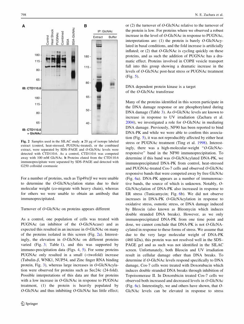
Turnover of O-GlcNAc on proteins appears different
As a control, one population of cells was treated with
PUGNAc (an inhibitor of the O-GlcNAcase) and as
expected this resulted in an increase in O-GlcNAc on many
of the proteins isolated in this screen (Fig. 2a). Interest-
ingly, the elevation in O-GlcNAc on different proteins
varied (Fig. 3; Table 1), and this was supported by
immuno-precipitation data (Figs. 4, 5). For some proteins
PUGNAc only resulted in a small (\twofold) increase
(Tubulin-b, WNK1, NUP54, and Zinc finger RNA binding
protein, Fig. 3), whereas large increases in O-GlcNAcyla-
tion were observed for proteins such as Sec24c (24-fold).
Possible interpretations of this data are that for proteins
with a low increase in O-GlcNAc in response to PUGNAc
treatment, (1) the protein is heavily populated by
O-GlcNAc and thus inhibiting O-GlcNAc has little effect;
or (2) the turnover of O-GlcNAc relative to the turnover of
the protein is low. For proteins where we observed a robust
increase in the level of O-GlcNAc in response to PUGNAc,
interpretations are: (1) the protein is barely O-GlcNAcy-
lated in basal conditions, and the fold increase is artificially
inflated; or (2) that O-GlcNAc is cycling quickly on these
proteins, and as such the addition of PUGNAc has a dra-
matic effect. Proteins involved in COPII vesicle transport
fall into this group showing a dramatic increase in the
levels of O-GlcNAc post-heat stress or PUGNAc treatment
(Fig. 5).
DNA dependent protein kinase is a target
of the O-GlcNAc transferase
Many of the proteins identified in this screen participate in
the DNA damage response or are phosphorylated during
DNA damage (Table 3). As O-GlcNAc levels are known to
increase in response to UV irradiation (Zachara et al.
2004), we investigated a role for O-GlcNAc in mediating
DNA damage. Previously, NF90 has been reported to bind
DNA-PK and while we were able to confirm this associa-
tion (Fig. 5), it was not reproducibly affected by either heat
stress or PUGNAc treatment (Ting et al. 1998). Interest-
ingly, there was a high-molecular-weight ‘‘O-GlcNAc-
responsive’’ band in the NF90 immunoprecipitation. To
determine if this band was O-GlcNAcylated DNA-PK, we
immunoprecipitated DNA-PK from control, heat-stressed
and PUGNAc-treated Cos-7 cells and observed O-GlcNAc
responsive bands that were competed away by free GlcNAc
(Fig. 6a). DNA-PK appears as a number of immunoreac-
tive bands, the source of which is unknown. Notably, O-
GlcNAcylation of DNA-PK also increased in response to
ER stress (Tunicamycin; Fig. 6b). We did not observe
increases in DNA-PK O-GlcNAcylation in response to
oxidative stress, osmotic stress, or DNA damage induced
by Bleocin (also known as Bleomycin which induces
double stranded DNA breaks). However, as we only
immunoprecipitated DNA-PK from one time point and
dose, we cannot conclude that DNA-PK is not O-GlcNA-
cylated in response to these forms of stress. We assume that
due to the very large molecular weight of DNA-PK
(460 kDa), this protein was not resolved well in the SDS–
PAGE gel and as such was not identified in the SILAC
screen. Unfortunately, both Bleocin and UV irradiation
result in cellular damage other than DNA breaks. To
determine if O-GlcNAc levels respond specifically to DNA
damage, Cos-7 cells were treated with Doxorubucin which
induces double stranded DNA breaks through inhibition of
Topoisomerase II. In Doxorubicin treated Cos-7 cells we
observed both increased and decreased levels in O-GlcNAc
(Fig. 6c). Interestingly, we and others have shown, that O-
GlcNAc levels can be elevated in response to stress
A B
Extract Buffer
180 -
115 -
80 -
65 -
50 -
40 -
IP: GlcNAc
IB: CTD110.6
IB: CTD110.6+ GlcNAc
180 -
115 -
80 -
65 -
180 -
115 -
80 -
65 -
Con
trol
Hea
t Sho
ck
PU
GN
Ac
Com
bine
d
BS
A
BS
A-G
lcN
Ac
Fig. 2 Samples used in the SILAC study. a 20 lg of isotope labeled
extract (control, heat-stressed, PUGNAc-treated), or the combined
extract, were separated by SDS–PAGE and O-GlcNAc levels were
detected with CTD110.6. As a control, CTD110.6 was competed
away with 100 mM GlcNAc. b Proteins eluted from the CTD110.6
immunoprecipitate were separated by SDS–PAGE and detected with
G250 colloidal coomassie
798 N. E. Zachara et al.
123

followed by an decline (Jones et al. 2008). Together these
data suggest that O-GlcNAc levels change in response to
DNA damage and that O-GlcNAc may regulate the repair
of DNA damage through modification of DNA-PK.
Discussion
Recently, the modification of nuclear, cytoplasmic, and
mitochondrial proteins by O-GlcNAc has emerged as a
regulator of the cellular stress response (Champattanachai
et al. 2007; Chatham et al. 2008; Cheung and Hart 2008;
Fulop et al. 2007a, b, c; Guinez et al. 2008; Jones et al.
2008; Laczy et al. 2009; Liu et al. 2007, 2006; Ngoh
et al. 2009; Ngoh and Jones 2008; Ngoh et al. 2008; Ohn
et al. 2008; Sohn et al. 2004; Yang et al. 2006; Zachara and
Hart 2004a, b, 2006; Zachara et al. 2004). In response to
diverse forms of cellular injury, O-GlcNAc levels are
dynamically upregulated on myriad of proteins. Moreover,
increasing the O-GlcNAc modification before or after cel-
lular injury appears to regulate the ability of cells to survive
a lethal insult. Given that O-GlcNAc appears to protect cells
in models of heat stress, hypoxia, oxidative stress, ischemia
reperfusion injury, and trauma hemorrhage, it would appear
that O-GlcNAc is an integral component of the cellular
stress response (Champattanachai et al. 2007; Chatham
et al. 2008; Cheung and Hart 2008; Fulop et al. 2007a, b, c;
Guinez et al. 2008; Jones et al. 2008; Laczy et al. 2009; Liu
et al. 2007, 2006; Ngoh et al. 2009; Ngoh and Jones 2008;
Ngoh et al. 2008; Ohn et al. 2008; Sohn et al. 2004; Yang
0.0
1.0
2.0
3.0
4.0
5.0
1 2 3 4 5 6 7 8 9 10 11 12 13 14 15 16 17 18 19 20 21 22 23 24 25 26 27 28 29 30
Medium/light Heavy/light
23.7
Fol
d in
crea
se in
O-G
lcN
Ac
com
pare
d to
con
trol
PUGNAc/Control
1 1.2-1.99 2-2.99 3+
Heat Stress/Control
1 1.2-1.99 2+
A
B
Fig. 3 Numerous proteins
showed elevated levels of
O-GlcNAc in response to either
heat stress or PUGNAc
treatment. a The fold increase in
response to heat stress (blackbars) or PUGNAc treatment
(blue bars) are indicated. The
red dashed line represents a
25% increase or decrease, which
represents the error in the
experiment. Proteins are
numbered as in Table 1. b The
distribution of proteins with a
1.2–1.99 fold or 2? fold
increase in response to heat
stress are shown (left panel).Whereas proteins with a
1.2–1.99 fold, 2–2.99 fold, or
3? fold increase in response to
PUGNAc treatment (rightpanel) are indicated
The dynamic stress-induced ‘‘O-GlcNAc-ome’’ highlights functions 799
123

Ta
ble
1P
rote
ins
iden
tifi
edin
the
MS
scre
en
#k
Da
Pro
tein
nam
e(a
lter
nat
ive
nam
es)
Hea
t
sho
ck
PU
GN
Ac
Co
nfi
rmed
by
IP/W
B
Mo
difi
ed
inT
r-H
*
Pre
vio
usl
yID
’d
asO
-Glc
NA
cyla
ted
1100
Import
insu
bunit
bet
a-1
(Kar
yopher
insu
bunit
bet
a-1,
Nucl
ear
fact
or
p97,
Pore
targ
etin
g
com
ple
x9
7k
Da
sub
un
it,
Imp
ort
in-9
0)
0.8
0.7
24
2R
AE
1(m
RN
Aex
po
rtfa
cto
r,m
RN
A-a
sso
ciat
edp
rote
inm
rnp
41
,R
ae1
pro
tein
ho
mo
log
)0
.82
.04
31
20
OG
T(O
-lin
ked
Glc
NA
ctr
ansf
eras
eis
ofo
rm1
)0
.81
.04
44
4100
Tra
nsp
ort
in1
(Im
port
inbet
a-2,
Kar
yopher
inbet
a-2,
M9
regio
nin
tera
ctio
npro
tein
)0.8
0.7
59
5N
UP
98
(Nucl
eop
ori
n9
8k
Da)
0.9
2.0
44
61
50
Nic
e-4
(Ub
iqu
itin
asso
ciat
edp
rote
in2
)0
.90
.8
76
8H
SP
70
-8(H
eat
sho
ck7
0k
Da
pro
tein
8)
0.9
0.8
4
85
5eI
F1a
(Eu
kar
yo
tic
tran
slat
ion
elo
ng
atio
nfa
cto
r1
alp
ha
1)
1.0
0.8
96
0T
ub
uli
n,
alp
ha
1b
1.0
0.9
4
10
95
NU
P8
8(N
ucl
eop
ori
n8
8k
Da)
1.0
1.0
44
4
11
63
NU
P6
2(N
ucl
eop
ori
n6
2k
Da)
1.1
1.0
4
12
14
0H
CF
1(H
ost
cell
fact
or
C1
)1
.11
.9
13
12
0V
P-1
6(H
ost
cell
fact
or
C1
—V
P1
6-a
cces
sory
pro
tein
)1
.11
.9
14
60
NU
P5
4(N
ucl
eop
ori
n5
4k
Da)
1.1
1.5
44
4
15
24
0N
UP
21
4(N
ucl
eop
ori
n2
14
kD
a)1
.21
.04
44
16
46
Car
m-1
(Co
acti
vat
or-
asso
ciat
edar
gin
ine
met
hy
ltra
nsf
eras
e1
,P
rote
inar
gin
ine
N-m
eth
ylt
ran
sfer
ase
4)
1.2
1.9
4
17
55
Tu
buli
n,
bet
a5
1.3
1.3
18
15
0S
EC
31
-lik
e1
iso
form
11
.42
.34
4
19
15
0S
EC
24
b(S
EC
24
fam
ily
,m
emb
erB
)1
.41
.84
4
20
19
0W
NK
1(W
NK
lysi
ne
defi
cien
tp
rote
ink
inas
e1
,E
ryth
rocy
te6
5k
Da
pro
tein
)1
.41
.24
44
21
15
0N
ice-
4(U
biq
uit
inas
soci
ated
pro
tein
2-l
ike)
1.4
1.7
22
14
0Z
inc
Fin
ger
RN
Ab
ind
ing
pro
tein
(hZ
FR
;M
-ph
ase
ph
osp
ho
pro
tein
ho
mo
log
;Z
FR
pro
tein
;Z
inc
fin
ger
RN
A-b
ind
ing
pro
tein
)
1.4
4.1
4
23
14
0p
12
5i
(Sec
23
-in
tera
ctin
gp
rote
inp
12
5)
1.5
3.8
44
24
13
0S
EC
24
c(S
EC
24-r
elat
edp
rote
inC
)1
.62
3.7
44
4
25
46
NF
45
(In
terl
euk
inen
han
cer
bin
din
gfa
cto
r2
)1
.64
.34
26
55
Tip
49a
(Ru
vB
-lik
e;N
ucl
ear
mat
rix
pro
tein
23
8,
54
kD
aer
yth
rocy
tecy
toso
lic
pro
tein
,
TIP
60
-ass
oci
ated
pro
tein
54
-alp
ha,
INO
80
com
ple
xsu
bun
itH
1.7
2.0
4
27
19
0N
UP
15
3(N
ucl
eop
ori
n1
53
kD
a)1
.72
.14
28
190
HB
xA
gtr
ansa
ctiv
ated
pro
tein
2(B
AT
2l2
)1.7
1.7
29
24
0S
imil
arto
gen
etr
apan
ky
rin
repea
t2
.03
.5
30
55
Tip
49b
(RuvB
-lik
e2,
Rep
tin
48
kD
a,R
epre
ssin
gponti
n5,
51
kD
aer
yth
rocy
tecy
toso
lic
pro
tein
,T
IP60
-ass
oci
ated
pro
tein
54
-bet
a,IN
O80
com
ple
xsu
bun
itJ
2.4
2.6
4
*P
rote
ins
iden
tifi
edas
dif
fere
nti
ally
O-G
lcN
Acy
late
din
am
od
elo
ftr
aum
ah
emo
rrh
age
(Teo
etal
.2
01
0)
800 N. E. Zachara et al.
123

et al. 2006; Zachara and Hart 2004a, b, 2006; Zachara et al.
2004). In support of this hypothesis, we and others have
shown that O-GlcNAc can regulate the expression of heat
shock proteins—the sentinels of the stress response (Lim
and Chang 2006; Singleton and Wischmeyer 2008; Sohn
et al. 2004; Zachara et al. 2004). In order to determine how
O-GlcNAc mediates stress-tolerance at a molecular level,
we have identified proteins that are dynamically O-Glc-
NAc-modified in response to heat stress.
Using SILAC technology we have identified 15 proteins
that are dynamically O-GlcNAc modified (Tables 1, 3;
Fig. 3), and have validated these data by immunoprecipi-
tation and western blot (Figs. 4, 5). These proteins fall into
diverse groups, including proteins involved in DNA dam-
age, nuclear transport, transcriptional regulation, vesicle
transport, and microRNA biosynthesis (Table 3). Interest-
ingly, not every O-GlcNAc-modified protein appeared to
be a target of the O-GlcNAc transferase during heat stress
(Fig. 3). Recently, an independent study identified proteins
that were O-GlcNAc modified in response to trauma
hemorrhage. The overlap between O-GlcNAc-modified
proteins identified in each study is indicated in Table 1.
While some proteins are modified in response to both
forms of cellular injury, other proteins only appear O-
GlcNAcylated in response to one form of stress. While
further research needs to be performed, these data suggest
that different profiles of proteins are O-GlcNAcylated in
response to different injuries. Thus some O-GlcNAcylated
proteins may play a role in the central stress response
others may play more specific roles.
Two of the proteins identified in this SILAC screen were
Tip49a and Tip48b, which are components of many
nuclear complexes involved in chromatin remodeling (Jha
and Dutta 2009). Interestingly, Tip49b (also known as
reptin) is one component of the Polycomb group complex
PRC1. Polycomb group proteins repress transcription
though a number of mechanisms, one of which is methyl-
ation of histones. Notably, the polycomb group protein,
Supersexcombs has recently been shown to encode OGT.
O-GlcNAcylation appears important for the activity of the
PRC1 complex, of which Polyhomeotic (PH) appears to be
O-GlcNAc modified (Gambetta et al. 2009; Schwartz and
Pirrotta 2009; Sinclair et al. 2009). Tip49a and Tip48b are
also important for the DNA damage response at multiple
steps: (1) In association with Tip60, Tip49a appears
important for efficient autophosphorylation and acetylation
of the kinase ATM which not only phosphorylates H2AX
Table 2 O-GlcNAc modified peptides identified in this study
Protein Peptide
Zinc finger RNA
binding protein
AGYSQGATQYTQAQQTR
NUP214 LGELLFPSSLAGETLGSFSGLR
RAE1 KGGRTLQLPER
NUP214 ASSTSLTSTQPTK
Unassigned KVIITIQYQK
Triosephosphate
isomerase 1
MNGRKQSLGELIGTLNAAK
Sec24B SSPVVSTVLSGSSSTR
HCF1 VMSVVQTKPVQTSAVTGQASTGPVTQIIQTK
Unassigned ASQGVLRLLVQDR
Unassigned MSVAFPSARSR
IB: Actin
IB: Tubulin
IB: Tip49α
C HS P Ig CTD Ig CTD Ig CTD
Control Heat
Shock PUGNAcInput
IB: NF90
180 -
115 -
80 -
65 -
50 -
40 - IB: GlcNAc
IB: OGT
IB: Carm1
IB: Sec24
IB: p125i
IB: mAb 414 (nuclear pore proteins)
IB: HSP70
180 -
115 -
80 -
65 -
50 -
40 -
IB: NF45
Fig. 4 Confirmation of proteins identified by the SILAC screen.
Control, heat-stressed, or PUGNAc-treated cells were immunopre-
cipitated with either CTD110.6 or control IgM covalently coupled to
cyanogen bromide activated Sepharose. Cell extract and immunopre-
cipitates were separated by SDS–PAGE and nuclear pore proteins
(mAb414), NF-90, NF45, Carm1, Sec24, p125i, Tip49a, OGT, Actin,
Tubulin, and HSP70 were detected by immunoblot
The dynamic stress-induced ‘‘O-GlcNAc-ome’’ highlights functions 801
123

but also promotes the induction of cell cycle checkpoints;
(2) termination of the DNA damage signal through
dephosphorylation of H2AX requires Tip49a; and (3)
remodeling of chromatin around DNA damage to promote
repair also requires Tip49a/b (Jha and Dutta 2009). Our
data has highlighted a possible role for O-GlcNAc in reg-
ulating the DNA damage response. In support of O-Glc-
NAc playing a role in stabilizing DNA after stress, we have
shown that O-GlcNAc levels respond to DNA damaging
agents such as Bleocin and Doxorubicin. Moreover,
DNA-PK is O-GlcNAc modified in response to various
forms of cellular stress.
We isolated several proteins that are associated with
protein methylation (though not in a PcG dependent man-
ner): Carm1 and NF90. Carm1 methylates arginine resi-
dues and this has been implicated in regulating both protein
function and chromatin structure though modification of
histones (Barr et al. 2009; Chen et al. 2002; Cheng et al.
2007; Cheung et al. 2008; El Messaoudi et al. 2006;
Scoumanne and Chen 2008; Wysocka et al. 2006). The
Buf
fer
Con
trol
Hea
t Sho
c k
PU
GN
Ac
IP: p125i
IB: GlcNAc
IB: GlcNAc + 100mM GlcNAc
IB: p125i
Buf
fer
Con
trol
Hea
t Sho
ck
PU
GN
Ac
IP: Carm1
IB: GlcNAc
IB: GlcNAc + 100mM GlcNAc
IB: Carm1
Buf
fer
Con
trol
Hea
t Sho
ck
PU
GN
Ac
IP: NF90
IB: GlcNAc
IB: GlcNAc + 100mM GlcNAc
IB: NF90
Buf
fer
Con
trol
Hea
t Sho
ck
PU
GN
Ac
IP: Tubulin
IB: GlcNAc
IB: GlcNAc + 100mM GlcNAc
IB: tubulin
Buf
fer
Con
trol
Hea
t Sho
ck
PU
GN
Ac
IP: WNK1
IB: GlcNAc
IB: GlcNAc + 100mM GlcNAc
IB: WNK1
IB: GlcNAc
IB: GlcNAc + 100mM GlcNAc
Buf
fer
Con
trol
Hea
t Sho
ck
PU
GN
Ac
IB: Sec24
IP: Sec24
∗
Buf
fer
Con
trol
Hea
t Sho
ck
PU
GN
Ac
IP: OGT
IB: GlcNAc
IB: GlcNAc + 100mM GlcNAc
IB: OGT
Buf
fer
Con
trol
Hea
t Sho
ck
PU
GN
Ac
IP: HSP70
IB: GlcNAc
IB: GlcNAc + 100mM GlcNAc
IB: HSP70
∗∗
∗
IB: DNA-PK
Fig. 5 Numerous proteins
identified by the SILAC screen
are O-GlcNAcylated
dynamically in response to heat
stress. Individual proteins were
immunoprecipitated from
control, heat-stressed, or
PUGNAc-treated cells and the
levels of protein, or O-GlcNAc
were detected by immunoblot.
An immuno-precipitation
containing extraction buffer (B)
was performed with the
antibody of interest to confirm
that bands detected by
CTD110.6 did not arise from the
primary antibody. As a control,
CTD110.6 was competed away
with 100 mM GlcNAc. In cases,
such as NF-90, were numerous
O-GlcNAcylated proteins are
present the molecular weight of
the protein of interest is
indicated by an asterisk
802 N. E. Zachara et al.
123

Ta
ble
3F
un
ctio
ns
of
pro
tein
sO
-Glc
NA
cyla
ted
inre
spo
nse
toh
eat-
stre
ss
kD
aP
rote
in
nam
e
O-G
lcN
Acy
late
din
resp
on
seto
stre
ss
Co
mp
lex
esw
ith
O-G
lcN
Acy
late
d
pro
tein
s
Fu
nct
ion
Str
ess-
asso
ciat
edfu
nct
ion
s
60
NU
P5
4–
?N
ucl
ear
tran
spo
rtO
-Glc
NA
cyla
tio
nin
resp
on
seto
ox
idat
ive
stre
ssis
asso
ciat
edw
ith
inh
ibit
ion
of
nu
clea
rim
po
rtan
d
exp
ort
(Cra
mp
ton
etal
.2
00
9;
Ko
dih
aet
al.
20
09
)
24
0N
UP
21
4–
4N
ucl
ear
tran
spo
rtO
-Glc
NA
cyla
tio
nin
resp
on
seto
ox
idat
ive
stre
ssis
asso
ciat
edw
ith
inh
ibit
ion
of
nu
clea
rim
po
rtan
d
exp
ort
(Cra
mp
ton
etal
.2
00
9;
Ko
dih
aet
al.
20
09
)
46
CA
RM
14
–M
eth
yla
tes
his
ton
ere
sid
ues
CA
RM
1is
inv
olv
edin
DN
Ad
amag
e-in
du
ced
acti
vat
ion
of
GA
DD
45
(Wy
sock
aet
al.
20
06
);co
-
acti
vat
or
of
P5
3(S
cou
man
ne
and
Ch
en2
00
8);
enh
ance
sto
lera
nce
too
xid
ativ
est
ress
(Bau
eret
al.
20
10
);in
tera
cts
wit
hO
GT
(Ch
eun
get
al.
20
08
)
15
0S
EC
31
??
ER
toG
olg
iV
esic
letr
ansp
ort
;b
ind
sS
ec1
3
and
p2
50
—a
ph
osp
ho
lip
ase
ER
stre
ssre
du
ces
ER
exp
ort
(Am
od
ioet
al.
20
09
)
15
0S
EC
24
B4
–E
Rto
Go
lgi
Ves
icle
tran
spo
rt;
bin
ds
Sec
24
and
p1
25
i
En
han
ces
surv
ival
un
der
ox
idat
ive
stre
ssin
Ara
bid
op
sis
(Bel
les-
Bo
ixet
al.
20
00)
19
0W
NK
14
–P
rote
ink
inas
eIn
vo
lved
inre
nal
tran
spo
rtan
dh
yp
erte
nsi
on
(Wil
son
etal
.2
00
1;
Xu
etal
.2
00
5);
reg
ula
tio
no
f
TR
PV
4(F
uet
al.
20
06),
pro
tect
sC
.E
leg
ans
fro
m
hy
per
ton
icco
nd
itio
ns
(Ch
oe
and
Str
ang
e2
00
7),
reg
ula
ted
by
hy
per
osm
oti
cst
ress
(Zag
ors
ka
etal
.
20
07
),re
gu
late
sS
PA
Kan
dO
SR
1(V
itar
iet
al.
20
05
);A
ctiv
ates
ER
K5
(Xu
etal
.2
00
4);
WN
K3
reg
ula
tes
the
inte
ract
ion
of
casp
ase
3ac
tiv
atio
n
(Ver
issi
mo
etal
.2
00
6)
15
0N
ICE
4?
??
Ph
osp
ho
ryla
ted
up
on
DN
Ad
amag
e,p
rob
ably
by
AT
Mo
rA
TR
14
0Z
inc
fin
ger
RN
A-
bin
din
gp
rote
in
??
Inv
olv
edin
po
stim
pla
nta
tio
nan
dg
astr
ula
tio
n
stag
eso
fd
evel
op
men
t.In
vo
lved
inth
e
nu
cleo
cyto
pla
smic
shu
ttli
ng
of
ST
AU
2—
a
pro
tein
that
tran
spo
rts
and
targ
ets
mR
NA
En
han
ces
p5
3tr
ansc
rip
tle
vel
sd
uri
ng
stre
ss
14
0p
12
5i
4–
ER
toG
olg
iV
esic
letr
ansp
ort
;p
ho
sph
oli
pas
e
bin
ds
sec2
3
ER
stre
ssre
du
ces
ER
exp
ort
(Am
od
ioet
al.
20
09
)
13
0S
EC
24
-C4
–E
Rto
Go
lgi
Ves
icle
tran
spo
rt;
bin
ds
Sec
24
and
p1
25
i
En
han
ces
surv
ival
un
der
ox
idat
ive
stre
ssin
Ara
bid
op
sis
(Bel
les-
Bo
ixet
al.
20
00)
46
NF
45
–4
Can
reg
ula
tep
rote
inar
gin
ine
N-
met
hy
ltra
nsf
eras
e1
acti
vit
y.
May
reg
ula
te
tran
scri
pti
on
of
the
IL2
gen
ed
uri
ng
T-c
ell
acti
vat
ion
.C
anp
rom
ote
the
form
atio
no
f
stab
leD
NA
-dep
end
ent
pro
tein
kin
ase
ho
loen
zym
eco
mp
lex
eso
nD
NA
.
Neg
ativ
ely
reg
ula
tes
mir
oR
NA
pro
cess
ing
Neg
ativ
ely
reg
ula
tes
mir
oR
NA
pro
cess
ing
(Sak
amo
toet
al.
20
09
),m
icro
rna
exp
ress
ion
chan
ges
inre
spo
nse
tost
ress
(Leu
ng
etal
.2
00
6;
Mar
sit
etal
.2
00
6;
Rey
eset
al.;
Su
nk
ar;
Xu
etal
.
20
03
);b
ind
sP
KR
(Lan
gla
nd
etal
.1
99
9),
bin
ds
Ku
80
and
Ku
70
(Sh
iet
al.
20
07);
inte
ract
sw
ith
DN
A-P
K(T
ing
etal
.1
99
8)
The dynamic stress-induced ‘‘O-GlcNAc-ome’’ highlights functions 803
123

Ta
ble
3co
nti
nu
ed
kD
aP
rote
in
nam
e
O-G
lcN
Acy
late
din
resp
on
seto
stre
ss
Co
mp
lex
esw
ith
O-G
lcN
Acy
late
d
pro
tein
s
Fu
nct
ion
Str
ess-
asso
ciat
edfu
nct
ion
s
55
TIP
49a
?4
Ch
rom
atin
rem
od
elin
g.A
TP
-bin
din
gp
rote
ins
that
bel
on
gto
the
AA
A?
(AT
Pas
e
asso
ciat
edw
ith
div
erse
cell
ula
rac
tiv
itie
s)
fam
ily
of
AT
Pas
es
DA
ND
amag
ere
pai
r&
Ap
op
tosi
s(J
ha
and
Du
tta
20
09
)
19
0N
UP
15
34
–N
ucl
ear
tran
spo
rtO
-Glc
NA
cyla
tio
nin
resp
on
seto
ox
idat
ive
stre
ssis
asso
ciat
edw
ith
inh
ibit
ion
of
nu
clea
rim
po
rtan
d
exp
ort
(Cra
mp
ton
etal
.2
00
9;
Ko
dih
aet
al.
20
09
)
24
0S
imil
arto
gen
etr
ap
ank
yri
nre
pea
t
??
Th
isg
ene
enco
des
ap
rote
inw
ith
ank
yri
n
rep
eats
,w
hic
har
eas
soci
ated
wit
hp
rote
in–
pro
tein
inte
ract
ion
s;al
sob
ind
sR
NA
May
be
inv
olv
edin
ho
st-v
iru
sin
tera
ctio
ns
(Yeo
and
Ch
ow
20
07)
55
Tip
49b
??
See
Po
nti
n(A
bo
ve)
See
Po
nti
n(A
bo
ve)
Pro
tein
sId
enti
fied
asO
-Glc
NA
cyla
ted
inre
spo
nse
tost
ress
,b
ut
no
tid
enti
fied
inth
eS
ILA
Csc
reen
90
&1
10
NF
-90
44
Can
reg
ula
tep
rote
inar
gin
ine
N-
met
hy
ltra
nsf
eras
e1
acti
vit
y.
May
reg
ula
te
tran
scri
pti
on
of
the
IL2
gen
ed
uri
ng
T-c
ell
acti
vat
ion
.C
anp
rom
ote
the
form
atio
no
f
stab
leD
NA
-dep
end
ent
pro
tein
kin
ase
ho
loen
zym
eco
mp
lex
eso
nD
NA
.
Neg
ativ
ely
reg
ula
tes
mir
oR
NA
pro
cess
ing
Neg
ativ
ely
reg
ula
tes
mir
oR
NA
pro
cess
ing
(Sak
amo
toet
al.
20
09
),m
icro
rna
exp
ress
ion
chan
ges
inre
spo
nse
tost
ress
(Leu
ng
etal
.2
00
6;
Mar
sit
etal
.2
00
6;
Rey
eset
al.
20
10
;S
un
kar
20
10
;
Xu
etal
.2
00
3);
bin
ds
PK
R(L
ang
lan
det
al.
19
99),
bin
ds
Ku
80
and
Ku
70
(Sh
iet
al.
20
07
);in
tera
cts
wit
hD
NA
-PK
(Tin
get
al.
19
98)
46
0D
NA
-PK
44
Pro
tein
Kin
ase
Ser
ine/
thre
on
ine-
pro
tein
kin
ase
that
acts
asa
mo
lecu
lar
sen
sor
for
DN
Ad
amag
e.In
vo
lved
in
DN
An
on
ho
mo
log
ou
sen
djo
inin
g(N
HE
J)
req
uir
edfo
rd
ou
ble
-str
and
bre
ak(D
SB
)re
pai
ran
d
V(D
)Jre
com
bin
atio
n
4In
dic
ates
that
defi
nit
ive
evid
ence
for
this
op
tio
nw
aso
bta
ined
inth
est
ud
y
?In
dic
ates
that
sug
ges
tiv
eev
iden
cefo
rth
iso
pti
on
was
ob
tain
edin
the
stu
dy
-In
dic
ates
that
no
evid
ence
for
this
op
tio
n,
or
alte
rnat
ives
,w
aso
bta
ined
inth
est
ud
y
804 N. E. Zachara et al.
123

homolog of NF-90, ILF3, is thought to associate with
CARM1 and regulate its methyl transferase activity,
although the biological consequence of this interaction has
not been defined (Tang et al. 2000). Together, these data
suggest that O-GlcNAc may regulate transcription post
stress through regulating both the methylation and acety-
lation status of histones.
Monosaccharides of O-linked b-N-acetylglucosamine
may alter transcriptional profiles in a manner independent
of chromatin remodeling. Recently, two of the proteins
isolated from this study have been implicated in the
biogenesis of microRNAs: similar to gene trap ankyrin
repeat and NF90. While no role for O-GlcNAc in regula-
tion microRNA profiles has been defined, it has been
shown that various stressors including heat stress and
ischemia reperfusion injury results in profound changes in
the expression of micro RNA (Cheng et al. 2009; Kulsh-
reshtha et al. 2007a, b; Leung et al. 2006; Marsit et al.
2006; Phillips et al. 2007; Ren et al. 2009; Tang et al. 2009;
Xu et al. 2009, 2003; Yin et al. 2009, 2008). Moreover,
deletion of Argonaut a key protein in the biogenesis of
microRNAs makes cells more susceptible to various forms
of stress (Cheng et al. 2009; Tang et al. 2009; van Rooij
et al. 2007; Xu et al. 2003). This study suggests that a
complex network of proteins are modified and regulated by
O-GlcNAc in response to stress, and this study should
provide insight into the molecular basis of O-GlcNAc
mediated stress tolerance.
Acknowledgments The authors acknowledge the generous gift of
antibodies by Dr. Peter N. Kao (Anti-NF-90 and NF-45), Dr. Jean-
Pierre Paccaud (Anti-Sec24), Dr. Katsuko Tani (Anti-p125i), and Dr.
Taka-aki Tamura (anti-Tip49a, antiTip49b). The author’s work is
supported by NIH grants HD R37-13563, CA R01-42486, DK-R01-
61671, DK-R21/33-71280 (GWH), the National Heart, Lung, and
Blood Institute, National Institutes of Health, contract No. N01-HV-
28180 (GWH and AP), an NIH roadmap grant for Technology Cen-
ters of Networks and Pathways (U54RR020839), and an A*Star
Research Grant to Johns Hopkins Singapore (NEZ). Under a licensing
agreement between Covance Research Products and The Johns
Hopkins University, Dr. Hart receives a share of royalties received by
the university on sales of the CTD 110.6 antibody. The terms of this
arrangement are being managed by The Johns Hopkins University in
accordance with its conflict of interest policies.
References
Amanchy R, Kalume DE, Pandey A (2005) Stable isotope labeling
with amino acids in cell culture (SILAC) for studying dynamics
of protein abundance and posttranslational modifications. Sci
STKE 2005, p l2
Amodio G, Renna M, Paladino S, Venturi C, Tacchetti C, Moltedo O,
Franceschelli S, Mallardo M, Bonatti S, Remondelli P (2009)
Endoplasmic reticulum stress reduces the export from the ER
and alters the architecture of post-ER compartments. Int J
Biochem Cell Biol 41:2511–2521
Barr FD, Krohmer LJ, Hamilton JW, Sheldon LA (2009) Disruption
of histone modification and CARM1 recruitment by arsenic
represses transcription at glucocorticoid receptor-regulated pro-
moters. PLoS One 4:e6766
Bauer I, Graessle S, Loidl P, Hohenstein K, Brosch G (2010) Novel
insights into the functional role of three protein arginine
methyltransferases in Aspergillus nidulans. Fungal Genet Biol
Belles-Boix E, Babiychuk E, Montagu MV, Inze D, Kushnir S (2000)
CEF, a sec24 homologue of Arabidopsis thaliana, enhances the
survival of yeast under oxidative stress conditions. J Exp Bot
51:1761–1762
Champattanachai V, Marchase RB, Chatham JC (2007) Glucosamine
protects neonatal cardiomyocytes from ischemia-reperfusion
180 -
115 -
80 -
65 -50 -
40 -
C 4h 8h PN
2μM Dox
IB: GlcNAc
IB: Actin
IB: GlcNAc
IB: DNA-PK
IB: GlcNAc + 100mM GlcNAc
Buf
fer
Con
trol
Hea
t Str
ess
PU
GN
Ac
Buf
fer
Con
trol
Lysate
IP: DNA-PK IP: IgG
IB: DNA-PK
IP: DNA-PK
IB: GlcNAc
IB: GlcNAc + 100mM GlcNAc
IB: DNA-PK
IB: GlcNAc
IB: DNA-PK
180 -
115 -
80 -
65 -
50 -
40 -
Con
trol
H2O
2
Ble
ocin
Tun
icam
ycin
NaC
l
Hea
t Sho
ck
PU
GN
Ac
Buf
fer
Con
trol
H2O
2
Ble
ocin
Tun
icam
ycin
NaC
l
Hea
t Sho
ck
PU
GN
Ac
Con
trol
Buf
fer
IP: IgG
A C
B
Fig. 6 DNA-PK appears to be O-GlcNAc modified. a DNA-PK was
immunoprecipitated from control, heat-stressed, or PUGNAc-treated
Cos-7 cells. O-GlcNAc was detected by immunoblot. b DNA-PK
immunoprecipitated from cells treated sodium chloride (100 mM,
6 h), PUGNAc (50 lM, 8 h), H2O2 (500 lM 6 h), bleocin (2.5 lg/
ml, 6 h), and Tunicamycin (25 lg/ml, 18 h) was separated by SDS–
PAGE and O-GlcNAc and DNA-PK were detected by immunoblot.
As a control, O-GlcNAc was competed away with 100 mM GlcNAc.
c Total cell extracts from Cos-7 cells treated with Doxorubicin (2 lM,
4 or 8 h), was separated by SDS–PAGE, and O-GlcNAc and Actin
levels were detected by immunoblot
The dynamic stress-induced ‘‘O-GlcNAc-ome’’ highlights functions 805
123

injury via increased protein-associated O-GlcNAc. Am J Physiol
Cell Physiol 292:C178–C187
Champattanachai V, Marchase RB, Chatham JC (2008) Glucosamine
protects neonatal cardiomyocytes from ischemia-reperfusion
injury via increased protein O-GlcNAc and increased mitochon-
drial Bcl-2. Am J Physiol Cell Physiol 294:C1509–C1520
Chatham JC, Not LG, Fulop N, Marchase RB (2008) Hexosamine
biosynthesis and protein O-glycosylation: the first line of defense
against stress, ischemia, and trauma. Shock 29:431–440
Chen SL, Loffler KA, Chen D, Stallcup MR, Muscat GE (2002) The
coactivator-associated arginine methyltransferase is necessary
for muscle differentiation: CARM1 coactivates myocyte enhan-
cer factor-2. J Biol Chem 277:4324–4333
Cheng D, Cote J, Shaaban S, Bedford MT (2007) The arginine
methyltransferase CARM1 regulates the coupling of transcrip-
tion and mRNA processing. Mol Cell 25:71–83
Cheng Y, Liu X, Zhang S, Lin Y, Yang J, Zhang C (2009)
MicroRNA-21 protects against the H(2)O(2)-induced injury on
cardiac myocytes via its target gene PDCD4. J Mol Cell Cardiol
47:5–14
Cheung WD, Hart GW (2008) AMP-activated protein kinase and p38
MAPK activate O-GlcNAcylation of neuronal proteins during
glucose deprivation. J Biol Chem 283:13009–13020
Cheung WD, Sakabe K, Housley MP, Dias WB, Hart GW (2008) O-
linked beta-N-acetylglucosaminyltransferase substrate specificity
is regulated by myosin phosphatase targeting and other inter-
acting proteins. J Biol Chem 283:33935–33941
Choe KP, Strange K (2007) Evolutionarily conserved WNK and
Ste20 kinases are essential for acute volume recovery and
survival after hypertonic shrinkage in Caenorhabditis elegans.
Am J Physiol Cell Physiol 293:C915–C927
Chou TY, Hart GW, Dang CV (1995) c-Myc is glycosylated at
threonine 58, a known phosphorylation site and a mutational hot
spot in lymphomas. J Biol Chem 270:18961–18965
Comer FI, Hart GW (1996) Investigating the role of O-GlcNAc on
RNA polymerase II. FASEB J 10(6):A1110–A1119
Crampton N, Kodiha M, Shrivastava S, Umar R, Stochaj U (2009)
Oxidative stress inhibits nuclear protein export by multiple
mechanisms that target FG nucleoporins and Crm1. Mol Biol
Cell 20:5106–5116
El Messaoudi S, Fabbrizio E, Rodriguez C, Chuchana P, Fauquier L,
Cheng D, Theillet C, Vandel L, Bedford MT, Sardet C (2006)
Coactivator-associated arginine methyltransferase 1 (CARM1) is
a positive regulator of the Cyclin E1 gene. Proc Natl Acad Sci
USA 103:13351–13356
Fu Y, Subramanya A, Rozansky D, Cohen DM (2006) WNK kinases
influence TRPV4 channel function and localization. Am J
Physiol Renal Physiol 290:F1305–F1314
Fulop N, Marchase RB, Chatham JC (2007a) Role of protein O-linked
N-acetyl-glucosamine in mediating cell function and survival in
the cardiovascular system. Cardiovasc Res 73:288–297
Fulop N, Zhang Z, Marchase RB, Chatham JC (2007b) Glucosamine
cardioprotection in perfused rat heart associated with increased
O-Linked N-acetylglucosamine protein modification and altered
p38 activation. Am J Physiol Heart Circ Physiol
Fulop N, Zhang Z, Marchase RB, Chatham JC (2007c) Glucosamine
cardioprotection in perfused rat hearts associated with increased
O-linked N-acetylglucosamine protein modification and altered
p38 activation. Am J Physiol Heart Circ Physiol 292:H2227–
H2236
Gambetta MC, Oktaba K, Muller J (2009) Essential role of the
glycosyltransferase sxc/Ogt in polycomb repression. Science
325:93–96
Griffith LS, Schmitz B (1995) O-linked N-acetylglucosamine is
upregulated in Alzheimer brains. Biochem Biophys Res Com-
mun 213:424–431
Griffith LS, Schmitz B (1999) O-linked N-acetylglucosamine levels
in cerebellar neurons respond reciprocally to pertubations of
phosphorylation. Eur J Biochem 262:824–831
Guinez C, Mir AM, Dehennaut V, Cacan R, Harduin-Lepers A,
Michalski JC, Lefebvre T (2008) Protein ubiquitination is
modulated by O-GlcNAc glycosylation. FASEB J 22:2901–2911
Harsha HC, Molina H, Pandey A (2008) Quantitative proteomics
using stable isotope labeling with amino acids in cell culture. Nat
Protoc 3:505–516
Hart GW, Housley MP, Slawson C (2007) Cycling of O-linked beta-
N-acetylglucosamine on nucleocytoplasmic proteins. Nature
446:1017–1022
Heilig C, Brosius F, Siu B, Concepcion L, Mortensen R, Heilig K,
Zhu M, Weldon R, Wu G, Conner D (2003) Implications of
glucose transporter protein type 1 (GLUT1)-haplodeficiency in
embryonic stem cells for their survival in response to hypoxic
stress. Am J Pathol 163:1873–1885
Hoorens A, Van de Casteele M, Kloppel G, Pipeleers D (1996)
Glucose promotes survival of rat pancreatic beta cells by
activating synthesis of proteins which suppress a constitutive
apoptotic program. JClin Invest 98:1568–1574
Ibarrola N, Kalume DE, Gronborg M, Iwahori A, Pandey A (2003) A
proteomic approach for quantitation of phosphorylation using
stable isotope labeling in cell culture. Anal Chem 75:6043–6049
Jha S, Dutta A (2009) RVB1/RVB2: running rings around molecular
biology. Mol Cell 34:521–533
Jones SP, Zachara NE, Ngoh GA, Hill BG, Teshima Y, Bhatnagar A,
Hart GW, Marban E (2008) Cardioprotection by N-acetylgluco-
samine linkage to cellular proteins. Circulation 117:1172–1182
Kamemura K, Hart GW (2003) Dynamic interplay between O-
glycosylation and O-phosphorylation of nucleocytoplasmic pro-
teins: a new paradigm for metabolic control of signal transduc-
tion and transcription. Prog Nucleic Acid Res Mol Biol
73:107–136
Kodiha M, Tran D, Morogan A, Qian C, Stochaj U (2009) Dissecting
the signaling events that impact classical nuclear import and
target nuclear transport factors. PLoS One 4:e8420
Kulshreshtha R, Ferracin M, Negrini M, Calin GA, Davuluri RV, Ivan
M (2007a) Regulation of microRNA expression: the hypoxic
component. Cell Cycle 6:1426–1431
Kulshreshtha R, Ferracin M, Wojcik SE, Garzon R, Alder H, Agosto-
Perez FJ, Davuluri R, Liu CG, Croce CM, Negrini M et al
(2007b) A microRNA signature of hypoxia. Mol Cell Biol
27:1859–1867
Laczy B, Hill BG, Wang K, Paterson AJ, White CR, Xing D, Chen
YF, Darley-Usmar V, Oparil S, Chatham JC (2009) Protein O-
GlcNAcylation: a new signaling paradigm for the cardiovascular
system. Am J Physiol Heart Circ Physiol 296:H13–H28
Langland JO, Kao PN, Jacobs BL (1999) Nuclear factor-90 of
activated T-cells: a double-stranded RNA-binding protein and
substrate for the double-stranded RNA-dependent protein kinase,
PKR. Biochemistry 38:6361–6368
Lefebvre T, Alonso C, Mahboub S, Dupire MJ, Zanetta JP, Caillet-
Boudin ML, Michalski JC (1999) Effect of okadaic acid on O-
linked N-acetylglucosamine levels in a neuroblastoma cell line.
Biochim Biophys Acta 1472:71–81
Lefebvre T, Ferreira S, Dupont-Wallois L, Bussiere T, Dupire MJ,
Delacourte A, Michalski JC, Caillet-Boudin ML (2003) Evi-
dence of a balance between phosphorylation and O-GlcNAc
glycosylation of Tau proteins—a role in nuclear localization.
Biochim Biophys Acta 1619:167–176
Leung AK, Calabrese JM, Sharp PA (2006) Quantitative analysis of
Argonaute protein reveals microRNA-dependent localization to
stress granules. Proc Natl Acad Sci USA 103:18125–18130
Lim KH, Chang HI (2006) O-linked N-acetylglucosamine suppresses
thermal aggregation of Sp1. FEBS Lett 580:4645–4652
806 N. E. Zachara et al.
123

Liu J, Pang Y, Chang T, Bounelis P, Chatham JC, Marchase RB
(2006) Increased hexosamine biosynthesis and protein O-
GlcNAc levels associated with myocardial protection against
calcium paradox and ischemia. J Mol Cell Cardiol 40:303–312
Liu J, Marchase RB, Chatham JC (2007) Increased O-GlcNAc levels
during reperfusion lead to improved functional recovery and
reduced calpain proteolysis. Am J Physiol Heart Circ Physiol
293:H1391–H1399
Loberg RD, Vesely E, Brosius FC III (2002) Enhanced glycogen
synthase kinase-3beta activity mediates hypoxia-induced apopto-
sis of vascular smooth muscle cells and is prevented by glucose
transport and metabolism. J Biol Chem 277:41667–41673
Malhotra R, Brosius FC 3rd (1999) Glucose uptake and glycolysis
reduce hypoxia-induced apoptosis in cultured neonatal rat
cardiac myocytes. J Biol Chem 274:12567–12575
Marsit CJ, Eddy K, Kelsey KT (2006) MicroRNA responses to
cellular stress. Cancer Res 66:10843–10848
Moley KH, Mueckler MM (2000) Glucose transport and apoptosis.
Apoptosis 5:99–105
Musicki B, Kramer MF, Becker RE, Burnett AL (2005) Inactivation
of phosphorylated endothelial nitric oxide synthase (Ser-1177)
by O-GlcNAc in diabetes-associated erectile dysfunction. Proc
Natl Acad Sci USA 102:11870–11875
Nagy T, Champattanachai V, Marchase RB, Chatham JC (2006)
Glucosamine inhibits angiotensin II-induced cytoplasmic
Ca2 ? elevation in neonatal cardiomyocytes via protein-associ-
ated O-linked N-acetylglucosamine. Am J Physiol Cell Physiol
290:C57–C65
Ngoh GA, Jones SP (2008) New insights into metabolic signaling and
cell survival: the role of beta-O-linkage of N-acetylglucosamine.
J Pharmacol Exp Ther 327:602–609
Ngoh GA, Watson LJ, Facundo HT, Dillmann W, Jones SP (2008)
Non-canonical glycosyltransferase modulates post-hypoxic car-
diac myocyte death and mitochondrial permeability transition.
J Mol Cell Cardiol 45:313–325
Ngoh GA, Facundo HT, Hamid T, Dillmann W, Zachara NE, Jones
SP (2009) Unique hexosaminidase reduces metabolic survival
signal and sensitizes cardiac myocytes to hypoxia/reoxygenation
injury. Circ Res 104:41–49
Not LG, Marchase RB, Fulop N, Brocks CA, Chatham JC (2007)
Glucosamine administration improves survival rate after severe
hemorrhagic shock combined with trauma in rats. Shock
Ohn T, Kedersha N, Hickman T, Tisdale S, Anderson P (2008) A
functional RNAi screen links O-GlcNAc modification of ribo-
somal proteins to stress granule and processing body assembly.
Nat Cell Biol 10:1224–1231
Ong SE, Blagoev B, Kratchmarova I, Kristensen DB, Steen H,
Pandey A, Mann M (2002) Stable isotope labeling by amino
acids in cell culture, SILAC, as a simple and accurate approach
to expression proteomics. Mol Cell Proteomics 1:376–386
Pang Y, Hunton DL, Bounelis P, Marchase RB (2002) Hyperglycemia
inhibits capacitative calcium entry and hypertrophy in neonatal
cardiomyocytes. Diabetes 51:3461–3467
Phillips JR, Dalmay T, Bartels D (2007) The role of small RNAs in
abiotic stress. FEBS Lett 581:3592–3597
Ren XP, Wu J, Wang X, Sartor MA, Qian J, Jones K, Nicolaou P,
Pritchard TJ, Fan GC (2009) MicroRNA-320 is involved in the
regulation of cardiac ischemia/reperfusion injury by targeting
heat-shock protein 20. Circulation 119:2357–2366
Reyes JL, Arenas-Huertero C, Sunkar R (2010) Cloning of stress-
responsive microRNAs and other small RNAs from plants.
Methods Mol Biol 639:239–251
Sakamoto S, Aoki K, Higuchi T, Todaka H, Morisawa K, Tamaki N,
Hatano E, Fukushima A, Taniguchi T, Agata Y (2009) The
NF90-NF45 complex functions as a negative regulator in the
microRNA processing pathway. Mol Cell Biol 29:3754–3769
Schwartz YB, Pirrotta V (2009) A little bit of sugar makes polycomb
better. J Mol Cell Biol 1:11–12
Scoumanne A, Chen X (2008) Protein methylation: a new mechanism
of p53 tumor suppressor regulation. Histol Histopathol
23:1143–1149
Shi L, Qiu D, Zhao G, Corthesy B, Lees-Miller S, Reeves WH, Kao
PN (2007) Dynamic binding of Ku80, Ku70 and NF90 to the IL-
2 promoter in vivo in activated T-cells. Nucleic Acids Res
35:2302–2310
Sinclair DA, Syrzycka M, Macauley MS, Rastgardani T, Komljenovic
I, Vocadlo DJ, Brock HW, Honda BM (2009) Drosophila O-
GlcNAc transferase (OGT) is encoded by the Polycomb group
(PcG) gene, super sex combs (sxc). Proc Natl Acad Sci USA
106:13427–13432
Singleton KD, Wischmeyer PE (2008) Glutamine induces heat shock
protein expression via O-glycosylation and phosphorylation of
HSF-1 and Sp1. JPEN J Parenter Enteral Nutr 32:371–376
Sohn KC, Lee KY, Park JE, Do SI (2004) OGT functions as a
catalytic chaperone under heat stress response: a unique defense
role of OGT in hyperthermia. Biochem Biophys Res Commun
322:1045–1051
Sunkar R (2010) MicroRNAs with macro effects on plant stress
responses. Semin Cell Dev Biol
Tallent MK, Varghis N, Skorobogatko Y, Hernandez-Cuebas L,
Whelan K, Vocadlo DJ, Vosseller K (2009) In vivo modulation
of O-GlcNAc levels regulates hippocampal synaptic plasticity
through interplay with phosphorylation. J Biol Chem 284:174–181
Tang J, Kao PN, Herschman HR (2000) Protein-arginine methyl-
transferase I, the predominant protein-arginine methyltransferase
in cells, interacts with and is regulated by interleukin enhancer-
binding factor 3. J Biol Chem 275:19866–19876
Tang Y, Zheng J, Sun Y, Wu Z, Liu Z, Huang G (2009) MicroRNA-1
regulates cardiomyocyte apoptosis by targeting Bcl-2. Int Heart J
50:377–387
Teo CF, Ingale S, Wolfert MA, Elsayed GA, Not LG, Chatham JC,
Wells L, Boons GJ (2010) Glycopeptide-specific monoclonal
antibodies suggest new roles for O-GlcNAc. Nat Chem Biol
6:338–343
Ting NS, Kao PN, Chan DW, Lintott LG, Lees-Miller SP (1998)
DNA-dependent protein kinase interacts with antigen receptor
response element binding proteins NF90 and NF45. J Biol Chem
273:2136–2145
van Rooij E, Sutherland LB, Qi X, Richardson JA, Hill J, Olson EN
(2007) Control of stress-dependent cardiac growth and gene
expression by a microRNA. Science 316:575–579
Verissimo F, Silva E, Morris JD, Pepperkok R, Jordan P (2006)
Protein kinase WNK3 increases cell survival in a caspase-3-
dependent pathway. Oncogene 25:4172–4182
Vitari AC, Deak M, Morrice NA, Alessi DR (2005) The WNK1 and
WNK4 protein kinases that are mutated in Gordon’s hyperten-
sion syndrome phosphorylate and activate SPAK and OSR1
protein kinases. Biochem J 391:17–24
Wang Z, Pandey A, Hart GW (2007) Dynamic interplay between O-
linked N-acetylglucosaminylation and glycogen synthase kinase-
3-dependent phosphorylation. Mol Cell Proteomics 6:1365–1379
Wang Z, Gucek M, Hart GW (2008) Cross-talk between GlcNAcy-
lation and phosphorylation: site-specific phosphorylation dynam-
ics in response to globally elevated O-GlcNAc. Proc Natl Acad
Sci USA 105:13793–13798
Wang Z, Udeshi ND, O’Malley M, Shabanowitz J, Hunt DF, Hart
GW (2010a) Enrichment and site mapping of O-linked N-
acetylglucosamine by a combination of chemical/enzymatic
tagging, photochemical cleavage, and electron transfer dissoci-
ation mass spectrometry. Mol Cell Proteomics 9:153–160
Wang Z, Udeshi ND, Slawson C, Compton PD, Sakabe K, Cheung
WD, Shabanowitz J, Hunt DF, Hart GW (2010b) Extensive
The dynamic stress-induced ‘‘O-GlcNAc-ome’’ highlights functions 807
123
crosstalk between O-GlcNAcylation and phosphorylation regu-
lates cytokinesis. Sci Signal 3, ra2
Wilson FH, Disse-Nicodeme S, Choate KA, Ishikawa K, Nelson-
Williams C, Desitter I, Gunel M, Milford DV, Lipkin GW,
Achard JM et al (2001) Human hypertension caused by
mutations in WNK kinases. Science 293:1107–1112
Wysocka J, Allis CD, Coonrod S (2006) Histone arginine methylation
and its dynamic regulation. Front Biosci 11:344–355
Xu P, Vernooy SY, Guo M, Hay BA (2003) The Drosophila
microRNA Mir-14 suppresses cell death and is required for
normal fat metabolism. Curr Biol 13:790–795
Xu BE, Stippec S, Lenertz L, Lee BH, Zhang W, Lee YK, Cobb MH
(2004) WNK1 activates ERK5 by an MEKK2/3-dependent
mechanism. J Biol Chem 279:7826–7831
Xu BE, Lee BH, Min X, Lenertz L, Heise CJ, Stippec S, Goldsmith
EJ, Cobb MH (2005) WNK1: analysis of protein kinase
structure, downstream targets, and potential roles in hyperten-
sion. Cell Res 15:6–10
Xu CF, Yu CH, Li YM (2009) Regulation of hepatic microRNA
expression in response to ischemic preconditioning following
ischemia/reperfusion injury in mice. OMICS 13:513–520
Yang S, Zou LY, Bounelis P, Chaudry I, Chatham JC, Marchase RB
(2006) Glucosamine administration during resuscitation
improves organ function after trauma hemorrhage. Shock
25:600–607
Yeo WM, Chow VT (2007) The VP1 structural protein of enterovirus
71 interacts with human ornithine decarboxylase and gene trap
ankyrin repeat. Microb Pathog 42:129–137
Yin C, Wang X, Kukreja RC (2008) Endogenous microRNAs induced
by heat-shock reduce myocardial infarction following ischemia-
reperfusion in mice. FEBS Lett 582:4137–4142
Yin C, Salloum FN, Kukreja RC (2009) A novel role of microRNA in
late preconditioning: upregulation of endothelial nitric oxide
synthase and heat shock protein 70. Circ Res 104:572–575
Zachara NE, Hart GW (2004a) O-GlcNAc a sensor of cellular state:
the role of nucleocytoplasmic glycosylation in modulating
cellular function in response to nutrition and stress. Biochim
Biophys Acta 1673:13–28
Zachara NE, Hart GW (2004b) O-GlcNAc a sensor of cellular state:
the role of nucleocytoplasmic glycosylation in modulating
cellular functions in response to nutrition and stress. Biochim
Biophys Acta Gen Subj (in press)
Zachara NE, Hart GW (2006) Cell signaling, the essential role of O-
GlcNAc!. Biochim Biophys Acta 1761:599–617
Zachara NE, O’Donnell N, Cheung WD, Mercer JJ, Marth JD, Hart
GW (2004) Dynamic O-GlcNAc modification of nucleocyto-
plasmic proteins in response to stress. A survival response of
mammalian cells. J Biol Chem 279:30133–30142
Zagorska A, Pozo-Guisado E, Boudeau J, Vitari AC, Rafiqi FH,
Thastrup J, Deak M, Campbell DG, Morrice NA, Prescott AR
et al (2007) Regulation of activity and localization of the WNK1
protein kinase by hyperosmotic stress. J Cell Biol 176:89–100
Zou L, Yang S, Hu S, Chaudry IH, Marchase RB, Chatham JC (2007)
The protective effects of PUGNAc on cardiac function after
trauma-hemorrhage are mediated via increased protein O-
GlcNAc levels. Shock 27:402–408
808 N. E. Zachara et al.
123