the digestive system - city university of new yorkuserhome.brooklyn.cuny.edu/jsiegler/lecture 15 -...
TRANSCRIPT

1
The Digestive System
Overview1. Food breakdown
• CHO, Protein, Fat
2. Anatomy
3. Digestive process
Primary function: breakdown & transport nutrients, H2O, & electrolytes
1. Motility – propulsive or mixing movements
2. Secretion – energy requiring secretion of H2O, electrolytes, & enzymes, bile salts, or mucus
Figure 16.1Page 592
Lumen
Ductcells
Exocrinegland cells
Capillary
Secretoryproduct

2
Carbohydrates
Polysaccharides (starch and glycogens)
Amylase
Sucrase
Lactase
DisaccharidesSucrose
Lactose
Maltose
Maltase
Monosaccharides(glucose, galactose,
fructose)
Primary function: breakdown & transport nutrients, H2O, & electrolytes
3. Digestion – CHO
Table 16.1 (1) Page 593
Fats
Triglyceride
Lipase
MonoglycerideFreefatty acids
Primary function: breakdown & transport nutrients, H2O, & electrolytes
3. Digestion – proteins & fats
Primary function: breakdown & transport nutrients, H2O, & electrolytes
4. Absorption – primarily in small intestines
3
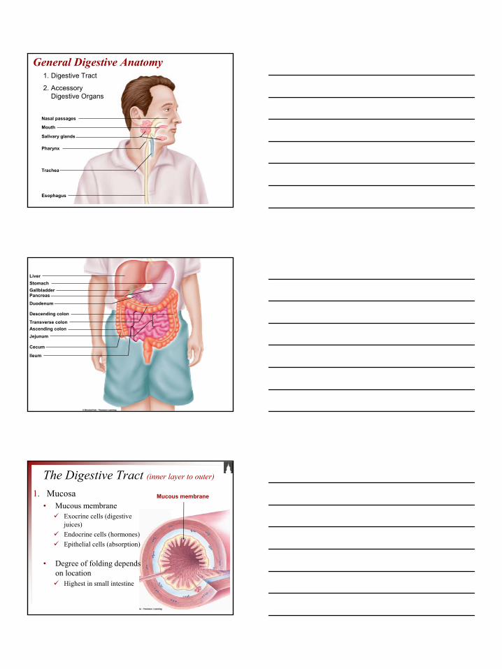
General Digestive Anatomy
Nasal passages
Mouth
Salivary glands
Pharynx
Trachea
Esophagus
1. Digestive Tract
2. Accessory Digestive Organs
LiverStomachGallbladderPancreas
Duodenum
Descending colon
Transverse colonAscending colonJejunum
Cecum
lleum
The Digestive Tract (inner layer to outer)
1. Mucosa• Mucous membrane
Exocrine cells (digestive juices)Endocrine cells (hormones)Epithelial cells (absorption)
• Degree of folding depends on location
Highest in small intestine
Mucous membrane
4
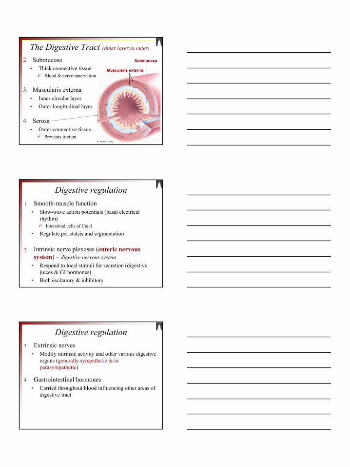
2. Submucosa• Thick connective tissue
Blood & nerve innervation
3. Muscularis externa• Inner circular layer• Outer longitudinal layer
4. Serosa• Outer connective tissue
Prevents friction
Submucosa
Muscularis externa
The Digestive Tract (inner layer to outer)
1. Smooth-muscle function• Slow-wave action potentials (basal electrical
rhythm)Interstitial cells of Cajal
• Regulate peristalsis and segmentation
2. Intrinsic nerve plexuses (enteric nervous system) – digestive nervous system
• Respond to local stimuli for secretion (digestive juices & GI hormones)
• Both excitatory & inhibitory
Digestive regulation
3. Extrinsic nerves• Modify intrinsic activity and other various digestive
organs (generally sympathetic &/or parasympathetic)
4. Gastrointestinal hormones• Carried throughout blood influencing other areas of
digestive tract
Digestive regulation

5
1. Chemoreceptors• Sense changes in chemical components within
lumen
2. Mechanoreceptors• Respond to stretch and tension
3. Osmoreceptors• Changes in osmolarity
Sensory Receptors
Figure 16.4Page 595
Externalinfluence
Local changes indigestive tract
Receptors in digestive tract
Intrinsicnerve plexuses
Extrinsicautomaticnerves
Gastrointestinalhormones
Smooth muscle(contraction for motility)
Exocrine gland cells(secretion of digestive juices)
Endocrine gland cells(secretion of gastrointestinaland pancreatic hormones)
The Digestive Process
6
The Digestion Process1. Mouth
• Chewing• Saliva secretion
3 major salivary glands• Salivary proteins
1. Amylase (CHO breakdown)Polysaccharides to disaccharides
2. Mucus (lubrication)3. Lysozyme (antibacterial)
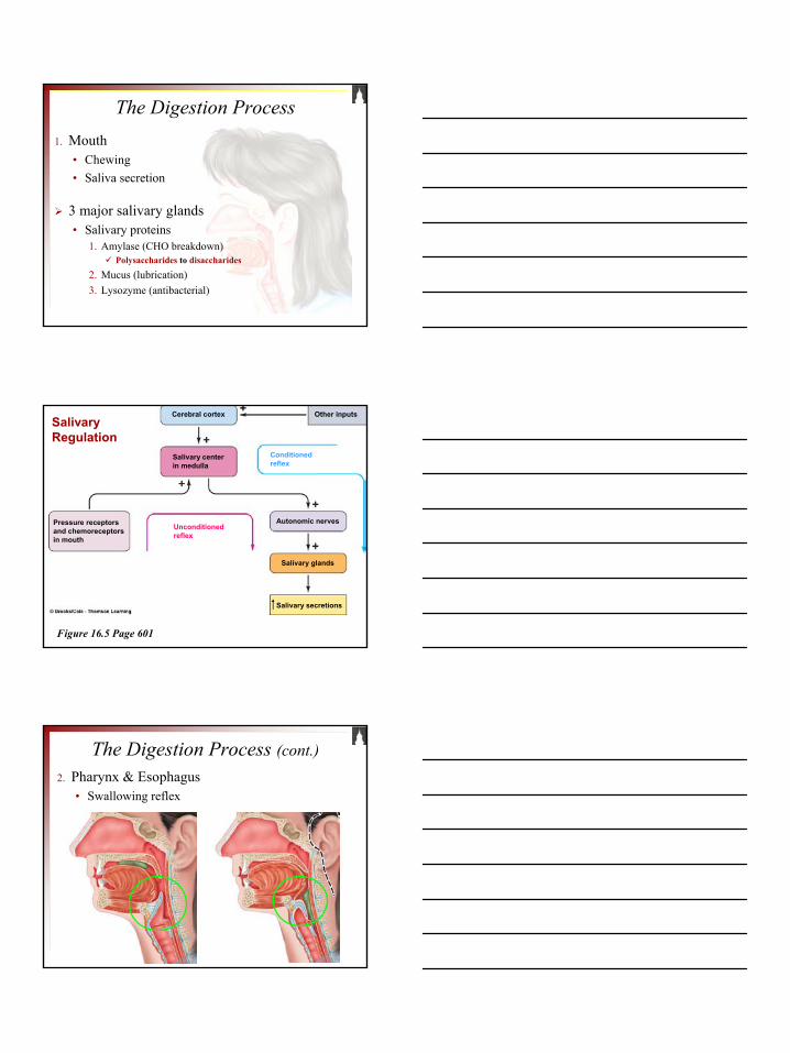
Figure 16.5 Page 601
Salivary centerin medulla
Conditionedreflex
Cerebral cortex Other inputs
Pressure receptorsand chemoreceptorsin mouth
Autonomic nerves
Salivary glands
Salivary secretions
Salivary Regulation
Unconditionedreflex
The Digestion Process (cont.)2. Pharynx & Esophagus
• Swallowing reflex

7
The Digestion Process (cont.)2. Pharynx & Esophagus
• Peristalsis
Figure 16.7
Page 603
Bolus
The Digestion Process (cont.)
Figure 16.8
Page 604
Gastroesophagealsphincter
Body
Duodenum
Antrum
Pyloricsphincter
FundusEsophagusStomach
Stomach Functions1. Storage
2. Gastric mixing & mucous secretion
3. Production of chyme
4. Secretes hydrochloric acid (HCl)• Reduces large food particles• Kills microorganisms ingested in food
5. Initial stages of protein breakdown• Pepsinogen forming pepsin

8
Figure 16.9 (1)Page 605
Duodenum
Direction ofmovementof peristalticcontraction
Pyloric sphincter
Peristalticcontraction
Gastric emptying
Basal electrical rhythm
~ 3 per minute
Figure 16.9 (2)Page 605
Gastric mixing
Peristalticcontraction
Regulation of Gastric EmptyingAmount of chyme
Neural response• Intrinsic nerve plexus (short reflex)• Autonomic nerves (long reflex)
Hormonal• Enterogastrones (secretin & cholecystoinin –
CCK) released from duodenal mucosaInhibit antral contractions
EnterogastricReflex

9
Regulation of Gastric EmptyingDuodenum
1. Fat ~ can only be processed in small intestine2. Acid (unneutralized)
Excess HCl not buffered by sodium bicarbonate
3. Hypertonicity ~ increased osmolarity due to abundance of amino acids and glucose
4. Distension
Emotions
Gastric Digestive Juices~ 2 liters/day
Responsibility of cells lining gastric mucosa
1. Oxyntic mucosa• Body• Fundus
2. Pyloric gland area (PGA)• Antrum
Table 16.4 (1) Page 609
Oxynticmucosa
Pyloricglandarea
Stomachlumen
Gastricpit
Mucosa
Submucosa
10
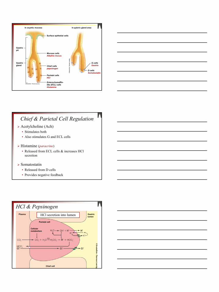
In oxyntic mucosa
Gastricpit
Gastricgland
Surface epithelial cells
Mucosa cellsAlkaline mucus
Chief cellspepsiongen
Parietal cellsHCl
Enterochomaffin-like (ECL) cellsHistamine
In pyloric gland area
G cellsGastrin
D cellsSomatostatin
Chief & Parietal Cell RegulationAcetylcholine (Ach)• Stimulates both• Also stimulates G and ECL cells
Histamine (paracrine)• Released from ECL cells & increases HCl
secretion
Somatostatin• Released from D cells• Provides negative feedback
Plasma Gastriclumen
Chief cell
Parietal cell
Cellularmetabolism
HCl & PepsinogenHCl secretion into lumen
11
Autocatalysis
Digestion
Protein
Peptide fragments
Gastriclumen
HCI
Pepsinogen Pepsin
HCl functions to:• Activate pepsinogen to form pepsin
• Breakdown of connective tissue• Denatures proteins• Kills microorganisms
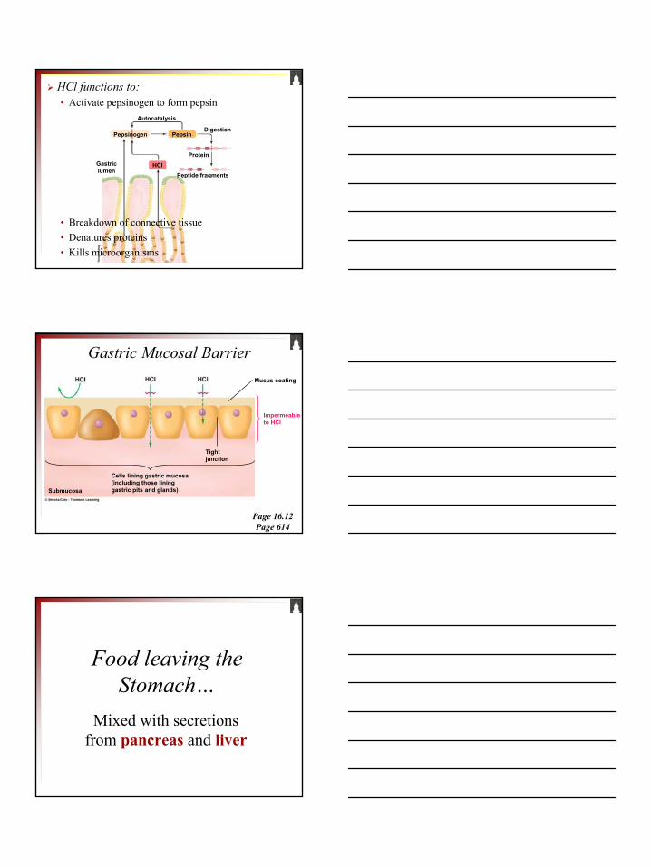
Gastric Mucosal Barrier
Page 16.12Page 614
Mucus coating
Impermeableto HCI
Cells lining gastric mucosa(including those lininggastric pits and glands)Submucosa
Tightjunction
Food leaving the Stomach…
Mixed with secretions from pancreas and liver

12
Page 16.13Page 616
DuodenumBile ductfrom liver Stomach
Hormones(insulin,glucagon)
Blood
Endocrine portionof pancreas(Islets of Langerhans)
The glandular portions ofthe pancreas are grosslyexaggerated
Duct cellssecrete aqueousNaHCO3 solution
Acinar cellssecrete digestiveenzymes
Exocrine portion of panaceas(Acinar and duct cells)
PancreasExocrine & endocrine tissue
1. Exocrine: secretes enzymes capable of breaking down CHO, fat, & protein
Proteolytic enzymes: proteinPancreatic amylase: CHOPancreatic lipase: fat
2. Endocrine (hormones): Insulin & glucagon
Regulated by secretin& cholecystokinin(CCK)
Acid induodenallumen
Fat and proteinproducts induodenal lumen
Secretion releasefrom duodenalmucosa
CCK releasefrom duodenalmucosa
(Secretin carriedby blood)
Pancreatic acinarcells
Secretion of aqueousNaHCO3 solution intoduodenal lumen
Secretion ofpancreatic digestiveenzymes intoduodenal lumen
Pancreatic ductcells
Neutralizes Digests(CCK carriedby blood)
Hormonal Regulation
13
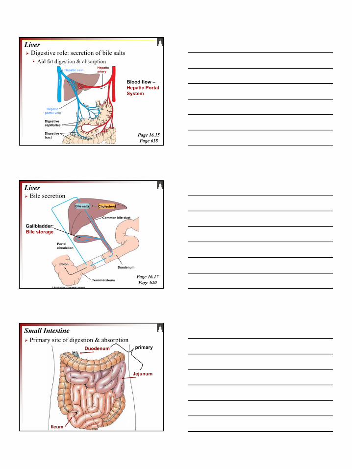
LiverDigestive role: secretion of bile salts• Aid fat digestion & absorption
Page 16.15Page 618
Hepatic arteryHepatic vein
Hepatic portal vein
Digestivecapillaries
Digestivetract
Blood flow –Hepatic Portal System
Liver
Bile salts Cholesterol
Common bile duct
Duodenum
Portalcirculation
Terminal ileum
Colon
Bile secretion
Page 16.17Page 620
Gallbladder: Bile storage
Primary site of digestion & absorption Small Intestine
Duodenum
Ileum
Jejunum
primary

14
Initiated by pacesetter cellsSegmentation of chyme
Small intestine – digestion & absorptionPotential for increased surface area
1. Extensive folding2. Villi & microvilli
Increased digestive enzyme release
Pancreatic enzymes• Fat reduced to FFA (with help of bile salts)
Lipase
• Proteins to AAAminopeptidases
• CHO to di- and monosaccharidesMaltase, sucrase, lactase
Lumen
Epithelial cellof villus
Capillary
Facilitateddiffusion
Na+- andenergy-dependentsecondaryactive transport
Energyrequired

15
Na+- andenergy-dependentabsorption
Energyrequired
Lumen
Epithelial cellof villus
Capillary
Lipid emulsion
Micelles
Epithelialcell of villus
Lumen
(Exocytosis)
Central lacteal
Aggregate andcoated withlipoprotien
Short ormediumchain Basement
membrane
Capillary
LumenMicellesdiffusion
Micelle
Microvillus
Fatty acids,monoglycerides
Passive absorption
1. Vitamin absorption (passive)• Water-soluble• Fat-soluble
2. Iron absorption – regulated• Absorbed into epithelial cells
Either used immediately for production of RBC orStored as ferritin
3. Calcium absorption – regulated• Active transport stimulated by Vitamin D
Small intestine – digestion & absorption

16
Biochemical BalanceDigestive tractlumen
Stomachparietal cell
Pancreaticduct cell
Intestinalepithelial cell
Blood
Large Intestine
Cecum
Colon
Rectum
Drying & storage