the cytotoxic effect and apoptosis rate of breast cancer
TRANSCRIPT

The cytotoxic effect and apoptosis rate of breastcancer stem cells treated with the novel thieno[2,3-b]pyridine anticancer compound
Willmen, Louisa
Master's thesis / Diplomski rad
2019
Degree Grantor / Ustanova koja je dodijelila akademski / stručni stupanj: University of Split, School of Medicine / Sveučilište u Splitu, Medicinski fakultet
Permanent link / Trajna poveznica: https://urn.nsk.hr/urn:nbn:hr:171:264930
Rights / Prava: In copyright
Download date / Datum preuzimanja: 2022-03-14
Repository / Repozitorij:
MEFST Repository

UNIVERSITY OF SPLIT
SCHOOL OF MEDICINE
Louisa Pauline Sofie Willmen
THE CYTOTOXIC EFFECT AND APOPTOSIS RATE
OF BREAST CANCER STEM CELLS TREATED WITH THE
NOVEL THIENO[2,3-B] PYRIDINE ANTICANCER COMPOUND
Diploma thesis
Academic year:
2018/2019
Mentor:
Assoc. Prof. Vedrana Čikeš Čulić, PhD
Split, July 2019
TABLE OF CONTENTS
ACKNOWLEDGEMENT
LIST OF ABBREVIATIONS
1. INTRODUCTION 1 ...........................................................................................................
1.1 Cancer 2 ................................................................................................................
1.2 Triple-negative breast cancer 3 ................................................................................
1.2.1 Definition of TNBC 3 ......................................................................................
1.2.2 Epidemiology and etiology 3 ...........................................................................
1.2.3 Pathophysiology 8 ............................................................................................
1.2.4 Clinical features and prognostics 9 ..................................................................
1.2.5 Diagnostics 13 ..................................................................................................
1.2.6 Classification of breast cancer 14 ....................................................................
1.2.7 Therapeutic options and advances 17 ..............................................................
2. OBJECTIVES AND HYPOTHESES 19 ..........................................................................
2.1 Objectives 20 ......................................................................................................
2.2 Hypotheses 20 .....................................................................................................
3. MATERIALS AND METHODS 21 ..................................................................................
3.1 Chemistry and cell line 22 .......................................................................................
3.2 Colorimetric MTT assay 24 .....................................................................................
3.3 Flow cytometric analysis 25 ....................................................................................
3.4 Statistical analysis 26 ..............................................................................................
4. RESULTS 27 .....................................................................................................................
4.1 Compound I: dose- and time-dependent cytotoxicity 28 ........................................
4.2 Compound I: cell death of breast cancer stem cells 29 ...........................................
4.3 Compound I: decrease in number of BCSC 30 .......................................................
5. DISCUSSION 31 ..............................................................................................................
6. CONCLUSIONS 35 ..........................................................................................................
7. REFERENCES 37 .............................................................................................................
8. SUMMARY 43 .................................................................................................................
9. CROATIAN SUMMARY 45 ............................................................................................
10. CURRICULUM VITAE 47 ............................................................................................
11. SUPPLEMENT 49..........................................................................................................

ACKNOWLEDGEMENT
First and foremost, I want to express my most sincere gratitude to my mentor,
Assoc. Prof. Vedrana Čikeš Čulić, PhD for her support, continuous guidance, encouragement,
and her overall excellent mentorship which made this diploma thesis possible.
Secondly, I want to express my heartfelt appreciation to my family and friends.
To my parents, Gottfried and Simone, thank you for believing in me, having my back, and giving me
the opportunity to fulfill my dream of becoming a doctor. To my siblings, Lukas and Laura, thank
you for always being there for me especially when I needed you the most.
I am truly grateful to call you my family.
Last but not least, this paper is dedicated to my late love Stefan, without whom I would have never
made it this far. Through his love, support, and unparalleled patience,
he helped make this degree possible.
I’ll see you again.
LIST OF ABBREVIATIONS- ASCO: American Society of Clinical Oncology
- ATCC: American Type Culture Collection
- BARD1: heterodimerizes with BRCA1
- BC: breast cancer
- BCC: breast cancer cell
- BCS: breast-conserving surgery
- BCSC: breast cancer stem cell
- BRCA1 / 2: mutated tumor suppressor genes
- CSC: cancer stem cell
- DMEM: Dulbecco’s Modified Eagle’s Medium
- DMSO: Dimethyl sulfoxide
- EGF: epidermal growth factor
- ER: estrogen receptor
- FBS: fetal bovine serum
- FISH: fluorescence in situ hybridization
- HER2: human epidermal growth factor receptor 2
- H&E: hematoxylin and eosin
- IBC: invasive breast cancer
- IHC: immunohistochemistry
- MDA-MB-231: triple-negative breast cancer stem cell line
- MRI: magnetic resonance imaging
- MTT: Method of Transcriptional and Translational assay,
3-(4,5-dimethylthiazol-2-yl)-2,5-diphenyltetrazolium bromide assay
- NST: neoadjuvant systemic treatment
- OC: oral contraceptive (pill)
- p53: TSG p53, TP53/tumor protein 53
- PALB2: partner and localizer of BRCA2
- PBS: phosphate-buffered saline
- PET: positron emission tomography
- PLC: phospholipase C
- PLC C-γ2: phospholipase C-γ2
- PR: progesterone receptor

- PV: pathologic variant
- RAD5D / C: DNA helicase
- RT: radiation therapy
- SC: stem cell
- SD: standard deviation
- TGF alpha: transforming growth factor alpha
- TNBC: triple-negative breast cancer
- TSG: tumor suppressor gene
- US: ultrasound
- WHO: World Health Organization
- WNT7B: oncogene

1. INTRODUCTION

1.1 Cancer
Cancer is with increasing significance a major global health problem, and it represents a
group of diseases which are most importantly characterized by abnormal cell growth with the
potential to invade and or metastasize to other sides in the human body. This distinguishes cancers
from benign tumors, which cannot spread across the side of its origin. However, several research
studies suggest that cancers are monoclonal in their origin, implying that the malignant
transformation started in one single cell. The cellular process of developing cancer cells is a
complex and long-lasting one, which requires several stages to turn healthy tissue into cancer. This
process is called tumor progression (1).
According to the newest model of ”The Hallmarks of Cancer” by Douglas Hanahan and
Robert Weinberg, all known cancers have ten traits in common which enable them to modulate
themselves from a normal to a cancerous cell, presented in the following list:
1. Self-sufficient in growth signals;
2. Insensitive to inhibitory anti-growth signals;
3. Resistant to apoptosis;
4. Limitless replication potential;
5. Stimulate angiogenesis;
6. Invade surrounding tissues and metastasize;
7. Disturbed metabolic pathways;
8. Evade the immune system;
9. Genomic instability;
10. Inflammation (2).
The differences in etiology, pathology, and clinical presentation of cancers lead to various
therapeutic angles, but unfortunately, it mostly results in the lack of a sufficient and standardized
treatment regime (1,2).
"2

1.2 Triple-negative breast cancer
1.2.1 Definition of TNBC
The term triple-negative breast cancer was first introduced in 2005, and is generally
abbreviated as TNBC. Overall it refers to a genomically heterogenic group of breast cancers which
make up 12-17% of all breast cancers and are defined by their unifying lack of expression of
estrogen receptor (ER) and progesterone receptor (PR), and the absence of human epidermal growth
factor receptor 2 amplification (HER2) (3,4). Furthermore, it has to be emphasized that TNBC is
not a synonym for basal-like cancer. Basal-like breast cancers are often used as a surrogate term for
triple-negative breast cancers even though only 80% of basal-like breast cancers are truly triple-
negative in their receptor expression. 10% are ER+/HER2- and 2% are even HER2+ (5).
1.2.2 Epidemiology and etiology
Breast cancer is a prevalent and significant disease in females with several potentially severe
adverse effects on a woman’s life. The importance of this cancer stressed by the fact that in 2018,
breast cancer was the most common type of cancer in females in 154 countries worldwide,
according to the World Health Organization (WHO), accounting for 2.08 million new cases in
women (Table 1). Furthermore, breast cancer is not only the most commonly diagnosed female
cancer type in the world, constituting for 24.2% out of 8.6 million cases of all newly diagnosed
female cancer patients, but also together with lung cancer, it is the most frequently diagnosed cancer
in general, as shown in Figure 1 A and B. The incidence of breast cancer cases in 2018, for males
and females together, is 11.6% out of 18.1 million new cancer cases, same as for lung cancer, but
99.12% of the new breast cancer cases are in females, highlighting the immense female
predominance of this cancer (Figure 1B, Table 1). Concerning cancer mortality, breast cancer is one
of the leading causes of cancer-related deaths. In 2018, breast cancer accounted for 0.6336 million
deaths. 99.43% of these were women, making it the 5th most lethal cancer in the world (Table 1).
Lung cancer is in the general population the leading cause of cancer-related deaths in humans being
responsible for 18.4% out of 9.6 million deaths, but breast cancer constitutes to the highest female
cancer mortality with 15% out of 4.2 million deaths and is, therefore, the principal cause of cancer-
related deaths in women in at least 103 countries according to the WHO (Figure 1, Table 1) (6).
"3

Figure 1. Pie charts depicting for the ten most common cancers the distribution of new cases and deaths in 2018 for (A) females and (B) both sexes. The area of the pie chart represents the proportion of the total number of new cases or deaths; the category ”others” includes nonmelanoma skin cancers. SOURCE: Globocan 2018, World Health Organization (6).
"4
A
B

Table 1. Cancer and breast cancer incidence and mortality in 2018.
The table above shows for both sexes and females the amount of people newly diagnosed with cancer and breast cancer and the mortality of cancer and breast cancer in 2018, as well as the percentage. SOURCE: Globocan 2018, World Health Organization.
The incidence of TNBC among breast cancer depends on several factors like, for instance,
sex, race, and age of the patient. Not only is breast cancer itself more common in females than in
males but also the TNBC type occurs mainly in females with an incidence of 13% and 6%
respectively. Taking into account the race of a patient it was noticed that for example, in the
African-American population, the incidence is the highest one yet determined accounting for 35%
of all breast cancers diagnoses of this group. To be more precise it is stated that it is most prominent
among non-Hispanic black and Hispanic patients (23.7% and 14.8%) and in the Caucasian
population it only makes up around 15% (7). The lowest occurrences so far measured are in Filipino
patients (8.9%), albeit Filipino women are known to be at a heightened risk for HER2+ cancers
(7,8). For all ethnic groups, it is deemed to be that younger patients are more prone to have TNBC,
with notable distinctions between Caucasian and African-American patients. As seen in Figure 2,
for patients younger than 30-years of age, the two racial groups start with a roughly similar overall
incidence of TNBC. In Caucasian patients, the TNBC percentage is highest for the age group
around 30 years, and then it continuously declines, while in African-American patients, there is no
significant decrease until the patients are older than 60-years, but even a slight increase during their
thirties. Nonetheless, the graph shown in Figure 2 points out that African-American women of all
age groups are at any time during their life more predisposed to have TNBC compared to Caucasian
patients (7).
2018Incidence of new cancer cases
Incidence of new breast cancer cases
Mortality of cancer
Mortality of breast cancer
Females 8.6 million 2.0812 million 4.2 million 0.63 million
Both sexes 18.1 million 2.0996 million 9.6 million 0.6336 million
Percentage of Females 47.5% 99.12 % 43.75 % 99.43%
"5

Figure 2. Percentage of TNBC based on patient age and race. TNBC: triple-negative breast cancer. SOURCE: Plasilova et al (7).
TNBC is of course also associated with genetic, pathologic variants (PV), which underline
how important it is to consider the race of a patient as the incidence of the different PVs varies
immensely depending on whether a patient is of Caucasian ancestry or African-American. For
example, an increased incidence in patients with germline BRCA1 mutations is commonly seen in
Caucasians with TNBC, the most striking PVs with the highest incidence are BRCA1 and PALB2
PVs which have a lifetime risk for TNBC of 18% and 10% respectively, followed by BARD1 with
7%, 6% BRCA2, and 5% RAD51D, as shown in Figure 3A and B. Even though the same
pathologic variants are found in African-American people, it has to be emphasized that the impact is
far more drastic on their TNBC risk estimates for overall breast cancer, with 81% BRCA1, 62%
BRCA2, 41% PALB2, and 39% BARD1 (Figure 3C and D) (9). Furthermore, around 70% of breast
cancers diagnosed in people with an inherited BRCA mutation, particularly BRCA1, are classified
as triple-negative (10). To summarize, one has to add that African-Americans have a large and
diverse mutation profile with more genetic variants on numerous genes per patient, while
Caucasians have PVs on fewer genes and also the deleterious PVs per patient are significantly less
prevalent in Caucasians (11).
"6

Figure 3. Absolute risk of specific pathologic variants for Caucasian (A and B) and African-American (C and D) accessed to age 85 the overall breast cancer and triple-negative breast cancer risk. (A) Age-related risk curves for Caucasian overall breast cancer for six genes as color lines. (B) Age-related risk curves for Caucasian TNBC for six genes as colored dashed lines. (C) Age-related risk curves for African-American overall breast cancer for four genes as color lines. (D) Age-related risk curves for African-American TNBC for four genes as colored dashed lines. TNBC: triple-negative breast cancer. SOURCES: http://www.seer.cancer.gov and Shimelis et al (9).
Another apparent risk factor which is still under investigation is the oral contraceptive pill
(OC). A study from 2009 compares the impact of taking OC on the risk of getting TNBC in women
who took birth control pills for less than one year or never. The article states that taking OCs for at
least one year causes a 2.5 times increase in risk for TNBC in females, and if the women are around
the age 40 the increase is even 4.2 times. Furthermore, they also concluded that the prolonged
duration of OC administration leads to an increased TNBC risk (12).
"7
A B
C D
However, in another study from 2011, which also covers the association of the patients’s
reproductive history and their oral contraceptive usage in relation to the risk of getting TNBC, the
increased risk of OCs for getting TNBC was not found. Albeit, it finds an association between
nulliparity and the risk of breast cancer, showing a decreased incidence in TNBC patients but an
increase in ER-positive ones (13). Another study from the same year strengthens the relevance of
number of pregnancies in breast cancer patients, as it also states that the frequency of nulliparity is
lowest in TNBC patients with 13% compared to other types of breast cancer, but it also says that
multiparty, more precisely ≥3 kids is more common in TNBC (14,15). Aside from that, another
known and relevant factor that decreases the risk of developing TNBC is breastfeeding for a
prolonged duration or breastfeeding several children (15).
Besides, metabolic syndrome, characterized by obesity and insulin resistance, and a higher
waist/hip ratio is also more prevalent in TNBC than it is in other breast cancers (15,16,17). Last but
not least, another study found that alcoholism has a decreasing effect on the risk of getting TNBC
and also concluded that smoking is not associated with TNBC (18).
1.2.3 Pathophysiology
The formation of breast cancer development is, like in most other cancers, set off by a
combination of multiple external environmental factors and their interaction in a genetically
susceptible host (1). The CD44+/CD24- phenotype, which is known to be numerously common in
cancer stem cells (CSC), represents the tumorigenic cells with stem cell-like properties, meaning
the ability to extensively self-renew and give rise to phenotypically different cells with a decreased
proliferative and developmental potential. The cell surface glycoprotein CD44 is crucial for the
quick cancer progression as it plays a critical role in the adhesion, migration, and invasion of breast
cancer cells (BCC), as well as in proliferation and metastasis (19,20). These CSC qualities do not
only lead to the production of tumorigenic cells but also to the production of several phenotypically
distinct nontumorigenic cells that make up the overall tumor mass. Due to the fact, that the only
unifying characteristic of TNBC is the lack of the expression of ER, PR, and HER2, and due to the
stem cell (SC) qualities of this cancer group, the creation of phenotypically heterogenic tumors is
expected (4,21). The latest definition presented by the American Society of Clinical Oncology
(ASCO) in their guidelines about the three main biomarkers ER, PR, HER2 states, that ER/PR is
considered positive if ≥1% immunohistochemistry (IHC) and HER2 is considered positive if there
"8

is protein expression of 3+, and/or HER2/neu gene amplification is ≥2.0 by fluorescence in situ
hybridization (FISH). This means that the contemporary proper pathologic definition of TNBC
actually is defined as 0% by IHC for ER and PR, and for HER2 negativity it states IHC expression
of 0-1+ or lack of HER2/neu gene amplification (FISH <2.0) (22).
The most known genes responsible for breast cancer development are BRCA mutations and
the germline mutations of the tumor protein 53 (p53) tumor suppressor gene (TSG). Even though
BRCA mutations are only responsible for 5-10% of breast cancer cases in women, 70% of these are
triple-negative, chiefly BRCA1 variants (10,23).
Besides, to the age of 80-years, these harmful mutations lead to an estimated cumulative risk
of getting breast cancer of 72% and 69%, for BRCA1 and BRCA2 respectively (24). On top of this,
the BRCA mutations form TSG complexes, where the BRCA1 modification combines with BARD1
and causes the heterodimer BRCA1-BARD1, which interacts with RAD51 and the TSG complex
BRCA2-PALB2. BRCA2 works together with PALB2, forming the TSG complex BRCA2-PALB2,
PALB2 stands for partner and localizer of BRCA2, and both combined mutations have increased
risks for breast cancer development and make targeted therapy even more difficult (25). Lastly,
TP53 causes Li-Fraumeni syndrome and is, therefore, responsible for 4% of breast cancers among
females under the age of 30-years (26).
1.2.4 Clinical features and prognostics
Table 2 states the most important clinical and diagnostic factors concerning TNBC, but for
further details, one can say that clinically, TNBC mostly presents as a palpable lump in the breast of
younger females and is therefore usually not detected by the standard breast cancer screenings,
which usually start around the age of 50. For women who attend regular screening programs, TNBC
is known to typically present as an interval cancer, meaning between two mammographies. Withal,
at the time of detection it commonly first manifests in a more unfavorable histopathologic
condition, as it is often attended with lymphovascular invasion when it is diagnosed and the tumor
is generally more massive in size, tends to present with clinically metastatic disease, and overall
mostly in a higher cancer stage. Strictly speaking at the time of diagnosis, a striking majority of
79.8% are grade III and the mean tumor size is 2.78 cm (Table 3). Only as little as 2.4% are grade I
at the time of their first presentation. (7,27). Until in 2016, the newer study by Plasilova et al. was
"9

published, it was believed that there is no correlation between increasing tumor size and lymph
node involvement in TNBC, unlike in other breast cancer types, but as shown in Figure 4, positive
lymph nodes are generally less common in TNBC, even though in bother BC groups the overall
percentage of lymph node positivity is the same (7,28,29). Furthermore, it usually presents more
aggressively and preferentially spreads hematogenously, leading to frequent visceral relapses in the
brain and lungs (16).
Table 2. Key points about TNBC characteristics.
SOURCES: Plasilova et al (7); Kumar et al (27); Dent et al (28).
Speaking of visceral relapses, the pattern of recurrences shows a significant difference in
TNBC compared to other BCs. First of all, distant metastases are more common than locoregional
ones and also considerably more common in TNBC (33.9%) than in different subtypes (20.4%).
After surgery, the relapse rate as shown in Figure 5, shows a striking increase in the first three years
with the mean time to distant metastasis being 2.6 years compared to five years in other BCs, and
interestingly the rate for distant metastases decreases quickly until it is rather uncommon eight years
following the diagnosis. Furthermore, distal recurrences are rarely preceded by local ones, only in
25% of women treated with breast-conserving surgery, which are only slightly more common in
TNBC than in other types of breast cancers, but occur notedly sooner in TNBC after an estimated
2.8 years versus 4.2 years respectively (27,28).
Main characteristic features of TNBC phenotype:
Younger women (<50 years)
More frequent in African-American and black ethnicities
Presents as interval cancer
Less likely to have lymph node involvement
Steep increase in risk of recurrence after diagnosis
Peak risk of recurrence at 1-3 years
Distal recurrence more common than local ones; no distant relapse after 8 years
Higher mortality rate first 5 years; all deaths occur within 10 years of diagnosis
Short survival after distant recurrence
More aggressive with increase incidence of visceral metastasis (brain, lung)
"10

Table 3. Specific characteristic differences for TNBC compared to other types of breast cancers.
*P values were calculated using the x2 test. SE=standard error; LVI, lymphovascular invasion. Full table shown in supplements (Table 2). SOURCE: Plasilova et al (7).
Figure 4. Comparison of TNBC and non-TNBC regarding their tumor size and lymph node positivity in percentage (%). Abbreviations: TNBC, triple-negative breast cancer. SOURCE: Plasilova et al (7).
Characteristic: TNBC n (%) P*
T stage: T1 T2 T3
T4
46.4% 38.1% 8.4% 7.1%
<0.001
N stage: N0 N1 N2 N3
72% 20.1% 4.7% 3.3%
<0.001
M stage: M0 M1
94.1% 5.9%
<0.001
Mean tumor size (cm±SE): 2.78±0.012 <0.001
Tumor grade I II III
2.4% 17.8% 79.8%
<0.0001
"11
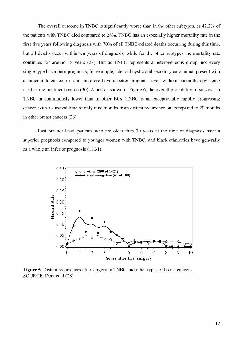
The overall outcome in TNBC is significantly worse than in the other subtypes, as 42.2% of
the patients with TNBC died compared to 28%. TNBC has an especially higher mortality rate in the
first five years following diagnosis with 70% of all TNBC-related deaths occurring during this time,
but all deaths occur within ten years of diagnosis, while for the other subtypes the mortality rate
continues for around 18 years (28). But as TNBC represents a heterogeneous group, not every
single type has a poor prognosis, for example, adenoid cystic and secretory carcinoma, present with
a rather indolent course and therefore have a better prognosis even without chemotherapy being
used as the treatment option (30). Albeit as shown in Figure 6, the overall probability of survival in
TNBC in continuously lower than in other BCs. TNBC is an exceptionally rapidly progressing
cancer, with a survival time of only nine months from distant recurrence on, compared to 20 months
in other breast cancers (28).
Last but not least, patients who are older than 70 years at the time of diagnosis have a
superior prognosis compared to younger women with TNBC, and black ethnicities have generally
as a whole an inferior prognosis (11,31).
Figure 5. Distant recurrences after surgery in TNBC and other types of breast cancers. SOURCE: Dent et al (28).
"12
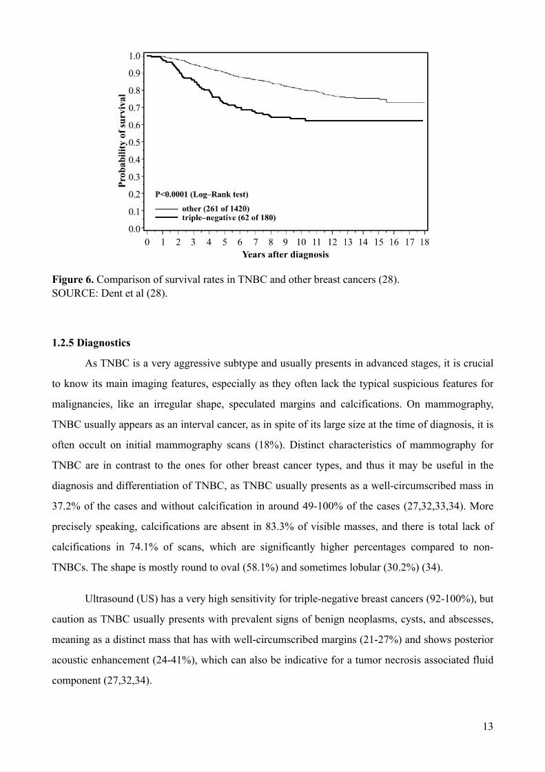
Figure 6. Comparison of survival rates in TNBC and other breast cancers (28). SOURCE: Dent et al (28).
1.2.5 Diagnostics
As TNBC is a very aggressive subtype and usually presents in advanced stages, it is crucial
to know its main imaging features, especially as they often lack the typical suspicious features for
malignancies, like an irregular shape, speculated margins and calcifications. On mammography,
TNBC usually appears as an interval cancer, as in spite of its large size at the time of diagnosis, it is
often occult on initial mammography scans (18%). Distinct characteristics of mammography for
TNBC are in contrast to the ones for other breast cancer types, and thus it may be useful in the
diagnosis and differentiation of TNBC, as TNBC usually presents as a well-circumscribed mass in
37.2% of the cases and without calcification in around 49-100% of the cases (27,32,33,34). More
precisely speaking, calcifications are absent in 83.3% of visible masses, and there is total lack of
calcifications in 74.1% of scans, which are significantly higher percentages compared to non-
TNBCs. The shape is mostly round to oval (58.1%) and sometimes lobular (30.2%) (34).
Ultrasound (US) has a very high sensitivity for triple-negative breast cancers (92-100%), but
caution as TNBC usually presents with prevalent signs of benign neoplasms, cysts, and abscesses,
meaning as a distinct mass that has with well-circumscribed margins (21-27%) and shows posterior
acoustic enhancement (24-41%), which can also be indicative for a tumor necrosis associated fluid
component (27,32,34).
"13
Magnetic resonance imaging (MRI) is the most sensitive method to detect TNBC, with a
visualization rate of 100%. The lesions appear with smooth margins, but also with rim
enhancement, which is a highly predictive characteristic of malignancy. Furthermore, features are
persistent enhancement patterns and, on T2-weighted images, a high intratumoral signal intensity
(35). A study from 2007 with a rather small sample 29 patients states that at the time of diagnosis, in
21% of the cases multifocality is visible, as well as in 34% prominent skin enhancement indicating
dermal lymphatic invasion in tumors smaller than five centimeters. The mean tumor of this study is
size 4.1 ± 2.7 cm and 79% of the cases presented at least at grade II (36,37). Dynamic MRI shows
vascular differences between TNBC and the other subgroups, observing a significantly higher
outflow rate and smaller leakage space in TNBCs (38). To sum up, the TNBC visualization rates of
the three main diagnostic methods, mammography, US, and MRI are very high, 91%, 93% and
100% respectively (34).
Another imaging procedure, rather than a primary diagnostic method, that can be performed
is [18F]2-fluoro-2-deoxy-D-glucose positron emission tomography (18F-FDG-PET), it is useful for
detecting metastasis and to follow-up the response to chemotherapy (32,39).
1.2.6 Classification of breast cancer
According to the WHO classification from 2012, there are for the time being 21
morphologically distinct subtypes of invasive breast cancer (IBC), which make it a very
heterogeneous group with distinct pathological and molecular characteristics. The whole list of all
acknowledged breast cancers is added in the supplement section, Figure 2. The most common IBC
type is the invasive carcinoma of no special type, also known as invasive ductal carcinoma, which
makes up between 40 to 75% in published series (40). Invasive ductal carcinoma comprises even a
more significant percentage among TNBCs with a 90% majority of TNBCs being unifocal, invasive
ductal carcinomas. All the other histological types of TNBC are classified as either medullary,
secretory, adenoid cystic, apocrine, metaplastic, invasive lobular carcinoma, or microglandular
adenosis carcinomas (27).
Up to this point, the morphological diagnosis and the contemporary staging systems are
rather inadequate and disorganized; consequently, they are currently evolving into a multi-
classification system which integrates the morphological diagnosis of cancer, immunohistochemical
assessment, DNA microarray analysis, and CIRCOS Plot to improve the therapy response rate and
"14
overall outcomes (41). However, for the time being, the guidelines for breast cancer therapy focus
on a combination of pathological classification, the histological grade of cancer, ER, PR, and HER2
status (42). But, due to the advancement of molecular procedures, such as DNA microarray analysis
which focuses on gene expression profiling, the concept of heterogeneity of breast cancer has
become accepted and the importance in diagnosis shifted to ”Molecular Classification” of breast
cancers — leading to the development of targeted therapeutics and individualized treatment (43).
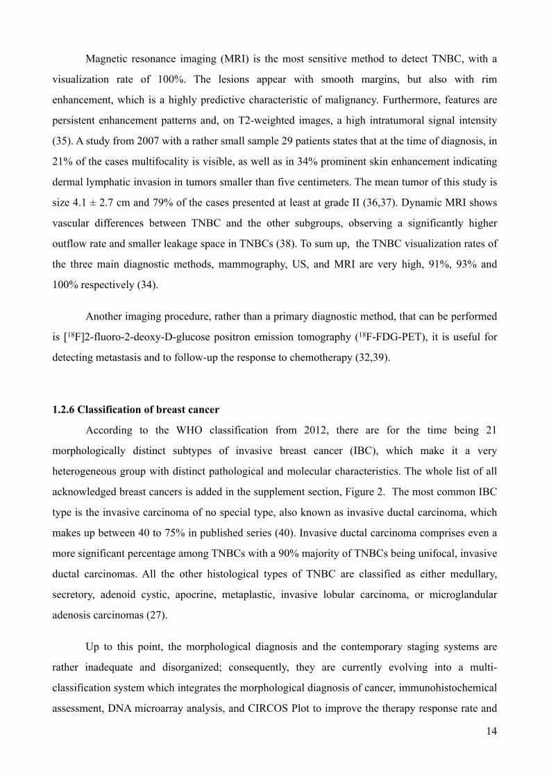
Figure 7 shows eleven distinct histopathological pictures of eight different types of breast
cancers stained with hematoxylin and eosin (H&E). The different classes show slight differences,
but the cancer cells are highly coherent. Histopathological slides show significant variations making
differentiation of breast cancer types hard (44).
Figure 7. Histopathological images of the eight most common breast cancer types at a magnification factor of 400. SOURCE: Han et al (44).
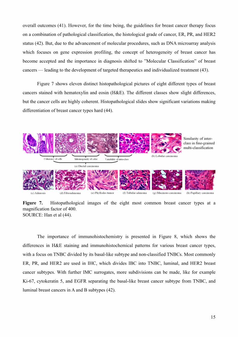
The importance of immunohistochemistry is presented in Figure 8, which shows the
differences in H&E staining and immunohistochemical patterns for various breast cancer types,
with a focus on TNBC divided by its basal-like subtype and non-classified TNBCs. Most commonly
ER, PR, and HER2 are used in IHC, which divides IBC into TNBC, luminal, and HER2 breast
cancer subtypes. With further IMC surrogates, more subdivisions can be made, like for example
Ki-67, cytokeratin 5, and EGFR separating the basal-like breast cancer subtype from TNBC, and
luminal breast cancers in A and B subtypes (42).
"15
Figure 8. Histological images with hematoxylin-eosin stain (H&E) and immunohistochemical patterns for seven molecular breast cancer subtypes.
Abbreviations: BC, breast cancer; BLBC, basal-like subtype; CK5, high–molecular weight cytokeratin expressed in normal myoepithelial cells; EGFR, epidermal growth factor receptor; ER, estrogen receptor; HER2, human epidermal growth factor receptor-2; NCBC, nonclassified subtype; PR, progesterone receptor; TNBC, triple-negative breast cancer. SOURCE: Tang et al (42).
Subclassifying the TN phenotype is somewhat tricky, considering that the only unifying
feature being the lack of three biomarkers. Figure 9 emphasizes this heterogeneity by showing
various histopathological TNBC subtypes and their degree of low- to high-grade cancers. Although
progression to high-grade cancers is typical in TNBC it is with varying rates depending on the
histological type, i.e., prevalent in acidic cell carcinoma and rare in salivary gland-like breast
tumors, resulting in some subgroups having a low histologic grade and rather indolent behavior,
even though in general TNBC is known for its aggressive presentation (45). For example, adenoid
cystic and secretory carcinomas of the breast, are TNBCs with an indolent course and especially a
better prognosis even without chemotherapy (29).
"16

Figure 9. Spectrum of low to high-grade TNBCs.Various histopathological subtypes of TNBC are connected to their known specific genetic alterations. Progression from low to high-grade TNBC is common but occurs at different rates. *Evidence for polymorphous carcinoma of the breast mutation PRKD1 E710D or PRKD1/2/3 rearrangements still needs to be documented. Magnification factor of 200. Abbreviations: PI3K, phosphatidylinositol 3-kinase.; TNBC, triple-negative breast cancer. SOURCE: Geyer et al (45).
1.2.7 Therapeutic options and advances
Due to the fact that there are no proper standard treatment regimens or even options
available for TNBC, there are various therapeutic approaches, ranging from local therapy, meaning
surgery and radiation to systemic approaches involving chemotherapy, targeted therapies,
immunotherapeutic approaches, to the option of breast cancer vaccines, but most potentially more
potent therapeutics are still under investigation so far in clinical or like our compound in preclinical
trials (27).
"17

Local therapy for TNBC is applied in a comparable way as to other IBCs and involves
removal of the cancerous lump by either breast-conserving surgery (BCS) or mastectomy supported
by radiotherapy (RT), as there are no TNBC-specific local therapy recommendations. The breast-
conserving procedure remains the standard therapy for small T1 and T2 BCs, and mastectomy is for
more extensive, multifocal cancers, or in patients with positive margins after BCS (8,27). A study
from 2017 highlights the point that there is insufficient data, to imply that mastectomy is better than
BCS, but also points out that systemic recurrence remains higher in TNBC patients emphasizing the
importance of more efficient systemic therapeutic options (46). There are no distinguished
guidelines for adjuvant RT in TNBC, and the better outcome in BCS is determined by the fact that
three-year relapse-free survival is 79.6% in patients treated with RT compared to 57.9% in patients
without RT, even though the patients with RT had a higher cancer stage (27).
Since appropriate targets in TNBC for systemic approaches are missing, chemotherapy
remains the primary treatment for TNBC, with no difference in the outcome whether or not the
therapy is started pre- or postoperative in early-stage TNBCs, but several other systemic treatment
options are momentarily under investigation (27,47). Even though the response to chemotherapy in
TNBC is superior compared to other BC types, the five-year survival is by less than 30% of women
with metastatic disease (48). So far anthracycline-taxane-based chemotherapy remains the standard
neoadjuvant systemic treatment (NST), but the addition of platinum-based agents in NST is
considered, in defiance of the added toxicity several papers say that it shows better clinical response
to systemic therapy and could possibly improve local therapy, however some state that it does not
improve survival (47,48).
Many promising therapies are under investigations to allow personalized treatment strategies
in this heterogeneous disease, like immunotherapeutic approaches and the oncolytic vaccinia virus,
which does not only show promising results in preventing metastasis but also in treating it (27).
Another approach is targeted therapies concentrating on, for example, PARP inhibitors, anti-
androgen therapy, PI3K inhibitors, MEK inhibitors, and inhibitors of cancer stem cells. This study
focuses on a novel therapeutic compound to counter the BCSC population, which is known to be
more resistant to chemotherapy than cancers without stem cells (48).
"18

2. OBJECTIVES AND HYPOTHESES
2.1 Objectives
The aim of this study is to determine the effects of treating breast cancer stem cells with the
newly synthesized thieno[2,3-b]pyridine anticancer agent, by focusing on its cytotoxic and
apoptotic effects on the investigated human cell line MDA-MB-231.
2.2 Hypotheses
1. Treating the MDA-MB-231 cell line with compound 1 will have a cytotoxic effect on the breast
cancer stem cells.
2. Compound 1 will lead to an increased apoptotic rate in the MDA-MB-231 breast cancer stem
cell line.
3. The percentage of the breast cancer stem cell subpopulation will be decreased following the
treatment with compound 1.
4. The effects of compound 1 will be in a dose- and time-dependent manner.
"20

3. MATERIALS AND METHODS

3.1 Chemistry and cell line
3-Amino-N-(3-chloro-2-methylphenyl)-5-oxo-5,6,7,8-tetrahydrothieno[2,3-b]quinoline-2-
carboxamide (compound 1) (Figure 10) was dissolved in dimethyl sulfoxide (DMSO). Cells were
purchased from the American Type Culture Collection (ATCC, LGC Standards), the cell
micrograph is shown in Figure 11. The MDA-MB-231 breast cancer stem cells were grown in a
Dulbecco’s Modified Eagle’s Medium (DMEM, Sigma-Aldrich, Steinheim, Germany) with 10%
fetal bovine serum (FBS, EuroClone) supplemented with 1% antibiotics (Penicillin-Streptomycin,
EuroClone). The culture conditions were in a humidified incubator at 37°C with an atmosphere of
5% CO2. The known characteristics of the cell line are presented in the table below (Table 4).
Figure 10. The structure of the newly synthesized anticancer agent (compound 1). Note: Compound 1, 3-Amino-N-(3-chloro-2-methylphenyl)-5-oxo-5,6,7,8-tetrahydrothieno[2,3-b]quinoline-2-carboxamide.
Figure 11. Cell micrograph SOURCE: https://www.lgcstandards-atcc.org (49).
"22

Table 4. Characteristics of MDA-MB-231 cell lines
SOURCE: https://www.lgcstandards-atcc.org (49).
Characteristics:
Organism: Homo sapiens, human
Tissue: mammary gland/breast; derived from metastatic site: pleural effusion
Disease: adenocarcinoma
Cell Type: epithelial
Age: 51 years adult
Gender: female
Ethnicity: Caucasian
Morphology: epithelial
Culture Properties: adherent
Biosafety Level: 1Biosafety classification is based on U.S. Public Health Service Guidelines
Product Format: frozen
Applications: these cells are a suitable transfection host
Storage Conditions: liquid nitrogen vapor phase
Receptor Expression: epidermal growth factor (EGF), expressed transforming growth factor alpha (TGF alpha), expressed
Oncogene: the cells express the WNT7B oncogene
"23

3.2 Colorimetric MTT assay
A tetrazolium-based colorimetric MTT assay was performed, to analyze the cell viability
and in doing so determining the cytotoxic effect of the novel anticancer compound on triple-
negative breast cancer stem cells. The cell proliferation was measured by first diluting the cells in a
solution of trypan blue and Neubauer chambers counted by MOTIC AE30, an inverted binocular
microscope. The cell number was calculated according to the formula: number of counted cells x 10
× 104/mL. A homogenous number of cells were then seeded in 96-well plates at a density of 104
cells/100 µL and incubated during the night to allow attachment (Figure 12). The cells were treated
in a complete medium and the experiments performed in triplicate, for 4, 24, 48 and 72 h with
specific solutions of compound 1 at a concentration of 50 nM, 250 nM, 500 nM, 1 µM and 5 µM.
Thereupon, the MTT 3-(4,5-dimethylthiazol-2-yl)-2,5-diphenyl tetrazolium bromide] assay was
performed, and the treated cells were incubated with 0.5 g MTT/L at 37°C for 1 hour, the medium
was removed and in the end dimethylsulphoxide (10% DMSO) was added and incubated for
another 10 minutes at 37°C with shaking. Absorbance was measured photometrically at a signal
wavelength of 570 nm and depends on the degree of formazan formation (Figure 13), which
indicates cell viability and their metabolic activity. The collected data was calculated in comparison
to the untreated control (100%) from three independent measurements.
Figure 12. MDA-MB-231 in 96-well plates and treated with compound 1. SOURCE: Biochemistry Laboratory, Medical Faculty of Split.
"24

Figure 13. Enzymatic formazan formation in MTT. SOURCE: Kuete et al (50).
3.3 Flow cytometric analysis
A homogenous number of cells were plated onto six-well plates at a density of 104 cells/100
µL and treated with 2 µM compound 1 for 48 h and afterward analyzed for apoptosis. The combined
staining of propidium iodide and Annexin-V-FITC makes it possible to properly distinguish
between early (Annexin-V+/PI-) and late (Annexin-V+/PI-) apoptotic cells, necrotic cells, and live
cells. Following the treatment with compound 1 the cells were trypsinized, then washed with
phosphate-buffered saline (PBS) and at last resuspended in 100 µl of the binding buffer containing
5 µl of propidium iodide and/or 5 µl Annexin-V-FITC (Annexin-V-FITC Apoptosis Detection Kit I,
BD Biosciences, San Jose, CA, USA). Then, the cells were incubated in the dark at room
temperature for 15 minutes and after that flow cytometric analysis was carried out (BD Accuri C6,
BD Biosciences). Using the FlowLogic Software (Inivai), the degree of apoptosis (Annexin-V
positive cells) was analyzed and presented as mean ± SD.
Besides, to detect the percentage of CD44+/CD24- cells, the cells administered with
compound 1 and the controls were trypsinized and washed with PBS. Furthermore, the cells were
prepared with an Fc receptor-blocking reagent (Miltenyi Biotec GmbH, Bergisch Gladbach,
Germany) to avoid the occurrence of nonspecific binding from happening. Following another
incubation at room temperature for 15 minutes with diluted anti-CD15s (BD Biosciences), the cells
were additionally diluted with anti-CD44-FITC (BD Biosciences), anti-CD24-PE (eBioscience,
Inc., San Diego, CA, USA) and secondary antibody conjugated with eFluor 660 fluorochrome
(eBioscience, Inc.) and anew incubated for 15 minutes in the dark. Eventually, the cells were
resuspended in PBS and then analyzed by flow cytometry (BD Accuri C6; BD Biosciences).
"25

3.4 Statistical analysis
The results were statistically analyzed using the statistical software Statistica issued for
Windows version 7.0 (Stat Soft, Tulsa, OK, USA), where P<0.05 indicates a statistically significant
difference.
The colorimetric MTT assay results were statistically analyzed by a one-way ANOVA
followed by post-hoc Tukey test (MTT results after 4, 48, and 72 hours) or post-hoc Dunn test
(MTT results after 24 hours). Data represent the percentage of live cells and mean ± SD.
From the flow cytometric analysis the results are presented as mean ± SD of Annexin-V+/PI-
cells and CD44+/CD24- cells. Percentage of early apoptosis, as well as the percentage of CD44+/
CD24- cells is determined using a two-tailed paired t test.
"26

4. RESULTS

4.1 Compound I: dose- and time-dependent cytotoxicity
In this study, we analyzed cell viability by MTT assay after 4, 24, 48, and 72 hours treated
with compound 1 is shown in Figure 10. Cytotoxic effect of compound 1 is already observable after
4 hours of treatment with a concentration of 0.5 µM. Even ten times lower concentrations (50 nM)
are effective after a prolonged period of 48 hours. Maximal cytotoxicity was obtained after 72 hours
treatment with 5 µM compound 1, for merely 47% of cells.
Figure 14. Cell viability after drug treatment. Notes: Compound 1 produces a dose- and time-dependent metabolic defect in MDA-MB-231 cell line. Cells were treated with a dose-dependent curve of 1 as shown in the Figure for 4, 24, 48 and 72 h and cell metabolism evaluated by the MTT assay. Data represent the mean from experiment performed in triplicate ± SD. Columns, mean of viable cells; bars, SD; *P<0.05; **P<0.01; ***P<0.001.Abbreviations: SD, standard deviation; SP, control.
"28

4.2 Compound I: cell death of breast cancer stem cells
To determine the cause of the MTT findings, we subsequently determined the type of cell
death induced by 48-h treatment with 2µM compound 1. Compound 1 shows a prominent increase
in early apoptosis in MDA-MB-231 cells compared to non-treated cells, as the percentage of early
apoptosis in treated cells is around 9% and in untreated cells less than 2%. Therefore we can
conclude that cell death mainly occurs due to treatment-induced apoptosis (Figure 15).
Figure 15. Apoptosis after drug treatment. Notes: Apoptosis after treatment with 2µM compound 1 (inhibitor) for 48 h in MDA-MB-231. Data represent the mean ± SD of Annexin-V+/PI- cells. Percentage of early apoptosis is given in bar charts for treatment compared to the control. Columns, mean of cells; bars, SD; *P<0.05. Abbreviations: SD, standard deviation.
"29
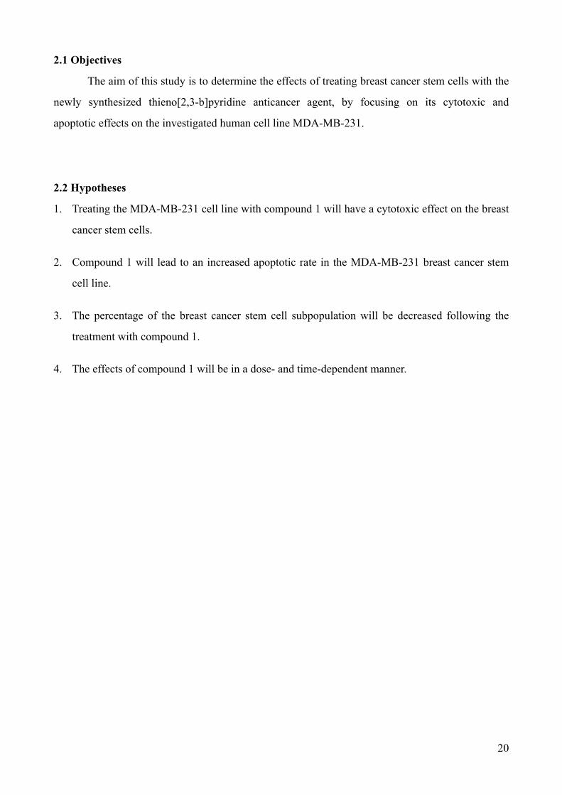
4.3 Compound I: decrease in number of BCSC
In the TNBC cell line MDA-MB-231, the marker combination CD44+/CD24- is known to be
numerously common in CSC as mentioned before. The treatment with compound 1 shows a
statistically significant decrease of the CD44+/CD24- subpopulation, with 89,86% in untreated
control and 55.54% after treatment with 2µM compound 1 for 48 hours (t=8.324 df=5 P <0.001).
Figure 16. CSCs (CD44+/CD24-) after drug treatment. Notes: Percentage of CSCs after treatment with 2µM compound 1 for 48 h in MDA-MB-231. Data represent the mean ± SD. Percentage of CSC is given in bar charts for treatment compared to the control. Columns, mean of cells; bars, SD; *P<0.001. Abbreviations: CSCs, cancer stem cells; SD, standard deviation.
"30
Contro
l
Compo
und 1
0
20
40
60
80
100
% C
SC
*

5. DISCUSSION

Acknowledging that targeting CSC is an auspicious strategy to defeat tumor recurrence and
immunity to chemotherapy, the focus of this study was to determine, whether the newly synthesized
putative phospholipase C (PLC) inhibitor, 3-amino-N-(3-chloro-2-methylphenyl)-5-oxo-5,6,7,8-
tetrahydrothieno[2,3-b]quinoline-2-carboxamide (compound 1; Figure 10), has cytotoxic properties
and is able to induce cell death and therefore a potentially new therapeutic approach to treat TNBC
(51). As it is ascertained that thieno[2,3-b]pyridine works against the phospholipase C-γ2 (PLC-γ2)
isoform, a novel therapeutic target for molecular anticancer treatments which is an efficient
anticancer agent against various cancer cell lines, we wanted to determine its potency against
TNBC (52).
Performing a colorimetric MTT assay to analyze the cell viability of TNBC cells treated
with different concentrations for various time intervals, we found that the newly synthesized
anticancer compound shows a dose- and time-dependent cytotoxicity for breast MDA-MB-231
cancer cells. The novel compound under investigation is already cytotoxic at the lowest
concentration tested after a prolonged period of 48 hours of treatment compared to the ten times
higher concentration which is already cytotoxic after only four hours, the maximal cytotoxicity was
achieved after treatment with maximal concentration at the longest time interval, but only for 47%
of cells (Figure 14). This shows how increasing the dose and time raises the cytotoxic effect of
compound 1 on MDA-MB-231. The performance of this experiment was done in triplicate to
exclude any errors., however, the MTT assay used in our investigation, does not take into account
whether the viable cells measured undergo active cell division or not. This means, we cannot
exclude cytostatic qualities of the novel compound, that it for example, induces G2/M phase cell
cycle arrest, and measured discrepancy is probably due to different analytical methods.
To determine the type of cell death, flow cytometric analysis was performed and we tested
the compound at a concentration of 2 µM after 48 hours. The results show a significant increase in
early apoptosis in MDA-MB-231 cancer cells in comparison to non-treated cells, meaning that the
majority of cells died by treatment-induced apoptosis (Figure 15). The same test was used to find
out whether or not the percentage of BCSC is also declining, showing a prominent decrease in the
CD44+/CD24- subpopulation, which represent the tumorigenic stem cell-like properties. The high
level of treatment-induced apoptosis and the significantly lowered BCSC subpopulation is so
relevant because SCs are responsible for extensive self-renewal, proliferation, quick cancer
progression, as well as metastasis disease in cancer patients, and up to this point no satisfying
"32
therapy exists for TNBC, so it seems promising for treating TNBC, especially the metastatic form
(19,20). However, in the context of this diploma thesis, we did not take into account the
glycosphingolipid expression on the surface of CSCs which are responsible for cancer relapse and
therapy resistance in TNBC, so we cannot say which receptors are associated and working together
with the new compound and mediating the treatment-induced apoptosis and therefore also lowering
the percentage of BCSCs (53).
In a similar study, another compound from the thieno[2,3-b]pyridine class was also used on
the MDA-MB-231 cell line, showing comparable results. For the MTT assay the same time
intervals were used but coupled with higher concentrations, 0.5 µM, 1 µM, 5 µM, 10 µM, and 25
µM of the thieno[2,3-b]pyridine compound. Anyways, it was also noticed that the compound
produces a dose- and time-dependent cytotoxicity with regular growth retardation for MDA-
MB-231 cells. Furthermore, it was also investigated whether the MTT results are caused by cell
death or cell cycle arrest. Determination of the type of cell death was done by flow cytometric
analysis with the same standards that were used in this study and it shows that compound-induced
cell death occurs also mostly by early apoptosis, but it destroys a higher percentage than the
compound tested in this study. Moreover, the decrease in BCSC was also observed but was not as
prominent as in this study. However, they were also able to associate increased levels of GM3
expression on MDA-MB-231 cells to a higher apoptotic rate and a lower percentage of BCSC
subpopulation.
Limiting factors to the study must be considered when interpreting and applying the
conclusion. However, no major limitations to this study are established as every experiment was
performed in triplicate. Albeit, one could test this novel compound on another TNBC cell line to
confirm its therapeutic effects and compare the results to this study, allowing better comparison of
the cytotoxic strength of this compound on TNBC stem cells.
This study conclusively confirms that the novel thieno[2,3-b]pyridine compound has a
cytotoxic effect on the MDA-MB-231 TNBC stem cell line in not only a time- but also a dose-
dependent manner. The most significant results obtained by this research concluded that the
treatment-induced cell death is mainly due to an increased apoptotic rate. Furthermore, compound 1
considerably decreases the CSC subpopulation percentage found in TNBC.
"33

Taking into account that several studies working with compounds of the thieno[2,3-
b]pyridine class described efficient anticancer qualities against several types of cancers, these
compounds targeting PLC-γ2 should be paid more attention to, especially for TNBC which has no
sufficient therapeutic regime yet (52,53). However, so far only in vitro trials on cancer cell lines
were done and for the future the compounds have to be tested for whether or not they are only
cytotoxic for cancerous cells or also for healthy tissue, to exclude any unwanted side effects.
"34

6. CONCLUSIONS

• According to the results we obtained in this study, we were able to demonstrate that the treatment
of human MDA-MB-231 TNBC with compound 1 works and has cytotoxic effects on the
investigated cell line.
• The results show that compound 1 leads to a higher level of apoptosis and a significantly lowered
percentage of the CSC subpopulation.
• Furthermore, to be more precise the results concluded that compound 1 affects TNBC in a dose-
and time-dependent cytotoxic manner.
• This study conclusively shows that if one considers the fact that TNBC’s characteristic property
is an increased percentage of BCSCs and knowing its connection to being at a higher risk for
metastatic disease and mortality, the thieno[2,3-b]pyridine class of compound 1 definitely
deserves further attention as a potentially new therapeutic approach for treating TNBC.
"36

7. REFERENCES
1. Alt-Epping B, Fuxius S, Wedding U. Onkologie Das Wichtigste für Ärzte aller Fachrichtungen.
1st ed. Urban & Fischer Verlag/Elsevier GmbH; 2017.
2. Hanahan D, Weinberg RA. Hallmarks of Cancer: The Next Generation. Cell. 2011;144:646-74.
3. Brenton JD, Carey LA, Ahmed AQ, Caldas C. Molecular classification and molecular
forecasting of breast cancer: ready for clinical application? J. Clin. Oncol. 2005;23:7350-60.
4. Foulkes WD, Smith IE, Reis-Filho JS. Triple-negative breast cancer. N Engl J Med.
2010;363:1938-48.
5. Koboldt DC, Fulton RS, McLellan MD, Schmidt H, Kalicki-Veizer J, McMichael JF et al.
Comprehensive molecular portraits of human breast tumors. Nature. 2012;490:61-70.
6. Bray F, Ferlay J, Soerjomataram I, Siegel RL, Torre LA, Jemal A. Global Cancer Statistics
2018: GLOBOCAN Estimates of Incidence and Mortality Worldwide for 36 Cancers in 185
Countries. CA Cancer J Clin. 2018;68:394-424.
7. Plasilova ML, Hayse B, Killelea BK, Horowitz NR, Chagpar AB, Lannin DR. Features of
triple-negative breast cancer: Analysis of 38,813 cases from the national cancer database.
Medicine (Baltimore). 2016;95:e4614.
8. Gangi A, Chung A, Mirocha J, Liou DZ, Leong T, Giuliano AE. Breast-conserving therapy for
triple-negative breast cancer. JAMA Surg 2014;149:252–8.
9. Shimelis H, LaDuca H, Hu C, Hart SN, Na J, Thomas A et al. Triple-Negative Breast Cancer
Risk Genes Identified by Multigene Hereditary Cancer Panel Testing. JNCI. 2018;110:855-62.
10. Breastcancer.org [Internet]. Triple-Negative Breast Cancer.; [updated 2019 March 13; cited
2019 April 27]. Available from: http://breastcancer.org/.
11. Chang CS, Kitamura E, Johnson J, Bollag R, Hawthorn L. Genomic analysis of racial
differences in triple negative breast cancer. Genomics. 2018. doc:10.1016/j.ygeno.2018.10.010
12. Doyle JM, Daling JR, White E, Brinton LA, Doody DR, Porter PL et al. Risk Factors for Triple-
Negative Breast Cancer in Women Under the Age of 45 Years. AACR. 2009;18:1157-66.
"38
13. Phipps AI, Chlebowski RT, Prentice R, McTiernan A, Wactawski-Wende J, Kuller LH, et al.
Reproductive history and oral contraceptive use in relation to risk of triple-negative breast
cancer. J Natl Cancer Inst. 2011;103:470-77.
14. Yang XR, Chang-Claude J, Goode EL, Couch FJ, Nevanlinna H, Milne RL et al. Associations
of breast cancer risk factors with tumor subtypes: a pooled analysis from the Breast Cancer
Association Consortium studies. J Natl Cancer Inst. 2011;103:250-63.
15. Millikan RC, Newman B, Tse CK, Moorman PG, Conway K, Dressler LG et al. Epidemiology
of basal-like breast cancer. Breast Cancer Res Treat. 2008;109:123-39.
16. Maiti B, Kundranda MN, Spiro TP, Daw HA. The association of metabolic syndrome with
triple-negative breast cancer. Breast Cancer Res Treat. 2010;121:479-83.
17. Pichard C, Plu-Bureau G, Neves-E Castro M, Gompel A. Insulin resistance, obesity and breast
cancer risk. Maturitas. 2008;60:19-30
18. Kabat GC, Kim M, Phipps AI, Li CI, Messina CR, Wactawski-Wende J et al. Smoking and
alcohol consumption in relation to risk of triple-negative breast cancer in a cohort of
postmenopausal women. Cancer Causes Control. 2011;22:775-83.
19. Bozorgi, A; Khazaei, M; Khazaei, MR. New findings on breast cancer stem cells. Journal of
Breast Cancer.2015;18:303-12.
20. Rangaswami, H; Bulbule, A; Kundu, GC. Osteopontin: role in cell signaling and cancer
progression. Trends in Cell Biology. 2006;16:79-87.
21. Al-Hajj M, Wicha MS, Benito-Hernandez A, Morrison SJ, Clarke MF. Prospective
identification of tumorigenic breast cancer cells. Proc Natl Acad Sci U S A. 2003;100:3983-8.
22. Anders CK, Ambrason V, Tan T, Dent R. The Evolution of Triple-Negative Breast Cancer:
From Biology to Novel Therapeutics. Am Soc Clin Oncol Educ Book. 2016;35:34-42.
23. Bickerstaff H. Gynaecology by Ten Teachers. United Kingdom: CRC Press; 2017. p. 330.
24. Kuchenbäcker KB, Hopper JL, Barnes DR, Phillips KA, Mooji TM, Roos-Bloom MJ et al.
Risks of Breast, Ovarian, and Contralateral Breast Cancer for BRCA1 and BRCA2 Mutation
Carriers. JAMA. 2017;317:2402-2416. "39

25. Zhao W, Seinfeld JB, Liang F, Cheng X, Maranon DG, Jian Ma C et al. BRCA1-BARD1
promotes RAD51-mediated homologous DNA pairing. Nature. 2017; 550:360-65.
26. Joi ML, Ora GK. Positive results: making the best decisions when you're at high risk for breast
or ovarian cancer. Amherst, N.Y.: Prometheus Books; 2010. p. 337-40.
27. Kumar P, Aggarwal R. An overview of triple-negative breast cancer. Arch Gynecol Obstet.
2016;293:247-69.
28. Dent R, Trudeau M, Pritchard KI, Hanna WM, Kahn HK, Sawka CA et al.Triple-negative breast
cancer: clinical features and patterns of recurrence. Clin Cancer Res. 2007;13:4429-34.
29. Gangi A, Mirocha J, Leong T, Giuliano AE.. Triple-negative breast cancer is not associated with
increased likelihood of nodal metastases. Ann Surg Oncol. 2014;21:4098-103.
30. Azoulay S, Laé M, Fréneaux P, Merle S, Al Ghuzlan A, Chnecker C et al. KIT is highly
expressed in adenoid cystic carcinoma of the breast, a basal-like carcinoma associated with a
favorable outcome. Mod Pathol. 2005;18:1623-31.
31. Aapro M, Wildiers H. Triple-negative breast cancer in the older population. Ann Oncol.
2012;23:vi52-5.
32. Dogan BE, Turnbull LW. Imaging of triple-negative breast cancer. Ann Oncol. 2012;23:vi23-9.
33. Gao B, Zhang H, Zhang SD, Cheng XY, Zheng SM, Sun JH et al. Mammographic and
clinicopathological features of triple-negative breast cancer. Br J Radiol. 2014 doi: 10.1259/bjr.
20130496.
34. Dogan BE, Gonzalez-Angulo AM, Gilcrease M, Dryden MJ, Yang WT.. Multimodality
imaging of triple receptor-negative tumors with mammography, ultrasound, and MRI. AJR Am
J Roentgenol. 2010;194:1160-6.
35. Uematsu T, Kasami M, Yuen S. Triple-negative breast cancer: correlation between MR imaging
and pathologic findings. Radiology. 2009;250:638-47.
36. Chen JH, Agrawal G, Feig B, Baek HM, Carpenter PM, Mehta RS et al. Triple-negative breast
cancer: MRI features in 29 patients. Ann Oncol. 2007;18:2042-3.
"40

37. Gigli S, Amabile MI, David E, De Luca A, Grippo C, Manganaro L et al.Morphological and
Semiquantitative Kinetic Analysis on Dynamic Contrast Enhanced MRI in Triple Negative
Breast Cancer Patients. Acad Radiol. 2018. doi:10.1016/j.acra.2018.06.014.
38. Li SP, Padhani AR, Taylor J, Beresford MJ, Ah-See ML, Stirling JJ et al. Vascular
characterisation of triple negative breast carcinomas using dynamic MRI. Eur Radiol.
2011;21:1364-73.
39. Groheux D, Giacchetti S, Moretti JL,Porcher R, Espié M, Lehmann-Che J et al. Correlation of
high 18F-FDG uptake to clinical, pathological, and biological prognostic factors in breast
cancer. Eur J Nucl Med Mol Imaging. 2010;38:426-35.
40. Lakhani SR, Ellis IO, Schnitt SJ, et al. World Health Organization Classification of Tumours of
the Breast. Lyon, France: IARC Press; 2012.
41. Baselga J, Norton L. Focus on breast cancer. Cancer Cell. 2002;1:319-22.
42. Tang P, Tse GM. Immunohistochemical Surrogates for Molecular Classification of Breast
Carcinoma: A 2015 Update. Arch Pathol Lab Med. 2016;140:806-14.
43. Eliyatkın N, Yalçın E, Zengel B, Aktaş S, Vardar E. Molecular Classification of Breast
Carcinoma: From Traditional, Old-Fashioned Way to A New Age, and A New Way. J Breast
Health. 2015;11:59-66.
44. Han Z, Wei B, Zheng Y, Yin Y, Li K, Li S. Breast Cancer Multi-classification from
Histopathological Images with Structured Deep Learning Model. Sci Rep. 2017;23;7:4172.
45. Geyer FC, Pareja F, Weigelt B, Rakha E, Ellis IO, Schnitt SJ et al. The Spectrum of Triple-
Negative Breast Disease High- and Low-Grade Lesions. Am J Pathol. 2017;187:2139-51.
46. Grubb W, Young R, Efird J, Jindal C, Biswas T. Local therapy for triple-negative breast cancer:
a comprehensive review. Future Oncol. 2017;13:1721-30.
47. Omarini C, Guaitoli G, Pipitone S, Moscetti L, Cortesi L, Cascinu S, Piacentini F. Neoadjuvant
treatments in triple-negative breast cancer patients: where we are now and where we are going.
Cancer Manag Res. 2018;10:91-103.
"41
48. Bianchini G, Balko JM, Mayer IA, Sanders ME, Gianni L. Triple-negative breast cancer:
challenges and opportunities of a heterogeneous disease. Nat Rev Clin Oncol. 2016;13:674-90.
49. Lgcstandards-atcc.org [Internet]. MDA-MB-231 (ATCC HTB-26).; [cited 2019 Jun 19].
Available from: https://www.lgcstandards-atcc.org/products/all/HTB-26.aspx/.
50. Kuete V, Karaosmanoğlu O, Sivas H. Chapter 10 - Anticancer Activities of African Medicinal
Spices and Vegetables. In: Medicinal Spices and Vegetables from Africa Therapeutic Potential
Against Metabolic, Inflammatory, Infectious and Systemic Diseases. Elsevier; 2017. p. 271-97.
51. Leung E, Hung JM, Barker D, Reynisson J. The effect of a thieno[2,3-b] pyridine PLC-
[gamma] inhibitor on the proliferation, morphology, migration and cell cycle of breast cancer
cells. Med Chem Comm. 2014;5:99-106.
52. Zafar A, Sari S, Leung E,3 Pilkington LI,1 van Rensburg M, Barker D et al. GPCR Modulation
of Thieno[2,3-b]pyridine Anti-Proliferative Agents. Molecules. 2017; 22:2254.
53. Mastelić A, Čikeš Čulić V, Režić Mužinić N, Vuica-Ross M, Barker D, Leg EY et al.
Glycophenotype of breast and prostate cancer stem cells treated with thieno[2,3-b]pyridine
anticancer compound. Drug Des Devel Ther. 2017;11:759-69.
"42

8. SUMMARY

Objectives: The purpose of this study is to determine the effects of treating breast cancer stem cells
with the newly synthesized thieno[2,3-b]pyridine anticancer agent, by focusing on its cytotoxic and
apoptotic effects on the investigated human cell line MDA-MB-231.
Methods: The MDA-MB-231 triple-negative breast cancer cell line was treated with a newly
developed thienopyridine anticancer compound (3-amino-N-(3-chloro-2-methylphenyl)-5-
oxo-5,6,7,8-tetrahydrothieno[2,3-b]quinoline-2-carboxamide, 1) to determine its cytotoxic effect on
triple negative breast cancer, the type of cell death it causes, and the cancer stem cell percentage
after treatment. The 3-(4,5-dimethylthiazolyl-2)-2,5-diphenyltetrazolium bromide (MTT) assay was
performed, to analyze the cellular metabolic activity and determine the cytotoxic effect. Flow
cytometric analysis was used in combination with Annexin-V-FITC and propidium iodide staining
to assess the type of cell death after 48h of treatment with compound 1 (2 µM). Furthermore, flow
cytometry also provided the percentage of CD44+/CD24- cancer stem cells after treatment.
Results: Compound 1 was cytotoxic for breast cancer cells in a dose- and time-dependent manner;
cell death occurs mainly by apoptosis. The percent of cancer stem cells decreased four times.
Conclusion: Due to its cytotoxic effect on the percentage of triple negative breast cancer stem cells
compound 1 can be a potential treatment for triple-negative breast cancer.
"44

9. CROATIAN SUMMARY
Citotoksični učinak i apoptoza matičnih stanica karcinoma dojke tretiranih
novosintetiziranim protutumorskim spojem tieno [2,3-b] piridinom
Ciljevi: Cilj ovog istraživanja je utvrditi učinke tretmana matičnih stanica karcinoma dojke
novosintetiziranim protutumorskim spojem tieno [2,3-b] piridinom, s naglaskom na njegove
citotoksične i apoptotske učinke na ispitivanu staničnu liniju MDA-MB-231.
Materijali i metode: Trostruko negativna stanična linija karcinoma dojke MDA-MB-231 tretirana
je novosintetiziranim tieno-piridinskim spojem (3-amino-N-(3-kloro-2-metilfenil)-5- okso-5,6,7,8-
tetrahidrotieno[2,3-b]kinolin-2-karboksamid, 1) kako bi se utvrdio citotoksični učinak, tip stanične
smrti i postotak matičnih stanica karcinoma nakon tretmana. Napravljen je MTT (3-(4,5-
dimetiltiazolil-2)-2,5-difeniltetrazolij bromid) test kako bi se analizirala stanična metabolička
aktivnost i utvrdio citotoksični učinak. Protočna citometrija u kombinaciji s bojanjem aneksin-V-
FITC-om i propidij jodidom, korištena je kako bi se utvrdio tip stanične smrti nakon 48h tretmana
spojem 1 (2 µM). Nadalje, protočnom citometrijom je također utvrđen postotak CD44+/CD24-
matičnih stanica karcinoma nakon tretmana.
Rezultati: Spoj 1 bio je citotoksičan za stanice karcinoma dojke te citotoksičnost korelira s
koncentracijom spoja i vremenom inkubacije. Stanična smrt je nastupila prvenstveno zbog
apoptoze. Postotak matičnih stanica karcinoma smanjio se četiri puta.
Zaključci: Zbog citotoksičnog učinka na postotak matičnih stanica trostruko negativnog karcinoma
dojke, spoj 1 može biti potencijalna terapija za trostruko negativni karcinom dojke.
"46

10. CURRICULUM VITAE

Personal Information:
Name and surname: Louisa Pauline Sofie Willmen
Telephone number: +49 173 5196856
E-mail address: [email protected]
Date of birth: 3rd March 1995
Nationality: German
Education:
2005 — 2013: Luise-von-Duesberg Gymnasium, Kempen, Germany
2010 — 2011: International American School of Cancún, Mexico
WS 2012/13:International Managemant, Akademie für Unternehmensmanagemant, Mohnheim, Germany
2013 — 2019: University of Split, School of Medicine, Medical Studies in English
Clinical traineeships:
August 2018: Krankenhaus Spittal/Drau GmbH: Internal medicine
September 2018: Krankenhaus Spittal/Drau GmbH: Anesthesiology and intensive care
January 2019: Krankenhaus Spittal/Drau GmbH: General surgery and trauma surgery
Other activities:
2010 — 2013:Privatpraxis G. Willmen, Kempen, Germany: Assistance at a general practitioners office
2011 — 2013: Tutoring of pupils: Maths, English, German, Latin, and Spanish
SS 2015:University of Split, School of Medicine, Medical Studies in English: Anatomy tutor
Skills:
Languages:Proficient in German, English, and Spanish Qualification in Latin Basics in Chinese and Croatian
Sport: Skiing and horse riding
"48

11. SUPPLEMENT

Figure 1. 1.2.2 Epidemiology and etiology. World maps illustrating the most common type of cancer (A) incidence and (B) mortality by country in 2018 among females. SOURCE: Globocan 2018, World Health Organization (6).
"50
A
B

Table 1. Frequency of PVs in African-American and Caucasian American.
Notes: 1.2.2 Epidemiology and etiology. Blue highlights the highest percentage of PVs found in both groups, red stands for mutations exclusively found in African-Americans, and green stands for PVs found only in Caucasian-Americans. Abbreviations: PV, pathologic variant; AA, African-American; CA, Caucasian-American.SOURCE: Chang et al (11).
"51

Table 2. Specific characteristic differences for TNBC compared to other types of breast cancers.
Notes: 1.2.4 Clinical features and prognostics. TNBC compared to nonTNBC except the rows with odds ratios, where the P value represents TNBC compared to the reference, HR+ Her2. Abbreviations: TNBC, triple-negative breast cancer; CI, confidence interval; HR, hormone receptor; Her2, human epidermal growth factor receptor; OR, odds ratio; SE,standard error. SOURCE: Plasilova et al (7).
"52
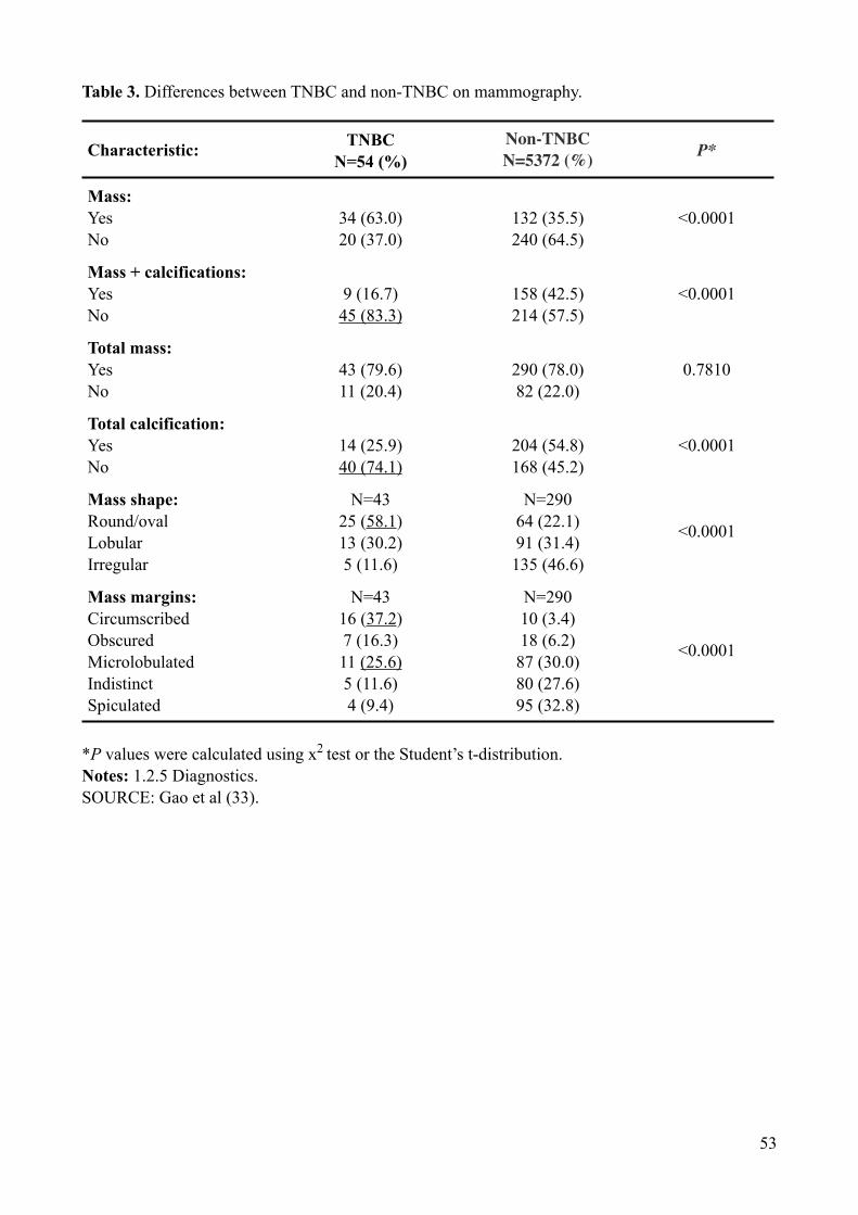
Table 3. Differences between TNBC and non-TNBC on mammography.
*P values were calculated using x2 test or the Student’s t-distribution. Notes: 1.2.5 Diagnostics. SOURCE: Gao et al (33).
Characteristic: TNBC N=54 (%)
Non-TNBC N=5372 (%) P*
Mass:YesNo
34 (63.0) 20 (37.0)
132 (35.5) 240 (64.5)
<0.0001
Mass + calcifications: YesNo
9 (16.7) 45 (83.3)
158 (42.5) 214 (57.5)
<0.0001
Total mass:YesNo
43 (79.6) 11 (20.4)
290 (78.0) 82 (22.0)
0.7810
Total calcification: YesNo
14 (25.9) 40 (74.1)
204 (54.8) 168 (45.2)
<0.0001
Mass shape:Round/ovalLobular Irregular
N=43 25 (58.1) 13 (30.2) 5 (11.6)
N=290 64 (22.1) 91 (31.4) 135 (46.6)
<0.0001
Mass margins:Circumscribed Obscured Microlobulated IndistinctSpiculated
N=43 16 (37.2) 7 (16.3) 11 (25.6) 5 (11.6) 4 (9.4)
N=290 10 (3.4) 18 (6.2) 87 (30.0) 80 (27.6) 95 (32.8)
<0.0001
"53

Figure 2. WHO Classification of tumours of the breast.
Notes: 1.2.6 Classification of breast cancer. SOURCE: Makhani et al (40)
"54

Table 4. MTT results.
Notes: 4.1 Compound I: dose- and time-dependent cytotoxicity.
SP 50 nM 250 nM 500 nM 1 µM 5 µM
After 4 hours 0,132 0,135 0,133 0,106 0,109 0,102
0,144 0,128 0,12 0,08 0,115 0,083
0,126 0,135 0,088 0,078 0,113 0,077
0,132 0,12 0,101 0,102 0,124 0,052
0,154 0,108 0,128 0,12 0,123 0,049
Average 0,1376 0,1252 0,114 0,0972 0,1168 0,0726
Mean 1 0,909883 0,828488 0,706395 0,848837 0,527616
SD 0,081835 0,083084 0,137794 0,130247 0,047210 0,161412
After 24 hours 0,152 0,079 0,116 0,083 0,107 0,108
0,139 0,089 0,126 0,124 0,074 0,128
0,131 0,145 0,126 0,109 0,119 0,112
0,131 0,148 0,142 0,118 0,107 0,111
0,122 0,138 0,137 0,111 0,118 0,095
Average 0,135 0,1198 0,1294 0,109 0,105 0,1108
Mean 1 0,887407 0,958518 0,807407 0,777777 0,820740
SD 0,083312 0,244971 0,075831 0,116298 0,135273 0,087237
After 48 hours 0,267 0,209 0,198 0,19 0,17 0,142
0,287 0,212 0,195 0,165 0,158 0,139
0,274 0,187 0,198 0,175 0,177 0,149
0,287 0,2013 0,209 0,172 0,184 0,141
0,29 0,226 0,208 0,178 0,169 0,12
Average 0,281 0,1198 0,2016 0,176 0,1716 0,1382
Mean 1 0,736868 0,717437 0,626334 0,610676 0,491814
SD 0,035498 0,051029 0,022870 0,032713 0,034558 0,038608
After 72 hours 0,232 0,187 0,139 0,149 0,144 0,114
0,225 0,161 0,138 0,133 0,141 0,12
0,221 0,157 0,158 0,15 0,13 0,115
0,216 0,167 0,156 0,162 0,135 0,132
0,237 0,169 0,153 0,152 0,146 0,131
0,242 0,15 0,154 0,146 0,158 0,122
Average 0,228833 0,165166 0,149666 0,148666 0,142333 0,122333
Mean 1 0,721777 0,654042 0,649672 0,621995 0,534595
SD 0,043297 0,055580 0,038561 0,041149 0,042338 0,033699
"55