the cytosolic protein response as a subcomponent … · the cytosolic protein response as a...
TRANSCRIPT

The Cytosolic Protein Response as a Subcomponent of theWider Heat Shock Response in Arabidopsis W
Akiko Sugio, Rene Dreos, Frederic Aparicio,1 and Andrew J. Maule2
John Innes Centre, Colney, Norwich NR4 7UH, United Kingdom
In common with a range of environmental and biological stresses, heat shock results in the accumulation of misfolded
proteins and a collection of downstream consequences for cellular homeostasis and growth. Within this complex array of
responses, the sensing of and responses to misfolded proteins in specific subcellular compartments involves specific
chaperones, transcriptional regulators, and expression profiles. Using biological (ectopic protein expression and virus
infection) and chemical triggers for misfolded protein accumulation, we have profiled the transcriptional features of the
response to misfolded protein accumulation in the cytosol (i.e., the cytoplasmic protein response [CPR]) and identified the
effects as a subcomponent of the wider effects induced by heat shock. The CPR in Arabidopsis thaliana is associated with
the heat shock promoter element and the involvement of specific heat shock factors (HSFs), notably HSFA2, which appears
to be regulated by alternative splicing and non-sense-mediated decay. Characterization of Arabidopsis HSFA2 knockout
and overexpression lines showed that HSFA2 is one of the regulatory components of the CPR.
INTRODUCTION
Protein homeostasis is central to normal cellular function. In
sessile plants, this is a major challenge since they are subjected
to a plethora of diverse biotic and abiotic challenges, many of
which impact upon protein stability. To deal with this, plants have
evolved a complex regulatory network of transcription factors
that interact with specific promoter elements to transcribe mul-
tiple families of heat shock proteins (HSPs) that, in turn, act as
chaperones for protein repair or targeted degradation. These are
not ubiquitous processes within the cell but are compartmental-
ized. Hence, the unfolded protein response (UPR), which is
induced by the accumulation of misfolded proteins in the endo-
plasmic reticulum (ER), recruits specific genes and pathways to
regulate protein repair in that compartment (Ron and Walter,
2007), and a parallel process, the cytosolic protein response
(CPR), operates in the cytosol (Aparicio et al., 2005). Although
these processes are essential for maintaining normal cellular
function under adverse conditions, how they are integrated into
the wider response has been little investigated in higher eukary-
otes. For example, heat shock is a pervasive stress with diverse
impacts on cellular organization and function. It results in the
denaturation of extant proteins, a block to further protein trans-
lation, destabilization of cellular membranes, and the activation
of oxidative stresses (Morimoto, 1998). It is widely held that the
flux of denatured proteins acts as a trigger for many of these
effects (Craig and Gross, 1991), but how the various functional
aspects relate to different subcellular compartments is not well
understood.
The eukaryotic heat shock response involves the induction of
HSPs and the action of specific subsets of their transcriptional
regulators, heat shock factors (HSFs) (Miller and Mittler, 2006;
Swindell et al., 2007). The main role of HSPs in prokaryotes,
plants, and animals relates to the protective repair of proteins,
although the association of some HSPs with ubiquitin-mediated
protein degradation (Qian et al., 2006) also points to a function
with respect to protein turnover. Protein homeostasis is main-
tained through an interaction between HSPs and HSFs, which is
disturbed when HSPs are recruited by misfolded proteins. The
release of HSFs allows them to trimerize to form the active
transcription factor complex required for induced expression of
HSPs. Transcription is activated following the binding of the HSF
complex to promoter upstream elements, notably the heat shock
element (HSE; a palindromic motif of nGAAn). Unlike budding
yeast (Saccharomyces cerevisiae) and fruitfly (Drosophila mela-
nogaster), in which a single HSF regulates the expression of heat
shock response genes (e.g., HSP70) following heat shock or
other stresses, in plants there are multiple HSFs; for example,
Arabidopsis thaliana encodes 21 HSF-like genes. These are
categorized into three classes by their structures. Class A HSF
proteins consist of a DNA binding domain, an oligomerization
domain, nuclear localization domains, and transcriptional acti-
vation domains (Nover et al., 2001). By contrast, classes B and C
lack activation domains. It is thought that this proliferation of
HSFs in plants is an adaptation to a sessile lifestyle coping with a
dynamic environment (Busch et al., 2005). In addition to the large
number of HSFs, further flexibility is provided by the use of both
homo- and heterotrimers in the formation of the transcription
factor complex (Nover et al., 2001; Bharti et al., 2004; Baniwal
et al., 2007).
1 Current address: Instituto de Biologıa Molecular y Celular de Plantas,Universidad Politecnica de Valencia, Avenida de los Naranjos s/n, 46022Valencia, Spain.2 Address correspondence to [email protected] author responsible for distribution of materials integral to thefindings presented in this article in accordance with the policy describedin the Instructions for Authors (www.plantcell.org) is: Andy J. Maule([email protected]).WOnline version contains Web-only data.www.plantcell.org/cgi/doi/10.1105/tpc.108.062596
The Plant Cell, Vol. 21: 642–654, February 2009, www.plantcell.org ã 2009 American Society of Plant Biologists

The UPR is triggered experimentally by treatment with the
antibiotic tunicamycin that blocks N-terminal glycosylation, in-
terfering with correct protein folding. Tunicamycin treatment
results in the recruitment of BiP (ER-located homologs of HSP70)
by misfolded proteins and the induction of larger suites of
corrective chaperones. Processes for the proteosome-mediated
degradation of terminally misfolded proteins are also activated
(Meusser et al., 2005). The CPR is induced by the accumulation
of unstable or misfolded proteins in the cytosol and is associated
with the induction of a specific subset of HSP70 genes. For
example, virus infection in the cytosol in plant or animal cells
triggers the induction of HSPs (Whitham et al., 2003), a process
that is exacerbated when the virus-encoded proteins are unsta-
ble (Jockusch et al., 2001; Jockusch and Wiegand, 2003). The
Arabidopsis genome encodes five soluble cytosolic HSP70s,
HSP70 (referred to as HSP70A in this article) and HSP70B, and
three constitutively expressed HSPs, HSC70-1, HSC70-2, and
HSC70-3. All of these genes are induced by heat shock treat-
ment, whereas all but HSP70B are induced in the CPR (Aparicio
et al., 2005).
We hypothesized that it should be possible to identify specific
responses to misfolded proteins within the overall heat shock
response and to use this information to dissect the transcrip-
tional features of the CPR. Using the proline analog L-azetidine-
2-carboxylic acid (AZC) to generate misfolded proteins (Trotter
et al., 2002) throughout the cell, we compared the transcript
profiles following heat shock treatment with profiles associated
with AZC (inducing the UPR and CPR) and tunicamycin treat-
ment (UPR alone). From our analyses, we identified the HSE as
being essential for the induction of the CPR and that the process
was partly regulated by HSFA2. In addition, we identified a novel
alternative splicing product of HSFA2 RNA with the potential to
modulate HSFA2 activity.
RESULTS
Transcript Profiling the CPR
Previously, the CPR has been induced by ectopic protein accu-
mulation following agroinfiltration-mediated transient expression
or by virus infection. To avoid the ancillary biological effects
associated with these treatments, we created misfolding of
nascent polypeptides in vivo by treating Arabidopsis leaves
with AZC. In parallel, leaves were treated with tunicamycin or
heat shock at 378C. The optimal timing for these treatments with
respect to known responses was assessed using quantitative
RT-PCR (qRT-PCR) for HSP70A, luminal binding protein 3,
(BiP3), and basic leucine zipper 60 (bZIP60) RNAs (see Supple-
mental Figure 1 online). HSP70A RNA, typically induced in the
CPR and in response to heat shock, showed maximal accumu-
lation after 1 and 3 h for heat shock and AZC treatments,
respectively. BiP3 RNA, typically increased in response to
misfolded proteins in the ER, showed maximum accumulation
3 h after treatment with tunicamycin and >6 h after treatment with
AZC. bZIP60, a key regulator of the UPR (Iwata and Koizumi,
2005), appeared to show an earlier but weaker response to
tunicamycin than BiP. Since we were primarily interested in the
contribution the CPR and its regulatory pathway made to the
overall heat shock response, 1 h (heat shock) and 3 h (AZC and
tunicamycin) were selected as treatment periods prior to the
transcript analysis using Arabidopsis ATH1 microarrays. Un-
treated, and proline or dimethyl formamide (tunicamycin solvent)
treated tissues were included as controls. Thus, we reasoned
that changes fromcontrol samples in commonbetween heat and
AZC but not present in the tunicamycin treatments would identify
a subclass of transcriptional responses that represented the
CPR as a subcomponent of the heat shock response. Three
biological replicates for each treatment were analyzed. As
tunicamycin-treated samples showed a relatively high degree
of variation between replicates, we performed hybridizations
with two further biological replicates for tunicamycin and control
treatments. The microarray data are summarized in Venn dia-
gram form in Figure 1A; detailed lists of transcripts showing
significant (P < 0.05 and $ twofold change) alteration are
included in Supplemental Data Set 1 online; P values were
adjusted using the Benjamini-Hochberg (false discovery)
method.
Heat shock, AZC, and tunicamycin treatments showed de-
creasing impact on transcript levels, assessed as the total
number of genes showing upregulation and downregulation in
each case (Figure 1A). Specific to the CPR (common area
between heat shock and AZC response minus tunicamycin
response), there were 153 upregulated genes and 90 down-
regulated genes. Intuitively, we speculated that positive re-
sponses to applied stresses might involve the recruitment of
new biological processes seen as transcriptional upregulation,
whereas downregulated genes might represent more the con-
sequences of the imposed stress. Focusing on the specific
overlap between heat shock and AZC (minus tunicamycin)
treatments, we found that the 153 upregulated genes fell into
several broad functional classes. Notably, for genes with func-
tional annotation, the classes included (1) a large set of chaper-
ones, including many HSPs (categorized as heat stress genes;
with respect to the CPR, these included inducible HSP70 genes
and excluded HSP70B); (2) a group of transcription and splicing
factors; and (3) a group of genes involved in protein degradation,
especially the ubiquitin-mediated degradation system (Figure
1B; see Supplemental Table 1 online). Of the remaining genes,
the majority had no known functional annotation.
To validate our strategy and data, we used three approaches.
First, we compared our microarray data with a published micro-
array data set of heat-treated Arabidopsis shoots using a cluster
analysis (see Supplemental Figure 2 online). Our data showed
that the heat shock and AZC-treated samples clustered with the
published responses at 1 and 3 h after treatment, while the
tunicamycin-treated samples tended to be closer to the earliest
times (0.25 and 0.5 h) of the heat shock response. This may
indicate that heat is sensed first within the ER, but more exper-
imentation will be needed to verify this. Second, we asked
whether genes known to be induced in the UPR (Urade, 2007)
appeared in the overlapping zone between heat shock, AZC, and
tunicamycin. Of the 13 upregulated genes, six were previously
reported as genes involved in the UPR. This included bZIP60.
Interestingly, the upregulated genes for the UPR excluded all of
the Arabidopsis HSFs, so identifying the heat shock–induced
Cytosolic Misfolded Protein Response 643

HSFs as part of the transcriptional activation response restricted
to the cytosol. Third, we selected 13 genes from the transcrip-
tion/splicing factor and protein degradation classes of genes
upregulated in response to heat shock and AZC and performed
qRT-PCR using three biological replicates (see Supplemental
Table 2 online). Our qRT-PCR data confirmed the microarray
data with most of the genes being significantly induced by both
heat shock and AZC treatment. For most of these genes, the
extent of change tended to be greater following heat shock
treatment.
A Canonical HSE Promoter Element Is Involved in the CPR
A key feature of the CPR is the selective induction of four of the
five HSP70 genes. These four genes have the canonical HSE
palindromic nGAAn motif in their upstream promoter regions,
whereas this is missing in the noninduced HSP70B, although all
these genes are induced by heat. Hence, we asked whether the
HSE was highly represented within the promoters of the heat
shock/AZC (minus tunicamycin) upregulated class of genes.
Bioinformatic analysis of sequences 1000 bp upstream of the
transcription start site of genes showed that the HSE was
significantly overrepresented in this class (15.03%), as opposed
to all genes in the heat-shock-only class (4.46%) or all genes in
the Arabidopsis genome (1.66%) (see Supplemental Table 3
online). Gene ontology analysis of the 15.03% of genes showed
that 38% of them encoded transcription factors.
To test the apparent relationship between the CPR and HSEs
more formally, we undertook a comparative functional analysis of
the promoter regions ofHSP70A andHSP70B. The promoters of
the two genes were examined using promoter:reporter con-
structs expressing b-glucuronidase (GUS). Previously, an;2 kb
upstream promoter region from each gene was fused to GUS
within an Agrobacterium tumefaciens binary vector and was
assayed following transient expression after infiltration of Agro-
bacterium carrying this plasmid into Nicotiana benthamiana
leaves. The GUS activity faithfully reported the response of these
promoters to heat, virus infection, and ectopic protein expres-
sion (Aparicio et al., 2005). (It has not been possible in our hands
to obtain quantitatively reproducible ectopic protein expression
by agroinfiltration into the leaves of Arabidopsis [Aparicio et al.,
2005].) We focused our attention on smaller promoter fragments
forHSP70A andHSP70B that similarly faithfully reported the heat
shock and misfolded protein response in the same transient
expression system; an ;340-bp promoter region upstream of
the start codon of HSP70A (proHSP70A337:GUS) and HSP70B
(proHSP70B340:GUS) showed the same degree and pattern of
responses to heat and protein accumulation, suggesting that the
regions contain the essential elements for the HSP70A and
HSP70B regulation. This minimal HSP70A promoter (337 bp)Figure 1. The Differentially Regulated Genes after Three Treatments.
(A) Venn diagram of the differentially regulated genes after three treat-
ments. The numbers of genes showing altered regulation following heat
shock (HS), AZC, or tunicamycin treatment in comparison to their
respective controls are shown in Venn diagrams. Numbers at the bottom
right indicate genes showing no significant (P$ 0.05) changes for any of
the treatments.
(B) Functional categorization of differentially regulated genes by CPR.
CPR upregulated or downregulated genes were grouped according to
their known or predicted function using the MapMan functional classi-
fication system (Thimm et al., 2004). The three main categories (stress,
protein, and RNA) belong to MapMan level 1, while the others belong to
level 3. Black bars represent upregulated genes, and white bars repre-
sent downregulated genes.
644 The Plant Cell

includes a canonical HSE (GAACGTTCTCGAA), while the mini-
mal HSP70B promoter (340 bp) includes only an imperfect HSE
(HSE*; GAACTcTCTTGtA; Figure 2A).When the canonical HSE in
the HSP70A promoter was mutated to tAACGcTCTCtAA (A
knockout), the induced response to either heat or protein over-
expression was lost (P value > 0.05) (Figures 2A and 2B).
However, the mutation did not completely abolish the response
of the promoter to heat or protein stress, indicating that there are
other sequences that contribute to the activation of the promoter.
The minimal HSP70B promoter (proHSP70B340:GUS) shows a
basal response to protein accumulation. Changing the imperfect
HSE* to tAACTcTCTTtTA (B knockout) showed that HSE* was
required for the heat shock response (Figure 2C) and showed no
change in the basal response to protein accumulation. Interest-
ingly, replacement of theHSP70BHSE*withHSP70AHSE (BtoA)
furnished the HSP70B promoter with the ability to respond to
protein accumulation and heat shock (see BtoA; Figure 2C).
Replacement of the canonical HSE in theHSP70A promoter with
HSE* reduced the response of the HSP70A promoter to both
heat and protein accumulation but did not completely abolish the
response to either stress (see AtoB; Figure 2B). The results show
that a complete HSE is required for the response to protein
accumulation. They also indicate that different factors are re-
quired for the complete heat or CPR responses and that specific
factors required for the latter cannot recognize the HSP70B
promoter due to its imperfect HSE*.
HSFA7, HSFA2, and Alternatively Spliced HSFA2 RNAs Are
Induced in the CPR
Our microarray data (see Supplemental Data Set 1 online)
showed that of 15 class A HSFs in Arabidopsis, only HSFA2
and HSFA7a were induced by the CPR (common to both AZC
and heat shock); class B HSFs (HSFB1, HSFB2a, and HSFB2b)
were also induced. In addition, HSFA3, HSFA1e, HSFA1d, and
HSFA7bwere induced by heat shock only. With the exception of
HSFA1e, these data confirm previous reports of heat shock
induction of HSFs in aerial tissues (Schramm et al., 2008).
To confirm the nature of the class A HSFs induced in the CPR,
we concentrated on the two CPR-induced HSFs and cloned and
sequenced the cDNAs for HSFA2 and A7a from heat shock and
AZC-treated tissues. HSFA7a cDNAs from both treatments and
HSFA2 cDNA from heat shock tissues revealed protein coding
sequences conforming to predictions based upon the presence
of a single intron within all HSFs (Nover et al., 2001). By contrast,
the HSFA2 cDNA from AZC-treated tissues revealed some
cloneswith a novel sequence (see Supplemental Figure 3 online),
generated by alternative splicing of the intron (Figure 3). This
novel splicing pattern introduced an additional 31 bp mini-exon
fromwithin the conserved intron in the DNA binding domain. The
mini-exon resulted from new 39 (tttag:a) and 59 (g:gttagt) splicesites (Figure 3). The additional mini-exon introduced an in-frame
stop codon that would terminate translation after the N-proximal
DNA binding domain.
To determine whether there was a temporal correlation in the
expression patterns for these HSFs and HSP70A, as a marker of
the CPR, heat shock– and AZC-treated Arabidopsis plants were
analyzed by qRT-PCR (Figure 4). Specific primer sets were used
to differentiate the AZC form of HSFA2 (HSFA2-II) from the
normally spliced HSFA2 RNA. Despite some quantitative varia-
bility in the response, the accumulations ofHSFA2,HSFA7a, and
HSP70Awere consistent acrossmultiple experiments. After heat
shock, HSFA7a had increased rapidly at 30 min then decreased
to the end of the experiment (3 h). Changes in HSFA2 and
HSFA2-II RNAs were delayed relative to HSFA7a, showing
maximum accumulation at 1 h after treatment (Figure 4), match-
ing the changes in HSP70A (see Supplemental Figure 1 online).
The pattern of induction indicated that the heat stress is sensed
most strongly during the first 60 min of treatment and that during
the subsequent period (up to 3 h of total treatment) the tissues
acclimate to survival at the higher temperature. Features of this
early response include a rapid and transient induction ofHSFA7a
and a subsequent differential accumulation of HSFA2 and
HSFA2-II RNA. The latter accumulated to a maximum of 15%
of the level seen for HSFA2 RNA. In AZC-treated tissues, HSF
RNAs reached a maximum in parallel with the peak of HSP70A
RNA accumulation at 3 h after treatment (cf. Figure 4 and
Supplemental Figure 1 online). Most notable in the comparisons
with heat shock treatment was that the relative proportions of
HSFA2 and HSFA2-II RNAs were reversed following AZC treat-
ment, with the level ofHSFA2RNA at only 17%ofHSFA2-IIRNA.
Extremely low levels of HSFA2 expression prevented an accu-
rate determination of the ratio for untreated tissues. Since AZC
activates the HSP response primarily through the induction of
misfolded proteins and HSP70A was not induced in response to
tunicamycin (UPR), it appeared that the presence of HSFA2,
HSFA7a, and accumulation of HSFA2-II identified the CPR.
HSFA2 Is Functionally Important for the CPR
Our transcriptional data identified the profile of HSFA2,HSFA7a,
and HSFA2-II RNA accumulation as a signature of the CPR.
Since HSFA2 and HSFA7a are Class A HSFs that have tran-
scription activation domains, theymight regulate the induction of
other CPR-associated genes (e.g., HSP70s and other chaper-
ones) that correct the accumulation of misfolded proteins. To
examine the regulatory effect of HSFA2 and HSFA7a during the
CPR, we have examined the induction of HSP70A in insertional
mutant lines. Using qRT-PCR to assay HSP70A expression, an
Arabidopsis insertional mutant knockout line (SALK_008979) for
HSFA2 (Alonso et al., 2003; Nishizawa et al., 2006) and a mutant
line for HSFA7a (SALK_080138; T-DNA insertion just upstream
of the AHA transcriptional activation domain) (Larkindale and
Vierling, 2008) were compared with wild-type Arabidopsis
Columbia-0 (Col-0) after treatment with AZC. In repeated exper-
iments, the analysis showed a strong trend (P value=0.057)
toward weaker induction of HSP70A following AZC treatment of
the hsfA2mutant linewhen comparedwithwild-typeArabidopsis
Col-0 (Figure 5A); the reduction of HSP70A induction in the
hsfA2 mutant was statistically significant (P < 0.05) at 6 h after
AZC infiltration (see Supplemental Figure 4 online). By contrast,
HSP70A expression in the hsfA7a mutant was unchanged (Fig-
ure 5B).
The accumulation of HSFA2-II RNA is also a feature of the
CPR. The location of a translational stop codon that eliminated
essential functions downstream of the N-proximal DNA binding
Cytosolic Misfolded Protein Response 645

domain made it unlikely that HSFA2-II RNA could be involved in
activating HSP70A expression. To confirm this, a complemen-
tary experiment inN. benthamianawas performedwhereHSFA2,
HSFA2-II, or an artificial constructHSFA2DADwith a termination
codon before the C-terminal activation domain, were expressed
from the cauliflower mosaic virus (CaMV) 35S promoter. Activa-
tion of the proHSP70A337:GUS reporter, as ameasure ofHSP70A
induction, was tested after cotransient expression with each of
the HSF constructs following agroinfiltration into leaf tissues.
Again, HSFA2 expression showed significantly (P value < 0.05)
increasedHSP70A induction to a level close to that seen for heat
shock itself, while expression of genes for the truncated HSFA2
proteins showed no change (Figure 5C).
HSFA2 is induced by heat shock and a diverse range of other
stresses (Miller and Mittler, 2006), and HSFA2-overexpressing
lines can confer improved tolerance to high temperature and light
conditions (Nishizawa et al., 2006). Hence, we also tested
whether transgenic lines ectopically expressing HSFA2 con-
ferred protection against the damaging effects of AZC treatment
bymeasuring growth in the presence of sublethal concentrations
of AZC (Figure 5E). While the HSFA2 knockout line showed less
tolerance to AZC, three independent homozygous HSFA2 trans-
genic lines with varying levels of transgene expression (Figure
5D) showed higher tolerance (Figure 5E). HSFA7a knockout and
overexpression lines showed variable and mostly insignificant
changes in tolerance to AZC (see Supplemental Figure 5 online).
HSFA2-II RNA Is Not Active and Is Degraded by
Non-Sense-Mediated Decay
HSFA2-II is specifically induced in but does not itself induce the
CPR (Figures 4 and 5C). The introduction of the premature
termination codon downstream of the N-proximal activation
domain would probably result in a nonfunctional protein. This
organization of HSFA2-II RNA has features in common with
transcripts processed via the non-sense-mediated decay (NMD)
pathway. NMD is a conserved RNA quality control system in
Figure 2. Response of HSP70A and HSP70B Promoters and Their
Variants to Heat Shock and Protein Stress.
(A) Promoter:reporter constructs and HSE mutants. Promoter:reporter
Agrobacterium binary constructs were made for HSP70A (A337) and
HSP70B (B340) using 337 and 340 bp, respectively, of the upstream
genomic DNA (gray arrow) fused to GUS and the CaMV 19S terminator
(term). Promoter variants were created by site-directed mutagenesis of
the HSE in the HSP70A promoter and the HSE-like (HSE*) sequence of
the HSP70B promoter. The original sequences are shown at the top. The
consensus sequence of HSEs is shown in bold, and the relative positions
are underlined. The mutated nucleotides are shown in lowercase.
(B) Promoter responses in N. benthamiana measured as GUS activity
following Agrobacterium infiltration of the constructs. Promoter con-
structs were coinfiltrated with pB3, which overexpresses a cytosolically
targeted Arabidopsis protein (At5g08290) to induce the CPR or with an
empty vector (pG; nonstressed) as a negative control. Infiltrated tissues
were also heat shocked (HS) as a positive control for HSP70A and
HSP70B induction. The responses of the HSP70A HSE knockout (AKO)
mutation or the HSP70A promoter mutant that substituted HSE with HSE*
(AtoB) to the three treatments in comparison with the wild-type minimal
promoter for HSP70A (A337) are shown. Assays were performed in
triplicate on individual extracts of two infiltrated leaves. Bars indicate6SD.
(C) Similar experiments to those in (B) but showing the responses to the
three treatments of the HSE* knockout mutation in theHSP70B promoter
(BKO) or the substitution of the HSE* with HSE in the HSP70B promoter
(BtoA) in comparison with the HSP70B (B340) promoter. Responses of
the A337 promoter to protein expression (pB3) or heat shock (HS) were
included (left panel) as positive controls in the same experiment. Assays
were performed in triplicate on individual extracts of two infiltrated
leaves. Bars indicate 6SD. Statistical comparisons were made with the
coinfiltrated empty plasmid (pG) in each case. P values < 0.05 are
indicated with asterisks.
646 The Plant Cell

eukaryotes that degrades mRNAs with a premature termination
codon. In plants, mRNAs that have an extended noncoding
distance between the premature termination codon and 39 end of
the mRNA, or which have a downstream splice junction, are
recognized by the NMD system and targeted for degradation
(Kertesz et al., 2006; Hori and Watanabe, 2007; Kerenyi et al.,
2008). To test whether HSFA2-II is subject to NMD, a T-DNA
insertion mutant in the NMD machinery (upf1-5; Arciga-Reyes
et al., 2006) was comparedwithwild-type plants for the induction
ofHSFA2 andHSFA2-IIRNAs following heat or AZC treatment. If
HSFA2-II was a substrate for NMD, the ratio of HSFA2-II to
HSFA2 RNAs would be higher in the upf1-5 mutant. Following
heat treatment for 1 h or AZC treatment for 3 h, RNA levels were
analyzed by qRT-PCR. In both situations, the ratios of HSFA2-II
to HSFA2 RNAs were higher in the NMD mutant line (Figure 6;
Supplemental Figure 6 online shows the quantitative transcript
values).
The reversed ratio in the accumulation ofHSFA2-II andHSFA2
RNAs after heat shock and AZC could point to differential
regulation of HSFA2 after the two treatments or a stimulation or
suppression of NMD after heat shock or AZC treatment, respec-
tively. To test the latter hypothesis, we examined the effect of
heat shock or AZC treatment on the accumulation of two specific
RNAs (transcripts from pseudogenes At5g40920 and
At1g01060) shown to be regulated by the NMD (Arciga-Reyes
et al., 2006). In neither case was the accumulation significantly
affected by the treatments when compared with controls (see
Supplemental Figure 7 online).
Biological Induction of the CPR Also Induces HSFA2,
HSFA2-II, and HSFA7a
AZC is pervasive in its effects and generates terminally misfolded
proteins throughout the cell. Consequences of AZC action are
Figure 3. HSFA2 Is Alternatively Spliced upon AZC Treatment.
The structural organizations of the two forms of the mature HSFA2
transcript are shown. In the HSFA2 mRNA, the single 324-nucleotide
intron is removed. By contrast, the HSFA2-II RNA has an additional mini-
exon (uppercase bold) included through the recognition of additional
splice sites (lowercase) within the intron. The new stop codon is under-
lined. DBD, DNA binding domain; HR-A/B, oligomerization domain;
NLS, nuclear localization signal; AHA, activation domain, notation taken
from (Nover et al., 2001). Inverted triangle marks the position of the
T-DNA insertion in the Arabidopsis insertional knockout (SALK_008978)
line.
Figure 4. Time Course of HSF Induction during Heat and AZC Treat-
ment.
(A) Expression level of selected HSFs, relative to EF1a, were examined
by qRT-PCR. Following AZC infiltration, leaf samples (triplicate) were
harvested after the indicated incubation times. Bars inicate 6SD.
(B) Similar experiment to that in (A), but samples (triplicate) were
harvested after heat shock treatment. Bars indicate 6SD.
Cytosolic Misfolded Protein Response 647

the induction of a large number of genes, amongwhich are genes
associated with transcription and transcript splicing (Figure 1).
Accordingly, HSFA2, HSFA7a, and HSFA2-II were identified
following AZC treatment. Although our analyses relate these
RNAs to the CPR, it remains possible that pleiotropic effects
associated with AZC have led to the induction of theseHSFs and
especially to the formation of HSFA2-II RNA. Induction of the
CPR, characterized by the differential induction ofHSP70 genes,
occurs in cells actively involved in virus genome replication and
virus gene expression. To test whether the appearance of
HSFA2-II RNA correlated with the CPR rather than being a
secondary consequence of AZC incorporation, we assayed HSF
expression pattern in virus-infected Arabidopsis tissues. Leaves
were infected with Turnip mosaic virus (TuMV; genus Potyvirus)
or Turnip crinkle virus (TCV; genus Carmovirus), and the newly
emerged systemically infected leaves were harvested 7 d after
inoculation. The samples were first subjected to RT-PCR to
confirm the virus infection and then subjected to qRT-PCR to
quantify the expression level of HSFs. The CPR response asso-
ciated with virus infection is highly localized, being focused on
cells actively involved in virus multiplication and virus and virus
genome expression (Aranda et al., 1996). As such, the level of
induction of HSFs was less than observed in response to AZC or
heat shock. Nevertheless, as shown in Figure 7, virus-infected
Arabidopsis leaves showed strong accumulation of HSFA2-II,
variable induction of HSFA2, and slight induction of HSFA7a but
no significant induction of HSP70B or HSFA3; in our microarray
experiments, HSFA3 was also not induced in the CPR (using
AZC), although it was induced by heat (see Supplemental Data
Figure 5. Functional Roles of HSFA2 and HSFA2-II RNAs.
(A) Expression analysis (qRT-PCR) of HSP70A in the insertional null (KO)
mutant for HSFA2 (Nishizawa et al., 2006) and a wild-type (Col-0) control
plant 3 h after treatment with AZC. The data, expressed as values relative
to the expression of EF1a, are the mean (6SD) of three biological and
three technical replicates. HSP70A induction in the HSFA2 mutant was
;50% of that in the control, significant at P value = 0.057. Significant
differences were observed at later times (6 h after infiltration with AZC;
see Supplemental Figure 4 online).
(B) Similar experiment to that in (A) but for the HSFA7a insertional
mutant. The data, expressed as values relative to the expression of
EF1a, are the mean (6SD) of three biological and three technical
replicates. In this case, there was no significant difference from the
control plant.
(C) The ability of different HSFs to induce the HSP70A 337-bp promoter
was tested. N. benthamiana leaves were infiltrated with mixed cultures of
Agrobacterium carrying proHSP70A337:GUS and a CaMV pro35S:HSF
construct. Induction of each promoter was measured as GUS activity;
data are the mean of three biological replicates; bars indicate 6SD;
asterisks show significant (P value < 0.05) differences from the empty
vector treated control. HS, noninfiltrated heat shock control; HSFA2DAD,
HSFA2 truncated before the AD. The experiment was repeated twice
with similar results.
(D) RNA gel blot detection (top panel) of HSFA2 RNA in the transgenic
Arabidopsis lines (2-1, 4-3, and 8-2) overexpressing pro35S:HSFA2.
Bottom panel shows rRNA staining used as a loading control.
(E) The growth of Arabidopsis lines (Col-0 wild type, HSFA2 knockout
mutant, and three HSFA2 overexpression lines) in media with the
indicated concentration of AZC. The percentage of growth was calcu-
lated by comparison to the growth of the lines cultured without AZC. The
data show the average of three sets of plants; bars indicate 6SD. The
lines showing significant change in growth rate with each concentration
of AZC compared with Col-0 (Student’s t test; P value < 0.05) are
indicated by asterisks. Two asterisks show significant growth reduction,
and one asterisk shows significant growth increase.
648 The Plant Cell
Set 1 online). The samegene induction patternswere observed in
both TuMV- and TCV-infected tissues. The relatively high ratio of
HSFA2-II toHSFA2 RNAs by virus infection mimics the response
to AZC and confirms that alternative splicing of HSFA2 is an
integral part of the CPR.
DISCUSSION
Eukaryotes have evolved complex defense pathways to cope
with biotic and abiotic stresses, and in many cases these stress
responses overlap. This is particularly true for responses involv-
ing genes for HSPs and their HSF transcriptional regulators
where specific subsets of gene classesmay be involved uniquely
or collectively in responding to biotic and abiotic stresses (Miller
and Mittler, 2006; Swindell et al., 2007). A good example is the
range of complex consequences and responses associated with
temperature stress, which impacts plant cells and tissues inde-
pendent of spatial organization. A primary consequence of heat
stress is the accumulation of misfolded proteins, which trigger
responses, notably the CPR and UPR, located within discrete
subcellular compartments. The aim of this work was to identify
the separate contribution of the CPR to the overall heat stress
response and to identify key transcriptional features of the CPR.
To aid the dissection of the CPR, we employed the proline
analog AZC. Incorporation of AZC leads to terminally misfolded
proteins in both the soluble and ER compartments of the cytosol
but is distinct fromUPR induction by tunicamycin in that the latter
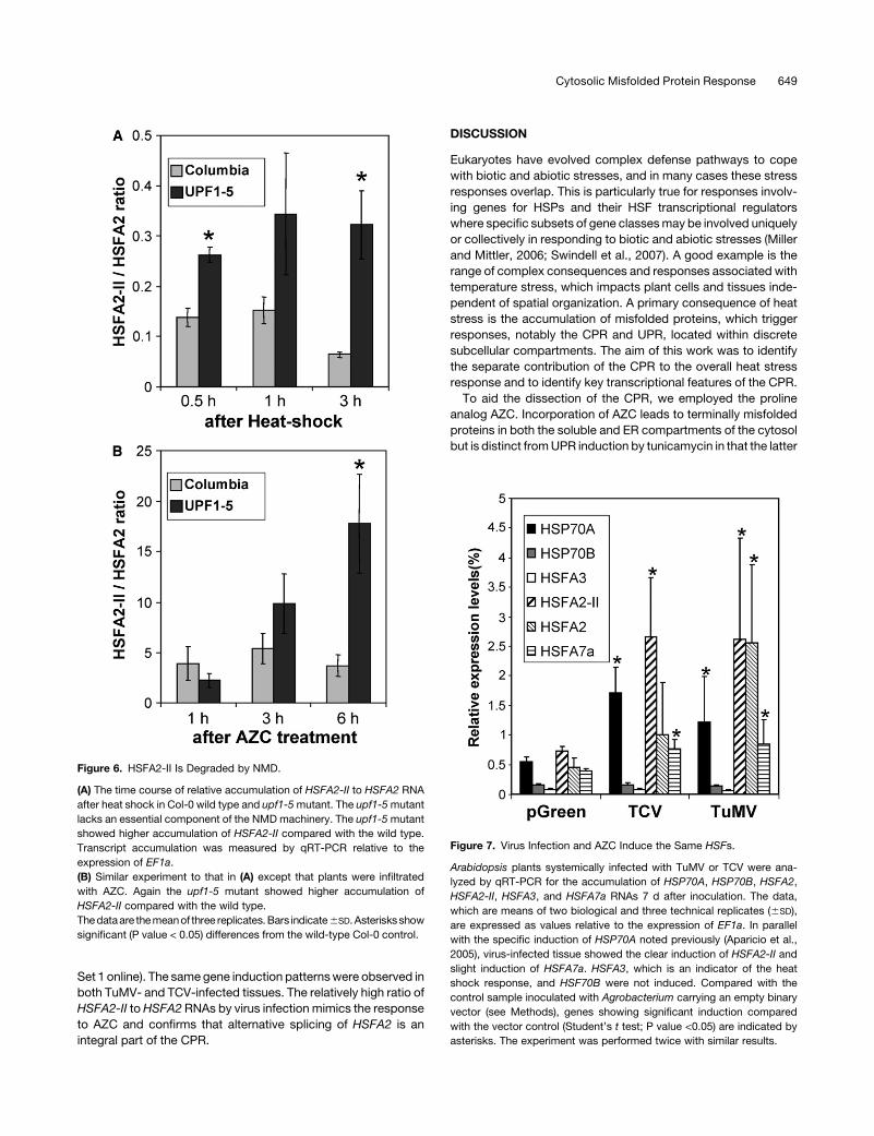
Figure 6. HSFA2-II Is Degraded by NMD.
(A) The time course of relative accumulation of HSFA2-II to HSFA2 RNA
after heat shock in Col-0 wild type and upf1-5mutant. The upf1-5mutant
lacks an essential component of the NMDmachinery. The upf1-5mutant
showed higher accumulation of HSFA2-II compared with the wild type.
Transcript accumulation was measured by qRT-PCR relative to the
expression of EF1a.
(B) Similar experiment to that in (A) except that plants were infiltrated
with AZC. Again the upf1-5 mutant showed higher accumulation of
HSFA2-II compared with the wild type.
Thedataare themeanof three replicates.Bars indicate6SD.Asterisksshow
significant (P value < 0.05) differences from the wild-type Col-0 control.
Figure 7. Virus Infection and AZC Induce the Same HSFs.
Arabidopsis plants systemically infected with TuMV or TCV were ana-
lyzed by qRT-PCR for the accumulation of HSP70A, HSP70B, HSFA2,
HSFA2-II, HSFA3, and HSFA7a RNAs 7 d after inoculation. The data,
which are means of two biological and three technical replicates (6SD),
are expressed as values relative to the expression of EF1a. In parallel
with the specific induction of HSP70A noted previously (Aparicio et al.,
2005), virus-infected tissue showed the clear induction of HSFA2-II and
slight induction of HSFA7a. HSFA3, which is an indicator of the heat
shock response, and HSF70B were not induced. Compared with the
control sample inoculated with Agrobacterium carrying an empty binary
vector (see Methods), genes showing significant induction compared
with the vector control (Student’s t test; P value <0.05) are indicated by
asterisks. The experiment was performed twice with similar results.
Cytosolic Misfolded Protein Response 649

fails to induce genes for the cytosolic HSP70s or HSFs. There-
fore, unlike AZC, the effect of tunicamycin is spatially restricted.
Therefore, comparison of the effects of AZC and tunicamycin
treatment allowed us to identify factors and processes that were
attributable to the CPR alone.
CPR as a Subcomponent of the Heat Shock Response
By integrating the transcript profiling data from heat shock (CPR +
UPR + pleiotropic effects), AZC treatment (CPR + UPR + other
pleiotropic effects), and tunicamycin treatment (UPR + pleiotro-
pic effects), we were able to identify groups of genes that were
upregulated or downregulated in association with the CPRwithin
the heat shock response (Figure 1; see Supplemental Data Set
1 online). Existing data for heat shock–treated Arabidopsis
(AtGenExpress, http://www.Arabidopsis.org/info/expression)
(Schramm et al., 2008) are based upon different experimental
approaches from ours. Nevertheless, there was a good corre-
spondence between our data set and the public data sets. For
the UPR, we identified fewer changes in expression than previ-
ously reported (Martinez and Chrispeels, 2003). However, both
the experimental approaches and technical array platform dif-
fered. Nevertheless, the appearance of bZIP60 and five other
known UPR genes, among the 13 genes overlapping between
heat shock, AZC, and tunicamycin treatment, gave us confi-
dence that the microarray analysis provided an accurate picture
of changes associated with the UPR.
Analysis of the correspondence between the treatments iden-
tified 153 upregulated genes associated with the CPR within the
heat shock response (see Supplemental Table 1 online). Appro-
priately, this list included CPR-induced HSP70 genes but not
HSP70B. It also included genes for chaperones, HSFs, and
components of the splicingmachinery, together with a number of
unannotated genes (27 expressed or hypothetical proteins). The
list provides a key resource for the further dissection of the CPR
in relation to other compartmentalized stress responses (e.g.,
UPR) or responses to more general stress conditions (e.g., heat
shock).
CPR Is Partly Regulated through Canonical HSE
A large number of genes that respond to heat shock contain the
canonical palindromic motif of nGAAn in their upstream regula-
tory domains, but the preponderance of the motif in the CPR
class of genes identified in the microarray analysis (see Supple-
mental Table 3 online) implies that this regulatory sequence is
more related to the sensing and transduction of responses to
misfolded protein than it is to other aspects of the broader heat
shock response. Of the 15% of CPR upregulated genes that
contain an HSE in the promoter region, ;40% encode tran-
scription factors. Among these transcription factors were several
HSFs, including HSFA2 and HSFA7a. We would predict that
someof the other induced factorswould in turn activate their own
downstream targets, some of which could be in the 85% of
induced genes that lack an HSE. This remains to be tested.
The induction of HSP70A, but not HSP70B, in response to
ectopic protein accumulation provided the opportunity to ex-
perimentally analyze the transcriptional elements required for the
CPR. Mutagenesis showed that the HSE within the HSP70A
minimal promoter was necessary and that it could confer protein
responsiveness upon HSP70B (Figure 2). Since HSP70B is also
heat inducible, it raises further the question as to whether the
canonical HSE is primarily a heat- or a protein-responsive
element.
CPR Induces a Unique Spectrum of HSFs
Arabidopsis deals with the impact of imposed stresses through
the recruitment of different combinations of HSFs. Of 15 Class A
HSF genes in our microarray analysis, few showed significant
induction in response to any of the stresses applied. Only
HSFA2, A3, and A7a were significantly induced in response to
heat shock, while only HSFA2 and HSFA7a were induced in
response to AZC, and none were induced in response to
tunicamycin (UPR). Intriguingly, in contrast with the situation
following AZC treatment, HSF expression changes in response
to heat shock appeared to be phased: HSFA7a showed the
earlier response andHSFA2/A2-II a later response (Figure 4). The
role of HSFA7a in the heat shock response is reported to be
involved in heat acclimation (Larkindale and Vierling, 2008).
Unlike HSFA7a, HSFA2 induction was strongly correlated with
the induction of HSP70A. However, considering AZC was used
as a specific activator of the CPR, it might be seen as surprising
that HSFA2 was more strongly induced by heat than by AZC
(Figure 4). This may relate to the misfolding of extant proteins by
heat shock but, for AZC, only the misfolding of nascent poly-
peptides during translation. Accordingly, the HSFA2/HSFA2-II
ratio, which potentially affects the amount of available active
HSFA2, was high in heat shocked samples and low in AZC-
treated samples. Crucially, however, the same profile of HSF
induction (i.e., induction of HSFA7a, HSFA2, and HSFA2-II and
no induction of HSFA3) was observed following biological acti-
vation of the CPR by virus infection (Figure 7).
Potential Regulation of the CPR by Alternative Splicing
of HSFA2
Overall, HSFA2 is the most widely induced HSF in response to
environmental stresses, indicating its central role in cell homeo-
stasis (Miller and Mittler, 2006). hsfA2 knockout plants show
reduced tolerance to heat shock treatment (Schramm et al.,
2006) and, in our experiments, reduced responsiveness to
misfolded protein in the cytosol as measured by the induction
of the HSP70A promoter. Although we experienced a large
variation between biological replicates, probably due to the
pressure infiltration of AZC, we observed the reduction of
HSP70A in the hsfA2 knockout plant (Figure 5). In turn, our
HSFA2-overexpressing lines showed increased tolerance to the
toxic effects of AZC, an effect not seen in the HSFA2-II–over-
expressing lines. Many of the genes induced in HSFA2-over-
expressing plants (Nishizawa et al., 2006) are also represented in
the category of genes overexpressed in response to heat shock
and AZC in our microarray experiment. These functions of
HSFA2 have a biological impact as shown by the altered growth
characteristics of the knockout and overexpressing lines in the
presence of AZC (Figure 5). Therefore, we could conclude that
650 The Plant Cell
HSFA2 partially regulates the CPR. What then is the purpose of
the alternatively spliced HSFA2-II RNA, apparently induced
uniquely as part of the CPR?
The novel mini-exon within HSFA2-II introduces a stop codon
toward the end of the DNA binding domain, hence excluding a
complete DNA binding domain, the nuclear localization signal,
and the oligomerization and activation domains, and likely re-
moving any functionality from the encoded protein (Figure 2).
However, the spliced RNA has features of aberrant mRNAs that
make it a substrate for NMD and, accordingly, HSFA2-II RNA
accumulated to a higher level in the NMD upf-1mutant (Figure 6).
The specific NMD-mediated degradation of HSFA2-II RNA was
confirmed from our experiments that showed that the NMD
pathway was not altered generally by heat shock or AZC treat-
ments (see Supplemental Figure 7 online). Therefore, we favor
the hypothesis that alternate splicing of HSFA2 RNA coupled
with NMD represents a mechanism for posttranscriptional reg-
ulation for the production of active HSFA2 protein. Accordingly,
our transcript profile analysis showed that the CPR induced a
number of genes encoding splicing factors (Figure 1). Since
HSFA2 is induced in response to many environmental stresses,
this posttranscriptional regulation offers a subtle second level of
control. Why then should heat show a lower ratio of HSFA2-II to
HSFA2 RNA? Heat denatures proteins throughout the cell, while
only newly synthesized proteins are misfolded in AZC-treated
cells. Thus, our interpretation is that heat-shocked cells induced
and required larger amounts of active HSFA2, while AZC- treated
cells had a lesser demand for HSFA2 and therefore showed
higher levels of alternative splicing to give HSFA2II RNA.
In parallel, we also examined the importance of HSFA7a
function in CPR. We did not detect a reduction in HSP70A
expression in the hsfA7a knockout line, and HSFA7a overex-
pression lines did not show increase of tolerance to AZC (see
Supplemental Figure 5 online). However, we cannot completely
exclude some involvement of HSFA7a in the regulation of the
CPR. The induction of HSFA7a in the CPR and the fact that the
hsfA2 knockout line did not eliminate HSP70A reporter expres-
sion suggests that HSFA7amay play some role. It is possible that
HSFA7a plays a contributory role (with HSFA2) in the CPR.
HSF RNA Alternative Splicing
Alternative splicing of Arabidopsis HSF RNAs has been recorded
elsewhere as EST sequences (http://www.plantgdb.org/ASIP/
EnterDB.php) although none of these events has been associ-
ated with functions. Many ESTs reveal intron retention (e.g.,
HSFA3,HSFA7b,HSFB2a, andHSFC1) or 39 untranslated region
(e.g., HSFA1b, HSFA1e, and HSFB1) alternative splicing events.
HSFA1dRNA has a similar alternative splicing pattern toHSFA2-
II. The alternative splicing of HSFA1d (mRNA from hormone-
treated callus) has the same donor site in the DNA binding
domain region as normal splicing but introduces a new exon and
intron and a new acceptor site, resulting in multiple new stop
codons. Medicago sativa HSF1 RNA is also alternatively spliced
within the conserved intron in the DNA binding domain coding
region. This introduces one or two small exons, including pre-
mature stop codons. The expression levels of these transcripts
were low and were thought to be degraded by NMD (He et al.,
2007). The features in common with HSFA2-II suggest that
alternative splicing of the HSF intron may be a conserved
mechanism in HSF regulation. The combination of alternative
splicing and NMD seems to be a broadly conserved regulatory
system in eukaryotes (Jaillon et al., 2008). In plants,;20%of the
expressed genes are alternatively spliced, and 36% (rice) to 43%
(Arabidopsis) of alternatively spliced RNAs are predicted to be
degraded by NMD (Wang and Brendel, 2006). Alternative splic-
ing (HSFA2-II) is a notable feature of the response to heat shock
(Figure 4) and correlates with the upregulation of a number of
splicing-related genes as common responses to both heat shock
and AZC treatment in our microarray analysis.
CPR for Protein Homeostasis in Eukaryotes
Protein misfolding in the soluble cytosol occurs as a conse-
quence of a range of induced stresses. These can be environ-
mental, chemical, or a consequence of ectopic protein
expression associated, for example, with virus infection. The
parallels between our data and related observations in yeast
(Trotter et al., 2002) point to the CPR as being a fundamental
process in wider eukaryotes designed to maintain protein ho-
meostasis in the cytosol. In both cases, the CPR is shown to be
distinct from the UPR in the use of HSF- and HSE-mediated
induction of HSP70 genes. Although heat shock has some
impact upon the ER, the spatial and functional separation of
the CPR and UPR provides additional precision in cellular reg-
ulation especially when protein misfolding in the ER is commonly
associated with posttranslational modification and maturation.
METHODS
Plants, Bacteria, and Viruses
Arabidopsis thalianaCol-0 andNicotiana benthamiana plants were grown
in a growth cabinet at 208C with a photoperiod of 8 h light/16 h dark.
All the intermediate DNA constructsweremaintained inEscherichia coli
DH5a cells. Binary plasmids were transformed into Agrobacterium
tumefaciens strain C58C1 with (for pGreen-based constructs) and with-
out (for pB7WG2-based constructs) the pSoup helper plasmid (Hellens
et al., 2000). Virus infections for TuMV and TCV were as described
previously (Aparicio et al., 2005). Briefly, Agrobacterium binary clones
pGreen-TuMV, containing a full-length TuMV genome (Dunoyer et al.,
2004), and pBIN61-TCV, containing a full-length genome of TCV (Oh
et al., 1995), were grown in Luria broth and resuspended in 10mMMgCl2.
The optical density of the suspension was adjusted to 0.5 at 600 nm, and
the bacterial suspension was infiltrated into six leaves of 4- to 5-week-old
Arabidopsis plants. Six newly emerged and systemically infected leaves
per plant were harvested 1 week after the infiltration and subjected to
cDNA synthesis.
Promoter Activity Assays
To assayHSP70 promoter activities,Agrobacterium carrying theHSP70A
orHSP70B promoter:GUS constructs (Aparicio et al., 2005), or the empty
vector, were grown in Luria broth overnight with appropriate antibiotics,
and the cells were resuspended in 10 mM of MgCl2 solution at an optical
density of 0.5 at 600 nm (OD600) before infiltrating into fully expanded
leaves from 5- to 6-week-old N. benthamiana plants using a needleless
syringe. For protein expression constructs, the cells were resuspended in
Cytosolic Misfolded Protein Response 651

10 mM MgCl2 solution at an OD600 of 0.5 and mixed in a 1:1 ratio by
volume with cells containing the reporter construct before infiltration. The
plantsweremaintained under the same growth conditions, and the leaves
were harvested 3 d after the infiltration. Two harvested leaves were
ground in liquid nitrogen and extracted using 400 mL of extraction buffer
(50 mM sodium phosphate, pH 7.0, 10 mM Triton X-100, 10 mM
N-lauroylsarcosine, and 1 mM 2-mercaptoethanol). The samples were
centrifuged for 5 min at 48C, and supernatants were recovered for GUS
assay. GUS activity was assayed using a buffer containing 4-methyl-
umbelliferyl-b-glucuronide (Sigma-Aldrich) (Aparicio et al., 2005). Fluo-
rescence was measured by a Wallac 1420 VICTOR multilabel counter
fluorometer (Wallac Oy) fitted with an excitation filter of 355 nm and an
emission filter of 460 nm.
Physical and Chemical Inducers
AZC (10 mM) or L-proline (10 mM), tunicamycin (2 mg/L in 50% DMF), or
50%DMF (control) were infiltrated into expanded Arabidopsis leaves. For
heat shock, detached plant leaves were incubated at 378 or 208C (as a
control) for the indicated times.
RNA Analysis
Four to six Arabidopsis leaves were used for RNA extraction using TRI
reagent (Sigma-Aldrich) following the manufacturer’s instructions. Five
micrograms of total RNA was treated with DNase I, and 1 mg of total RNA
was subjected to cDNA synthesis using M-MLV reverse transcriptase
(Invitrogen). RT-PCR was performed using the primers listed in Supple-
mental Table 4 online. Real-time PCRwas performed using a DNA Engine
Opticon 2 (MJ Research) and SYBR Green JumpStart Taq ReadyMix
(Sigma-Aldrich). (Primer sequences for all this work are listed in Supple-
mental Table 4 online.) Each reaction was triplicated, and an average
threshold cycle (Ct) was used to determine the fold change of gene
expression. Expression level of elongation factor 1-alpha (EF1a;
At5g60390) was used as an internal control. For the validation of micro-
array data, At2g15130 was used as an internal control.
RNA gel blot analysis was performed as described (Jones et al., 1998).
Six micrograms of total RNA was separated by 1% agarose gel electro-
phoresis and transferred to Hybond-NX membranes. The HSFA2 probe
was PCR amplified using primers attB1HsfA2FLF and attB1HsfA2FLR,
and the HSFA2 full-length cDNA was cloned into pDonor207. The
amplified PCR fragment was labeled with 32P-dCTP and used for hybrid-
ization.
Promoter Mutant Analysis
To create the constructs pHSP70A-337:GUS and pHSP70B-340:GUS,
337 and 340 bp of genomic DNA upstream from the ATG codon of the
Arabidopsis HSP70A and HSP70B coding region, respectively, were
amplified by PCR using the primers FA27 and FA6, and FA35 and FA8,
respectively. The primers included NcoI and KpnI sites to enable direct
replacement of the 2-kb promoter of the pHSP70:GUS vector (Aparicio
et al., 2005). To create the promoter mutants, the minimal promoter
fragments were cloned into the pGEM-T vector (Promega). Mutations
were generated by PCR using the Quickchange site-directed mutagen-
esis kit (Stratagene) and the primers specified in Supplemental Table 4
online. The promoter regions of the pHSP70A-337:GUS and pHSP70B-
340:GUS constructs were replaced by the mutated promoter fragments
using the NcoI and KpnI sites.
HSF Constructs
The HSF constructs were made following Gateway technology (Karimi
et al., 2002). Specific primers were designed to include a partial sequence
of the attB1 or attB2 Gateway recombination sites. HSF full-length cDNA
fragments were amplified by PCR using a cDNA library generated from
heat- or AZC-treated Arabidopsis leaves. The fragments were further
amplified using primers that added the complete attB1 and attB2 se-
quences at the ends of the fragments. The fragments were polyethylene
glycol precipitated and cloned into pDONOR 207 (Invitrogen) using
Gateway BP Clonase II (Invitrogen). The fragments in pDONOR 207
were then cloned into the pB7WG2 expression vector (Karimi et al., 2002)
using Gateway LR Clonase II (Invitrogen). The HSF clones were se-
quenced and compared with the published sequences in GenBank. The
pB7WG2-HSFA2 construct was used to transform Arabidopsis to create
HSFA2 overexpression lines.
Growth Inhibition Assay
SterileArabidopsis seedlingswere grownonMurashige andSkoogmedia
at 228Cwith 16 h of lighting. After 7 d, plantlets were transferred to 2mL of
liquid media with or without AZC. Plantlet weight was determined after a
further 7 d of incubation at 228C, with an 18-h light cycle. The weights of
five sets of two plantlets (total 10 plantlets) were taken for each condition
tested, but highest and lowest data for each line were discarded.
Transcript Profile Analysis
Total RNA was extracted from treated and control plants and was
processed according to the GeneChip standard protocol (Eukaryotic
Target Preparation; Affymetrix). Statistical analysis was done using the
open source software Bioconductor (Gentleman et al., 2004). Raw data
were normalized with the Robust Multichip Average function implemented
in the affy package (Irizarry et al., 2003). This function background-
corrected the perfect match values using the nonlinear Robust Multichip
Averagemethod, normalized them using quantile normalization, and finally
summarized them resulting in a set of log2-transformed expression mea-
sures.Comparisonsbetween treatedplants and their relative controlswere
performed using the Limma package (Smith, 2005). Differentially ex-
pressed genes were identified using the false discovery rate corrected t
test (Benjamini and Hochberg, 1995). Raw and normalized data are
available from http://www.ncbi.nlm.nih.gov/geo/query/acc.cgi?token=
rxinhueeqiuaenkandacc=GSE11758. Cluster analysis of our microarray
data with public data of responses following heat shock (Kilian et al., 2007;
Gene Expression Omnibus [GEO] accession number GSE5628; Control.
GEO accession number GSE5620) was computed bymultiscale bootstrap
resampling (10,000 iterations; Shimodaira, 2004). The distancematrix was
evaluated using the Pearson correlation coefficient, and clusters were
created using the complete linkage method.
Promoter Analysis
The 1000-bp regions located upstream of the genes of interest were used
for analysis with the Patmatch tool available from The Arabidopsis
Information Resource website (http://www.Arabidopsis.org/cgi-bin/
patmatch/nph-patmatch.pl).
Gene Ontology Analysis
Gene ontologies were taken from the MapMan functional classification
(Thimm et al., 2004).
Accession Numbers
Sequence data from this article can be found in the Arabidopsis Genome
Initiative or GenBank/EMBL databases under the following accession
numbers: HSFA2, NM_128173; HSFA2-II, EG494778; and HSFA7a,
NM_115050.
652 The Plant Cell

Supplemental Data
The following materials are available in the online version of this article
Supplemental Figure 1. Time Course of CPR and UPR Marker Gene
Expression in Response to Heat Shock.
Supplemental Figure 2. Clustal Analysis of Public and Recorded
Microarray Data Sets.
Supplemental Figure 3. Nucleotide Sequence of Alternatively
Spliced HSFA2-II.
Supplemental Figure 4. Later HSP70A Responses in the hsfa2
Knockout Line Showing CPR.
Supplemental Figure 5. Characterization of HSFA7a Knockout and
Overexpression Lines.
Supplemental Figure 6. Time Course of HSFA2 and HSFA2-II
Transcript Accumulation in Wild-Type and NMD Defective Plants.
Supplemental Figure 7. Expression of NMD Target RNAs after Heat
Shock or AZC Treatment.
Supplemental Table 1. List of the Genes Upregulated by CPR.
Supplemental Table 2. Validation of Selected Microarray Gene
Expression Data by qRT-PCR.
Suplemental Table 3. HSE Representation in Genes Upregulated by
Both HS and AZC.
Supplemental Table 4. Methods: Primers Used for PCR and Cloning.
Supplemental Data Set 1. List of Genes Showing Differential
Responses to HS, AZC, and Tunicamycin Treatment.
ACKNOWLEDGMENTS
We thank John Brown and Phil Wigge for their critical reviews of the
article prior to submission, Shigeoka Shegeru for seeds of the HSFA2
knockout line, and Peter Shaw for seeds of upf1-5 line. A.S. was funded
from the European Union Marie Curie IIF Programme (Contract MIF-CT-
2006-022085). F.A. was funded by a Spanish Government Fellowship.
The John Innes Centre is grant-aided by the Biotechnology and Bio-
logical Sciences Research Council.
Received August 11, 2008; revised January 6, 2009; accepted January
14, 2009; published February 24, 2009.
REFERENCES
Alonso, J.M., et al. (2003). Genome-wide insertional mutagenesis of
Arabidopsis thaliana. Science 301: 653–657.
Aparicio, F., Thomas, C.L., Lederer, C., Niu, Y., Wang, D., and Maule,
A.J. (2005). Virus induction of heat shock protein 70 reflects a general
response to protein accumulation in the plant cytosol. Plant Physiol.
138: 529–536.
Aranda, M.A., Escaler, M., Wang, D., and Maule, A.J. (1996). Induc-
tion of HSP70 and polyubiquitin expression associated with plant
virus replication. Proc. Natl. Acad. Sci. USA 93: 15289–15293.
Arciga-Reyes, L., Wootton, L., Kieffer, M., and Davies, B. (2006).
UPF1 is required for nonsense-mediated mRNA decay (NMD) and
RNAi in Arabidopsis. Plant J. 47: 480–489.
Baniwal, S.K., Chan, K.Y., Scharf, K.D., and Nover, L. (2007). Role of
heat stress transcription factor HsfA5 as specific repressor of HsfA4.
J. Biol. Chem. 282: 3605–3613.
Benjamini, Y., and Hochberg, Y. (1995). Controlling the false discovery
rate—a practical and powerful approach to multiple testing. J. R. Stat.
Soc. B 57: 289–300.
Bharti, K., Von Koskull-Doring, P., Bharti, S., Kumar, P., Tintschl-
Korbitzer, A., Treuter, E., and Nover, L. (2004). Tomato heat
stress transcription factor HsfB1 represents a novel type of general
transcription coactivator with a histone-like motif interacting with
the plant CREB binding protein ortholog HAC1. Plant Cell 16: 1521–
1535.
Busch, W., Wunderlich, M., and Schoffl, F. (2005). Identification of
novel heat shock factor-dependent genes and biochemical pathways
in Arabidopsis thaliana. Plant J. 41: 1–14.
Craig, E.A., and Gross, C.A. (1991). Is hsp70 the cellular thermometer?
Trends Biochem. Sci. 16: 135–140.
Dunoyer, P., Thomas, C., Harrison, S., Revers, F., and Maule, A.
(2004). A cysteine-rich plant protein potentiates Potyvirus movement
through an interaction with the virus genome-linked protein VPg. J.
Virol. 78: 2301–2309.
Gentleman, R.C., et al. (2004). Bioconductor: open software develop-
ment for computational biology and bioinformatics. Genome Biol.
5: R80.
He, Z.S., Xie, R., Zou, H.S., Wang, Y.Z., Zhu, J.B., and Yu, G.Q.
(2007). Structure and alternative splicing of a heat shock transcription
factor gene, MsHSF1, in Medicago sativa. Biochem. Biophys. Res.
Commun. 364: 1056–1061.
Hellens, R.P., Edwards, E.A., Leyland, N.R., Bean, S., and
Mullineaux, P.M. (2000). pGreen: A versatile and flexible binary Ti
vector for Agrobacterium-mediated plant transformation. Plant Mol.
Biol. 42: 819–832.
Hori, K., and Watanabe, Y. (2007). Context analysis of termination
codons in mRNA that are recognized by plant NMD. Plant Cell
Physiol. 48: 1072–1078.
Irizarry, R.A., Hobbs, B., Collin, F., Beazer-Barclay, Y.D., Antonellis,
K.J., Scherf, U., and Speed, T.P. (2003). Exploration, normalization,
and summaries of high density oligonucleotide array probe level data.
Biostatistics 4: 249–264.
Iwata, Y., and Koizumi, N. (2005). An Arabidopsis transcription factor,
AtbZIP60, regulates the endoplasmic reticulum stress response in a
manner unique to plants. Proc. Natl. Acad. Sci. USA 102: 5280–5285.
Jaillon, O., et al. (2008). Translational control of intron splicing in
eukaryotes. Nature 451: 359–362.
Jockusch, H., and Wiegand, C. (2003). Misfolded plant virus proteins:
Elicitors and targets of ubiquitylation. FEBS Lett. 545: 229–232.
Jockusch, H., Wiegand, C., Mersch, B., and Rajes, D. (2001). Mutants
of tobacco mosaic virus with temperature-sensitive coat proteins
induce heat shock response in tobacco leaves. Mol. Plant Microbe
Interact. 14: 914–917.
Jones, A.L., Johansen, I.E., Bean, S.J., Bach, I., and Maule, A.J.
(1998). Specificity of resistance to pea seed-borne mosaic potyvirus
in transgenic peas expressing the viral replicase (Nlb) gene. J. Gen.
Virol. 79: 3129–3137.
Karimi, M., Inze, D., and Depicker, A. (2002). GATEWAY vectors for
Agrobacterium-mediated plant transformation. Trends Plant Sci. 7:
193–195.
Kerenyi, Z., Merai, Z., Hiripi, L., Benkovics, A., Gyula, P., Lacomme,
C., Barta, E., Nagy, F., and Silhavy, D. (2008). Inter-kingdom
conservation of mechanism of nonsense-mediated mRNA decay.
EMBO J. 27: 1585–1595.
Kertesz, S., Kerenyi, Z., Merai, Z., Bartos, I., Palfy, T., Barta, E., and
Silhavy, D. (2006). Both introns and long 39-UTRs operate as
cis-acting elements to trigger nonsense-mediated decay in plants.
Nucleic Acids Res. 34: 6147–6157.
Kilian, J., Whitehead, D., Horak, J., Wanke, D., Weinl, S., Batistic, O.,
Cytosolic Misfolded Protein Response 653

D’Angelo, C., Bornberg-Bauer, E., Kudla, J., and Harter, K. (2007).
The AtGenExpress global stress expression data set: protocols,
evaluation and model data analysis of UV-B light, drought and cold
stress responses. Plant J. 50: 347–363.
Larkindale, J., and Vierling, E. (2008). Core genome responses in-
volved in acclimation to high temperature. Plant Physiol. 146:
748–761.
Martinez, I.M., and Chrispeels, M.J. (2003). Genomic analysis of the
unfolded protein response in Arabidopsis shows its connection to
important cellular processes. Plant Cell 15: 561–576.
Meusser, B., Hirsch, C., Jarosch, E., and Sommer, T. (2005). ERAD:
The long road to destruction. Nat. Cell Biol. 7: 766–772.
Miller, G., and Mittler, R. (2006). Could heat shock transcription factors
function as hydrogen peroxide sensors in plants? Ann. Bot. (Lond.)
98: 279–288.
Morimoto, R.I. (1998). Regulation of the heat shock transcriptional
response: Cross talk between a family of heat shock factors, molec-
ular chaperones, and negative regulators. Genes Dev. 12: 3788–3796.
Nishizawa, A., Yabuta, Y., Yoshida, E., Maruta, T., Yoshimura, K.,
and Shigeoka, S. (2006). Arabidopsis heat shock transcription factor
A2 as a key regulator in response to several types of environmental
stress. Plant J. 48: 535–547.
Nover, L., Bharti, K., Doring, P., Mishra, S.K., Ganguli, A., and
Scharf, K.D. (2001). Arabidopsis and the heat stress transcription
factor world: How many heat stress transcription factors do we need?
Cell Stress Chaperones 6: 177–189.
Oh, J.W., Kong, Q.Z., Song, C.Z., Carpenter, C.D., and Simon, A.E.
(1995). Open reading frames of turnip crinkle virus involved in satellite
symptom expression and incompatibility with Arabidopsis thaliana
ecotype Dijon. Mol. Plant Microbe Interact. 8: 979–987.
Qian, S.B., McDonough, H., Boellmann, F., Cyr, D.M., and Patterson,
C. (2006). CHIP-mediated stress recovery by sequential ubiquitination
of substrates and Hsp70. Nature 440: 551–555.
Ron, D., and Walter, P. (2007). Signal integration in the endoplasmic
reticulumunfoldedprotein response.Nat.Rev.Mol.Cell Biol.8:519–529.
Schramm, F., Ganguli, A., Kiehlmann, E., Englich, G., Walch, D., and
von Koskull-Doring, P. (2006). The heat stress transcription factor
HsfA2 serves as a regulatory amplifier of a subset of genes in the heat
stress response in Arabidopsis. Plant Mol. Biol. 60: 759–772.
Schramm, F., Larkindale, J., Kiehlmann, E., Ganguli, A., Englich, G.,
Vierling, E., and von Koskull-Doring, P. (2008). A cascade of
transcription factor DREB2A and heat stress transcription factor
HsfA3 regulates the heat stress response of Arabidopsis. Plant J.
53: 264–274.
Shimodaira, H. (2004). Approximately unbiased tests of regions using
multistep-multiscale bootstrap resampling. Ann. Stat. 32: 2616–2641.
Smith, G.K. (2005). Limma: Linear Models for Microarray Data. (New
York: Springer).
Swindell, W.R., Huebner, M., and Weber, A.P. (2007). Transcriptional
profiling of Arabidopsis heat shock proteins and transcription factors
reveals extensive overlap between heat and non-heat stress response
pathways. BMC Genomics 8: 125.
Thimm, O., Blasing, O., Gibon, Y., Nagel, A., Meyer, S., Kruger, P.,
Selbig, J., Muller, L.A., Rhee, S.Y., and Stitt, M. (2004). MAPMAN: A
user-driven tool to display genomics data sets onto diagrams of
metabolic pathways and other biological processes. Plant J. 37:
914–939.
Trotter, E.W., Kao, C.M., Berenfeld, L., Botstein, D., Petsko, G.A.,
and Gray, J.V. (2002). Misfolded proteins are competent to mediate a
subset of the responses to heat shock in Saccharomyces cerevisiae.
J. Biol. Chem. 277: 44817–44825.
Urade, R. (2007). Cellular response to unfolded proteins in the endo-
plasmic reticulum of plants. FEBS J. 274: 1152–1171.
Wang, B.B., and Brendel, V. (2006). Genomewide comparative analysis
of alternative splicing in plants. Proc. Natl. Acad. Sci. USA 103: 7175–
7180.
Whitham, S.A., Quan, S., Chang, H.S., Cooper, B., Estes, B., Zhu, T.,
Wang, X., and Hou, Y.M. (2003). Diverse RNA viruses elicit the
expression of common sets of genes in susceptible Arabidopsis
thaliana plants. Plant J. 33: 271–283.
654 The Plant Cell

DOI 10.1105/tpc.108.062596; originally published online February 24, 2009; 2009;21;642-654Plant Cell
Akiko Sugio, René Dreos, Frederic Aparicio and Andrew J. MauleArabidopsis
The Cytosolic Protein Response as a Subcomponent of the Wider Heat Shock Response in
This information is current as of October 10, 2018
Supplemental Data /content/suppl/2009/02/12/tpc.108.062596.DC1.html
References /content/21/2/642.full.html#ref-list-1
This article cites 43 articles, 12 of which can be accessed free at:
Permissions https://www.copyright.com/ccc/openurl.do?sid=pd_hw1532298X&issn=1532298X&WT.mc_id=pd_hw1532298X
eTOCs http://www.plantcell.org/cgi/alerts/ctmain
Sign up for eTOCs at:
CiteTrack Alerts http://www.plantcell.org/cgi/alerts/ctmain
Sign up for CiteTrack Alerts at:
Subscription Information http://www.aspb.org/publications/subscriptions.cfm
is available at:Plant Physiology and The Plant CellSubscription Information for
ADVANCING THE SCIENCE OF PLANT BIOLOGY © American Society of Plant Biologists