the crystal structure of an aliphatic amidase...
TRANSCRIPT

i
The Crystal structure of an aliphatic amidase from
Geobacillus pallidus RAPc8
Serah Kimani
A minithesis submitted in partial fulfilment of the requirements for the degree of
Master of Science (Structural Biology) at the University of Cape Town
Supervisors:
Associate Professor B. T. Sewell and Dr. Muhamed Sayed
February 2007

ii
KEYWORDS
Active site
Amidase
Amides
Catalytic triad
Dimer
Geobacillus pallidus RAPc8
Hexamer
Homologue
Molecular replacement
Nitrilase
Nitriles
Superfamily
Trimer
iii
ABBREVIATIONS 1D - 1-dimensional
2D - 2-dimensional
3D - 3-dimensional
6D - 6-dimensional
BSA - Bovine Serum Albumin
CC - correlation coefficient
CCP4 - Collaborative Computational Project, Number 4
DCase - N-carbamyl-D-amino acid amidohydrolase from Agrobacterium sp. KNK712
DM - Density modification
DTT - dithiothreitol
EM - Electron microscopy
FOM - Figure of Merit
HOMSTRAD - Homologous Structures Alignment Database
LLG - The Log Likelihood Gain
MSA - Multivariate Statistical Analysis
mM – millimolar
MR - molecular replacement
NCS - Non-Crystallographic Symmetry
NHase - Nitrile hydratase
Nit – Nitrilase domain of the C. elegans NitFhit fusion protein
ORF - Open Reading Frame
PamiE - A nitrilase-related aliphatic amidase from Pseudomonas aeruginosa
PCA - Principal Component Analysis
PC - Patterson correlation
PDB - Protein Data Base
RMSD - Root Mean Square Deviation
SDS-PAGE - sodium dodecyl sulfate-polyacrylamide gel electrophoresis
S/N - Signal-to-Noise
Vm - Matthews coefficient

iv
ABSTRACT
The crystal structure of an aliphatic amidase from Geobacillus pallidus
RAPc8
S. Kimani
M.Sc. minithesis, Department of Molecular & Cell Biology, University of Cape Town
Amidases are a group of carbon-nitrogen hydrolysing enzymes that catalyze the conversion of amides to corresponding carboxylic acids and ammonia. These enzymes are of great interest in synthetic industries where they are used for mass production of acidic products. Aliphatic amidase from Geobacillus pallidus RAPc8 (RAPc8 amidase), which belongs to the nitrilase superfamily of enzymes, has recently been characterised biochemically. It shows both amide hydrolysis and acyl transfer activities, and also exhibits stereoselectivity for some enantiomeric substrates. This enzyme can therefore be exploited in large-scale production of enantio-pure compounds. Structural characterization of this amidase would yield insights into the basis of this substrate selectivity and activity. This would inform future experiments that aims at modifying this enzyme to alter its substrate specificity. This work presents structural characterization of RAPc8 amidase. Gel filtration chromatography and electron microscopic analyses provided useful information on the quaternary structure of RAPc8 amidase. Crystals were grown, and an X-ray diffraction dataset to 1.9 Å collected using an in-house X-ray source. The space group of this data was determined to be primitive cubic P4232, and the structure was solved by molecular replacement using the backbone of the hypothetical protein PH0642 from Pyrococcus horikoshii (PDB ID, 1j31) that had all non-identical side chains substituted with alanines, as a search probe. The molecular replacement rotational and translational searches were performed using PHASER. The model was rebuilt with PHENIX before refinement using REFMAC5. The final model was of high quality with minimal errors. RAPc8 amidase is homohexameric in solution and has a four-layer α-β-β-α structural fold that highly resembles nitrilase superfamily enzymes. It has an extended C-terminal tail that is essential for strengthening the interacting dimer interfaces by participating in domain swapping. The active site pocket has Glu, Lys, Cys catalytic triad that is conserved in the nitrilase superfamily. The substrate binding pocket is small in size, explaining the specificity of this enzyme for short aliphatic amides. These findings have made steps towards understanding the catalytic mechanism, and the basis for substrate specificity in this enzyme. It has also provided useful information on the overall structure, as well as the structure of the active site, not only for RAPc8 amidase but also for related enzymes, which will form the basis for designing future structural characterization work in the nitrilase-related amidases. February 2007

v
DECLARATION I declare that The crystal structure of an aliphatic amidase from Geobacillus pallidus
RAPc8 is my own work, that it has not been submitted for any degree or examination in
any other university, and that all the sources I have used or quoted have been indicated
and acknowledged by complete references.
Serah Kimani 27th February 2007
Signed:

vi
ACKNOWLEDGEMENTS
I would like to thank the following:
My supervisor, Associate Professor Trevor Sewell, for expert guidance and support
My co-supervisor Dr. Muhammed Sayed and fellow scientists who have contributed to
this work; Vinod Agarkar for purifying and crystallizing RAPc8 amidase; Brandon
Weber for performing gel filtration analysis and for preparing electron micrographs;
Jason Von Rooyen for performing electron micrographs analysis
Jean Watermayer for continued support, encouragement and proofreading
All structural biology students and staff
Carnegie Foundation of the New York for supporting structural biology program, Polio
Research Foundation and the University of Cape Town for funding
My family, for prayers, support, patience and encouragement
God almighty, the giver of all knowledge and wisdom

vii
CONTENTS
Title page i
Keywords ii
Abbreviations iii
Abstract iv
Declaration v
Acknowledgements vi
Contents vii
CHAPTER 1 LITERATURE REVIEW 1
1.1. General Introduction 2
1.1.1. Nitrile metabolism and nitrile-degrading enzymes 3
1.1.2. Applications of nitrile-degrading enzymes 5
1.2. The Nitrilase superfamily enzymes 7
1.3. The aliphatic amidases 15
1.3.1. Classification of amidases 16
1.3.1.1. Signature amidase enzymes 16
1.3.1.2. Nitrilase-related amidases 21
1.3.2. Mechanism of catalysis by amidases 24
1.3.3. Geobacillus pallidus RAPc8 amidase 26
1.3.3.1. Substrate specificity in RAPc8 amidase 28
1.4. Motivation and study objectives 32
CHAPTER 2 MATERIALS AND METHODS 35
2.1 Biological unit molecular weight determination 36
2.1.1. Gel filtration 36
2.1.2. Electron microscopy and image analysis 36
2.2. Secondary structure prediction 37
2.3. Protein purification and crystallization 37
2.4. X-ray diffraction data collection 38
viii
2.5. Data processing and characterization 38
2.6. Checking for the quality of diffraction data 39
2.7. Molecular replacement 39
2.7.1 Search for homologues 39
2.7.2 Preparation of the search models for molecular
replacement
40
2.7.3 Molecular replacement rotational and translational
searches
40
2.8. Model rebuilding and refinement 40
2.9. Final model validation 41
2.10. RAPc8 amidase structure analysis 41
CHAPTER 3 RESULTS AND DISCUSSION 42
3.1. Molecular weight of RAPc8 amidase 43
3.1.1. Gel filtration 43
3.1.2. Electron microscopy 44
3.2. Predicted secondary structure for RAPc8 amidase 48
3.3. X-ray diffraction data collection 50
3.4. Processing of X-ray diffraction data and space
group determination
52
3.4.1. Characterization of RAPc8 amidase datasets 53
3.5. Assessment of diffraction data quality 63
3.6. Molecular replacement 68
3.6.1. Search for homologues 69
3.6.2. Rotational and translational molecular replacement
searches
73
3.6.2.1. Verifying the obtained molecular replacement
solution
79
3.6.2.2. Packing of the P4232 unit cell with the MR solution 80
3.6.3. Rigid-body refinement and phase angles
calculation
80
3.6.3.1. Initial electron density map calculation 81
3.7. Model rebuilding and refinement 83
ix
3.8. Final Model Validation 94
3.9. Analysis of the RAPc8 amidase structure 98
CHAPTER 4 CONCLUSIONS 119
REFERENCES 122

1
CHAPTER 1.: LITERATURE REVIEW

2
1.1. General Introduction
The formation and cleavage of carbon-nitrogen (C-N) bonds are central processes in
both eukaryotic and prokaryotic organisms. While the processes of peptide bond
formation by ribosomes (Moore, Steitz, 2002) and non-ribosomal peptide synthetases
(Keating et al., 2002), and the cleavage of the same through proteolytic activities of
proteases (Rawlings et al., 2002) are well documented, the metabolism of non-peptide
C-N bonds is still being investigated, and a body of knowledge is beginning to
emerge. Non-peptide C-N bond hydrolysis reactions occur in plants, animals and
fungi where they play an important role in the production of natural substances such
as auxin and biotin, etc which are required for deamination of protein and amino acid
substrates (Pace, Brenner, 2001). In plants particularly, these activities are implicated
in nutrient metabolism, as well as in the degradation of toxic cyanogenic compounds
(Piotrowski et al., 2001). On the other hand, C-N bond condensation reactions are
important in biochemical processes, including post-translational modification of
amino acids, proteins and other compounds. C-N bond reactions are also observed in
bacteria and archea, particularly those that have an ecological relationship with plants
and animals (Pace, Brenner, 2001).
C-N bond containing compounds are widespread in nature and they include among
others, organic cyanides or nitriles (R-C≡N), inorganic cyanides (H-C≡N), acid
amides [R-C(=O)-NH2], secondary amides [R-C(=O)NH-R’] and N-carbamyl amides
[R-NH-C(=O)-NH2]. The hydrolysis of these compounds is mainly performed by
nitrilase superfamily enzymes (Pace and Brenner, 2001) that attack either the cyano
carbon of a linear nitrilase substrate or the planar carbon of an amide substrate, using
a conserved cysteine residue (Stevenson et al., 1990; Bork, Koonin, 1994). Enzymes
from other families, including signature amidases (Chebrou et al., 1996; Patricelli,
Cravatt, 2000), N-terminal nucleophile hydrolases and amidotransferases (Zalkin,
Smith, 1998) also acts on C-N bond-containing substrates, although they are
structurally and mechanistically unrelated to nitrilase superfamily members (Brenner,
2002).

3
1.1.1. Nitrile metabolism and nitrile-degrading enzymes
Although nitrile-metabolism activities are relatively infrequent in plants and animals,
they are commonly observed in bacteria (including Acinetobacter, Corynebacterium,
Arthrobacter, Pseudomonas, Klebsiella, Norcadia, Bacillus, Rhocococcus, etc), that
metabolize nitriles as a sole source of carbon and nitrogen (Banerjee et al., 2002).
However the physiological role of nitrile-degrading enzymes in these microbes is not
fully understood. Nitriles, which are products of aldoxime degradation (observed in
plants; (Kato et al., 2000), as well as abiotic conversion of metal cyanides (Banerjee et
al., 2002), enter a number of metabolic pathways (Figure 1-1), including hydrolysis,
oxidation (oxygenase; (Sawyer et al., 1984) and reduction (nitrogenase; (Liu et al.,
1997) by various enzymes.
Nitrile hydrolysis (Figure 1-1) in microbes follows two pathways: (1) A single
enzymatic pathway that is catalyzed by nitrilases and that involves conversion of
organic nitriles to corresponding acids and ammonia, and (2), a bi-enzymatic pathway
that involves hydration of nitriles to corresponding amides by nitrile hydratases
(NHases), followed by conversion of amides to corresponding organic acids and
ammonia by amidases. Cyanide dihydratase and cyanide hydratase enzymes are
closely related to nitrilases in terms of amino acid sequence similarities and protein
structure, but unlike nitrilase enzymes which have a wide substrate specificity, these
enzymes only use inorganic cyanide (H-C≡N) substrates efficiently to produce acid
and amide products, respectively (O'Reilly, Turner, 2003).

4
Figure 1-1: Pathways of nitrile metabolism. Enzymes from the nitrilase superfamily are circled in green
and various nitrile metabolism products involving nitrilase superfamily enzymes are labelled. The
Figure was taken from Banerjee et al. (2003) and modified slightly.

5
1.1.2. Applications of nitrile-degrading enzymes
Micro-organisms containing nitrile-metabolizing enzymes have a great potential as
synthetic biocatalysts in chemical industries, as well as in environmental
bioremediation (Banerjee et al., 2002). The in vitro use of nitrile-degrading enzymes
is being explored.
Nitrile compounds are used in synthetic chemical industries as important
intermediates for providing amides, amines, amidines, carboxylic acids, esters, drug
intermediates and pharmaceuticals (Banerjee et al., 2002; Fournand, Arnaud, 2001),
among other compounds. They are also useful for the manufacture of a variety of
polymers including polyacrylonitrile (acrylonitriles) and nylon-6:6 (adiponitriles)
(Banerjee et al., 2002). The possibility of utilizing the biotransformation capabilities
of nitrile-degrading enzymes has been explored, as a replacement of the traditional
chemical-based nitrile conversion methods (Figure 1-2) which have several
drawbacks: Aside from being cost-ineffective, chemical hydrolysis of nitriles requires
harsh conditions (Fournand, Arnaud, 2001; Banerjee et al., 2002) such as heating at
strong acidic or alkaline pH, which makes selective transformation unachievable
particularly in cases of labile substrates and products. In addition, formation of by-
products such as toxic inorganic cyanides and salts (Banerjee et al., 2002) impedes the
production of pure products. Biocatalytic conversions are therefore attractive, as the
hydrolysis proceeds at mild pH and temperature conditions. The observed chemo-,
regio-, and enantio-selective properties (Yamamoto et al., 1990; Yamamoto et al.,
1991; Banerjee et al., 2002) of these enzymes can be utilized to produce enantio-pure
products. An example of production mass biotransformation is the application of the
nitrile hydratase from Rhodococcus rhodochrous J1 by the Nitto Chemistry Industry
Company Ltd in Japan to produce 30 000 tons of acrylamide annually as reported by
Yamada and Kobayashi (1996). Moreau and colleagues (1993) also reported the use
of a nitrile hydratase and amidase couple in Rhodococcus sp. R312 strain to produce
adipic acid from adiponitrile. Adipic acid is one of the raw materials in the
manufacture of nylon-6:6.

6
Most nitrile compounds are reported to be highly toxic, carcinogenic and mutagenic
(Pollak et al., 1991), and as industrial wastewater ends up in the environment, these
toxic compounds pose a danger to both humans and animals. The use of micro-
organisms containing nitrile-degrading enzymes would constitute a cost-effective
way of detoxifying the environment. For example, the use of a mixed culture of
bacteria containing nitrilases, NHases and amidases to biodegrade acrylonitrile-
containing effluent from acrylonitrile-manufacturing industries has been reported
(Wyatt & Knowles, 1995). Several soil micro-organisms have also been reported that
degrade nitrile-containing herbicides in the soil, ensuring that these herbicides do not
accumulate in foods, where they could result in disease conditions in humans as
reported by Freyssinet and others (1996). Another example is the soil bacterium,
Agrobacterium radiobacter, which has the potential of degrading bromoxynil
herbicide (Muller, Gabriel, 1999).
Figure 1-2: Chemical pathway of nitrile
hydrolysis: The activation of carbon-
nitrogen triple bond is achieved by either
hydrogen protons or metal cationic
species (M+). The conversion of amide to
acid is more resistant to hydrolytic
cleavage, hence requires harsh
conditions of temperature and pH for
total hydrolysis. The figure was taken
from Fournand and Arnaud (2001).

7
1.2. The Nitrilase superfamily enzymes
Initially, members of the nitrile-hydrolyzing superfamily were reported to include
nitrilases, cyanide hydratases, aliphatic amidases, β-ureidopropionases, β-alanine
synthases and N-carcamyl-D-amino acid amidohydrolases (Bork, Koonin, 1994). On
the basis of sequence similarity and domain fusion characteristics, Pace and Brenner
(2001) re-classified nitrilase superfamily enzymes into 4 major groups (nitrilases,
amidases, carbamylases and N-acyltransferases), distributed in 13 different branches.
The four major reactions involving nitrilase superfamily enzymes are shown in Figure
1-3 below.
Branch 1 enzymes have nitrilase activity and consist of nitrilases, cyanide
dihydratases and cyanide hydratases (O'Reilly, Turner, 2003; Pace, Brenner, 2001).
Eight of the other branches (branches 2, 3, 4, 5, 6, 7, 8 and 9) consist of amidases with
varying substrate specificities: branches 2, 3, and 4 consist of aliphatic amidases,
amino-terminal amidases and biotinidases; branches 5 and 6 are amidases with
carbamylase activity, comprising of β-ureidopropionases and carbamylases
respectively; branches 7 and 8 are fusion proteins, with a nitrilase-related domain that
has amidase activity specific for glutamine hydrolysis in prokaryotes (branch 7) and
eukaryotes (branch 8); branch 9 consists of apolipoprotein N-acyltransferase enzymes
that perform an amidase condensation reaction, transferring a fatty acid to polypeptide
amino terminus. Branch 10 is likely to consist of fusion proteins. The only known
member of this branch is the Nit domain of the worm (C. elegans) NitFhit “Rosetta
stone” fusion protein, whose function is not known (Pace et al., 2000). The function
of branch 11 enzymes is not very clear, but a new member of the group, N-carbamyl
putrescine amidohydrolase that catalyzes the metabolism of arginine into spermidine
and succinate (Nakada et al., 2001b) has been identified. This enzyme is related to the
β–ureidopropionases of branch 5 (Nakada et al., 2001b). The function of branch 12
has not been confirmed, but these enzymes are thought to play a role in protein post-
translational modification based on the fusion of their nitrilase-related domain with
RimI N-terminal acetyltransferases (Brenner, 2002). Branch 13 consists of non-fused

8
outliers with no known function. A summary of the activity and the domain structure
for the 13 branches of the nitrilase superfamily is found in Table 1-1.
Figure 1-3: Reaction types in the four groups of nitrilase superfamily enzymes. Nitrilase activity is
observed in branch 1 enzymes. Amidase activity is observed in branches 2-4 and in nitrilase-related
domains of branch 7 and 8 enzymes. Carbamylase activity is observed in branches 5 and 6. Amidase
condensation is observed in branch 9. All reactions have been proposed to proceed through acylenzyme
intermediates as depicted. The figure was taken from Pace and Brenner (2001).
Four crystal structures of enzymes in different branches of the nitrilase superfamily
are now available. These are: the Nit domain of the worm NitFhit fusion protein (Nit;
PDB ID, 1ems); (Pace et al., 2000), Agrobacterium N-carbamyl-D-amino acid
amidohydrolase (DCase; PDB ID, 1erz); (Nakai et al., 2000), putative C-N hydrolase
from yeast (PDB ID, 1f89); (Kumaran et al., 2003) and hypothetical protein PH0642
from Pyrococcus horishii (PH0642; PDB ID, 1j31); (Sakai et al., 2004). Only the
carbamylase (DCase, in branch 6) has been well characterized both structurally and
biochemically (Chen et al., 2003), with a number of substrate-bound active site

9
mutant structures (1uf4, 1uf5, 1uf7 and 1uf8) (Hashimoto et al., unpublished data)
available in the Brookhaven Protein Data Bank (PDB). The remaining three structures
(Nit, yeast C-N hydrolase and PH0642) have not been characterized in their
enzymatic context, and their functions still remain to be determined; however,
Mueller and colleagues (2006) have recently characterized an enzyme that is similar
to the hypothetical PH0642 protein (1j31), as having nitrilase activity. The four
nitrilase superfamily structures possess a novel four-layer α-β-β-α sandwich fold that
is depicted in Figure 1-4.
Although the four proteins have relatively low average sequence identities (26%),
several conserved motifs exist in the structures. This includes a novel Glu, Lys, Cys
catalytic triad, which is similarly coordinated and positioned in all structures. In their
biological context, the four nitrilase superfamily enzymes have a quaternary structure
as summarised in Table 1-2 below. An interesting feature of the association of
monomers in the formation of biological complexes is that a conserved dimer
interface (Figure 1-5) exists in all four structural homologues, where the monomers
involved in the dimer formation are related by 2-fold symmetry. An active site cleft
exists in each monomer, as revealed by the analysis of the DCase structure (Nakai et
al., 2000).

10
Nitrilase Branch number & name
Enzymatic reaction Domain structure
Representative member of the branch Example specific reaction
1-Nitrilase Nitrilase
Rhodococcus Nit A (Kobayashi et al., 1992) Acrylonitrile + 2H2O → Acrylic acid + NH3
2-Aliphatic amidase Amidase
Pseudomonas amiE (Novo et al., 2002) Asparagine + H2O → Aspartic acid + NH3
3-Amino-terminal amidase
Amidase
Saccharomyces NTA1 (Baker et al., 1995) N-terminal asparagine + H2O → N-terminal aspartic acid + NH3
4-Biotinidase
Amidase/ secondary amidase
Human BTD (Hymes & Wolf, 1996) Biocytin + H2O → Biotin + lysine
5-β-Ureidopropionase Amidase
Rat BAS (Kvalnes-Krick & Traut, 1993) N-carbamyl-β-alanine + 2H2O → β-alanine + CO2 + NH3
6-Carbamylase Amidase
Agrobacterium DCase (Nakai et al., 2000) N-carbamyl-D-methionine + H2O → D-methionine + CO2 + NH3
7-Prokaryote Gln-dependent NAD synthase
Amidase Saccharomyces QNS1 (Pace & Brenner, 2001) Glutamine + H2O → Glutamic acid + NH3
8-Eukaryote Gln-dependent NAD Amidase
Similar reaction to number 7 above

11
synthase
9-Apolipoprotein N-acetyltransferase
Reverse amidase
Pseudomonas cutE (Piotrowski et al., 2001) N-terminal DAG-modified Cys + palmitate → N-palmitylated protein + H2O
10-Nit unknown
Worm NitFhit (Pace et al., 2000) Unknown reaction
11-N-carbamyl putrescine amidohydrolase
Predicted amidase
Pseudomonas AguB (Nakada et al., 2001) N-carbamyl putrescine + H2O → Putrescine + CO2 +NH3
12-NB12 unconfirmed
Sphingomonas 1dhX (Pace & Brenner, 2001) Unknown reaction
13-Non-fused outliers unknown Unknown reaction
Table 1-1: A summary of the 13 branches of the nitrilase superfamily enzymes. Enzymes in seven branches have nitrilase-related domain (coloured purple) fused to other domains. Domains with a black star are only found in some members of the branch. The information in the table is consolidated from nitrilase superfamily classification papers, Pace and Brenner (2001) and Brenner (2002).

12
Figure 1-4: Structural folds of the four nitrilase superfamily structures as depicted by their topology
diagrams. Helices are shown as circles, while β strands are shown as triangles. (A) is the topology
diagram of 1ems, (B) of 1erz, (C) of 1f89 and (D) of 1j31. The four-layer α-β-β-α sandwich fold of the
conserved hydrophobic core is clearly evident. The topology diagrams were generated with TOPS
(Westhead et al., 1997).
(A) (B)
(C) (D)
1ems 1erz
1f89 1j31

13

14
Table 1-2: Characteristics of the existing structures in the nitrilase superfamily. All the four proteins
have a quaternary structure in solution.
PDB ID
of the
protein
Branch in the
nitrilase
superfamily
Function/activity Quaternary
structure
1erz 6
N-carbamyl -D-amino acid +
H2O D-amino acid +
CO2 +NH4
Homotetramer
1ems 10 Unknown Tetramer
1f89 Unknown Unknown Homodimer
1j31 Unknown Unknown Homodimer
NHases (Huang et al., 1997), N-terminal nucleophile hydrolases and triad
amidotransferases (Zalkin, Smith, 1998), and signature amidases (Patricelli, Cravatt,
2000) are not members of the nitrilase superfamily, despite the fact that they all share
common substrates.
15
1.3. The aliphatic amidases
Amidases belong to the hydrolase family, which is a subclass of acrylamide
amidohydrolases, which are specific for linear amides (EC 3.5.1) and cyclic amides
(EC 3.5.2). As mentioned earlier (Figure 1-1), amidases catalyze the hydrolysis of
carboxylic acid amides to their corresponding free carboxylic acids and ammonia.
They occur in both prokaryotes and eukaryotes, where in most cases they are coupled
with nitrile hydratases (NHases), to drive the hydrolysis of nitriles in a bi-enzymatic
pathway. Although this group of enzymes has not been sufficiently investigated,
bacterial aliphatic amidases are the most extensively characterised, particularly due to
their potential in the large scale production of acrylic as well as other acidic products
in industry (Hughes et al., 1998).
Apart from the widely documented amide hydrolysis activity, some amidases are
capable of transferring an acyl moiety to hydroxylamine to form hydroxamates
(Figure 1-6) (Fournand, Arnaud, 2001). A good example is a study by Thiery and
colleagues (1986) that demonstrated the ability of Rhodococcus species R312 wide
spectrum aliphatic amidase to transfer acyl groups of amides, acids and esters to
hydroxylamine, allowing the formation of the corresponding hydroxamic acids and
ammonia. This enzyme also catalyzes the transfer of acyl moieties from short-chain
amides to hydrazine (NH2NH2), leading to formation of acid hydrazines
(RCONHNH2). Other reactions that have been observed in a number of characterized
amidases include acid transfer, ester hydrolysis and ester transfer (Fournand et al.,
1998a; Fournand et al., 1998b; Fournand et al., 1997). These reactions however are
much slower, leaving amide hydrolysis and amide transfer reactions as the only
industrially interesting amidase reactions (Fournand, Arnaud, 2001). As information
on the biochemical characteristics of amidases continues to emerge, it is becoming
clearer that amidases exhibit a wide range of substrate specificities; some are specific
for aliphatic amides (Asano et al., 1982), others act on aromatic acid amides
(Hirrlinger et al., 1996), while still others hydrolyze amides of α- or ω-amino acids
(Stelkes-Ritter et al., 1995). Stereo-selectivity is an important aspect that is observed
in some amidases (Mayaux et al., 1990; Mayaux et al., 1991; Hashimoto et al., 1991;

16
Hirrlinger et al., 1996); this can be exploited to allow production of enantiopure acids
that would be very difficult to produce by other methods.
1.3.1. Classification of amidases
Although attempts to group amidases have been reported in a number of studies
(Chebrou et al., 1996; Pace, Brenner, 2001; Fournand, Arnaud, 2001), their
classification is not definitively formulated (Pertsovich et al., 2005). However, based
on the amino acid sequence and structural organization, bacterial aliphatic amidases
can now be broadly divided into two groups: Group 1 consists of amidases that are
structurally related to the nitrilase superfamily enzymes, while group 2 mainly
represents amidases belonging to the signature amidase family. These are structurally
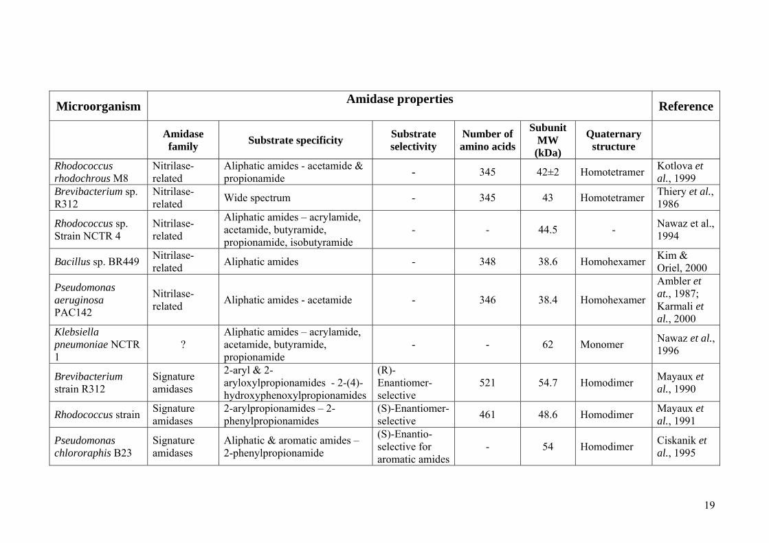
un-related to nitrilases (Pertsovich et al., 2005; Banerjee et al., 2002). Table 1-3
presents detailed information on some of the characterized bacterial amidases.
1.3.1.1. Signature amidase enzymes
Signature amidases are characterized by the presence of an invariant GGSS (Gly-Gly-
Ser-Ser) signature motif in their primary sequence (Chebrou et al., 1996; Kobayashi et
Figure 1-6: Reactions that are catalysed by amidases. Most of the characterized amidases are able exhibit both amide hydrolysis and amide transfer; these are the only reactions that are of interest in industry, as the rest are too slow to be of any economical use. The figure was taken from Fournand and Arnaud (2001).

17
al., 1997). They also have aspartate and serine residues (Asp191 and Ser195 in the
Rhodococcus J1 amidase numbering (Kobayashi et al., 1997) in the active site, in
place of the nucleophilic cysteine (Novo et al., 1995) that is observed in the nitrilase
superfamily. Amidases from this group exhibit a wide substrate specificity including
aliphatic and aromatic amides, as well as amides of α-substituted carboxylic acids.
Importantly, most of them exhibit stereoselectivity (Hirrlinger et al., 1996; Kobayashi
et al., 1997). Most of the structurally characterized signature amidase enzymes have
quaternary structure (Table 1-3), forming homodimeric and homooctameric
complexes in solution (Kobayashi et al., 1997; Novo et al., 1995; Chebrou et al.,
1996; Mayaux et al., 1991). Representative members of this group include amidases
from R. rhodochrous J1 (Kobayashi et al., 1997), Rhodococcus sp. R312 (Fournand et
al., 1998b; Chebrou et al., 1996), Sulfolobus solfataricus (Cilia et al., 2005) and P.
chlororaphis B23 (Ciskainik et al., 1995).
Aside from C-N bond metabolism in prokaryotes, over 200 signature amidase
enzymes are also found in archea and eukaryotic organisms forms (Labahn et al.,
2002). Curnow and colleagues (1997) reported a signature amidase that transfers NH3
from glutamine, resulting in the generation of properly charged tRNAGln in
eubacteria. Cravatt and others (1996) also reported a mammalian amidase that was
involved in the degradation of neuromodulatory fatty acid amides. This enzyme was
found to belong to an “amidase signature (AS) family” of enzymes that is
characterized by a conserved stretch (approximately 130 amino acids) that is rich in
glycine and serine residues, referred to as the “amidase signature sequence” (Mayaux
et al., 1991; Chebrou et al., 1996). Further studies by Patricelli and Cravatt (2000)
revealed that this amidase represented a large class of serine-lysine catalytic dyad
hydrolases that closely resembled serine hydrolases (Boger et al., 2000).
Several crystal structures of signature amidase enzymes have been reported to-date.
Figure 1-7 below presents details of the structural features of a peptide amidase from
Stenotrophomonas maltophilia (Pam; PDB ID, 1m22); (Labahn et al., 2002). This is a
regio-selective signature amidase that is involved in the hydrolysis of the C-terminal
amide bond in peptide amides (Labahn et al., 2002). Pam has clear α/β sandwich fold,
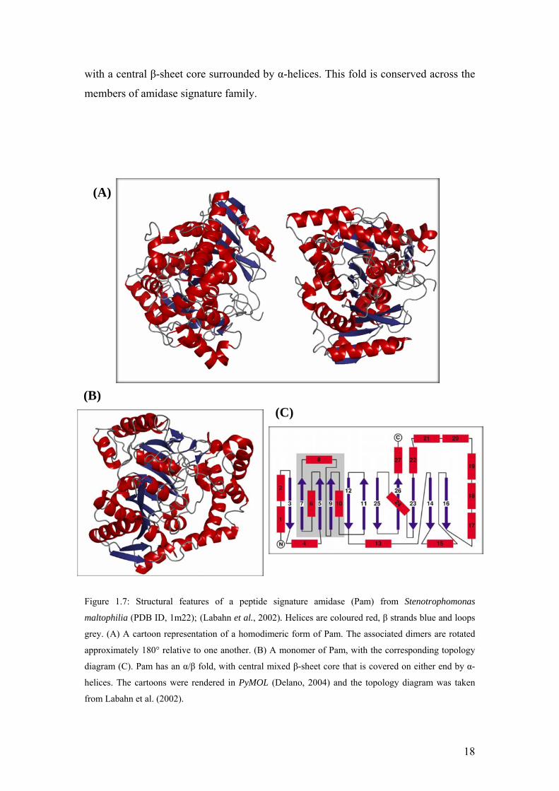
18
with a central β-sheet core surrounded by α-helices. This fold is conserved across the
members of amidase signature family.
Figure 1.7: Structural features of a peptide signature amidase (Pam) from Stenotrophomonas
maltophilia (PDB ID, 1m22); (Labahn et al., 2002). Helices are coloured red, β strands blue and loops
grey. (A) A cartoon representation of a homodimeric form of Pam. The associated dimers are rotated
approximately 180° relative to one another. (B) A monomer of Pam, with the corresponding topology
diagram (C). Pam has an α/β fold, with central mixed β-sheet core that is covered on either end by α-
helices. The cartoons were rendered in PyMOL (Delano, 2004) and the topology diagram was taken
from Labahn et al. (2002).
(A)
(B) (C)

19
19
Microorganism Amidase properties Reference
Amidase family Substrate specificity Substrate
selectivity Number of amino acids
Subunit MW (kDa)
Quaternary structure
Rhodococcus rhodochrous M8
Nitrilase-related
Aliphatic amides - acetamide & propionamide - 345 42±2 Homotetramer Kotlova et
al., 1999 Brevibacterium sp. R312
Nitrilase-related Wide spectrum - 345 43 Homotetramer Thiery et al.,
1986
Rhodococcus sp. Strain NCTR 4
Nitrilase-related
Aliphatic amides – acrylamide, acetamide, butyramide, propionamide, isobutyramide
- - 44.5 - Nawaz et al., 1994
Bacillus sp. BR449 Nitrilase-related Aliphatic amides - 348 38.6 Homohexamer Kim &
Oriel, 2000
Pseudomonas aeruginosa PAC142
Nitrilase-related Aliphatic amides - acetamide - 346 38.4 Homohexamer
Ambler et at., 1987; Karmali et al., 2000
Klebsiella pneumoniae NCTR 1
? Aliphatic amides – acrylamide, acetamide, butyramide, propionamide
- - 62 Monomer Nawaz et al., 1996
Brevibacterium strain R312
Signature amidases
2-aryl & 2-aryloxylpropionamides - 2-(4)-hydroxyphenoxylpropionamides
(R)-Enantiomer-selective
521 54.7 Homodimer Mayaux et al., 1990
Rhodococcus strain Signature amidases
2-arylpropionamides – 2-phenylpropionamides
(S)-Enantiomer-selective 461 48.6 Homodimer Mayaux et
al., 1991
Pseudomonas chlororaphis B23
Signature amidases
Aliphatic & aromatic amides – 2-phenylpropionamide
(S)-Enantio-selective for aromatic amides
- 54 Homodimer Ciskanik et al., 1995

20
Agrobacterium tumefaciens strain d3
Signature amidases
2-arylpropionamides – 2-phenylpropionamide
(S)-Enantiomer-selective 517 63 Homooctamer Trott et al.,
2001
Rhodococcus erythropolis strain MP50
Signature amidases
Aromatic amides – 2-phenilpropionamides, α-chlorophenylacetamide
(S)-Enantiomer-selective 525 55 Homooctamer Trott et al.,
2002
Rhodococcus erythropolis MP50
Signature amidases
Broad spectrum, including 2-arylpropionamides - 2-phenylpropionamide
(S)-enantiomer-selective 525 61 Homooctamer Hirrlinger et
al., 1996
Ochrobactrum anthropi SV3 ? D-amino acids with aromatic or
hydrophobic side chains D-stereo-selective 363 40 Monomer Komeda &
Asano, 2000
Pseudomonas sp. MCI3434 ?
Piperazine-2-tert-butylcarboxamine, -alaninamide, piperazine-2-carboxamide
R-stereo-selective 274 30.1 Monomer Komeda et
al., 2004
Table 1.2: Properties of some of the characterized amidases in the literature. Question marks indicate cases where the classification is not certainly confirmed, while dashes indicate
cases where no information on that particular property is available. Most of the nitrilase-related amidases are homotetrameric and homohexameric in solution while the signature
amidase family enzymes are mostly homodimeric or homooctameric in solution. The amidase from Klebsiella pneumonae NCTR 1 (Nawaz et al., 1996) has all the characteristics
of nitrilase-related amidases including a nucleophilic cysteine, except that it has metal ions in the active site and is monomeric in solution. No other characterized amidase has been
found to have coordinated metal ions in the active site, which makes this enzyme unique. Amidases from Ochrobactrum anthropi SV3 (Komeda & Asano, 2000) and Pseudomonas
sp. MCI3434 (Komeda et al., 2004) which are monomeric in solution, show stereo-selectivity, but no information is available as to whether an “amidase signature sequence” has
been observed. Their substrates are slightly different from the signature amidases, which suggests that these enzymes might belong to a family of their own.

21
1.3.1.2. Nitrilase-related amidases
A combination of biochemical studies and primary sequence analyses has suggested that
amidases from this group are sulphydryl enzymes (Novo et al., 1995; Novo et al., 2002;
Farnaud et al., 1999) containing a conserved nucleophilic cysteine (Novo et al., 1995)
that is involved in catalysis. Only bacterial amidases have been extensively characterized
in this family. These include amidases from Rhodococcus erythropolis (Soubrier et al.,
1992), Rhodococcus sp. R312 (Fournand et al., 1998b; Soubrier et al., 1992; Thiery et al.,
1986), Bacillus stearothermophilis (Cheong, Oriel, 2000), Pseudomonas aeruginosa
(Novo et al., 1995), Bacillus sp. BR449 (Kim, Oriel, 2000), Helicobacter pylori
(Skouloubris et al., 1997) and our amidase of interest Geobacillus pallidus RAPc8
amidase (Cameron et al., 2005), among others. Unlike signature amidases which are
either homodimeric or homooctameric in their active form, most of the characterized
nitrilase-related aliphatic amidases form homotetrameric and homohexameric complexes
in solution (Pertsovich et al., 2005). These enzymes show substrate preference for short-
chain aliphatic amides (Fournand, Arnaud, 2001; Pertsovich et al., 2005). Details of some
of the characterized nitrilase-related amidases are given in Table 1-3.
Although no crystal structures of nitrilase-related amidases have been determined to-date,
a homology model of Pseudomonas aeruginosa amidase (PamiE; PDB ID, 1k17); (Novo
et al., 2002), based on the crystal structure of the Nit domain of worm NitFhit fusion
protein (Nit; PDB ID, 1ems); (Pace et al., 2000) now exists. The model (Figure 1-8)
shows a conserved α-β-β-α sandwich fold that resembles the conserved structural fold of
the nitrilase superfamily structures. Analysis of the three-dimensional (3D) PamiE model
identified a Glu59, Lys134, Cys166 catalytic triad, that was also supported by
mutagenesis studies (Farnaud et al., 1999; Novo et al., 2002). These catalytic residues
were found to align with the Glu54, Lys129, Cys169 triad of the Nit crystal structure,
after the two molecules were superimposed. Similar catalytic triad residues were also
observed in the models of R. erythropolis, H. pylori and B. stearothermophilus amidases
that were also based on Nit as the template (Novo et al., 2002). This confirmed the
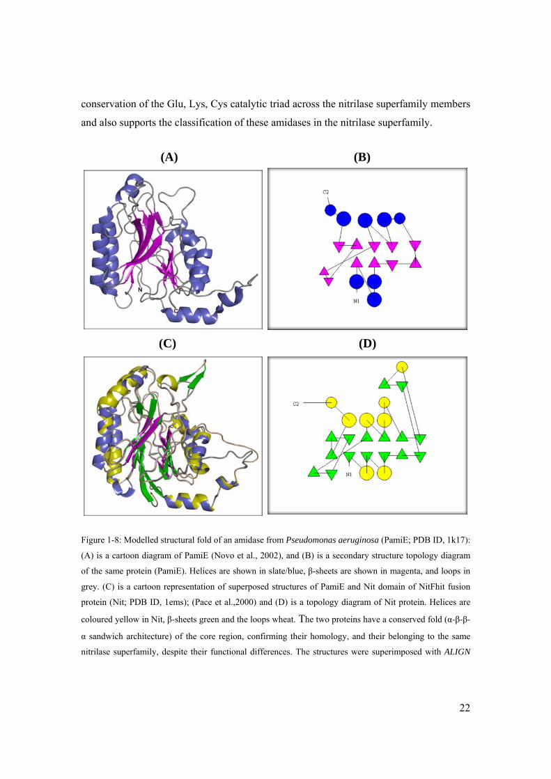
22
conservation of the Glu, Lys, Cys catalytic triad across the nitrilase superfamily members
and also supports the classification of these amidases in the nitrilase superfamily.
Figure 1-8: Modelled structural fold of an amidase from Pseudomonas aeruginosa (PamiE; PDB ID, 1k17):
(A) is a cartoon diagram of PamiE (Novo et al., 2002), and (B) is a secondary structure topology diagram
of the same protein (PamiE). Helices are shown in slate/blue, β-sheets are shown in magenta, and loops in
grey. (C) is a cartoon representation of superposed structures of PamiE and Nit domain of NitFhit fusion
protein (Nit; PDB ID, 1ems); (Pace et al.,2000) and (D) is a topology diagram of Nit protein. Helices are
coloured yellow in Nit, β-sheets green and the loops wheat. The two proteins have a conserved fold (α-β-β-
α sandwich architecture) of the core region, confirming their homology, and their belonging to the same
nitrilase superfamily, despite their functional differences. The structures were superimposed with ALIGN
(A) (B)
(C) (D)

23
(Cohen, 1997), the cartoons were rendered with PyMOL (Delano, 2004) and the topology diagrams were
generated using TOPS (Westhead et al., 1997).
Comparison of this modelled structure with that of the signature amidase Pam reveals
that although amidases catalyze similar reactions, the two groups are very different
structurally (Figure 1-9).
Figure 1-9: A side-by-side comparison of the structural fold differences between signature amidases and
nitrilase superfamily aliphatic amidases: (A), a topology diagram of a signature amidase from
Stenotrophomonas maltophilia (Pam; PDB ID, 1m22); (Labahn et al., 2002) and (B), a topology diagram of
a theoretical model of a nitrilase-related aliphatic amidase from Pseudomonas aeruginosa (PamiE; PDB
ID, 1k17); (Novel et al., 2002). Although the two have similar functions, they exhibit completely different
structural folds. The topology diagrams were generated with TOPS (Westhead et al., 1997).
Some organisms have been found to express a wide spectrum of amidases. An example is
Rhodococcus sp. R312 bacteria that has been reported to express at least four types of
amidases; an aliphatic amidase (Novo et al., 1995), an enantioselective amidase that
hydrolyzes aryloxy propionamides (Mayaux et al., 1990), a novel amidase hydrolyzing
dinitriles (Moreau et al., 1993) and an α-amino acid amidase that is specific for L-α-
amino amides (Banerjee et al., 2002; Fournand, Arnaud, 2001). It is also worth noting
that, although bacterial amidases function in the same nitrogen metabolism pathway as
NHases, the existence of metal ions (like iron and cobalt) in the active site of amidases is
(A) (B)

24
very rare. This has only been reported in an amidase from Klebsiella pneumoniae by
Nawaz and colleagues (1996).
1.3.2. Mechanism of catalysis by amidases
In 1998, Kobayashi and colleagues (Kobayashi et al., 1998) reported the interesting
finding that an amidase from Rhodococcus rhodochrous J1 also catalyzes the hydrolysis
of nitriles to corresponding acids and ammonia. They then proposed an amidase
hydrolytic mechanism that included nitrile hydrolysis, as shown in Figure 1-10 (A)
below. In the proposed mechanism, the carbonyl group of the amide undergoes
nucleophilic attack, resulting in the formation a tetrahedral intermediate. With the
removal of ammonia, the tetrahedral intermediate is converted to an acyl-enzyme
complex, which is then hydrolysed to an acid upon the addition of a water molecule. This
mechanism had previously been suggested by Maestracci and others (1986), who
proposed a similar mechanism (Figure 1-10 (B)) for acyl transfer activity that they had
observed in the aliphatic amidase from Rhodococcus sp. R312.

25
Figure 1-10: Catalytic mechanism of amidases: (A), amide and nitrile hydrolysis mechanism proposed by Kobayashi and others (1998). (B), amide hydrolysis and acyl transfer mechanisms proposed by Maestracci and colleagues (1986); the two mechanisms are proposed to proceed via the formation of an acyl-enzyme intermediate, followed by the transfer of the acyl group to either water (hydrolysis) or hydroxylamine (acyl transfer) co-substrates. Figure (A) is taken from Banerjee et al. (2002), while Figure (B) is taken from Fournand and Arnaud (2001).

26
1.3.3. Geobacillus pallidus RAPc8 amidase
Pereira and colleagues (1998) identified a thermostable amidase in a Bacillus isolate (sp.
RAPc8) that is currently named Geobacillus pallidus (RAPc8), based on its 16S RNA
sequence. An Open Reading Frame (ORF) encoding a putative 348-amino acid amidase
(MW 38.6 kDa) was located 127 bp upstream (Figure 1-11) of ORFs encoding the β and
α (in that order) subunits of a cobalt-containing nitrile hydratase (NHase). This gene
organization was also observed in Bacillus sp. BR449 (Kim, Oriel, 2000), which also
encodes a cobalt-containing NHase and a nitrilase-related aliphatic amidase. This differs
from the gene organization observed in organisms expressing a NHase-signature amidase
couple (Figure 1-11), the main difference being the order of the genes for α and β
subunits of NHases. The amidase from Geobacillus pallidus RAPc8 (RAPc8 amidase)
gene was cloned and over-expressed by Cameron and others (2005), who identified 100%
sequence identity between this amidase and an aliphatic amidase from Bacillus sp.
BR449 (Kim, Oriel, 2000). The DNA sequences however shared 99% identity due to six
silent nucleotide substitutions (Cameron et al., 2005).
Although the physiological role of RAPc8 amidase is still not clear, there is a high
likelihood that it might be involved in the metabolism of aldoximes (Figure 1-1), forming
the third stage in the three-step pathway that involves aldoxime dehydratase, NHase and
amidase. This speculation is based on the report by Kato and colleagues (1999), who
showed a link between the three enzymes (aldoxime dehydratase, NHase and amidase)
and aldoxime metabolism in Rhodococcus sp. YH3-3.
27
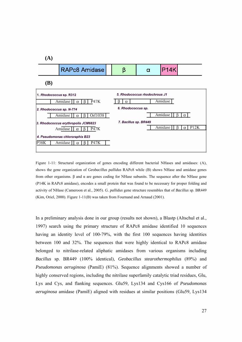
Figure 1-11: Structural organization of genes encoding different bacterial NHases and amidases: (A),
shows the gene organization of Geobacillus pallidus RAPc8 while (B) shows NHase and amidase genes
from other organisms. β and α are genes coding for NHase subunits. The sequence after the NHase gene
(P14K in RAPc8 amidase), encodes a small protein that was found to be necessary for proper folding and
activity of NHase (Cameroon et al., 2005). G. pallidus gene structure resembles that of Bacillus sp. BR449
(Kim, Oriel, 2000). Figure 1-11(B) was taken from Fournand and Arnaud (2001).
In a preliminary analysis done in our group (results not shown), a Blastp (Altschul et al.,
1997) search using the primary structure of RAPc8 amidase identified 10 sequences
having an identity level of 100-79%, with the first 100 sequences having identities
between 100 and 32%. The sequences that were highly identical to RAPc8 amidase
belonged to nitrilase-related aliphatic amidases from various organisms including
Bacillus sp. BR449 (100% identical), Geobacillus stearothermophilus (89%) and
Pseudomonas aeruginosa (PamiE) (81%). Sequence alignments showed a number of
highly conserved regions, including the nitrilase superfamily catalytic triad residues, Glu,
Lys and Cys, and flanking sequences. Glu59, Lys134 and Cys166 of Pseudomonas
aeruginosa amidase (PamiE) aligned with residues at similar positions (Glu59, Lys134
(A)
(B)

28
and Cys166) in RAPc8 amidase. The similarity between the primary structure of the
nitrilase superfamily amidases and RAPc8 amidase, as well as the existence of the
characteristic Glu, Lys, Cys catalytic triad in RAPc8 amidase, was a clear indication that
RAPc8 amidase was a member of the nitrilase superfamily.
1.3.3.1. Substrate specificity in RAPc8 amidase
Makhongela and colleagues (in press) have recently characterized RAPc8 amidase in
terms of activity, substrate specificity and the effect of immobilization on different
scaffolds. This is a first study of the kind on this enzyme. They have found that RAPc8
amidase is a highly thermostable enzyme, with activity between 50 and 60 °C and
maximum specific activity at pH 7.0. It exhibits both amide hydrolysis and acyl transfer
activities (Makhongela et al., in press), as observed with other characterised amidases.
Similar to the substrate specificities observed in other nitrilase-related amidases, RAPc8
actively hydrolyzes (Table 1-4) short-chain aliphatic amides such as acrylamide,
propionamide and acetamide. It is however only moderately active on substituted short-
chain and mid-length aliphatic amides (such as diacetamide, lactamide, fluoroacetamide
and isobutyramide) and has no activity at all on long-chain aliphatic amides
(hexanoamide), aromatic substrates or urea amides (Makhongela et al., in press). This
enzyme has substantial level of chiral selectivity on D-enantiomers, particularly on D-
lactamide and to some extent D-alaninamide. No activity was observed on L-lactamide.
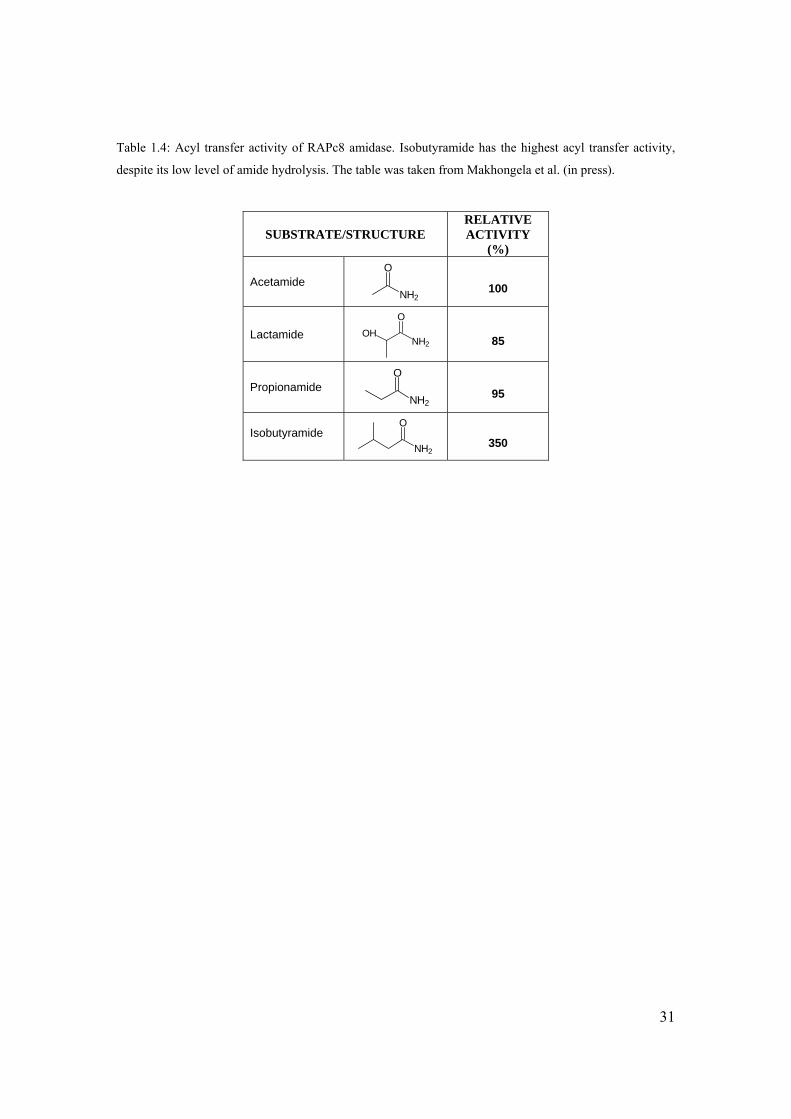
A similar trend of substrate preference was observed with the acyl transfer activity when
various amide substrates were used as acyl donors in the presence of hydroxylamine
reagents (Makhongela et al., in press). Similar to hydrolysis reactions, RAPc8 amidase
readily transfers acyl moieties from low molecular weight aliphatic amides (Table 1-5) to
hydroxylamine to form hydroxamic acids. The amidase is not active on long-chain
aliphatic amides (such as hexanamide), aromatic amides (benzamide) or L-isomers of
either short- or mid-length aliphatic amides (including L-alaninamide and L-
leucinamide). Among the amide substrates tested, isobutyramide showed highest relative

29
activity for acyl transfer (Table 1-5), which probably points to a shift in chain length
specificity from hydrolysis to acyl transfer (Makhongela et al., in press).
RAPc8 amidase is completely inhibited by oxidizing agents such as hydrogen peroxide
(H2O2), while the activity is reduced several fold by the presence of modifying metal ions
like Fe3+, Co2+, Cu2+, Zn2+ (Makhongela et al., in press). On the other hand, the presence
of reducing agents like dithiothreitol (DTT) at millimolar (mM) concentrations not only
restores the activity of the inactivated enzyme, but also stabilizes the acyl transfer activity
against oxidation; this explains why the enzyme activity was lost in the absence of DTT
during purification. All these observations are clearly consistent with the presence of a
catalytically active cysteine thiol group. The activity of RAPc8 amidase is however not
affected by the presence of either serine protease inhibitor PMSF or metal-chelating agent
EDTA. This is a strong indication that neither an active site serine residue nor
coordinated ions are present in the active site (Makhongela et al., in press).

30
Table 1-4: Hydrolytic activity of RAPc8 amidase on a number of tested amide substrates. High activity is
noted on the short-chain aliphatic amides, while there is no activity at all with long-chain and aromatic
amides. The table was borrowed from Makhongela et al. (in press).
SUBSTRATE /STRUCTURE RELATIVEACTIVITY
(%) SUBSTRATE /STRUCTURE
RELATIVE ACTIVITY
(%)
Nicotinamide
N
NH2
O
0 L-Alaninamide
CH3
O
NH2
NH2
3
Isonicotinamide
N
H2N O
0 Diacetamide
N H
O
O
14
Benzamide
CONH2
0 Isobutyramide
NH2
O
16
L-Asparagine -OOC NH2
NH2 O
0 Lactamide
NH2
O
OH
17
D-Asparagine -OOC NH2
NH2 O
0 Formamide
H NH2
O
30
DL-Phenylalanine
NH2
O
0 Flouroacetamide
NH2
O
F
165
L-Prolinamide
NHNH2
O
0 Propionamide
NH2
O
67
Urea H2N NH2
O
1.5 Acetamide
NH2
O
100
Hexanamide
CH3 O
NH2
0 Acrylamide
NH2
O
102
31
Table 1.4: Acyl transfer activity of RAPc8 amidase. Isobutyramide has the highest acyl transfer activity,
despite its low level of amide hydrolysis. The table was taken from Makhongela et al. (in press).
SUBSTRATE/STRUCTURE RELATIVE ACTIVITY
(%) Acetamide NH2
O
100
Lactamide NH2
O
OH
85
Propionamide NH2
O
95
Isobutyramide
NH2
O
350

32
1.4. Motivation and study objectives
Motivation
Although the physiological role of Geobacillus pallidus sp. RAPc8 amidase (RAPc8
amidase) is not clear, its recent biochemical characterization (Makhongela et al., in press)
has revealed the specificity of this enzyme for short-chain aliphatic amides, both in
hydrolysis and acyl transfer. RAPc8 amidase also shows stereoselectivity for a number of
tested enantiomeric substrates. However the molecular and structural basis of the
observed substrate specificity and selectivity is still not understood.
It is now known that members of the nitrilase superfamily form oligomeric complexes in
solution. The four enzymes whose crystal structures have been determined in the family
(PDB IDs; 1erz, 1ems, 1f89 & 1j31: Nakai et al., 2000, Pace et al., 2000, Kumaran et al.,
2003 & Sakai et al., 2004 respectively) have quaternary structure; 1erz and 1ems are
tetrameric while 1f89 and 1j31 are dimeric in their active forms. The characterized
amidases have an even number of subunits, ranging from 2 to 8 (Kotlova et al., 1999),
while the nitrilase branch of the superfamily are reported to form complexes of 1 to 26
subunits (O'Reilly, Turner, 2003; Banerjee et al., 2002). The quaternary structure of
RAPc8 amidase and its implications (if any) on the activity is still not known.
Members of the nitrilase superfamily of enzymes share a conserved Glu, Lys, Cys
catalytic triad (Brenner, 2002), yet they show significant differences in the reaction
(amidase, nitrilase, carbamylase, acyl transferase reactions among others) that they
catalyze, in substrate specificity and in catalytic activity. Understanding the features
responsible for these differences will require detailed structural comparison between
enzymes of different branches. Although four crystal structures of nitrilase superfamily
enzymes have been determined (a carbamylase (DCase) and 3 with unknown function),
there is none from the amidase branch.
33
Finally, sequence comparisons between members from different branches in the nitrilase
superfamily have revealed that RAPc8 amidase has an elongated C-terminal tail that is
also present in some members of the nitrilase branch, but absent in most of the members
in the superfamily. The role (either functional and/or structural) of this extended C-
terminal tail in these enzymes is not known.
Project aims
The overall objective of the study was to determine the crystal structure of Geobacillus
pallidus sp. RAPc8 amidase (RAPc8 amidase). The specific objectives were:
1. To determine the biological unit of the active RAPc8 amidase enzyme in solution
using gel filtration and electron microscopy,
2. To collect X-ray diffraction data from RAPc8 amidase crystals, and to use
crystallographic methods (specifically molecular replacement) to determine
crystal packing as well as to solve crystal structure of RAPc8 amidase,
3. To determine the role of the elongated C-terminal tail in RAPc8 amidase and
4. To examine the active site of RAPc8 amidase in order to determine the structural
basis of substrate specificity and selectivity and possibly the catalytic mechanism.
Significance and impact
In the past two decades, biotechnological industries have been exploring the enzymatic
hydrolysis of nitriles and amides as a method of obtaining a broad spectrum of useful
carboxylic acids. As mentioned elsewhere, this has been driven by attempts to find
alternatives to the traditional acid- or base-catalyzed amide and nitrile hydrolytic
methods, that involve harsh conditions of temperature and pH, and hence are
incompatible with the labile structures of many industrially relevant compounds,
introduce unwanted by-products, decrease product yield and increase the overall
production costs (Fournand, Arnaud, 2001; Banerjee et al., 2002; Mylerova, Martinkova,
2003). The potential of nitrile-degrading enzymes in catalyzing useful enantio-, chemo-
34
and regio-selective biotransformations has been discovered. The use of these enzymes in
chemical-free bioremediation of the environment has also been explored (Muller,
Gabriel, 1999; Banerjee et al., 2002; Wyatt, Knowles, 2003). Ongoing biochemical
characterization studies (Makhongela et al., in press) have revealed the capability of
RAPc8 amidase to drive both hydrolytic and acyl transfer activities with a significant
level of enantio-selectivity. The stereoselectivity of RAPc8 amidase will allow
production of enantiopure acids that are difficult to produce by traditional chemical
methods. Immobilization of RAPc8 amidase on Eupergit C beads (Makhongela et al., in
press) has been found to enhance its activity, increasing the production yield several fold.
This enzyme thus has great potential in synthetic industries.
Structural characterization of RAPc8 amidase will shed light on the basis of substrate
specificity and selectivity as well as the catalytic mechanism not only in this enzyme but
also in the nitrilase-related amidases. The long term focus will be to extend the utility of
this enzyme both in industries and probably in waste degradation, to fine-tune its activity
so that it is more selective, or even to alter its substrate specificity. Structural information
is needed to inform such experiments.
The research will in the long run have an impact in the biotechnological industries in
South Africa, and across the continent. The outcome of this will allow the generation of
highly efficient enzymes, whose use will ensure safe and effective production protocols,
high quality pure products and increased yields at lower production costs. In cases where
nitrile- or amide-containing industrial wastes present a challenge for safe disposal, there
is the potential to construct waste treatment systems using microorganisms that contain
amide-degrading enzymes, ensuring safe disposal of these wastes. This will go a long
way in preventing environmental degradation resulting from the accumulation of these
wastes in the environment.
35
CHAPTER 2.: MATERIALS AND METHODS
36
2.1. Biological unit molecular weight determination
The molecular weight of the amidase from Geobacillus pallidus RAPc8 amidase
(RAPc8 amidase) biological unit was determined by gel filtration and electron
microscopy (EM).
2.1.1. Gel filtration
Gel filtration was performed according to the published protocol of Makhongela et al.
(in press). In brief, an S-300 HR gel exclusion chromatography column (GE-
Healthcare) was pre-equilibrated with a buffer containing 150 mM NaCl and 50 mM
potassium phosphate buffer (pH 7.0). The protein sample was applied to the column
and eluted with 20 mM Tris, 150 mM NaCl, 1 mM DTT (pH 7.4) over a total volume
of 120 ml. Fractions were collected at 3 min intervals with a Gilson FC 203B fraction
collector at a flow rate of 0.5 ml/min. RAPc8 amidase homogeneity and sub-unit
molecular mass were determined by sodium dodecyl sulfate-polyacrylamide gel
electrophoresis (SDS-PAGE) and non-denaturing gel electrophoresis (native-PAGE)
using 15 % gels stained with Coomassie blue R-250. Samples were assayed for
protein concentration using a Bradford protein determination kit (BioRad) with
bovine serum albumin (BSA) as the standard.
2.1.2. Electron microscopy and image analysis
Negatively-stained electron micrographs preparation and EM images processing and
analysis were performed using the protocols that are published in Makhongela et al.
(in press). In summary, purified amidase (3 µl) at a concentration of 0.05 mg / ml was
applied to carbon films which had been glow-discharged for 30s, rinsed on two
droplets of water, blotted and negatively stained with 2% uranyl acetate. Images were
recorded at minimum dose (approx 100 el/A2) using a Zeiss 912 electron microscope
set for zero energy loss imaging at 120kV and 50,000x magnification on a Proscan 2k
x 2k CCD camera. The magnification on the CCD had previously been determined to
be 2.28Å per pixel under these conditions.

37
Projection images (12,698) of the putative RAPc8 amidase were manually selected
using Ximdisp graphical program (Crowther et al., 1996). The selected particles were
then boxed, filtered and normalized using SPIDER (Frank et al., 1996). The
normalized projection images were rotationally and translationally aligned using the
reference-free alignment method after which they were subjected to K-means
Multivariate Statistical Analysis (MSA) to put them in classes of similar views, using
SPIDER. The images in each class were then averaged to generate class averages with
improved Signal-to-Noise (S/N) ratio. The aligned images were also subjected to
Principal Component Analysis (PCA) using methods described by Frank (2006). This
was done to determine the symmetry of the elements that comprised the images.
2.2. Secondary structure prediction
Protein Structure Prediction Server (PSIPRED) (McGuffin et al., 2000) was used to
predict the secondary structure profile for RAPc8 amidase primary sequence.
2.3. Protein purification and crystallization
RAPc8 amidase protein was purified and crystallized using materials and methods
that we have published (Agarkar et al., 2006). After expression in E. coli BL21 (DE3)
cells and cell lysis, the protein was heated to 45 °C for 20 min in a buffer containing
10 mM DTT, 1 mM EDTA, 200 mM NaCl and 10% glycerol. The precipitants were
removed by centrifugation. The supernatant was applied to a HighLoad 16/10 Phenyl-
Sepharose column (Amersham Biosciences) pre-equilibrated in a buffer containing
1.7 M ammonium sulphate, 50 mM potassium phosphate, 5 mM DTT, pH 7.4. The
bound protein was eluted with a linear gradient of ammonium sulphate (1.7-0 M).
This was followed by another chromatographic step using a Hiprep 16/10 Q-
sepharose FF column (Amersham Biosciences) pre-equilibrated with a potassium
phosphate buffer (20 mM) that also contained 2 mM DTT and 1 M NaCl at pH 7.4.
The column was eluted with a linear gradient of NaCl (0.1-1 M). The fractions
containing pure protein were pooled, and concentrated, then taken through a gel
filtration chromatography step as described earlier.

38
The enzyme was crystallized using the hanging-drop vapor diffusion method, using 4
µl of protein and 4 µl reservoir solution. After a number of crystallization conditions
screens and optimization steps, diffraction quality crystals were grown at 22 °C in the
presence of 1.2 M sodium citrate, 400 mM NaCl and 100 mM sodium acetate at pH
5.6.
2.4. X-ray diffraction data collection
Crystals were visually inspected for quality, and their dimensions were measured
using an in-lens graticule on a stereomicroscope (Leica Microsystems, Wetzlar,
Germany). Images were acquired on a Leica Z16 APO (KLI500 LCD)
stereomicroscope connected to a computer, using the IM500 program (Leica
Microsystems, Wetzlar, Germany).
The X-ray data were collected on the in-house X-ray diffraction machine at the
University of Western Cape, South Africa. The X-ray diffraction machine consists of
a Rigaku RUH3R copper rotating-anode X-ray source operated at 40 kV, 22 mA; a
Rigaku R-axis IV+ image plate camera (Rigaku MSC, Tokyo, Japan); an X-stream
2000 low-temperature system (Rigaku MSC, Houston TX, USA); and an AXCO
PX50 glass capillary optic with a 0.1 mm focus (Australian X-Ray Capillary Optics,
Parkville VIC, Australia). Data from crystals mounted on a cryoloop (Hampton
Research) were collected at a temperature of 100 K with a crystal-to-detector distance
of 157.1 mm.
Images data frames covering an oscillation angle of 0.5° per frame and spanning a
range of 48° (between 25° and 73°) were collected for 55 minutes per frame, to make
a total of 96 images.
2.5. Data processing and characterization
The indexing, integration, scaling and merging of the raw X-ray diffraction data were
performed using Denzo and SCALEPACK (Otwinowski, Minor, 1997). Determination

39
of the Laue group and subsequently the space group was were attempted using various
strategies as implemented with the crystallographic programs, Denzo and
SCALEPACK, POINTLESS (Collaborative Computational Project Number 4 (CCP4).,
1994; Evans, 2006) and PHASER (CCP4, 1994; (Read, 2001). The measured data
were characterized in terms of asymmetric unit contents by calculating a Matthews
coefficient (Vm) (Matthews, 1968) using PHASER. The program POLARRFN (CCP4,
1994) was used to calculate a fast self-rotation function in order to discern the
presence of a local rotation axis and its orientation in the asymmetric unit.
2.6. Checking for the quality of diffraction data
In addition to data quality statistics obtained from the merging and scaling step in
SCALEPACK (Otwinowski, Minor, 1997), the Xtriage program of the PHENIX
(Python-Based Hierarchical Environment for Integrated Xtallography) suite (Adams
et al., 2002) was used to check for crystal twinning, translational pseudo-symmetry,
possible outlier reflections (Read, 1999) and ice rings-related problems in the merged
RAPc8 amidase data.
2.7. Molecular replacement
2.7.1. Search for homologues
A search for distant structural homologues of RAPc8 amidase was performed using
the structure-sequence threading tool, GenTHREADER (Jones, 1999). Structural
alignments between the identified homologues and RAPc8 amidase sequence were
performed manually with the guidance of the GenTHREADER output, the predicted
RAPc8 amidase secondary structure profile and the pair-wise superposition of the
structures using ALIGN (Cohen, 1997).

40
2.7.2. Preparation of the search models for molecular replacement
The coordinate files of the identified homologues were edited manually before
molecular replacement (MR) trials. Parts of the models that had a high likelihood of
being different from RAPc8 amidase structure were removed, including non-protein
atoms such as water molecules and metal ions, regions containing deletions and/or
insertions, and extra domains that were not homologous to RAPc8 amidase. Extra
subunits were also removed from the models leaving only regions of the model that
were consistent with the molecular content of RAPc8 amidase asymmetric unit. A
series of search models were prepared for use in different MR trials; these included
models lacking the variable loop regions, mutated models with RAPc8 amidase side
chains, and mixed poly-alanine models with alanines replacing non-identical residues.
Mutation of the models’ side chains was performed using both SCWRL (Canutescu et
al., 2003) and CHAINSAW (Schwarzenbacher et al., 2004), and was guided by
structural alignments between RAPc8 amidase and the homologues.
2.7.3. Molecular replacement rotational and translational searches
Since RAPc8 amidase MR searches were not trivial, several strategies as discussed in
the Results and Discussion section were attempted. Various MR programs were used,
including MOLREP (CCP4, 1994; Vagin, Teplyakov, 1997), AMoRE (CCP4, 1994);
Navaza, 1994), EPMR (Kissinger et al., 1999), REPLACE suite (GLRF and TF)
(Tong, 1993) and PHASER (CCP4, 1994; Read, 2001). Confirmation of molecular
replacement (MR) solution and rigid body refinement were performed using
PHASER.
2.8. Model rebuilding and refinement
Rigid body refinement was performed with PHASER (CCP4, 1994; Read, 2001), and
initial sigma-A weighted electron density maps were generated using REFMAC5
(CCP4, 1994; Murshudov et al., 1997). Since model fitting, rebuilding and refinement
process was not straight-forward, several strategies were employed as discussed in the
Results and Discussion chapter. These included (among other strategies) solvent

41
flattening of the initial electron density map using RESOLVE (Terwilliger, 2004a).
The initial model (substituted with RAPc8 amidase side chains) was rebuilt in
PHENIX (Adams et al., 2002). This was followed by alternating iterative cycles of
manual model building, fitting and rebuilding using the crystallographic graphical
program O (Jones et al., 1990) and restrained refinement using REFMAC5.
2.9. Final model validation
The accuracy, precision and correctness of the refined RAPc8 amidase model was
assessed using various validation tools, including PROCHECK (CCP4, 1994;
Laskowski et al., 1993), WHATCHECK (Hooft et al., 1996) and MOLPROBITY
(Lovell et al., 2003).
2.10. RAPc8 amidase structure analysis The online CASTp (Computed Atlas of Surface Topography of proteins) server
(Binkowski et al., 2003) was used to analyze the pockets and cavities on the surface or
interior of the complete RAPc8 amidase model. BAVERAGE program (CCP4, 1994)
was used to calculate average B-factors for the final model, both for the main chain
and the side chains. The topology diagrams were generated with TOPS (Westhead et.
al., 1999) and modified using TOPDRAW (Bond, 2003).
Unless otherwise stated all molecular diagrams were rendered with PyMOL (Delano,
2004).

42
CHAPTER 3: RESULTS AND DISCUSSION
43
3.1. Molecular weight of RAPc8 amidase
3.1.1. Gel filtration
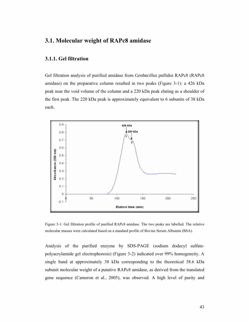
Gel filtration analysis of purified amidase from Geobacillus pallidus RAPc8 (RAPc8
amidase) on the preparative column resulted in two peaks (Figure 3-1): a 426 kDa
peak near the void volume of the column and a 220 kDa peak eluting as a shoulder of
the first peak. The 220 kDa peak is approximately equivalent to 6 subunits of 38 kDa
each.
Figure 3-1: Gel filtration profile of purified RAPc8 amidase. The two peaks are labelled. The relative
molecular masses were calculated based on a standard profile of Bovine Serum Albumin (BSA).
Analysis of the purified enzyme by SDS-PAGE (sodium dodecyl sulfate-
polyacrylamide gel electrophoresis) (Figure 3-2) indicated over 99% homogeneity. A
single band at approximately 38 kDa corresponding to the theoretical 38.6 kDa
subunit molecular weight of a putative RAPc8 amidase, as derived from the translated
gene sequence (Cameron et al., 2005), was observed. A high level of purity and

44
homogeneity is necessary for crystallization as impurities and heterogeneity can
hinder successful crystallization.
Figure 3-2: SDS-PAGE of purified RAPc8 amidase. Lane M: molecular weight markers; lane 1: A
fraction of peak 1 from gel filtration chromatography; lane 2: A fraction of peak 2 from gel filtration
chromatography. The single bands at 38 kDa indicate over 98% purity, with each peak representing a
different multimeric form of the amidase.
The electrophoretic mobility of active enzyme on a native acrylamide gel
electrophoresis suggested a complex of approximately 230 kDa, consistent with a
homohexameric native structure (Results not shown).
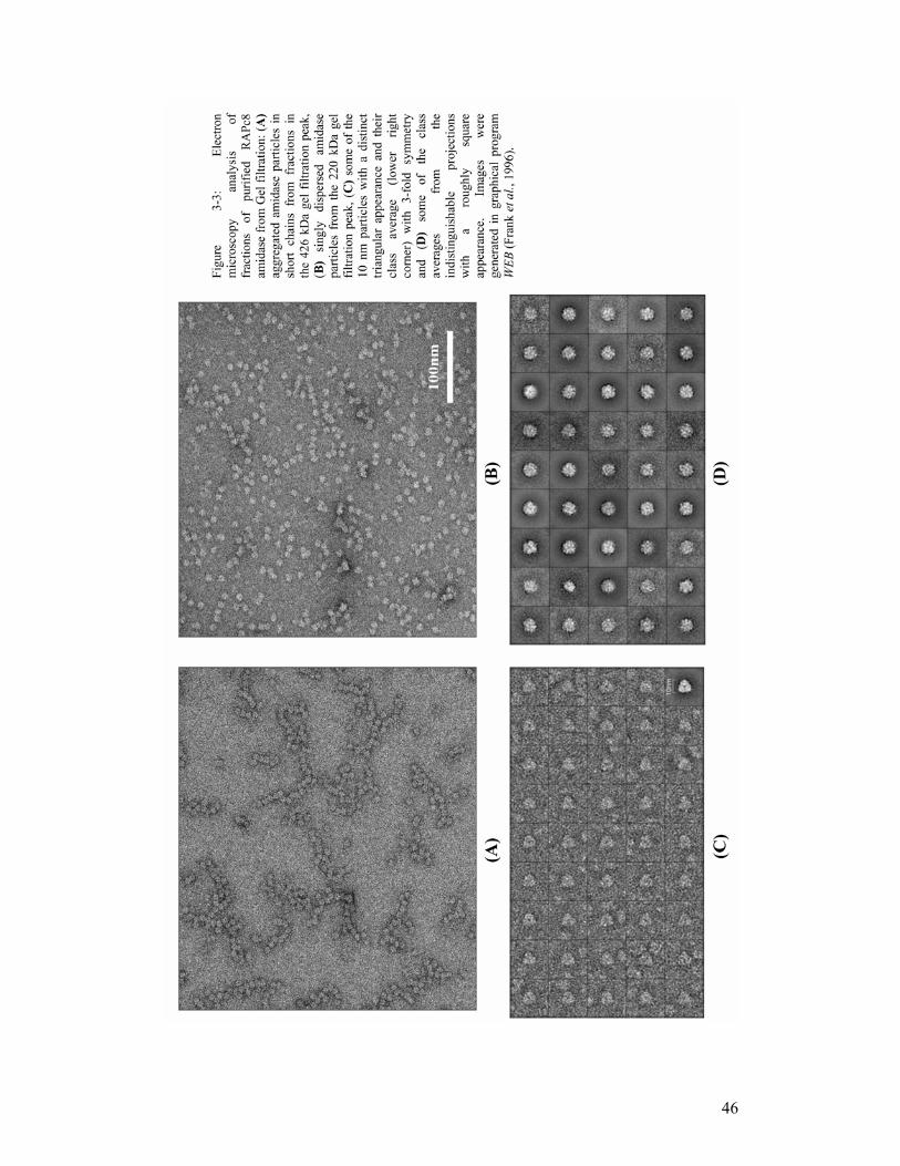
3.1.2. Electron microscopy
Electron microscopy (EM) (Figure 3-3) of various protein-containing fractions from
the two gel filtration peaks showed that the 426 kDa peak consisted mainly of
aggregates of 10 nm particles arranged in short chains of various sizes while the 220
kDa peak contained 10 nm particles which were dispersed as single particles. The 220
kDa particles correspond to a complex of approximately six 38 kDa subunits,
suggesting a homohexameric form of RAPc8 amidase. A large number of particles in
the 220 kDa peak were roughly square in shape while a few had a distinct triangular

45
appearance, making them easy to identify. Following reference-free alignment and k-
means based multivariate statistical analysis classification and averaging of single
particles using SPIDER (Frank et al., 1996), projections with the triangular shape
clustered together giving a clear class average with a 3-fold symmetry (Figure 3-
3(C)).
Due to the difficulty in satisfactory classification of the images with the approximate
square appearances, the aligned particles were subjected to Principal Component
Analysis (PCA) (Bretaudiere, Frank, 1986). In brief, PCA is a linear transformation
method in which 2-dimensional (2D) projection image matrices are first transformed
into 1-dimensional (1D) image vectors. The corresponding 1D eigenvectors are then
computed, after which 2D eigenimages are reconstituted from the chosen
eigenvectors. The presence of symmetry in the 2D eigen images indicates underlying
symmetry in the components of the images. The aligned projection images gave rise
to eigenimages (Figure 3-4) with clear 2-, 3-, and 6- fold symmetry, among many
other shapes. The presence of 6-fold eigenimages indicated that the particles had D3
point group symmetry. It was therefore concluded that the roughly square images, of
which the 2-fold eigenimage is a component, were projections perpendicular to the
triangular projections. This is consistent with the RAPc8 amidase being
homohexameric in solution, and indicates the possible arrangement of the subunits in
the complex; i.e. trimers of dimers possibly arranged around a 3-fold axis, to give rise
to D3 point group symmetry.
46

47
Figure 3-4: Eigenimages from Principal Component Analysis of the aligned image projections. 1.
Average of all the images; 2: eigenimage having 2-fold symmetry; 8 and 11: 3-fold symmetric
eigenimages 10: eigenimage with 6-fold symmetry. The other numbers represent other shapes. The
image was prepared using WEB (Frank et al., 1996).

48
3.2. Predicted secondary structure for RAPc8 amidase Advances in technology through bioinformatics approaches have made it possible to
predict the secondary structure of a protein from its sequence only, with varying
degrees of success resulting from the use of different methods. The secondary
structure of RAPc8 amidase was predicted using a secondary structure prediction
server, PSIPRED (McGuffin et al., 2000), a program that utilizes neural networks that
have been trained to recognize certain patterns like solvation parameters (Jones,
1999b) in the sequence in order to predict the secondary structure.
The secondary structure elements of RAPc8 amidase were found to follow a pattern
(Figure 3-5) that resembled that of the nitrilase superfamily homologues, suggesting a
similar conserved 3-dimensional (3D) fold of the core region. The C-terminal region
(which is more extended relative to other nitrilase superfamily enzymes) was
predicted to have secondary structure elements including helices, which indicated that
the C-terminus in the crystal was possibly highly ordered, most likely with a structural
role.

49
Figure 3-5: Predicted secondary structure prediction of RAPc8 amidase.

50
3.3. X-ray diffraction data collection
Diffraction-quality crystals were obtained using the protocol we have described
(Agarkar et al., 2006). The crystals (Figure 3-6), which had a tetrahedral shape with
dimensions of approximately 0.2 mm x 0.1 mm x 0.1 mm, were selected for X-ray
diffraction experiments. They diffracted well, producing visible spots beyond 1.8 Å
resolution and having a mosaicity of 0.375°. A typical X-ray diffraction image is
shown in Figure 3-7 below.
Figure 3-6: The image of a crystal from RAPc8 amidase.

51
Figure 3-7: RAPc8 amidase crystal X-ray diffraction image. The spots are visible beyond 1.8 Å
resolution.
52
3.4. Processing of X-ray diffraction data and space group
determination
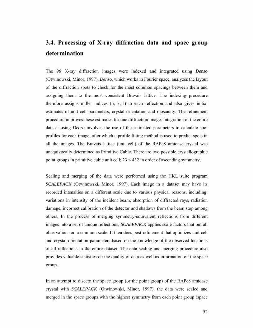
The 96 X-ray diffraction images were indexed and integrated using Denzo
(Otwinowski, Minor, 1997). Denzo, which works in Fourier space, analyzes the layout
of the diffraction spots to check for the most common spacings between them and
assigning them to the most consistent Bravais lattice. The indexing procedure
therefore assigns miller indices (h, k, l) to each reflection and also gives initial
estimates of unit cell parameters, crystal orientation and mosaicity. The refinement
procedure improves these estimates for one diffraction image. Integration of the entire
dataset using Denzo involves the use of the estimated parameters to calculate spot
profiles for each image, after which a profile fitting method is used to predict spots in
all the images. The Bravais lattice (unit cell) of the RAPc8 amidase crystal was
unequivocally determined as Primitive Cubic. There are two possible crystallographic
point groups in primitive cubic unit cell; 23 < 432 in order of ascending symmetry.
Scaling and merging of the data were performed using the HKL suite program
SCALEPACK (Otwinowski, Minor, 1997). Each image in a dataset may have its
recorded intensities on a different scale due to various physical reasons, including:
variations in intensity of the incident beam, absorption of diffracted rays, radiation
damage, incorrect calibration of the detector and shadows from the beam stop among
others. In the process of merging symmetry-equivalent reflections from different
images into a set of unique reflections, SCALEPACK applies scale factors that put all
observations on a common scale. It then does post-refinement that optimizes unit cell
and crystal orientation parameters based on the knowledge of the observed locations
of all reflections in the entire dataset. The data scaling and merging procedure also
provides valuable statistics on the quality of data as well as information on the space
group.
In an attempt to discern the space group (or the point group) of the RAPc8 amidase
crystal with SCALEPACK (Otwinowski, Minor, 1997), the data were scaled and
merged in the space groups with the highest symmetry from each point group (space

53
group P213 for 23 point group and space groups P4132, P4232 and P4332 for 432 point
group). The merging statistics were good, with reasonable numbers of outliers in all
cases. SCALEPACK output a list of likely axial reflections in all cases. However, most
of the listed reflections in all the four space groups were too strong (with high signal-
to-noise ratio) for truly systematically absent reflections. At this stage, it was hard to
tell if the strong axial reflections were due to background noise. Since neither the
presence (or absence) of systematic absences nor the merging statistics could offer
reliable guide in distinguishing between the space groups, the data were merged in the
two space groups with the lowest symmetry (P23 and P432) and subjected to further
analysis.
3.4.1. Characterization of RAPc8 amidase datasets
(A) - Matthews coefficient (volume) calculation
The Matthews coefficient (Vm) (Matthews, 1968) allows estimation of the number of
macromolecular subunits in the asymmetric unit. It is calculated as:
Vm = V / MW * Z * X where,
V is the volume of the unit,
MW is the molecular mass of the molecule,
Z is the number of the asymmetric units in the unit cell, and
X is the number of molecules in the asymmetric unit.
The program PHASER (Collaborative Computational Project Number 4 (CCP4).,
1994; Read, 2001) was used to calculate Vm for both P23- and P432-merged datasets
to 2 Å resolution.
P23-merged data had a Matthews coefficient of 2.41 with 2 amidase molecules of
38,596 Daltons each and 48.90% solvent in the asymmetric unit. P432-merged data on
the other hand, had a Matthews coefficient of 2.41 with 1 amidase molecule of 38,596
Daltons and 48.90% solvent in the asymmetric unit. The Vm values in both datasets
were within the empirically observed range as reported by Matthews (1968).

54
(B) - Self rotation function calculation
If the Matthews coefficient suggests 2 or more molecules in the asymmetric unit, a
self rotation function of the native data is usually calculated. This is important in
determining if the molecules in the asymmetric unit are related by a rotational
symmetry axis, and if so, the orientation of the axis in the unit cell (direction of the
rotation axis). It is a useful criterion with which to check for the presence of Non-
Crystallographic Symmetry (NCS). In principal, the self rotation function compares a
native Patterson with itself, with the peaks corresponding to the rotation that
superimposes multiple copies of the molecule in the asymmetric unit. Peaks
corresponding to crystallographic symmetry are similar in height to the origin peak.
The program POLARRFN (CCP4, 1994) that calculates fast self-rotation function in
polar angles was used to calculate a self rotation function for the RAPc8 amidase P23
and P432 datasets. Even though there was no possibility of NCS in the P432
asymmetric unit, the dataset was included in the calculations for comparison purposes.
Peaks (Figure 3-8) corresponding to crystallographic four-fold, three-fold and two-
fold axes (kappa = 90°, 120°, and 180° respectively) were observed in P432 dataset as
expected. The same peaks were observed in P23 dataset but in this case, the four-fold
peaks were non-crystallographic. Four-fold NCS in the P23 dataset was not consistent
with two molecules in the asymmetric unit, as suggested by the Matthews coefficient.
These results clearly indicated that the amidase crystal belonged to the 432 point
group; consistent with the crystallographic four-, three- and two-fold axes, and one
molecule in the asymmetric unit.
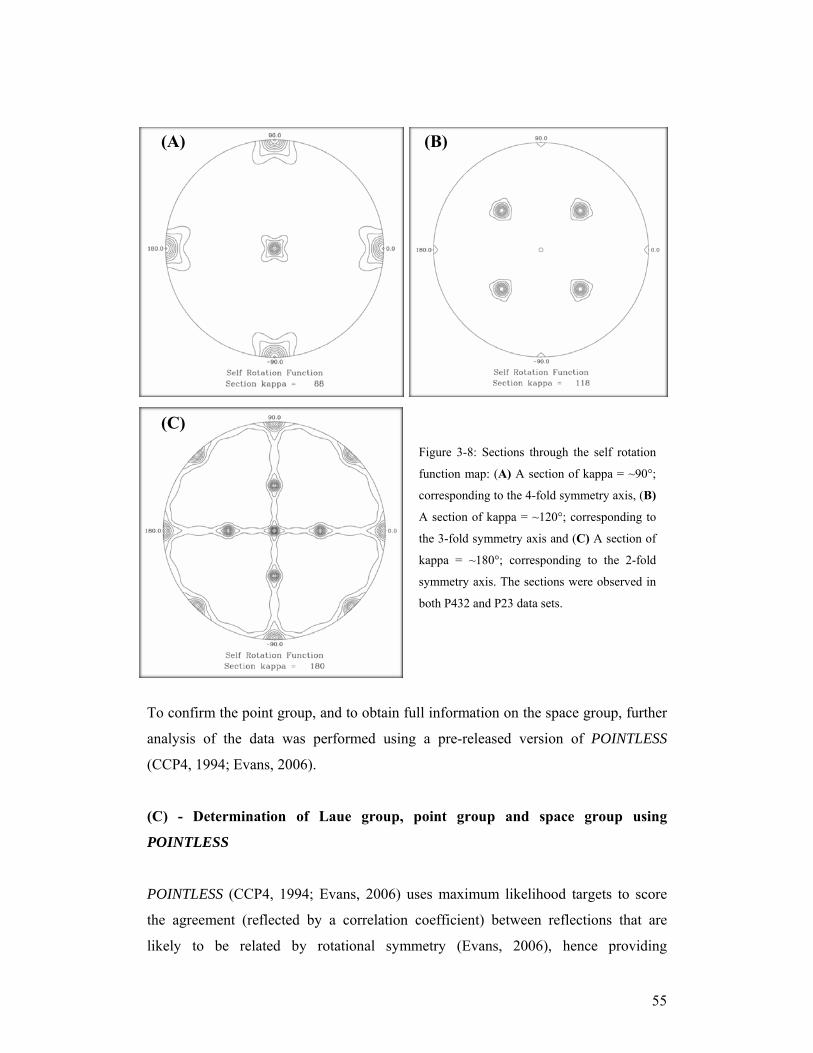
55
To confirm the point group, and to obtain full information on the space group, further
analysis of the data was performed using a pre-released version of POINTLESS
(CCP4, 1994; Evans, 2006).
(C) - Determination of Laue group, point group and space group using
POINTLESS
POINTLESS (CCP4, 1994; Evans, 2006) uses maximum likelihood targets to score
the agreement (reflected by a correlation coefficient) between reflections that are
likely to be related by rotational symmetry (Evans, 2006), hence providing
Figure 3-8: Sections through the self rotation
function map: (A) A section of kappa = ~90°;
corresponding to the 4-fold symmetry axis, (B)
A section of kappa = ~120°; corresponding to
the 3-fold symmetry axis and (C) A section of
kappa = ~180°; corresponding to the 2-fold
symmetry axis. The sections were observed in
both P432 and P23 data sets.
(A) (B)
(C)
56
information on the Laue group of the diffraction data. As clearly described by Evans
(2006), POINTLESS reads unmerged intensities, determines the Lattice with the
highest symmetry from the unit cell parameters, scores each rotational symmetry
element separately, groups the symmetry elements into subgroups and compares the
symmetry subgroups to obtain a net score for each subgroup. Upon successful
determination of Laue and point group symmetries, POINTLESS proceeds to test for
systematic absences through Fourier analysis of signal intensity-to-noise ratio (I/σ(I)
for each zone in which some reflections are likely to be systematically absent.
Unmerged RAPc8 amidase crystal data that had been indexed, integrated and scaled
in the P1 space group using SCALEPACK (Otwinowski, Minor, 1997) having unit cell
parameters a = b = c = 130.5Ǻ, α = β = γ = 90°, were used. The m-3m Laue group had
the highest score (Z-score = 13.81) and all the expected rotational symmetry elements
(2-, 3- & 4-fold axes) were clearly present (Table 3-1). Since 432 is the only point
group corresponding to m-3m Laue group (Suh et al., 1993), 432 was therefore
confirmed to be the point group for RAPc8 amidase crystal.

57
Table 3-1: Scores for each symmetry element in Laue group m-3m. Correlation coefficient Z-score (Z-
CC) was used to score the agreement of the symmetry-related observations in each rotational symmetry
element. The 2-fold, 3-fold and 4-fold axes are clearly present in RAPc8 amidase crystal, suggesting
m-3m Laue group.
Symmetry
element &
operator
Correlation
coefficient
Z-scores (Z-
CC)
Correlation
coefficient
(CC)
1 Identity 13.82 0.96
2 2-fold [101] 13.89 0.96
3 2-fold [10-1] 13.98 0.97
4 2-fold [01-1] 13.92 0.97
5 2-fold [011] 13.70 0.95
6 2-fold [1-10] 13.79 0.96
7 2-fold k [010] 13.94 0.97
8 2-fold [110] 13.93 0.97
9 2-fold h [100] 13.93 0.97
10 2-fold l [001] 14.07 0.98
11 3-fold [111] 13.79 0.96
12 3-fold [1-1-1] 13.72 0.95
13 3-fold [1-11] 13.69 0.95
14 3-fold [11-1] 13.65 0.95
15 4-fold h [100] 13.62 0.94
16 4-fold k [010] 13.90 0.96
17 4-fold l [001] 13.74 0.95
A test of the m-3m Laue group for systematic absences, suggested a possibility of
axial reflections on the 4(2) [b] screw axis (Table 3-2). However, most of the flagged
reflections (list not shown) had very high (I/σ(I)) values; similar to the axial
reflections that had been flagged by SCALEPACK during data scaling and merging.

58
Table 3-2: Possibility of systematic absences in zones 4(2) and 4(1). Peak height is the value in Fourier
space at the relevant screw (translation) point relative to the origin. This should be close to 1.0 if
systematic absences are present. Probability is the likelihood for the symmetry element to be present.
Zone Peak
Height
Standard
Deviation
Probability Reflection Condition
Screw axis 4(2) [b] 0.659 0.138 0.129 0k0 k=2n
Screw axis 4(1) [b] -0.164 0.086 0.000 0k0 k=2n
A negative peak height and a probability of 0 for the systematic absences along 4(1)
screw axis (Table 3-2), ruled out the possibility of having P4132 or P4332 as the space
groups for the RAPc8 amidase. POINTLESS (CCP4, 1994) could not distinguish
between P432 and P4232 space groups.
(D) - Molecular replacement success as a strategy for distinguishing between
P432 and P4232 as the possible space groups for RAPc8 amidase crystal.
Based on the assumption that the right space group would yield a correct molecular
replacement (MR) solution. MR searches were performed using the programs
MOLREP (CCP4, 1994; Vagin, Teplyakov, 1997), AMoRe (CCP4, 1994; Navaza,
1994) and EPMR (Kissinger et al., 1999) with both P432- and P4232-merged datasets,
using four nitrilase superfamily homologues as search probes. No clear MR solution
was obtained with either of the two datasets (data not shown). The RAPc8 amidase
crystal space group could therefore not be determined using this strategy.
(E) - Manual packing of the P432 cubic unit cell
Following the insights obtained from the electron microscopy (EM) analysis of the
RAPc8 amidase biological complex (i.e. the likelihood of RAPc8 amidase being
homohexameric in solution, with each hexamer being comprised of trimers of dimers
with a clear 3-fold axis), it was found necessary to explore the packing of P432 and
P4232 unit cells manually. Trimers of dimers can exist in either of these two space
groups.

59
A P432 cubic unit cell (Figure 3-9) has 3-fold axes at the corners of the cube directed
towards the centre, 2-fold axes at the middle of the sides of the cube, also directed
towards the centre and 4-fold axes on the faces of the cube also pointing towards the
centre. The structurally-related nitrilase superfamily enzymes have a conserved dimer
axis that is likely to be present in RAPc8 amidase complex. Assuming one subunit per
asymmetric unit (from the Matthews coefficient), the dimer interface would lie on the
crystallographic 2-fold axis. The arrangement of the subunits to form a hexamer
comprising of trimers of dimers, would place three dimers around the crystallographic
3-fold axis. With this arrangement in place, the orienting and positioning of the
model(s) in the unit cell is reduced from a 6-dimensional (6D) to a 2D search
problem; i.e. distance (translation) of the dimer from the centre of the cube and
rotation of the dimer around the 2-fold axis. A sample of a packed P432 unit cell, with
a 7 Å translation towards the centre of the cube and a rotational angle of -35° around
the 2-fold axis is shown in figure 3-10 below.
Figure 3-9: A sample of P432 cubic unit cell with the 4-fold axes on the faces of the cube (green) and
the 3-fold axes on the corners of the cubes (red). The 2-fold axes (not marked) are located in the middle
of the sides of the cube. The figure was taken from Dale Minerals International website (Online).
A simulation of all possible combinations of translation distance(s) and rotation
angle(s) using one of the nitrilase superfamily homologues as the search probe did not
give rise to a packing arrangement (Figure 3-10) similar to the observed 3-fold

60
symmetric projection images (Figure 3-3 (c)) from the electron micrographs. In all
cases, unlikely biological packing in which the 2-fold axes intersected with the 3-fold
axes at a position remote from perpendicular was observed. A check of clashes
between molecules in the packed unit cell, calculation of phases and electron density
maps confirmed that the RAPc8 amidase homohexamer could not pack in a P432 unit
cell.
Figure 3-10: A P432 cubic unit cell packed with 24 molecules of a RAPc8 amidase homologue (1f89),
with a translation distance of 7 Ǻ towards the centre of the cube and a -35° rotation around the 2-fold
axis, as viewed along the 3-fold axis. The six subunits forming one hexamer (trimer of dimers) are
rendered as cartoons. The hexameric packing does not resemble the 3-fold symmetric projections
observed in the electron micrographs (inset). The packed unit cell was rendered with UCSF-Chimera
(Pettersen et al., 2004).
61
Packing in the P4232 unit cell was not exhaustively attempted. However the space
group of the RAPc8 amidase crystal was confirmed to be P4232 when the MR
solution was obtained. This solution was determined after the phasing program
PHASER (CCP4, 1994; Read, 2001) was set to perform exhaustive exploration of
translations of many rotation function peaks (including smaller ones) in all alternative
space groups, as described later in the MR section. There are only ten deposited
structures with the space group P4232 in the Brookhaven Protein Data Base (PDB),
six of which are viruses.
(F) - Visualization of systematic absences in the RAPc8 amidase P4232 space
group
Systematically-absent reflections on the 4(2) screw axis were visualized as pseudo-
precession images (Figure 3-11) of the hk0 zone in reciprocal space using HKLVIEW
(CCP4, 1994). Merged RAPc8 amidase structure factor amplitudes were used to
generate the images. All odd reflections in both h and k principal axes were found to
be extremely weak (absent), relative to the even ones. This lead to the conclusion that,
the high signal-to-noise ratio that was observed in the detected axial reflections was
partly a contribution from background noise.

62
Figure 3-11: hk0 zone for RAPc8 amidase diffraction data. The darkness of the spots (square) is a
consequence of the amplitudes.

63
3.5. Assessment of diffraction data quality
The quality of a 3D crystal structure is dependent on the information content of the
diffraction data (Weiss, 2001). The overall quality of diffraction data can be deduced
from the analysis of the agreement between equivalent reflections after scaling, which
also helps in identifying parts of the data that agree poorly with the rest of the data
(Evans, 2006). This allows for omission of the ‘bad’ data during the structure
determination process.
Aside from the data quality statistics generated by SCALEPACK (Otwinowski, Minor,
1997) upon scaling and merging of symmetry-related reflections, merged RAPc8
amidase data was also analysed using the Xtriage program of the PHENIX suite
(Adams et al., 2002). Xtriage is a command line utility that combines twin analysis
with other data quality indicators to automatically assess the diffraction data. The
following parameters were analysed:
Checking for bad parts of the data
A plot of merging R-factor (Rsym) against batch/frame number (Evans, 2006) (Figure
3-12) showed no parts of the data that were problematic; the Rsym values were low
(11.1% on average), with no big shifts between the images. All 96 images were good,
and all data were usable.
Figure 3-12: A plot of Rsym against batch number showed no problematic frames. The images had an
overall Rsym of 11.1%.

64
A plot of scale- and B-factors against Batch/frame number (Evans, 2006) (Figure 3-
13) showed constant scale factors but a slight increase in B-factors with increasing
exposure time (batch number). The slight increase in B-factors was possibly as a
result of mild radiation damage during the data collection process, which is expected.
The radiation damage-associated deterioration was minimal and therefore unlikely to
affect the quality of the collected RAPc8 amidase data.
Figure 3-13: A plot of scale factors (Blue squares) and B-factors (pink squares) against batch number.
The slight increase in B-factors is an indication of crystal deterioration as more frames were collected.
A Wilson plot (mean intensity as a function of resolution) (Figure 3-14) of all the
observed RAPc8 amidase data was normal, with a reasonably straight line for data
above 4 Ǻ (corresponding to scattering from ordered atoms) and an expected dip at
about 5 Å (where the disordered solvent contribution to structure factor amplitudes is
high).

65
Figure 3-14: A Wilson plot of all RAPc8 amidase data. The plot was ‘normal’ with no bad region (s)
identified.
Other quality checks
Analysis of the merged data with the Xtriage program found no possible outliers
among acentric and centric reflections (Read, 1999). A check by the same program
did not detect any ice rings-related problems in the ice ring-sensitive resolution
ranges. Although no twinning was expected in the P4232 space group, crystal
twinning analysis found no merohedral or pseudo-merohedral twin operators as
expected. Native Patterson peaks analysis showed no probability of translational
pseudo symmetry in RAPc8 amidase data.
Data collection, crystallographic parameters and overall data quality statistics as
assessed by various indicators are presented in Table 3-3 below.
Overall, the collected RAPc8 amidase data was of high quality (Table 3-3), likely to
yield a high quality model upon structure determination. An average B-factor of 12.70
Å2 was indicative of a highly ordered crystal, unlikely to have many regions of high
flexibility, mobility and/or disorder. Although diffraction spots had been observed
beyond 1.8 Å on the images, a nominal resolution (extent to which features that are
close to each other in space can be differentiated) cut-off of 1.90 Ǻ was decided on,
based on the signal-to-noise ratio as deduced from (I/σ(I)), the merging R-factor

66
(Rsym) and the χ2 value of the outer resolution shell. This ensured that very weak
reflections were omitted from the data set.
An overall merging R-factor (Rsym) of 11% indicated high accuracy in the
measurement of identical reflections, while a χ2 value of close to unity (1.04) was an
indication of how well the declared scaling error model matched the actual errors in
the reflections data. The data was of high redundancy (7.9) suggesting high accuracy
in the averaged measurements, an indication of intrinsically high quality data (Weiss,
2001). A completeness level of 99.3% indicates that almost all of the theoretically
expected unique reflections had been measured.

67
Table 3-3: Crystallographic parameters, data collection statistics and quality indicators of RAPc8
amidase diffraction data. Under the data quality parameters, both low resolution (inner) and high
resolution (outer) data shells are presented.
Data Collection Statistics: Crystal parameters
Wave length (Ǻ) 1.5418
Rotation range (°) 48
Space group P4232
Unit cell Parameters a = b = c = 130.39Ǻ; α = β = γ = 90.00°
Unit cell Volume (Ǻ3) 2216322
Matthews Coefficient (Ǻ3 Da-1) 2.41
Solvent content (%) 48.90
Number of molecules per asymmetric
unit
1
Data Collection statistics: Data quality parameters
Nominal Resolution Range (Ǻ) (inner
shell; outer shell)
46.1 – 1.90 (46.1 - 4.09; 1.97 - 1.90)
Total observations (unique) 240879 (30300)
Completeness (%) (inner shell; outer
shell)
99.30 (99.70; 94.80)
Redundancy (inner shell; outer shell) 7.90 (8.30; 3.90)
Signal-to-noise ratio (I/σ(I)) (inner shell;
outer shell)
18.27 (43.12; 3.23)
Rsym† (%) (inner shell; outer shell) 11.0 (6.10; 31.5)
χ2 (inner shell; outer shell) 1.04 (1.33; 0.59)
Wilson plot average B-factor (Ǻ2) 12.70
†Rsym = ∑ │I - <I>│ ⁄ ∑ <I> in which I is a measured intensity and <I> is the average intensity
from multiple measurements of symmetry-related reflections.

68
3.6. Molecular replacement
The phase information of X-ray diffraction data is lost during data collection.
Molecular replacement (MR) (Rossmann, 1990) is an appropriate method for
obtaining the phase when known homologous structures exist. MR encompasses
techniques which aim at orienting and positioning the search model (of a previously
solved structure) so that it coincides with the position of the unknown (target) protein
in the crystal. If the search molecule is correctly orientated and positioned in the target
cell, a preliminary model of the target structure (MR solution) is obtained, which
provides estimated phase information for the target protein, allowing electron density
maps to be calculated. The MR solution is optimized by rigid body refinement after
which the structure is taken through alternating cycles of map calculation, model
fitting, rebuilding and refinement to help obtain the correct positions of atoms in the
target protein and also to remove the bias introduced by the starting model.
Orienting the search model requires three rotational parameters (α, β, γ) while
positioning it relative to the origin requires 0 (for P1 space group), 2 (for polar space
groups) or 3 (for all other space groups) translational parameters. The MR search is
therefore a 6D problem for most space groups, with billions of possible combinations
(Hazes, 2003). To simplify the MR procedure, a “divide and conquer” approach is
used where the problem is broken into a 3D rotational search to obtain the orientation
of the molecule, followed by a 3D translational search to position the model in the
unit cell. In the traditional MR methods, the rotational search compares intramolecular
vectors in the Patterson map (an interatomic vector Fourier map calculated using the
square of the structure factor amplitudes and a phase of zero) of the observed data
with those calculated from the search model, as the model is rotated to match the
observed data. The translational searches on the other hand are performed by two
main methods traditionally: (1) by comparing intermolecular vectors in the Patterson
map of the target protein with those of the model as the model is moved around in the
unit cell or (2) by an R-factor search method which basically involves the calculation
of an R-factor as the search model and its symmetry-related molecules are moved
through the unit cell of the target protein. The correct position gives the lowest R-
69
factor. A correlation coefficient (CC) is also calculated in some cases, to measure the
agreement between calculated and observed structure factors as the search model is
moved in the unit cell.
The success of MR is largely dependent on the similarity (percentage sequence
identity) between the search model and the target protein (Hazes, 2003). In addition to
similarity, the completeness of the search model (extent to which the search model
represents the target protein) also plays a substantial role. Of great importance too, is
the completeness and overall quality of the observed data.
The MR procedure for RAPc8 amidase comprised of four main steps as described by
Rossmann (1990). These were: (1) finding solved structures that are homologous to
RAPc8 amidase for use as search models, (2) determining the relative orientation of
the search probe in RAPc8 amidase unit cell, (3) defining the position of the model in
the unit cell relative to the origin and (4) calculating the phases using the correctly-
oriented and -positioned model.
3.6.1. Search for homologues
Since no known structure of Geobacillus pallidus RAPc8 amidase in any other form
(in another functional state or in different space group) was available,
GenTHREADER (Jones, 1999a) was used to search for existing homologous
structures in the Brookhaven Protein Data Base (PDB) (Berman et al., 2000).
GenTHREADER is a protein fold recognition tool that uses a protein sequence-to-
structure threading technique to identify pairs of proteins (either homologous or
analogous) that have similar folds. It first utilizes the PSIPRED server (McGuffin et
al., 2000) to predict the sequence of the secondary structure elements in the target
protein and then uses HOMSTRAD (Homologous Structures Alignment Database)
(Mizuguchi et al., 1998) to pull out related homologues.
A search for RAPc8 amidase structural homologues produced four independent
crystal structures with certainty (average score of 0.97): the hypothetical protein
PH0642 from Pyrococcus horikoshii (PDB ID, 1j31; Sakai et al., 2004), a putative C-

70
N hydrolase from yeast (PDB ID, 1f89; Kumaran et al., 2003), the N-carbamyl-D-
amino acid amidohydrolase (DCase) from Agrobacterium sp. KNK712 (PDB ID,
1erz; Nakai et al., 2000) and the Nit domain of the worm NitFhit fusion protein (Nit)
(PDB ID, 1ems; Pace et al., 2000). The structures had varying percentage sequence
identity to the amidase, as presented in Table 3-4 below. All the four homologues
belong to the nitrilase superfamily, one of which (DCase; 1erz) has known function. A
homology model of an aliphatic amidase from Pseudomonas aeruginosa (PamiE)
(PDB ID, 1k17; Novo et al., 2002) based on the crystal structure of Nit (1ems) also
exists, which is 81.4% identical to RAPc8 amidase.
Table 3-4: A comparison between RAPc8 amidase and solved structures of distant homologues in the
nitrilase superfamily. The number of residues in each protein is highlighted in bold (black) whereas the
number of identical residues at conserved positions and percentage sequence identity are highlighted in
red and blue respectively. The four homologues are on average 19.2% identical to RAPc8 amidase. All
comparisons were done using GenTHREADER. Protein/PDB ID
RAPc8 Amidase
1j31 1f89 1erz 1ems
RAPc8 amidase
348 61 59 56 58
1j31
21.4% 262 71 80 67
1f89
20.7% 27.1% 271 59 85
1erz
18.8% 30.5% 21.4% 303 62
1ems
15.8% 25.6% 31.4% 20.4% 296
To identify the structurally-conserved features, coordinates from the four homologues
were first superimposed (Figure 3-15) by pair-wise α-carbon-atom alignment using
ALIGN (Cohen, 1997). A structural alignment (Figure 3-16) between RAPc8 amidase
and the four homologues was then generated manually, guided by the predicted
RAPc8 amidase secondary structure elements (Figure 3-5), as well as by
GenTHREADER (Jones, 1999) and ALIGN pair-wise alignments.

71
Figure 3-15: A stereo view of cartoon representation of the superimposed monomers from the four
nitrilase superfamily crystal structures: 1j31 is shown in green, 1f89 in blue, 1erz (DCase) in purple and
1ems (Nit) in yellow. The four proteins have a conserved α-β-β-α hydrophobic core, with major
variability being observed in the external loop regions. The cartoon was rendered using PyMOL
(Delano, 2004).
The predicted secondary structure elements in RAPc8 amidase (Figure 3-5) coincide
with the conserved regions in the homologues (Figure 3-16). Although the sequence
identity between RAPc8 amidase and the distant homologues is only 19.2% (on
average), the α-β-β-α structural fold is conserved in these proteins; consistent with the
idea that structure is better conserved than sequence during evolution, especially the
hydrophobic core and the enzymatic active site.
Before rotational and translational MR searches were performed, the four homologues
(1j31, 1f89, 1erz & 1ems) were superimposed using ALIGN (Cohen, 1997) to put
them on the same orientation prior to MR trials. This was necessary to allow the
rotation function with different models to be directly comparable.

72
73
3.6.2. Rotational and translational molecular replacement searches
The MR searches were likely to be very difficult and/or unsuccessful due to the
following reasons:
1. Since the success of MR is largely dependent on the sequence identity
between the search model and the target protein (Hazes, 2003), the low
similarity (19.2% on average) between RAPc8 amidase and the nitrilase
homologues was clearly a predicted bottle-neck. Experimental phasing would
have been useful in this case, but attempts to soak the crystals or to co-
crystallize with a number of heavy metals (mercury, platinum, uranium)
introduced disorder in the crystals rendering them non-diffracting.
Selenomethionine-containing RAPc8 amidase protein on the other hand did
not crystallize. MR was therefore the only method of choice. It was hoped
that, the high quality dataset, to 1.9 Å, would partly compensate for the low
sequence identity.
2. The complexity provided by the high symmetry of the space group was
another major problem. With a cubic unit cell, which has 24 copies of the
asymmetric unit, a relatively complete model represents only 1/24th of the
atoms in the unit cell (Hazes, 2003). In addition to this, the number of unique
reflections relative to the total number of reflections is small in a cubic space
group. These two factors were likely to present difficulties in obtaining the
rotation function. Further more, the rotation function is more compact in cases
of high symmetry, resulting in increased noise levels in the rotational searches.
Thus a problem of reduced signal-to-noise ratio was predicted for the RAPc8
amidase.
3. The extent to which the four homologous structures represented RAPc8
amidase (in terms of completeness) was likely to further complicate the MR
searches. RAPc8 amidase had 348 residues whereas the homologues had 240
residues on average; representing only 70% of RAPc8 amidase sequence.
Most of the missing residues were at the C-terminus.

74
Most of the existing MR programs perform both rotational and translational searches
in an automated manner; but the parameter settings can also be changed in difficult
cases to increase the sensitivity of MR. MR searches were attempted using the
Patterson-based programs MOLREP (CCP4, 1994; Vagin, Teplyako,1997), AMoRe
(CCP4, 1994; Navaza, 1994) and REPLACE (Tong, 1993), as well as the evolutionary
6D search program EPMR (Kissinger et al., 1999). No MR solution was obtained in
any of the attempted runs (data not shown). Therefore a program with a different
approach to MR searches was considered.
PHASER (CCP4, 1994; Read, 2001) is a general phasing program that performs
likelihood-based MR utilizing the rotational likelihood function (Storoni et al., 2004)
to orient the model and then the translational likelihood function (McCoy et al., 2005)
to position the model in the unit cell. The maximum likelihood approach statistically
measures the agreement between the model and the observed data, with the best
model being the one with the highest probability of giving rise to the observed data
(Read, 2001). The quality of the search model (as determined from the correlations
between sequence identity and the coordinates’ errors (Read, 2001)) is accounted for
in the likelihood function, making likelihood-based scores more sensitive and accurate
(with improved signal-to-noise ratio) than traditional Patterson-based targets,
particularly in cases of low homology and multiple molecules in the unit cell. The
increased sensitivity of MR using likelihood targets has been reported (Read, 2001;
Yano et al., 2000) to allow the solution of difficult structures, which could not be
solved by other methods. Due to these advantages, PHASER was considered to be an
ideal program for attempting RAPc8 amidase MR searches, in the light of the low
sequence identity, the incompleteness of the models and the high degree of symmetry
in the unit cell; which may have presented difficulties for the other programs.
The following strategies were employed using the four nitrilase homologues to search
for RAPc8 amidase MR solution using PHASER (CCP4, 1994; Read, 2001):
1. With the default settings of PHASER Automated Molecular Replacement
mode (MR_AUTO), and with each of the four homologues having all their
side chains and the external loops intact, MR was attempted with the RAPc8

75
amidase data to resolution limit (1.9 Å). Alteration of adjustable search
parameters was not attempted at this stage as likelihood-based targets optimize
the MR searches based on the structure factors and the quality of the models,
which is deduced from the declared sequence identity (Read, 2001). None of
the models was successful in determining the MR solution for RAPc8 crystal;
all the searches were very noisy, yielding a series of possible 6D solutions that
were indistinguishable (data not shown). The Log Likelihood Gain (LLG),
which measures how well the data can be predicted with the model, was very
low and in most cases even negative, pointing to the low sequence identity
between RAPc8 amidase and the four homologues. Comparing the
performance of the four homologues as judged from the LLG values, 1j31 was
found to be slightly better than the other three models. This could be attributed
to the slightly higher sequence identity (21.4%) between 1j31 and RAPc8
amidase.
2. All the search models (intact with side chains and loops) were used
simultaneously (as an ensemble) for MR searches with the MR_AUTO mode
in PHASER. This was to decrease the weighting of the least conserved regions
between various models (Figure 3-15 above) while enhancing the conserved
regions. This strategy was also not successful in obtaining a clear MR solution
(data not shown).
3. As an alternative to strategy number 2 above, the least conserved external
loops (Figure 3-14) were trimmed from each model, leaving only the
conserved core. The models were used separately as well as all simultaneously
as an ensemble for MR searches using MR_AUTO mode in PHASER. As the
sensitivity of the MR runs may have been further affected by the reduced
completeness of the models (due to the removal of residues in the least
conserved regions), the searches were even noisier than in case 2 above, with
no MR solution obtained (data not shown).
4. The side chains of all four homologues were mutated and replaced with those
of RAPc8 amidase using SCWRL (Canutescu et al., 2003), so that the models
had the same sequence as the target RAPc8 amidase protein. This was in an

76
attempt to minimize the overall differences between the homologues and
RAPc8 amidase protein in the crystal, hence improving the quality of the
searches. MR searches were attempted both with separate models as well as
with the ensemble. No clear MR solution was obtained (data not shown) with
any of the four models nor with the ensemble.
5. Mixed poly-alanine models (where all non-identical side chains longer than
alanine were replaced with alanines) were generated for the four homologues,
first by alignment of the RAPc8 amidase with the homologues using a
sophisticated FFAS alignment protocol as suggested by Schwarzenbacher and
colleagues (2004), followed by mutation of non-identical side chains to
alanines using CHAINSAW (Schwarzenbacher et al., 2004). This was an
attempt to alleviate the problem of low sequence identity by minimizing the
differences between the target protein and the homologues. MR searches were
performed with these mixed models from each homologue. None of the
searches was successful in obtaining a clear MR solution (data not shown).
Judging from the LLG values, mixed poly-Alanine models were better than
any other models that were used in the searches.
6. Although in maximum likelihood MR approaches data at high resolution are
automatically down-weighted where necessary (Read, 2001), it became
necessary to optimize the MR searches by using data at different resolution
ranges. Very low resolution data (d > 20) were omitted during the searches to
minimize solvent-related systematic errors that are stronger at low resolution,
due to the differences in solvent environment between the model (in a
vacuum) and the crystal (in solvent) (Hazes, 2003). High resolution data (d <
3) were also omitted to minimize the differences between the search models
and the target protein. MR searches were performed with different models
(models intact with their side chains, conserved hydrophobic cores only,
RAPc8 amidase side chains-substituted models and mixed poly-alanine
models) with the observed data at varying resolution ranges. However, using
data to different resolution cut-offs was not sufficient to allow successful
determination of the RAPc8 amidase MR solution (data not shown).

77
7. The modes of PHASER were run separately with all the models from the four
homologues (models intact with their side chains, conserved hydrophobic
cores only, RAPc8 amidase side chains-substituted models and mixed poly-
alanine models); i.e. the rotation function, followed by translation function,
rigid body refinement and phasing. Both fast and brute force rotation and
translation functions were attempted with several non-default inputs. This
strategy was considered in order to control and enhance each step separately,
and to monitor the exact stage at which MR was failing. In most cases, the
rotation function was very noisy with a very flat peaks’ distribution, resulting
in numerous numbers of possible 3D solutions, which had extremely low
rotation function Z-scores (the number of standard deviations over the mean).
This partly demonstrated the effect of poor search models (significantly
incomplete, and with coordinates errors arising from low similarities) on the
rotation function – which makes worse the generally low signal-to-noise ratio
that is associated with rotational searches owing to low correlations between
mistranslated search model and the observed data (Kissinger et al., 1999).
Performing exhaustive translation searches on all the low signal rotation
function solutions gave no clear 6D solution (data not shown). The translation
searches were noisy in all cases, with many 6D solutions being rejected by the
packing tests. In many cases, the LLG value decreased from the rotational to
translational searches; another indication of the poor quality of the search
models. As observed in earlier trials, the mixed poly-alanine models were
better than the other models.
8. To increase the sensitivity of the MR searches, automated MR runs were
performed with all the available models and with different non-default
parameters, including: change in resolution limits, change in peak selection
criteria particularly during the rotation function searches, enabling or disabling
the peaks clustering function before rotation function peaks are re-scored,
choosing whether or not to re-score rotation function peaks before final peaks
selection, change in the packing criteria to allow a few clashes between α-
carbon atoms after the translation function step, and exhaustive searches in all
the alternative space groups having the m-3m Laue group. Most of changed
parameters were aimed at improving the signal-to-noise ratio in the rotation

78
function searches in an attempt to make up for the severe effects of the poor
search models.
A clear single MR solution (Table 3-5) in the space group P4232 was obtained using a
mixed poly-alanine model (comprising 235 residues) of 1j31 with a combination of a
number of non-default parameters in PHASER that included: (1) using data in the
resolution range 3.5-20 Å, (2) generating many rotational orientations to allow for
exhaustive searches in the subsequent translation function calculations; this was
achieved through the selection of all high peaks without clustering, and by changing
the peak selection criteria from the default 75% to 65% of peaks above the mean; and
(3) carrying out exhaustive translational searches in all alternative space groups in m-
3m Laue group (See section on space group determination).
Table 3-5: Details of RAPc8 amidase 6D MR solution from a successful MR_AUTO run in PHASER.
RFZ is the RF Z-score, TFZ is the TF Z-score and LLG is the Log Likelihood Gain. A mixed 1j31
poly-alanine model was used with non-default MR search parameters in PHASER as explained above.
A single solution in space group P4232 was obtained, which had no alpha carbon clashes on unit cell
packing, after the translation function step.
Rotation Function (RF) Translation Function (TF)
RFZ RF-LLG TFZ TF-LLG Cα Clashes
3.0 42 7.2 44 0
6-D solution: Orientation (Euler) (°) : 334.955, 126.380, 8.007
Position (Fractional) (Å) : 0.19654, -0.08927, -0.55872
Space Group: P4232
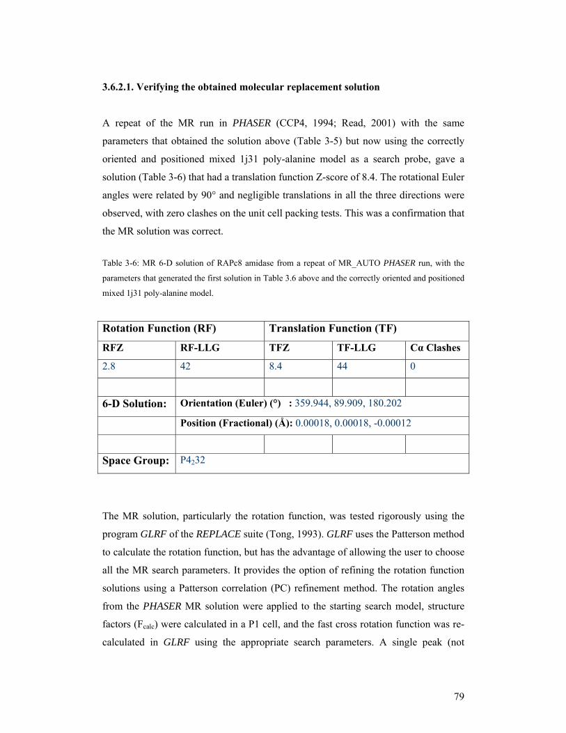
79
3.6.2.1. Verifying the obtained molecular replacement solution
A repeat of the MR run in PHASER (CCP4, 1994; Read, 2001) with the same
parameters that obtained the solution above (Table 3-5) but now using the correctly
oriented and positioned mixed 1j31 poly-alanine model as a search probe, gave a
solution (Table 3-6) that had a translation function Z-score of 8.4. The rotational Euler
angles were related by 90° and negligible translations in all the three directions were
observed, with zero clashes on the unit cell packing tests. This was a confirmation that
the MR solution was correct.
Table 3-6: MR 6-D solution of RAPc8 amidase from a repeat of MR_AUTO PHASER run, with the
parameters that generated the first solution in Table 3.6 above and the correctly oriented and positioned
mixed 1j31 poly-alanine model.
Rotation Function (RF) Translation Function (TF)
RFZ RF-LLG TFZ TF-LLG Cα Clashes
2.8 42 8.4 44 0
6-D Solution: Orientation (Euler) (°) : 359.944, 89.909, 180.202
Position (Fractional) (Å): 0.00018, 0.00018, -0.00012
Space Group: P4232
The MR solution, particularly the rotation function, was tested rigorously using the
program GLRF of the REPLACE suite (Tong, 1993). GLRF uses the Patterson method
to calculate the rotation function, but has the advantage of allowing the user to choose
all the MR search parameters. It provides the option of refining the rotation function
solutions using a Patterson correlation (PC) refinement method. The rotation angles
from the PHASER MR solution were applied to the starting search model, structure
factors (Fcalc) were calculated in a P1 cell, and the fast cross rotation function was re-
calculated in GLRF using the appropriate search parameters. A single peak (not

80
shown) was observed at the origin of the β=0 map sections, indicating that the rotation
function was correct.
The lack of α-carbon clashes on packing the model in the unit cell was a further
confirmation that the translation function solution was correct.
3.6.2.2. Packing of the P4232 unit cell with the MR solution
The ultimate test for the correctness of the obtained MR solution was the consistency
of the packed unit cell with the RAPc8 amidase homohexameric complex features as
revealed by EM analysis. A packed P4232 unit cell (Figure 3-17(A)) had four
hexamers arranged in a tetrahedral manner. The conserved dimer interface that is
found in the four nitrilase superfamily distant homologues was indeed preserved by
the crystallographic 2-fold symmetry in the MR solution (Figure 3-17(B)). In
addition, three dimers packed around the P4232 crystallographic 3-fold axis (Figure 3-
17(C)), with D3 point group symmetry. Projection of the model was remarkably
similar to the EM 3-fold symmetry projections.
3.6.3. Rigid body refinement and phase angles calculation
Although the MR solution model is normally optimised and the phases calculated
using the PHASER (CCP4, 1994; Read, 2001) MR_AUTO mode, an independent
rigid body refinement and phase calculation step was performed to ensure and
confirm the accuracy of the MR solution before an electron density map was
calculated. The MR_RNP mode in PHASER was used to perform rigid body
refinement of the correctly-oriented and positioned mixed 1j31 poly-alanine model
against the RAPc8 amidase data to 3.5 Å resolution. The model was found to be
optimal as no changes were observed in either its orientation (rotational Euler angles)
or its position (translation) in the unit cell. The calculated phases however were
extremely poor, with an overall phasing Figure of Merit (FOM, a measure of
effectiveness, efficiency and performance) of 0.191.
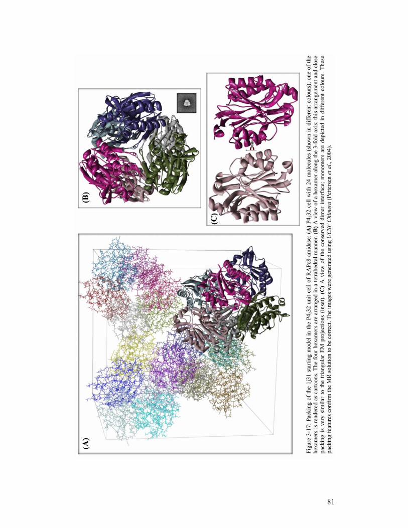
81

82
3.6.3.1. Initial electron density map calculation
A sigma-A weighted 2Fobs-Fcalc electron density map was generated using the
calculated phases (from the correctly oriented and positioned mixed 1j31 poly-alanine
model) and structure factor amplitudes from the observed RAPc8 amidase data to 3.5
Å resolution. The resultant map, although noisy, was interpretable in most regions; the
density for some of the side chains that had been replaced with alanines in the model
had re-appeared (Figure 3-18). Continuous density with considerable amount of noisy
was also observed on the C-terminal side where over 80 residues were missing in the
model. Despite the poor phase information, the emergence of features missing in the
model, in the electron density map was a further confirmation that the MR solution
was correct.
Figure 3-18: A stereo view of a portion of the initial 2Fobs - Fcalc electron density map generated with
mixed 1j31-poly-alanine phases and RAPc8 amidase structure factor amplitudes, calculated at 3.5 Å
and contoured at 0.8σ. The mixed 1j31 poly-alanine model is displayed in the density. Though the map
has a considerable amount of noise, density for some of the side chains that had been replaced with
alanines is visible. An example is the Met41 side chain (orange, with labelled density). The picture was
rendered using PyMOL (Delano, 2004).

83
3.7. Model rebuilding and refinement
Model rebuilding and refinement is an interactive and iterative process that aims at
improving the agreement between the atomic model and the observed data (Pannu,
Read, 1996). While model rebuilding focuses on fitting the model to the electron
density map, either manually or in a semi-automated manner, refinement minimizes
the difference between the atomic model and the observed data by making small
movements of the model atoms, which ultimately result in the improvement of the fit
between the model and the electron density.
Major differences existed between the starting atomic model and the observed RAPc8
amidase data; aside from being only 68% complete, the mixed 1j31 poly-alanine
model had over 75% of all the side chains mutated to alanines. Although the initial
electron density map was interpretable in most regions, a significant amount of bias
towards the starting model bias could not be ruled out. The process of model
rebuilding and refinement was therefore bound to be difficult and complicated with
model-bias creating a bottle-neck. The graphical program O (Jones et al., 1990) was
used for manual rebuilding and fitting of the model to the electron density while
REFMAC5 (CCP4, 1994; Murshudov et al., 1997) was used for restrained refinement.
REFMAC5 uses maximum likelihood targets that take into account errors and
uncertainties (standard deviations) in both the experimental data and the atomic
model, as well any prior phase information (Murshudov et al., 1997), to minimize the
differences between the observed data and the atomic model. The following strategies
were employed in the refinement of RAPc8 amidase structure.
1. The alanines in the mixed 1j31 poly-alanine MR solution were replaced with
RAPc8 amidase side chains using SCWRL (Canutescu et al., 2003), to generate
a model that had the same sequence as the targeted RAPc8 amidase structure.
The resultant model was used to calculate phases which together with
observed RAPc8 amidase data were used to generate a sigma-A weighted
2Fobs–Fcalc electron density map to 3.5 Å resolution. The generated map was
heavily biased towards the model; the electron density almost perfectly

84
matched the model, with no new features observed. The model-biased map
could not allow for further model rebuilding and refinement. This clearly
emphasises the fact that phases have more structural information than the
structure factor amplitudes (Taylor, 2003).
2. A sigma-A weighted 2Fobs–Fcalc electron density map calculated with the
phases from the mixed 1j31 poly-alanine model and the measured RAPc8
amidase amplitudes to 3.5 Å resolution, was used for model fitting. The MR
solution model whose poly-alanines had been replaced with RAPc8 amidase
side chains (as described in number 1 above) was manually rebuilt and fitted
to the interpretable regions of the density using O, alternating with a few
cycles of restrained refinement in REFMAC5 after each rebuilding cycle.
Although the R-factor (measure of the agreement between the model and the
observed data) and phasing FOM showed slight improvements with each cycle
of rebuilding and refinement, the electron density map did not improve
sufficiently to reveal clear density for most of the features that were noisy and
less determined in the initial map. With more cycles of rebuilding and
refinement, the model’s structural elements slowly started to disintegrate
(Figure 3-19). This was attributed to the possibility of over-fitting the model to
electron density features that were not necessarily true (due to starting poor
phases), eventually leading to the disruption of the hydrogen-bonding network
and other interactions that hold secondary structure elements together. The
electron density thus became more and more biased towards the model,
reducing the possibility of building a correct model and making further model
refinement impossible.
3. Density modification (DM) through solvent flattening using RESOLVE
(Terwilliger, 2004a) was performed in an attempt to improve the initial
electron density map prior to rebuilding. RESOLVE uses the principle of
maximum likelihood to maximize the total probability of the phases
(calculated from the map-probability function as described by Terwilliger in
2001), by combining information from the experimental data and the expected
consistency of the map with the flat solvent region (Terwilliger, 1999).
Solvent flattening of the initial electron density map (from mixed 1j31 poly-

85
alanine model phases) was attempted with 50% solvent content (determined
from the calculated Matthew’s coefficient). Although the phasing FOM
showed slight improvement after solvent flattening, the bias ratio (representing
the extent to which the map is biased towards the starting model phases) was
significantly high (3.91) and the map was even noisier. More runs were
performed with gradually increasing levels (in steps of 5%) of solvent content
in an attempt to account for regions that were likely to be highly disordered in
the crystal. DM did not seem to improve the maps even with as much as 70%
solvent. This was most likely an indication that, although the initial phases
were relatively accurate, they were likely to be heavily model-biased.
4. In an attempt to reduce the bias introduced by using a single model, phases
were calculated from an ensemble of the four homologues; prepared by
superimposing (using ALIGN (Cohen et al., 1997)) the homologues (intact
with their side chains) onto the mixed poly-alanine MR solution from
PHASER. This strategy was aimed at enhancing the conserved regions in the
four homologues while down-weighting the variable loop regions. Rigid body
refinement of the ensemble against RAPc8 amidase data to 3.5 Å, resulted in
an increase in LLG value from 44 to 56, and very minimal changes (almost
negligible) on the rotational and translational parameters. The increase in LLG
value was an indication that the ensemble agreed better with the data than a
single model. The overall phasing FOM improved from 0.191 (with single
mixed poly-alanine model) to 0.306. The resultant electron density map was
much improved, and new features could clearly be observed. These new
phases were used for model rebuilding using the prime-and-switch phasing
technique (Terwilliger, 2004b) as implemented in RESOLVE (Terwilliger,
2004a), within PHENIX (Adams et al., 2002). In prime-and-switch phasing,
the crystallographic phases (which might be model-biased) calculated from the
atomic model are used to ‘prime’ the DM process, which then ‘switches’ to a
map-probability function (Terwilliger, 2001) in order to iteratively adjust and
improve the phases so that they are more correct and devoid of the memory of
the initial model (Terwilliger, 2004b). The process of phase improvement
involves cross-validation procedures (that remove model bias. In the model
rebuilding step, the phases calculated from the ensemble as described above,

86
were used together with the measured amplitudes (between 45 and 1.9 Å) from
the RAPc8 amidase crystal and a 1j31 model that contained RAPc8 amidase
side chains. 20 iterative cycles of model rebuilding (in-place) with three
refinement cycles after each rebuilding cycle and 50% solvent content were
performed using prime-and-switch phasing with PHENIX. Over 94% of all the
residues in the model were rebuilt. Prime-and-switch phasing was successful
in significantly reducing model bias; the rebuilt model had atoms shifted
significantly (Figure 3-20) to their correct positions, the phases had
dramatically improved with an increase in phasing FOM from 0.306 to 0.552
and the electron density map (Figure 3-21) was clear with interpretable density
for most of the residues that were absent in the starting model.
Figure 3-19: Cartoon and transparent surface representations of the models: (A), before rebuilding and
refinement and (B), after several cycles of rebuilding in O and restrained refinement in REFMAC5. The
model structural elements slowly disintegrated (helices and beta sheets breaking up) with continued
fitting, rebuilding and refinement. Images rendered in PyMOL (Delano, 2004).
(A) (B)

87
Figure 3-21: A stereo view of a portion of a simple map generated with phases after the rebuilding step
with PHENIX, calculated at 1.9 Å and contoured at 1.2 σ. The 1j31 model substituted with RAPc8
amidase side chains before rebuilding is shown in pink sticks while in green sticks is the model after
rebuilding with PHENIX. The rebuilt model is greatly shifted and the map features are quite clear, with
reduced bias towards the starting model. Met41 (labelled) is now in well-defined density; and so are the
other amino acids in that portion of the map. The picture was rendered using PyMOL (Delano, 2004).
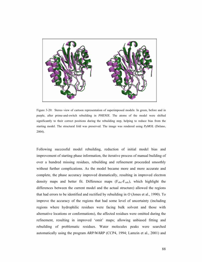
88
Figure 3-20: Stereo view of cartoon representation of superimposed models: In green, before and in
purple, after prime-and-switch rebuilding in PHENIX. The atoms of the model were shifted
significantly to their correct positions during the rebuilding step, helping to reduce bias from the
starting model. The structural fold was preserved. The image was rendered using PyMOL (Delano,
2004).
Following successful model rebuilding, reduction of initial model bias and
improvement of starting phase information, the iterative process of manual building of
over a hundred missing residues, rebuilding and refinement proceeded smoothly
without further complications. As the model became more and more accurate and
complete, the phase accuracy improved dramatically, resulting in improved electron
density maps and better fit. Difference maps (Fobs-Fcalc), which highlight the
differences between the current model and the actual structure) allowed the regions
that had errors to be identified and rectified by rebuilding in O (Jones et al., 1990). To
improve the accuracy of the regions that had some level of uncertainty (including
regions where hydrophilic residues were facing bulk solvent and those with
alternative locations or conformations), the affected residues were omitted during the
refinement, resulting in improved ‘omit’ maps; allowing unbiased fitting and
rebuilding of problematic residues. Water molecules peaks were searched
automatically using the program ARP/WARP (CCP4, 1994; Lamzin et al., 2001) and

89
rationalised manually to ensure availability of possible hydrogen bonding network. As
model refinement proceeded, more water molecules were incorporated in the
structure, improving the quality and accuracy of the model each time. The model was
manually inspected and analysed after every cycle of refinement.
The progress of the refinement process as well as the overall accuracy of the atomic
model was judged through the crystallographic R-factor (which represents the average
fractional error in the model of the molecular structure compared to the observed
diffraction data from which it has been derived from). A free R-factor (R-free) was
calculated from 5% cross-validation data (test set) that was omitted from the
refinement process. R-free was introduced by Brünger (1992) and it represents a
statistical measure that is essentially free from the artifactual lowering that can be
caused by over-fitting. Figure 3-22 below shows the behaviour of crystallographic R-
factor, R-free and phasing FOM during the rebuilding and refinement of RAPc8
amidase structure model. Since the crystallographic R-factor is only a global indicator
of model quality (Kleywegt, 2000) (and does not highlight local errors in the
structure), structure validation programs as well as manual evaluation of the fit of the
model to the electron density were used as ultimate checks for problematic local
regions in the model, especially towards the end of the refinement. Refinement was
assumed to have converged globally when all the density belonging to the structural
features had been accounted for, the difference maps were featureless and parameter
changes in the model were negligibly small in further refinement cycles.
A portion of the electron density map at the end of the refinement, with the refined
atomic model fitted in is shown in Figure 3-23 below.

90
Key:
Step Action
A 1j31 MR solution - substituted with RAPc8 amidase side chains, before rebuilding in
PHENIX
B After prime-and-switch phasing and rebuilding in PHENIX
C On correction of major errors as well as building and rebuilding residues 1-308
D All 340 residues built
E 137 water molecules added automatically with ARP/WARP
F 100 more water molecules added manually; most errors corrected
G Residues with alternative locations were built and 34 water molecules added
H Most of the detectable errors corrected, and all justifiable waters added
I Refinement converges
Figure 3-22: Progress in rebuilding and restrained refinement of RAPc8 amidase structure. R-factor is
shown in blue circles, R-free in purple squares and phasing FOM in green triangles. As the model
agreed better with the observed data, the crystallographic R-factor and R-free dropped consistently,
while the phasing FOM increased in a similar but opposite trend. Refinement convergence was reached
when the model was not improving much further and the refinement statistics had reached a plateau.
Rebuilding the model in PHENIX caused a large increase in the phasing FOM as a result of improved
accuracy in the phases.

91
Figure 3-23: A stereo view of a portion of a 2Fobs-Fcalc map at the end of the refinement calculated at
1.9 Å and contoured at 1.3 σ. The refined model is displayed in the density. The picture was rendered
using PyMOL (Delano, 2004).
The final model had 1-340 out of 348 (from the theoretically translated sequence)
residues. The 8 residues that were missing on the C-terminus were likely to have been
absent from the protein during crystallization, as opposed to them being disordered; as
there was no density at all for these residues. The model was of good quality as can be
observed from the refinement statistics presented in Table 3-7 below. The relatively
high resolution data (to 1.9 Å) allowed for the location of most of the protein chain-
associated water molecules, resulting in a total of 304 waters in the final structure.
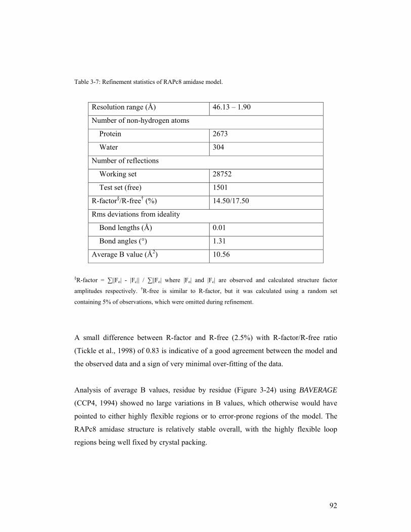
92
Table 3-7: Refinement statistics of RAPc8 amidase model.
Resolution range (Å) 46.13 – 1.90
Number of non-hydrogen atoms
Protein 2673
Water 304
Number of reflections
Working set 28752
Test set (free) 1501
R-factor§/R-free† (%) 14.50/17.50
Rms deviations from ideality
Bond lengths (Å) 0.01
Bond angles (°) 1.31
Average B value (Å2) 10.56
§R-factor = ∑||Fo| - |Fc|| / ∑||Fo| where |Fo| and |Fc| are observed and calculated structure factor
amplitudes respectively. †R-free is similar to R-factor, but it was calculated using a random set
containing 5% of observations, which were omitted during refinement.
A small difference between R-factor and R-free (2.5%) with R-factor/R-free ratio
(Tickle et al., 1998) of 0.83 is indicative of a good agreement between the model and
the observed data and a sign of very minimal over-fitting of the data.
Analysis of average B values, residue by residue (Figure 3-24) using BAVERAGE
(CCP4, 1994) showed no large variations in B values, which otherwise would have
pointed to either highly flexible regions or to error-prone regions of the model. The
RAPc8 amidase structure is relatively stable overall, with the highly flexible loop
regions being well fixed by crystal packing.

93
Figure 3-24: An all atom plot of average B value per residue. Apart from the C-terminal Glu340, which
had an average B value of 29.5 Å2, the rest of the residues had relatively low average B factors, and
indication of a relatively stable overall structure.
94
3.8. Final Model Validation
Aside from the global indicators of the quality model indicators; R-factor and R-free
(that measure the agreement between the model and the data), the overall and local
accuracy of the final model is usually assessed by various validation methods. These
methods utilize aspects of the model that are independent of the information that is
optimised during model rebuilding and refinement (Kleywegt, 2000; Richardson,
2003). Torsion angles for instance, are not optimised by the refinement process, thus
they form the basis for most geometric validation methods. In most cases, validation
approaches are entirely model-based and they utilize information derived from highly
accurate structures from the databases, to identify local outlier regions that deviate
from ideality (Richardson, 2003).
The program PROCHECK (CCP4, 1994; Laskowski et al., 1993) was used to assess
the molecular geometry, particularly the backbone (φ and ψ angles) and the side
chains (mainly side chain torsion angles) conformations. The program WHATCHECK
(Hooft et al., 1996) was used for a number of geometric checks, analysis of residues
environment and assessment of hydrogen bonding network. MOLPROBITY (Lovell et
al., 2003) was used for geometric validation, particularly in analysing the complete α-
carbon geometry (Lovell et al., 2003), including backbone φ and ψ angles and the
deviation of the β-carbon positions (that provide information on the backbone-side
chain incompatibilities). MOLPROBITY generates accurate φ, ψ Ramachandran plots
(Ramachandran et al., 1963; Laskowski et al., 1993; Lovell et al., 2003), with
redefined allowed and disallowed regions, based on current information from highly
accurate structures (Lovell et al., 2003). It performs all-atom contacts analysis (Word
et al., 1999), providing information on hydrogen bonding network, van der Waals
contacts, and unfavourable atomic clashes thus highlighting local structural
inaccuracies (Lovell et al., 2003). Table 3-8 provides a summary of the identified
outliers in the RAPc8 amidase final model.

95
Table 3-8: Outliers in the refined RAPc8 amidase structure
Pairs of residues with atomic clashes (12; 1 clash per pair)
Ramachandran outliers 0
Rotamer outliers 6
Cβ outliers 0
Residues with distorted geometry 1
Residues with unusual backbone
conformations 30
Residues in abnormal packing
environment 9
Buried unsatisfied hydrogen bond
donors and acceptors 4
Figure 3-25: Backbone φ, ψ angles distribution for the refined RAPc8 amidase structure: (A) A general
case for non-glysine, non-proline and non-preproline residues, generated using MOLPROBITY. (B) An
all residues case; generated with PROCHECK.
(A) (B)

96
The Ramachandran plot (Figure 3-25) statistics from PROCHECK (CCP4, 1994;
Laskowski et al., 1993) had 99.6% of all residues in the favoured (90%) and allowed
(9.6%) regions, and 0.3% (1 residue, cysteine 166) in the disallowed region. Cysteine
166 is found in the allowed but rare region (type II’-turn conformation) of the
MOLPROBITY (Lovell et al., 2003) Ramachandran plot. In the recently defined type
II’-tight turns conformation, the second of the four residues in a turn adopts φ, ψ
angles near 50°, -125°, resulting in steric overlap between the β-carbon and carbonyl
oxygen. This conformation is common in the active sites of various protein families,
including the serine of the catalytic triad of all α/β hydrolases and lipases (Uppenberg
et al., 1994). The cysteine of the Glu, Lys, Cys catalytic triad in the four nitrilase
superfamily structures is also found in the type II’-turn conformation region of the
Ramachandran plot, indicating a functional role in mediating activity in these
enzymes.
No β-carbon position outliers (Table 3-8) were found in the RAPc8 amidase structure,
which indicates good compatibility between the backbone and the side chains.
However, some unusual features were observed (Table 3-8) despite clear interpretable
density in the highlighted regions. The 12 atomic clashes involved hydrogen atoms of
neighbouring residues. Most of the overlapping hydrogen atoms are found in the
tightly constrained active site, while a number of clashes involve residues in sharp
bends. Of the 6 residues that had unusual side chain rotamers, 2 (Tyr192 and Met193)
are in the highly constrained active site, 3 are in the tightly packed environment while
the last one is the Glu340 residue that is facing the bulk solvent.
Only one residue had distorted geometry; a cis-conformation proline 195, that is
found in a tight bend. Although the environment could partly explain the constraint
on this residue, any efforts to improve the geometry (including simulated annealing
and molecular dynamics) were unsuccessful. Of the four buried residues that had
deficiencies in hydrogen-bonding network, two are neighbouring cysteine residues
possibly forming weak cysteine hydrogen bonds, one is in the active site; while one is
in a bend, where it is likely to be stabilised by neighbouring hydrogen bonding
environment. The residues in the abnormal environment and those with unusual
backbone conformations, are mostly found in the bends, some are on the edges, a

97
number are in the highly constrained active site and a few are on the interfaces,
interacting with symmetry-related molecules.
Overall, the refined RAPc8 amidase model was of high quality, showing reasonably
good agreement with the high quality diffraction data and minimal errors.
98
3.9. Analysis of the RAPc8 amidase structure
(A) - Geobacillus pallidus RAPc8 amidase has an α-β-β-α sandwich fold
The monomer of RAPc8 amidase is an α/β globular protein that consists of 11 helices
and 14 β strands, interconnected with external loops. A cartoon representation of a
RAPc8 amidase monomer, and the topology of β sheets and α helices are shown in
Figure 3-26 below. Helices α1, α2 and α3 form one outer helical layer while helices
α4, α5 and α6 form the other. β strands β1, β2, β3, β4, β5, and β14 form one β sheet
while β strands β6, β7, β8, β9, β12 and β13 form the other sheet. The two six-stranded
β sheets are sandwiched between the two helical layers to form a four-layer α-β-β-α
sandwich architecture (Orengo et al., 1997). The two β sheet layers have a similar
alternation of parallel and anti-parallel β strands; the three central β strands of each β
sheet (β1, β2, β3 and β7, β8, β9) form a parallel β sheet, while the rest of the strands
are arranged in an anti-parallel configuration. The interface between the two β sheets
is tightly packed with hydrophobic residues. Helices α8, α9, α10, and α11 are found
on the extended C-terminal tail, while β strands β10 and β11 are on a long external
loop that links β9 and β12. Four residues form a 3-10 helix (shown in cyan on the
topology diagram) at the active site, between β7 and α5. The two α/β halves are
connected by two loops, one at each end; a 20 residues loop between β5 and β6 on
one end and an 8 residues loop between β13 and β14 on the other end.
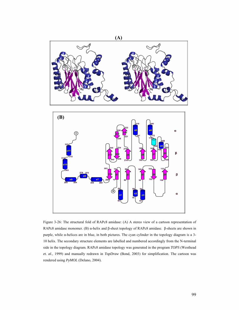
99
Figure 3-26: The structural fold of RAPc8 amidase: (A) A stereo view of a cartoon representation of
RAPc8 amidase monomer. (B) α-helix and β-sheet topology of RAPc8 amidase. β-sheets are shown in
purple, while α-helices are in blue, in both pictures. The cyan cylinder in the topology diagram is a 3-
10 helix. The secondary structure elements are labelled and numbered accordingly from the N-terminal
side in the topology diagram. RAPc8 amidase topology was generated in the program TOPS (Westhead
et. al., 1999) and manually redrawn in TopDraw (Bond, 2003) for simplification. The cartoon was
rendered using PyMOL (Delano, 2004).
(A)
(B)

100
(B) - Geobacillus pallidus RAPc8 amidase is a member of the nitrilase
superfamily
There are 14 existing topology classes under the α/β class with four-layer sandwich
architecture (3.6.0) in the CATH (class, architecture, topology, homologous
superfamily) database (Orengo et al., 1997). These topologies are found in a number
of enzyme families, including hydrolases, lyases, ligases, transferases,
oxidoreductases, and isomerases. A structure similarity search of the DALI database
(Holm & Sander, 1993) with RAPc8 amidase identified the four nitrilase superfamily
structures (PDB IDs; 1j31, 1f89, 1erz & 1ems) with high structural similarity (average
Z-score of 32.0). The structural similarity with other reported four-layer sandwich
proteins was poor. This was a further confirmation that RAPc8 amidase is a C-N
hydrolase belonging to the nitrilase superfamily. The fold similarity between RAPc8
amidase and putative C-N Hydrolase from yeast (1f89; Kumaran et al., 2003) is
shown in Figure 3-27 below. Although the two structures are only 20.7% similar in
sequence, they share a common four-layer sandwich architecture; the secondary
structure element topology is highly similar in the conserved core with only a few
minor differences such as the position of α4 helix (which is between β5 and β6 in
1f89 and links the two α/β halves while in RAPc8 amidase, α4 helix is between β6
and β7) and the insertion of the two small β strands (β10 and β11) on the external loop
of RAPc8 amidase. Outside the α-β-β-α conserved core, RAPc8 has an extended C-
terminal tail that is completely helical. The same topology similarity and structural
fold are shared with 1j31, 1erz and the Nit domain of 1ems as shown in Figure 3-28
below.
(C) - Geobacillus pallidus RAPc8 amidase is homohexameric is solution
Gel filtration chromatography and electron microscopy (EM) had indicated that
RAPc8 amidase is homohexameric in solution. These findings have been confirmed
by the crystal structure, where RAPc8 amidase exists as an associated homohexamer
(Figure 3-29), comprised of trimers of dimers.
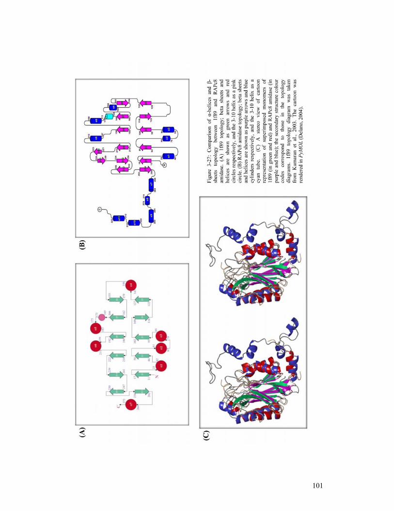
101
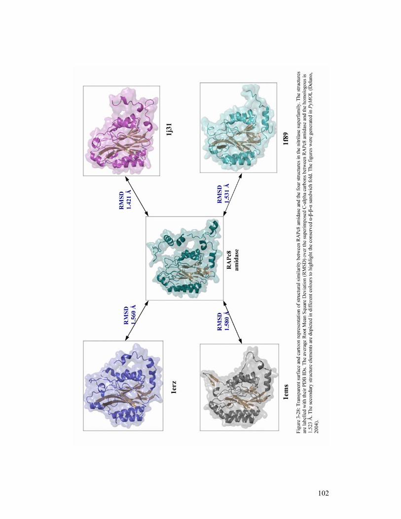
102
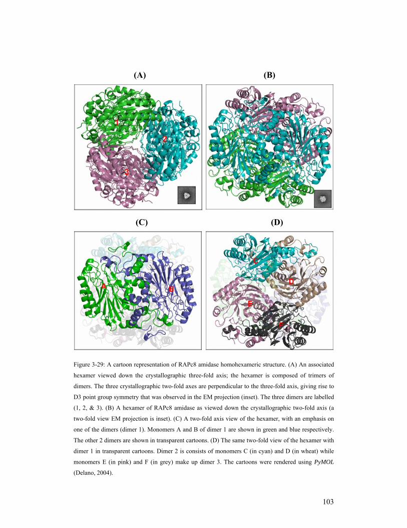
103
Figure 3-29: A cartoon representation of RAPc8 amidase homohexameric structure. (A) An associated
hexamer viewed down the crystallographic three-fold axis; the hexamer is composed of trimers of
dimers. The three crystallographic two-fold axes are perpendicular to the three-fold axis, giving rise to
D3 point group symmetry that was observed in the EM projection (inset). The three dimers are labelled
(1, 2, & 3). (B) A hexamer of RAPc8 amidase as viewed down the crystallographic two-fold axis (a
two-fold view EM projection is inset). (C) A two-fold axis view of the hexamer, with an emphasis on
one of the dimers (dimer 1). Monomers A and B of dimer 1 are shown in green and blue respectively.
The other 2 dimers are shown in transparent cartoons. (D) The same two-fold view of the hexamer with
dimer 1 in transparent cartoons. Dimer 2 is consists of monomers C (in cyan) and D (in wheat) while
monomers E (in pink) and F (in grey) make up dimer 3. The cartoons were rendered using PyMOL
(Delano, 2004).
(A) (B)
(C) (D)

104
The interaction of two monomers along the crystallographic two-fold axis to form a
dimer in the crystal structure, results in an eight-layered α-β-β-α:α-β-β-α sandwich
structure (Figure 3-30 (A)). This dimer interface (also denoted as A-surface by Sewell
and colleagues (2003)) is conserved in the four nitrilase superfamily crystal structures
(PDB IDs: 1j31, 1f89, 1erz, & 1ems; Sakai et al., 2004, Kumaran et al., 2003, Nakai
et al., 2000 & Pace et al., 2000, respectively). Similar to the homologous structures,
the conserved dimeric interface in RAPc8 amidase is formed by helices α5 (residues
166-186) and α6 (residues 195-213) from both monomers, as well as the C-terminal
tails (residues 273-340) that fold over the entire A-surface. The interfacing helices (α5
and α6) are anti-parallel and they form compact interactions that are mainly
hydrophobic, in addition to 4 salt bridges involving Glu173 and Arg176 of each
monomer (Figure 3-30(C)). Two hydrogen bonds also exist between Trp209 and
Asn170. Some of the residues (atoms) interacting on the dimer interface are listed in
table 3-9 below
Table 3-9: Interactions at the conserved dimer interface.
Monomer A Monomer B Distance (Ǻ)
Arg176 NH2 Glu173 OE1 2.89
Arg176 NE Glu173 OE2 2.80
Trp209 NE1 Asn170 OD1 2.88
Met202 SD Lys205 Cβ 3.40
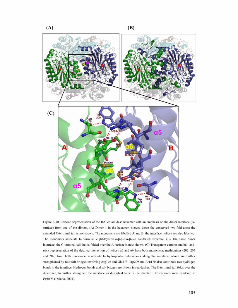
105
Figure 3-30: Cartoon representation of the RAPc8 amidase hexamer with an emphasis on the dimer interface (A-
surface) from one of the dimers. (A) Dimer 1 in the hexamer, viewed down the conserved two-fold axes; the
extended C-terminal tail is not shown. The monomers are labelled A and B, the interface helices are also labelled.
The monomers associate to form an eight-layered α-β-β-α:α-β-β-α sandwich structure. (B) The same dimer
interface; the C-terminal tail that is folded over the A-surface is now shown. (C) Transparent cartoon and ball-and-
stick representation of the detailed interaction of helices α5 and α6 from both monomers; methionines (202, 203
and 207) from both monomers contribute to hydrophobic interactions along the interface, which are further
strengthened by four salt bridges involving Arg176 and Glu173. Trp209 and Asn170 also contribute two hydrogen
bonds in the interface. Hydrogen bonds and salt bridges are shown in red dashes. The C-terminal tail folds over the
A-surface, to further strengthen the interface as described later in the chapter. The cartoons were rendered in
PyMOL (Delano, 2004).
(A) (B)
(C)

106
The interaction of helices α6 on the conserved dimer interface shows a class II helix-
helix packing pattern (Chothia et al., 1977) where the angle between the packing
helices is approximately -60°. This packing is illustrated in Figure 3-31 below.
Figure 3-31: A stereo diagram of a cartoon representation of the helix-helix packing pattern at the
conserved dimer interface in RAPc8 amidase. Helices from chain A are coloured green while those
from Chain B are blue, they are all labelled. The helices are viewed from a direction perpendicular to
the crystallographic two-fold axes. Helices α6 from both monomers pack against one another by
crossing over to form a class II helix-helix pattern. The image was generated using PyMOL (Delano,
2004).
In the hexameric complex (Figure 3-29), one dimer interacts with the other two
dimers at two-fold interfaces (which we have termed F-surfaces) that are opposite the
conserved dimer interfaces (A-surface). Specifically, chain A of dimer 1 interacts
with chain F of dimer 3, chain B of dimer 1 with chain C of dimer 2 while chain D of
dimer 2 interacts with chain E of dimer 3, so that interface A-B is opposite D-E, C-D
is opposite F-A and E-F is opposite B-C. The F-surface is formed by helices α7 and
the N-terminal loops of the two interacting monomers. These interactions are
predominantly electrostatic with a number of salt bridges and a few hydrophobic
interactions. Figure 3-32 shows a 2D cylindrical projection of the RAPc8 amidase
hexamer density, which makes it easy to visualize the interacting subunits. Table 3-10

107
shows a list of the interacting residues on the F-surface while Figure 3-33 shows the
details of these interactions.
Figure 3-32: A 2D cylindrical projection of the density of the RAPc8 amidase hexamer, with the
cylinder axis and the three-fold axis aligned. The density of each subunit was projected separately,
coloured and then combined to form the composite image. The subunits are labeled. The conserved A-
surfaces exist between A-B, C-D and E-F, which constitute dimers 1, 2, and 3 respectively, while the
novel F-surfaces links A-F, B-C and D-E. The figure was generated using UCSF Chimera (Pettersen et
al., 2004)
Table 3-10: Interactions at the two-fold interface (F-surface) that links one dimer to another in RAPc8
amidase hexameric complex.
Monomer B Monomer C Distance (Ǻ)
Arg2 NH1 Gln262 OE1 2.94
Arg2 NH2 Asp265 OD2 2.95
His3 N Asp239 OD2 2.94
Asp5 OD1 Gln273 N 2.71
Asp5 OD2 Gln271 N 2.85
Ser7 OG Asn269 O 2.98
Ser9 OG Asp265 OD2 2.67
Arg241 NH1 Thr242 O 2.98
A
B
C
D
E
F

108
Figure 3-33: Cartoon images of the two-fold interface (F-surface) that links one dimer to another in the RAPc8 amidase hexamer: (A) Interaction between monomer B (blue) in dimer 1 and monomer C (cyan) in dimer 2 as viewed down the F-surface two-fold axes. Helices α7 and the N-terminal loops are labelled. The curved arrows (labelled A) are pointing to the conserved dimer interface (A-surface) that involves helices α5 and α6, which are labelled. (B) Detailed interactions of helices α7 and the N-terminal loops on the F-surface crystallographic two-fold axis. The interacting monomers are shown in cyan (labelled C) and blue (labelled B). The black straight arrow is pointing to the two-fold axis. This interface is stabilised mainly by electrostatic interactions, with at least two salt bridges between Asp265 and Arg2 of both monomers. The cartoon images were generated using PyMOL (Delano, 2004).
(A)
(B)
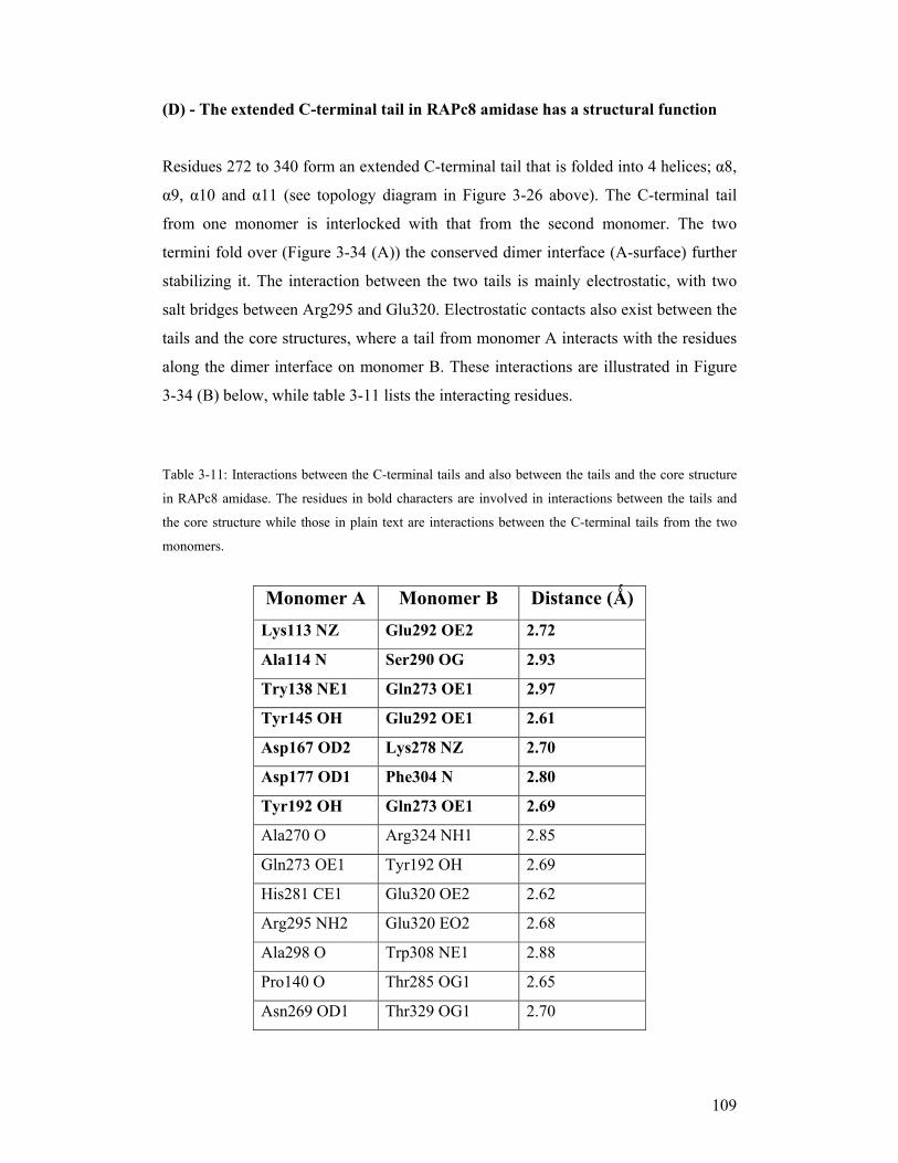
109
(D) - The extended C-terminal tail in RAPc8 amidase has a structural function
Residues 272 to 340 form an extended C-terminal tail that is folded into 4 helices; α8,
α9, α10 and α11 (see topology diagram in Figure 3-26 above). The C-terminal tail
from one monomer is interlocked with that from the second monomer. The two
termini fold over (Figure 3-34 (A)) the conserved dimer interface (A-surface) further
stabilizing it. The interaction between the two tails is mainly electrostatic, with two
salt bridges between Arg295 and Glu320. Electrostatic contacts also exist between the
tails and the core structures, where a tail from monomer A interacts with the residues
along the dimer interface on monomer B. These interactions are illustrated in Figure
3-34 (B) below, while table 3-11 lists the interacting residues.
Table 3-11: Interactions between the C-terminal tails and also between the tails and the core structure
in RAPc8 amidase. The residues in bold characters are involved in interactions between the tails and
the core structure while those in plain text are interactions between the C-terminal tails from the two
monomers.
Monomer A Monomer B Distance (Ǻ) Lys113 NZ Glu292 OE2 2.72
Ala114 N Ser290 OG 2.93
Try138 NE1 Gln273 OE1 2.97
Tyr145 OH Glu292 OE1 2.61
Asp167 OD2 Lys278 NZ 2.70
Asp177 OD1 Phe304 N 2.80
Tyr192 OH Gln273 OE1 2.69
Ala270 O Arg324 NH1 2.85
Gln273 OE1 Tyr192 OH 2.69
His281 CE1 Glu320 OE2 2.62
Arg295 NH2 Glu320 EO2 2.68
Ala298 O Trp308 NE1 2.88
Pro140 O Thr285 OG1 2.65
Asn269 OD1 Thr329 OG1 2.70
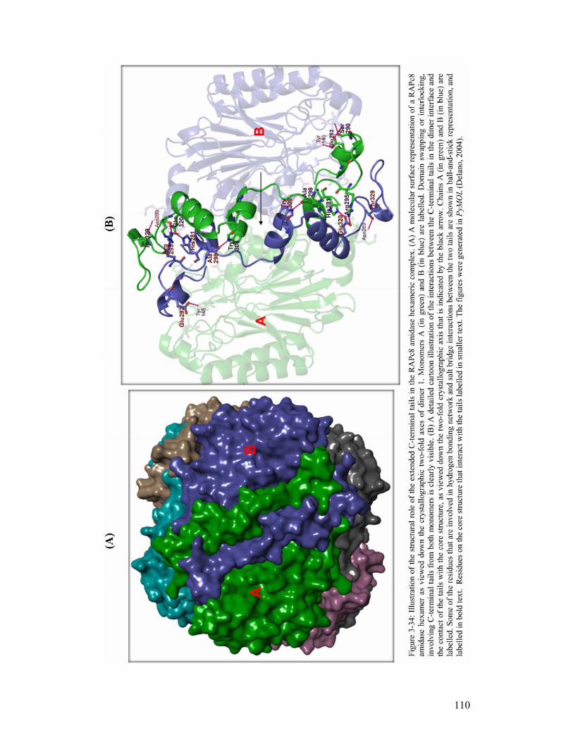
110

111
Aside from the overall strengthening of the A-surface, the domain swapping of the C-
terminal tails could also play the role of linking the monomers in the process of dimer
formation. This role was first proposed by Nakai and colleagues (2000), in the
structure of N-carbamyl-D-amino acid amidohydrolase (DCase: PDB ID, 1erz).
RAPc8 amidase C-terminal tails however are unique in that they are much longer
which probably emphasizes their role in driving the process of hexamer formation, as
well as in holding the hexameric complex together.
(E) - The RAPc8 amidase active site framework is similar to nitrilase
superfamily enzymes
RAPc8 amidase shares structural homology to the members of the nitrilase
superfamily. One of the conserved motifs in the nitrilase enzymes is the cysteine,
glutamate and lysine (Glu, Lys, Cys) catalytic triad (Novo et al., 1995; Pace, Brenner,
2001) that is involved in the hydrolysis of the carbon-nitrogen bond. In RAPc8
amidase, the catalytic triad is composed of Glu59, Lys143 and Cys166. The
coordination of these residues is similar to that of the four nitrilase superfamily
structural homologues, as shown in Figure 3-35 below.
A buried cleft corresponding to the binding pocket was identified and characterised in
each monomer of the RAPc8 amidase structure using CASTp (Binkowski et al., 2003)
online server. The pocket is formed by residues in the loops between β-strand β2 and
the small helix α2, strands β5 and β6, strand β7 and helix α5, as well as strand β8 and
helix α6. It is located near the edge of the dimer interface. This active site location is
similar to all α/β four-layer sandwich structures including nitrilase superfamily
enzymes. Figure 3-36 below shows the location of active site clefts of both RAPc8
amidase and N-carbamyl-D-amino acid amidohydrolase (DCase) (Nakai et al., 2000).

112
Figure 3-35: A transparent cartoon image showing the coordination of the catalytic triad residues in
RAPc8 amidase as compared to the nitrilase superfamily homologous structures (PDB IDs, 1j31, 1f89,
1erz, and 1ems). 1j31 is in yellow, 1ems in green, 1erz in cyan, 1f89 in magenta and RAPc8 amidase in
blue. The catalytic triad residues (Glu, Lys, Cys) have similar coordination and position in all five
structures indicating their role in driving the catalysis in the superfamily. This further confirms the
structural relationship between RAPc8 amidase structure and the existing nitrilase superfamily
structures. The five structures were superimposed with ALIGN (Cohen, 1997) and the figure was
rendered in PyMOL (Delano, 2004).
Figure 3-36: The location of the active site clefts in both RAPc8 amidase and nitrilase superfamily
enzyme, DCase (Nakai et al., 2000): (A) Cartoon representation of a RAPc8 amidase dimer; the
monomers are shown in green (monomer A) and blue (monomer B). (B) A cartoon representation of a
DCase dimer; monomers A (in cyan) and B (in brown) are labelled. The dimer interfaces are labelled
and the atoms lining the binding sites are rendered as spheres. The active-site pocket exists in each
monomer in both cases with the pockets having similar locations. DCase pocket is however many times
larger and deeper than that of RAPc8 amidase. The figures were generated using PyMOL (Delano,
2004).
(A) (B)
Amidase DCase

113
The RAPc8 amidase active site cleft is lined with residues Glu59, Tyr60, Trp138,
Glu142, Cys166, Gly191, Tyr192 and Met193 as depicted in Figure 3-37 below. A
hydrogen bonding network exists that plays a role in maintaining a highly constrained
configuration of the residues in the substrate binding site. The catalytic residue
Cys166 is located on a β strand-turn-helix structural motif, which is formed by strand
β7 and helix α5 (Figure 3-37 (B)). This motif is termed a nucleophile elbow
(Kumaran et al., 2003) and is found in many α/β four-layer sandwich hydrolases
(Schrag & Cygler, 1997). Similar to the catalytic cysteines in the other C-N hydrolase
structures, Cys166 has a rare but allowed combination of peptide bond dihedral
angles, falling in the recently described type II’-turn conformation (Lovell et al.,
2003) region of the Ramachandran plot. The carbonyl oxygen of Cys166 forms
hydrogen bonds with the NE (3.02 Å) and NH2 (2.83 Å) atoms of Arg188; this
hydrogen bonding network may play a role in maintaining the orientation of Cys166
side chain in the active site.
A second glutamate residue (Glu142) is present in the active site. This may play a role
in making Glu59 a better nucleophile, which together with an electrophilic Lys134,
assists Cys166 in attacking the carbonyl carbon of an amide substrate. Figure 3-38
presents a catalytic mechanism of RAPc8 amidase that was proposed in our research
group. Upon the nucleophilic attack of the carbonyl group by Cys166, an unstable
tetrahedral intermediate (number [1] in the diagram) is formed. With the removal of
ammonia, a nucleophilic Lys134 and a water molecule assist in the generation of a
second tetrahedral intermediate [3], which then decomposes into an acidic product
and a regenerated amidase active site environment.

114
(A)
(B) Figure 3-37: RAPc8 amidase active site structure. (B) A stereo view of the residues lining the active site pocket; Glu59, Lys134 and Cys166 are the catalytic triad residues, and they are shown in blue ball-and-sticks. The hydrogen bonds between atoms are shown in purple dashes. The brown arrow points to the possible route into the cleft. (B) A cartoon diagram of the nucleophile elbow where the catalytic Cys166 is located. Cys166 is shown as ball-and-stick. Similar to other nitrilase superfamily structures, Cys166 backbone adopts type II’-turn conformation with a positive phi angle. This is important in maintaining a favourable side chain orientation for catalysis. The images were rendered using PyMOL (Delano, 2004).

115

116
Unlike in the other nitrilase superfamily structures where the active site clefts are
reported to be large and deep (Kumaran et al., 2003; Nakai et al., 2000; Sakai et al.,
2004), the RAPc8 amidase binding pocket is relatively shallow, with a molecular
volume of 53.59 Å3 and a solvent accessible area of 4.683 Å2. The DCase substrate-
binding pocket for instance has a molecular volume of 1134.7 Å3 and a solvent
accessible area of 425.8 Å2, making it over 20 times bigger than RAPc8 amidase
pocket. The small size of the active site pocket may explain the limited specificity of
RAPc8 amidase for short acyl-chained aliphatic amides as revealed by recent
biochemical characterization (Makhongela et al., in press). A comparison of the
RAPc8 amidase pocket (Figure 3-39) with that of a substrate-bound DCase mutant
structure (PDB ID: 1fu5, Hashimoto et al., unpublished data) rationalized the
specificity of RAPc8 amidase for small un-branched amide compounds and also
revealed the role of some active site residues in restricting the size of amide
substrates. The bulky side chain from Trp138 partly covers the binding pocket from
the outside, hence acting as a ‘filter’, that only allows small un-branched substrates to
access the active site. The presence of the Trp138 side chain at the gateway to the
substrate binding pocket may also explain why CASTp (Binkowski et al., 2003) could
not locate any mouth openings for the RAPc8 amidase binding pocket.

117
Figure 3-39: Surface and cartoon representation of a comparison between RAPc8 amidase active site
pocket and that of a substrate-bound DCase mutant (PDB ID: 1fu5, Hashimoto et al., unpublished
data). RAPc8 amidase is shown in green while DCase is in violet. The N-carbamyl-D-methionine
substrate bound in the active site pocket of DCase is shown in yellow sticks and labelled. The catalytic
triad residues (shown in ball-and-stick) are in similar positions in the two structures. The Trp138 side
chain of RAPc8 amidase clashes with the bulky substrate of DCase. This means that the amide
substrates of RAPc8 amidase would have to be of a smaller size in order to access the pocket for
hydrolysis. The two structures were superimposed using ALIGN (Cohen, 1997) and the image was
rendered with PyMOL (Delano, 2004).
(F) - The catalytic Cys166 is modified
Unlike other nitrilase superfamily structures in which a number of bound water
molecules, believed to participate in catalysis, are present in the active sites, no waters
were found in the active site pocket of RAPc8 amidase. However, both 2Fobs–Fcalc and
positive difference electron density maps indicated that Cys166 was modified,
possibly by oxidation in the crystal (Figure 3-40). Attempts to fit this density either as

118
a sulfinic acid or a sulfenic acid were unsuccessful. We therefore suggest that the
Cys166 oxidative modification is a mixture of species. The importance (if any) of this
modification is uncertain, but any form of oxidation would decrease the
nucleophilicity of Sγ atom in the modified cysteine residue; this partly explains why
RAPc8 amidase enzyme progressively lost activity in the absence of reducing agents
during purification (Makhongela et al. in press).
Figure 3-40: A stereo view of the electron density maps around the active site residues Glu59, Lys 134,
and Cys166. A sigma-A weighted 2Fobs–Fcalc map contoured at 1.3 σ is shown in blue while a sigma-A
weighted Fobs–Fcalc difference map contoured at 3.0 σ and is shown in red. The positive difference
electron density around Sγ atom of Cys166 suggests oxidative modification probably to a mixture of
species, including sulfinic acid. The image was rendered using PyMOL (Delano, 2004).

119
CHAPTER 4: CONCLUSIONS

120
The successful determination of the Geobacillus pallidus RAPc8 amidase crystal
structure was a combination of skills and techniques from various fields, each of
which contributed unique information to the process.
Gel filtration analysis was the first to shed light onto the possible biological unit of the
RAPc8 amidase enzyme. A 220 kDa peak on a preparative gel exclusion
chromatography column indicated an approximate hexameric complex, of 38 kDa
molecular weight per subunit. Electron microscopy analysis allowed for the
visualization of these complexes, and provided crucial information on the symmetry
of the molecule, which strongly guided the X-ray crystallography work. The
prediction of the RAPc8 amidase secondary structure elements revealed the structural
relationship between this enzyme and the members of the nitrilase superfamily,
despite the low sequence homologies that exist between these enzymes.
High protein purity levels as well as homogeneity were important in allowing
crystallization, leading to highly ordered crystals that diffracted well, and that
generated a high quality dataset, which led to the determination of RAPc8 amidase
structure. The power of maximum likelihood-based crystallographic software cannot
be under-estimated in this work. A new generation of software is now emerging that is
allowing determination of difficult structures. The high symmetry of RAPc8 amidase
crystal and the low sequence identity between RAPc8 amidase and the nitrilase
superfamily distant homologues, presented huge obstacles in the search for the
molecular replacement solution, and in the model refinement. These were however
overcome by a combination of maximum likelihood crystallographic programs
including POINTLESS, PHASER, PHENIX and REFMAC5 among others, that
allowed assignment of the correct space group, determination of the molecular
replacement solution, and rebuilding of the starting model prior to model fitting and
refinement.
The crystal of RAPc8 amidase has revealed high structural homology to between this
enzyme and the determined structures of other nitrilase superfamily enzymes (PDB
IDs, 1j31, 1f89, 1erz and 1f89). Apart from the highly conserved four-layer α–β–β–α
sandwich fold, RAPc8 amidase also has Glu, Lys, Cys catalytic triad that has a similar
121
framework to other nitrilase superfamily enzymes. This is despite that fact that,
enzymes in the family have varying substrate specificities. Aside from the conserved
dimer interface that is observed across the members in the family, A novel dimer
interface (termed as F-surface in this work) has been identified in RAPc8 amidase
structure that allows the association of dimers to form a hexamer. RAPc8 amidase has
a longer C-terminal sequence than the other solved structures in the superfamily that
forms an interlock by domain swapping. This, apart from strengthening the dimer
interface, is thought to play the role of a linker in the process of dimer formation, and
to hold to the active hexameric enzyme in the catalytic conformation.
A pocket corresponding to the active site cleft of RAPc8 amidase has been identified,
that is small enough to explain the specificity of this enzyme for short aliphatic
amides.
Future work
Biochemical work that is underway has revealed specificity of RAPc8 amidase for a
number of short acyl-chained substrates. Future work will examine the active site cleft
to rationalize this specificity. It will also focus on defining the catalytic mechanism
that allows amide hydrolysis, acyl transfer and D-lactamide enantioselectivity. To
achieve this, a number of active site mutants will need to be prepared, and structures
of these mutants in complex with substrates will need to be determined.

122
REFERENCES

123
Adams PD, Grosse-Kunstleve RW, Hung LW, Ioerger TR, McCoy AJ, Moriarty NW,
Read RJ, Sacchettini JC, Sauter NK, Terwilliger TC. 2002. PHENIX: building new
software for automated crystallographic structure determination. Acta Crystallogr D
Biol Crystallogr 58:1948-1954.
Agarkar VB, Kimani SW, Cowan DA, Sayed MF-R, Sewell BT. 2006. The
quaternary structure of the amidase from Geobacillus pallidus sp. RAPc8 is revealed
by its crystal packing. Acta Cryst. F62:1174-1178.
Altschul SF, Madden TL, Schaffer AA, Zhang J, Zhang Z, Miller W, Lipman DJ.
1997. Gapped BLAST and PSI-BLAST: a new generation of protein database search
programs. Nucleic Acids Res 25:3389-3402.
Ambler RP, Auffret AD, Clarke PH. 1987. The amino acid sequence of the aliphatic
amidase from Pseudomonas aeruginosa. FEBS Lett 215:285-290.
Asano Y, Yasuda T, Tani Y, Yamada H. 1982. Microbial-degradation of nitrile
compounds .7. A new enzymatic method of acrylamide production. Agricultural and
Biological Chemistry 46:1183-1189.
Baker RT, Varshavsky A. 1995. Yeast N-terminal amidase. A new enzyme and
component of the N-end rule pathway. J Biol Chem 270:12065-12074.
Banerjee A, Sharma R, Banerjee UC. 2002. The nitrile-degrading enzymes: current
status and future prospects. Appl Microbiol Biotechnol 60:33-44.
Berman HM, Westbrook J, Feng Z, Gilliland G, Bhat TN, Weissig H, Shindyalov IN,
Bourne PE. 2000. The Protein Data Bank. Nucleic Acids Research 28: 235-242.
Binkowski TA, Naghibzadeg S, and Liang J. 2003. CASTp: computed atlas of surface
topography of proteins. Nucleic Acid Research 31:3352-3355.
Boger DL, Fecik RA, Patterson JE, Miyauchi H, Patricelli MP, Cravatt BF. 2000.
Fatty acid amide hydrolase substrate specificity. Bioorg Med Chem Lett 10:2613-
2616.

124
Bond CS. 2003. TopDraw: a sketchpad for protein structure topology cartoons.
Bioinformatics 19:311-312.
Bork P, Koonin EV. 1994. A new family of carbon-nitrogen hydrolases. Protein Sci
3:1344-1346.
Brenner C. 2002. Catalysis in the nitrilase superfamily. Curr Opin Struct Biol 12:775-
782.
Bretaudiere JP, Frank J. 1986. Reconstitution of molecule images analysed by
correspondence analysis: a tool for structural interpretation. J Microsc 144:1-14.
Brünger AT. 1992. Free R value: a novel statistical quantity for assessing the accuracy
of crystal structures. Nature 355: 472-475.
Cameron RA, Sayed M, Cowan DA. 2005. Molecular analysis of the nitrile
catabolism operon of the thermophile Bacillus pallidus RAPc8. Biochim Biophys Acta
1725:35-46.
Canutescu AA, Shelenkov AA, Dunbrack RL, Jr. 2003. A graph-theory algorithm for
rapid protein side-chain prediction. Protein Sci 12:2001-2014.
Chebrou H, Bigey F, Arnaud A, Galzy P. 1996. Study of the amidase signature group.
Biochim Biophys Acta 1298:285-293.
Chen CY, Chiu WC, Liu JS, Hsu WH, Wang WC. 2003. Structural basis for catalysis
and substrate specificity of Agrobacterium radiobacter N-carbamoyl-D-amino acid
amidohydrolase. J Biol Chem 278:26194-26201.
Cheong TK, Oriel PJ. 2000. Cloning of a wide-spectrum amidase from Bacillus
stearothermophilus BR388 in Escherichia coli and marked enhancement of amidase
expression using directed evolution*. Enzyme Microb Technol 26:152-158.
Chothia C, Levitt M, Richardson D. 1977. Structure of proteins: Packing of α-helices
and pleated sheets. Chemistry 74(10):4130-4134.
Cilia E, Fabbri A, Uriani M, Scialdone GG, Ammendola S. 2005. The signature
amidase from Sulfolobus solfataricus belongs to the CX3C subgroup of enzymes

125
cleaving both amides and nitriles - Ser195 and Cys145 are predicted to be the active
site nucleophiles. Febs Journal 272:4716-4724.
Ciskanik LM, Wilczek JM, Fallon RD. 1995. Purification and Characterization of an
Enantioselective Amidase from Pseudomonas chlororaphis B23. Appl Environ
Microbiol 61:998-1003.
Cohen GH. 1997. ALIGN: a program to superimpose protein coordinates, accounting
for insertions and deletions. J. Appl. Cryst. 30:1160-1161.
Collaborative Computational Project Number 4 (CCP4). 1994. The CCP4 suite:
programs for protein crystallography. Acta Crystallogr D Biol Crystallogr 50:760-
763.
Cravatt BF, Giang DK, Mayfield SP, Boger DL, Lerner RA, Gilula NB. 1996.
Molecular characterization of an enzyme that degrades neuromodulatory fatty-acid
amides. Nature 384:83-87.
Crowther RA, Henderson R, Smith JM. 1996. MRC image processing programs. J
Struct Biol 116:9-16.
Curnow AW, Hong K, Yuan R, Kim S, Martins O, Winkler W, Henkin TM, Soll D.
1997. Glu-tRNAGln amidotransferase: a novel heterotrimeric enzyme required for
correct decoding of glutamine codons during translation. Proc Natl Acad Sci U S A
94:11819-11826.
Dale Minerals International (Online) available:
http://webmineral.com/crystal/Iso_432.shtml
Delano WL. 2004. The PyMOL Molecular Graphics System. Delano Scientific, San
Carlos CA, USA. http://www.pymol.org.
Evans P. 2006. Scaling and assessment of data quality. Acta Crystallogr D Biol
Crystallogr 62:72-82.
Farnaud S, Tata R, Sohi MK, Wan T, Brown PR, Sutton BJ. 1999. Evidence that
cysteine-166 is the active-site nucleophile of Pseudomonas aeruginosa amidase:

126
crystallization and preliminary X-ray diffraction analysis of the enzyme. Biochem J
340 ( Pt 3):711-714.
Fournand D, Arnaud A. 2001. Aliphatic and enantioselective amidases: from
hydrolysis to acyl transfer activity. J Appl Microbiol 91:381-393.
Fournand D, Arnaud A, Galzy P. 1998a. Study of the acyl transfer activity of a
recombinant amidase overproduced in an Escherichia coli strain. Application for
short-chain hydroxamic acid and acid hydrazide synthesis. Journal of Molecular
Catalysis B-Enzymatic 4:77-90.
Fournand D, Bigey F, Arnaud A. 1998b. Acyl transfer activity of an amidase from
Rhodococcus sp. strain R312: formation of a wide range of hydroxamic acids. Appl
Environ Microbiol 64:2844-2852.
Fournand D, Bigey F, Ratomahenina R, Arnaud A, Galzy P. 1997. Biocatalyst
improvement for the production of short chain hydroxamic acids. Enzyme and
Microbial Technology 20:424-431.
Frank J. 2006. Three-Dimensional Electron Microscopy of Macromolecular
Assemblies: Visualization of Biological Molecules in Their Native State, Oxford
University Press, New York.
Frank J, Radermacher M, Penczek P, Zhu J, Li Y, Ladjadj M, Leith A. 1996. SPIDER
and WEB: processing and visualization of images in 3D electron microscopy and
related fields. J Struct Biol 116:190-199.
Freyssinet G, Peleissier B, Freyssinet M, Delon R. 1996. Crops resistant to oxynils:
from the laboratory to the market. Field Crops Res 45:125–133.
Hashimoto H, Aoki M, Shimizu T, Nakai T, Morikawa H, Ikenaka Y, Takahashi, S,
Sato M. (unpublished data). Crystal structures of substrate-complexed N-carbamyl-D-
amino acid amidohydrolase active site mutants (1uf4, 1uf5, 1uf7 & 1uf8).
Hashimoto Y, Nishiyama M, Ikehata O, Horinouchi S, Beppu T. 1991. Cloning and
characterization of an amidase gene from Rhodococcus species N-774 and its
expression in Escherichia coli. Biochim Biophys Acta 1088:225-233.

127
Hazes B. 2003. Biochem609: Molecular replacement class. (Online) Available:
http://eagle.mmid.med.ualberta.ca/tutorials/MR/MR.html
Hirrlinger B, Stolz A, Knackmuss HJ. 1996. Purification and properties of an amidase
from Rhodococcus erythropolis MP50 which enantioselectively hydrolyzes 2-
arylpropionamides. J Bacteriol 178:3501-3507.
Holm L and Sander C. 1993. Protein structure comparison by alignment of distance
matrices. J. Mol. Biol. 233:123-138.
Hooft RW, Vriend G, Sander C, and Abola EE. 1996. Errors in protein structures.
Nature. 381:272-272.
Huang W, Jia J, Cummungs J, Nelson M, Schneder G, Lindqvist Y. 1997. Crystal
structure of nitrile hydratase reveals a novel iron center in a novel fold. Structure
5:691-699.
Hughes J, Armitage YC, Symes KC. 1998. Application of whole cell rhodococcal
biocatalysts in acrylic polymer manufacture. Antonie Van Leeuwenhoek 74:107-118.
Hymes J, Wolf B. 1996. Biotinidase and its roles in biotin metabolism. Clin Chim
Acta 255:1-11.
Jones DT. 1999a. GenTHREADER: an efficient and reliable protein fold recognition
method for genomic sequences. J Mol Biol 287:797-815.
Jones DT. 1999b. Protein secondary structure prediction based on position-specific
scoring matrices. J Mol Biol 292:195-202.
Jones T, Bergdoll M and Kjeldgaard M. 1990. O: A macromolecular modelling
environment. In: Crystallographic and Modeling Methods in Molecular Design. Eds.:
C. Bugg & S. Ealick. Springer-Verlag Press: 189-195.
Karmali A, Tata R, Brown PR. 2000. Substitution of Glu-59 by Val in amidase from
Pseudomonas aeruginosa results in a catalytically inactive enzyme. Mol Biotechnol
16:5-16.

128
Kato Y, Ooi R, Asano Y. 2000. Distribution of aldoxime dehydratase in
microorganisms. Appl Environ Microbiol 66:2290-2296.
Kato Y, Tsuda T, Asano Y. 1999. Nitrile hydratase involved in aldoxime metabolism
from Rhodococcus sp. strain YH3-3 purification and characterization. Eur J Biochem
263:662-670.
Keating TA, Marshall CG, Walsh CT, Keating AE. 2002. The structure of VibH
represents nonribosomal peptide synthetase condensation, cyclization and
epimerization domains. Nat Struct Biol 9:522-526.
Kim S, Oriel P. 2000. Cloning and expression of the nitrile hydratase and amidase
genes from Bacillus sp. BR449 into Escherichia coli. Enzyme Microb Technol 27:492-
501.
Kissinger CR, Gehlhaar DK, Fogel DB. 1999. Rapid automated molecular
replacement by evolutionary search. Acta Crystallogr D Biol Crystallogr 55:484-491.
Kleywegt GJ. 2000. Validation of protein crystal structures. Acta Crystallogr D Biol
Crystallogr 56:249-265.
Kobayashi M, Fujiwara Y, Goda M, Komeda H, Shimizu S. 1997. Identification of
active sites in amidase: evolutionary relationship between amide bond- and peptide
bond-cleaving enzymes. Proc Natl Acad Sci U S A 94:11986-11991.
Kobayashi M, Goda M, Shimizu S. 1998. The catalytic mechanism of amidase also
involves nitrile hydrolysis. FEBS Lett 439:325-328.
Kobayashi M, Komeda H, Yanaka N, Nagasawa T, Yamada H. 1992. Nitrilase from
Rhodococcus rhodochrous J1. Sequencing and overexpression of the gene and
identification of an essential cysteine residue. J Biol Chem 267:20746-20751.
Kotlova EK, Chestukhina GG, Astaurova OB, Leonova TE, Yanenko AS, Debabov
VG. 1999. Isolation and primary characterization of an amidase from Rhodococcus
rhodochrous. Biochemistry (Mosc ) 64:384-389.
Kumaran D, Eswaramoorthy S, Gerchman SE, Kycia H, Studier FW, Swaminathan S.
2003. Crystal structure of a putative CN hydrolase from yeast. Proteins 52:283-291.
129
Kvalnes-Krick KL, Traut TW. 1993. Cloning, sequencing, and expression of a cDNA
encoding beta-alanine synthase from rat liver. J Biol Chem 268:5686-5693.
Labahn J, Neumann S, Buldt G, Kula MR, Granzin J. 2002. An alternative mechanism
for amidase signature enzymes. J Mol Biol 322:1053-1064.
Lamzin VS, Perrakis A and Wilson KS. 2001. The ARP/WARP suite for automated
construction and refinement of protein models. In Int. Tables for Crystallography.
Vol. F: Crystallography of biological macromolecules (Rossmann, M.G. & Arnold, E.
eds.), Dordrecht, Kluwer Academic Publishers, The Netherlands, pp. 720-722.
Laskowski RA, MacArthur MW, Moss DS and Thornton JM. 1993. PROCHECK: a
program to check the stereochemical quality of protein structures. J. Appl. Cryst.
26:283-291.
Liu JK, Liu CH, Lin CS. 1997. The role of nitrogenase in a cyanide-degrading
Klebsiella oxytoca strain. Proc Natl Sci Counc Repub China B 21:37-42.
Lovell SC, Davis IW, Arendall WB, III, de Bakker PI, Word JM, Prisant MG,
Richardson JS, Richardson DC. 2003. Structure validation by C-alpha geometry:
phi,psi and C-beta deviation. Proteins 50:437-450.
Maestracci M, Thiery A, Arnaud A, Galzy P. 1986. A study of the mechanism of the
reactions catalyzed by the amidase from Brevibaterium sp. R312. Agricultural and
biological chemistry. 50:2237-2241.
Makhongela HS, Glowacka AE, Agarkar VB, Sewell BT, Weber B, Cowan D &
Burton SG. (in press). A novel thermostable nitrilase superfamily amidase from
Geobacillus pallidus with acyl transfer activity. Applied Microbiology and
Biotechnology.
Matthews BW. 1968. Solvent content of protein crystals. J Mol Biol 33:491-497.
Mayaux JF, Cerbelaud E, Soubrier F, Yeh P, Blanche F, Petre D. 1991. Purification,
cloning, and primary structure of a new enantiomer-selective amidase from a

130
Rhodococcus strain: structural evidence for a conserved genetic coupling with nitrile
hydratase. J Bacteriol 173:6694-6704.
Mayaux JF, Cerebelaud E, Soubrier F, Faucher D, Petre D. 1990. Purification,
cloning, and primary structure of an enantiomer-selective amidase from
Brevibacterium sp. strain R312: structural evidence for genetic coupling with nitrile
hydratase. J Bacteriol 172:6764-6773.
McCoy AJ, Grosse-Kunstleve RW, Storoni LC, Read RJ. 2005. Likelihood-enhanced
fast translation functions. Acta Crystallogr D Biol Crystallogr 61:458-464.
McGuffin LJ, Bryson K, Jones DT. 2000. The PSIPRED protein structure prediction
server. Bioinformatics 16:404-405.
Mizuguchi K, Deane CM, Blundell TL, Johnson MS, Overington JP. 1998. JOY:
protein sequence-structure representation and analysis. Bioinformatics 14:617-623.
Moore PB, Steitz TA. 2002. The involvement of RNA in ribosome function. Nature
418:229-235.
Moreau JL, Bernet N, Arnaud A, Galzy P. 1993. Isolation of Brevibacterium sp. R312
mutants potentially useful for the enzymatic production of adipic acid. Canadian
Journal of Microbiology 39:524-528.
Mueller P, Egorova K, Vorgias CE, Boutou E, Trauthwein H, Verseck S, Antranikian
G. 2006. Cloning, overexpression, and characterization of a thermoactive nitrilase
from the hyperthermophilic archaeon Pyrococcus abyssi. Protein Expr Purif.
47(2):672-81.
Muller D, Gabriel J. 1999. Bacterial degradation of the herbicide bromoxynil by
Agrobacterium radiobacter in biofilm. Folia Microbiol (Praha) 44:377-379.
Murshudov GN, Vagin AA, Dodson EJ. 1997. Refinement of macromolecular
structures by the maximum-likelihood method. Acta Crystallogr D Biol Crystallogr
53:240-255.
Mylerova V & Martinkova L. 2003. Synthetic applications of nitrile-converting
enzymes. Current Organic Chemistry 7: 1-17.

131
Nakada Y, Jiang Y, Nishijyo T, Itoh Y, Lu CD. 2001b. Molecular characterization
and regulation of the aguBA operon, responsible for agmatine utilization in
Pseudomonas aeruginosa PAO1. J Bacteriol 183:6517-6524.
Nakada Y, Jiang Y, Nishijyo T, Itoh Y, Lu CD. 2001a. Molecular characterization and
regulation of the aguBA operon, responsible for agmatine utilization in Pseudomonas
aeruginosa PAO1. J Bacteriol 183:6517-6524.
Nakai T, Hasegawa T, Yamashita E, Yamamoto M, Kumasaka T, Ueki T, Nanba H,
Ikenaka Y, Takahashi S, Sato M and Tsukihara T. 2000. Crystal structure of N-
carbamoyl-D-amino acid amidohydrolase with a novel catalytic framework common
to amidohydrolases. Structure 8:729–739.
Navaza J. 1994. AMoRe: an Automated Package for Molecular Replacement. Acta
Cryst. A50:157-163.
Nawaz MS, Khan AA, Bhattacharayya D, Siitonen PH, Cerniglia CE. 1996. Physical,
biochemical, and immunological characterization of a thermostable amidase from
Klebsiella pneumoniae NCTR 1. J Bacteriol 178:2397-2401.
Novo C, Farnaud S, Tata R, Clemente A, Brown PR. 2002. Support for a three-
dimensional structure predicting a Cys-Glu-Lys catalytic triad for Pseudomonas
aeruginosa amidase comes from site-directed mutagenesis and mutations altering
substrate specificity. Biochem J 365:731-738.
Novo C, Tata R, Clemente A, Brown PR. 1995. Pseudomonas aeruginosa aliphatic
amidase is related to the nitrilase/cyanide hydratase enzyme family and Cys166 is
predicted to be the active site nucleophile of the catalytic mechanism. FEBS Lett
367:275-279.
Orengo CA, Michie AD, Jones S, Jones DT, Swindells MB, Thornton JM. 1997.
CATH: A hierarchic classification of protein domain structures. Structure 5:1093-
1108.
O'Reilly C, Turner PD. 2003. The nitrilase family of CN hydrolysing enzymes - a
comparative study. J Appl Microbiol 95:1161-1174.

132
Otwinowski Z and Minor W. 1997. Processing of X-ray Diffraction Data Collected in
Oscillation Mode. Methods in Enzymology Volume 276: Macromolecular
Crystallography, part A, p.307-326,C.W. Carter, Jr. & R.M. Sweet, Eds., Academic
Press.
Pace HC, Brenner C. 2001. The nitrilase superfamily: classification, structure and
function. Genome Biol 2:REVIEWS 0001.1-0001.9.
Pace HC, Hodawadekar SC, Draganescu A, Huang J, Bieganowski P, Pekarsky Y,
Croce CM, Brenner C. 2000. Crystal structure of the worm NitFhit Rosetta Stone
protein reveals a Nit tetramer binding two Fhit dimers. Curr Biol 10:907-917.
Pollak P, Romender G, Hagedorn F, Gelbke H-P. 1991. In: Elvers B, Hawkins S,
Schulz G (eds) Ullman’s encyclopedia of industrial chemistry, 5th edn, vol A17.
Wiley-VCH, Weinheim. pp 363–376.
Pannu NS and Read RJ. 1996. Improved structure refinement through maximum
likelihood. Acta Cryst. A52:659-668.
Patricelli MP, Cravatt BF. 2000. Clarifying the catalytic roles of conserved residues in
the amidase signature family. J Biol Chem 275:19177-19184.
Pereira RA, Graham D, Rainey FA, Cowan DA. 1998. A novel thermostable nitrile
hydratase. Extremophiles 2:347-357.
Pertsovich SI, Guranda DT, Podchernyaev DA, Yanenko AS, Svedas VK. 2005.
Aliphatic amidase from Rhodococcus rhodochrous M8 is related to the
nitrilase/cyanide hydratase family. Biochemistry (Mosc ) 70:1280-1287.
Pettersen EF, Goddard TD, Huang CC, Couch GS, Greenblatt DM, Meng EC, Ferrin
TE. 2004. UCSF Chimera: a visualization system for exploratory research and
analysis. J Comput Chem 25:1605-1612.

133
Piotrowski M, Schonfelder S, Weiler EW. 2001. The Arabidopsis thaliana isogene
NIT4 and its orthologs in tobacco encode beta-cyano-L-alanine hydratase/nitrilase. J
Biol Chem 276:2616-2621.
Ramachandran GN, Ramakrishnan C, Sasisekharan V. 1983. Stereochemistry of
polypeptide chain configurations. J Mol Biol 7:95-99.
Rawlings ND, O'Brien E, Barrett AJ. 2002. MEROPS: the protease database. Nucleic
Acids Res 30:343-346.
Read RJ. 1999. Detecting outliers in non-redundant diffraction data. Acta Crystallogr
D Biol Crystallogr 55:1759-1764.
Read RJ. 2001. Pushing the boundaries of molecular replacement with maximum
likelihood. Acta Crystallogr D Biol Crystallogr 57:1373-1382.
Richardson JS. 2003. All-atoms contacts: A new approach to structure validation. In
Structural Bioinformatics (Philip EB & Weissig H. Eds.). Wiley-Liss, Inc. pp 303-
317.
Rossmann MG. 1990. The molecular replacement method. Acta Crystallogr A 46 ( Pt
2):73-82.
Sakai N, Tajika Y, Yao M, Watanabe N, Tanaka I. 2004. Crystal structure of
hypothetical protein PH0642 from Pyrococcus horikoshii at 1.6A resolution. Proteins
57:869-873.
Sawyer DT, Sugimoto H, Calderwood TS. 1984. Base (O.-2, e-, or OH-)-induced
autoxygenation of organic substrates: a model chemical system for cytochrome P-
450-catalyzed monoxygenation and dehydrogenation by dioxygen. Proc Natl Acad
Sci U S A 81:8025-8027.
Schrag JD, Cygler M. 1997. Lipases and α/β hydrolase fold. In: Rubin B, Dennis EA,
editors. Methods Enzymol, vol 284. New York: Academic Press; pp 85–107.

134
Schwarzenbacher R, Godzik A, Grzechnik SK, Jaroszewski L. 2004. The importance
of alignment accuracy for molecular replacement. Acta Crystallogr D Biol Crystallogr
60:1229-1236.
Suh I-H, Kim K-J, Choo G-H and Lee J-H. 1963. The asymmetric unit of X-ray
intensity data of the seven crystal systems. Acta Cryst. A49:369-371.
Sewell BT, Thuku RN, Zhang X, Benedik MJ. 2005. Oligomeric structure of
nitrilases: effect of mutating interfacial residues on activity. Ann N Y Acad Sci
1056:153-159.
Skouloubris S, Labigne A, De Reuse H. 1997. Identification and characterization of
an aliphatic amidase in Helicobacter pylori. Mol Microbiol 25:989-998.
Soubrier F, Levy-Schil S, Mayaux JF, Petre D, Arnaud A, Crouzet J. 1992. Cloning
and primary structure of the wide-spectrum amidase from Brevibacterium sp. R312:
high homology to the amiE product from Pseudomonas aeruginosa. Gene 116:99-104.
Stelkes-Ritter U, Wyzgol K, Kula MR. 1995. Purification and characterization of a
newly screened microbial peptide amidase. Appl Microbiol Biotechnol 44:393-398.
Stevenson DE, Feng R, Storer AC. 1990. Detection of covalent enzyme-substrate
complexes of nitrilase by ion-spray mass spectroscopy. FEBS Lett 277:112-114.
Storoni LC, McCoy AJ, Read RJ. 2004. Likelihood-enhanced fast rotation functions.
Acta Crystallogr D Biol Crystallogr 60:432-438.
Taylor G. 2003. The phase problem. Acta Crystallogr D Biol Crystallogr 59:1881-
1890.
Terwilliger T. 2004a. SOLVE and RESOLVE: automated structure solution, density
modification and model building. J Synchrotron Radiat 11:49-52.
Terwilliger TC. 2004b. Using prime-and-switch phasing to reduce model bias in
molecular replacement. Acta Crystallogr D Biol Crystallogr 60:2144-2149.

135
Terwilliger TC. 1999. Reciprocal-space solvent flattening. Acta Crystallogr D Biol
Crystallogr 55:1863-1871.
Terwilliger TC. 2001. Map-likelihood phasing. Acta Crystallogr D Biol Crystallogr
57:1763-1775.
Thiery A, Maestracci M, Arnaud A, Galzy P. 1986. Acyltransferase activity of the
wide spectrum amidase of Brevibacterium sp R312. Journal of General Microbiology
132:2205-2208.
Tickle IJ, Laskowski RA, Moss DS. 1998. Rfree and the Rfree ratio. I. Derivation of
expected values of cross-validation residuals used in macromolecular least-squares
refinement. Acta Crystallogr D Biol Crystallogr 54:547-557.
Tong L. 1993. REPLACE, a suite of computer programs for molecular-replacement
calculations. J. Appl. Cryst. 26:748-751.
Uppernberg J, Hansen MT, Patkar S, Jones TA. 1994. The sequence, crystal structure
determination and refinement of two crystal forms of lipase B from Candida
Antarctica. Structure 2: 293-308.
Vagin A and Teplyakov A. 1997. MOLREP: an automated program for molecular
replacement. J. Appl. Cryst. 30:1022-1025.
Weiss MS. 2001. Global indicators of X-ray data quality. J. Appl. Cryst. 34:130-135.
Westhead DR, Slidel TW, Flores TP and Thornton JM. 1999. Protein structural
topology: automated analysis and diagrammatic representation. Protein Sci. 8: 897-
904.
Word JM, Lovell SC, LaBean TH, Taylor HC, Zalis ME, Presley BK, Richardson JS,
Richardson DC. 1999. Visualization and quantifying molecular goodness-of-fit:
small-probe contact dots with explicit hydrogens. J Mol Biol 285:1711-1733.

136
Wyatt J, Knowles C. 1995. Microbial degradation of acrylonitrile waste effluents: the
degradation of effluents and condensates from the manufacture of acrylonitrile. Int
Biodeterior Biodegrad 35:227–248.
Yamada H, Kobayashi M. 1996. Nitrile hydratase and its application to industrial
production of acrylamide. Biosci Biotechnol Biochem 60:1391-1400.
Yamamoto K, Oishi K, Fujimatsu I, Komatsu K. 1991. Production of R-(-)-mandelic
acid from mandelonitrile by Alcaligenes faecalis ATCC 8750. Appl Environ
Microbiol 57:3028-3032.
Yamamoto K, Ueno Y, Otsubo K, Kawakami K, Komatsu K. 1990. Production of S-
(+)-ibuprofen from a nitrile compound by Acinetobacter sp. strain AK226. Appl
Environ Microbiol 56:3125-3129.
Yano JK, Koo LS, Schuller DJ, Li H, Ortiz de Montellano PR, Poulos TL. 2000.
Crystal structure of a thermophilic cytochrome P450 from the archaeon Sulfolobus
solfataricus. J Biol Chem 275:31086-31092.
Zalkin H, Smith JL. 1998. Enzymes utilizing glutamine as an amide donor. Adv
Enzymol Relat Areas Mol Biol 72:87-144.