the coracoid transfer for recurrent dislocation of the ... latarjet.pdf · the coracoid transfer...
TRANSCRIPT

The PDF of the article you requested follows this cover page.
This is an enhanced PDF from The Journal of Bone and Joint Surgery
1983;65:926-934. J Bone Joint Surg Am.L Hovelius, L Korner, B Lundberg, C Akermark, P Herberts, T Wredmark and E Berg
Technical aspects of the Bristow-Latarjet procedureThe coracoid transfer for recurrent dislocation of the shoulder.
This information is current as of October 24, 2010
Reprints and Permissions
Permissions] link. and click on the [Reprints andjbjs.orgarticle, or locate the article citation on
to use material from thisorder reprints or request permissionClick here to
Publisher Information
www.jbjs.org20 Pickering Street, Needham, MA 02492-3157The Journal of Bone and Joint Surgery

Copyright 983 by The Journal of Bone and Joint Surgery. Ituorporated
926 THE JOURNAL OF BONE AND JOINT SURGERY
The Coracoid Transfer for Recurrent Dislocation of the Shoulder
TECHNICAL ASPECTS OF THE BRISTOW-LATARJET PROCEDURE
BY L. HOVELIUS, M.D.*, GAVLE, L. KORNER, M.D.t, GOTEBORG, B. LUNDBERG, M.D.*, GAVLE, C. I{KERMARK, M.D4,
STOCKHOLM, P. HERBERTS, M.D.t, GOTEBORG, T. WREDMARK, M.D4, STOCKHOLM, AND E. BERG, M.D.�,
LUDVIKA, SWEDEN
From Gay/c Hospital, Gdvle; Eastern Hospital, Gdteborg; Huddinge Hospital, Stockholm: and Ludvika Hospital, Ludvika
Ai�sm�c’r: One hundred and twelve shoulders with
recurrent anterior dislocation were treated with the
Bristow-Latarjet procedure and had a two to five-year
follow-up after surgery. The incidence of redislocation
was 6 per cent, and an additional 7 per cent of the pa-
tients reported occasional subluxation. In 106 shoulders,
a radiographic study was carried out in order to deter-
mine the importance of factors such as healing and posi-
tion of the transferred coracoid process with regard to
the postoperative clinical results. No redislocation or
subluxation occurred in the forty patients in whom thetransplant showed osseous or fibrous union at the
scapula and was located inferior to the equator of the
glenoid and less than one centimeter medial to its rim. Inshoulders in which either the transplant had migrated
more than 1.5 centimeters from that position or was
placed one centimeter or more medial to the glenoid rim,
the incidence of redis!ocation or subluxation was sig-
nificantly increased.
Transfer of the coracoid process through the sub-
scapulanis tendon as a method of treating recurrent anterior
instability of the shoulder was described in 1954 by Latar-
jet, who used a screw to secure fixation of the coracoid to
the scapular neck. The Bmistow procedure, in which the
coracoid process is merely sutured to the anterior part of the
scapular neck through a transversely sectioned sub-
scapularis muscle, was described by Helfet in 1958. The
modifications of the Bristow pmoceduret�9�1#{176}�1t are in fact
exactly the same procedure as was first described by Latar-
jet. It therefore seems proper to give credit to Latarjet in the
naming of the operation.
To date, papers on the Bristow procedure have given
little information about the importance of certain technical
aspects of the surgery affecting the final position of the
transferred coracoid process. Sweeney et al. reported bone
union of the transferred coracoid process to the scapular
neck in 85 pen cent of their patients, and were of the opinion
that migration of the transplant did not influence the post-
operative stability of the shoulder. Others5 have come to the
opposite conclusion. In one account transfer of the trans-
* Orthopaedic Department, G#{228}vleHospital, 801 17 Ghvle, Sweden.Please address reprint requests to Dr. Hovelius.
t Eastern Hospital , G#{246}teborg, Sweden.� Huddinge Hospital, Stockholm, Sweden.§ Ludvika Hospital, Ludvika, Sweden.
plant to the anteroinferior part of the neck was recom-
mended2”6, and it has been claimed that the action of the
transplant need not necessarily be that of a bone-block16,
contrary to previous ideas �
The purpose of the present study was to analyze the
bearing of several technical factors on the clinical results, to
answer the following questions. Is postoperative redisloca-
tion related to: (1) solid fusion of the transplant, (2) its posi-
tion medial to the glenoid rim (so-called longitude position),
or (3) its position superoinferior to the glenoid rim (so-
called altitude position)? Is solid fusion of the transplant me-
lated to: (1) fracture of the transplant at surgery, (2) duration
of postoperative immobilization of the shoulder, or (3)
length of the screw and its penetration of the posterior part
Of the cortex of the scapular neck?
The study was based on the first 1 12 Bnistow-Latarjet
procedures performed at four Swedish hospitals. In another
article we analyzed the same series of patients with regard to
the clinical results after two to five years of follow-up6.
Seven (6 per cent) of the dislocations had recurred post-
operatively, and one patient had an injury to the mus-
cuhocutaneous nerve. The limitation of outward rotation as
compared with the other shoulder averaged 19 degrees in
adduction and 2 1 degrees in abduction . There was a
measurable loss in strength ( 10 per cent) as judged by corn-
parison of the two shoulders. One hundred and one (90 per
cent) of the patients considered the result to be excellent or
good; eight, fair; and three, poor. Prior to injury, thirty of
the total of 1 12 patients had engaged in competitive sports
(ice hockey, soccer, rugby, and so on) and fifty had engaged
in recreational sports activities. Ninety per cent of these pa-
tients returned to their preinjury level of athletic par-
Material and Methods
Between January 1975 and November 1979, 113
Bnistow-Latarjet procedures were performed in four
Swedish hospitals6. Before then this operative repair had
not been used in these hospitals, and during 1975 to 1977 itwas adopted as the only operative procedure for recurrent
anterior instability of the shoulder. Since one patient was
lost to follow-up, the study includes 1 12 shoulders. Some
details on the selection of patients and surgical treatment in
the four hospitals (Table I) summarize and supplement our
previous report6. Twenty of the dislocations were classified

CORACOID TRANSFER FOR RECURRENT DISLOCATION OF THE SHOULDER 927
TABLE I
DATA ON THE PATIENTS
Hospital I Hospital II Hospital III Hospital IV
No. of shoulders 36 35 30 ( 1 bilat.) 10
Type of dislocation (no.) 3 spont. , 5 spont. , I 1 spont. , 1 spont.,
33 traum. 30 traum. 20 traum. 9 traum.
Duration of surgery (mins.) 54 (35-90) 67 (25- 100) 89 (50- 185) 73 (55- 1 10)
Screw most often used Malleolar Malleolar Cortical , and Malleolar orwasher cancellous
Length of screw (cm) 3.5-4.5 2-3.5 3-6 3-5
Joint opened or not 33 yes, 3 no Yes 18 yes, 13 no Yes
Coracoid fractured (no.) 1 5 9 1
No. of surgeons 2 2 6 1
No. with radiographs at follow-up 36/36 30/35 31/31 9/10
Bone-healing confirmed by Yes No Yes Nofluoroscopy
Bone-healing (no.) 24/36 1 1/30 14/3 1 6/9
Fibrous union (no.) 10 7 11 2
Migration of transplant (no.)
�l.5cm 2 3 4 1
>1.5cm 0 6 1 0
as spontaneous, meaning that the initial dislocation was thejoint was not opened, and in fifty-seven ofthe ninety-six
caused by insignificant trauma which a normal shoulder or- shoulders in which the joint was opened the surgeon de-
dinarily would tolerate, such as an imprudent movement of scnibed a Bankart lesion. In twenty-nine shoulders no
the arm when throwing a ball. No shoulders with voluntary lesion of the articulation was found and in ten shoulders
12 were included in the study. there was no information available in this respect. Two
The surgical technique essentially followed the pninci- shoulders had osteoarthnitis, and in one shoulder loose
pIes described by Latarjet and later by May, Lombardo et bodies were removed from the joint.
a!. , and Collins and Wilde. The aim was to secure the trans- A malleolar screw about four centimeters long was
ferred tip of the coracoid process to the anterior part of the used most often. A cortical screw with a washer was used in
scapular neck medial to the glenoid rim. In seventy-one fewer than one-third of the shoulders. Altogether eleven
shoulders the subscapularis muscle was divided along its surgeons were involved, all but one of them senior sun-
fibers. In forty-one shoulders (all in Hospital I) an geons. Postoperatively the shoulder was immobilized with
additional, more or less transverse, division was made. No the arm against the body for two to six weeks on was merely
capsular imbrication was performed. In sixteen shoulders held in a sling. In sixteen patients the transplant was acci-
TABLE II
PATIENTS WITH FAILED SURGERY PRIOR TO THE BRISTOW-LATARJET PROCEDURE*
Type of Age at Time fromInitial Initial Type of Surgery to Result of
Case Sex Dislocation Surgery Failed Procedure Recurrence Bristow-Latarjet Repair(Yrs.)
I M Spontaneous 22 Putti-Platt 13 mos. Poor
2 F Traumatic 20 Putti-Platt 4 yrs. Excellent
25 Eden-Hybbinette <1 yr.
3 M Spontaneous 28 Putti-Platt 1 yr. Good
30 Putti-Platt 6 mos.
4 M Traumatic 21 Putti-Platt 18 mos. Good
5 M Traumatic 25 Putti-Platt 18 mos. Poor
6 M Traumatic 31 Putti-Platt 2 yrs. Excellent
7 M Traumatic 17 Putti-Plau 4 mos. Excellent
8 M Traumatic 20 Putti-Platt 6 mos. Excellent
9 M Traumatic 17 Putti-Platt 6 mos. Excellent
10 M Traumatic 25 Nicola 3 mos. Excellent
1 1 M Traumatic 24 Putti-Platt 1 wk. Good
25 Putti-Platt I yr.
12 M Traumatic 24 Putti-Platt 6 mos. Good
26 Eden-Hybbinette 1 1 mos.
* Cases 1, 5, and 12 are documented further in Table VI.
VOL. 65.A, NO. 7, SEPTEMBER 1983

FIG. 1-A
Osseous union.
FIG. 1-B
Fibrous union.
928 L. HOVELIUS ET AL.
THE JOURNAL OF BONE AND JOINT SURGERY
dently fractured during surgery. The shoulder was im-
mobilized postoperatively for five weeks in eight of these
patients, for four weeks in four, and for three weeks in
three. A sling was used for two weeks in one patient. At
follow-up these patients did not differ from the whole series
regarding range of movement of the shoulder.
In twelve shoulders, previous surgery had failed (Table
II).
The minimum follow-up was two years (mean, thirty
months; range, two to five years), at which time a clinical
and radiographic examination was done, with special atten-
tion to reports of postoperative redislocation or subluxation.
The shoulders were classified as stable, subluxated, on redis-
located. Subluxation was defined as one or more specific
transient episodes when the patient felt the shoulder to be
almost dislocated, with immediate spontaneous reduction.
The patient usually did not feel pain. Redislocation was
defined as an incident that required manipulative reduction.

Fio. 1-C
A migrated transplant.
CORACOID TRANSFER FOR RECURRENT DISLOCATION OF THE SHOULDER 929
VOL. 65-A, NO. 7, SEPTEMBER 1983
The radiographic examination, which was performed
on 106 of the 1 12 shoulders , included four different pro-
jections (frontal, subcoracoid, side, and axillany)7. Bone
union of the transplant to the scapular neck was considered
to be present when there was no visible radiolucent zone be-
tween the transplant and the scapular neck on the four
radiographs (Fig. 1-A). This was routinely confirmed by
fluoroscopy in two of the four hospitals. We did not use
fluoroscopy to establish whether the transplant was stable.
Fibrous union was considered to be present when the trans-
plant showed separation from the scapular neck by a
radiolucent zone no widen than five millimeters (Fig. 1-B).
This group was divided into two subgroups (stable and un-
stable) as indicated by signs of instability of the screw. In
the unstable subgroup the nadiographs showed a nadiolucent
zone of one millimeter or more around the intrascapular partof the screw, and we inferred that the transplant as well as
the screw was unstable. In the stable subgroup, the radio-
graph showed no signs of osteolysis around the screw.When a nadiolucent zone of more than five millimeters
between the transplant and the scapular neck was demon-
strated on one on more of the four radiognaphs, the implant
was classified as migrated (Fig. 1-C). Slight migration
meant that the interval was 1 .5 centimeters on less and se-
vene migration, more than 1 .5 centimeters. Generally we
could not establish if this interval was the result of malposi-
tioning of the bone-block at operation or if the coracoid had
moved in the postoperative period. We use the term ‘ ‘mi-
gration’ ‘ because we consider the last circumstance to be
more probable.
In our analysis of the bearing of the position of the
transplant on the clinical results, the eighty-five shoulders
that had only osseous or fibrous union were studied (Table
IV). The analysis was carried out with regard to position
with reference to two axes: medial-lateral (the longitude
position) and superior-inferior (the altitude position). For
the longitude position, the shoulders were assigned to two
groups: those in which the transplant was one centimeter on
more medial to the glenoid rim comprised onegroup and the
remaining shoulders, the other group (Figs. 2-A and 2-B).
For the altitude position, the shoulders were divided into
three groups: high, middle, and low altitude (Fig. 3).

FIG. 2-A
930 L. HOVELIUS ET AL.
THE JOURNAL OF BONE AND JOINT SURGERY
Discniminant analysis including analysis of variance
and the chi-square test with Yates correction were used for
the statistical evaluation.
Results
At follow-up, seven shoulders (6 per cent) had redis-
located. Another eight shoulders (7 per cent) had had one
or two subluxations.
Five of the seven patients with redislocation and
seven of the eight with subluxation had the complication
within the first postoperative year. In the remaining three
patients the recurrences or subluxations occurred during
the second postoperative year. There were no redisloca-
tions during the two to four-year follow-up (twenty-three
patients) on the four to five-year follow-up (seven pa-
tients).
FIG. 2-B
Figs. 2-A and 2-B: Longitude position. Axillary radiographs demonstrating a transplant close to the joint (Fig. 2-A) and one that is situated onecentimeter medial to the glenoid rim (Fig. 2-B).

CORACOID TRANSFER FOR RECURRENT DISLOCATION OF THE SHOULDER 931
VOL. 65-A, NO. 7. SEPTEMBER 983
TABLE III
RESULTS OBTA INED AT THE FOUR HOSPITALS
Trau matic Dislocations ( No.) Sponta neous Dislocations ( No.)..-
No Recurrence Recurrence SubluxationNo Recurrence Recurrence Subluxation
Hospital I
Hospital II
Hospital III
Hospital IV
Total
30
25
18
9
82
1
1
2
0
4 (4�)
2
4
0
0
6 (7�T)
3
4
7
1
15
0
0
3
0
3 (15%)
0
1
1
0
2(10%)
Postoperative dislocation or subluxation was only
once caused by significant trauma (Case 2, Table VI), and
this patient was the only one who had to visit a hospital to
have the shoulder reduced. Consequently the diagnosis of
a redislocation or subluxation in the majority of patients
(fourteen of fifteen) was based on information from the
FIG. 3
Altitude position. Radiograph (side view) demonstrating a transplantsituated above the equator ( 1). The arrow indicates the middle positionon the equator (2), and (3) shows the low position (below the equator).
patient that was not confirmed radiognaphically.
With regard to the traumatic and spontaneous dislo-
cations, the results obtained in each of the four hospitals
(Table III) revealed that even though there were more me-
dislocations and subluxations in the group in which the
index operation had been performed after spontaneous dis-
location (five of twenty) than in the group with traumatic
dislocation (ten of eighty-two), the difference was not
statistically significant.
Healing and Migration ofthe Transplant (Table IV)
Of 106 shoulders, in 52 per cent the transferred
coracoid showed bone union; in 28 per cent, fibrous union;
and in 16 pen cent, migration. Seven shoulders (7 pen cent)
had more than 1 .5 centimeters of migration from the
presumed original position. In this group of seven severely
migrated transplants, two shoulders had recurrence and
two had subluxation, a significant (p < 0.01) difference
from the incidence in the other patients (eleven of ninety-
five). The difference regarding recurrences among slightly
migrated transplants (one of ten) compared with severely
migrated transplants (four of seven) was not statistically
significant.
In the group with a stable transplant, seven ( 10 per
cent) of sixty-seven shoulders had symptoms of instability
(subluxation on dislocation). The corresponding figure for
the thirty-five patients with an unstable transplant was
eight (23 per cent). Even though this difference may be
important, especially when redislocations only are con-
sidened, it is not statistically significant.
Distance Of the Transplant from the Glenoid
Ritn (Longitude Position) (Table V)
In seven shoulders the transplant was ten millimeters
or more medial to the rim. Two of the shoulders had redis-
located and two others had subluxated. The prevalence of
instability was significantly greater in these shoulders than
among those in which the transplant was situated closer to
the joint (six of seventy-eight) (p < 0.01).
Position oJ� the Transplant tt’ith Respect to the
Superior-Inferior Axis (Altitude) (Table V)
The majority of subluxations (four of six) and the four
recurrences occurred among the forty-two shoulders in
which the transplant was at on above the equator. Discnim-
inant analysis emphasizes the significance of too media! a
position (F = 17.9, p < 0.01) and also shows that there is
932 L. HOVELIUS ET AL.
THE JOURNAL OF BONE AND JOINT SURGERY
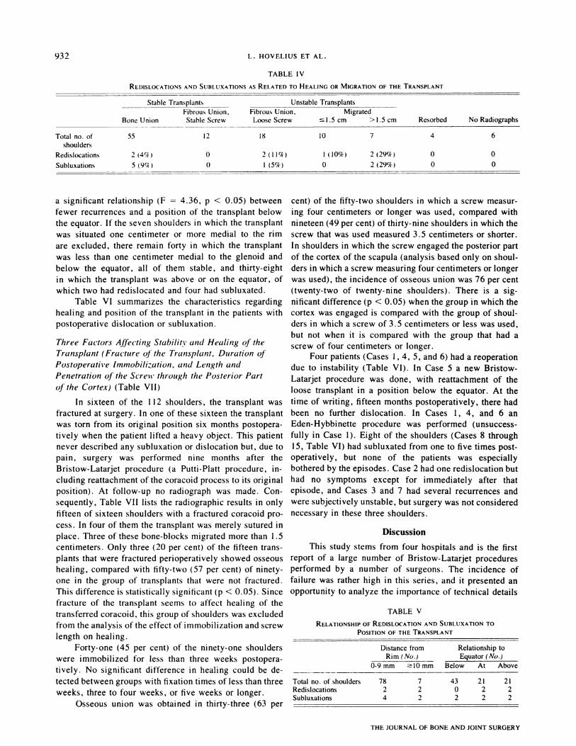
TABLE IV
REDISLOCATIONS AND SUBLUXATIO NS AS RELATED TO HEALING OR MIGRATION OF THE TRANSPLANT
Stable Transplants Unstable Transplants
Fibrous Union, Fibrous Union, Migrated
Bone Union Stable Screw Loose Screw � 1 .5 cm > 1 .5 cm Resorbed No Radiographs
Total no. of 55 12 18 10 7 4 6
shoulders
Redislocations 2 (4C/r) 0 2 (11%) 1 (10%) 2 (29%) 0 0
Subluxations 5 (9C/� ) 0 1 (5%) 0 2 (29%) 0 0
a significant relationship (F = 4.36, p < 0.05) between
fewer recurrences and a position of the transplant below
the equator. If the seven shoulders in which the transplant
was situated one centimeter or more media! to the rim
are excluded, there remain forty in which the transplant
was less than one centimeter medial to the glenoid and
below the equator, all of them stable, and thirty-eight
in which the transplant was above or on the equator, of
which two had redislocated and four had subluxated.
Table VI summarizes the characteristics regarding
healing and position of the transplant in the patients with
postoperative dislocation or subluxation.
Three Factors Affecting Stability and Healing of the
Transplant (Fracture of the Transplant, Duration of
Postoperative I,n,nobilization , and Length and
Penetration of the Screw through the Posterior Part
ofthe Cortex) (Table VII)
In sixteen of the 1 12 shoulders, the transplant was
fractured at surgery. In one of these sixteen the transplant
was torn from its original position six months postopera-
tively when the patient lifted a heavy object. This patient
never described any subluxation or dislocation but, due to
pain, surgery was performed nine months after the
Bristow-Latanjet procedure (a Putti-Platt procedure, in-
cluding reattachment of the coracoid process to its original
position). At follow-up no radiograph was made. Con-
sequently, Table VII lists the radiographic results in only
fifteen of sixteen shoulders with a fractured coracoid pro-
cess. In four of them the transplant was merely sutured in
place. Three of these bone-blocks migrated more than 1.5
centimeters. Only three (20 per cent) of the fifteen trans-
plants that were fractured perioperatively showed osseous
healing, compared with fifty-two (57 per cent) of ninety-
one in the group of transplants that were not fractured.
This difference is statistically significant (p < 0.05). Since
fracture of the transplant seems to affect healing of the
transferred coracoid, this group of shoulders was excluded
from the analysis of the effect of immobilization and screw
length on healing.
Forty-one (45 per cent) of the ninety-one shoulders
were immobilized for less than three weeks postopera-
tively. No significant difference in healing could be de-
tected between groups with fixation times of less than three
weeks, three to four weeks, or five weeks or longer.
Osseous union was obtained in thirty-three (63 pen
cent) of the fifty-two shoulders in which a screw measur-
ing four centimeters on longer was used, compared with
nineteen (49 pen cent) of thirty-nine shoulders in which the
screw that was used measured 3.5 centimeters on shorter.
In shoulders in which the screw engaged the posterior part
of the cortex of the scapula (analysis based only on shoul-
ders in which a screw measuring four centimeters or longer
was used), the incidence of osseous union was 76 pen cent
(twenty-two of twenty-nine shoulders). There is a sig-
nificant difference (p < 0.05) when the group in which the
cortex was engaged is compared with the group of shoul-
dens in which a screw of 3.5 centimeters on less was used,
but not when it is compared with the group that had a
screw of four centimeters on longer.Four patients (Cases 1 , 4, 5, and 6) had a neoperation
due to instability (Table VI). In Case 5 a new Bnistow-
Latanjet procedure was done, with neattachment of the
loose transplant in a position below the equator. At the
time of writing, fifteen months postoperatively, there had
been no further dislocation. In Cases 1 , 4, and 6 an
Eden-Hybbinette procedure was performed (unsuccess-
fully in Case 1). Eight of the shoulders (Cases 8 through
15, Table VI) had subluxated from one to five times post-
operatively, but none of the patients was especially
bothered by the episodes. Case 2 had one redislocation but
had no symptoms except for immediately after that
episode, and Cases 3 and 7 had several recurrences and
were subjectively unstable, but surgery was not considered
necessary in these three shoulders.
Discussion
This study stems from four hospitals and is the first
report of a large number of Bristow-Latarjet procedures
performed by a number of surgeons. The incidence of
failure was rather high in this series, and it presented an
opportunity to analyze the importance of technical details
TABLE V
RELATIONSHIP OF REDISLOCATION AND SUBLUXATION TO
POSITION OF THE TRANSPLANT
Distance from Relationship to
Rim (No.) _ Equator (No.)
0-9 mm �mm Below At Above
Total no. of shoulders 78 7 43 2 1 21
Redislocations 2 2 0 2 2
Subluxations 4 2 2 2 2

CORACOID TRANSFER FOR RECURRENT DISLOCATION OF THE SHOULDER 933
VOL. 65-A, NO. 7, SEPTEMBER 1983
TABLE VI
PATIENTS WITH RECURRENCE OF DISLOCATION OR WITH SUBLUXATION
Type ofOriginal Fracture of
Case Sex , Age
( Yrs.)Dislocation Transplant Screw Size
(tin)Healing* Altitudet Remarks
Dislocation
1 M, 24 Spontaneous No 3.5 B ( I .5)1 Above Reoperation (see
Table II)
2 M. 34 Traumatic Yes None used M (2-3)� I redisloc.
3 F, 29 Spontaneous Yes 2 M (3)� Minor disability
4 M, 32 Traumatic Yes 4 M ( 1 .5)t Reoperation
5 M, 28 Traumatic No 4 F (0)� At Reoperation (see
Table II)
6 ‘sI, 22 Spontaneous Yes 3 B ( I .3)� At Reoperation
7 F, 27 Traumatic No 3.2 F (0.5)1 At Minor disability
Subluxation
8 M, 19 Traumatic No 3 B (0) Above� 2 sublux.
9 M, 27 Spontaneous No 3 B (0) Above4 5 sublux.
10 M, 21 Traumatic No 3 B (0) At I sublux.
1 1 M, 25 Spontaneous No 3.5 F ( I)1 Below 2 sublux.
12 M, 28 Traumatic No 4.5 B (0) At 2 sublux. (see
Table II)
13 M, 37 Traumatic No 4 B ( l)� Below 2 sublux.
14 F, 30 Traumatic Yes None used M (3-4)� 2 sublux.
15 M, 27 Traumatic No 2.5 M (2)� 2 sublux.
.� B = bone union and F = fibrous union in good position but unstable, with total transplant-to-rim distance (in centimeters) in parentheses; and
M = migration, with distance from the glenoid neck (in centimeters) in parentheses.1� Above, at, or below the equator.
:� Presumed reason for failure.
regarding the coracoid transfer. Even though it has been
reported that the Bristow procedure may be successful
even when the transplant has migrated two to three cen-
timeters#{176}11, that complication has not been studied
previously, nor have the other pertinent technical features
of the procedure been analyzed.
If all shoulders showing ( 1 ) a migrated transplant, (2)
a bone-block in a position one centimeter on more medial
to the glenoid rim, and finally (3) a transplant situated at
the equator or higher on the scapular neck were excluded
from the present study, then the series would consist of
forty shoulders with no postoperative redislocations or
subluxations. Accordingly, we consider the orientation of
the transplant in relation to the glenoid rim and obtained
healing of the bone-block to be important. In the latest
edition of Cwnphell’s Operative Orthopaedics 16 and
elsewhere9 it was stated that the transferred coracoid pro-
cess need not provide a bone-block effect. Therefore a
more media! position of the transplant than that on the rim
of the glenoid ought to be tolerated. Judging from our ex-
perience, this deviation in technique should be avoided.
Our data show that there seems to be a limit for the lon-
gitudinal position of the transplant of about ten millimeters
from the glenoid rim. Beyond that limit the incidence of
recurrences increases (Table V). On the basis of our exper-
ience, we therefore recommend that the transplant be po-
sitioned one to five millimeters medial to the glenoid rim.
We cannot exclude the possibility that the bone-block ef-
fect of the transplant is responsible for some of the success
of the Bristow-Latanjet repair.
Our findings confirm those of AIlman that the best me-
sults are obtained if the position of the transplant is below
the mid-point of the scapular neck. An experimental study
by Tunkel et al. explained why this subequatonial position
is best. They found that when the shoulder is abducted 90
degrees and externally notated, the inferior glenohumeral
ligament prevents the humera! head from dislocating. With
the coracoid transplant positioned below the equator, the
conjoined tendon is in the best position to take over the
function of the inferior glenohumenal ligament. Hill et a!.
reported a fracture of the bone-block in seven (7 per cent)
of 107 shoulders. Four of the fractured transplants were
merely sutured to the subscapular tissue, and all seven
shoulders eventually were stable. This is not in accordance
with the findings in our series, in which the sixteen shoul-
dens with a fractured bone-block showed several instances
of migration or non-union of the transplant and a high rate
of postoperative instability.
From the results of this study, we further conclude
that an important factor in obtaining adequate healing of
the transferred coracoid process to the glenoid neck is the
method of stabilizing it. We recommend the use of a ma!-
leolar or a cancellous bone screw, very seldom shorter
than four centimeters and preferably penetrating the pos-
tenor pant of the cortical bone of the scapular neck.
Postoperative immobilization for three weeks or more

934 L. HOVELIUS ET AL.
THE JOURNAL OF BONE AND JOINT SURGERY
TABLE VII
HEALING AND MIGRATION OF THE BONE-B LOCK IN REL ATION TO 5EV ERAL OTHER FACTORS
NotVisible at Bone Fibrous Migration of
Follow-up Union Union Transplant Total(No.) (No.) (No.) (No.) (No.)
Perioperative fracture
of transplantFixed by suture 1 0 0 3(3 > 1.5cm) 4Fixedbyscrew I 3 2 5(2>1.5cm) Il
Postoperative immobilization<3wks. 0 25 11 5(1>1.5cm) 41
3to4wks. 1 17 7 3(0>1.5cm) 28�Swks. 1 10 10 1(1>1.5cm) 22
Length of screw
�3.5cm I 19 13 6(2>1.5cm) 39�4 cm I 33 15 3(0> 1.5cm) 52
Screw ( �4 cm) through posteriorpart of wall of cortexof scapular neck
Yes 1 22 5 1(0> 1.5cm) 29No 0 1 1 9 2 (0 > 1 .5 cm) 22Extracted 1 1
does not seem to promote healing of the transplant. The
postoperative immobilization can thus be short. We agree
with Tagliabue and Esposito that one of the advantages of
this type of repair is that the postoperative morbidity is so
short.
Conclusions
This study indicates that optimum results of the
Bnistow-Latarjet procedure are obtained if the bone-block
heals to the neck of the glenoid and the position of the
bone-block is less than one centimeter medial to the
glenoid rim and below the equator of the scapular neck. To
ensure adequate healing, the bone-block should not be
fractured during surgery, and should be securely fixed with
a screw long enough to engage the posterior part of the con-
tex of the scapula. Divergence from these guidelines,
however, does not exclude excellent or good results in
some shoulders.
References
I. AKERMARK, C.; HOVELIUS, L.; HERBERTS, P.; WREDMARK, T.; GUSTAVSSON, B.; K#{246}RNER, L.; and BERG, E.: Results of the Bristow Repair:Sports Participation after Surgery. In Shoulder Surgery, pp. 92-94. Edited by J. I. Bayley and Lippmann Kessel. Berlin, Springer, 1982.
2. ALLMAN, FRED: Symposium on Sports Injuries to the Shoulder. Contemp. Surg. , 7: 82, Sept. 1975.3. COLLINS, H. R., and WILDE, A. H.: Shoulder Instability in Athletics. Orthop. Clin. North America, 4: 759-774, 1973.4. HELFET, A. J.: Coracoid Transplantation for Recurring Dislocation of the Shoulder. J. Bone and Joint Surg., 40.B(2): 198-202, 1958.5. HILL, H. A.; LOMBARDO, S. J.; KERLAN, R. K.; JOBE, F. W.; CARTER, V. S.; SHIELDS, C. L., JR.; COLLINS, H. R.; and YOCUM, L. A.: The
Modified Bristo�w-Helfet Procedure for Recurrent Anterior Shoulder Subluxations and Dislocations. Am. J. Sports Med., 9: 283-287, 1981.6. HOVELIUS, L.; AKERMARK, C.; ALBREKTSSON, B.; BERG, E.; K#{246}RNER, L.; LUNDBERG, B.; and WREDMARK, T.: The Bristow-Latarjet Procedure
for Recurrent Dislocation of the Shoulder. A 2-5 Year Follow-up Study on the Results of 1 12 Cases. Acta Orthop. Scandinavica, 54: 284-290,1983.
7. LAMM, C. R.; ZACHRISSON, B. E.; and K#{246}RNER, L.: Radiography ofthe Shoulder after Bristow Repair. Acta Radiol. Diag., 23: 523-528, 1982.8. LATARJET, M.: A propos du traitement des luxations r#{233}cidivantes de l’epaule. Lyon chir., 49: 994-997, 1954.9. LOMBARDO, S. J.; KERLAN, R. K.; JOBE, F. W.; CARTER, V. S.; BLAZINA, M. E.; and SHIELDS, C. L., JR.: The Modified Bristow Procedure for
Recurrent Dislocation of the Shoulder. J. Bone and Joint Surg. , 58-A: 256-261 , March 1976.10. MAY, J. V., JR.: A Modified Bristow Operation for Anterior Recurrent Dislocation of the Shoulder. J. Bone and Joint Surg. , 52-A: 1010-1016,
July 1970.1 1 . POST, MELVIN: The Shoulder: Surgical and Nonsurgical Management, p. 452. Philadelphia, Lea and Febiger, 1978.12. ROWE, C. R.; PIERCE, D. S.; and CLARK, J. G.: Voluntary Dislocation ofthe Shoulder. A Preliminary Report on a Clinical, Electromyographic,
and Psychiatric Study of Twenty-six Patients. J. Bone and Joint Surg. , 55-A: 445-460, April 1973.13. SWEENEY, H. J.; MEAD, N. C.; DAWSON, W. J.; and FITZSIMMONS, PHILIP: Fourteen Years’ Experience with the ModifIed Bristow Procedure
for Recurrent Anterior Dislocation of the Shoulder. Read at the Annual Meeting of The American Academy of Orthopaedic Surgeons, SanFrancisco, California, March 5, 1975.
14. TAGLIABUE, D., and ESPOSITO, A.: L’intervento di Latarjet nella lussazione recidivante di spalla dello sportivo. Italian J. Sports Traurnat., 2:91-100, 1980.
15. TURKEL, S. J.; PANIO, M. W.; MARSHALL, J. L.; and GIRGIS, F. G.: Stabilizing Mechanisms Preventing Anterior Dislocation of the Glenohu-meral Joint. J. Bone and Joint Surg., 63-A: 1208-1217, Oct. 1981.
16. WRIGHT, P. E.: Bristow Procedure. In Campbell’s Operative Orthopaedics, edited by A. S. Edmonson and A. H. Crenshaw. Ed. 6, pp. 486-488.St. Louis, C. V. Mosby, 1980.