the contribution of central and peripheral vision in scene categorization: a study on people with...
TRANSCRIPT

Vision Research 98 (2014) 46–53
Contents lists available at ScienceDirect
Vision Research
journal homepage: www.elsevier .com/locate /v isres
The contribution of central and peripheral vision in scene categorization:A study on people with central vision loss
http://dx.doi.org/10.1016/j.visres.2014.03.0040042-6989/� 2014 Elsevier B.V. All rights reserved.
⇑ Corresponding author. Address: CHRU Lille, Hôpital Roger Salengro, LaboratoireNeurosciences et Pathologies Fonctionnelles, 59037 Lille, France. Fax: +33 3 20 4467 32.
E-mail address: [email protected] (M. Boucart).
Miguel Thibaut a, Thi Ha Chau Tran a,b, Sebastien Szaffarczyk a, Muriel Boucart a,⇑a Laboratoire Neurosciences et Pathologies Fonctionnelles, Université Lille Nord de France, CNRS, Franceb Service d’Ophtalmologie, Hôpital Saint Vincent de Paul, Lille, France
a r t i c l e i n f o a b s t r a c t
Article history:Received 5 January 2014Received in revised form 5 March 2014Available online 20 March 2014
Keywords:Scene perceptionPeripheral visionAge-related macular degenerationLow vision
Studies in normally sighted people suggest that scene recognition is based on global physical propertiesand can be accomplished by the low resolution of peripheral vision. We examine the contribution ofperipheral and central vision in scene gist recognition in patients with central vision loss and age-matched controls. Twenty-one patients with neovascular age related macular degeneration (AMD), witha visual acuity lower than 20/50, and 15 age-matched normally sighted controls participated in a natural/urban scene categorization task. The stimuli were colored photographs of natural scenes presented ran-domly at one of five spatial locations of a computer screen: centre, top left, top right, bottom left and bot-tom right at 12� eccentricity. Sensitivity (d0) and response times were recorded. Normally sighted peopleexhibited higher sensitivity and shorter response times when the scene was presented centrally than forperipheral pictures. Sensitivity was lower and response times were longer for people with AMD than forcontrols at all spatial location. In contrast to controls patients were not better for central than for periph-eral pictures. The results of normally sighted controls indicate that scene categorization can be accom-plished by the low resolution of peripheral vision but central vision remains more efficient thanperipheral vision for scene gist recognition. People with central vision loss likely categorized scenes onthe basis of low frequency information both in normal peripheral vision and in low acuity central vision.
� 2014 Elsevier B.V. All rights reserved.
1. Introduction
The gist of a scene includes all levels of processing, from low-le-vel features (color, spatial frequency, orientation. . .) to intermedi-ate image properties (surface, volumes, texture) and high levelinformation (semantic knowledge) (Oliva, 2005). Studies on nor-mally sighted people have shown that scene gist recognition is par-ticularly robust, even in conditions of limited presentation time(around 20 ms) (Greene & Oliva, 2009a; Joubert et al., 2007; Rousselet,Joubert, & Fabre-Thorpe, 2005), limited spatial frequency informa-tion (Oliva & Schyns, 2000), limited attentional allocation (Fei-Feiet al., 2005) and large visual eccentricities (Boucart et al., 2013;Thorpe et al., 2001). The question of the contribution of centralversus peripheral vision on natural scene perception has been ad-dressed in normally sighted observers and in pathologies inducinga visual field loss. For instance, Thorpe et al., (2001) have lookedat performance of young normally-sighted people for object
categorization at large eccentricities. Photographs of naturalscenes were randomly presented on a hemispheric screen from0� (central) to 75� eccentricity. Surprisingly, they found that per-formance to detect an animal in a natural scene was above 70%at 60� eccentricity though participants claimed to perform the taskby guessing. This ‘‘perception without awareness’’ at large eccen-tricities has been confirmed and extended by Boucart et al.(2010). They reported both implicit recognition (measured bypriming effects) and explicit recognition (measured by recognitionof previously seen pictures) of colored photographs of objects at30� eccentricity. Only non conscious implicit recognition occurredat 50� eccentricity in normally sighted people and in 4 patientswith Stargardt disease (a juvenile maculopathy inducing central vi-sion loss). Larson and Loschky (2009) examined the contribution ofcentral versus peripheral vision on scene gist recognition in a ver-ification task (a matching between a word and a photograph). Theypresented participants with central photographs of real worldscenes (27 � 27� of visual angle) for 106 ms each. Performancewas compared in two conditions: a window condition showingthe central portion of the scene and blocking peripheral informa-tion and a scotoma condition blocking out the central portionand showing only the periphery. The radii of the window and

Table 1Demographic and clinical data of the study population.
AMD participants n = 21Mean age (years) (mean, SD, range) 79 ± 5.7 (66–89)Gender (M/F) 6 Males/15
femalesMean MMSE (SD) 27 ± 2Mean VA (LogMAR) 0.6 ± 0.22Mean greatest linear lesion diameter (mm) (mean, SD,
range)3.57 ± 1.38 (1.3–7.5)
Mean area of the lesion (mm2) (mean, SD, range) 8 ± 6.8 (0.6–30.6)
Controls n = 15Mean age (years) (mean, SD, range) 74.6 ± 6 (66–83)Gender (M/F) 7 Males/8 femalesMean VA (LogMAR) 0.06 ± 0.04Mean MMSE (SD) 28.7 ± 1.9
VA = Visual Acuity; MMSE = Mini Mental State Examination; SD = StandardDeviation.
M. Thibaut et al. / Vision Research 98 (2014) 46–53 47
scotoma were 1�, 5�, 10.8� and 13.6�. Performance was barelyabove chance in the 1� window condition and, when all informa-tion was eliminated from foveal and parafoveal vision (in the 5�scotoma condition), accuracy was no worse than when the entireimage was shown. This suggests that central vision is not necessaryfor recognizing scene gist. Accuracy increased as the radius of thewindow increased or the radius of the scotoma decrease. Theauthors suggested that peripheral (and parafoveal vision) is moreuseful than high resolution foveal vision for scene gist recognition.However, a control experiment showed that the advantage of theperiphery resulted from a difference in the size of the viewing field.When viewing field size was equalized there was an advantage forcentral vision in their study. A further control study showed thatcentral vision required less than half as many pixels as peripheralvision required to achieve the same gist accuracy, suggesting thatcentral vision was more efficient at extracting scene gist.
Tran et al., (2010) investigated scene gist recognition in peoplewith central vision loss resulting from macular degeneration(AMD). Colored photographs of scenes (15 � 15� of visual angle)were centrally displayed for 300 ms. People with AMD and normallysighted age-matched controls were asked to categorize the sceneseither as natural versus urban or as indoor versus outdoor in a go/nogo task (i.e., half of the participants in each group pressed a keyfor a pre-defined target (e.g., the natural, the urban, the indoor orthe outdoor scene depending on the participant) and refrained fromresponding for the other category). It was found that people withAMD performed with high accuracy in both categories of scenes(84% hits for natural/urban and 79% hits for indoor/outdoor scenes).As people with AMD had a central vision loss, these results are con-sistent with studies on normally sighted people (Larson & Loschky,2009) showing that scene recognition can be accomplished withthe low resolution of peripheral vision. However, in the Tran et al.,(2010) study, the pictures were always displayed at the same spatiallocation, in the centre of the computer screen. Therefore, as the loca-tion was predictable, it might be that people with AMD orientedtheir gaze in such a way that the images fell in their preferred retinallocation (PRL). When the macular scotoma affects the fovea, the vi-sual system develops preferred retinal loci (PRLs) as a ‘‘pseudofovea’’to perform visual tasks (Crossland et al., 2005). The PRL refers to oneor several retinal areas used for fixation. It is task specific (Crossland,Crabb, & Rubin, 2011a), and it is used on repeated testing (Crossland,Engel, & Legge, 2011b). The PRL tends to develop in a functional ret-inal area near the edge of the scotoma (Cheung & Legge, 2005;Crossland et al., 2005).
The present study was designed to compare scene gist recogni-tion in central and in peripheral vision in people with central visionloss and normally sighted age-matched observers. In addition tothe previous study (Tran et al., 2010) the spatial location of the pic-tures was unpredictable, appearing randomly at one of five spatiallocations on a computer screen (centre, top left, top right, bottomleft and bottom right). Also, in the Tran et al., (2010) study imageswere displayed for 300 ms. Though that duration does not allow vi-sual exploration it does provide enough time for two moderate(150 ms) fixations. In the present study images were displayed ata duration that was shorter for a saccade at 12� eccentricity. If areliable scene representation can be built from low level features(Larson & Loschky, 2009; Torralba & Oliva, 2003) then peripheralpresentation should not impair performance, both in people withAMD and in normally sighted controls.
2. Method
2.1. Participants
Twenty-one patients with neovascular age-related maculardegeneration were included. Due to the asymmetry of the
pathology only one eye of each patient was tested. In cases of bilat-eral AMD, we tested eye with the best corrected visual acuity. Ifboth eyes had equal acuity, one eye was randomly selected. Bestcorrected visual acuity (BCVA) was determined using Early Treat-ment Diabetic Retinopathy Study (ETDRS) charts at a distance of4 m, which was converted to logMAR visual acuity for statisticalpurpose. Slit lamp examination, intraocular pressure, and fundus-copy were performed in all patients and controls. The diagnosisof neovascular AMD was confirmed by fluorescein angiography,using a confocal scanning laser ophthalmoscope (HeidelbergRetina Angiograph, HRA2; Heidelberg Engineering, Dossenheim,Germany). The area of the lesion (mm2) and the greatest lineardiameter of the lesion were measured from digital angiogramsby outlining the lesion, using image analysis software (HeidelbergEye Explorer, Heidelberg Engineering) (Barbazetto et al., 2003;Hogg et al., 2003). Clinical assessment and experiments wereperformed at the same visit in the hospital.
The age-matched control group, with normal visual acuity, wascomposed of 15 volunteers. Control participants had no history ofophthalmologic or neurological diseases and no cognitive impair-ment. They were either relatives of participants with AMD or pa-tients who underwent successful cataract surgery with normalvisual acuity ranging from 20/25 to 20/20. Controls were testedmonocularly on their preferred eye. Clinical and demographic dataare provided in Table 1.
Both participants with AMD and controls were recruited in theOphthalmology department of Saint Vincent the Paul hospital,Lille, France. The study was approved by the ethical committee ofLille, in accordance with the tenets of the Declaration of Helsinski.Written informed consent was obtained from all participants.
2.2. Stimuli and apparatus
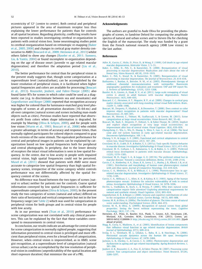
The stimuli were displayed on a 30 in color monitor (Dell) con-nected to a computer (Dell T 3400). The stimuli were photographsof natural scenes. Two categories were represented: natural (de-serts, forests, mountains, rivers) versus urban scenes (cities, sky-scrapers, streets and highways). Examples are shown in Fig. 1.The amplitude spectra were computed for the two categories ofscenes to assess whether they differed on the orientation (horizon-tal/vertical) of low, medium and high spatial frequency compo-nents. As shown in Table 2, the difference (ratio) in vertical andhorizontal components between the two categories of scenes wassmall for low spatial frequencies. It increased for medium and highspatial frequencies. The angular size of the photographs was15� � 15� at a viewing distance of 1 m. The participant’s headwas not fixed. The scenes were displayed on a light gray

Fig. 1. Examples of natural and urban scenes used in the experiment.
Table 2Horizontal and vertical components of the image as a function of spatial frequency range (low, medium and high spatial frequencies) and categories of pictures. The ratio wascomputed as (Horizontal � Vertical)/(Horizontal + Vertical) � 100.
SF = Spatial Frequency. In bold: Mean values for the four categories of man-made and the four categories of natural scenes.
48 M. Thibaut et al. / Vision Research 98 (2014) 46–53
background (56.2 cd/m2). The software was developed by the lab’sengineer in C++. Responses were recorded by keypress on a re-sponse box connected to the computer.
2.3. Procedure
A black (5�) central fixation cross was displayed for 500 ms, fol-lowed by a blank interval of 500 ms, and followed by a single pho-tograph of a scene. The picture appeared randomly and equally atone of 5 spatial locations: the centre of the screen or one on thefour quadrants surrounding the fixation cross at an eccentricity(centre of the picture) of 12�. An example of the paradigm is pre-sented in Fig. 2. A go/no-go paradigm was used. Participants wereasked to press a key when they saw their target and to refrain fromresponding when the photograph did not correspond to their tar-get. The target was a natural scene for half of the participantsand an urban scene for the other half. There were 140 trials
determined by 28 photographs (14 natural and 14 urbanscenes) � 5 spatial locations. The 70 natural scenes were randomlyselected, by software, from a set containing 400 photographs andthe 70 urban scenes were randomly selected from a set containing200 photographs. As we wanted to investigate peripheral visionthe exposure duration was fixed at a duration lower than that ofa saccade. The mean duration of a saccade varies between 175and 200 ms for young people (Rayner, 1995). It is longer for olderadults (on average 264 ms) (Irving et al., 2006). Based on pilotstudies to get performance above chance, but not at ceiling for nor-mally sighted controls, the exposure time was fixed at 100 ms forcontrols and at 200 ms for patients as a majority of patientsclaimed that they saw nothing with a 100 ms exposure time. Re-sponse times were triggered on stimulus onset. A picture was dis-played every 2 s. Responses were recorded on the basis of thesignal detection theory with correct detections of the target desig-nated as hits, detection of a target when there was none designated

M. Thibaut et al. / Vision Research 98 (2014) 46–53 49
as false alarms, failure to detect the target when it was present des-ignated as omission and no response when the target was absentdesignated as correct rejections. In order to avoid the problem ofinfinite z score values for 100% hits and 0% false alarms in casesof perfect discriminability, a correction (Macmillan & Creelman,1991) was applied: the proportion of hits and false alarms wereset at 0.99 (for 100%) and 0.01 (for 0%). Based on these data, a d0
index of sensitivity was computed for each participant and eachspatial location of the target. Analyses of variance were conductedon the d0 index of sensitivity and on correct response times (RTs).The factors were the group (people with AMD vs. normally sightedcontrols), the two categories of scenes (natural/urban) and the fivespatial locations. Correlations between performance (d0) and clini-cal parameters (logMAR visual acuity, greatest diameter of the le-sion, and the area surface) were performed by using Pearson’scorrelation coefficient (r) and the matching significance of the cor-relation (p). For tests of statistical significance, alpha is set top < 0.05. All data were analyzed using the software Statistica(Version 8, Stat soft, France).
3. Results
The results are presented in Fig. 3 for sensitivity and RTs.Individual results are presented in Table 3. There was no signifi-cant main effect of the category of scene (urban/natural)(F(1,32) = 0.3 ns for d0 and F(1,32) = 1.6 ns for RTs) and no interac-tion between category of scene and other variables.
Sensitivity was higher for controls than for people with AMD(F(1,32) = 11.7, p < .002) but RTs did not differ significantly be-tween groups (F < 1). A significant main effect of spatial locationwas observed both on sensitivity (F(4,128) = 12, p < .0001) andon RTs (F(4,128) = 5.2, p < .001), with centrally located scenes pro-ducing higher sensitivity and faster RTs. Group interacted signifi-cantly with spatial location for sensitivity (F(4,128) = 4.7,p < .001) but not for RTs (F(4,128) = 0.6, ns). The interaction re-sulted from a better sensitivity when the scene was displayed cen-trally than peripherally for controls (F(4, 52) = 11.2, p < .0001)whilst no significant difference between the spatial locations wasfound for people with AMD (F(4, 76) = 1.5 ns). RTs were also
Fig. 2. Illustration of the paradigm: a central cross (500 ms) was followed by a blank interfive spatial locations during 100 ms for controls and 200 ms for patients.
significantly shorter for central than for peripheral pictures in thecontrol group (F(4,52) = 6.7, p < .001) but not for people withAMD (F(4,76) = 1.6, ns). A comparison between patients andcontrols for each spatial location showed that sensitivity was sig-nificantly higher for controls than for patients with AMD at eachspatial location except bottom-left (centre: F(1,34) = 24.7,p < .001, top-right: F(1,34) = 11.4, p < .001), bottom-right:F(1,34) = 6.6, p < .014, top-left: F(1,34) = 6.2, p < .018, bottom-left:F(1,34) = 3.17, p < .08). No significant difference between patientsand controls was observed for RTs (centre: F(1,34) = 1.6, ns,top-right: F(1,34) = 0.44, ns), bottom-right: F(1,34) = 0.002, ns,top-left: F(1,34) = 0.13, ns, bottom-left: F(1,34) = 0.31, ns). Individ-ual data, presented in Table 3, show that sensitivity was better forcentrally displayed pictures in 4/21 patients (5, 9, 12 and 21). Itwas better on the top-left for 2/21 patients (7 and 17). Sensitivitywas equivalent at the 5 spatial locations for the other patients andit was close to chance for centrally displayed pictures in 4/21patients (2, 6, 15 and 19) (see Table 3).
No significant correlation was found between performance (interms of response times and d0) at the five spatial locations (centre,top right, bottom right, top left, bottom left) and any clinicalparameters (distance visual acuity, lesion size).
4. Discussion
Models of scene recognition (Oliva & Torralba, 2001; Torralba &Oliva, 2003) suggest that the initial scene representation is basedon holistic descriptors and statistic regularities that can be rapidlyextracted at an early perceptual stage (Greene & Oliva, 2009b).Numerous evidence from behavioral studies on normally sightedpeople indicate that rapid scene categorization is based on globalscene properties like color (e.g., to categorize a natural scene ashot or cold landscape), orientation (e.g., to discriminate betweena forest and a field), depth, texture density. . . (e.g., Greene & Oliva,2009a, 2009b; Oliva & Torralba, 2006; Serre et al., 2007). It hasbeen shown that, with a single glance at a scene (20 ms exposuretime) normally sighted people are able to categorize scenes at asuper-ordinate level (e.g., natural versus man made) and at a basiclevel (e.g., river, forest, highway, city. . .) with high accuracy (above
val of 500 ms and then by a single picture of a scene (natural versus urban) in one of

Table 3Individual d0s and response times as a function of the group of participants (AMD vs controls), the spatial location of the target (centre, top right, top left, bottom right and bottomleft) and the category of scenes (natural vs urban scene). On the right: individual clinical data for patients: visual acuity, surface area of the lesion (in mm2) and diameter of thelesion (in mm).
Centre Top right Bottom right Top left Bottom left LogMAR visual acuity Surface area (mm2) Greatest linear diameter (mm)
AMD groupTarget: Natural
P1 1.56 1.09 1.28 1.10 1.23 0.40 2.69 2.40P2 0.80 1.16 1.03 0.90 1.52 0.50 4.67 3.20P2 1.06 0.77 1.88 1.29 1.52 0.70 8.00 3.70P4 1.29 1.67 0.84 2.20 1.71 0.40 3.00 2.20P5 3.29 1.81 1.94 0.52 1.52 0.60 3.50 2.70P6 0.13 0.25 0.68 0.36 1.39 0.60 7.30 4.00P7 3.97 3.29 3.97 4.65 3.97 0.40 4.84 2.76P8 2.36 1.66 1.04 2.32 2.32 1.12 11.70 4.50P9 3.29 2.17 1.68 2.56 1.90 0.40 0.57 1.28P10 1.42 1.67 0.84 0.51 0.38 0.70 30.60 7.50
Target: UrbanP11 2.68 2.32 1.81 2.49 2.32 0.70 4.50 2.90P12 3.00 1.61 1.96 1.52 2.12 0.40 5.08 3.10P13 1.92 1.81 1.56 1.64 1.16 0.70 0.98 1.29P14 2.12 1.42 1.88 1.96 1.23 0.70 9.60 3.60P15 0.84 2.20 1.52 1.56 0.78 0.50 7.20 4.52P16 1.16 1.11 1.28 1.10 0.13 1.00 8.70 4.40P17 1.12 2.20 2.33 3.17 1.82 1.00 20.00 5.50P18 2.68 2.49 2.68 2.93 2.68 0.40 10.15 3.90P19 0.64 0.51 0.97 1.33 0.97 0.40 7.00 3.80P20 1.42 1.26 0.48 1.37 1.37 0.40 13.00 4.10P21 3.61 2.17 1.96 2.85 2.49 0.50 6.31 3.70
Controls groupTarget: Natural
C1 2.93 2.03 0.84 1.56 0.64 0.05C2 4.65 3.00 2.03 3.97 2.68 0.05C3 3.17 2.32 1.37 1.35 1.55 0.10C4 2.68 1.41 1.77 1.77 0.90 0.10C5 4.65 3.00 3.00 3.17 2.33 0.05C6 3.97 4.65 3.97 6.16 3.29 0.10C7 3.61 3.29 3.29 2.56 2.03 0.05C8 3.97 1.16 1.59 1.90 2.33 0.10
Target: UrbanC9 2.12 1.56 2.56 1.88 2.32 0.10C10 3.97 3.61 2.49 2.93 2.56 0.00C11 4.65 3.97 4.65 3.67 3.03 0.10C12 3.36 1.64 1.67 1.41 1.71 0.10C13 2.32 1.52 0.63 2.33 0.93 0.10C14 3.97 3.00 2.85 3.36 2.32 0.00C15 3.61 3.61 3.97 3.97 3.97 0.00
Centre Top right Bottom right Top left Bottom left
AMD groupTarget: Natural
P1 488.79 656.36 568.90 656.08 688.38P2 667.91 698.56 736.25 764.86 810.67P2 529.08 640.75 579.47 568.58 520.50P4 643.08 868.77 872.20 865.67 921.67P5 596.60 450.07 477.86 562.60 776.78P6 724.11 637.33 515.00 510.67 617.25P7 541.48 538.32 567.25 592.50 597.60P8 454.83 350.18 546.70 461.00 478.24P9 507.90 558.00 527.50 531.94 569.67P10 680.53 711.85 778.70 593.00 630.45
Target: UrbanP11 524.42 578.80 548.07 610.06 594.27P12 441.20 452.72 433.00 493.40 471.31P13 828.65 712.29 988.29 966.90 913.45P14 466.25 510.31 463.75 530.87 476.93P15 638.44 785.33 646.33 708.57 703.75P16 682.28 665.39 719.94 648.81 590.20P17 540.17 839.33 683.40 694.31 559.83P18 573.12 545.19 521.71 540.17 458.26P19 492.62 510.55 480.09 526.00 531.36P20 443.46 584.86 419.67 437.00 404.69P21 546.63 584.14 499.89 553.14 530.00
50 M. Thibaut et al. / Vision Research 98 (2014) 46–53

Fig. 3. Index of sensitivity (d0) and response times (RTs) as a function of the spatiallocation of the target photograph on the screen and the group of participants(people with AMD/controls). The vertical lines represent standard deviations.
Controls groupTarget: Natural
C1 636.94 625.69 659.10 640.79 743.33C2 515.19 589.07 548.62 553.42 512.00C3 487.10 551.67 636.36 512.33 485.06C4 549.84 592.18 619.45 720.45 708.00C5 617.70 717.93 685.13 755.00 707.60C6 589.65 639.10 631.05 732.00 633.84C7 370.60 443.11 389.32 449.67 398.37C8 554.10 576.45 675.08 647.67 604.20
Target: UrbanC9 588.72 696.21 651.22 615.63 634.88C10 454.95 497.90 474.81 514.53 476.56C11 445.30 469.05 518.95 490.94 516.69C12 558.18 632.80 650.92 574.45 646.60C13 608.50 650.89 663.13 691.90 634.75C14 503.90 576.47 518.21 482.94 522.29C15 505.90 566.05 660.95 548.90 589.95
M. Thibaut et al. / Vision Research 98 (2014) 46–53 51
90% correct) and fast RTs (below 400 ms) (Greene & Oliva, 2009a;Joubert et al., 2007; Rousselet, Joubert, & Fabre-Thorpe, 2005). Cat-egorization at a super-ordinate level can even be shorter and moreaccurate than categorization at a basic level at short target-maskSOAs (Loschky & Larson, 2010). Even if severe spatial filtering (4–8 cycles per image) is applied to the image, enough structural cuesare provided to allow the categorization of a scene as indoor/out-door (Oliva & Schyns, 2000) suggesting that the initial scene repre-sentation constructed by the visual system is based on coarseglobal information.
We examined scene categorization in high resolution centraland in low resolution peripheral vision in normally sighted observ-ers and in people with degraded central vision who must rely ontheir peripheral vision. The results show (1) that both people withAMD and normally sighted age-matched controls were able tocategorize scenes in peripheral vision at 12� eccentricity. (2)
Sensitivity was higher for normally sighted controls than for peo-ple with AMD at all spatial locations even though controls sawscenes for durations half as long as patients. (3) Response timeswere shorter, and sensitivity was higher, for central than forperipheral scenes in normally sighted people but not in patientswhose performance (both RTs and sensitivity) was equivalent forcentral and peripheral scenes.
Even though the density of cone photoreceptors, responsible forhigh resolution perception, decreases considerably as eccentricityincreases from the fovea (Curcio et al., 1991) the results in bothgroups of participants show that scene categorization can beaccomplished with the low resolution of peripheral vision. This re-sult is consistent with Larson and Loschky (2009) and Boucart et al.(2013). Tran et al., (2010) showed that people with central visionloss were able to categorize scenes with high accuracy but, in theirstudy, photographs were always displayed at the same spatial loca-tion and for a duration allowing two fixations. It might be thatthese two conditions allowed people with AMD to place the photo-graphs in their preferred retinal location (PRL), a region of higheracuity developed by people with a central scotoma. With a spatiallocation of the photographs made unpredictable and a shorterexposure duration people with AMD were still able to categorizescenes highly above chance suggesting that they used efficientlytheir peripheral vision to categorize scenes, both for peripherallyand for centrally displayed pictures as the angular size of the pho-tographs was larger than the scotoma. However, likely due to theshorter exposure time accuracy was lower for central pictures inthe present study (hits: 75.5%) than in the Tran et al., (2010) studyfor patients (84.4%) but not for controls (96% hits in both studies).
Exposure duration was longer for patients than for age-matchedcontrols (200 vs 100 ms) yet performance was better for controls atall spatial locations. This result suggests (1) that the patients’peripheral vision is not as efficient as that of normally sightedage-matched controls and (2) that peripheral vision does not im-prove with central vision loss as a result of plasticity. Histopatho-logic studies of human donor retinas have shown that, althoughboth rods and cones degenerate in AMD, rod loss precedes coneloss in 75% of early and late AMD eyes. The maximum loss occursin the parafovea, 1–3 mm from the fovea (i.e., 3.5–10 degrees fromfixation), beginning inferior to the fovea and culminating in anannulus of deepest loss at 0.5–3 mm eccentricity (Adler et al.,1999; Changzheng et al., 2004; Curcio, Medeiros, & Millican,1996; Curcio et al., 1993; Jackson, Owsley, & Curcio, 2002; Neelamet al., 2009). Consistent with this, a majority of patients with AMDexhibit more scotopic than photopic sensitivity loss (see Neelamet al for a review). In the present study the angular size of the pic-tures was 15� � 15� and peripheral pictures were displayed at an
52 M. Thibaut et al. / Vision Research 98 (2014) 46–53
eccentricity of 12� (centre to centre). Both central and peripheralpictures appeared in the area of maximum receptor loss thusexplaining the lower performance for patients than for controlsat all spatial locations. Regarding plasticity, conflicting results havebeen reported in studies investigating cerebral re-organization inpatients with central vision loss. Some neuroimaging studies pointto cerebral reorganization based on retinotopic re-mapping (Bakeret al., 2005, 2008) and changes in cortical gray matter density con-secutive to AMD (Boucard et al., 2009; Hernowo et al., 2013) whilstothers failed to show any changes (Baseler et al., 2011; Sunness,Liu, & Yantis, 2004) or found incomplete re-organization depend-ing on the age of disease onset (juvenile vs age related maculardegeneration) and therefore the adaptation period (Liu et al.,2010).
The better performance for central than for peripheral vision inthe present study suggests that, though scene categorization at asuperordinate level (natural/urban), can be accomplished by thelower resolution of peripheral vision, it is facilitated when higherspatial frequencies and colors are available for processing (Boucartet al., 2013). Rousselet, Joubert, and Fabre-Thorpe (2005) alsofound faster RTs for scenes in which color could be used as a diag-nostic cue (e.g. to discriminate between sea and mountains) andGegenfurtner and Rieger (2000) reported that recognition accuracywas higher for colored than for luminance-matched grey level pho-tographs of scenes at all presentation durations (16–64 ms) andall-over categories (natural scenes and scenes including man-madeobjects such as cities). Previous studies have reported that observ-ers profit from colors when shape information is degraded, forexample by blurring (Oliva & Schyns, 2000), and for people withlow vision (Wurm et al., 1993; Boucart et al., 2008) who exhibita greater advantage, in terms of accuracy and response times, thannormally sighted participants for colored objects compared to graylevels versions of the same stimuli. The equivalent performance forcentral and peripheral images in patients can be explained by cat-egorization based on low spatial frequencies both for peripheraland central photographs. In periphery, due to the lower densityof receptors the intact visual information is seen in low spatial fre-quency. In the centre, due to low acuity in people with impairedcentral vision, high spatial frequencies could not be perceived.Musel et al. (2011) showed that patients with AMD were moreaccurate to categorize low spatial frequency than high spatial fre-quency scenes, irrespective of the scene category whilst controlsperformance was not differentially affected by the spatial fre-quency content of the scenes.
No difference was found between the two types of scenes (nat-ural vs urban) neither for patients nor for controls. Coarse spatialinformation conveyed by low spatial frequencies is sufficient forsuperordinate categorization (Oliva & Schyns, 2000). In the presentstudy the two categories of scenes (natural and urban) differed ontheir ratio of horizontal and vertical components but not in the lowfrequency range (see Table 2) which was used for categorization inperipheral vision for both groups and in central vision for peoplewith low acuity.
As in our previous work (Tran et al., 2010) performance forscene categorization was not correlated with any clinical parame-ters. This can be explained by the fact that these variables corre-spond to measurements in central vision.
In conclusion, our results indicate an advantage for central visionfor scene categorization in normally sighted people, suggesting thatinformation presented to central vision is privileged and more effi-cient than peripheral vision, even for a broad level of categorization.However, when central vision is impoverished, as in AMD, scenegist recognition, at a superordinate level of categorization (naturalversus urban) can be accomplished by the low resolution of periph-eral vision in conditions (unpredictability of the spatial location andshort exposure duration) that minimize the use of a PRL.
Acknowledgments
The authors are grateful to Aude Oliva for providing the photo-graphs of scenes, to Sandrine Delord for computing the amplitudespectra of natural and urban scenes and to Steven Ola for checkingthe English of the manuscript. The study was funded by a grantfrom the French national research agency (ANR Low vision) tothe last author.
References
Adler, R., Curcio, C., Hicks, D., Price, D., & Wong, F. (1999). Cell death in age-relatedmacular degeneration. Molecular Vision, 3, 5–31.
Baker, C., Dilks, D., Peli, E., & Kanwisher, N. (2008). Reorganization of visualprocessing in macular degeneration: Replication and clues about the role offoveal loss. Vision Research, 48(18), 1910–1919.
Baker, C., Peli, E., Knouf, N., & Kanwisher, N. (2005). Reorganization of visualprocessing in macular degeneration. The Journal of Neuroscience, 25, 614–618.
Barbazetto, I., Burdan, A., Bressler, N. M., et al. (2003). Photodynamic therapy ofsubfoveal choroidal neovascularization with verteporfin: fluoresceinangiographic guidelines for evaluation and treatment–TAP and VIP report No.2. Archives of Ophthalmology, 121(9), 1253–1268.
Baseler, H. A., Gouws, A., Haak, K. V., et al. (2011). Large-scale remapping of visualcortex is absent in adult humans with macular degeneration. NatureNeuroscience, 14(5), 649–655.
Boucard, C. C., Hernowo, A. T., Maguire, R. P., et al. (2009). Changes in cortical greymatter density associated with long-standing retinal visual field defects. Brain.,132(Pt 7), 1898–1906.
Boucart, M., Despretz, P., Hladiuk, K., & Desmettre, T. (2008). Does context or colorimprove object recognition in patients with macular degeneration? VisualNeuroscience, 25, 685–691.
Boucart, M., Moroni, C., Thibaut, M., Szaffarczyk, S., & Greene, M. (2013). Scenecategorization at large visual eccentricities. Vision Research, 86C, 35–42.
Boucart, M., Naili, F., Despretz, P., Defoort, S., & Fabre-Thorpe, M. (2010). Implicitand explicit object recognition at very large visual eccentricities: noimprovement after loss of central vision. Visual Cognition, 18(6), 839–858.
Changzheng, C., Wu, L., Wu, D., Huang, S., Wen, F., Luo, G., et al. (2004). The localcone and rod system function in early age-related macular degeneration.Documenta Ophthalmologica, 109, 1–8.
Cheung, S. H., & Legge, G. E. (2005). Functional and cortical adaptations to centralvision loss. Visual Neuroscience, 22(2), 187–201.
Crossland, M. D., Crabb, D. P., & Rubin, G. S. (2011a). Task-specific fixation behaviorin macular disease. Investigative Ophthalmology & Visual Science, 52(1), 411–416.
Crossland, M. D., Culham, L. E., Kabanarou, S. A., & Rubin, G. S. (2005). Preferredretinal locus development in patients with macular disease. Ophthalmology,112(9), 1579–1585.
Crossland, M. D., Engel, S. A., & Legge, G. E. (2011b). The preferred retinal loci inmacular disease: Toward a consensus definition. Retina, 31(10), 2109–2114.
Curcio, C. A., Kimberley, A. A., Sloan, K. R., Lerea, C. L., Hurley, J. B., Klkock, I. B., et al.(1991). Distribution and morphology of human cone photoreceptors stainedwith anti-blue opsin. Journal of Comparitive Neurology, 312, 610–624.
Curcio, C. A., Medeiros, N. E., & Millican, C. L. (1996). Photoreceptor loss in age-related macular degeneration. Investigative Ophthalmology & Visual Science, 37,1236–1249.
Curcio, C. A., Millican, C. L., Allen, K. A., & Kalina, R. E. (1993). Aging of the humanphotoreceptor mosaic: Evidence for selective vulnerability of rods in centralretina. Investigative Ophthalmology & Visual Science, 34, 3278–3296.
Fei-Fei, L., VanRullen, R., Koch, C., & Perona, P. (2005). Why does natural scenecategorization require little attention? Exploring attentional requirements fornatural and synthetic stimuli. Visual Cognition, 12(6), 893–924.
Gegenfurtner, K. R., & Rieger, J. (2000). Sensory and cognitive contributions of colorto the recognition of natural scenes. Current Biology, 10(13), 805–808.
Greene, M. R., & Oliva, A. (2009a). The briefest of glances: The time course of naturalscene understanding. Psychological Science, 20(4), 464–472.
Greene, M. R., & Oliva, A. (2009b). Recognition of natural scenes from globalproperties: Seeing the forest without representing the trees. CognitivePsychology, 58(2), 137–176.
Hernowo, A.T., Prins, D., Baseler, H.A., Plank, T., Gouws, A.D., Hooymans, J.M.,Morland, A.B., Greenlee, M.W., Cornelissen, F.W. (2013). Cortex. pii:S0010-9452(13)00006-3 http://dx.doi.org/10.1016/j.cortex.2013.01.003. [Epubahead of print].
Hogg, R., Curry, E., Muldrew, A., et al. (2003). Identification of lesion componentsthat influence visual function in age related macular degeneration. BritishJournal of Ophthalmology, 87(5), 609–614.
Irving, E. L., Steinbach, M. J., Lillakas, L., Babu, R. J., & Hutchings, N. (2006).Horizontal saccade dynamics across the human life span. InvestigativeOphthalmology & Visual Science, 47, 2478–2484.
Jackson, G. R., Owsley, C., & Curcio, C. A. (2002). Photoreceptor degeneration anddysfunction in ageing and age-related maculopathy. Ageing Research Reviews, 1,381–396.
Joubert, O. R., Rousselet, G. A., Fize, D., & Fabre-Thorpe, M. (2007). Processing scenecontext: Fast categorization and object interference. Vision Research, 47(26),3286–3297.

M. Thibaut et al. / Vision Research 98 (2014) 46–53 53
Larson, A. M., & Loschky, L. C. (2009). The contributions of central versus peripheralvision to scene gist recognition. Journal of Vision, 9(10), 6, 1–16.
Liu, T., Cheung, S., Schuchard, R. A., et al. (2010). Incomplete cortical reorganizationin macular degeneration. Visual Neurophysiology, 51(12), 6826–6834.
Loschky, L. C., & Larson, A. M. (2010). The natural/man-made distinction is madeprior to basic-level distinctions in scene gist processing. Visual Cognition, 18(4),513–536.
Macmillan, N. A., & Creelman, C. D. (1991). Detection theory: A user guide.Cambridge: Cambridge University Press.
Musel, B., Hera, R., Chokron, S., Alleysson, D., et al. (2011). Residual abilities in age-related macular degeneration patients to process spatial frequencies duringnatural scenes categorization. Visual Neuroscience, 28(6), 529–541.
Neelam, K., Nolan, J., Chakravarthy, U., & Beatty, S. (2009). Psychophysical functionin age-related maculopathy. Survey of Ophthalmology, 54(2), 167–210.
Oliva, A. (2005). Gist of the scene. In L. Itti, G. Rees, & J. K. Tsotsos (Eds.), Theencyclopedia of neurobiology of attention (pp. 251–256). San Diego, CA:Elsevier.
Oliva, A., & Schyns, P. (2000). Diagnostic colors mediate scene recognition. CognitivePsychology, 41, 176–210.
Oliva, A., & Torralba, A. (2001). Modeling the shape of the scence: A holisticrepresentation of the spatial envelope. International of Computer Vision, 42,145–175.
Oliva, A., & Torralba, A. (2006). Building the gist of a scene: The role of global imagefeatures in recognition. Progress in Brain Research: Visual Perception, 155, 23–36.
Rayner, K. (1995). Eye movements and cognitive processes in reading, visual search,and scene perception. In J. M. Findlay, R. Walker, & R. W. Kentridge (Eds.), Eyemovement research: Mechanisms, processes and applications (pp. 3_22).Amsterdam: Elsevier.
Rousselet, G. A., Joubert, O. R., & Fabre-Thorpe, M. (2005). How long to get the gist ofreal world natural scenes. Visual Cognition, 6(12), 852–877.
Sunness, J. S., Liu, T., & Yantis, S. (2004). Retinotopic mapping of the visual cortexusing functional magnetic resonance imaging in a patient with central scotomasfrom atrophic macular degeneration. Ophthalmology, 111(8), 1595–1598.
Thorpe, S. J., Gegenfurtner, K. R., Fabre-Thorpe, M., & Bulthoff, H. H. (2001).Detection of animals in natural images using far peripheral vision. Eur JNeurosci., 14(5), 869–876.
Torralba, A., & Oliva, A. (2003). Statistics of natural image categories. Network:Computational in Neural Systems, 14(3), 391–412.
Tran, T. H. C., Rambaud, C., Despretz, P., & Boucart, M. (2010). Scene perception inage-related macular degeneration. Invest Ophthalmol Vis Sci., 51(12),6868–6874.
Wurm, L. H., Legge, G. E., Isenberg, L. I., & Luebker, A. (1993). Color improves objectrecognition in normal and low vision. J Exp Psychol Hum Percept Perform., 19,899–911.