the compromised tooth: conservative treatment or … and... · the compromised tooth: conservative...
TRANSCRIPT

The compromised tooth:conservative treatment orextraction?DOMENICO RICUCCI & ANTONIO GROSSO
In everyday practice, the dentist is not seldom faced with the dilemma of assessing teeth troubled by a combination
of endodontic, periodontal, and reconstructive problems. As attempts to saving such teeth may carry risks for failure
in the mid and long term, a multidisciplinary team approach to a treatment decision is required. Recent
advancement of implantology techniques has brought about a useful option to treacherous tooth-saving efforts in
that severly compromised teeth, following extraction, may be replaced by implants. Such an opportunity, however,
involves the inherent risk for overuse to the detriment of the basic thrust of dental care to conserve the
natural dentition when damaged or diseased. By referring to a variety of clinical treatment scenarios, this article
discusses the factors that may assist the clinician to weigh maintenance against extraction of teeth with guarded
prognosis.
Introduction
In spite of an increasing awareness of the value of
preventive measures to maintain oral health, the effects
of caries and periodontal disease are not always dealt
with in a timely manner. The presence of pain to alert
the individual of a pending dental problem is, in
addition, rather the exception than the rule (1).
Therefore, the dentist may not seldom be confronted
with badly compromised teeth, which require massive
treatment efforts for salvage, and that may carry
uncertain mid- or long-term prognosis for survival.
In terms of endodontically treated teeth, all present
with needs for reconstructive work. While some are
easily repaired, others are more challenging because of
great losses of tooth structure. This is especially true in
cases with subgingival caries or crown fracture. In order
to meet the requirements of proper access and canal
instrumentation, healthy tooth substance has to be
sacrificed as well. In order to ensure long-term tooth
maintenance in such cases, crown therapy may be the
only reasonable restorative option (2, 3). Nevertheless,
the long-term prognosis may be impaired by a variety
of factors including the need to place a post where
length, width, form, and material type are critical
considerations, besides the position of the tooth in the
dental arch, the location of the tooth in a complex
prosthetic restoration, and the quantity of residual
tooth structure. The significance of such factors has
been assessed in both in vitro and in clinical follow-up
studies (4–11). In fact, endodontically treated teeth are
more often lost because of reconstructive failure than
because of failure to meet the treatment objectives of
endodontics (12, 13).
A treatment decision may also be complicated by the
presence of periodontal disease. Maintenance of the
dentition depends here, among many factors, on how
successfully the patient can be motivated to effective
oral hygiene measures (see article by Wennstrom &
Tomasi in this volume of Endodontic Topics). In
multirooted teeth, one also has to consider the
With kind permission of the editor this article is adapted from a
previous publication in Dental Cadmos of August 2005 entitled
‘Valutazione dell’elemento gravemente compromesso: conserva-zione o implantologia.’
108
Endodontic Topics 2006, 13, 108–122All rights reserved
Copyright r Blackwell Munksgaard
ENDODONTIC TOPICS 20061601-1538

problem with an exposed furcation, which, if not
properly managed, may result in infectious sequels and
progressive loss of periodontal tissue support.
Earlier, tooth extraction was a common remedy for
teeth gravely affected by caries and/or periodontal
disease and reconstructive opportunities were limited
to bridgeworks and removable dentures. Today, with
the advent of implantology techniques, a new, valuable
option has been introduced to dentistry to replace lost
teeth or teeth that have a hopeless prognosis. From
being exclusively a treatment to be carried out by
specially trained dentists (surgeons and prosthodon-
tists), implant dentistry has now become widely
adopted in the general practice of dentistry. However,
such a development has been viewed with concern, as
there are risks for abuse in that perfectly restorable teeth
are extracted and replaced by implants for mere
lethargy and pecuniary reasons. While an uncritical
and indiscriminate use of implants should be discour-
aged, there is an obvious need to discuss under which
circumstances one is better off to propose extraction to
the patient than to engage in a conservative monitoring
of teeth with doubtful prognosis. Hence, it has long
been advised that a multidisciplinary team approach be
used when single-tooth elements are seriously com-
promised by loss of tooth substance and with en-
dodontic or periodontal pathology or both. The
collaboration between dental specialists in these situa-
tions brings about the standard of excellence in
operative dentistry.
By referring to a variety of clinical treatment
scenarios, this article discusses factors that may assist
the clinician to weigh maintenance against extraction of
severely compromised teeth. Specifically, this article
addresses the conditions of significance for the estab-
lishment of a proper treatment plan and describes cases
in which extraction may be the preferred treatment of
choice.
Formulating a treatment plan
Evaluating the potentials of endodontics
Critical to the selection of cases for conservative
treatment is many times the assessment of the potential
to conduct a successful endodontic treatment. On
consulting the endodontic literature, one will find
numerous clinical follow-up studies, commencing with
the one by Strindberg (14), that have evaluated both
the outcome and the most significant treatment factors
that may determine outcome (15–18). Even based on
very stringent criteria (absence of postoperative signs of
apical periodontitis at recall), these studies have
reported very high success rates of endodontics and
in the range of 90% with somewhat less successful
outcomes for teeth with primary root canal infections.
Thus, it is important to keep in mind that the success
rates vary, depending on the status of the pulp (vital,
necrotic, infected necrosis with apical periodontitis)
before the endodontic treatment. Sjogren (18) re-
ported for example that all 30 roots observed with a
vital pulp condition were without apical periodontitis at
the 8–10-year follow-up, whereas in teeth with necrotic
pulps and apical periodontitis, lesions resolved in 86%
of the analyzed cases. This observation reflects why
occasionally it is not possible to eliminate completely an
endodontic infection to the extent that apical period-
ontitis is resolved. Interestingly, the success rate for
orthograde retreatment of previously root-filled teeth
plunged to 62% when a periapical lesion had been
present at the outset. It should be pointed out that
reduced success rates for teeth with apical periodontitis
do not imply that such teeth necessarily are candidates
for extraction. In fact, if tooth survival is used as a
measure for success of endodontics, there will be very
few failures. Salehrabi & Rotstein (19) estimated from a
survey of nearly 1.5 million teeth, endodontically treated
within a dental insurance plan, that 97% were fully
functional 8 years after initial non-surgical treatment.
The data on endodontic success rates that emerge
from the cited literature must be interpreted with
caution. Often, longitudinal follow-up studies have
been based on treatment effected by or supervised by
endodontic specialists. The picture changes radically
when cross-sectional epidemiological studies are taken
into consideration. Cohorts of individuals with en-
dodontic treatments carried out by general dental
practitioners have now been examined in many
countries (e.g. (20–32)). Often, such studies have
been conducted on the basis of full sets of radiographs
with assessments of prevalences and quality of endo-
dontic treatments conducted as well as of presence or
absence of periapical radiolucencies. These studies have
consistently demonstrated that more than half of the
teeth are inadequately treated and approximately 30–
50% show radiographic signs of apical periodontitis. A
recent longitudinal follow-up of endodontically treated
The compromised tooth
109

teeth by practitioners in Denmark (33) demonstrated
that the periapical status assessed in cross-sectional
studies is not necessarily a stable condition. While apical
bone lesions in some root-filled teeth may be in the
process of healing, others may develop radiographic
signs of apical periodontitis over time. For example, of
endodontically treated teeth without apical period-
ontitis, 6 years earlier, 20% had developed apical
periodontitis. The risk for deteriorated apical status
seems higher for teeth with fillings of substandard
quality (34).
From these data, important considerations emerge
for a dental treatment plan. Firstly, high-quality
endodontics represents a fundamental step in the
multidisciplinary treatment of the gravely compro-
mised tooth, whose value cannot be overemphasized.
Fig. 1. The maxillary second premolar of a 50-year-old patient had been treated endodontically ca. 10 years before, andrestored with a cast post and a ceramo-metallic crown. The patient did not return to the clinic for any follow-ups until 10years later when an abscess had emerged in the area. A radiograph demonstrated severe periodontal tissue destructionand the tooth was deemed non-treatable (A). Radiograph of the tooth after extraction (B) indicates that all treatmentsperformed were carried out properly: an endodontic treatment with an adequately tapered canal preparation and denseroot filling to the correct apical level; a cast post of adequate length, extending mainly in the palatal canal; good precisionof the crown, and correct profiles. The mesial view of the tooth shows calculus and plaque covering the entire root surfaceto the apex (C). In order to examine the different components of the restoration, the extracted tooth was ground down ina bucco-lingual plan (D). The crown ends buccally on a 901 shoulder, and on a bevelled shoulder lingually, grabbing asufficient quantity of residual tooth substance satisfying the ferrule effect. Consequently, this tooth was not lost becauseof endodontic or reconstructive failure, but for periodontal disease.
Ricucci & Grosso
110

Secondly, the clinical competency plays an important
role in the treatment outcome. Thus, inexperienced
dentists ought to consider the possibility of referring
for specialist treatment, especially teeth that are crucial
to the success of a comprehensive treatment plan.
Thirdly, even in the hands of an endodontic specialist,
there exists a margin for failure, which is greater in the
presence of apical periodontitis.
Evaluating the quantity of residual toothsubstance
In the deliberation of whether or not to maintain a
compromised tooth, the amount of remaining tooth
structure is critical. As already stated, reconstruction of
a severely broken down tooth with a full crown may
more often than not be necessary to ensure long-term
maintenance. Then, a post and core often has to be
placed. At the same time, there should be coronal
dentine remaining to obtain the so-called ferrule effect.
A ferrule is a metal ring or cap intended to embrace the
tooth structure cervically to achieve root strengthening
and prevent shattering of the root ((35), see also the
article by Kishen in this volume of Endodontic Topics).
Rosen (36) was the first to propose the concept of
extra coronal reinforcement in 1961. To be efficient,
the ferrule must be uniform around the cervical
circumference of the tooth (Fig. 1). In clinical practice,
Fig. 2. The patient is a 25-year-old man with insufficient endodontic and reconstructive treatments on teeth 14, 15, and16. A periapical radiolucency is seen on tooth 15 (A). The treatment plan included endodontic retreatment of the teeth,followed by coronal restorations with cast posts and ceramo-metallic crowns. A follow-up radiograph taken after 4 yearsshows healing of the lesion and good precision of the restorations (B). After 5 years, the patient presented with loss of thecrown on tooth #15 (C, D). The post remained cemented to the crown (E). These images demonstrate the insufficientretention obtained in the presence of a short and conic post, and complete absence of a ferrule. Under thesecircumstances, the post and the crown may be recemented. Prognosis does not seem good, considering that occlusalforces may decement the restoration over again. Also, they may have induced undetected cracks in the root dentine. Inconclusion, a conservative approach with high-quality endodontics resolved the periapical pathology, but the absence offerrule effect most likely caused the restorative failure.
The compromised tooth
111

Fig. 3. The maxillary second premolar was in a 26-year-old man who had received endondontic treatment of this toothseveral years before. The root filling is incomplete but there is no periapical lesion (A). The crown is destroyed by recurrentcaries, which extended apically to the gingival margin (B). A treatment plan including crown lengthening, endodonticretreatment, and a restoration with cast post and a ceramo-metallic crown was rejected as attaining a circumferential ferrulewould require substantial alveolar bone removal to the detriment of neighboring teeth. An immediate post-extraction implantwas carried out. The root was removed after careful sectioning (C, D). After preparing the apical seat, a self-tapping implantwas inserted, and the gap was filled with hemocollagene (E). After 3 months of tissue healing (F), the implant was loaded (G).
Ricucci & Grosso
112
this may sometimes be hard to achieve. While in vivo
studies supporting the concept is scarce (35), it has
indeed been demonstrated in vitro that teeth restored
with a fused post and core and a crown with a uniform
ferrule of 2 mm are more resistant to fracture than teeth
restored with a non-uniform height of ferrule, varying
from 0.5 to 2 mm (37). Both groups of teeth in that
study, with uniform and non-uniform ferrules, were
more resistant to fracture compared with teeth without
a ferrule.
Fracture is not the only complication that may occur
after a restorative procedure. In the absence of a ferrule,
when the crown relies on a post only for its retention, it
is not unusual for a post and core to decement (Fig. 2).
Based on the results of in vitro experiments, Sorensen
& Engelman (38) demonstrated that as much as
possible of the tooth substance should be conserved
between the post and core and the preparation margin.
For decoronated teeth, surgical crown lengthening can
provide coronal dentine from the crown margin.
However, such a procedure has limitations as the
periodontal tissue support of neighboring teeth may be
compromised.
On deciding the merits of a tooth for restoration, it is
clear that the clinician must assess whether the quantity
of residual supragingival tooth substance is sufficient. If
a ferrule cannot be obtained, the patient should be
informed of a questionable outcome of the crown
restoration and be presented with an alternative
treatment option, such as extraction and replacement
with a traditional bridge, an implant, partial denture,
or no replacement (Fig. 3). It should also be kept in
mind that, even if the clinician seeks to obtain a ferrule
effect, the risk for ‘vertical fracture’ may not be ruled
out (Fig. 4).
Evaluating the integrity of residual toothsubstance
It is logical that not only the quantity but also the
quality of the residual supragingival tooth substance be
evaluated. Yet, an accurate assessment by simple clinical
inspection or radiographic examination may not always
be possible due to the presence of caries or restoration.
It is therefore crucial to remove completely all carious
tissues and all restorative materials present to allow
proper assessment. This precaution is particularly
important when a root fracture is suspected or is seen
at a coronal level, as its presence may imply poor
prognosis for tooth survival. In the case illustrated in
Fig. 5, exploration of the base of the cavity, after
removal of the restorative material and carious tissue,
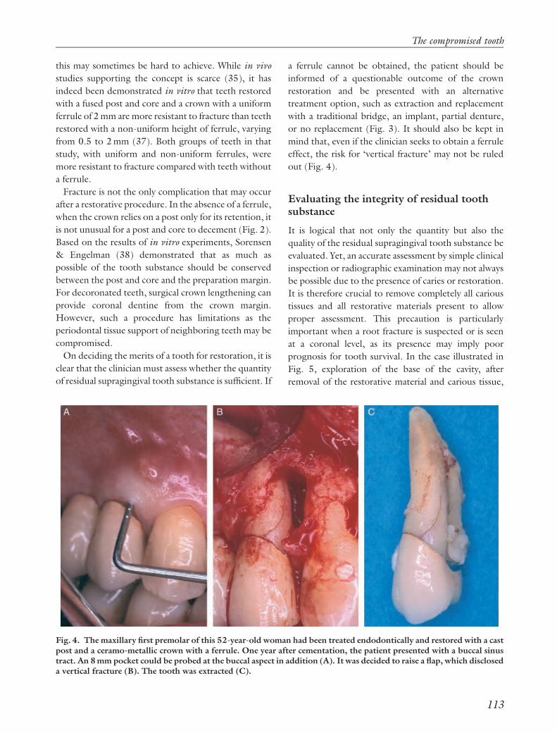
Fig. 4. The maxillary first premolar of this 52-year-old woman had been treated endodontically and restored with a castpost and a ceramo-metallic crown with a ferrule. One year after cementation, the patient presented with a buccal sinustract. An 8 mm pocket could be probed at the buccal aspect in addition (A). It was decided to raise a flap, which discloseda vertical fracture (B). The tooth was extracted (C).
The compromised tooth
113

permitted the many fracture lines responsible for pulpal
symptoms to be seen. However, it was only after
uncovering the floor of the pulpal chamber that the
extension of the fracture lines could be fully appre-
ciated. At the time of examination, there was no sign of
periodontal involvement at this level, but an apical
propagation of the distal fracture line could be
imminent. In a situation like this, the quandary is
whether to treat or suggest extraction with replacement
using an implant. Either way, the decision should be
taken together with the patient. It is stressed that a
proper diagnosis and thorough communication with
the patient is imperative to maintaining a good rapport
and to reduce the risk of medico-legal actions. After an
analogous diagnostic procedure in the case displayed in
Fig. 6, the presence of a fracture line was seen on the
floor of the pulp chamber and resulted in a decision to
extract the tooth.
While overextended access opening preparations for
endodontic treatment should be avoided, direct
straight-line accesses allow fracture lines on the floor
of the pulp chamber to be revealed. When fracture lines
involve the orifices of the root canals, conservation of
the tooth must be deemed highly questionable (see case
in Fig. 7). On superficial examination, the clinician
could have easily presumed that a simple post and core
and a porcelain-bonded crown would have resolved the
esthetic problem of tooth #25. Removal of the
Fig. 5. This case is about tooth #37 in a 38-year-old woman. The patient reported pain to chewing, and episodes ofspontaneous pain. Thermal sensitivity testing gave exaggerated responses. Clinical inspection disclosed cracks in theenamel both buccally and lingually, as well as also in a distal direction involving the distal marginal crest (A). A periapicalradiograph indicated no periapical pathology (B). Having arrived at a diagnosis of irreversible pulpitis, the tentativecause of a root fracture remained to be established. Removing the restoration to allow direct inspection of the underlyingdentine was the first step to assess the depth of the cracks. This revealed carious tissue on the cavity floor (C). Afterremoval of caries, a crack line running in a mesio-distal direction, together with a second crack running transversally, wasobserved (D). The next step was the removal of the pulp chamber roof (E). The distal fracture line appeared to be moresevere (F). Its course in an apical direction seriously questioned conservation of the tooth. While endodontic treatmentcertainly is possible, the main problem is now periodontal, as an apical progression of the fracture may cause aperiodontal lesion. At this point in time, no probable deep pocket was revealed. Prognosis is nevertheless not good andthe patient was adviced extraction. In spite of the poor prognosis, the patient opted for a conservative treatment of thetooth. Endodontic treatment was completed (G), followed by a build-up with resin-composite, and a ceramo-metalliccrown to hold the tooth pieces together. A radiograph taken after cementation of the crown shows no periodontalinvolvment (H). As cases like these carry a highly guarded prognosis, periodical follow-ups are mandatory.
Ricucci & Grosso
114

restorative material revealed the true amount of sound
tooth structure and also the presence of a fracture line,
thus lessening the chance for long-term maintenance.
It should be stressed that all diagnostic procedures in
this context are best carried out under a rubber dam
using magnification and good light.
Evaluating the periodontal tissues
When caries or a fracture has advanced to such an extent
that the periodontal ligament and the crestal bone
below the gingival margin are involved, restoration of
the tooth presents with more advanced problems than
those already described. In all cases, it is imperative that
a restoration is completed on sound tooth structure. If
gingival tissue extends over the tooth margins, the
tooth cannot be restored properly unless the period-
ontal tissues are apically repositioned. Of course,
lengthening of the clinical crown may be considered
in such cases, as long as it does not necessitate excessive
removal of bone tissue from the adjacent teeth. The
final restoration must be placed in such a position that it
does not encroach on the periodontal tissues, thus
allowing for the width of a sufficient amount of
attached gingiva (ca. 3 mm). Owing to this reason,
meticulous clinical evaluation and radiographic exam-
ination are crucial. Aspects such as root length,
proximity to the neighboring teeth (in the case of
interproximal defects), the distance between the
margin of the defect and the furcation area (in
multirooted teeth), and above all, the distance between
the apical limit of the defect and the bone must be
evaluated. It is imperative that radiographs be taken
with as little distortion as possible.
On crown lengthening, it is important to decide how
much bone should be removed. Of course, all carious
tooth substance should be excavated first to reveal the
level of sound tooth substance. This is illustrated in the
case displayed in Fig. 8, where the precise amount of
bone to be removed distally to the canine could be
assessed chairside after having eliminated the hyper-
plasic gingival tissue in the cavity and the carious lesion.
Multirooted teeth with loss of attachment in the
furcation have special problems in that the prognosis is
less favorable unless the area can be properly managed
by oral hygiene measures. Hemisection and resection
Fig. 6. The case is a 40-year-old man presenting with severe pain to chewing in the mandibular right quadrant. A bite-wing radiograph displayed amalgam restorations and recurrent caries (B). It was decided to remove restorations toevaluate the residual dentine. After anesthesia, isolation, and removal of filling material, large quantities of carious tissueappeared (A). A fracture also crossing in a bucco-lingual direction appeared on the floor of tooth #47 (C). In order toexplore the depth of the fracture, the pulp chamber was opened and revealed involvment of the pulp chamber floor (D),which cautioned the option of attempting a conservative treatment. The tooth was extracted.
The compromised tooth
115

procedures are used in an attempt to maintain multi-
rooted teeth that present with advanced periodontal
involvement (39). This may be considered when one
root is affected by non-treatable periodontal disease,
whereas the others are only moderately affected
(Fig. 9).
There are few longitudinal studies over long periods
assessing the outcome of this type of treatment, and it is
difficult to compare the results of those published due
to the differences in the periods of observation as well
as criteria used for evaluation. Blomlof et al. (44), in a
longitudinal study conducted over 10 years, found that
Ricucci & Grosso
116

the prognosis for root-resected posterior teeth was not
worse than that for single-rooted teeth with similar
periodontal involvement. Also, other longitudinal
studies have revealed high percentages of success of
root-resected teeth (40, 41). Buhler (42) examined
available longitudinal studies in the literature and
recorded an overall failure rate of 13%. This percentage
figure was compared with the results of studies on
implants. It became apparent that the failure rate was
quite similar. Fugazzotto (43) followed 701 resected
molars and 1472 posterior implants for 15 years.
Resection of the distal root of mandibular molars
demonstrated the lowest percentage of success (75%).
Other roots had a success rate of 95–100%. Implants in
the position of the second molar, as posterior support
for bridges, had the lowest success rate (85%). All other
Fig. 7. The case describes the diagnostic procedure and treatment of tooth #25 in a 38-year-old woman. The mainproblem was the unesthetic appearance of the maxillary second premolar (A). The tooth had been treated endodonticallyseveral years before and the crown had been restored with amalgam (B). A radiograph showed that the apex was incontinuity with the sinus floor, and no periapical lesion was present (C). During preparation for the entrance of thecanals, a significant quantity of tooth structure had been removed mesially. It is evident that clinical examination andradiograph alone were not sufficient to guide the treatment decision. After isolation and removal of all restorativematerials, the residual tooth structure could be inspected (D). Only at this point the large loss of tooth structure wasevident (only undermined enamel remained). A fracture line also became obvious at the distal aspect of the root.Conservation of the tooth through a cast post and a ceramo-metallic crown therefore did not seem worthwhile. Thetreatment plan included extraction and its substitution with an implant (E–J). In the absence of a periapical lesion, it wasdecided to place an immediate postextraction implant (H). To allow this procedure, great care must be exercised inremoving the tooth to preserve the integrity of the alveolar margins, particularly at the buccal aspect. The extractionprocedure included cutting the tooth into pieces (E–G). The radiographs show the integrity of the alveolus afterextraction of the root (K), freshly inserted implant (L), and the 6-month follow-up (M). The maintenance of the gingivalarchitecture and an optimum integration of the prosthesis from both a dental and a periodontal aspect is shown in (N).
The compromised tooth
117

implants in molar positions were near 100% successful
(97–99%). Thus, hemisection, being a relative simple
procedure, inexpensive, and having a relatively good
prognosis in carefully selected cases, is a viable
treatment alternative to tooth extraction. Outcome
certainly depends on how well each step of the
treatment is performed including endodontic, period-
ontal, and prosthetic treatments (40, 44, 45).
Fig. 8. The maxillary left canine in a 30-year-old woman had the clinical crown totally destroyed, and only the enamelremained buccally (A). A palatal view shows that the destruction and gingival tissue has grown into the defect, whosemargins are not visible (B). The radiograph confirmed the severity of the tooth tissue destruction (C). It was evident thata proper root canal treatment to conserve this tooth would require removal of a certain quantity of bone at the distalaspect. The patient was informed about the possibility of extraction, followed by an implant-supported crown, but sheopted for a conservative treatment. Crown lengthening was carried out following caries excavation to sound dentin andexposure of a vital pulp (D). Endodontic treatment could subsequently be carried out properly under adequate asepsis.(E, F) Finally a cast post and core and a ceramo-metallic crown was placed (G). The condition was stable at 4-year follow-up (H).
Ricucci & Grosso
118

Reflection on the use of dentalimplants for replacement ofcompromised teeth
Once the decision has been taken to extract a given
tooth with bleak potentials for conservative treatment,
replacement using an osseointegrated implant may be a
very useful option (Figs 3 and 7). Within the dental
profession, recent years have certainly witnessed a
tremendous surge in the use of this technology spurred
by prospective multi-center studies demonstrating very
high rates of success for both single and multiple
implants (46–57). In fact, failures registered have been
few and when occurring most implant losses have been
The compromised tooth
119

observed during the first year of function (58, 59).
Nevertheless, it would be ill-advised to promise, before
extraction, that replacement using an implant invari-
ably can be carried out successfully. In fact, there are a
number of conditions that should caution the clinician
to suggest such a treatment. These include whether or
not the patient is a smoker, and whether or not there is
non-treated periodontal disease. Such factors are now
known to impair the prognosis for a successful long-
term outcome (60, 61). Furthermore, the amount of
bone resorption that may occur around an implant
following extraction is not possible to predict, as
implants are no bone-preserving vehicles (62). In the
treatment-planning phase, one must also recognize the
condition of neighboring teeth and any pathology
these teeth may be affected by and that ought to be
addressed. Further considerations include whether or
not one shall attempt a direct procedure following
extraction or wait for soft and bone tissue healing
before insertion. In other words, for a successful
outcome of tooth replacement using implants proper
case selection and skillful management are indeed
decisive factors (63).
Concluding remarks
The decision to maintain or extract a gravely compro-
mised tooth is a challenging task, especially today, with
the access to the potential of replacing the tooth with a
dental implant-based restoration. The clinical cases
selected for this article demonstrate that in some
clinical situations, the decision may be to attempt
salvaging the tooth using a multidisciplinary team
approach. Yet, in other cases, a simple extraction would
be equally correct. An appropriate treatment plan puts
to test the diagnostic ability of the clinician, his/her
clinical experience and skill as well the ability to
communicate his/her assessment with the patient. Of
course, as a responsible clinician, one must put aside
preconceived ideas and not let financial needs dictate
the advice. Resorting to implants whenever a case
appears complicated should be discouraged. For
example, a general dentist not overly experienced in
the field of endodontics, who comes across a tooth with
a complex anatomy and a periapical lesion, may be
tempted to take the easier route of extraction. Similarly,
an operator who does not master the means to cure
periodontal disease or techniques of crown lengthen-
ing, root amputation as well as root resection may more
readily prefer the implant alternative. By the same
token, it is necessary to avoid the other extreme, that is,
to being overly conservative and proceed with a
treatment without critically evaluating the potentials
for a successful outcome. Indeed, balancing these
options is delicate. In these extreme cases, care must
be taken to carry out every diagnostic procedure
available, even those more invasive. This means that
before arriving to a definitive diagnosis and treatment
plan, a conscientious operator should obtain the
patient’s consent to remove a restoration in order to
analyze the residual tooth structure and assess the
potential to carry out a reliable treatment. When it does
seem possible to maintain a compromised tooth with
multidisciplinary intervention, the patient must be
informed of the feasibility and the margins of success of
each treatment option.
Acknowledgments
We are grateful to Dr Gunnar Bergenholtz for editing this
manuscript. We also thank Dr Ulf Lekholm for providing
helpful critiques, and Dr Anna Bate for translating the
original manuscript.
Fig. 9. Tooth #26 had been restored with a crown ca. 5years before. Because of symptoms attributed to anendodontic problem, the tooth had recently beentreated endodontically with an access prepared throughthe crown. Symptoms, however, did not resolve. Tooth#27 had been extracted several years earlier because ofperiodontal disease. At clinical examination, a gingivalrecession and a sinus tract were present (A). Probingdistally revealed a more than 10 mm pocket (B). Aradiograph taken in an orthoradial view wasinconclusive (C). A long metal probe was inserted intothe pocket, and an angulated radiograph subsequentlytaken (D) showed penetration beyond the root apex,confirming a large bone defect around the distal root. Theother two roots had normal pocket probing depths.Hemisection with extraction of the distobuccal root wassuggested and carried out following removal of the crown(E). A temporary resin crown was placed and after 6months, the periodontal tissues appeared to be in asatisfactory condition (F). (G) shows the crown justcemented. After 1 year, a follow-up radiograph (K)revealed normal periodontal tissues. The extracted root(H) was processed for study under a light microscope. Themicrophotograph in (I) shows an overview of the apexstained with a modified Brown & Brenn to disclosebacterial profiles. High-power view of the apical rootsurface (J) shows resorption areas covered by a bacterialbiofilm, confirming the cause of the condition as beingperiodontis.
Ricucci & Grosso
120

References
1. Taintor JF, Langeland K, Valle GF, Krasny RM. Pain: apoor parameter of evaluation in dentistry. Oral Surg1981: 3: 299–303.
2. Sorensen JA, Martinoff JT. Intracoronal reinforcementand coronal coverage: a study of endodontically treatedteeth. J Prosthet Dent 1984: 51: 780–784.
3. Ricucci D, Saulo V. Il restauro dei denti trattatiendodonticamente. Studio clinico longitudinale. DentalCadmos 2000: 19: 15–33.
4. Newburg RE, Pameijer CH. Retentive properties of postand core systems. J Prosthet Dent 1976: 36: 636.
5. Nayyar A, Walton RE, Leonard RA. An amalgam coronalradicular dowel and core technique for endodonticallytreated posterior teeth. J Prosthet Dent 1980: 43: 511–515.
6. Sorensen JA, Martinoff JT. Clinically significant factors indowel design. J Prosthet Dent 1984: 52: 28–35.
7. Morgano SM, Milot P. Clinical success of cast metal postsand cores. J Prosthet Dent 1993: 70: 11–16.
8. Randow K, Glantz PO, Zoger B. Technical failures andsome related clinical complications in extensive fixedprosthodontics. An epidemiological study of long-termclinical quality. Acta Odontol Scand 1986: 44: 241–255.
9. Glantz PO, Nilner K. Patient age and long term survivalof fixed prosthodontics. Gerodontology 1993: 10: 33–39.
10. Palmqvist S, Soderfeldt B. Multivariate analyses of factorsinfluencing the longevity of fixed partial dentures,retainers, and abutments. J Prosthet Dent 1994: 71:245–250.
11. Goodacre CJ, Bernal G, Rungcharassaeng K, Kan JY.Clinical complications in fixed prosthodontics. J ProsthetDent 2003: 90: 31–41.
12. Johnson JK, Schwartz NL, Blackwell RT. Evaluation andrestoration of endodontically treated posterior teeth. JAm Dent Assoc 1976: 93: 597–695.
13. Aquilino SA, Caplan DJ. Relationship between crownplacement and the survival of endodontically treatedteeth. Prosthet Dent 2002: 87: 256–263.
14. Strindberg LZ. Dependence of the results of pulptherapy on certain factors. An analytical study based onradiographic and clinical follow-up examination. ActaOdontol Scand 1956: 14.
15. Kerekes K, Tronstad L. Long-term results of endodontictreatment performed with a standardized technique. JEndod 1979: 5: 83–90.
16. Swartz DB, Skidmore AE, Griffin JA. Twenty years ofendodontic success and failure. J Endod 1983: 9: 198–202.
17. Molven O, Halse A. Success rate for gutta-percha andkloroperka N-Ø root fillings made by undergraduatestudents: radiographic findings after 10–17 years. IntEndod J 1988: 21: 243–250.
18. Sjogren U, Hagglund B, Sundqvist G, Wing K. Factorsaffecting the long-term results of endodontic treatment.J Endod 1990: 16: 498–504.
19. Salehrabi R, Rotstein I. Endodontic treatment outcomesin a large patient population in the USA: an epidemio-logical study. J Endod 2004: 30: 846–850.
20. Odesjo B, Hellden L, Salonen L, Langeland K.
Prevalence of previous endodontic treatment, technicalstandard and occurrence of periapical lesions in a
randomly selected adult, general population. EndodDent Traumatol 1990: 6: 265–272.
21. Imfeld TN. Prevalence and quality of endodontictreatment in an elderly urban population of Switzerland.
J Endod 1991: 17: 604–607.22. De Cleen MJ, Schuurs AH, Wesselink PR, Wu MK.
Periapical status and prevalence of endodontic treatment
in an adult Dutch population. Int Endod J 1993: 26:112–119.
23. Buckley M, Spangberg LSW. The prevalence andtechnical quality of endodontic treatment in an American
subpopulation. Oral Surg Oral Med Oral Pathol OralRadiol Endod 1995: 79: 92–100.
24. Weiger R, Hitzler S, Hermle G, Lost C. Periapical status,quality of root canal fillings and estimated endodontic
treatment needs in an urban German population. EndodDent Traumatol 1997: 13: 69–74.
25. Saunders WP, Saunders EM, Sadiq J, Cruickshank E.
Technical standard of root canal treatment in an adultScottish sub-population. Br Dent J 1997: 182: 382–386.
26. De Moor RJ, Hommez GM, De Boever JG, Delme KI,Martens GE. Periapical health related to the quality of
root canal treatment in a Belgian population. Int Endod J2000: 33: 113–120.
27. Kirkevang LL, Orstavik D, Horsted-Bindslev P, Wenzel
A. Periapical status and quality of root fillings and coronalrestorations in a Danish population. Int Endod J 2000:
33: 509–515.28. Boucher Y, Matossian L, Rilliard F, Machtou P. Radio-
graphic evaluation of the prevalence and technical qualityof root canal treatment in a French subpopulation. IntEndod J 2002: 35: 229–238.
29. Kabak Y. Abbott Prevalence of apical periodontitis andthe quality of endodontic treatment in an adult
Belarusian population. Int Endod J 2005: 38: 238–245.30. Loftus JJ, Keating AP, McCartan BE. Periapical status
and quality of endodontic treatment in an adult Irishpopulation. Int Endod J 2005: 38: 81–86.
31. Tsuneishi M, Yamamoto T, Yamanaka R, Tamaki N,Sakamoto T, Tsuji K, Watanabe T. Radiographic evalua-
tion of periapical status and prevalence of endodontictreatment in an adult Japanese population. Oral SurgOral Med Oral Pathol Oral Radiol Endod 2005: 100:631–635.
32. Siqueira JF Jr, Rocas IN, Alves FR, Campos LC.
Periradicular status related to the quality of coronalrestorations and root canal fillings in a Brazilian
population. Oral Surg Oral Med Oral Pathol Oral RadiolEndod 2005: 100: 369–374.
33. Kirkevang LL, Vaeth M, Horsted-Bindslev P, Wenzel A.Longitudinal study of periapical and endodontic status in
a Danish population. Int Endod J 2006: 39: 100–107.34. Petersson K, Hakansson R, Hakansson J, Olsson B,
Wennberg A. Follow-up study of endodontic status in an
adult Swedish population. Endod Dent Traumatol 1991:7: 221–225.
The compromised tooth
121

35. Stankiewicz NR, Wilson PR. The ferrule effect: aliterature review. Int Endod J 2002: 35: 575–581.
36. Rosen H. Operative procedures on mutilated endodon-tically treated teeth. J Prosthet Dent 1961: 11: 973–986.
37. Tan PL, Aquilino SA, Gratton DG, Stanford CM, TanSC, Johnson WT, Dawson D. In vitro fracture resistanceof endodontically treated central incisors with varyingferrule heights and configurations. J Prosthet Dent 2005:93: 331–336.
38. Sorensen JA, Engelman MJ. Ferrule design and fractureresistance of endodontically treated teeth. J Prosthet Dent1990: 63: 529–536.
39. Carnevale G, Pontoriero R, Lindhe J. Treatment offurcation involved teeth. In: Lindhe J, Karring T, LangNP, eds. Clinical Periodontology and Implant Dentistry.Blackwell Munksgaard, 2003: 705–730.
40. Bergenholtz A. Radeectomy of multirooted teeth. J AmDent Assoc 1972: 85: 870–875.
41. Babay NA, Almas K. A four-year clinical follow-up ofnonvital root resection in maxillary molar teeth. Indian JDent Res 1996: 7: 29–32.
42. Buhler H. Survival rates of hemisected teeth: an attemptto compare them with survival rates of alloplasticimplants. Int J Period Restor Dent 1994: 14: 536–543.
43. Fugazzotto PA. A comparison of the success of rootresected molars and molar position implants in functionin a private practice: results of up to 15-plus years. JPeriodontol 2001: 72: 1113–1123.
44. Blomlof L, Jansson L, Appelgren R, Ehnevid H,Lindskog S. Prognosis and mortality of root-resectedmolars. Int J Period Restor Dent 1997: 17: 190–201.
45. Buhler H. Evaluation of root-resected teeth. Results after10 years. J Periodontol 1988: 59: 805–810.
46. Adell R, Eriksson B, Lekholm U, Branemark PI, Jemt T.Long-term follow-up study of osseointegrated implantsin the treatment of totally edentulous jaws. Int J OralMaxillofac Implants 1990: 5: 347–359.
47. Henry PJ, Laney WR, Jemt T, Harris D, Krogh PH,Polizzi G, Zarb GA, Herrmann I. Osseointegratedimplants for single-tooth replacement: a prospective 5-year multicenter study. Int J Oral Maxillofac Implants1996: 11: 450–455.
48. Romeo E, Chiapasco M, Ghisolfi M, Vogel G. Long-term clinical effectiveness of oral implants in thetreatment of partial edentulism. Seven-year life tableanalysis of a prospective study with ITI dental implantssystem used for single-tooth restorations. Clin OralImplants Res 2002: 13: 133–143.
49. Lambrecht JT, Filippi A, Kunzel AR, Schiel HJ. Long-term evaluation of submerged and nonsubmerged ITIsolid-screw titanium implants: a 10-year life table analysisof 468 implants. Int J Oral Maxillofac Implants 2003:18: 826–834.
50. Fugazzotto PA, Vlassis J, Butler B. ITI implant use inprivate practice: clinical results with 5 526 implantsfollowed up to 721months in function. Int J OralMaxillofac Implants 2004: 19: 408–412.
51. Sulzer TH, Bornstein MM, Buser D. Indications for oralimplantology in a referral clinic. A three-year retro-spective analysis of 737 patients with 1176 implants.Schweiz Monatsschr Zahnmed 2004: 114: 444–450.
52. Karoussis IK, Salvi GE, Heitz-Mayfield LJ, Bragger U,Hammerle CH, Lang NP. Long-term implant prognosisin patients with and without a history of chronicperiodontitis: a 10-year prospective cohort study of theITI Dental Implant System. Clin Oral Implants Res2003: 14: 329–339.
53. Mayer TM, Hawley CE, Gunsolley JC, Feldman S. Thesingle-tooth implant: a viable alternative for single-toothreplacement. J Periodontol 2002: 73: 687–693.
54. Priest G. Single-tooth implants and their role inpreserving remaining teeth: a 10-year survival study.Int J Oral Maxillofac Implants 1999: 14: 181–188.
55. Simon RL. Single implant-supported molar and pre-molar crowns: a ten-year retrospective clinical report. JProsthet Dent 2003: 90: 517–521.
56. Covani U, Crespi R, Cornelini R, Barone A. Immediateimplants supporting single crown restoration: a 4-yearprospective study. J Periodontol 2004: 75: 982–988.
57. Chen ST, Wilson TG Jr, Hammerle CH. Immediate orearly placement of implants following tooth extraction:review of biologic basis, clinical procedures, and out-comes. Int J Oral Maxillofac Implants 2004: 9(Suppl):12–25.
58. Esposito M, Hirsch JM, Lekholm U, Thomsen P.Biological factors contributing to failures of osseointe-grated oral implants. (I). Success criteria and epidemiol-ogy. Eur J Oral Sci 1998: 106: 527–551.
59. Snauwaert K, Duyck J, van Steenberghe D, Quirynen M,Naert I. Time dependent failure rate and marginal boneloss of implant supported prostheses: a 15-year follow-upstudy. Clin Oral Invest 2000: 4: 13–20.
60. Esposito M, Hirsch JM, Lekholm U, Thomsen P.Biological factors contributing to failures of osseointe-grated oral implants. (II). Etiopathogenesis. Eur J OralSci 1998: 106: 721–764.
61. Hardt CR, Grondahl K, Lekholm U, Wennstrom JL.Outcome of implant therapy in relation to experiencedloss of periodontal bone support: a retrospective 5-yearstudy. Clin Oral Implants Res 2002: 13: 488–494.
62. Araujo MG, Sukekava F, Wennstrom JL, Lindhe J. Ridgealterations following implant placement in fresh extrac-tion sockets: an experimental study in the dog. J ClinPeriodontol 2005: 32: 645–652.
63. Lekholm U. The surgical site. In: Lindhe J, Karring T,Lang NP, eds. Blackwell Munksgaard, 2003: 852–865.
Ricucci & Grosso
122