the combined alt–groin flap for the mutilated and degloved ... · representative presentation of...
TRANSCRIPT

Injury, Int. J. Care Injured 46 (2015) 1591–1596
Contents lists available at ScienceDirect
Injury
journa l homepage: www.e lsevier .com/ locate / in jury
The combined ALT–groin flap for the mutilated and degloved hand
Jonathan A. Zelken, Nai-jen Chang, Fu-chan Wei, Cheng-hung Lin *
Department of Plastic and Reconstructive Surgery, Chang Gung Memorial Hospital, Chang Gung Medical College and Chang Gung University, Taipei, Taiwan
A R T I C L E I N F O
Article history:
Accepted 8 May 2015
Keywords:
Degloving
Hand reconstruction
Anterolateral thigh flap
Mutilated hand
A B S T R A C T
Background: Degloving and mutilation of the hand is a rare but formidable challenge. When replantation
is not possible, we rely on distant pedicled flaps. We present a technique using pedicled anterolateral
thigh (ALT) and groin flaps to sandwich and resurface the degloved hand. The purpose of this study is to
describe the rationale, indications, methods and outcomes of combined pedicled ALT and groin flap
reconstruction of the degloved hand.
Methods: Five injuries were treated at this center between 2011 and 2014. Charts were retrospectively
reviewed and outcomes evaluated. Four ALT–groin flaps were performed in a single stage for degloving,
crush and combined injuries. In one case, partial necrosis of a tight groin flap necessitated secondary ALT
coverage at a second stage.
Results: Flaps survived after division at 4 weeks, and venous congestion was not observed at any point.
Debulking, syndactyly release and toe transfer followed reconstruction to enhance outcomes.
Conclusions: The combined ALT–groin flap is safe and feasible for the reconstruction of the degloved or
mutilated hand when replantation is not an option. It is attractive for familiar donor anatomy, donor-site
morbidity and the quantity and composition of the tissue it provides.
� 2015 Elsevier Ltd. All rights reserved.
Background
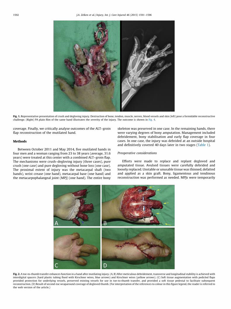
Degloving injuries of the hand are rare but alarming (Supple-mental Digital Content: Video 1). As the soft tissue envelope suppliesblood to the distal hand, ischemia and necrosis of the denuded partsmay be imminent without expedient vascularised tissue transfer. Inthe spirit of replacing like with like, replantation is ideal but oftenimpossible [1–4]. Even when amputated and degloved parts areavailable, neurovascular trauma at multiple levels and an extensivezone of injury deem flap survival and functional restoration unlikely.If degloving is accompanied by amputation (Fig. 1), dependabletissue coverage provides a platform for digital reconstruction andtoe-to-hand transfer (Fig. 2A–D) [5–7].
The perfect reconstructive strategy would restore intricate handfunctions and a specialised anatomy; the current methods, includingreplantation, fall short in both categories. Distant pedicled optionsfrom the groin and abdomen have been favoured since the 1970s.These methods do not require a microsurgical technique and theypermit a two-team approach. Groin flaps are reliable, provide ample
* Corresponding author at: Department of Plastic and Reconstructive Surgery,
Chang Gung Memorial Hospital, 5, Fu-Hsing St. Kuei-Shan, Taoyuan 333, Taiwan.
Tel.: +886 3 3281200x2946; fax: +886 3 3289582.
E-mail address: [email protected] (C.-h. Lin).
http://dx.doi.org/10.1016/j.injury.2015.05.022
0020–1383/� 2015 Elsevier Ltd. All rights reserved.
tissue, are easy to harvest and exhibit minimal donor-site morbidity.However, they may not cover extensive defects that involve thedorsal and palmar hand, particularly without a time-consumingdelay procedure or tissue expansion [8]. Paired groin and abdomenflaps have been described since the 1970s [2,5,9–18], which arebetter suited to extensive and three-dimensional wounds.
Because degloving injuries of the hand are infrequent, theliterature scarcely supports a single technique for secondaryreconstruction and no comprehensive reviews or meta-analysesexist. There seems to have been little evolution in reconstructivetechnique since the 1970s, and to our knowledge no gold standardexists. The anterolateral thigh (ALT) flap has a long-standingheritage as a workhorse free [19] and pedicled flap [20], yet it wasonly recently that the pedicled ALT flap was described for handreconstruction. Tuncer was the first to use it in upper-extremityreconstruction in 2008 [21]. Three years later, Senda combined apedicled ALT and groin flap, providing additional volume andimproved coverage for complex defects [17].
Senda described the only report of the combined use of ALT andgroin flaps in the literature, to the author’s knowledge [17]. Wemodified Senda’s technique to enhance reliability and outcomes.The purpose of this study is to retrospectively review a series ofcases where pedicled ALT and groin flaps were used forreconstruction following degloving injury. We aim to providerationale, indications and methods for combined ALT–groin flap
[(Fig._1)TD$FIG]
Fig. 1. Representative presentation of crush and degloving injury. Destruction of bone, tendon, muscle, nerves, blood vessels and skin (left) pose a formidable reconstructive
challenge. (Right) PA plain film of the same hand illustrates the severity of the injury. The outcome is shown in Fig. 4.
J.A. Zelken et al. / Injury, Int. J. Care Injured 46 (2015) 1591–15961592
coverage. Finally, we critically analyse outcomes of the ALT–groinflap reconstruction of the mutilated hand.
Methods
Between October 2011 and May 2014, five mutilated hands infour men and a woman ranging from 23 to 38 years (average, 31.6years) were treated at this center with a combined ALT–groin flap.The mechanisms were crush–degloving injury (three cases), purecrush (one case) and pure degloving without bone loss (one case).The proximal extent of injury was the metacarpal shaft (twohands), wrist crease (one hand), metacarpal base (one hand) andthe metacarpophalangeal joint (MPJ) (one hand). The entire bony[(Fig._2)TD$FIG]
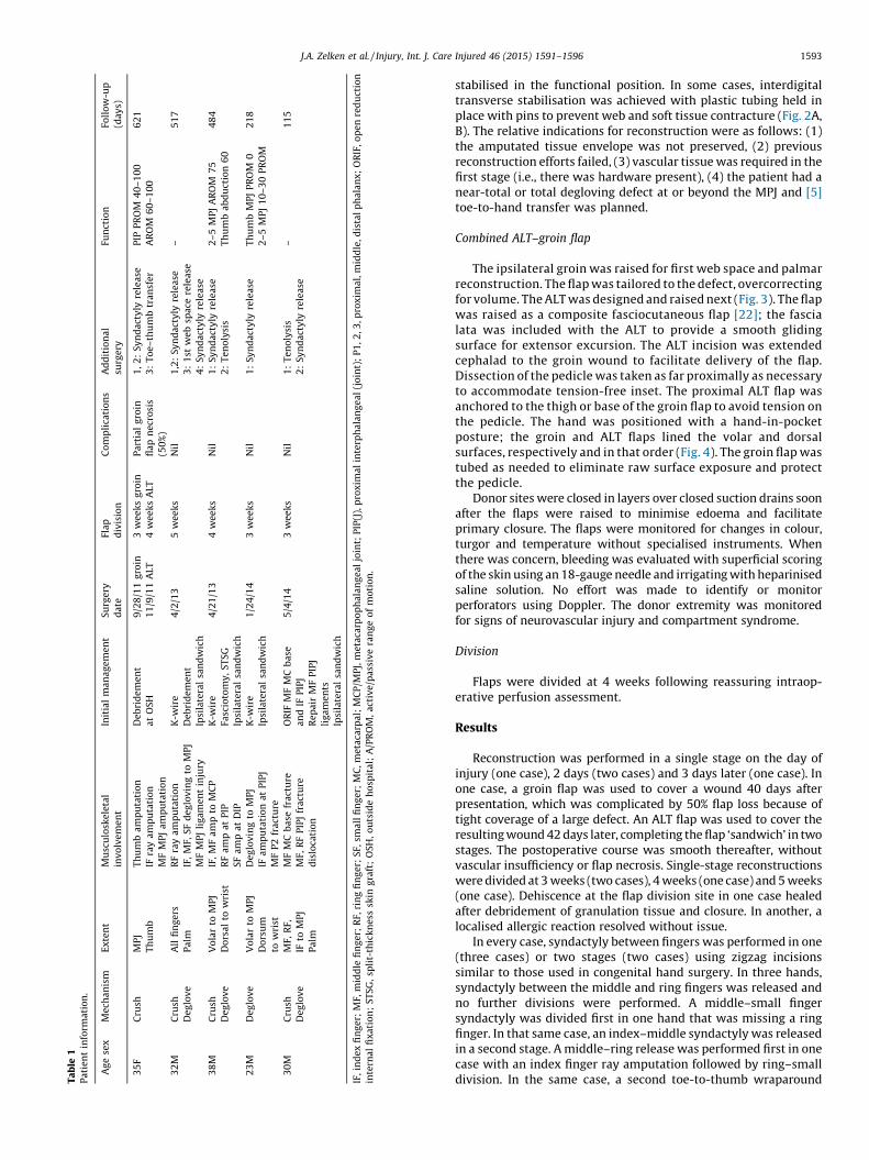
Fig. 2. A toe-to-thumb transfer enhances function in a hand after mutilating injury. (A, B)
interdigital spacers (hard plastic tubing fixed with Kirschner wires, blue arrows) and
provided protection for underlying vessels, preserved existing vessels for use in toe
reconstruction. (D) Result of second-toe wraparound coverage of degloved thumb. (For in
the web version of the article.)
skeleton was preserved in one case. In the remaining hands, therewere varying degrees of bony amputation. Management includeddebridement, bony stabilisation and early flap coverage in fourcases. In one case, the injury was debrided at an outside hospitaland definitively covered 40 days later in two stages (Table 1).
Preoperative considerations
Efforts were made to replace and replant degloved andamputated tissue. Avulsed tissues were carefully debrided andloosely replaced. Unstable or unusable tissue was thinned, defattedand applied as a skin graft. Bony, ligamentous and tendinousreconstruction was performed as needed. MPJs were temporarily
After meticulous debridement, transverse and longitudinal stability is achieved with
Kirschner wires (yellow arrows). (C) Soft tissue augmentation with pedicled flaps
-to-thumb transfer, and provided a soft tissue pedestal to facilitate subsequent
terpretation of the references to colour in this figure legend, the reader is referred to
Ta
ble
1P
ati
en
tin
form
ati
on
.
Ag
ese
xM
ech
an
ism
Ex
ten
tM
usc
ulo
ske
leta
l
inv
olv
em
en
t
Init
ial
ma
na
ge
me
nt
Su
rge
ry
da
te
Fla
p
div
isio
n
Co
mp
lica
tio
ns
Ad
dit
ion
al
surg
ery
Fun
ctio
nFo
llo
w-u
p
(da
ys)
35
FC
rush
MP
J
Th
um
b
Th
um
ba
mp
uta
tio
n
IFra
ya
mp
uta
tio
n
MF
MP
Ja
mp
uta
tio
n
De
bri
de
me
nt
at
OS
H
9/2
8/1
1g
roin
11
/9/1
1A
LT
3w
ee
ks
gro
in
4w
ee
ks
ALT
Pa
rtia
lg
roin
fla
pn
ecr
osi
s
(50
%)
1,
2:
Sy
nd
act
yly
rele
ase
3:
To
e–
thu
mb
tra
nsf
er
PIP
PR
OM
40
–1
00
AR
OM
60
–1
00
62
1
32
MC
rush
De
glo
ve
All
fin
ge
rs
Pa
lm
RF
ray
am
pu
tati
on
IF,
MF,
SF
de
glo
vin
gto
MP
J
MF
MP
Jli
ga
me
nt
inju
ry
K-w
ire
De
bri
de
me
nt
Ipsi
late
ral
san
dw
ich
4/2
/13
5w
ee
ks
Nil
1,2
:S
yn
da
cty
lyre
lea
se
3:
1st
we
bsp
ace
rele
ase
4:
Sy
nd
act
yly
rele
ase
–5
17
38
MC
rush
De
glo
ve
Vo
lar
toM
PJ
Do
rsa
lto
wri
st
IF,
MF
am
pto
MC
P
RF
am
pa
tP
IP
SF
am
pa
tD
IP
K-w
ire
Fasc
ioto
my
,S
TS
G
Ipsi
late
ral
san
dw
ich
4/2
1/1
34
we
ek
sN
il1
:S
yn
da
cty
lyre
lea
se
2:
Te
no
lysi
s
2–
5M
PJ
AR
OM
75
Th
um
ba
bd
uct
ion
60
48
4
23
MD
eg
lov
eV
ola
rto
MP
J
Do
rsu
m
tow
rist
De
glo
vin
gto
MP
J
IFa
mp
uta
tio
na
tP
IPJ
MF
P2
fra
ctu
re
K-w
ire
Ipsi
late
ral
san
dw
ich
1/2
4/1
43
we
ek
sN
il1
:S
yn
da
cty
lyre
lea
seT
hu
mb
MP
JP
RO
M0
2–
5M
PJ
10
–3
0P
RO
M
21
8
30
MC
rush
De
glo
ve
MF,
RF,
IFto
MP
J
Pa
lm
MF
MC
ba
sefr
act
ure
MF,
RF
PIP
Jfr
act
ure
dis
loca
tio
n
OR
IFM
FM
Cb
ase
an
dIF
PIP
J
Re
pa
irM
FP
IPJ
lig
am
en
ts
Ipsi
late
ral
san
dw
ich
5/4
/14
3w
ee
ks
Nil
1:
Te
no
lysi
s
2:
Sy
nd
act
yly
rele
ase
–1
15
IF,
ind
ex
fin
ge
r;M
F,m
idd
lefi
ng
er;
RF,
rin
gfi
ng
er;
SF,
sma
llfi
ng
er;
MC
,m
eta
carp
al;
MC
P/M
PJ,
me
taca
rpo
ph
ala
ng
ea
ljo
int;
PIP
(J),
pro
xim
al
inte
rph
ala
ng
ea
l(j
oin
t);
P1
,2
,3,p
rox
ima
l,m
idd
le,d
ista
lp
ha
lan
x;
OR
IF,
op
en
red
uct
ion
inte
rna
lfi
xa
tio
n;
ST
SG
,sp
lit-
thic
kn
ess
skin
gra
ft;
OS
H,
ou
tsid
eh
osp
ita
l;A
/PR
OM
,a
ctiv
e/p
ass
ive
ran
ge
of
mo
tio
n.
J.A. Zelken et al. / Injury, Int. J. Care Injured 46 (2015) 1591–1596 1593
stabilised in the functional position. In some cases, interdigitaltransverse stabilisation was achieved with plastic tubing held inplace with pins to prevent web and soft tissue contracture (Fig. 2A,B). The relative indications for reconstruction were as follows: (1)the amputated tissue envelope was not preserved, (2) previousreconstruction efforts failed, (3) vascular tissue was required in thefirst stage (i.e., there was hardware present), (4) the patient had anear-total or total degloving defect at or beyond the MPJ and [5]toe-to-hand transfer was planned.
Combined ALT–groin flap
The ipsilateral groin was raised for first web space and palmarreconstruction. The flap was tailored to the defect, overcorrectingfor volume. The ALT was designed and raised next (Fig. 3). The flapwas raised as a composite fasciocutaneous flap [22]; the fascialata was included with the ALT to provide a smooth glidingsurface for extensor excursion. The ALT incision was extendedcephalad to the groin wound to facilitate delivery of the flap.Dissection of the pedicle was taken as far proximally as necessaryto accommodate tension-free inset. The proximal ALT flap wasanchored to the thigh or base of the groin flap to avoid tension onthe pedicle. The hand was positioned with a hand-in-pocketposture; the groin and ALT flaps lined the volar and dorsalsurfaces, respectively and in that order (Fig. 4). The groin flap wastubed as needed to eliminate raw surface exposure and protectthe pedicle.
Donor sites were closed in layers over closed suction drains soonafter the flaps were raised to minimise edoema and facilitateprimary closure. The flaps were monitored for changes in colour,turgor and temperature without specialised instruments. Whenthere was concern, bleeding was evaluated with superficial scoringof the skin using an 18-gauge needle and irrigating with heparinisedsaline solution. No effort was made to identify or monitorperforators using Doppler. The donor extremity was monitoredfor signs of neurovascular injury and compartment syndrome.
Division
Flaps were divided at 4 weeks following reassuring intraop-erative perfusion assessment.
Results
Reconstruction was performed in a single stage on the day ofinjury (one case), 2 days (two cases) and 3 days later (one case). Inone case, a groin flap was used to cover a wound 40 days afterpresentation, which was complicated by 50% flap loss because oftight coverage of a large defect. An ALT flap was used to cover theresulting wound 42 days later, completing the flap ‘sandwich’ in twostages. The postoperative course was smooth thereafter, withoutvascular insufficiency or flap necrosis. Single-stage reconstructionswere divided at 3 weeks (two cases), 4 weeks (one case) and 5 weeks(one case). Dehiscence at the flap division site in one case healedafter debridement of granulation tissue and closure. In another, alocalised allergic reaction resolved without issue.
In every case, syndactyly between fingers was performed in one(three cases) or two stages (two cases) using zigzag incisionssimilar to those used in congenital hand surgery. In three hands,syndactyly between the middle and ring fingers was released andno further divisions were performed. A middle–small fingersyndactyly was divided first in one hand that was missing a ringfinger. In that same case, an index–middle syndactyly was releasedin a second stage. A middle–ring release was performed first in onecase with an index finger ray amputation followed by ring–smalldivision. In the same case, a second toe-to-thumb wraparound

[(Fig._3)TD$FIG]
Fig. 3. Design. (A) Representative layout and vascular axis identification (B) for combined ALT–groin flap. The purple dashed line (yellow arrow) creates a continuous plane
between the suprafascial groin flap and ALT flap wounds, permitting delivery of the ALT flap through the proximal wound (C). This is intended to increase ALT flap mobility,
efficiency and pedicle length. (For interpretation of the references to colour in this figure legend, the reader is referred to the web version of the article.)
J.A. Zelken et al. / Injury, Int. J. Care Injured 46 (2015) 1591–15961594
flap was transferred in a third stage (Fig. 2). No additionalcomplications occurred. An average of 1.8 revision procedureswere performed (range, 1–4 procedures) per case. Other refine-ments included tenolysis (two cases) and soft tissue contracturerelease (two cases).
Joint motion was evaluated in three of five cases at the mostrecent follow-up (Table 1). In one case, a reconstructed rightproximal interphalangeal (PIP) demonstrated 40–1008 of passivemotion and 60–1008 of active motion at 1.7 years. In another injurythat involved all the digits, the thumb had no MPJ motion, and theremaining MP joints were severely restricted to 10–308 of activemotion at 8 months. In a third case, the second through fifth MPjoints had 758 of active flexion and the thumb could adduct at theMPJ to 608 at 1.3 years. Average duration of follow-up fromreconstruction to the most recent follow-up was 391 days (range115–621 days). Wide variations in functional outcomes reflectedthe severity of initial trauma.
[(Fig._4)TD$FIG]Fig. 4. Inset and division. (A) Crush and degloving injury as seen in Fig. 1, following debr
(red arrow) pedicle was dissected as proximally as possible (C), delivered through the w
wound coverage. (C) The tissues were closed in a tension-free manner. (D) Postopera
references to colour in this figure legend, the reader is referred to the web version of
Discussion
Degloving injuries may result in extensive tissue loss, fewavailable recipient vessels, disfigurement and significant function-al impairment. Goals of reconstruction are preservation of length,improvement of appearance and restoration of function. Toachieve this, adequate soft tissue coverage should be establishedearly. Secondary procedures such as toe-to-hand transplantationenhance appearance, enable key pinch and improve grip strength.We currently favour the use of combined pedicled ALT and groinflaps to reconstruct degloving injuries. We describe the rationale,indications, methods and outcomes of our preferred strategy.
The important benefit of pedicled flap coverage of the mutilatedhand is preservation of existing blood supply. Pedicled flaps do notnecessitate a microsurgical technique and specialised postsurgicalcare, and they permit a two-team approach for debridement andflap harvest. The pedicled groin flap is reliable and boasts a large
idement. (B) The pliable groin flap (blue arrow) provided palmar coverage. The ALT
ound extension (yellow arrow) and gently draped over the dorsum, providing total
tive view after two syndactyly releases and debulking. (For interpretation of the
the article.)

J.A. Zelken et al. / Injury, Int. J. Care Injured 46 (2015) 1591–1596 1595
skin paddle, facile harvest, minimal donor-site morbidity and aninconspicuous scar. However, this flap will not cover extensivedorsal and volar hand defects, particularly without time-consum-ing delay procedures or tissue expansion [8]. Extensive tissue losswarrants multiple-flap reconstruction [9,10,12,14,15,17,18]. Thepedicled ALT flap was not described in hand reconstruction beforethis decade [21,23]. However, historical references [17,21] and ourexperience support the feasibility of the ALT in pedicled handcoverage. The combination of groin and ALT flaps provides asubstantial amount of tissue.
Candidates for surgery suffered traumatic degloving injuries,often with a crush component whose replantation efforts, ifattempted, had failed. We strived to create a pliable and sensateenvelope that preserved joint excursion, blood supply and digitallength. Manifestations of degloving injury are different from blastand pure crush injuries as the soft tissue envelope is dispropor-tionately injured. In some cases, soft tissue is all that needsreplacement: nerves, tendons, pulleys, ligaments and skeletaltissue proximal to the DIP joints are often preserved. When softtissue coverage is replaced early, the functional length of thefingers and range of motion (ROM) of MPJs may be maximised, andbasic hand function can be achieved with fewer subsequentprocedures. The alternative options to this technique includeamputation and prosthesis, free flaps and local pedicled flaps.
From a sociocultural prospective, the Taiwanese typically preferlimb preservation to amputation, as do most Asian populations.There are important economic considerations as well. In the lower-extremity literature, the cost of salvage was shown to be lowerthan amputation at 2 years. The disparity widens over the lifetimeof the patient, where prostheses eventually cost three times asmuch as reconstruction [24]. Many patients we treat for deglovinginjuries are young labourers who cannot afford prohibitive lifetimecosts of maintaining and replacing prostheses. When there isincomplete degloving, or at least one digit is preserved, sensoryoutcomes are always superior to amputation and prosthesis.Ultimately, the decision to amputate is the responsibility of theconsultant surgeon and it should reflect the patient’s other injuries,quantity and function of the salvaged tissue, patient motivationand wishes, surgeon expertise and resources. We cannot claimwhether functional return after reconstruction is superior to earlyamputation and prosthesis fitting.
This technique relies on intact and uncompromised donortissue, which may not be available. In theory, the contralateral hipmay be used if the ipsilateral tissue is absent or unreliable.Neumeister et al. provide an exhaustive summary of alternativesfor mutilated hand reconstruction [25]. Free tissue options such asthe latissimus dorsi are capable of covering large areas, and theserratus flap is effective for smaller defects. The temporal parietalflap is classically described for dorsal hand coverage for itspliability, dependability and amenability to tendon excursion[26,27]. Numerous free tissue options are available.
We do not favour free tissue transfer, because we prefer not toexploit vessels or compromise perfusion. Local pedicled optionsinclude the reverse radial forearm flap, interosseous flaps and theBecker (ulnar artery) flap. The forearm flap entails proximaldivision of the radial artery, and the latter options are small andmay not reach. Other options including the omentum, abdomenand spare parts may play a role in future applications, but theyhave not been widely used in the literature or at our institution.Distant pedicled flaps can be ergonomic, dependable and generousin quantity. For reasons we mentioned, a combination of ALT andgroin flaps is an attractive option.
Combining pedicled groin and ALT flaps provides abundanttissue that exceeds other combinations in that region. Sendaintroduced this concept [17], but we present the first series andmodified the design. Venous congestion and partial necrosis
described in Senda’s report probably resulted from a vulnerable,bare pedicle that kinked at the flap–thigh junction. To optimise theALT pedicle position, we first inset the pliable groin flap in thepalm, and then gently draped the ALT flap over the dorsum [21].We avoided folding the ALT to optimise flap perfusion andefficiency. We anchored the ALT to the thigh and groin flap withheavy-gauge suture after mobilisation and inset to minimisefurther risk of mechanical trauma and kinking.
No delay procedures for flap training were needed in our series,and the vascular pedicle was patent upon division in every case.We divided flaps at 3–4 weeks with success, as did Tuncer andSenda [17,21]. There is room for improvement in flap coverage ofthe degloved and mutilated hand; superthin [28–32] elevation andprimary defatting may enhance cosmetic and functional outcomes.We acknowledge that the final product is not always cosmeticallyappealing and functional outcomes are equivocal at best. This isevidenced by unsatisfactory parameters documented after threesuccessful reconstructions. Aesthetic results may be enhancedwith staged excision and liposuction [33], but excessive debulkingcan further impair mobility and perfusion.
The series is important in establishing the feasibility ofcombined ALT and groin flaps for coverage of extensive andcomplex defects. It is the first series to review these flaps. Weclearly identify the rationale of a modified design and suggeststrategies for subsequent refinements, such as toe transfer anddivision of syndactylous digits. The important limitations of thisseries include small sample size, varying degrees and patterns ofinjury and failure to evaluate patient satisfaction. Most impor-tantly, the outcomes were not compared with alternativestrategies that are currently in use. This can largely be attributedto the rarity of degloving injuries encountered globally.
Future studies may compare the economic, cosmetic, functionaland psychological aspects of pedicled versus free flap reconstruc-tion, salvage versus amputation, vascularised composite allotrans-plantation, etc. For now, the combined ALT–groin flap is ourpreferred method when enough tissue remains to restore basichand function. We endorse this straightforward and feasiblestrategy using workhorse flaps that offer acceptable donor-sitemorbidity, abundant tissue and a concealable scar.
Conflicts of interest and source of funding
None of the authors has a financial interest in any of theproducts, devices, or drugs mentioned in this manuscript.
This work has not been presented at any meetings or symposia.
Acknowledgements
The authors would like to thank Dr. Aaron Huang and Dr. SabaLee for assistance in data collection. There is no other source ofsupport to acknowledge.
Appendix A. Supplementary data
Supplementary data associated with this article can be found, in
the online version, at http://dx.doi.org/10.1016/j.injury.2015.05.022.
References
[1] Hsu WM, Wei FC, Lin CH, Chen HC, Chuang CC, Chen HT. The salvage of adegloved hand skin flap by arteriovenous shunting. Plast Reconstr Surg1996;98(July (1)):146–50.
[2] Thomas BP, Katsarma E, Tsai TM. Replantation of total degloving of the hand:case report. J Reconstr Microsurg 2003;19(May (4)):217–20.
[3] Rodriguez-Lorenzo A, Lin CH, Lin CH, Ching WC, Lin YT. Replantation of adegloved hand with added arteriovenous anastomoses: report of two cases. JHand Surg Am 2009;34(Dec (10)):1864–7.

J.A. Zelken et al. / Injury, Int. J. Care Injured 46 (2015) 1591–15961596
[4] Lo S, Lin YT, Lin CH, Wei FC. A new classification to aid the selection ofrevascularization techniques in major degloving injuries of the upper limb.Injury 2013;44(March (3)):331–5.
[5] Buchman SJ, Eglseder Jr WA, Robertson BC. Pedicled groin flaps for upper-extremity reconstruction in the elderly: a report of 4 cases. Arch Phys MedRehabil 2002;83(June (6)):850–4.
[6] Goertz O, Kapalschinski N, Daigeler A, Hirsch T, Homann HH, Steinstraesser L,et al. The effectiveness of pedicled groin flaps in the treatment of hand defects:results of 49 patients. J Hand Surg Am 2012;37(Oct (10)):2088–94.
[7] McGregor IA, Jackson IT, The groin flap, Br J. Plast Surg 1972;25(Jan (1)):3–16.[8] DeHaan MR, Hammond DC, Mann RJ. Controlled tissue expansion of a groin
flap for upper extremity reconstruction. Plast Reconstr Surg 1990;86(Nov(5)):979–82.
[9] Cowen NJ, Giannotto RP. Multiple flap coverage for degloving hand injuries.South Med J 1978;71(March (3)):281–8.
[10] Henry M, Levaro F, Masson M, Clifton J. Simultaneous three-flap reconstruc-tion of extensive hand and finger degloving injury: case report. J ReconstrMicrosurg 2002;18(July (5)):387–91.
[11] Kleinman WB, Dustman JA. Preservation of function following completedegloving injuries to the hand: use of simultaneous groin flap, randomabdominal flap, and partial-thickness skin graft. J Hand Surg Am1981;6(Jan (1)):82–9.
[12] Miura T, Nakamura R. Use of paired flaps to simultaneously cover the dorsaland volar surfaces of a raw hand. Plast Reconstr Surg 1974;54(Sep (3)):286–9.
[13] Pshenisnov K, Minachenko V, Sidorov V, Hitrov A. The use of island and freeflaps in crush avulsion and degloving hand injuries. J Hand Surg Am1994;19(Nov (6)):1032–7.
[14] Watson AC, McGregor JC. The simultaneous use of a groin flap and a tensorfasciae latae myocutaneous flap to provide tissue cover for a completelydegloved hand. Br J Plast Surg 1981;34(July (3)):349–52.
[15] Choi JY, Chung KC. The combined use of a pedicled superficial inferiorepigastric artery flap and a groin flap for reconstruction of a dorsal and volarhand blast injury. Hand (N Y) 2008;3(Dec (4)):375–80.
[16] Saint-Cyr M, Wong C. The split pedicle groin flap: new refinement in groin flapapplication and technique for combined thumb and dorsal hand defects. PlastReconstr Surg 2012;129(Feb (2)):396e–7e.
[17] Senda H, Muro H, Terada S, Okamoto H. A case of degloving injury of the wholehand reconstructed by a combination of distant flaps comprising an ante-rolateral thigh flap and a groin flap. J Reconstr Microsurg 2011;27(June(5)):299–302.
[18] Brooks TM, Jarman AT, Olson JL. A bilobed groin flap for coverage of traumaticinjury to both the volar and dorsal hand surfaces. Can J Plast Surg2007;15(Spring (1)):49–51.
[19] Song YG, Chen GZ, Song YL. The free thigh flap: a new free flap concept basedon the septocutaneous artery. Br J Plast Surg 1984;37(April (2)):149–59.
[20] Zelken JA, Hsu CC, Chang NJ, Lin CH, Lin CH. Algorithmic approach to lowerabdominal, perineal, and groin reconstruction using anterolateral thighflaps. Microsurgery 2014;(Dec [2_TD$DIFF] 8). http://dx.doi.org/ [3_TD$DIFF]10.1002/micr.22354[Epub ahead of print].
[21] Tuncer S, Findikcioglu K, Ayhan S. Upper extremity reconstruction withpedicled anterolateral thigh perforator flap: a simple modification for adifficult situation. J Plast Reconstr Aesthet Surg 2008;61(Sep (9)):1119–21.
[22] Wei FC, Celik N, Jeng SF. Application of ‘‘simplified nomenclature for com-pound flaps’’ to the anterolateral thigh flap. Plast Reconstr Surg2005;;115(April (4)):1051–5 [discussion 6–7].
[23] Ng RW, Chan JY, Mok V, Li GK. Clinical use of a pedicled anterolateral thigh flap.J Plast Reconstr Aesthet Surg 2008;61(2):158–64.
[24] MacKenzie EJ, Jones AS, Bosse MJ, Castillo RC, Pollak AN, Webb LX, et al. Health-care costs associated with amputation or reconstruction of a limb-threateninginjury. J Bone Joint Surg Am 2007;89(Aug (8)):1685–92.
[25] Neumeister M, Hegge T, Amalfi A, Sauerbier M. The reconstruction of themutilated hand. Semin Plast Surg 2010;24(Feb (1)):77–102.
[26] Brent B, Upton J, Acland RD, Shaw WW, Finseth FJ, Rogers C, et al. Experiencewith the temporoparietal fascial free flap. Plast Reconstr Surg 1985;76(Aug(2)):177–88.
[27] Rogachefsky RA, Ouellette EA, Mendietta CG, Galpin P. Free temporoparietalfascial flap for coverage of a large palmar forearm wound after hand replan-tation. J Reconstr Microsurg 2001;17(Aug (6)):421–3.
[28] Dabernig J, Sorensen K, Shaw-Dunn J, Hart AM. The thin circumflex scapularartery perforator flap. J Plast Reconstr Aesthet Surg 2007;60(10):1082–96.
[29] Kimura N, Saitoh M, Hasumi T, Sumiya N, Itoh Y. Clinical application andrefinement of the microdissected thin groin flap transfer operation. J PlastReconstr Aesthet Surg 2009;62(Nov (11)):1510–6.
[30] Narushima M, Yamasoba T, Iida T, Yamamoto T, Yoshimatsu H, Hara H, et al.Pure skin perforator flap for microtia and congenital aural atresia usingsupermicrosurgical techniques. J Plast Reconstr Aesthet Surg 2011;64(Dec(12)):1580–4.
[31] Vinh VQ, Van Anh T, Nam L, Hyakusoku H, Ogawa R. Reconstruction of acid-injured face with occipitocervicodorsal super-thin flaps. Plast Reconstr Surg2009;124(July (1)):167e–9e.
[32] Xiong SH, Cheng XD, Xu DC, Li N, Yan L, Zhao TL, et al. Facial subdermalvascular network flap: anatomic study and clinical application. Surg RadiolAnat 2002;24(Dec (5)):258–64.
[33] Askouni EP, Topping A, Ball S, Hettiaratchy S, Nanchahal J, Jain A. Outcomes ofanterolateral thigh free flap thinning using liposuction following lower limbtrauma. J Plast Reconstr Aesthet Surg 2012;65(April (4)):474–81.