the clinical, neuroanatomical, and neuropathologic … the clinical, neuroanatomical, and...
TRANSCRIPT
Alzheimer’s & Dementia: Diagnosis, Assessment & Disease Monitoring 6 (2017) 75-81
Genetics
The clinical, neuroanatomical, and neuropathologic phenotype ofTBK1-associated frontotemporal dementia: A longitudinal case report
Carolin A. M. Koriatha, Martina Bocchettab, Emilie Brotherhoodb, Ione O. C. Woollacottb,Penny Norsworthya, Javier Sim�on-S�anchezc, Cornelis Blauwendraatd, Katrina M. Dickb,
Elizabeth Gordonb, Sophie R. Hardingb, Nick C. Foxb, Sebastian Crutchb, Jason D. Warrenb,Tamas Revesze, Tammaryn Lashleye, Simon Meada, Jonathan D. Rohrerb,*
aDepartment of Neurodegenerative Disease, MRC Prion Unit, UCL Institute of Neurology, London, UKbDepartment of Neurodegenerative Disease, Dementia Research Centre, UCL Institute of Neurology, London, UK
cGenetics and Epigenetics of Neurodegeneration, Hertie Institute for Clinical Brain Research (HIH), T€ubingen, GermanydApplied Genomics for Neurodegenerative Diseases, German Centre for Neurodegenerative Diseases (DZNE), T€ubingen, Germany
eQueen Square Brain Bank for Neurological Disorders, Department of Molecular Neuroscience, UCL Institute of Neurology, University College London,
London, UK
Abstract Introduction: Mutations in the TANK-binding kinase 1 (TBK1) gene have recently been shown to cause
*Corresponding au
E-mail address: j.r
http://dx.doi.org/10.10
2352-8729/ � 2016 T
(http://creativecommo
frontotemporal dementia (FTD). However, the phenotype of TBK1-associated FTD is currently unclear.Methods: We performed a single case longitudinal study of a patient who was subsequently found tohave a novel A705fs mutation in the TBK1 gene. Hewas assessed annually over a 7-year period with aseries of clinical, cognitive, and magnetic resonance imaging assessments. His brain underwent path-ological examination at postmortem.Results: The patient presented at the age of 64 years with an 18-month history of personality changeincluding increased rigidity and obsessiveness, apathy, loss of empathy, and development of a sweettooth. His mother had developed progressive behavioral and cognitive impairment from the age of57 years. Neuropsychometry revealed intact cognition at first assessment. Magnetic resonance imag-ing showed focal right temporal lobe atrophy. Over the next few years his behavioral problems pro-gressed and he developed cognitive impairment, initially with anomia and prosopagnosia.Neurological examination remained normal throughout without any features of motor neurone dis-ease. He died at the age of 72 years and postmortem showed TDP-43 type A pathology but withan unusual novel feature of numerous TAR DNA-binding protein 43 (TDP-43)–positive neuriticstructures at the cerebral cortex/subcortical white matter junction. There was also associated argyr-ophilic grain disease not previously reported in other TBK1 mutation cases.Discussion: TBK1-associated FTD can be associated with right temporal variant FTD with progres-sive behavioral change and relatively intact cognition initially. The case further highlights the benefitsof next-generation sequencing technologies in the diagnosis of neurodegenerative disorders and theimportance of detailed neuropathologic analysis.� 2016 The Authors. Published by Elsevier Inc. on behalf of the Alzheimer’s Association. This is anopen access article under the CC BY license (http://creativecommons.org/licenses/by/4.0/).
Keywords: TBK1; Neurogenetics; Neuropathology; Frontotemporal dementia
thor. Tel.: 0845-155-5000.
16/j.dadm.2016.10.003
he Authors. Published by Elsevier Inc. on behalf of the Alzh
ns.org/licenses/by/4.0/).
1. Introduction
Frontotemporal dementia (FTD) is a frequent cause ofyoung-onset dementia with around one-third of cases beingfamilial. Patients may present clinically with behavioral or
eimer’s Association. This is an open access article under the CC BY license
C.A.M. Koriath et al. / Alzheimer’s & Dementia: Diagnosis, Assessment & Disease Monitoring 6 (2017) 75-8176
language problems, with around 10% to 15% also devel-oping motor neurone disease (MND). Mutations in severalgenes have been linked to FTD and MND with C9orf72 be-ing the most commonly associated gene. However, recentstudies have identified mutations in the TANK-binding ki-nase 1 (TBK1) as a novel cause of both FTD and MND[1–5]. Patients with TBK1 mutations have been describedwith the clinical syndrome of FTD (usually the behavioralvariant), MND (usually amyotrophic lateral sclerosis), orthe combination of both [6], but few details are currentlyknown about the clinical phenotype, atrophy pattern, andtime-course of the disease. In this study, we present a longi-tudinal case report of a patient with a novel TBK1 mutationassessed over several years.
2. Methods
The patient had consented to be part of a longitudinalstudy at the Dementia Research Centre, UCL Institute ofNeurology, approved by the Local Ethics Committee. Aspart of the study he underwent a standardized clinical historyand examination, neuropsychometric testing, and three-dimensional T1-weighted magnetic resonance imaging(MRI), initially on a 1.5GE Signa scanner (first four scans)and then on a 3T Siemens Trio scanner. Using the volumetricMRI, we calculated cortical volumes using an automatedsegmentation method as previously described [7]. We alsomanually segmented the caudate, hippocampus, amygdala,and hypothalamus [8–10]. All brain volumes werecorrected for total intracranial volume, which wascalculated using SPM12 (www.fil.ion.ucl.ac.uk/spm). Weused the SPM12 Serial Longitudinal Registration tool toestimate the percentage of volumetric contraction andexpansion for each voxel across the different follow-upvisits.
The patient consented to brain donation and after deathhis brain was assessed using standard pathological methodsat the Queen Square Brain Bank for Neurological Disorders,UCL Institute of Neurology. Tissue sections of 7-mm thick-ness were immunostained using commercially available an-tibodies to the following proteins: TDP-43 (1:800; 2E2-D3;Abnova); p62 (1:200; BD Transduction Laboratories, Ox-ford, UK); ubiquitin (1:200; Dako, Ely, UK); a-synuclein(1:1000; 42/syn; BD Biosciences), tau (1:600; AT8;Thermo), or Ab (1:100; 6F/3D; DAKO) as previouslydescribed [11]. Briefly, immunohistochemistry for all anti-bodies required pressure cooker pretreatment in citratebuffer, pH 6.0. Endogenous peroxidase activity was blockedwith 0.3% H2O2 in methanol and nonspecific binding with10% dried milk solution. Tissue sections were incubatedwith the primary antibodies, followed by biotinylated anti-mouse immunoglobulin G (1:200, 30 minutes; DAKO) andABC complex (30 minutes; DAKO). Color was developedwith diaminobenzidine/H2O2.
The patient was tested for mutations in the C9orf72 geneand also using a panel examining 17 genes linked to neuro-
degeneration (APP, CHMP2B, CSF1R, FUS, GRN, ITM2B,MAPT, NOTCH3, PRNP, PSEN1, PSEN2, SERPINI1,SQSTM1, TARDBP, TREM2, TYROBP, and VCP) [12].These were negative. Whole-exome sequencing was subse-quently carried out using the Agilent SureSelect HumanAll Exon v2 target enrichment kit (Agilent, Santa Clara,CA) followed by paired-end sequencing performed on an Il-lumina HiSeq2000 (Illumina, San Diego, CA), achieving anaverage 30-fold depth-of-coverage of target sequence. Aftertrimming and quality control, sequencing reads were alignedto the human reference genome (hg19) using Burrows-Wheeler Aligner [13], followed by variant calling and reca-libration by Genome Analysis Toolkit [14] and annotationwith SnpEff [15]. Variants of interest were validated bySanger sequencing. This revealed a novel 13 base pair dele-tion causing a frameshift mutation in the TBK1 gene(c.G2114del-CTGAAAATAACCA; p.A705fs).
3. Results
A retired right-handed gentleman presented at the age of64 years with an 18-month history of personality changeincluding increased rigidity and obsessiveness, apathy, lossof empathy, and development of a sweet tooth. His motherhad developed progressive behavioral and cognitive impair-ment from the age of 57 years but without a formal diag-nosis. At his first assessment his Mini-Mental StateExamination was 30/30 and neuropsychometry revealedintact cognition (Table 1). Over the next few years his behav-ioral problems progressed with worsening of his initialsymptoms and the development of disinhibition. His cogni-tion also started to become impaired: by 2 years after hisinitial visit he had developed anomia and prosopagnosia,with impairment on tests of naming and face memory(Table 1). Over the next few visits he subsequently devel-oped impairment of executive function and verbal episodicmemory, followed by visuoperceptual problems, and finallyimpaired single word comprehension and dyscalculia(Table 1). Neurological examination remained normalthroughout without any features of MND. He died at theage of 72 years after 9 years of illness.
His initial MRI scan showed evidence of focal right tem-poral lobe atrophy, particularly affecting the anterior tempo-ral lobe and the amygdala (Table 1, Fig. 1). Over a period oftime, atrophy spread from the right temporal lobe to involvethe right frontal lobe (particularly the orbitofrontal cortex),and more posteriorly to involve the right posterior temporallobe, hippocampus, and anterior parietal lobe. Subcorticalinvolvement including the caudate and hypothalamus wasalso seen (Table 1). Atrophy progressed over time to involvethe left hemisphere, following a similar pattern to the right,focused initially on the anterior and medial temporal lobe(Fig. 1). However, atrophy remained asymmetricalthroughout the disease process, being greater on the rightthan on the left side (Table 1, Fig. 1).
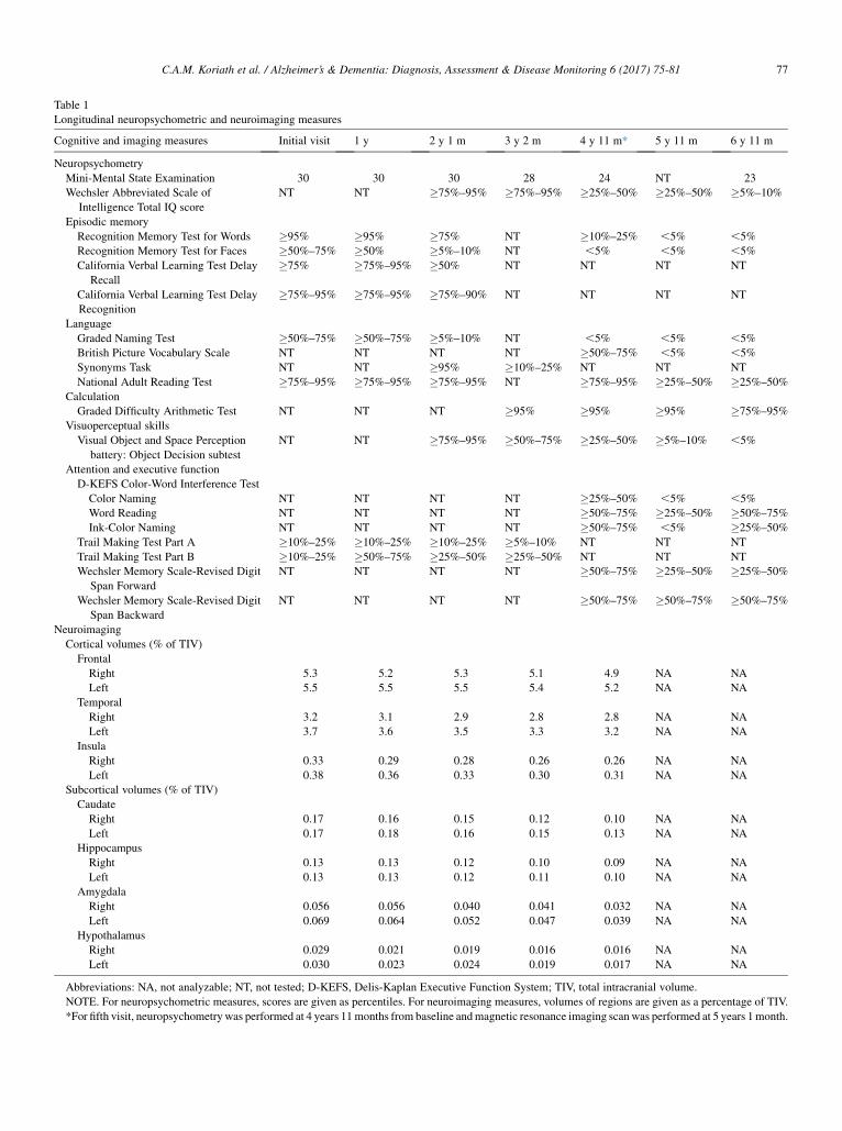
Table 1
Longitudinal neuropsychometric and neuroimaging measures
Cognitive and imaging measures Initial visit 1 y 2 y 1 m 3 y 2 m 4 y 11 m* 5 y 11 m 6 y 11 m
Neuropsychometry
Mini-Mental State Examination 30 30 30 28 24 NT 23
Wechsler Abbreviated Scale of
Intelligence Total IQ score
NT NT �75%–95% �75%–95% �25%–50% �25%–50% �5%–10%
Episodic memory
Recognition Memory Test for Words �95% �95% �75% NT �10%–25% ,5% ,5%
Recognition Memory Test for Faces �50%–75% �50% �5%–10% NT ,5% ,5% ,5%
California Verbal Learning Test Delay
Recall
�75% �75%–95% �50% NT NT NT NT
California Verbal Learning Test Delay
Recognition
�75%–95% �75%–95% �75%–90% NT NT NT NT
Language
Graded Naming Test �50%–75% �50%–75% �5%–10% NT ,5% ,5% ,5%
British Picture Vocabulary Scale NT NT NT NT �50%–75% ,5% ,5%
Synonyms Task NT NT �95% �10%–25% NT NT NT
National Adult Reading Test �75%–95% �75%–95% �75%–95% NT �75%–95% �25%–50% �25%–50%
Calculation
Graded Difficulty Arithmetic Test NT NT NT �95% �95% �95% �75%–95%
Visuoperceptual skills
Visual Object and Space Perception
battery: Object Decision subtest
NT NT �75%–95% �50%–75% �25%–50% �5%–10% ,5%
Attention and executive function
D-KEFS Color-Word Interference Test
Color Naming NT NT NT NT �25%–50% ,5% ,5%
Word Reading NT NT NT NT �50%–75% �25%–50% �50%–75%
Ink-Color Naming NT NT NT NT �50%–75% ,5% �25%–50%
Trail Making Test Part A �10%–25% �10%–25% �10%–25% �5%–10% NT NT NT
Trail Making Test Part B �10%–25% �50%–75% �25%–50% �25%–50% NT NT NT
Wechsler Memory Scale-Revised Digit
Span Forward
NT NT NT NT �50%–75% �25%–50% �25%–50%
Wechsler Memory Scale-Revised Digit
Span Backward
NT NT NT NT �50%–75% �50%–75% �50%–75%
Neuroimaging
Cortical volumes (% of TIV)
Frontal
Right 5.3 5.2 5.3 5.1 4.9 NA NA
Left 5.5 5.5 5.5 5.4 5.2 NA NA
Temporal
Right 3.2 3.1 2.9 2.8 2.8 NA NA
Left 3.7 3.6 3.5 3.3 3.2 NA NA
Insula
Right 0.33 0.29 0.28 0.26 0.26 NA NA
Left 0.38 0.36 0.33 0.30 0.31 NA NA
Subcortical volumes (% of TIV)
Caudate
Right 0.17 0.16 0.15 0.12 0.10 NA NA
Left 0.17 0.18 0.16 0.15 0.13 NA NA
Hippocampus
Right 0.13 0.13 0.12 0.10 0.09 NA NA
Left 0.13 0.13 0.12 0.11 0.10 NA NA
Amygdala
Right 0.056 0.056 0.040 0.041 0.032 NA NA
Left 0.069 0.064 0.052 0.047 0.039 NA NA
Hypothalamus
Right 0.029 0.021 0.019 0.016 0.016 NA NA
Left 0.030 0.023 0.024 0.019 0.017 NA NA
Abbreviations: NA, not analyzable; NT, not tested; D-KEFS, Delis-Kaplan Executive Function System; TIV, total intracranial volume.
NOTE. For neuropsychometric measures, scores are given as percentiles. For neuroimaging measures, volumes of regions are given as a percentage of TIV.
*For fifth visit, neuropsychometry was performed at 4 years 11months from baseline andmagnetic resonance imaging scanwas performed at 5 years 1month.
C.A.M. Koriath et al. / Alzheimer’s & Dementia: Diagnosis, Assessment & Disease Monitoring 6 (2017) 75-81 77
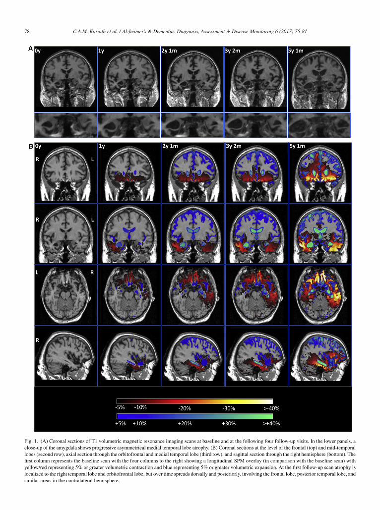
Fig. 1. (A) Coronal sections of T1 volumetric magnetic resonance imaging scans at baseline and at the following four follow-up visits. In the lower panels, a
close-up of the amygdala shows progressive asymmetrical medial temporal lobe atrophy. (B) Coronal sections at the level of the frontal (top) and mid-temporal
lobes (second row), axial section through the orbitofrontal and medial temporal lobe (third row), and sagittal section through the right hemisphere (bottom). The
first column represents the baseline scan with the four columns to the right showing a longitudinal SPM overlay (in comparison with the baseline scan) with
yellow/red representing 5% or greater volumetric contraction and blue representing 5% or greater volumetric expansion. At the first follow-up scan atrophy is
localized to the right temporal lobe and orbitofrontal lobe, but over time spreads dorsally and posteriorly, involving the frontal lobe, posterior temporal lobe, and
similar areas in the contralateral hemisphere.
C.A.M. Koriath et al. / Alzheimer’s & Dementia: Diagnosis, Assessment & Disease Monitoring 6 (2017) 75-8178
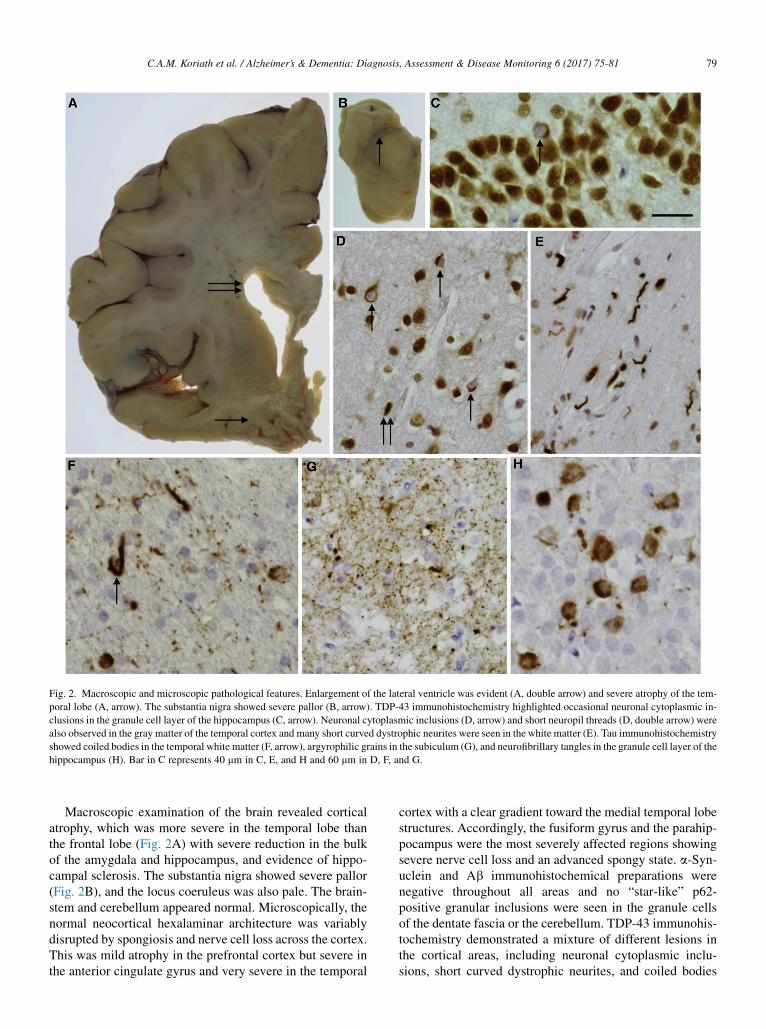
Fig. 2. Macroscopic and microscopic pathological features. Enlargement of the lateral ventricle was evident (A, double arrow) and severe atrophy of the tem-
poral lobe (A, arrow). The substantia nigra showed severe pallor (B, arrow). TDP-43 immunohistochemistry highlighted occasional neuronal cytoplasmic in-
clusions in the granule cell layer of the hippocampus (C, arrow). Neuronal cytoplasmic inclusions (D, arrow) and short neuropil threads (D, double arrow) were
also observed in the gray matter of the temporal cortex and many short curved dystrophic neurites were seen in the white matter (E). Tau immunohistochemistry
showed coiled bodies in the temporal white matter (F, arrow), argyrophilic grains in the subiculum (G), and neurofibrillary tangles in the granule cell layer of the
hippocampus (H). Bar in C represents 40 mm in C, E, and H and 60 mm in D, F, and G.
C.A.M. Koriath et al. / Alzheimer’s & Dementia: Diagnosis, Assessment & Disease Monitoring 6 (2017) 75-81 79
Macroscopic examination of the brain revealed corticalatrophy, which was more severe in the temporal lobe thanthe frontal lobe (Fig. 2A) with severe reduction in the bulkof the amygdala and hippocampus, and evidence of hippo-campal sclerosis. The substantia nigra showed severe pallor(Fig. 2B), and the locus coeruleus was also pale. The brain-stem and cerebellum appeared normal. Microscopically, thenormal neocortical hexalaminar architecture was variablydisrupted by spongiosis and nerve cell loss across the cortex.This was mild atrophy in the prefrontal cortex but severe inthe anterior cingulate gyrus and very severe in the temporal
cortex with a clear gradient toward the medial temporal lobestructures. Accordingly, the fusiform gyrus and the parahip-pocampus were the most severely affected regions showingsevere nerve cell loss and an advanced spongy state. a-Syn-uclein and Ab immunohistochemical preparations werenegative throughout all areas and no “star-like” p62-positive granular inclusions were seen in the granule cellsof the dentate fascia or the cerebellum. TDP-43 immunohis-tochemistry demonstrated a mixture of different lesions inthe cortical areas, including neuronal cytoplasmic inclu-sions, short curved dystrophic neurites, and coiled bodies
C.A.M. Koriath et al. / Alzheimer’s & Dementia: Diagnosis, Assessment & Disease Monitoring 6 (2017) 75-8180
(Fig. 2D). An unusual feature was the presence of numerousTDP-43–positive neuritic structures and also oligodendrog-lial inclusions at the cerebral cortex/subcortical white matterjunction (Fig. 2E). Only occasional TDP-43–positiveneuronal cytoplasmic inclusions were seen in the granulecells of the dentate fascia (Fig. 2C) and only occasionalneuritic structures in the amygdala. TDP-43–positive inclu-sions corresponded to frontotemporal lobar degeneration(FTLD)-TDP type A pathology. Lower motor neuronswere investigated in the 12th nerve nucleus for TDP-43 pa-thology: no pathological inclusions were seen and normalTDP-43 immunohistochemistry was observed with TDP-43 found in the nucleus, i.e. there was no pathological evi-dence of MND. Tau immunohistochemistry demonstratednot only occasional neuropil threads (NTs) in the prefrontaland parietal cortices but also neurofibrillary tangles, pretan-gles, NTs, and coiled bodies (Fig. 2F) in the temporal cortex,particularly in the medial temporal region. Severe tau pa-thology was observed in the hippocampal formation and par-ahippocampus, where pretangles and NTs were seenthroughout and grain-like structures observed in the subicu-lum (Fig. 2G) consistent with argyrophilic grain disease.Pretangles and neurofibrillary tangles were seen in thegranule cells of the dentate fascia (Fig. 2H). Tau-positivecoiled bodies, NTs, and an occasional tufted astrocyte-likestructure were seen in the pontine tegmentum. There wasminimal Purkinje cell loss in the cerebellar cortex and occa-sional NTs were present in the dentate nucleus.
4. Discussion
We describe the clinical, cognitive, and neuroanatomicalprogression over 9 years of a patient with FTD due to a novelTBK1 mutation who was found to have FTLD-TDP type Apathology. This case highlights a number of important andnovel aspects of TBK1 mutations as well as the descriptionof a novel mutation, it also reveals the clinical and neuroan-atomical phenotype that can be associated with TBK1 muta-tions, and the nature of disease progression.
The 13 base pair deletion causes a frameshift introducinga premature stop codon and is therefore likely to be patho-genic even in the absence of supporting segregation or func-tional data. Haploinsufficiency is a known diseasemechanism for TBK1, and loss-of-function mutations inTBK1 have already been described in patients with FTD,MND, and FTD-MND [1–5].
The most common clinical phenotype associated withTBK1 mutations is FTD-MND rather than FTD alone, withthe frequency in patient cohorts 3.0% to 4.5% and 0.5% to1.1%, respectively [3,4]. However, despite close clinicalfollow-up, our patient exhibited no signs of MND. In termsof the specific FTD phenotype, most patients described inthe literature so far have presented with behavioral variantFTD (bvFTD), rather than the language variant, consistentwith the case here, although there appears to be a high prev-alence of early memory impairment [3,6]. Despite our
patient complaining of subjective cognitive symptomsearly in the disease, his neuropsychometric testingremained normal initially with the development of anomiaand prosopagnosia only a few years into the illness. Thedisease duration in our patient was 9 years with an onset at63 years, which is not dissimilar from findings of a recentstudy showing a mean onset of 66.3 years and a diseaseduration of 8.2 years [6].
Previous studies have not detailed the neuroanatomicalphenotype of TBK1mutations. Here, we have shown an asso-ciation with right temporal lobe atrophy. This anatomicalvariant of FTD has been found to be caused by a number ofpathologies, with patients presenting with bvFTDmore likelyto have tau pathology, and those with the semantic dementiaphenotype likely to have TDP-43 pathology [16,17]. Here, wedescribe a casewith bvFTDwho has FTLD-TDP typeA and aTBK1 mutation, a novel association with right temporal lobevariant FTD. Progression of atrophy over time was consistentwith a number of other right temporal lobe cases who remainwith an asymmetrical pattern of involvement but develop asimilar pattern of focal temporal lobe involvement in theopposite temporal lobe as the disease progresses.
Little is known about the neuropathology of TBK1 muta-tions but previous cases have shown either FTLD-TDP typeA or type B pathology, with the histological findings in ourcase being consistent with type A. Although it fits criteria forthis subtype, therewere nonetheless some unusual novel fea-tures with the presence of numerous TDP-43–positiveneuritic structures at the cerebral cortex/subcortical whitematter junction. This case also had associated tau pathologyconsistent with argyrophilic grain disease, which has notbeen seen in other TBK1 mutation cases.
Further research is needed to understand the completephenotype of patients with TBK1 mutations but it shouldbe considered as a cause of right temporal variant FTD,and in patients who present with familial FTD, whetherMND is present or not.
Acknowledgments
The authors thank the patient and his family, who took partin this research.Funding: This work was funded by the Leonard WolfsonFoundation and the Medical Research Council. The authorsacknowledge the support of the NIHR Queen Square De-mentia Biomedical Research Unit, Leonard Wolfson Exper-imental Neurology Centre, and the University CollegeLondon Hospitals NHS Trust Biomedical Research Centre.The Dementia Research Centre is an Alzheimer’s ResearchUK coordinating centre and has also received equipmentfunded by Alzheimer’s Research UK and Brain ResearchTrust. I.O.C.W. is supported by an MRC Clinical ResearchTraining Fellowship (MR/M018288/1). N.C.F. is an NIHRsenior investigator. J.D.W. is supported by a Wellcome TrustSenior Clinical Fellowship (091673/Z/10/Z). J.D.R. is anMRC Clinician Scientist (MR/M008525/1) and has received
C.A.M. Koriath et al. / Alzheimer’s & Dementia: Diagnosis, Assessment & Disease Monitoring 6 (2017) 75-81 81
funding from the NIHR Rare Diseases TranslationalResearch Collaboration (BRC149/NS/MH).
RESEARCH IN CONTEXT
1. Systematic review: TBK1 mutations have recentlybeen linked to frontotemporal dementia (FTD). Toput our findings into context, we reviewed the litera-ture on Pubmed Central relating to other publishedTBK1 cases and research.
2. Interpretation: Our findings detail the natural long-term progression in a case of TBK1-associated FTDand further characterize the related neuropathologicfindings. In addition, the case emphasizes the bene-fits of next-generation sequencing technologies inthe diagnosis of neurodegenerative disorders.
3. Future directions: Further functional studies of TBK1and other genes linked to neurodegenerative diseasesare needed to fully comprehend how loss-of-functionand other mutations affect cellular pathways andcause disease.
References
[1] Cirulli ET, Lasseigne BN, Petrovski S, Sapp PC, Dion PA,
Leblond CS, et al. Exome sequencing in amyotrophic lateral sclerosis
identifies risk genes and pathways. Science 2015;347:1436–41.
[2] Freischmidt A, Wieland T, Richter B, Ruf W, Schaeffer V, M€uller K,
et al. Haploinsufficiency of TBK1 causes familial ALS and fronto-
temporal dementia. Nat Neurosci 2015;18:631–6.
[3] Gijselinck I, Van Mossevelde S, van der Zee J, Sieben A, Philtjens S,
Heeman B, et al., BELNEU Consortium. Loss of TBK1 is a frequent
cause of frontotemporal dementia in a Belgian cohort. Neurology
2015;85:2116–25.
[4] Le Ber I, De Septenville A, Millecamps S, Camuzat A, Caroppo P,
Couratier P, et al., French Clinical and Genetic Research Network
on FTLD/FTLD-ALS. TBK1 mutation frequencies in French fronto-
temporal dementia and amyotrophic lateral sclerosis cohorts. Neuro-
biol Aging 2015;36:3116.e5–8.
[5] Pottier C, Bieniek KF, Finch N, van de Vorst M, BakerM, Perkersen R,
et al. Whole-genome sequencing reveals important role for TBK1 and
OPTN mutations in frontotemporal lobar degeneration without motor
neuron disease. Acta Neuropathol 2015;130:77–92.
[6] Van Mossevelde S, van der Zee J, Gijselinck I, Engelborghs S,
Sieben A, Van Langenhove T, et al., Belgian Neurology consortium.
Clinical features of TBK1 carriers compared with C9orf72, GRN
and non-mutation carriers in a Belgian cohort. Brain 2016;139(Pt
2):452–67.
[7] Rohrer JD, Nicholas JM, Cash DM, van Swieten J, Dopper E,
Jiskoot L, et al. Presymptomatic cognitive and neuroanatomical
changes in genetic frontotemporal dementia in the Genetic Frontotem-
poral dementia Initiative (GENFI) study: a cross-sectional analysis.
Lancet Neurol 2015;14:253–62.
[8] Boccardi M, Bocchetta M, Aronen HJ, Repo-Tiihonen E, Vaurio O,
Thompson PM, et al. Atypical nucleus accumbens morphology in psy-
chopathy: another limbic piece in the puzzle. Int J Law Psychiatry
2013;36:157–67.
[9] Boccardi M, Bocchetta M, Apostolova LG, Barnes J, Bartzokis G,
Corbetta G, et al., for the EADC-ADNIWorking Group on the Harmo-
nized Protocol for Manual Hippocampal Segmentation. Delphi defini-
tion of the EADC-ADNI harmonized protocol for hippocampal
segmentation on magnetic resonance. Alzheimers Dement 2015;
11:126–38.
[10] Bocchetta M, Gordon E, Manning E, Barnes J, Cash DM,
Espak M, et al. Detailed volumetric analysis of the hypothalamus
in behavioral variant frontotemporal dementia. J Neurol 2015;
262:2635–42.
[11] Lashley T, Rohrer JD, Bandopadhyay R, Fry C, Ahmed Z, Isaacs AM,
et al. A comparative clinical, pathological, biochemical and genetic
study of fused in sarcoma proteinopathies. Brain 2011;134:2548–64.
[12] Beck J, Pittman A, Adamson G, Campbell T, Kenny J, Houlden H,
et al. Validation of next-generation sequencing technologies in genetic
diagnosis of dementia. Neurobiol Aging 2014;35:261–5.
[13] Li H, Durbin R. Fast and accurate short read alignment with Burrows-
Wheeler transform. Bioinformatics 2009;25:1754–60.
[14] McKenna A, Hanna M, Banks E, Sivachenko A, Cibulskis K,
Kernytsky A, et al. The genome analysis toolkit: a MapReduce frame-
work for analyzing next-generation DNA sequencing data. Genome
Res 2010;20:1297–303.
[15] Cingolani P, Platts A, Wang le L, Coon M, Nguyen T, Wang L,
et al. A program for annotating and predicting the effects of single
nucleotide polymorphisms, SnpEff: SNPs in the genome of
Drosophila melanogaster strain w1118; iso-2; iso-3. Fly (Austin)
2012;6:80–92.
[16] Clark CN, Lashley T, Mahoney CJ, Warren JD, Revesz T, Rohrer JD.
Temporal variant frontotemporal dementia is associated with globular
glial tauopathy. Cogn Behav Neurol 2015;28:92–7.
[17] Josephs KA, Whitwell JL, Knopman DS, Boeve BF, Vemuri P,
SenjemML, et al. Two distinct subtypes of right temporal variant fron-
totemporal dementia. Neurology 2009;73:1443–50.