the centrosomal e3 ubiquitin ligase fbxo31-scf · pdf fileregulates neuronal morphogenesis and...
TRANSCRIPT
The Centrosomal E3 Ubiquitin Ligase FBXO31-SCFRegulates Neuronal Morphogenesis and MigrationMayur Vadhvani1,2, Nicola Schwedhelm-Domeyer1, Chaitali Mukherjee1,2, Judith Stegmuller1,2*
1 Cellular and Molecular Neurobiology, Max Planck Institute of Experimental Medicine, Gottingen, Germany, 2 Center for Nanoscale Microscopy and Molecular Physiology
of the Brain (CNMPB), Gottingen, Germany
Abstract
Neuronal development requires proper migration, polarization and establishment of axons and dendrites. Growingevidence identifies the ubiquitin proteasome system (UPS) with its numerous components as an important regulator ofvarious aspects of neuronal development. F-box proteins are interchangeable subunits of the Cullin-1 based E3 ubiquitinligase, but only a few family members have been studied. Here, we report that the centrosomal E3 ligase FBXO31-SCF (Skp1/Cullin-1/F-box protein) regulates neuronal morphogenesis and axonal identity. In addition, we identified the polarity proteinPar6c as a novel interaction partner and substrate targeted for proteasomal degradation in the control of axon but notdendrite growth. Finally, we ascribe a role for FBXO31 in dendrite growth and neuronal migration in the developingcerebellar cortex. Taken together, we uncovered the centrosomal E3 ligase FBXO31-SCF as a novel regulator of neuronaldevelopment.
Citation: Vadhvani M, Schwedhelm-Domeyer N, Mukherjee C, Stegmuller J (2013) The Centrosomal E3 Ubiquitin Ligase FBXO31-SCF Regulates NeuronalMorphogenesis and Migration. PLoS ONE 8(2): e57530. doi:10.1371/journal.pone.0057530
Editor: Kin-Sang Cho, Schepens Eye Research Institute, Harvard Medical School, United States of America
Received August 9, 2012; Accepted January 23, 2013; Published February 28, 2013
Copyright: � 2013 Vadhvani et al. This is an open-access article distributed under the terms of the Creative Commons Attribution License, which permitsunrestricted use, distribution, and reproduction in any medium, provided the original author and source are credited.
Funding: This work was supported by the Max Planck Society, the Deutsche Forschungsgemeinschaft (DFG), the Gottingen Graduate Schools for Neurosciences,Biophysics and Molecular Biosciences (GGNB) and the Cluster of Excellence and DFG Research Center for Nanoscale Microscopy and Molecular Physiology of theBrain, Gottingen. The funders had no role in study design, data collection and analysis, decision to publish, or preparation of the manuscript.
Competing Interests: The authors have declared that no competing interests exist.
* E-mail: [email protected]
Introduction
During brain development neurons acquire a typical polarized
morphology, which is fundamental to proper functioning of the
network. Both extrinsic as well as intrinsic programs contribute to
neuronal morphogenesis. The ubiquitin proteasome system (UPS)
has emerged as a crucial intrinsic regulator of neuronal
morphogenesis and other aspects of neuronal development [1–3].
E3 ubiquitin ligases are the most numerous components of the
UPS. They specifically recruit the substrate and the E2 ubiquitin-
conjugating enzyme, which brings in the highly conserved small
protein ubiquitin [4]. This interaction triggers the ubiquitination
of the substrate and brings about degradation or functional
modification of the target protein. Most E3 ligases belong to the
RING (really interesting new gene)-type ligases, which share the
E2-binding RING domain. RING ligases can either act as single
molecule or as multi-subunit ligases [5]. The Cullin-1 based E3
ligase SCF (Skp1, Cullin-1, F-box protein) complex belongs to the
latter; while the subunit Rbx1/Roc1 harbors the RING domain,
the F-box protein represents an interchangeable subunit respon-
sible for substrate recognition and recruitment. Interestingly, F-
box proteins comprise a large family of approximately 70
members but only a few of them have been characterized in
depth, mostly in the context of cell cycle regulation and thus
cancer research [6–9]. F-box proteins have been classified into
FBXW, FBXL and FBXO; while they share the F-box domain,
the W-group harbors several WD40 repeats, the L-group leucine-
rich repeats and the O-group other domains [6].
The quest for neuronal F-box proteins has only recently
begun and revealed important functions for F-box proteins in
the brain including stem cell differentiation, neuronal cell fate,
cerebellar development, axon tract development, dendrite
patterning, and synapse formation [10–16]. Here, we report
that the centrosomal E3 ligase FBXO31-SCF controls neuronal
morphogenesis and axonal identity. We identified the polarity
protein Par6c as a novel substrate of FBXO31-SCF and
established an FBXO31/Par6c pathway of axonal but not
dendritic growth. In addition, we found that FBXO31 is
required for dendrite growth and migration of neurons in the
developing cerebellar cortex.
Materials and Methods
Ethics StatementAll experiments involving live animals have been conducted
according to the animal protocol approved by the ‘‘Verbrau-
cherschutz und Lebensmittelsicherheit’’ of Lower Saxony, Ger-
many (33.11.42502-04-059/08).
Plasmids and AntibodiesA DNA-based template method was used to express short
hairpin RNAs. The sequences for shRNAs targeting FBXO31 are
as follows: FBXO31 RNAi#1:59 GGATGAGTTCTCCAC-
CAAGT 39; FBXO31 RNAi#2:59 AGTCAGTACGA-
CAACTGCCT 39 and FBXO31
RNAi#3:59AGGGGCACCAAGATCACGGG 39. The antibod-
ies used for the study were the following: rb a-FBXO31 (Novus
Biologicals), ms a-c-tubulin (Sigma), ms a-AnkG (Neuromab), rb
a-GFP (Invitrogen), ms a-myc (Santa Cruz), ms a-Flag (Santa
PLOS ONE | www.plosone.org 1 February 2013 | Volume 8 | Issue 2 | e57530
Cruz), ms a-14-3-3ß (Santa Cruz), a-ubiquitin (DAKO), a-K48/
K63 linkage-specific polyubiquitin (Millipore).
Immunoprecipitation293T cells, transfected with indicated plasmids, were lysed in
Co-IP buffer (1% NP40, 150 mM NaCl, 20 mM Tris pH 7.4,
1 mM EDTA, 10% glycerol, protease inhibitors) and 5% of lysates
were set aside as input. The remaining lysate (1 mg) was incubated
with the primary antibody, followed by incubation with protein A
sepharose. The precipitates were analyzed with SDS-PAGE
followed by immunoblotting.
Cell-based Ubiquitination AssayTransfected HEK 293T cells were lysed in RIPA buffer (50 mM
Tris-HCl pH 7.5, 150 mM NaCl, 1% NP40, 1% sodium
deoxycholate and 5 mM EDTA) supplemented with fresh protease
inhibitors (1 mg/mL pepstatin, 3 mg/mL aprotinin and 1 mg/mL
leupeptin) and 10 mM N-ethylmaleimide (NEM). 1 mg of total
protein was incubated with anti-myc antibody for immunoprecip-
itation, followed by incubation with protein A sepharose. The
precipitates were washed twice with RIPA buffer, twice with lysis
buffer (50 mM HEPES pH 7.5, 150 mM NaCl, 10% glycerol,
1.5 mM MgCl2, 1% Triton X-100) and boiled with SDS-sample
buffer prior to immunoblotting analysis.
Transfection of Primary NeuronsCerebellar granule neurons were cultured and transfected 8
hours after plating using a modified calcium phosphate protocol as
described earlier [33]. The calcium phosphate method ensures a
86%–95% co-transfection efficiency of multiple plasmids [34] To
rule out the possibility that genetic manipulation (RNAi or
overexpression) affects axon or dendrite length due to poor health,
we co-transfected granule neurons in all experiment with a
plasmid encoding BCL-XL to stimulate neuronal survival.
Expression of BCL-XL does not affect neuronal morphology
[33]. Neurons were fixed with paraformaldehyde after 3 to 4 days
in vitro and subjected to immunocytochemistry.
Centrosomal PurificationTransfected cerebellar granule neurons were treated with
cytochalasin D (1 mg/mL) and nocodazole (0.2 mM) for 1 hour
and lysed in lysis buffer (1 mM HEPES pH 7.2, 0.5% NP40,
0.5 mM MgCl2, 0.1% ß-mercaptoethanol) supplemented with
protease inhibitors (1 mg/mL pepstatin, 1 mg/mL aprotinin, 1 mg/
mL leupeptin and 1 mM PMSF). Cell lysates were subjected to
gradient centrifugation and centrosomal fractions were purified
using a discontinuous sucrose density gradient centrifugation
(40%, 50% and 70% sucrose). The fractions were further analyzed
using immunoblotting.
In vivo ElectroporationP4 rat pups were electroporated as described previously
(Konishi et al., 2004). Briefly, DNA together with Fast Green
was injected into the cerebellar cortex of isofluorane-anesthetized
P4 rat pups using a Hamilton syringe. Pups were subjected to 5
electrical pulses (160–170 V, 50 ms pulses, 950 ms pulse intervals)
using a Harvard Apparatus ECM 830 square wave electroporator.
Pups were sacrificed at P9 and 40 mm coronal sections of GFP-
positive cerebella were analyzed.
MorphometryImages of transfected GFP-positive neurons were captured in a
blinded manner using a Nikon Eclipse epifluorescence microscope.
Axons and dendrites were manually traced to determine their
lengths using ImageJ. For in vivo analysis, confocal z-stacks were
processed in ImageJ and the cells were counted in individual layers
and relative distance of migration was analyzed. Confocal images
were captured using a Leica SP2 were processed using Imaris
software (Bitplane) to determine the dendrite lengths of neurons in
3D.
Results
The Centrosomal E3 Ubiquitin Ligase FBXO31-SCFRegulates Neuronal Morphology
The E3 ligase FBXO31-SCF has been introduced as a tumor
suppressor and cell cycle regulator [17,18]. Interestingly, different
sources suggest a brain-enriched expression of FBXO31 [17]
(CDT-DB at Riken). However FBXO31’s role in neurons remains
elusive. To characterize the function of FBXO31, we started out
by quantifying the expression of FBXO31 in distinct brain regions
including cortex, hippocampus, cerebellum and olfactory bulb
using quantitative PCR and found a relative enrichment of
FBXO31 expression in postnatal and adult rat brain as compared
to non-neural tissue (Figure S1A–C, Methods S1).
We then determined the localization of FBXO31 in neurons
and discovered that FBXO31 localizes to the centrosome in both
cerebellar granule neurons and in hippocampal neurons
(Figure 1A). Co-staining with the centrosomal protein -tubulin
supported FBXO31’s centrosomal localization (Figure 1A). In
addition, we confirmed the specificity of the immunostaining in
neurons by preincubation of the antibody with recombinant
FBXO31 protein (Figure S1D). Interestingly, a previous study
described the SCF complex subunit Skp1 and Cullin-1 as
components of the centrosomal material [19], which prompted
us to determine if the binding to Skp1 or Cullin-1 is required to
recruit FBXO31 to the centrosome. An FBXO31 mutant lacking
the F-box domain, which mediates the binding to Skp1 and
Cullin-1 (Figure S2) [17,20], still localizes to the centrosome.
Further mapping analysis revealed that a stretch in the N-terminal
region of FBXO31 is required for its recruitment to the
centrosome (Figure S3).
As a component of the centrosomal material, we reasoned
that FBXO31 might play a role in neuronal morphogenesis or
migration. We used an RNAi approach to acutely knockdown
FBXO31 in neurons. Validation of FBXO31 RNAi plasmids in
heterologous cells demonstrated that two of the RNAi plasmids
efficiently downregulate FBXO31, while one had no effect and
served as a negative control thereafter (Figure S4A). We then
transfected neurons with the control vector or different
FBXO31 RNAi plasmids together with the GFP expression
plasmid. We found that while the non-functional RNAi plasmid
has little or no effect on axon (longest process) and dendrite
growth, the functional RNAi plasmids lead to a decrease in
both axonal and dendritic length (Figure 1C–E, Figure S4B–D). To ensure that the morphological FBXO31 RNAi
phenotype is specific and to rule out off-target effects, we
constructed an RNAi-resistant form of FBXO31 (FBXO31-
Rescue = FBXO31-Res). We introduced silent mutations into
the RNAi target region and validated the FBXO31-Res
encoding plasmid in heterologous cells (Figure 1B). We
observed that while FBXO31 RNAi decreases axon (longest
process) and dendrite length, expression of FBXO31-Res leads
to significantly longer axons and dendrites as compared to
FBXO31 knockdown neurons, supporting the specific FBXO31
RNAi-induced phenotype (Figure 1C–E). In addition, we
analyzed whether FBXO31 knockdown affects neuronal surviv-
FBXO31-Controlled Neuromorphology and Migration
PLOS ONE | www.plosone.org 2 February 2013 | Volume 8 | Issue 2 | e57530
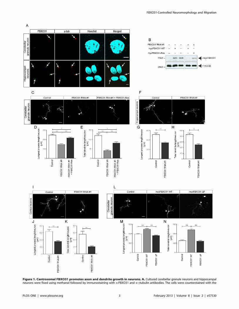
Figure 1. Centrosomal FBXO31 promotes axon and dendrite growth in neurons. A. Cultured cerebellar granule neurons and hippocampalneurons were fixed using methanol followed by immunostaining with a-FBXO31 and a-ctubulin antibodies. The cells were counterstained with the
FBXO31-Controlled Neuromorphology and Migration
PLOS ONE | www.plosone.org 3 February 2013 | Volume 8 | Issue 2 | e57530
al. Here, we transfected neurons with control vector or
FBXO31 RNAi plasmids together with the ß-Galactosidase
expression plasmid and assessed neuronal survival. We found a
slight increase in apoptosis in FBXO31 knockdown neurons as
compared to control (Figure S4E) and concluded that
FBXO31 plays a rather subordinate role in neuronal survival.
To determine if FBXO31-mediated neuronal morphogenesis is
a generalizable mechanism, we examined FBXO31 knockdown
in hippocampal and in cortical neurons and found that
FBXO31 promotes axon (longest process) and dendrite growth
in both neuronal cell types (Figure 1F–K). These data indicate
that FBXO31 promotes axon and dendrite growth.
In gain-of function analyses, we examined the effect of
overexpression of FBXO31. Conversely to the loss-of-function
phenotype, we found that FBXO31 overexpression results in
longer axons and dendrites, underscoring the role of FBXO31 in
neuronal morphogenesis (Figure 1L–N). Furthermore, we found
that ligase activity of FBXO31-SCF is required to promote
neuronal morphogenesis, since the aforementioned FBXO31 DF-
box mutant fails to promote elongation of the processes
(Figure 1L–N). These data bolster our finding that FBXO31
acts as a regulator of neuronal morphogenesis.
FBXO31-SCF Controls Axonal IdentityAside from the enhanced of neurite growth, we noticed that a
larger number of FBXO31-overexpressing granule neurons
appeared non-polarized as compared to control neurons. We
carried out morphological analysis and defined a neuron as
polarized when the longest process was at least twice as long as the
second longest. When we quantified the number of non-polarized
neurons, we found that while 18% of control neurons appear non-
polarized, 38% of FBXO31-overexpressing neurons display a non-
polarized morphology (Figure 2A, 2B). We then used molecular
markers to examine a possible defect. Axons harbor several unique
features including the axon initial segment (AIS), which is
characterized by the presence of AnkyrinG. AnkyrinG is
responsible for the organization of the AIS and the maintenance
of neuronal polarity [21,22]. We subjected control vector-, wild
type FBXO31- and FBXO31 DF-expressing hippocampal neurons
to immunocytochemistry and found that a significantly larger
number of wild type FBXO31-expressing neurons harbor two or
more AnkG-positive processes while control or FBXO31 DF
neurons display mostly one AnkG-positive axon (Figure 2C, 2D).
Conversely, when we triggered FBXO31 knockdown, a large
percentage of longest processes failed to display the axonal marker
AnkyrinG (Figure 2E, 2F). These data suggest that FBXO31
controls not only neuronal morphogenesis but also axonal identity.
Par6c is a Novel Target of FBXO31-SCF in the Control ofAxon Growth
To identify targets of FBXO31-SCF and owing to FBXO31’s
centrosomal localization, we took a candidate approach and found
that FBXO31 interacts with Par6c, a previously identified
centrosomal protein [23,24]. Par6 is known to regulate epithelial
cell polarity [25,26] and neuronal polarity [27,28]. In the nervous
system, Par6c represents the predominantly enriched Par6 family
member [29]. We determined the interaction of exogenous
FBXO31 and Par6c in heterologous cells by immunoprecipitating
FBXO31 followed by immunoblotting for Par6c (Figure 3A). In a
reciprocal experiment, we immunoprecipitated Par6c and im-
munoblotted for FBXO31 (Figure 3B). Mapping analyses of
FBXO31 and Par6c revealed that Par6c’s PDZ domain mediates
the interaction with FBXO31 (Figure 3C, Figure S5A). These
experiments establish the FBXO31-Par6c interaction.
To determine if Par6c is a potential substrate of FBXO31-SCF,
we first examined if Par6c levels respond to proteasome inhibition.
We transfected neurons with the Par6c expression plasmid and
subjected them to treatment with the proteasome inhibitor
lactacystin or vehicle as control. We analyzed the lysates and
found that proteasome inhibition results in the accumulation of
Par6c (Figure 3D). In further experiments, we analyzed if Par6c
levels respond to presence or absence of FBXO31. We expressed
the Par6c plasmid together with control vector or the FBXO31
plasmid in heterologous cells and found that overexpression of
FBXO31 results in decreased Par6c levels while the control
protein 14-3-3ß remains unaffected (Figure S5B). Conversely, the
expression of the Par6c and FBXO31 plasmids together with
control vector or FBXO31 RNAi plasmid results in an accumu-
lation of Par6c in response to low FBXO31 levels but no change in
14-3-3ß (Figure 3E). To corroborate this finding in neurons, we
compared Par6c levels in neurons upon FBXO1 overexpression
and knockdown, respectively. Immunoblotting of neuronal lysates
revealed that Par6c levels are reduced when FBXO31 is
overexpressed (Figure S5C) and increased upon FBXO31 RNAi
(Figure 3F). For the latter we included a non-functional FBXO31
RNAi plasmid as control and observed no change of Par6c levels
(Figure 3F). In further experiments, we carried out centrosomal
purification analysis to determine if centrosomal Par6c is affected
by FBXO31 knockdown. We induced FBXO31 RNAi in neurons
and subjected the lysate to an ultracentrifugation procedure to
enrich for centrosomes. In control conditions, we find that Par6c
appears together with the centrosomal protein gamma2tubulin in
the bottom fractions. It is also here where we observe the
accumulation of Par6c upon FBXO31 knockdown (Figure 3G).
We do not detect any Par6c in the supernatant, where non-
centrosomal proteins including 14-3-3ß are present (Figure S5D).
DNA dye bisbenzimide Hoechst 33258. Arrows indicate centrosomes. Scale bar equals 5 mm. B. Cell lysates of HEK 293T cells transfected withindicated plasmids were probed with a-myc antibody. 14-3- ß served as a loading control. C. Representative images of cerebellar granule neuronstransfected with empty control vectors, FXO31 RNAi #1 plasmid or FBXO31 RNAi #1 together with mycFBXO31-Res at DIV 0 and analyzed at DIV 4.Arrowheads indicate granule neuron cell bodies. Scale bar equals 50 mm. D. Quantification of longest process lengths of granule neurons shown in C(N = 3, n = 296, mean6SEM, one-way ANOVA *p,0.05, ***p,0.001). E. Quantification of total dendrite lengths of granule neurons shown in C (N = 3,n = 291, mean6SEM, one-way ANOVA, *p,0.05, ***p,0.001). F. Representative images of cultured hippocampal neurons transfected with controlvector or FBXO31 RNAi #1 plasmids at DIV 1 and analyzed at DIV 5. Arrowheads indicate hippocampal neuron cell bodies. Scale bar equals 50 mm. G.Quantification of longest process lengths of hippocampal neurons shown in F (N = 3, n = 190, mean6SEM, unpaired t-test, ***p,0.001). H.Quantification of total dendrite lengths of hippocampal neurons shown in F (N = 3, n = 184, mean6SEM, unpaired t-test, **p,0.01). I. Representativeimages of cultured cortical neurons transfected with control vector or FBXO31 RNAi #1 plasmids at DIV 1 and analyzed at DIV 5. Arrowheads indicatecortical neuron cell bodies. Scale bar equals 50 mm. J. Quantification of longest process lengths of cortical neurons shown in I (N = 3, n = 164,mean6SEM, unpaired t-test, ***p,0.001). K. Quantification of total dendrite lengths of cortical neurons shown in I (N = 3, n = 147, mean6SEM,unpaired t-test, ***p,0.001). L. Representative images of cerebellar granule neurons transfected with empty control vector, mycFBXO31 wild type(WT) plasmid or mycFBXO31 DF mutant plasmid at DIV 0 and analyzed at DIV 3. Arrowheads indicate granule neuron cell bodies. Scale bar equals50 mm. M. Quantification of longest process lengths of granule neurons shown in L (N = 3, n = 381, mean6SEM, one-way ANOVA ***p,0.001). N.Quantification of total dendrite lengths of granule neurons shown in L (N = 3, n = 341, mean6SEM, one-way ANOVA, ***p,0.001).doi:10.1371/journal.pone.0057530.g001
FBXO31-Controlled Neuromorphology and Migration
PLOS ONE | www.plosone.org 4 February 2013 | Volume 8 | Issue 2 | e57530

Figure 2. FBXO31 regulates axonal identity in neurons. A. Representative images of cerebellar granule neurons transfected with controlvector or mycFBXO31 WT plasmid at DIV 0 and analyzed at DIV 3. Arrowheads indicate granule neuron cell bodies. Scale bar equals 50 mm. B.Quantification of percentage of non-polarized granule neurons shown in A. (N = 3, n = 256, mean6SEM, unpaired t-test, **p,0.01). C. Representativeimages of cultured hippocampal neurons transfected at DIV 1 with control vector, plasmids encoding mycFBXO31 WT or mycFBXO31 DF togetherwith the GFP plasmid and immunostained at DIV 7 with a-GFP and a-AnkG antibodies and counterstained with Hoechst. Arrows indicate axon initialsegment. Scale bar equals 10 mm. D. Quantification of number of axons in C. A total of 169 cells were analyzed (N = 3, mean6SEM, two-way ANOVA***p,0.001). E. Representative images of cultured hippocampal neurons from E18 rat embryos transfected with control vector or FBXO31 RNAi#1/CMVGFP plasmid at DIV 1 and immunostained at DIV 6 with a-GFP and a-AnkG antibody and counterstained with Hoechst. Arrows indicate axoninitial segment. Scale bar equals 10 mm. F. Quantification of number of axons in E. A total of 121 cells were analyzed (N = 3, mean6SEM, two-wayANOVA ***p,0.001).doi:10.1371/journal.pone.0057530.g002
FBXO31-Controlled Neuromorphology and Migration
PLOS ONE | www.plosone.org 5 February 2013 | Volume 8 | Issue 2 | e57530
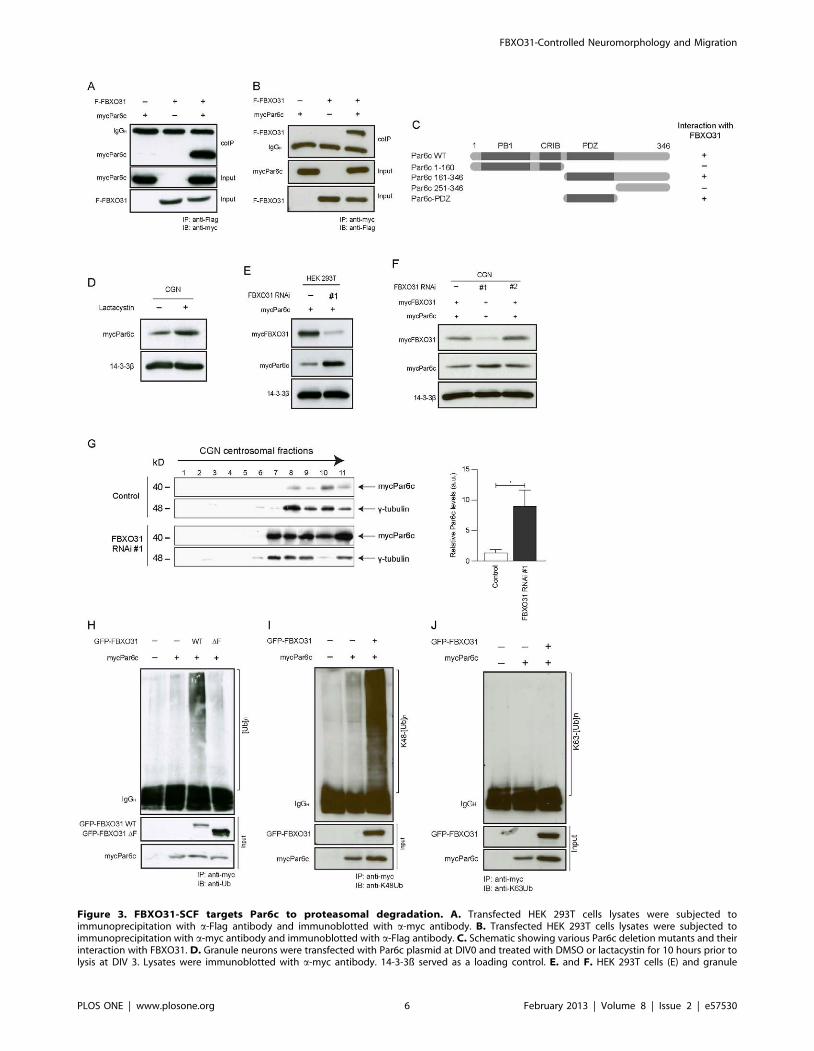
Figure 3. FBXO31-SCF targets Par6c to proteasomal degradation. A. Transfected HEK 293T cells lysates were subjected toimmunoprecipitation with a-Flag antibody and immunoblotted with a-myc antibody. B. Transfected HEK 293T cells lysates were subjected toimmunoprecipitation with a-myc antibody and immunoblotted with a-Flag antibody. C. Schematic showing various Par6c deletion mutants and theirinteraction with FBXO31. D. Granule neurons were transfected with Par6c plasmid at DIV0 and treated with DMSO or lactacystin for 10 hours prior tolysis at DIV 3. Lysates were immunoblotted with a-myc antibody. 14-3-3ß served as a loading control. E. and F. HEK 293T cells (E) and granule
FBXO31-Controlled Neuromorphology and Migration
PLOS ONE | www.plosone.org 6 February 2013 | Volume 8 | Issue 2 | e57530
This could mean that cytoplasmic Parc6c is below detection-level
or that Par6c is solely present at the centrosome at the time when
we examine the neurons. Collectively, these data suggest that
centrosomal Par6c is targeted for degradation by FBXO31.
To examine if FBXO31-SCF is the ligase responsible for Par6c
ubiquitination, we expressed Par6c together with control vector, a
plasmid expressing wild type FBXO31 or the ligase-dead mutant
FBXO31 DF-box. The lysates were subjected to immunoprecip-
itation for Par6c and immunoblotted with the ubiquitin antibody.
While we found Par6c to be sparsely ubiquitinated in control or
FBXO31-DF conditions, wild type FBXO31 potently stimulates
polyubiquitination of Par6c (Figure 3H). To confirm that the
polyubiquitination of Par6c contributes to its proteasomal
turnover, we examined the linkage of the polyubiquitination chain
associated with Par6c. Ubiquitin chains can be assembled via
different lysines in ubiquitin. Ubiquitin chains that are linked via
lysine 48 (K48) are known to trigger the degradation of proteins,
while K63-linkage of ubiquitin represents a non-proteolytic
modification [30,31]. In cell-based ubiquitination assays, we found
that FBXO31 triggers the assembly of a K48-linked but not K63-
linked polyubiquitin chain of Par6c (Figure 3I, 3J). In addition,
we carried out this cell based-ubiquitination assay using denatured
lysates to exclude that Par6c interactors are being detected by
ubiquitin. As demonstrated before, we found that Par6c is
modified by K48-linked ubiquitin chains only in the presence of
wild type but not ligase-dead FBXO31 (Figure S5E, MethodsS2). Collectively, our data indicate that Par6c is targeted for
proteasomal degradation by FBXO31-SCF.
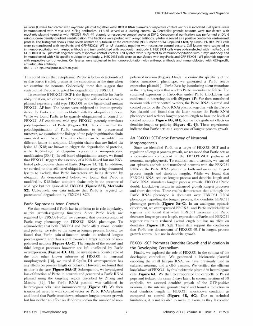
Par6c Suppresses Axon GrowthWe then examined if Par6c has in addition to its role in polarity,
neurite growth-regulating functions. Since Par6c levels are
regulated by FBXO31-SCF, we reasoned that overexpression of
Par6c may phenocopy the FBXO31 RNAi phenotype. To
acknowledge that both FBXO31 and Par6c affect axonal identity
and polarity, we refer to the axon as longest process. Indeed, we
found that Par6c gain-of-function results in reduced longest
process growth and thus a shift towards a larger number of non-
polarized neurons (Figure 4A–C). The lengths of the second and
third longest processes however are left unaffected by Par6c
overexpression. (Figure 4D, 4E). To investigate a possible role of
the only other known substrate of FBXO31 in neuronal
morphogenesis [18], we tested if Cyclin D1 overexpression has
any effects on process length or polarity. However, we found that
neither is the case (Figure S6A–D) Subsequently, we investigated
loss-of-function of Par6c in neurons and generated a Par6c RNAi
plasmid using the targeting region described by Zhang and
Macara [32]. The Par6c RNAi plasmid was validated in
heterologous cells using immunoblotting (Figure 4F). We then
transfected neurons with control vector or Par6c RNAi plasmid
and found that Par6c knockdown enhances longest process growth
but has neither an effect on dendrites nor on the number of non-
polarized neurons (Figure 4G–J). To ensure the specificity of the
Par6c knockdown phenotype, we generated a Par6c rescue
expression plasmid ( = Par6c-Res) by introducing silent mutations
in the targeting region that renders Par6c insensitive to RNAi. The
sustained expression of Par6c-Res under Par6c knockdown was
confirmed in heterologous cells (Figure 4F). We then transfected
neurons with either control vectors, the Par6c RNAi plasmid and
control vector or the Par6c RNAi plasmid together with the Par6c-
Res plasmid and found that the latter rescues the Par6c RNAi
phenotype and reduces longest process length to baseline levels of
control neurons (Figure 4G, 4H), but has no significant effects on
dendrite length or polarity (Figure 4I, 4J). These experiments
indicate that Par6c acts as a suppressor of longest process growth.
An FBXO31-SCF/Par6c Pathway of NeuronalMorphogenesis
Since we identified Par6c as a target of FBXO31-SCF and a
regulator of longest process growth, we reasoned that Par6c acts as
a downstream component in the FBXO31-SCF pathway of
neuronal morphogenesis. To establish such a cascade, we carried
out epistasis analysis and transfected neurons with the FBXO31
RNAi or the Par6c RNAi plasmid or both and measured longest
process length and dendritic lengths. While we found that
FBXO31 RNAi reduces longest process and dendrite length and
Par6c RNAi stimulates longest process growth, FBXO31/Par6c
double knockdown results in enhanced growth longest processes
and short dendrites. These results demonstrate that although the
Par6c RNAi phenotype is dominant over FBXO31 RNAi
phenotype regarding the longest process, the dendritic FBXO31
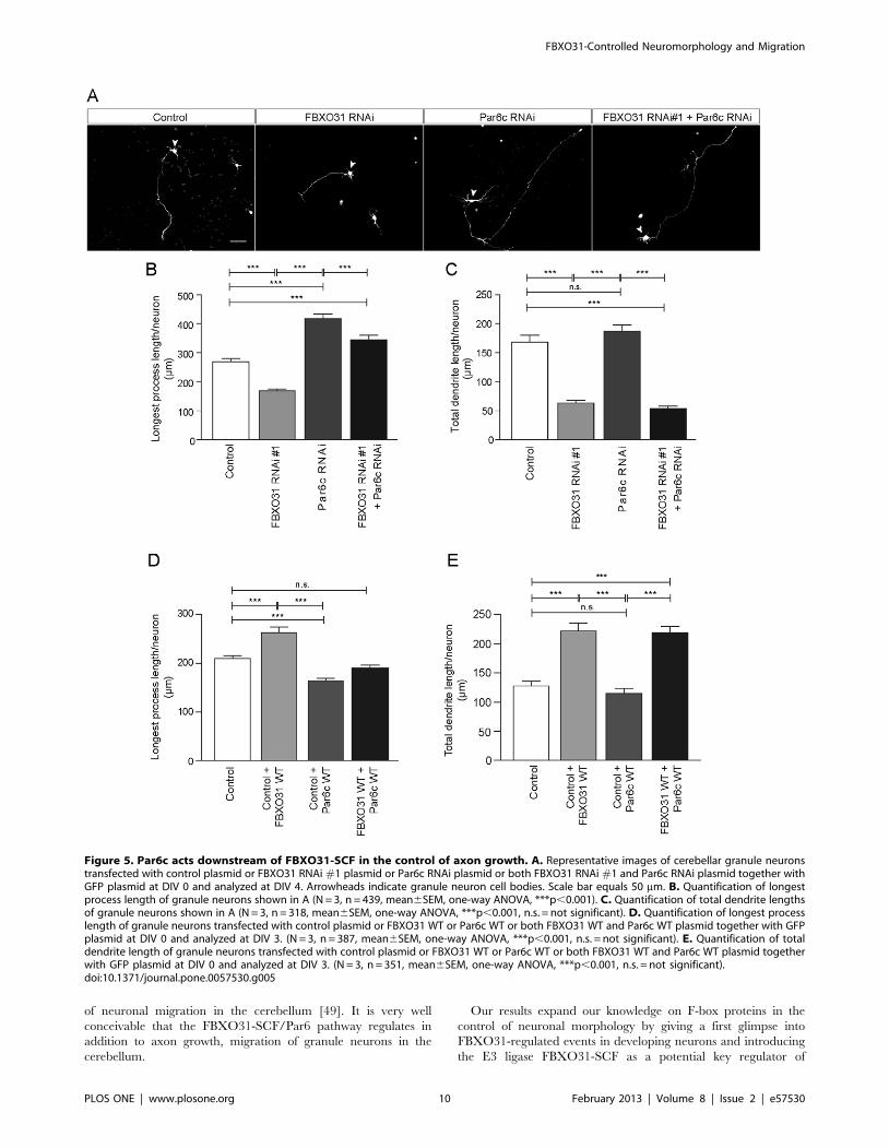
phenotype prevails (Figure 5A–C). In an analogous epistasis
experiment, we overexpressed FBOX31 and Par6c individually or
together and found that while FBXO31 increases and Par6c
decreases longest process length, expression of Par6c and FBXO31
together results in reduced axonal length but has no effect on
dendrites (Figure 5D, 5E). These data support the conclusion
that Par6c acts downstream of FBXO31-SCF in longest process
growth control, but not in dendrite growth.
FBXO31-SCF Promotes Dendrite Growth and Migration inthe Developing Cerebellum
Finally, we explored the role of FBXO31 in the context of the
developing cerebellum. We generated a bicistronic plasmid
encoding the small hairpin RNA, we have previously used in
cultured neurons, and a GFP cassette. We verified the efficient
knockdown of FBXO31 by this bicistronic plasmid in heterologous
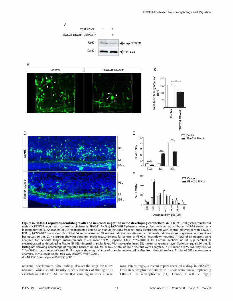
cells (Figure 6A). We then electroporated the cerebella of P4 rat
pups and isolated the tissue 5 days later. In coronal sections of P9
cerebella, we assessed dendrite growth of the GFP-positive
neurons in the internal granular layer and found a reduction in
total dendritic length in FBXO31 knockdown condition as
compared to control (Figure 6B, 6C). Due to technical
limitations, it is not feasible to measure axons as they fasciculate
neurons (F) were transfected with mycPar6c plasmid together with FBXO31 RNAi plasmids or respective control vectors as indicated. Cell lysates wereimmunoblotted with a-myc and a-Flag antibodies. 14-3-3ß served as a loading control. G. Cerebellar granule neurons were transfected withmycPar6c plasmid together with FBXO31 RNAi #1 plasmid or respective control vector at DIV 2. Centrosomal purification was performed at DIV 6using sucrose density gradient centrifugation. The fractions were probed with a-myc antibody. c-tubulin served as a positive control for centrosomalprotein. The histogram shows Par6c levels relative to c-tubulin at the centrosome (N = 3, mean6SEM, unpaired t-test, *p,0.05). H. HEK 293T cellswere co-transfected with mycPar6c and GFP-FBXO31 WT or DF plasmids together with respective control vectors. Cell lysates were subjected toimmunoprecipitation with a-myc antibody and immunoblotted with a-ubiquitin antibody. I. HEK 293T cells were co-transfected with mycPar6c andGFP-FBXO31 WT plasmids together with respective control vectors. Cell lysates were subjected to immunoprecipitation with a-myc antibody andimmunoblotted with K48-specific a-ubiquitin antibody. J. HEK 293T cells were co-transfected with mycPar6c and GFP-FBXO31 WT plasmids togetherwith respective control vectors. Cell lysates were subjected to immunoprecipitation with anti-myc antibody and immunoblotted with K63-specificanti-ubiquitin antibody.doi:10.1371/journal.pone.0057530.g003
FBXO31-Controlled Neuromorphology and Migration
PLOS ONE | www.plosone.org 7 February 2013 | Volume 8 | Issue 2 | e57530
FBXO31-Controlled Neuromorphology and Migration
PLOS ONE | www.plosone.org 8 February 2013 | Volume 8 | Issue 2 | e57530
in the molecular layer into untraceable fibers. Strikingly, we also
found that while more than 80% of transfected control neurons
descend rapidly into the internal granular layer, nearly 50% of
FBXO31 RNAi neurons fail to migrate and remain in the external
granule layer/molecular layer (Figure 6D, 6E). We further
quantified the migration defect and measured the distance of
transfected neurons from the pial surface and found that the
migration of FBXO31 knockdown neurons is markedly stalled
(Figure 6F). These results indicate that FBXO31 promotes
dendritic morphogenesis and migration of granule neurons in the
developing cerebellum.
Discussion
In this study, we described that the novel centrosomal E3 ligase
FBXO31-SCF regulates neuronal morphogenesis by targeting
Par6c for proteasomal degradation. In addition, we found that
FBXO31 is required for proper axonal identity and the efficient
migration of neurons in the cerebellar cortex.
The E3 ligase FBXO31-SCF has previously been implicated in
cell cycle regulation as it emerged as a tumor suppressor and
regulator of DNA repair by degrading cyclin D1 [17,18]. Its
abundant expression in the nervous system has been reported [17],
which is consistent with our data suggesting a relative enrichment
of FBXO31 in neural tissue.
We identified FBXO31 as a regulator of axonal and dendritic
growth. FBXO31-SCF follows the lead of the E3 ligase Cdh1-
APC, which is a crucial cell cycle regulator with an unexpected
role in axon growth regulation in neurons by targeting the
substrates SnoN and Id2 for proteasomal degradation [33–35].
Like FBXO31-SCF, the E3 ligase FBXW7-SCF also operates in
the cerebellum, where it controls cerebellar size, Purkinje cell
number and axonal arborization [11]. In addition, other F-box
proteins were identified to support the proper functioning of
neurons including brain-specific FBXO45, which is required for
formation of axon tracts and neuromuscular junctions in
mammals, its C. elegans homologue FSN-1, which regulates
presynaptic differentiation [13,15], and lin-23 (FBXW1) that
regulates the abundance of glutamate receptors [14]. Further-
more, the F-box protein FBXW8, which forms a Cullin-7 based
E3 ligase, regulates the morphology of the Golgi complex and
dendrites by degrading the Golgi protein Grasp65 [16]. These
studies and our report support the notion of an elaborate interplay
of F-box proteins in neurons.
The finding that F-box protein FBXO31 is a centrosomal
protein in neurons is in agreement with FBXO31 being a
regulator of neuronal morphogenesis, axon specification and
migration. The centrosome has been previously identified as a
cornerstone of neuronal polarity allowing the prediction of the
positioning of the future axon [36]. Also, the centrosome’s
function as microtubule organizing center (MTOC) marks not
only the site of axonal outgrowth but is intimately connected with
the coordination of neuronal migration [37–39]. FBXO31-SCF
joins a small number of E3 ligases, which are positioned at the
centrosome including dendrite-regulating Cdc20-APC and the
Parkinson’s disease protein Parkin [40–42], supporting an essential
role for centrosomal E3 ubiquitin ligases in neuronal development
and disease [43,44].
FBXO31’s centrosomal position led us to discover the polarity
protein Par6c as an interactor and novel substrate. Par6c has
previously been described as a component of the centrosome,
which recruits other centrosomal proteins [24]. A key function of
Par6, which forms the Par complex together with aPKC and
Par3b, is its role in neuronal polarity [27,28,45,46]. Also, the Par6
complex has been implicated in stimulation-induced local control
of axon growth [47]. Yet another study proposed that Par6 acts on
its own as an effector of TGF beta-induced axon initiation and
growth via type II TGF-beta receptor [48]. Our finding that Par6c
inhibits axon growth but has no effect on dendrites ascribes a
specific axon growth-suppressing role to Par6c. These results are
supported by previous work that revealed a Par6-mediated
inhibition of axon growth [49]. We believe that Par6c acts in an
alternative complex in axon growth control at the centrosome
since we did not find an interaction of FBXO31 with Par3b
(neither in the presence nor absence of Par6c; data not shown).
Our results also underscore a dual role for Par6c in neuronal
polarity and in axon growth control. The latter may require other
Par6c interactors than Par3b. A study by Zhang and Macara
revealed that dendritic spine development is yet another aspect in
neurons that is regulated by Par6 but not Par3b [32].
Apart from FBXO31-SCF, Par6c is degraded by the E3 ligase
Smurf1 in axon initiation [50]. Here, Smurf1 promotes Par6c
degradation to initiate axons but then switches to RhoA
degradation for axon elongation as a consequence of trophic
factor-induced phosphorylation of Smurf1. In contrast to
FBXO31, Smurf1 has not been described as a centrosomal E3
ligase. FBXO31 together with Smurf1 and TGFß signaling is likely
to orchestrate axonal initiation and growth by keeping the
common substrate Par6 in check at different subcellular localiza-
tions and in response to various extrinsic cues.
The finding that FBXO31 controls dendritic growth but not via
Par6c also prompted us to conclude that FBXO31-SCF is likely to
target another protein for degradation in the control of dendrite
growth. Our in vivo analyses underscored FBXO31’s importance
in proper dendrite development in the cerebellum. Furthermore,
our results implicate that FBXO31-SCF promotes neuronal
migration in the cerebellum, which is consistent with the finding
of Solecki and colleagues who reported abated migration of
granule neurons in Par6 gain-of-function analyses in organotypic
cerebellar slices and hence identified Par6 as a negative regulator
Figure 4. Par6c acts as an axon growth suppressor. A. Representative images of granule neurons transfected with control vector or plasmidencoding mycPar6c WT together with the GFP plasmid at DIV 0 and analyzed at DIV 3. Arrowheads indicate granule neurons cell bodies. Scale barrepresents 50 mm. B. Quantification of longest process length of granule neurons shown in A. (N = 3, n = 160, mean6SEM, unpaired t-test, *p,0.05).C. Quantification of percentage of non-polarized granule neurons shown in A. (N = 3, n = 226, mean6SEM, unpaired t-test, *p,0.05). D. and E.Quantification of 2nd longest (D) and 3rd longest process length (E) of granule neurons shown in A. (N = 3, n = 160, mean6SEM, unpaired t-test,*p,0.05). F. HEK 293T cell lysates transfected with mycPar6c WT or mycPar6c-Res plasmids together with control or Par6c RNAi plasmids wereimmunoblotted with a-myc antibody. 14-3-3ß served as a loading control. G. Representative images of granule neurons transfected with controlvector or Par6c RNAi or Par6c RNAi and Par6c-Res together with the GFP plasmid at DIV 0 and analyzed at DIV 4. Arrowheads indicate granule neuroncell bodies. Scale bar equals 50 mm. H. Quantification of longest process length of granule neurons transfected with control vector or Par6c RNAiplasmid or both Par6c RNAi plasmid and mycPar6c-Res plasmid together with GFP plasmid at DIV 0 and analyzed at DIV 4 (N = 3, n = 309, mean6SEM,one-way ANOVA, ***p,0.001) I. Quantification of total dendrite lengths of granule neurons transfected with control vector or Par6c RNAi plasmidtogether with GFP plasmid at DIV 0 and analyzed at DIV 4 (N = 3, n = 255, mean6SEM, one-way ANOVA, n.s. = not significant). J. Quantification ofpercentage of non-polarized granule neurons transfected with control vector or Par6c RNAi plasmid together with GFP plasmid at DIV 0 and analyzedat DIV 4. (N = 3, n = 313, mean6SEM, one-way ANOVA, n.s. = not significant).doi:10.1371/journal.pone.0057530.g004
FBXO31-Controlled Neuromorphology and Migration
PLOS ONE | www.plosone.org 9 February 2013 | Volume 8 | Issue 2 | e57530
of neuronal migration in the cerebellum [49]. It is very well
conceivable that the FBXO31-SCF/Par6 pathway regulates in
addition to axon growth, migration of granule neurons in the
cerebellum.
Our results expand our knowledge on F-box proteins in the
control of neuronal morphology by giving a first glimpse into
FBXO31-regulated events in developing neurons and introducing
the E3 ligase FBXO31-SCF as a potential key regulator of
Figure 5. Par6c acts downstream of FBXO31-SCF in the control of axon growth. A. Representative images of cerebellar granule neuronstransfected with control plasmid or FBXO31 RNAi #1 plasmid or Par6c RNAi plasmid or both FBXO31 RNAi #1 and Par6c RNAi plasmid together withGFP plasmid at DIV 0 and analyzed at DIV 4. Arrowheads indicate granule neuron cell bodies. Scale bar equals 50 mm. B. Quantification of longestprocess length of granule neurons shown in A (N = 3, n = 439, mean6SEM, one-way ANOVA, ***p,0.001). C. Quantification of total dendrite lengthsof granule neurons shown in A (N = 3, n = 318, mean6SEM, one-way ANOVA, ***p,0.001, n.s. = not significant). D. Quantification of longest processlength of granule neurons transfected with control plasmid or FBXO31 WT or Par6c WT or both FBXO31 WT and Par6c WT plasmid together with GFPplasmid at DIV 0 and analyzed at DIV 3. (N = 3, n = 387, mean6SEM, one-way ANOVA, ***p,0.001, n.s. = not significant). E. Quantification of totaldendrite length of granule neurons transfected with control plasmid or FBXO31 WT or Par6c WT or both FBXO31 WT and Par6c WT plasmid togetherwith GFP plasmid at DIV 0 and analyzed at DIV 3. (N = 3, n = 351, mean6SEM, one-way ANOVA, ***p,0.001, n.s. = not significant).doi:10.1371/journal.pone.0057530.g005
FBXO31-Controlled Neuromorphology and Migration
PLOS ONE | www.plosone.org 10 February 2013 | Volume 8 | Issue 2 | e57530
neuronal development. Our findings also set the stage for future
research, which should identify other substrates of this ligase to
establish an FBXO31-SCF-controlled signaling network in neu-
rons. Interestingly, a recent report revealed a drop in FBXO31
levels in schizophrenic patients with short term illness, implicating
FBXO31 in schizophrenia [51]. Hence, it will be highly
Figure 6. FBXO31 regulates dendrite growth and neuronal migration in the developing cerebellum. A. HEK 293T cell lysates transfectedwith mycFBXO31 along with control or bi-cistronic FBXO31 RNAi #1/CMV-GFP plasmids were probed with a-myc antibody. 14-3-3ß served as aloading control. B. Snapshots of 3D-reconstructed cerebellar granule neurons from rat pups electroporated with control plasmid or with FBXO31RNAi #1/CMV-GFP bi-cistronic plasmid at P4 and analyzed at P9. Arrows indicate dendrites and arrowheads indicate axons of granule neurons. Scalebar equals 50 mm. C. Histogram showing dendrite length measurements for control or FBXO31 knockdown neurons. A total of 84 neurons wereanalyzed for dendrite length measurements (n = 3, mean6SEM, unpaired t-test, ***p,0.001). D. Coronal sections of rat pup cerebellumelectroporated as described in Figure 4B. IGL = internal granular layer, ML = molecular layer, EGL = external granular layer. Scale bar equals 50 mm. E.Histogram showing percentage of migrated neurons in EGL, ML or IGL. A total of 3637 neurons were analyzed. (n = 3, mean6SEM, two-way ANOVA***p,0.001, n.s. = not significant). F. Histogram showing distance of granule neuron cell bodies from the pial surface. A total of 681 neurons wereanalyzed. (n = 3, mean6SEM, two-way ANOVA ***p,0.001).doi:10.1371/journal.pone.0057530.g006
FBXO31-Controlled Neuromorphology and Migration
PLOS ONE | www.plosone.org 11 February 2013 | Volume 8 | Issue 2 | e57530
informative to determine how systemic or conditional deletions of
FBXO31 affect brain development and if defects and phenotypes
resulting thereof are reminiscent of developmental brain disorders
including schizophrenia.
Supporting Information
Figure S1 FBXO31 expression and localization. A. to C.quantitative PCR analysis of FBXO31 expression in various tissues
of postnatal day (P) 6 rat pup (A), P12 rat pup (B) and adult rat (C).
Data was normalized to b-actin and values indicated are relative to
cortex for each group. D. Hippocampal neurons cultured from
E18 rat embryos were immunostained with a-FBXO31 antibody
with or without pre-incubation with recombinant FBXO31
protein recognized by the FBXO31 antibody. Arrows indicate
centrosome. Scale bar equals 10 mm.
(TIF)
Figure S2 FBXO31 associates with Cullin1 and Skp1through its F-box domain. HEK 293T cells were co-
transfected with mycSkp1 and GFP-FBXO31 WT or DF plasmids
together with respective control vectors. Cell lysates were subjected
to immunoprecipitation with a-GFP antibody and immunoblotted
with a-Cul1 and a-myc antibodies.
(TIF)
Figure S3 The F-box domain of FBXO31 is not essentialfor its centrosomal localization. A. Schematic of FBXO31
deletion mutants and their respective sub-cellular localization. B.HEK 293T cells were transfected with indicated GFP-FBXO31
deletion mutant constructs together with Flag-DISC1 plasmid and
immunostained with anti-GFP and anti-Flag antibodies. The cells
were counterstained with the DNA dye bisbenzimide Hoechst
33258. Scale bar equals 10 mm.
(TIF)
Figure S4 FBXO31 promotes axon and dendrite growthin cerebellar granule neurons. A. HEK 293T cell lysates
transfected with mycFBXO31 along with control or FBXO31
RNAi #1, #2 or #3 plasmids were probed with a-myc antibody.
14-3-3ß served as a loading control. Note that FBXO31 RNAi #2
is non-functional. B. Representative images of granule neurons
transfected with empty control vectors, FBXO31 RNAi #1, #2 or
#3 plasmid at DIV 0 and analyzed at DIV 4. Arrowheads indicate
granule neuron cell bodies. Scale bar equals 50 mm. C. and D.Quantification of longest process lengths (C) (N = 3, n = 539,
mean6SEM, one-way ANOVA, ***p,0.001) and total dendrite
length (D) (N = 3, n = 526, mean6SEM, one-way ANOVA,
***p,0.001) of cerebellar granule neurons transfected with control
vector or FBXO31 RNAi#1, #2 or #3 plasmids together with
GFP plasmid at DIV 0 and analyzed at DIV 4. E. Quantification
of percentage of apoptotic granule neurons transfected with
control, FBXO31 RNAi #1, #2 or #3 plasmids together with ß-
galactosidase plasmid at DIV 2 and analyzed at DIV 6 (N = 3,
n = 1585, mean6SEM, one-way ANOVA, ***p,0.001,
**p,0.01, *p,0.05, n.s. = not significant).
(TIF)
Figure S5 Biochemical characterization of the FBXO31-Par6c interaction. A. Lysates of HEK 293T cells transfected
with plasmids encoding GFP-FBXO31 and Par6c deletion
mutants were subjected to immunoprecipitation with a-myc
antibody and immunoblotted with anti-GFP antibody. B. andC. HEK 293T cells (B) and granule neurons (C) were transfected
with mycPar6c plasmid along with FBXO31 WT plasmids or
respective control vectors as indicated. Cell lysates were immuno-
blotted with a-myc and a-Flag antibodies. 14-3-3ß served as a
loading control. D. Lysates of granule neurons transfected with
control vector or FBXO31 RNAi #1 plasmid were subjected to
centrosomal purification. Shown here is the immunoblotting
analysis of non-centrosomal protein-containing supernatant of the
first ultracentrifugation step revealing the presence of the
cytoplasmic protein 14-3-3ß. E. HEK 293T cells were co-
transfected with mycPar6c and GFP-FBXO31 WT or DF
plasmids along with respective control vectors. Cell lysates were
denatured and subjected to immunoprecipitation with anti-myc
antibody and immunoblotted with K48 linkage-specific anti-
ubiquitin antibody.
(TIF)
Figure S6 The FBXO31-SCF target Cyclin D1 does notinfluence axon and dendrite growth in cerebellargranule neurons. A. Representative images of cerebellar
granule neurons transfected with empty control vector or GFP-
Cyclin D1 plasmid at DIV 0 and analyzed at DIV 3. Arrowheads
indicate granule neuron cell bodies. Scale bar equals 50 mm. B.Quantification of longest process lengths of granule neurons shown
in A (N = 3, n = 197, mean6SEM, unpaired t-test, n.s. = not
significant). C. Quantification of total dendrite lengths of granule
neurons shown in A (N = 3, n = 194, mean6SEM, unpaired t-test,
n.s. = not significant). D. Quantification of percentage of non-
polarized granule neurons shown in A. (N = 3, n = 200, mean6-
SEM, unpaired t-test, n.s. = not significant).
(TIF)
Methods S1 Quantitative RT-PCR. cDNA synthesized from
RNA isolated from various tissues (cortex, hippocampus, cerebel-
lum, olfactory bulb, liver, lung, heart, spleen and kidney) of P4,
P12 and 4 months old adult rat, was used for quantitative PCR
(Roche light cycler). The primers used for FBXO31 gene were:
sense 59 CCACTGTTTTAGAATCCATCTGATGGA 39 and
anti-sense 59 ACTTGGTGGAGAACTCGTCCC 39 while the
primers used for b-actin were: sense 59 CTTCCTCCCTGGA-
GAAGAGC 39 and antisense 59 ATGCCACAGGATTCCA-
TACC 39. The FBXO31 levels were normalized to b-actin and
represented relative to the cortex values for each age group.
(DOCX)
Methods S2 Cell-based ubiquitination assay. Transfected
HEK 293T cells were lysed in RIPA buffer (50 mM Tris-HCl
pH 8.0, 150 mM NaCl, 1% NP40, 0.5% sodium deoxycholate,
0.1% SDS and 5 mM EDTA) supplemented with fresh protease
inhibitors (1 mg/mL pepstatin, 3 mg/mL aprotinin and 1 mg/
mLleupeptin) and 10 mM NEM. 1 mg of total protein was boiled
in 1% SDS for 5 minutes at 95 uC, diluted 10x in lysis buffer
(50 mM HEPES pH 7.5, 150 mM NaCl, 10% glycerol, 1.5 mM
MgCl2, 1% Triton X-100) to dilute the SDS and immunoprecip-
itated with anti-myc antibody for 2 hours at 4 uC. 50 mL of Protein
A sepharose beads were added to the lysates and incubated for 1
hour at 4 uC. The samples were washed twice with lysis buffer,
twice with HNTG buffer (20 mM HEPES pH 7.5, 150 mM
NaCl, 10% glycerol and 0.1% Triton X-100), once with PBS and
boiled with SDS sample buffer.
(DOCX)
Acknowledgments
Dr. Lidija Andonovic for help during the study, Dr. David Callen
(University of Adelaide, Australia) for providing the FBXO31 expression
plasmids.
FBXO31-Controlled Neuromorphology and Migration
PLOS ONE | www.plosone.org 12 February 2013 | Volume 8 | Issue 2 | e57530
Author Contributions
Conceived and designed the experiments: JS. Performed the experiments:
MV NSD CM JS. Analyzed the data: MV NSD CM. Wrote the paper: JS.
References
1. Kawabe H, Brose N (2011) The role of ubiquitylation in nerve cell development.
Nat Rev Neurosci 12: 251–268.
2. Stegmuller J, Bonni A (2010) Destroy to create: E3 ubiquitin ligases in
neurogenesis. F1000 Biology Reports 2.
3. Yi JJ, Ehlers MD (2007) Emerging roles for ubiquitin and protein degradation in
neuronal function. Pharmacol Rev 59: 14–39.
4. Hershko A, Ciechanover A (1998) The ubiquitin system. Annu Rev Biochem 67:425–479.
5. Deshaies RJ, Joazeiro CA (2009) RING domain E3 ubiquitin ligases. Annu RevBiochem 78: 399–434.
6. Jin J, Cardozo T, Lovering RC, Elledge SJ, Pagano M, et al. (2004) Systematicanalysis and nomenclature of mammalian F-box proteins. Genes Dev 18: 2573–
2580.
7. Silverman JS, Skaar JR, Pagano M (2012) SCF ubiquitin ligases in themaintenance of genome stability. Trends Biochem Sci 37: 66–73.
8. Cardozo T, Pagano M (2004) The SCF ubiquitin ligase: insights into a molecularmachine. Nat Rev Mol Cell Biol 5: 739–751.
9. Frescas D, Pagano M (2008) Deregulated proteolysis by the F-box proteinsSKP2 and beta-TrCP: tipping the scales of cancer. Nat Rev Cancer 8: 438–449.
10. Hoeck JD, Jandke A, Blake SM, Nye E, Spencer-Dene B, et al. (2010) Fbw7
controls neural stem cell differentiation and progenitor apoptosis via Notch andc-Jun. Nat Neurosci 13: 1365–1372.
11. Jandke A, Da Costa C, Sancho R, Nye E, Spencer-Dene B, et al. (2011) The F-box protein Fbw7 is required for cerebellar development. Dev Biol 358: 201–
212.
12. Westbrook TF, Hu G, Ang XL, Mulligan P, Pavlova NN, et al. (2008) SCFbeta-TRCP controls oncogenic transformation and neural differentiation through
REST degradation. Nature 452: 370–374.
13. Saiga T, Fukuda T, Matsumoto M, Tada H, Okano HJ, et al. (2009) Fbxo45
forms a novel ubiquitin ligase complex and is required for neuronaldevelopment. Mol Cell Biol 29: 3529–3543.
14. Dreier L, Burbea M, Kaplan JM (2005) LIN-23-mediated degradation of beta-
catenin regulates the abundance of GLR-1 glutamate receptors in the ventralnerve cord of C. elegans. Neuron 46: 51–64.
15. Liao EH, Hung W, Abrams B, Zhen M (2004) An SCF-like ubiquitin ligasecomplex that controls presynaptic differentiation. Nature 430: 345–350.
16. Litterman N, Ikeuchi Y, Gallardo G, O’Connell BC, Sowa ME, et al. (2011) AnOBSL1-Cul7Fbxw8 ubiquitin ligase signaling mechanism regulates Golgi
morphology and dendrite patterning. PLoS Biol 9: e1001060.
17. Kumar R, Neilsen PM, Crawford J, McKirdy R, Lee J, et al. (2005) FBXO31 isthe chromosome 16q24.3 senescence gene, a candidate breast tumor suppressor,
and a component of an SCF complex. Cancer Res 65: 11304–11313.
18. Santra MK, Wajapeyee N, Green MR (2009) F-box protein FBXO31 mediates
cyclin D1 degradation to induce G1 arrest after DNA damage. Nature 459: 722–
725.
19. Freed E, Lacey KR, Huie P, Lyapina SA, Deshaies RJ, et al. (1999) Components
of an SCF ubiquitin ligase localize to the centrosome and regulate thecentrosome duplication cycle. Genes Dev 13: 2242–2257.
20. Zheng N, Schulman BA, Song L, Miller JJ, Jeffrey PD, et al. (2002) Structure ofthe Cul1-Rbx1-Skp1-F boxSkp2 SCF ubiquitin ligase complex. Nature 416:
703–709.
21. Hedstrom KL, Ogawa Y, Rasband MN (2008) AnkyrinG is required formaintenance of the axon initial segment and neuronal polarity. J Cell Biol 183:
635–640.
22. Sobotzik JM, Sie JM, Politi C, Del Turco D, Bennett V, et al. (2009) AnkyrinG is
required to maintain axo-dendritic polarity in vivo. Proc Natl Acad Sci U S A106: 17564–17569.
23. Solecki DJ, Trivedi N, Govek EE, Kerekes RA, Gleason SS, et al. (2009) Myosin
II motors and F-actin dynamics drive the coordinated movement of thecentrosome and soma during CNS glial-guided neuronal migration. Neuron 63:
63–80.
24. Kodani A, Tonthat V, Wu B, Sutterlin C (2010) Par6 alpha interacts with the
dynactin subunit p150 Glued and is a critical regulator of centrosomal protein
recruitment. Mol Biol Cell 21: 3376–3385.
25. Etienne-Manneville S, Hall A (2003) Cell polarity: Par6, aPKC and cytoskeletal
crosstalk. Curr Opin Cell Biol 15: 67–72.26. Henrique D, Schweisguth F (2003) Cell polarity: the ups and downs of the Par6/
aPKC complex. Curr Opin Genet Dev 13: 341–350.27. Shi SH, Jan LY, Jan YN (2003) Hippocampal neuronal polarity specified by
spatially localized mPar3/mPar6 and PI 3-kinase activity. Cell 112: 63–75.
28. Yoshimura T, Arimura N, Kaibuchi K (2006) Signaling networks in neuronalpolarization. J Neurosci 26: 10626–10630.
29. Joberty G, Petersen C, Gao L, Macara IG (2000) The cell-polarity protein Par6links Par3 and atypical protein kinase C to Cdc42. Nat Cell Biol 2: 531–539.
30. Chen ZJ, Sun LJ (2009) Nonproteolytic functions of ubiquitin in cell signaling.
Mol Cell 33: 275–286.31. Ikeda F, Dikic I (2008) Atypical ubiquitin chains: new molecular signals. ’Protein
Modifications: Beyond the Usual Suspects’ review series. EMBO Rep 9: 536–542.
32. Zhang H, Macara IG (2008) The PAR-6 polarity protein regulates dendriticspine morphogenesis through p190 RhoGAP and the Rho GTPase. Dev Cell 14:
216–226.
33. Konishi Y, Stegmuller J, Matsuda T, Bonni S, Bonni A (2004) Cdh1-APCcontrols axonal growth and patterning in the mammalian brain. Science 303:
1026–1030.34. Stegmuller J, Konishi Y, Huynh MA, Yuan Z, Dibacco S, et al. (2006) Cell-
intrinsic regulation of axonal morphogenesis by the Cdh1-APC target SnoN.
Neuron 50: 389–400.35. Lasorella A, Stegmuller J, Guardavaccaro D, Liu G, Carro MS, et al. (2006)
Degradation of Id2 by the anaphase-promoting complex couples cell cycle exitand axonal growth. Nature 442: 471–474.
36. de Anda FC, Pollarolo G, Da Silva JS, Camoletto PG, Feiguin F, et al. (2005)
Centrosome localization determines neuronal polarity. Nature 436: 704–708.37. Tsai LH, Gleeson JG (2005) Nucleokinesis in neuronal migration. Neuron 46:
383–388.38. Barnes AP, Polleux F (2009) Establishment of axon-dendrite polarity in
developing neurons. Annu Rev Neurosci 32: 347–381.39. Kuijpers M, Hoogenraad CC (2011) Centrosomes, microtubules and neuronal
development. Mol Cell Neurosci 48: 349–358.
40. Kim AH, Puram SV, Bilimoria PM, Ikeuchi Y, Keough S, et al. (2009) Acentrosomal Cdc20-APC pathway controls dendrite morphogenesis in postmi-
totic neurons. Cell 136: 322–336.41. Puram SV, Kim AH, Ikeuchi Y, Wilson-Grady JT, Merdes A, et al. (2011) A
CaMKIIbeta signaling pathway at the centrosome regulates dendrite patterning
in the brain. Nat Neurosci.42. Jiang Q, Ren Y, Feng J (2008) Direct binding with histone deacetylase 6
mediates the reversible recruitment of parkin to the centrosome. J Neurosci 28:12993–13002.
43. Higginbotham HR, Gleeson JG (2007) The centrosome in neuronal develop-ment. Trends Neurosci 30: 276–283.
44. Megraw TL, Sharkey JT, Nowakowski RS (2011) Cdk5rap2 exposes the
centrosomal root of microcephaly syndromes. Trends Cell Biol 21: 470–480.45. Macara IG (2004) Parsing the polarity code. Nat Rev Mol Cell Biol 5: 220–231.
46. Ohno S (2001) Intercellular junctions and cellular polarity: the PAR-aPKCcomplex, a conserved core cassette playing fundamental roles in cell polarity.
Curr Opin Cell Biol 13: 641–648.
47. Hengst U, Deglincerti A, Kim HJ, Jeon NL, Jaffrey SR (2009) Axonal elongationtriggered by stimulus-induced local translation of a polarity complex protein. Nat
Cell Biol 11: 1024–1030.48. Yi JJ, Barnes AP, Hand R, Polleux F, Ehlers MD (2010) TGF-beta signaling
specifies axons during brain development. Cell 142: 144–157.49. Solecki DJ, Model L, Gaetz J, Kapoor TM, Hatten ME (2004) Par6alpha
signaling controls glial-guided neuronal migration. Nat Neurosci 7: 1195–1203.
50. Cheng PL, Lu H, Shelly M, Gao H, Poo MM (2011) Phosphorylation of E3ligase Smurf1 switches its substrate preference in support of axon development.
Neuron 69: 231–243.51. Narayan S, Tang B, Head SR, Gilmartin TJ, Sutcliffe JG, et al. (2008)
Molecular profiles of schizophrenia in the CNS at different stages of illness.
Brain Res 1239: 235–248.
FBXO31-Controlled Neuromorphology and Migration
PLOS ONE | www.plosone.org 13 February 2013 | Volume 8 | Issue 2 | e57530